surrogate models of orthodontic tooth movement
TRANSCRIPT
1
Seminar 14
Murray C Meikle DDSc, MSD, PhD, FDSRCS, FRCS Emeritus Professor of Orthodontics
Surrogate models of orthodontic tooth movement By the 1970s, numerous well-documented studies of the histology of tooth movement had been undertaken in various animal models. These had limitations, however, and if progress was to be made in understanding the cellular and molecular mechanisms involved, different experimental methods would be required. This provided the impetus for the development of in vitro cell and tissue culture systems, designed to answer questions not readily accessible from in vivo studies. Some of the experiments investigating cell signalling and mechanotransduction in which osteoblasts and PDL fibroblasts have been mechanically deformed in culture will be described later. The first section focuses on the use of cranial sutures to model tooth movement.
Culturing explants of neonatal rabbit calvarial sutures Melcher and Turnbull (1972) and Yen and Melcher (1978) developed an organ culture system for mouse molar explants, but the technical difficulties of isolating and maintaining the vitality of an explant composed of teeth and bone in vitro, and applying force to the PDL were formidable. In the summer of 1975, I started working as a part-time post-doc at the Strangeways Research Laboratory in Cambridge. One of our projects was the development of organ culture systems in which mechanical deformation could be applied to rabbit cranial sutures under controlled conditions (Meikle et al. 1979). The aim was to produce a simple experimental model that would mimic the deformation to which the periodontal and other sutural articulations of the craniofacial skeleton are exposed during orthodontic treatment, and was based on the assumption that remodelling mechanisms are similar for all fibrous joints (Slide 4). The advantage of an in vitro model was that synthesis and degradation could be quantified contemporaneously in both the suture and culture media using biochemical methods. Culturing explants in medium containing 3H-proline, for example, resulted in a 2–3-fold increase in suture protein synthesis, and a 2–fold increase in collagen synthesis detected after 6 hours. There was also a significant increase in 3H-thymidine incorporation, and a 3-fold increase in the DNA content of the sutures after 48 hours, indicating a stimulation of cell proliferation and an increase in the cell population (Meikle et al. 1979, 1984). While non-stressed sutures synthesized type I collagen, in stressed sutures 20 per cent of the collagen synthesized was type III (Meikle et al. 1982). Type III collagen, a fibrillar collagen comprising three α1(III) chains expressed in early embryos and throughout embryogenesis, is found in connective tissues with a high turnover, confirming the biomechanical environment of a cell is an important determinant of the collagen type synthesized (Slide 5). Unexpectedly, sutural fibroblasts under tensile strain did not preferentially synthesize structural proteins. Assays of the culture media showed that mechanical deformation also stimulated the synthesis of the proteolytic enzymes collagenase and stromelysin, as well as down-regulating the synthesis of their specific inhibitor TIMP (Meikle et al. 1980, 1984; Green et al. 1990 ). Collagenase was also immunolocalized within the suture, suggesting degradation of the extracellular matrix to accommodate the increase in cell population (Slide 6). Although rendered redundant by emerging technologies, and the increasing availability of cDNA probes and antibodies to mouse and rat proteins, in vivo studies in rats (Takahashi et al. 2003), and in vitro studies with PDL fibroblasts (Bolcato-Bellemin et al. 2000) have confirmed the above findings. Although requiring lateral thinking, calvarial sutures provide valuable information regarding the growth and remodelling of fibrous joints. In a mouse sagittal suture organ culture model for example, Ikegame et al. (2001) found that tensile mechanical stress increased Bmp-4
2
expression in preosteoblasts and fibroblasts, which in turn activated the expression of the osteoblast-specific transcription factor Cbfa-1/Runx2 required for osteoblast differentiation.
Mechanical strain activates multiple cell signalling pathways One area in which in vitro methods have made a significant impact is in the field of cell signalling. The earliest experiments showed that the immediate response of cells to mechanical strain was the generation of prostaglandins (PGs) and the second messengers 3′,5′ cyclic adenosine monophosphate (cAMP; Harell et al. 1977; Yeh and Rodan, 1984 ) and inositol phosphates (Sandy et al. 1989). Changes in intracellular [Ca2+] were later shown to occur via stretch-activated ion channels (Davidson et al. 1990; McDonald et al. 1996). Little was known at the time, however, about integrin receptors other than their role in cell adhesion. Activation of cAMP and inositol phosphate signalling pathways are downstream from the initial mechanoreception event at focal adhesions, where integrin receptors link the extracellular matrix to the cytoskeleton (Wang et al. 1993; DeMali et al. 2003). (Slide 7)
Prostaglandins and the cyclic AMP pathway The first evidence that PGs might be involved in mechanical force transduction came from cell culture studies by Hong et al. (1976), who observed that perturbing cell membranes by detaching cells mechanically from culture dishes caused an increase in PG synthesis. They suggested mechanical distortion alters the conformation of the cell membrane, thereby exposing membrane phospholipids to the action of phospholipases to release free arachidonic acid (Slide 8). These findings, plus research by Rodan et al. (1975) and Davidovitch and Shanfield (1975) showing mechanical deformation produced changes in intracellular cyclic nucleotides (Slide 9), provided the impetus for an in vitro study by Harell et al. (1977), who deformed Petri dishes with orthodontic screws cemented to the base on which osteoblast-like cells had been cultured (Slides 10, 11). They observed the following sequence of events: (1) activation of phospholipase A2 with the subsequent release of arachidonic acid resulted in a rapid (threefold) synthesis of PGE2 at 5 min; (2) this in turn activated adenylate cyclase and a transient increase in intracellular cAMP which peaked at 15 min, and (3) an increase in intracellular [Ca2+] and a stimulation of DNA synthesis. The ability to mimic or inhibit these changes by exogenous PGE2 or indomethacin [a cyclooxygenase-1 (COX-1) inhibitor] provided further evidence that the transduction of mechanical deformation into a biological response was mediated by PG production (Harell et al. 1977; Somjen et al. 1980 ). Other in vitro models have implicated PGs as mediators of mechanical stress in a number of cell types, including mechanically-deformed osteoblasts (Yeh and Rodan, 1984 ), gingival fibroblasts (Ngan et al. 1988 ), and PDL fibroblasts (Ngan et al. 1990).
The phosphoinositide pathway The phosphoinositide (PI) pathway had been investigated in the 1950s, but the importance of membrane phospholipids as cellular second messengers was not fully appreciated until the 1980s, when Streb et al. (1983) showed that products of inositol lipid breakdown caused a release of intracellular [Ca2+]. The PI pathway accounts for many of the changes seen in mechanically deformed tissues, including the elevation of intracellular [Ca 2] from the endoplasmic reticulum, and increased DNA synthesis (Slide 12). PGE2 and parathyroid hormone (PTH) elevate inositol phosphates as well as cAMP in mouse calvarial osteoblast cultures; however, the effect of PTH on inositol phosphate mobilization was not direct, being mediated indirectly by PGs in an autocrine/paracrine fashion (Farndale et al. 1988). Evidence that mechanical stress was also PG-mediated made it unlikely that only one pathway would be selectively mobilized, and subsequently, Sandy et al. (1989) found that both cAMP and inositol phosphates were elevated in mouse osteoblasts following intermittent mechanical strain (Slide 13). Activation of cAMP and inositol phosphates by PGE2 has been shown to induce the expression of transcription factors such as Erg-1 (early growth response gene-1) and the proto-oncogenes c-jun and c-fos (referred to as immediate early genes). These activate or repress the transcription of late-acting genes required for bringing about changes in phenotype. Erg-1 is involved in cell proliferation and has been shown to be elevated in rat osteoblasts by mechanical stretch within 15 minutes (Dolce et al. 1996 ).
3
Mechanotransduction through integrins Cells in culture are not uniformly attached to their substrate, but are ‘ spot welded ’ at specialized sites of cell attachment called focal adhesions. Focal adhesions are sites where integrin receptors physically link actin-associated cytoskeletal proteins (talin, vinculin, α-actinin, paxillin) with the extracellular matrix (ECM; Sastry and Burridge, 2000 ), as well as with adhesion molecules on the surface of adjacent cells (Slide 14). Integrins are therefore ideally sited to mediate the transmission of bidirectional forces across the cell membrane (Ingber, 1991). Integrins that are not bound to ligands in the ECM are distributed diffusely over the cell surface and appear not to be linked to the actin cytoskeleton; association with the actin cytoskeleton is induced by binding to the ECM (Wang et al. 1993 ). Integrins are composed of structurally distinct α and β subunits that combine to generate heterodimeric receptors with unique binding properties (Slide 15); for example, α2β1 integrin binds to collagen and laminin, and αvβ3 to vitronectin. Integrins bind to adhesive and structural proteins in the ECM via the peptide sequence RGD (arginine–glycine–aspartate). Integrin-mediated adhesive interactions play key roles in cell migration, proliferation, and differentiation, and also regulate intracellular signal transduction pathways that control adhesion-induced (outside–in) changes in cell physiology (Wang et al. 1993; Clarke and Brugge, 1995 ). Integrins thus function both as cell adhesion molecules and intracellular signalling receptors (Slide 16). The numerous signalling pathways activated by integrins are beyond the scope of this discussion, but the low-molecular weight guanosine triphosphatases, Rab and Rho, as well as mitogen-activated protein (MAP) kinase subtypes that are components of integrin-mediated signalling, have been shown to be altered in mechanically stretched PDL fibroblasts (Basdra et al. 1995) and osteoblasts (Peverali et al. 2001). MAP kinase phosphorylates and activates phospholipase A2, leading to the hydrolysis of glycerophospholipids to yield arachidonic acid. Furthermore, integrin-dependent increases in intracellular [Ca2+] appear to involve either inositol triphosphate-induced Ca2+ mobilization from the endoplasmic reticulum, or the transport of extracellular Ca2+ through mechanosensitive ion channels (Clarke and Brugge, 1995). In a study designed to determine whether cells sense mechanical signals through generalized membrane distortion or through integrins, Meyer et al. (2000) found that mechanical stress altered the cAMP signalling cascade and downstream gene transcription, through signals generated by activated integrin receptors, and in a G-protein-dependent manner. In an earlier report (Wilson et al. 1995 ), the proliferative response of cultured vascular smooth muscle cells to cyclic mechanical strain was found to be dependent on interactions between integrins and specific matrix proteins (collagen, fibronectin, vitronectin) since the response to strain was neutralized by antibodies to β3 and αvβ5 integrins. Carvalho et al. (1996) have further shown, that mechanical stimulation of bone cells results in a marked increase in β1 mRNA expression after 30 minutes ( αv expression did not change). Firm attachment to the ECM at focal adhesions is evidently necessary to enable mechanical distortion to be recognized by (1) the cytoskeleton and intracellular signalling pathways, and (2) mechanosensitive ion channels, phospholipids, and G-protein-coupled receptors in the cell membrane. (Slide 17)
Cytokines: cell–cell signalling molecules Arguably the most significant advances in connective tissue biology during the 1980s was the demonstration that many cytokines, in addition to mediating the host immunological response were also produced by connective tissue cells such as fibroblasts and osteoblasts, and were involved in normal physiological turnover and bone remodelling. It was an exciting time to be involved in the pathophysiology of connective tisse research with the discovery of numerous new molecules and new ideas. Cytokines are low-molecular weight proteins (mw < 25 kDa) produced by cells that act through cell-surface receptors that regulate or modify the action of other cells in an autocrine (acting on the cell of origin) or paracrine (acting on adjacent cells) manner. The definition includes the interleukins (ILs), tumour necrosis factors (TNFs), interferons, growth factors, and colony stimulating factors, and often regarded as locally acting hormones. A major difficulty in understanding cytokine biology is the sheer number and complexity of these factors. Another problem is several factors such as IL-1 and TNF exhibit overlapping biological activities (redundancy) and many have multiple biological effects (pleiotropy). (Slide 18)
4
The role of cytokines in cell–cell signalling in bone Cells of the osteoblast lineage play a pivotal role in bone remodelling, a process that involves interactions between osteoblasts and osteoclasts, systemic hormones, cytokines, and growth factors. Systemic hormones and mechanical stimuli influence the process via their ability to control the synthesis and/or action of cytokines. Since bone remodelling occurs at discrete sites throughout the skeleton, osteoblast-derived cytokines are ideally placed to regulate or modify the action of other cell types in bone including osteoclasts. The first cytokine shown to play a role in bone turnover was IL-1, which in addition to activating lymphocytes also proved to be a potent bone resorptive agent (Gowen et al. 1983; Heath et al. 1985). Not long afterwards, the TNFs were shown to stimulate bone resorption, and inhibit bone formation in vitro (Bertolini et al. 1986); subsequent studies have implicated numerous others. Experiments in which bone marrow cells had been cultured in the presence of bone resorptive agents such as PTH, 1,25(OH)2vitamin D3, or IL-1 had shown that osteoclast formation and function was dependent upon the presence of osteoblasts/stromal cells. This suggested that cells of the osteoblast lineage produced soluble factors that were involved in osteoblast–osteoclast signalling. After many attempts to purify, characterize, and sequence the biological activity known as osteoclast activating factor (OAF) from large quantities of conditioned culture media using biochemical methods, a cell-surface protein found to be identical to receptor activator of nuclear factor κ B ligand (RANKL, a member of the membrane-associated TNF ligand family) was simultaneously discovered by two research teams (Lacey et al. 1998; Yasuda et al. 1998 ) using molecular techniques. RANKL and its receptor RANK expressed on osteoclasts and their precursor cells, turned out to be the molecular determinants of osteoclast formation and function (Slide 19). Cell–cell signalling by RANKL is essential for the induction of osteoclast differentiation. PTH and other systemic hormones as well as cytokines, such as IL-1, TNF-α, and IL-6, stimulate bone resorption by their ability to up-regulate RANKL expression by osteoblasts/stromal cells. Another cytokine, osteoprotegerin (OPG; Simonet et al. 1997), also produced by osteoblasts and stromal cells, acts as an inhibitor of osteoclast function by competing with RANKL for the membrane receptor RANK. RANKL and OPG produced by PDL fibroblasts and osteoblasts, play important roles in regulating connective tissue turnover and bone resorption during orthodontic tooth movement. Early research into the effects of tensile strain on cellular metabolism showed with few exceptions, that whatever was measured always seemed to increase. These observations suggested that the cellular response to mechanical stress was part of a generalized increase in metabolic activity, rather than a specific response to mechanical strain. However, Rubin et al. (2000, 2002) showed that a tensile strain applied intermittently to mouse bone marrow stromal cells in vitro, decreased RANKL mRNA expression by approximately 60 per cent and suggested that strain-induced reductions in RANKL contributed to the maintenance of bone mass by physical activity. Additionally, a study of IL-10 and IL-12 expression (two cytokines that inhibit osteoclast formation) has shown that within 2 hours of the application of intermittent tensile strain to mouse calvarial osteoblasts in vitro, the synthesis of IL-10 was down-regulated (IL-10 also suppresses osteoblast differentiation) and IL-12 synthesis up-regulated (García-López et al. 2005). In addition to showing that the synthesis of some cytokines can be suppressed by mechanical stress, these data support the assumption that the initial response of osteoblastic cells to tensile strain is to promote osteogenesis. Cytokines and tooth movement At the Biology of Tooth Movement conference held at Farmington, Connecticut in November 1986, Meikle et al. (1989) proposed that cytokines such as IL-1 produced locally by mechanically activated cells, were responsible for mediating both the resorptive and formative phases of connective tissue remodelling. The immunolocalization of IL-1β in the periodontal tissues of cat canine teeth following the application of a tipping force (Davidovitch et al. 1988 ) provided the first experimental evidence in support of this hypothesis, subsequently confiirmed by numerous investigations, and reviewed by Meikle (2006). Clinical studies have also shown that IL-1β, TNF-
5
α, IL-6, and EGF (epidermal growth factor) are all elevated in gingival crevicular fluid collected from patients during the early phases of orthodontic tooth movement (Grieve et al. 1994; Lowney et al. 1995; Uematsu et al. 1996). From this information it is possible to construct a hypothetical model of the response of the cells on the tension side of the PDL during orthodontic tooth movement (Slide 20). Slide 20: Remodelling of the periodontium: tension side. In this hypothetical model: (1). PDL fibroblasts under tensile strain synthesize cytokines such as interleukin-1 (IL-1) and IL-6; (2). These in turn stimulate MMP and TIMP synthesis by PDL cells via autocrine and paracrine mechanisms; (3). VEGF produced by mechanically activated fibroblasts promotes angiogenesis, and degradation of the extracellular matrix by MMPs facilitates cell proliferation and capillary growth; (4). PDL cells synthesis collagens, proteoglycans and other noncollegenous proteins and osteoblasts, and (5). Bone-lining cells enter a biosynthetic phase with the synthesis of structural and other matrix molecules.
Remodelling the periodontium at sites of compression Connective tissue degradation and bone resorption are more complicated processes involving the interaction of several differentiated cell types. In situ hybridization studies in the rat have proved useful in understanding the molecular events involved in the recruitment and activation of osteoclasts, as well as the multinucleated giant cells involved in the resorption of hyalinized tissue during orthodontic tooth movement. The expression of IL1B and IL6 mRNAs (but not TNFA) has been shown to be up-regulated in both PDL cells and osteoblasts on the compression side (Alhashimi et al. 2001), as is MMP8 (collagenase-2) and MMP13 (collagenase-3; Takahashi et al. 2003). Both RANKL and OPG mRNAs are widely expressed in osteoblasts and PDL cells throughout the periodontal tissues and during tooth movement, and positive signals for RANKL and RANK were detected in multinucleate osteoclasts at sites of active bone resorption (Ogasawara et al. 2004). Images from these investigations are included in Seminar 13. Slide 21 represents another hypothetical model of the events involved in remodelling the periodontium at sites of compression during orthodontic tooth movement. Slide 21: Remodelling of the periodontium: compression side. In this hypothetical model (1). PDL cells under compressive strain synthesize interleukin-1 (IL-1) and IL-6; (2). IL-1 and IL-6 act in an autocrine and paracrine manner to up-regulate RANKL and MMP expression by PDL cells and osteoblasts; (3). Osteoblast-derived MMPs degrade the non-mineralized surface osteoid layer of bone, while MMPs produced by PDL cells degrade their extracellular matrix; (4) RANKL stimulates the formation and function of osteoclasts from mononuclear precursor cells which access the bone surface and degrade the mineralized matrix; (5) deformation of the alveolar bone up-regulates MMPs expression by osteocytes adjacent to the bone surface.
Response of human PDL cells to mechanical strain in vitro One of the advantages of the commercialization of molecular biology is that clinical departments can study genomics and proteomics without a large laboratory, and limited financial resources and technical help. The following discussion is research by orthodontic DClinDent students at the University of Otago, and doctoral research students at the National University of Singapore. In all experiments, human PDL cells were obtained from fragments of tissue on the roots of premolars extracted for orthodontic reasons, and expanded in culture flasks (Slides 24, 25), subjected to cyclic strain in a Flexercell FX-4000 Strain Unit, and the RNA extracted. The samples were then screened by Superarray Bioscience Corporation (Frederick, Maryland, USA) for the expression of genes of interest using real time RT-PCR, that enabled interacting cytokine networks to be studied instead of just individual mediators. Although significant progress has been made, our understanding of the complex sequence of events involved in orthodontic tooth movement remains far from complete. This is true of the remodelling dynamics of both the PDL and alveolar bone. Flexercell FX-4000 Strain Unit In the FlexercellTM system, a silicone membrane is stretched across a loading post by the application of vacuum pressure, and depending on the shape of the loading post, either a biaxial, or a uniaxial strain, applied to the cells (Slides 26, 27). Although widely used for delivering controlled mechanical strain to cells in vitro, the Flexercell system does have limitations, particularly when it comes to determining the physical characteristics of the strain. First, the
6
mechanical in-plane deformation applied to Uniflex platesTM does not produce a purely tensional strain – in fact, a purely tensile strain is impossible – tension in one plane is always accompanied by compressive and shear strains due to the Poisson effect (Vande Geeste et al. 2004; Matheson et al. 2006). Second, cells cultured on a Uniflex membrane will experience about half the applied substrate strain programmed into the computer (Wall et al. 2007). And third, the amount of cellular deformation will vary with the distance from the middle of the membrane; cells near the perimeter will experience greater deformation (Matheson et al. 2006). Nevertheless, despite the shortcomings of all existing model systems in precisely defining the strain profile, the method still remains the most effective way to screen cells for the expression of mechanoresponsive genes. In any event, 3-dimensional finite element analysis of the von Mises and principle stresses generated in different parts of the PDL when multirooted teeth are mechanically loaded by a masticatory force, indicates that the deformation profile of cells in vivo is also complex (Milne et al. 2009). Osteogenic gene expression by human PDL cells under cyclic tension New bone formation on the tension side of the tooth has traditionally been attributed to osteogenic cells originating from the alveolar bone. However, PDL cells are functionally heterogeneous and have been shown to: (1) exhibit osteogenic potential when cultured in the presence of triple supplement – ascorbic acid, sodium β-glycerophosphate, and dexamethasone (Cho et al. 1992; Basdra and Komposch, 1997); and (2) contain a subpopulation of cells able to produce the osteoblast-related matrix proteins osteopontin, ALP, and bone sialoprotein (BSP; Lekic et al. 2001; Murakami et al. 2003). It seemed likely that under strained conditions, PDL cells have the capacity to form bone as originally hypothesized by Roberts and Chase (1981), following experimental tooth movement in the rat. To evaluate the osteogenic potential of PDL cells, an in-plane 12% uniaxial cyclic tensile strain was applied for 5 sec (0.2 Hertz), every 90 sec for 6–24 h, to cultured human PDL cells in monolayer culture in a Flexercell FX-4000 Strain Unit (Slide 26). RNA was extracted and screened for the expression of 78 genes of osteogenic significance by Superarray Bioscience, using the RT2 Profiler PCR Array System. Expression profiles of the target genes were measured relative to the mean critical threshold (CT) values of 5 different calibrator genes (GAPDH, 2-microglobulin, -actin, HPRT1 and RPLI3A). Nineteen genes showed a statistically significant difference in expression, or T/C ratio of ±2, including genes consistent with an osteogenic (ALP, BMP2, BMP6); chondrogenic (SOX9, COL2A1) and fibroblastic (TGFB1, TGFB3, CTGF) response (Wescott DC et al. 2007). (Slides 28, 29).
Mechanoresponsive osteotropic cytokines and growth factors A role for cytokines and growth factors in mediating cellular and molecular events in orthodontic tooth movement is well established. The next project therefore was to expand our knowledge of the cytokines and growth factors expressed by human PDL cells, to identify new genes responsive to mechanical strain. Using the same regimen, 79 cytokine and growth factor genes were targeted; 41 were detected at critical threshold (CT) values < 35, and of these 15 showed a significant change in relative expression, including seven multifunctional (pleiotropic) interleukins. Among these were: IL1A, IL1F7, IL6 and IL7 (down) and IL8, IL11 and IL12 (up). Alterations ranged from a 2.11-fold downregulation of IL1A to a 3.37 fold upregulation of IL8 at 24 h. VEGFD and OPG an important regulator of bone resorption was downregulated. For discussion of the results see Pinkerton et al. (2008). (Slide 30)
Is orthodontic tooth movement an inflammatory process? The demonstration that cytokines, frequently referred to in the literature as inflammatory mediators or pro-inflammatory cytokines, are involved in remodelling the PDL has revived the concept that tooth movement is an inflammatory process (Davidovitch et al. 1988; Long et al. 2001; Alhashimi et al. 2001; Bletsa et al. 2006). This raises two questions: first, are cytokines unique to inflammation and second, to what extent can tooth movement be regarded as an inflammatory event? (Slide 31) The answer to the first question is no. The naming of cytokines has customarily been based on the biological activity by which it was first identified. For example, before being sequenced, IL-1

7
was known by several names including lymphocyte-activating factor (LAF), endogenous pyrogen, catabolin, mononuclear cell factor (MCF), osteoclast activating factor (OAF) depending on the bioassay in which it was active, and the research interests of the group that identified it. The first cytokines were discovered in lymphocytes and monocytes through their role in mediating various aspects of the inflammatory and immune response and called lymphokines, chemokines and monokines by immunologists. They were not referred to as cytokines until many including IL-1 and TNF-α (cachectin), were eventually shown to be produced by numerous cell types including connective tissue cells, and more importantly, were involved in normal physiological turnover and bone remodelling (Nature doesn’t invent new molecules for pathogenesis). The statement of Tuncay (2004) that tooth movement is the end-product of an inflammatory response because it is diminished by NSAIDs such as aspirin and ibuprofen (therefore not recommended for alleviating orthodontic tooth pain), merely shows that arachidonic acid metabolites are involved in PDL remodelling, and their activity can be blocked by COX inhibitors. Apart from the fact that tooth movement does not meet the four criteria for inflammation described by 2nd-century Greek philosopher Celsus, except perhaps for the pain, if this were true one might expect to find mRNAs for cytokines described in OMIM (Online Mendelian Inheritance in Man) as immunoregulatory cytokines. However, we could find no evidence for the expression of IL2–IL5 or IL17B at CT values less than 35 in PDL cells. The majority of the cytokines found to be expressed were in fact pleiotropic cytokines, such as IL1A, IL6, IL11 and IL12A, which have multiple biological activities. Describing orthodontic tooth movement as an inflammatory process creates the impression that it is a pathological event. For the most part it is not, unless the clinician or animal investigator happens to be applying excessive forces to the teeth. If one wishes to employ a label to describe the tissue response to tooth movement, I would suggest regarding it as an exaggerated form of physiological turnover, combined with foci of tissue repair or wound healing at sites where hyalinized tissue and adjacent bone and cementum are undergoing resorptive remodelling.
Mechanical strain, cell adhesion and apopotsis Alterations in cell–matrix and cell–cell adhesion play important roles not only in maintaining the structural integrity of connective tissues, but also in sensing changes in the biomechanical environment of cells, and mediating the transmission of bidirectional forces across their plasma membranes (Wang et al. 1993; Clarke and Brugge, 1995; Sastry and Burridge, 2000). Being from a mechanically active tissue, PDL cells offer an excellent experimental model for investigating the expression of genes involved in these interactions. All cells have a finite life span, and apoptosis, a form of programmed cell death in which cells are induced to activate their own death or suicide, plays an important role in many pathophysiologcal processes including tissue remodelling and homeostasis (Wylie et al. 1980; Elmore, 2007). Cultured bone and PDL cells deprived of the normal functional loading to which they are exposed in vivo are in a physiological default state, raising the question as to what effect mechanical stimuli might have on apoptosis-mediated cell death; the in vitro evidence to date is equivocal. Although complicated by the use of different model systems and strain regimens, unlike cultured bone cells where mechanical stimuli have typically been shown to inhibit apoptosis (Bakker et al. 2004; Aguirre et al. 2006), cyclic mechanical strain has been reported to transiently trigger an increase in apoptosis in cultured human PDL cells (Zhong et al. 2008; Hao et al. 2009). Saminathan et al. (2012) subjected cultured PDL cells to a cyclic in-plane tensile deformation of 12% for 5 sec (0.2 Hz) every 90 sec for 6–48 h in a Flexercell FX-4000 Strain Unit. The following parameters were measured: (i) cell viability by the MTT assay; (ii) caspase-3 and -7 activity; and (iii) the expression of 84 genes encoding adhesion-related molecules using real-time RT-PCR microarrays. Mechanical stress reduced the metabolic activity of deformed cells at 6 h, and caspase-3 and -7 activity at 6 and 12 h (Slide 33). Seventy-three genes were detected at CT values < 35. Sixteen showed a significant change in relative expression: Three coding collagen alpha chains COL11A1, COL6A1, COL8A1; two cell-
8
adhesion molecules ICAM1, NCAM1; and four integrin subunits ITGA3, ITGA6, ITGA8, ITGB1. All were down-regulated with the exception of the transmembrane protein ICAM1, upregulated 1.74-fold (Slide 34). Also showing statistically significant alterations (p < 0.05) in mRNA expression were four members of the MMP family of proteolytic enzymes ADAMTS1, MMP8, MMP11, MMP15, plus the fibroblast differentiation gene CTGF, and the extracellular matrix proteins, SPP1 (osteopontin) and the cell attachment protein VTN (vitronectin). (Slide 35) ADAMST1 and ITGA8 were the only genes altered across two time points, and just four were upregulated (ADAMTS1, CTGF, ICAM1 and SPP1). ITGA3, ITGA6, ITGA8 and ITGB1 were significantly downregulated at various points in the time scale. All are involved in cell attachment to collagen and other substrate adhesion molecules, and function to provide cell–matrix linkage. Integrin a3b1 is one of the integrin heterodimers that binds to type I collagen, while the adhesion of fibroblastic cells to laminin is mediated by both a3b1 and a6b1 integrins (Kikkawa et al. 2000). Integrin a8 also forms heterodimers with integrin b1 and functions as a receptor for tenascin, fibronectin and vitronectin (Schnapp et al. 1995). Reduction in the expression of these genes is therefore consistent with detachment and reorientation of the cells observed microscopically (Slide 36), as was downregulation in the expression of the NCAM1 and VTN genes. The next puzzle is how this might be mediated. Members of the MMP family of proteolytic enzymes are the most likely candidates. Sixteen members of the MMP family were constitutively expressed at CT values < 35,
Engineering 3-dimensional constructs of the PDL Traditional 2-dimensional culture systems for investigating the response of cells to mechanical strain have a number of limitations, not least the fact that PDL cells are normally surrounded by a complex network of collagens, proteoglycans and noncollagenous proteins – not grown on tissue culture plastic or films of matrix proteins such collagen or fibronectin. Because they have material properties that resemble naturally occurring extracellular matrices and bridge the gap between two-dimensional cell culture systems and animal models, hydrogels have been widely used to create three-dimensional tissue constructs for use in cell biology, bioengineering and regenerative medicine. Hydrogels are a class of polymer materials that can absorb large amounts of water without dissolving, due to the physical or chemical crosslinkage of the various hydrophilic polymer chains from which they are composed (Lee et al. 2001; Schacht, 2004). Not only do hydrogels mimic more closely the environment experienced by cells in vivo, they also confer beneficial effects on gene expression, cell adhesion, morphology and phenotype (Roskelley et al. 1994; Cukierman et al. 2001). (Slide 37) Collagen hydrogels have been widely used as tissue culture scaffolds for numerous cell types since first being described by Elsdale and Bard (1972) almost 50 years ago. Nevertheless, collagen is just one of the major structural macromolecules found in extracellular matrices, and collagen gels are subject to shrinkage after a few days. The challenge for engineering three-dimensional tissue constructs has been to replicate not only connective tissue complexity, but also the mechanical and viscoelastic characteristics of the native tissue – the intention being to enable the incorporated cells reproduce their parent tissue as closely as possible. To address the limitations of two-dimensional cultures, the aim of our next investigation was to engineer and characterize a tissue that more closely resembled the structure of the PDL by seeding cells into ExtracelTM, a commercially-available hydrogel matrix composed of hyaluronan (HA) and gelatin (GLN; Pike et al. 2006; Serban et al. 2008), and in a format that could be mechanically deformed (Saminathan et al. 2013). We first carried out preliminary experiments to compare the suitability of 2% agarose gels with Extracel. Agarose, a naturally occurring linear polysaccharide composed of galactose subunits obtained from agar, has been widely used in orthopaedic research due its ability to support the chondrocyte phenotype. The attraction of Extracel was that it is composed of two major structural molecules, HA and collagen (albeit in the denatured form of gelatin). It soon became apparent, however, that a marked difference existed between the two polymers in terms of cell survival; after 24 h in agarose, almost 50 percent of the PDL cells were non-vital compared to about 5 percent in Extracel (unpublished findings). There was an improvement in cell growth in agarose
9
cultures over a 4 wk time-course, but the added difficulty of spreading agarose into films that could be mechanically deformed led to our focus on Extracel.
PDL cells in 3-dimensional HA-GLN Extracel cultures For these experiments primary human PDL cells were purchased from a commercial source (ScienCell Research Laboratories, Carlsbad, CA, USA; Lot number: 5145), isolated from several donors (Slide 38). Our initial experiments were carried out on stationary cultures in 35 mm six-well culture plates – these were chosen because the wells are the same diameter as the Tissue Train plates, with the added advantage that laser scanning confocal microscopy can be used to visualize the cells. On being encapsulated into the HA–gelatin matrix, PDL cells remained rounded over the first few days, gradually acquiring a fibroblastic phenotype, and although initially distributed evenly throughout the gel, with cell growth the central two-thirds of the wells became densely populated with the peripheral one-third less so. When the constructs were viewed in profile, the cells were found to be initially distributed randomly throughout the gel, but by the second wk had became organized into a double cell layer, one at the gel–substrate interface, and the other near the surface (Slide 39). Laser scanning confocal microscopy and triple staining for vinculin, actin and DNA enabled images of the morphology and organization of the cells within the hydrogel matrix to be captured. At the gel–substrate interface the cells assumed an amoeboid-like morphology, and near the surface the characteristic elongated fusiform appearance of fibroblasts in two-dimensional culture (Slide 40). Laser scanning confocal microscopy and FDA/PI staining proved effective at distinguishing viable and nonviable cells (Slide 41). A progressive increase in the number of cells in stationary cultures occurred both with and without the addition of growth factors, which plateaued around week 2, the number of nonviable cells remaining fairly constant at approximately 5 percent. However, the addition of CTGF and FGF-2 resulted in a significant increase in the number of cells per field compared to controls. We also found that maintaining the cultures for up to 6 weeks had no significant effect on the number of viable cells, but increased the number undergoing apoptosis to about 14 percent in cultures with growth factors, and 12 percent in those without. While CTGF/FGF-2 stimulated cell proliferation, relative quantity values suggested modest effects on gene expression. Two transcription factors (RUNX2 and PPARG), two collagens (COL1A1, COL3A1), four MMPs (MMP-1–3, TIMP-1), TGFB1, RANKL, OPG and P4HB were detected by gel electrophoresis at CT values < 35. In mechanically active cultures, with the exception of P4HB, TGFB1 and RANKL, each was upregulated at some point in the time scale. Protein levels of MMP-1–3 and TIMP-1 measured by ELISAs in supernatants from mechanically stressed cultures in the absence of CTGF/FGF-2 were significantly upregulated at various points in the time-scale, with MMP-3 and TIMP-1 increased across all three time-points (Slide 42). Because the ELISAs only recognized latent proMMPs and free TIMP-1, it is not clear what proportion of the enzymes were in the active, latent, or complexed form. SOX9, MYOD, SP7, BMP2, BGLAP or COL2A1, genes associated with chondrogenesis were not detected in either stationary or mechanically active cultures (Saminathan et al. 2013). Unlike SOX9 and MYOD, which we failed to detect, expression of the adipocyte-related transcription factor PPARG and its responsiveness to mechanical strain was intriguing. PPAR-γ ligands induce bone marrow stem cell adipogenesis but also inhibit osteogenesis (Lecka-Czernick et al. 2002), and the secretion of adipocyte hormones such as leptin, which affect bone development, goes some way to explaining the poorly understood pathophysiological association between fat and bone (Zhao et al. 2009). The reasons for this in the present context are not immediately obvious, but with the PDL bordered on one side by a surface lined with osteoblasts and on the other by cementoblasts, inhibition of osteogenesis by peroxisome proliferator-activated receptor-γ ligands may form part of a mechanism preventing ossification of the PDL. The finding that PDL cells also express RANKL and OPG upregulated by compressive and tensile mechanical strain may also be related to limiting ossification of the ligament.
10
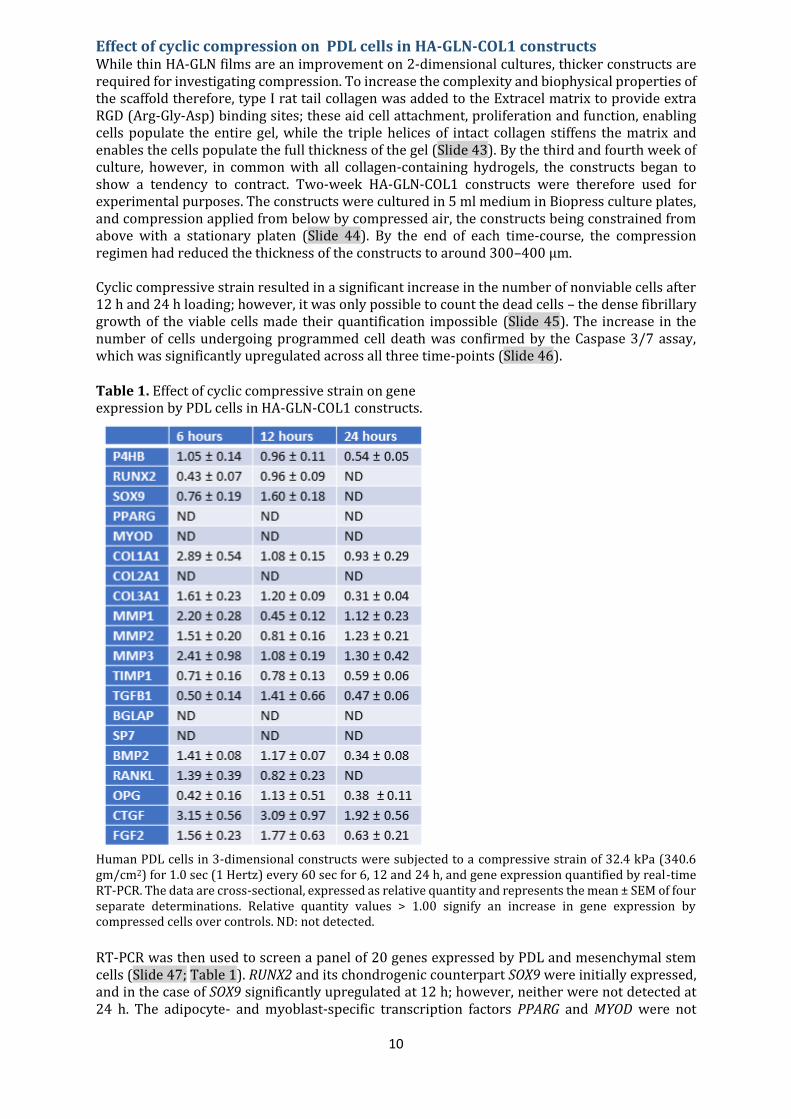
Effect of cyclic compression on PDL cells in HA-GLN-COL1 constructs While thin HA-GLN films are an improvement on 2-dimensional cultures, thicker constructs are required for investigating compression. To increase the complexity and biophysical properties of the scaffold therefore, type I rat tail collagen was added to the Extracel matrix to provide extra RGD (Arg-Gly-Asp) binding sites; these aid cell attachment, proliferation and function, enabling cells populate the entire gel, while the triple helices of intact collagen stiffens the matrix and enables the cells populate the full thickness of the gel (Slide 43). By the third and fourth week of culture, however, in common with all collagen-containing hydrogels, the constructs began to show a tendency to contract. Two-week HA-GLN-COL1 constructs were therefore used for experimental purposes. The constructs were cultured in 5 ml medium in Biopress culture plates, and compression applied from below by compressed air, the constructs being constrained from above with a stationary platen (Slide 44). By the end of each time-course, the compression regimen had reduced the thickness of the constructs to around 300–400 μm. Cyclic compressive strain resulted in a significant increase in the number of nonviable cells after 12 h and 24 h loading; however, it was only possible to count the dead cells – the dense fibrillary growth of the viable cells made their quantification impossible (Slide 45). The increase in the number of cells undergoing programmed cell death was confirmed by the Caspase 3/7 assay, which was significantly upregulated across all three time-points (Slide 46). Table 1. Effect of cyclic compressive strain on gene expression by PDL cells in HA-GLN-COL1 constructs.
Human PDL cells in 3-dimensional constructs were subjected to a compressive strain of 32.4 kPa (340.6 gm/cm2) for 1.0 sec (1 Hertz) every 60 sec for 6, 12 and 24 h, and gene expression quantified by real-time RT-PCR. The data are cross-sectional, expressed as relative quantity and represents the mean ± SEM of four separate determinations. Relative quantity values > 1.00 signify an increase in gene expression by compressed cells over controls. ND: not detected.
RT-PCR was then used to screen a panel of 20 genes expressed by PDL and mesenchymal stem cells (Slide 47; Table 1). RUNX2 and its chondrogenic counterpart SOX9 were initially expressed, and in the case of SOX9 significantly upregulated at 12 h; however, neither were not detected at 24 h. The adipocyte- and myoblast-specific transcription factors PPARG and MYOD were not
11
detected at any time point. P4HB a gene abundantly expressed by cells synthesing collagen and a useful marker of the fibroblast phenotype was expressd at 6 and 12 h at RQ values either side of 1.00. BGLAP, the gene encoding the bone matrix protein osteocalcin, SP7 (osterix) the osteoblast transcription factor acting downstream of RUNX2, and the cartilage specific collagen gene COL2A1, all failed to be identified. Of the 15 extracellular matrix genes screened, 12 were detected at CT values < 35. Although most genes were upregulated at some point with RQ values > 1.00 after 6 h and/or 12 h, all were downregulated at 24 h except for the MMPs, TIMP-1 and CTGF. Table 2. Effect of cyclic compressive strain on the expression of a panel apoptosis-related genes.
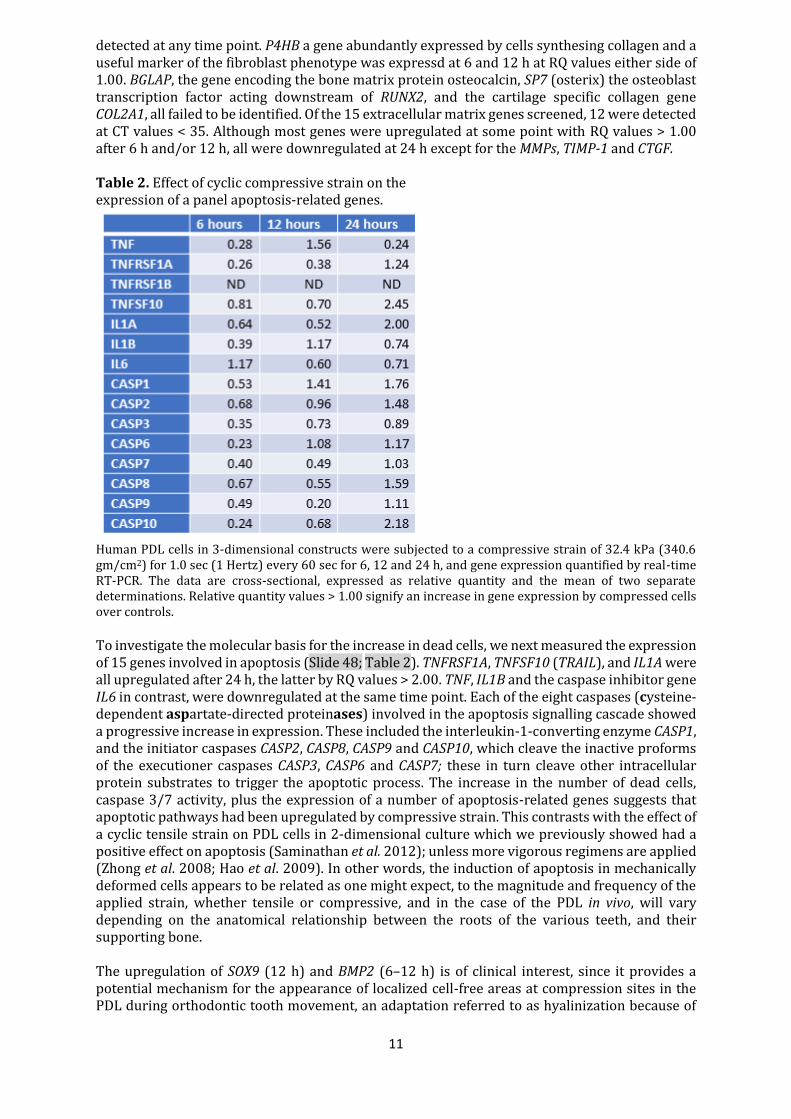
Human PDL cells in 3-dimensional constructs were subjected to a compressive strain of 32.4 kPa (340.6 gm/cm2) for 1.0 sec (1 Hertz) every 60 sec for 6, 12 and 24 h, and gene expression quantified by real-time RT-PCR. The data are cross-sectional, expressed as relative quantity and the mean of two separate determinations. Relative quantity values > 1.00 signify an increase in gene expression by compressed cells over controls.
To investigate the molecular basis for the increase in dead cells, we next measured the expression of 15 genes involved in apoptosis (Slide 48; Table 2). TNFRSF1A, TNFSF10 (TRAIL), and IL1A were all upregulated after 24 h, the latter by RQ values > 2.00. TNF, IL1B and the caspase inhibitor gene IL6 in contrast, were downregulated at the same time point. Each of the eight caspases (cysteine-dependent aspartate-directed proteinases) involved in the apoptosis signalling cascade showed a progressive increase in expression. These included the interleukin-1-converting enzyme CASP1, and the initiator caspases CASP2, CASP8, CASP9 and CASP10, which cleave the inactive proforms of the executioner caspases CASP3, CASP6 and CASP7; these in turn cleave other intracellular protein substrates to trigger the apoptotic process. The increase in the number of dead cells, caspase 3/7 activity, plus the expression of a number of apoptosis-related genes suggests that apoptotic pathways had been upregulated by compressive strain. This contrasts with the effect of a cyclic tensile strain on PDL cells in 2-dimensional culture which we previously showed had a positive effect on apoptosis (Saminathan et al. 2012); unless more vigorous regimens are applied (Zhong et al. 2008; Hao et al. 2009). In other words, the induction of apoptosis in mechanically deformed cells appears to be related as one might expect, to the magnitude and frequency of the applied strain, whether tensile or compressive, and in the case of the PDL in vivo, will vary depending on the anatomical relationship between the roots of the various teeth, and their supporting bone. The upregulation of SOX9 (12 h) and BMP2 (6–12 h) is of clinical interest, since it provides a potential mechanism for the appearance of localized cell-free areas at compression sites in the PDL during orthodontic tooth movement, an adaptation referred to as hyalinization because of
12
its histological appearance to hyaline cartilage. The role of SOX9 and its regulatory target BMP2 in determining chondrogenic lineage development is well established (Akiyama et al. 2002; Schmitt et al. 2003), and in the skeleton, cartilage is typically found at sites of compressive mechanical loading. The present findings, therefore, suggest that hyalization might arise from the differentiation of localized mesenchymal stem cells down the chondrogenic pathway, followedby their eventual removal by programmed cell death. Proteins levels of all three MMPs (matrixins), the major enzymes involved in extracellular matrix turnover, plus their specific inhibitor TIMP-1, were identified by ELISAs in culture supernatants, although the concentrations of MMP-2 and MMP-3 were low in comparison with MMP-1 and TIMP-1. Both MMP-1 and TIMP-1 proteins were significantly upregulated by cyclic compressive strain (Slide 49), although after 24h, the concentration of free TIMP-1 in compressed cultures was more than twice that of the pro-form of MMP-1. Both were significantly higher in 24 h cultures, despite TIMP1 expression remaining unchanged. However, because the ELISAs only recognize latent proMMPs and free TIMP-1, we were not in a position to say what proportion of the enzymes were in the active, latent, or complexed forms. The finding that PDL cells express RANKL and OPG, members of the RANKL/RANK/OPG triad that constitutes a ligand-receptor system directly regulating the final steps of the bone resorptive cascade, has resulted in their being widely investigated for possible roles in tooth support. RANKL which exists in both membrane-bound and soluble forms, stimulates the differentiation and function of osteoclasts, an effect mediated by RANK, a member of the TNF receptor family expressed primarily on cells of the monocyte/macrophage lineage, including osteoclasts and their precursor cells (Nakagawa et al. 1998). OPG is a secreted protein that inhibits bone resorption by acting as a decoy receptor, binding to and neutralizing both cell-bound and soluble RANKL (Simonet et al. 1997). Interestingly, in each of the studies in which static compression has been applied to PDL cells in 2-dimensional culture, RANKL expression was upregulated (Kanzaki et al. 2002; Tsuji et al. 2004; Yamamoto et al. 2006; Nakajima et al. 2008), which contrasts with the present findings, highlighting differences between culturing cells on rigid 2-dimensional substrates, and enclosed in a 3-dimensional microenvironment.; given the key role of integrin receptors in mediating outside-in signalling at focal adhesions, this will significantly influence their response to mechanical loading.
Summary Two-dimensional in vitro culture systems have provided valuable information regarding cell behaviour at its most reductionist level. However, they lack the physical and other environmental signals provided by 3-dimensional microenvironments that are important determinants of phenotype, proliferation, gene expression, and matrix turnover – in other words, information derived from 2-dimensional models is unlikely to reflect the complex physiology of the cells in vivo. There is clearly a difference between culturing cells on rigid 2-dimensional substrates, and enclosed within a 3-dimensional microenvironment; The challenge in engineering 3-dimensional constructs is to replicate not only tissue complexity, but also the mechanical and viscoelastic characteristics (resistance to elastic deformation or stiffness) of the native tissue, and further improvements in the composition of the present constructs are required. Although close to the minimum level recommended for use with the Flexercell Compression Plus system, a force of 32.4 kPa was at the limit appropriate for deforming the constructs in their present format, highlighting one of the disadvantages of hydrogels – their poor mechanical properties, resulting in tissue constructs with significantly poorer mechanical strength than the real tissue (Aherne et al. 2005). Analysis of the rheological properties of cross-linked HA-gelatin hydrogels for use in soft tissue engineering has shown that the elastic moduli range from 11 Pa to 3.5 kPa depending on the concentration of the HA; increasing the ratio of gelatin reduced gel stiffness by diluting the concentration of the HA component. Extracel has a shear elastic modulus of around 70 Pa (Vanderhooft et al. 2009), and although it has not been measured, the elastic modulus of the present constructs is likely to be similar to the 90 Pa reported recently for a 3-dimensional collagen gel populated with PDL cells (Kim et al. 2011).
13
While this may be suitable for imaging cell–cell and cell–matrix interactions in studies of tooth support by immunological methods and confocal microscopy, for tissue engineering applications a much higher stiffness is required. One problem in trying to reconstruct the biophysical properties of the human PDL, a complex fibre-reinforced tissue that responds to mechanical loading in a viscoelastic and nonlinear manner (Jónsdóttir et al. 2006), is that for all practical purposes the elastic modulus is unknown. The difficulty of examining a thin tissue (0.1–0.4 mm) sandwiched between bone and cementum has resulted in a lack of consistency regarding its elastic properties, highlighted by a recent systematic review of 23 studies that had used finite element analysis, which found that Young’s modulus ranged from 10 kPa to 1750 MPa, an astonishing difference approaching six orders of magnitude (Fill et al. 2011). In comparison, the elastic moduli of soft mammalian tissues range from near 100 Pa for soft organs such as the brain, to tens of thousands in muscle and around 300 MPa for Achilles tendon (Levental et al. 2006). Recapitulating the biophysical properties of the next generation of artificial PDL constructs, requires a much more robust extracellular matrix. Since type I collagen is the major structural protein of the PDL, rather than add collagen to the hydrogel matrix, a more rational approach would be to permeabilize preformed type I collagen scaffolds with a mixture of PDL cells and Extracel followed by gelation. A similar approach is suggestive for engineering artificial bone constructs for transplantation. Osteoblasts are anchorage-dependent cells, which require a stable substrate on which to lay down new bone matrix, not something that disappears under their focal adhesions – the fundamental reason why the various bioresorbable polymers used in bone regeneration have proved so disappointing; the result being mainly fibrosis with a few microscopic islands of bone. What is required is a method to recapitulate artificial bone in the laboratory, in sufficient qnantities to be transplanted minus the cells, that can be resorbed and replaced by the host in the course of normal bone remodelling. Having had my research career generously extended from 65 to 75 courtesy of the University of Otago, and the National University of Singapore, these are research projects for the next generation – mine now being time expired.!
References Aherne M, Yang Y, El Haj AJ, Then KY, Liu K-K (2005). Characterizing the viscoelastic properties of thin hydrogel-based constructs for tissue engineering applications. Journal of the Royal Society, Interface 2, 455–463. Akiyama H, Chaboissier MC, Martin JF, Schedl A, de Crobrugghe B (2002). The transcription factor Sox9 has essential roles in successive steps of the chondrocyte differentiation pathway and is required for expression of Sox5 and Sox6. Genes and Development 16, 2813–2828. Aguirre JI, Plotkin LI, Stewart SA, Weinstein RS, Parfitt AM, Manolagas SC et al. (2006). Osteocyte apoptosis is induced by weightlessness in mice and precedes osteoclast recruitment and bone loss. Journal of Bone and Mineral Research 21, 605–615. Alhashimi N, Frithiof L, Brudvik P, Bakhiet M (2001). Orthodontic tooth movement and de novo synthesis of proinflammatory cytokines. American Journal of Orthodontics and Dentofacial Orthopedics 119, 307–312. Bakker A, Klein-Nulend J, Burger E. (2004). Shear stress inhibits while disuse promotes osteocyte apoptosis. Biochemical and Biophysical Research Communications 320, 1163–1168. Basdra EK, Papavassiliou AG, Huber LA (1995) Rab and rho GTPases are involved in specific response of periodontal ligament fibroblasts to mechanical stretching. Biochimica et Biophysica Acta 1268, 209–213. Basdra EK, Komposch G (1997). Osteoblast-like properties ofhuman periodontal ligament cells: an in vitro analysis. European Journal of orthodontics 19, 615–621.
14
Bertolini DR, Nedwin GE, Bringham TS, et al. (1986). Stimulation of bone resorption and inhibition of bone formation in vitro by human tumour necrosis factors. Nature 319, 516–518. Bletsa A, Berggreen E, Brudvik P (2006). Interleukin-1α and tumor necrosis factor-α expression during the early phases of orthodontic tooth movement in rats. European Journal of Oral Sciences 114, 423–429. Bolcato-Bellemin AL, Elkaim R, Abehsera A, et al. (2000). Expression of mRNAs encoding for α and β integrin subunits, MMPs and TIMPs in stretched human periodontal ligament and gingival fibroblasts. Journal of Dental Research 79, 1712 –1716. Carvalho RS, Bumann A, Schwarzer C, Scott E, Yen EHK (1996). A molecular mechanism of integrin regulation from bone cells stimulated by mechanical forces. European Journal of Orthodontics 18, 227–235. Cho MI, Matsuda N, Lin WL, et al. (1992). In vitro formation of mineralized nodules by periodontal ligament cells from the rat. Calcified Tissue International 50, 459–467. Clarke EA, Brugge JS (1995). Integrins and signal transduction pathways: the road taken. Science 268, 233–239. Cukierman E, Pankov R, Stevens DR, Yamada KM (2001). Taking cell-matrix adhesions to the third dimension. Science 294, 1708–1712. Davidovitch Z (1995). Cell biology associated with tooth movement. In: Berkovitz B, Moxham B, Newman HN (Eds). The Periodontal Ligament in Health and Disease, 2nd edn. Mosby-Wolfe, Philadelphia, pp. 259–278. Davidovitch Z, Shanfield JL (1975). Cyclic AMP levels in alveolar bone of orthodontically-treated cats. Archives of Oral Biology 20, 567–574. Davidovitch Z, Nicolay OF, Ngan PW, Shanfield JL (1988) Neurotransmitters, cytokines and the control of alveolar bone remodeling in orthodontics. Dental Clinics of North America 32, 411–435. Davidson RM, Tatakis DW, Auerbach AL (1990). Multiple forms of mechanosensitive channels in osteoblast-like cells. Pflugers Archiv 416, 646–651. DeMali KA, Wennerberg K, Burridge K (2003). Integrin signaling to the actin cytoskeleton. Current Opinion in Cell Biology 15, 72–582. Dolce C, Kinniburgh AJ, Dziak R (1996). Immediate early-gene induction in rat osteoblastic cells after mechanical deformation. Archives of Oral Biology 41, 1101–1108. Elmore S. (2007). Apoptosis: a review of programmed cell death. Toxicologic Pathology 35, 495–516. Elsdale T, Bard J (1972). Collagen substrata for studies in cell behaviour. Journal of Cell Biology 54, 626–637. Farndale RW, Sandy JR, Atkinson SJ, Pennington SR, Meghji S, Meikle MC (1988). Parathyroid hormone and prostaglandin E2 stimulate both inositol phosphates and cyclic AMP accumulation in mouse osteoblast cultures. Biochemical Journal 252, 263–268. Fill T, Carey JP, Toogood RW, Major PW (2011). Experimentally determined mechanical properties of, and models for, the periodontal ligament: critical review of current literature. Journal of Dental Biomechanics. doi:10.4061/2011/312980.
15
García-López S, Meikle MC, Villanueva RE, et al. (2005). Mechanical deformation inhibits IL-10 and stimulates IL-12 production by mouse calvarial osteoblasts in vitro. Archives of Oral Biology 50, 449–452. Green DD, Hembry RM, Atkinson SJ, Reynolds JJ, Meikle MC (1990). Immunolocalization of collagenase and tissue inhibitor of metalloproteinases (TIMP) in mechanically deformed fibrous joints. American Journal of Orthodontics and Dentofacial Orthopedics 97, 281–288. Grieve WG, Johnson GK, Moore RN, Reinhardt RA, DuBois LM (1994) Prostaglandin E (PGE) and interleukin-1β (IL-1β ) levels in gingival crevicular fluid during human orthodontic tooth movement. American Journal of Orthodontics and Dentofacial Orthopedics 105, 369–374. Gowen M, Meikle MC, Reynolds JJ (1983). Stimulation of bone resorption in vitro by a non-prostanoid factor released by human monocytes in culture. Biochimica et Biophysica Acta 762, 471–474. Hao Y, Xu C, Sun S, Zhang F (2009). Cyclic stretching force induces apoptosis in human periodontal ligament cells via Caspase-9. Archives of Oral Biology 54, 864–870. Heath JK, Saklatvala J, Meikle MC, Atkinson SJ, Reynolds JJ (1985). Pig interleukin 1 (catabolin) is a potent stimulator of bone resorption in vitro. Calcified Tissue International 37, 95–97. Harell A, Dekel S, Binderman I (1977). Biochemical effect of mechanical stress on cultured bone cells. Calcified Tissue Research 22, 202–207. Hong S-C, Polsky-Cynkin R, Levine L (1976). Stimulation of prostaglandin biosynthesis by vasoactive substances in methylcholanthrene-transformed BALB/3T3 cells. Journal of Biological Chemistry 251, 776–780. Hynes RO (1992). Integrins: versatility, modulation, and signaling in cell adhesion. Cell 69, 11–25. Ikegame M, Ishobashi O, Yoshizawa T et al. (2001). Tensile stress induces bone morphogenetic protein 4 in preosteoblastic and fibroblastic cells, which later differentiate into osteoblasts leading to osteogenesis in the mouse calvariae in organ culture. Journal of Bone and Mineral Research 16, : 24–32. Ingber DE (1991). Integrins as mechanochemical transducers. Current Opinion in Cell Biology 3, 841–848. Jónsdóttir SH, Giesen EB, Maltha JC (2006). Biomechanical behaviour of the periodontal ligament of the beagle dog during the first 5 hours of orthodontic force application. European Journal of Orthodontics 28, 547–552. Kanzaki H, Chiba M, Shimizu Y, Mitani H (2002). Periodontal ligament cells under mechanical stress induces osteoclastogenesis by receptor activator of nuclear factor B ligand up-regulation via prostaglandin E2 synthesis. Journal of Bone and Mineral Research 17, 210–220. Kikkawa Y, Sanzen N, Fujiwara H, Sonnenberg A, Sekiguchi K (2000). Integrin binding specificity of laminin-10/11: laminin-10/11 are recognized by a3b1, a6b1 and a6b4 integrins. Journal of Cell Science 113, 859–876. Kim SG, Kim S-G, Viechnicki B, Kim S, Nah H-D (2011). Engineering of a periodontal ligament construct: cell and fibre alignment induced by shear stress. Journal of Clinical Periodontology 38, 1130–1136.
16
Lacey DL, Timms E, Tan H-L, et al. (1998). Osteoprotegerin ligand is a cytokine that regulates osteoclast differentiation and activation. Cell 93, 165–176. Lecka-Czernick B, Moerman EJ, Grant DF, Lehmann JM, Manolagas SC, Jilka RL (2002). Divergent effects of selective peroxisome proliferator-activated receptor-γ2 ligands on adipocyte versus osteoblast differentiation. Endocrinology 143, 2376–2384. Lee KY, Mooney DJ (2001). Hydrogels for tissue engineering. Chemical Reviews 101, 869–1879. Lekic P, Rojas J, Birek C, Tenenbaum H, McCulloch CAG (2001). Phenotypic comparison of periodontal cells in vivo and in vitro. Journal of Periodontal Research 36, 71–79. Levental I, Georges PC, Janmey PA (2006). Soft biological materials and their impact on cell function. Soft Matter 2, 1–9. Long P, Hu J, PiescoN, Buckley M, Agarwal S (2001). Low magnitude of tensile strain inhibits IL-1β-dependent induction of pro-inflammatory cytokines and induces synthesis of IL-10 in human periodontal ligament cells in vitro. Journal of Dental Research 80, 1416–1420. Lowney JJ, Norton LA, Shafer DM, Rossomondo EF (1995). Orthodontic forces increase tumor necrosis factor alpha in the human gingival sulcus. American Journal of Orthodontics and Dentofacial Orthopedics 108, 519–524. Matheson LA, Fairbank NJ, Maksym GN, Santerre JP, Labow RS (2006). Characterization of the FlexcellTM cyclic strain culture system with U937 macrophage-like cells. Biomaterials 27, 226–233. McDonald F, Somasundaram B, McCann TJ, Mason WT, Meikle MC (1996) Calcium waves in fluid flow stimulated osteoblasts are G protein mediated. Archives of Biochemistry and Biophysics 326, 31–38. Meikle MC, Reynolds JJ, Sellers A, Dingle JT (1979). Rabbit cranial sutures in vitro: a new experimental model for studying the response of fibrous joints to mechanical stress. Calcified Tissue International 28, 137–144. Meikle MC, Sellers A, Reynolds JJ (1980). Effect of tensile mechanical stress on the synthesis of metalloproteinases by rabbit coronal sutures in vitro. Calcified Tissue International 30, 77–82. Meikle MC, Heath JK, Hembry RM, Reynolds JJ (1982). Rabbit cranial suture fibroblasts under tension express a different collagen phenotype. Archives of Oral Biology 27, 609–613. Meikle MC, Heath JK, Reynolds JJ (1984). The use of in vitro models for investigating the response of fibrous joints to tensile mechanical stress. American Journal of Orthodontics 85, 141–153. Meikle MC, Heath JK, Atkinson SJ, Hembry RM, Reynolds JJ (1989). Molecular biology of stressed connective tissues at sutures and hard tissues in vitro. In: Norton LA, Burstone CJ (Eds). The Biology of Tooth Movement. CRC Press, Boca Raton, pp. 71–86. Meikle MC (2006). The tissue, cellular, and molecular regulation of orthodontic tooth movement: 100 years after Carl Sandstedt. European Journal of Orthodontics 28, 221–240. Melcher AH, Turnbull RS (1972). Organ culture in studies of the periodontium. In: Balls M, Monnickendam M (Eds). Organ Culture in Biomedical Research. Cambridge University Press, Cambridge, pp. 149 – 163. Meyer CJ, Alenghat FJ, Rim P, Fong JH, Fabry B, Ingber DE (2000). Mechanical control of cyclic AMP signaling and gene transcription through integrins. Nature Cell Biology 2, 666–668.
17
Milne TJ, Ichim I, Patel B, McNaughton A, Meikle MC.(2009). Induction of osteopenia during experimental tooth movement in the rat: alveolar bone remodelling and the mechanostat theory. European Journal of Orthodontics 31, 221–231. Murakami Y, Kojima T, Nasagawa T, Kobayashi H, Ishikawa I (2003). Novel isolation of alkaline phosphatase-positive subpopulation from periodontal ligament fibroblasts. Journal of Periodontology 74,780–786. Nakagawa N, Kinosaki M, Yamaguchi K, Shima N, Yasuda H, Yano K, et al. (1998). RANK is the essential signaling receptor for osteoclast differentiation factor in osteoclastogenesis. Biochemical and Biophysical Research Communications 253, 395–400. Nakajima R, Yamaguchi M, Kojima T, Takano M, Kasai K. (2008). Effects of compression force on fibroblast growth factor-2 and receptor activator of nuclear factor kappa B ligand production by periodontal ligament cells in vitro. Journal of Periodontal Research 43, 168–173. Ngan PW, Crock B, Vargese J, Lanese R, Shanfield J, Davidovitch Z (1988). Immunohistochemical assessment of the effect of chemical and mechanical stimuli on cAMP and prostaglandin E levels in human gingival fibroblasts in vitro. Archives of Oral Biology 33, 163–174. Ngan PW, Saito S, Saito M, Shanfield J, Davidovitch Z (1990). The interactive effects of mechanical stress and interleukin 1β on prostaglandin E and cyclic AMP production in human gingival fibroblasts in vitro : comparison with cloned osteoblastic cells of mouse (MC3T3-E1). Archives of Oral Biology 35, 717–725. Ogasawara T, Yoshimine Y, Kiyoshima T, et al. (2004) In situ expression of RANKL, RANK, osteoprotegerin and cytokines in osteoclasts of rat periodontal tissue. Journal of Periodontal Research 39, 42–49. Peverali FA, Basdra EK, Papavassiliou AG (2001). Stretch-mediated activation of selective MAPK subtypes and potentiation of AP-1 binding in human osteoblastic cells. Molecular Medicine 7, 68–78. Pike DB, Cai S, Pomraning KR, et al. (2006). Heparin-regulated release of growth factors in vitro and angiogenic response in vivo to implanted hyaluronan hydrogels containing VEGF and bFGF. Biomaterials 27, 5242–5251. Pinkerton MN, Wescott DC, Gaffey BJ, Beggs KT, Milne TJ, Meikle MC (2008). Cultured human periodontal ligament cells constitutively express multiple osteotropic cytokines and growth factors, several of which are mechanoresponsive to mechanical deformation. Journal of Periodontal Research 43, 343–351. Roberts WE, Chase DC (1981). Kinetics of cell proliferation and migration associated with orthodontically-induced osteogenesis. Journal of Dental Research 60, 174–181. Rodan GA, Bouret LA, Harvey A, Mensi T (1975). Cyclic AMP and cyclic GMP: mediators of the mechanical effects of bone remodeling. Science 189, 467–471. Roskelley CD, Desprez PY, Bissell MJ (1994). Extracellular matrix-dependent issue specific gene expression in mammary epithelial cells requires both physical and biochemical signal transduction. Proceedings of the National Academy of Sciences USA 91, 12378–12382. Rubin J, Murphy T, Nanes MS, Fan X (2000). Mechanical strain inhibits expression of osteoclast differentiation factor by murine stromal cells. American Journal of Physiology. Cell Physiology 278, C1126–C1132.
18
Rubin J, Murphy YC, Fan X, Goldschmidt M, Taylor WR (2002). Activation of extracellular signal-regulated kinase is involved in mechanical strain inhibition of RANKL expression in bone stromal cells. Journal of Bone and Mineral Research 17, 1452–1460. Saminathan A, Vinoth KJ, Wescott DC, Pinkerton MN, Milne TJ, Cao T, Meikle MC. (2012). The effect of cyclic mechanical strain on the expression of adhesion‐related genes by periodontal ligament cells in two‐dimensional culture. Journal of Periodontal Research 47, 212–221. Saminathan A, Vinoth KJ, Low HH, Cao T, Meikle MC (2013). Engineering three‐dimensional constructs of the periodontal ligament in hyaluronan–gelatin hydrogel films and a mechanically active environment. Journal of Periodontal Research 48, 790–801. Saminathan A, Sriram G, Vinoth JK, Cao T, Meikle (2015). Engineering the Periodontal Ligament in Hyaluronan–Gelatin–Type I Collagen Constructs: Upregulation of Apoptosis and Alterations in Gene Expression by Cyclic Compressive Strain. Tissue Engineering Part A 21, 518–529. Sandy JR, Meghji S, Farndale RW, Meikle MC (1989). Dual elevation of cyclic AMP and inositol phosphates in response to mechanical deformation of murine osteoblasts. Biochimica et Biophysica Acta 1010, 265–169. Sastry SK, Burridge K (2000). Focal adhesions: a nexus for intracellular signaling and cytoskeletal dynamics. Experimental Cell Research 261, 25–36. Schacht EH (2004). Polymer chemistry and hydrogel systems. Journal of Physics: Conference Series 3, 22–28. Schmitt B, Ringe J, Häupl T, Notter M, Manz R, Burmeister G-D et al. (2003). BMP2 initiates chondrogenic lineage development of adult human mesenchymal stem cells in high-density culture. Differentiation 76, 567–577. Schnapp LM, Hatch N, Ramos DM, Klimanskaya IV, Sheppard D, Pytela R (1995). The human integrin a8b1 functions as a receptor for tenascin, fibronectin and vitronectin. Journal of Biological Chemistry 270, 23196–23202. Serban MA, Scott A, Prestwich GD (2008). Use of hyaluronan-derived hydrogels for three-dimensional cell culture and tumor xenografts. Current Protocols in Cell Biology 40, 10.14.1–10.14.21. Simonet WS, Lacey DL, Dunstan CR, et al. (1997). Osteoprotegerin: a novel secreted protein involved in the regulation of bone density. Cell 89, 309–319. Somjen D, Binderman I, Berger E, Harell A (1980). Bone remodelling induced by physical stress is prostaglandin E2 mediated. Biochimica et Biophysica Acta 627, 91–100. Streb H, Irvine RF, Berridge MJ, Schulz I (1983) Release of Ca2+ from a nonmitochondrial intracellular store in pancreatic acinar cells by inositol-1,4,5-triphosphate. Nature 306, 67–69. Takahashi I, Nishimura M, Onodera K, et al. (2003). Expression of MMP-8 and MMP-13 genes in the periodontal ligament during tooth movement in rats. Journal of Dental Research 82, 646–651. Tsuji K, Uno K, Zhang G X, Tamura M (2004). Periodontal ligament cells under intermittent tensile stress regulates mRNA expression of osteoprotegerin and tissue inhibitor of metalloproteinase-1 and -2. Journal of Bone and Mineral Research 22, 94–103. Tuncay OC (2004). Taking stock: Hippocratic and Platonic thoughts on orthodontic tooth movement. Orthodontics and Craniofacial Research 7, 162–164.
19
Uematsu S, Mogi M, Deguchi T (1996). Interleukin (IL)-1β , IL-6, tumor necrosis factor-α, epidermal growth factor, and B2 -microglobulin levels are elevated in gingival crevicular fluid during human orthodontic tooth movement. Journal of Dental Research 75, 562–567. Vande Geeste JP, Di Martino ES, Vorp DA (2004). An analysis of the complete strain field within FlexercellTM membranes. Journal of Biomechanics 37, 1923–1928. Vanderhooft JL, Alcoutlabi M, Magda JJ, Prestwich GD (2009). Rheological properties of cross-linked hyaluronan-gelatin hydrogels for tissue engineering. Macromolecular Bioscience 9, 20–28. Wall ME, Weinhold PS, Siu T, Brown TD, Banes AJ (2007). Comparison of cellular strain with applied substrate strain in vitro. Journal of Biomechanics 40, 173–181. Wang N, Butler JP, Ingber DE (1993). Mechanotransduction across the cell surface and through the cytoskeleton. Science 269, 1124–1127. Wescott DC, Pinkerton MN, Gaffey BJ, Beggs KT, Milne TJ, Meikle MC (2007). Osteogenic gene expressiod by human periodontal cells under cyclic tesion. Journal of Dental Research 86, 1212–1216. Wilson E, Sudhir K, Ives HE (1995). Mechanical strain of rat vascular smooth muscle cells is sensed by specific extracellular matrix/integrin interactions. Journal of Clinical Investigation 96, 2364–2372. Wyllie AH, Kerr JFR, Currie AR (1980). Cell death: the significance of apoptosis. International Review of Cytology 68, 251–307. Yamamoto T, Kita M, Kimura I, Oseko F, Terauchi R, Takahashi K, et al. (2006). Mechanical stress induces expression of cytokines in human periodontal ligament cells. Oral Diseases 12, 171–175. Yasuda H, Shima N, Nakagawa N, et al. (1998). Osteoclast differentiation factor is a ligand for osteoprotegerin/osteoclastogenesis-inhibitory factor and is identical to TRANCE/RANKL. Proceedings of the National Academy of Sciences USA 95, 3597–3600. Yen EHK, Melcher AH (1978). A continuous flow culture system for organ culture of large explants of adult tissue. Effect of oxygen tension on mouse molar periodontium. In Vitro 14, 811–818. Yeh C-K, Rodan GA (1984). Tensile forces enhance prostaglandin E2 synthesis in osteoblastic cells grown on collagen ribbons. Calcified Tissue International 36, S67 –S71. Zhao L-J, Jiang H, Papasian CJ, et al. (2009). Correlation of obesity and osteoporosis: effect of fat mass on the determination of osteoporosis. Journal of Bone and Mineral Research 23, 17–29. Zhong W, Xu C, Zhang F, Jiang X, Zhang X, Ye D (2008). Cyclic stretching force-induced early apoptosis in human periodontal ligament cells. Oral Diseases 14, 270–276.