biologics in sports medicineuhealthsportsmedicine.com/documents/sports_med_and...biologics in sports...
TRANSCRIPT

Biologics in Sports MedicineBasic SciencePreparationApplications
Evidence Base
Paul Rothenberg, MDPGY-2 Orthopaedics
Jackson memorial Hospital/University of Miami
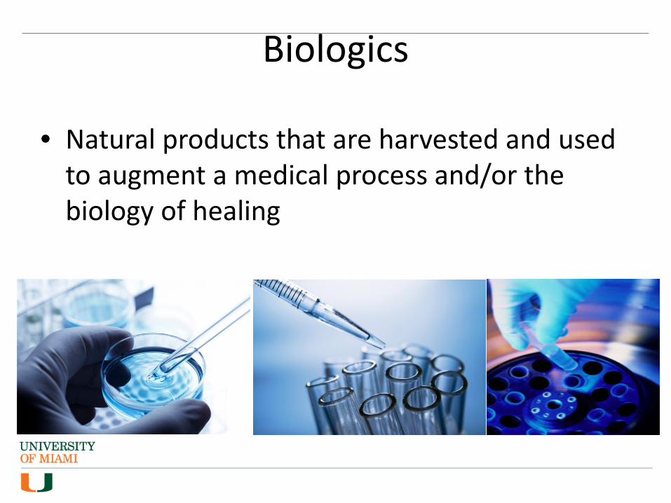
Biologics
• Natural products that are harvested and used to augment a medical process and/or the biology of healing

3 categories
– Growth factors• Harvest and delivery of GFs to site• Eg. PRP
– Cell therapies• Harvest and delivery of cells to a site• Eg. Autologous chondrocyte therapy for cartilage repair
– Tissue therapy• Use of tissue to replace damaged structures or
augment repair• Eg. Meniscal allograft transplantation

Biologics
• PRP• Stem Cells

Healing• Inflammation (day 1-5)
– Hematoma– Platelets activate– Fibrin-platelet matrix forms– Vasodilation– Migration of numerous cells
• Proliferative stage (day 3 – wk6)– Fibroblasts produce collagen
• Remodeling (wk 6 – 1 year)– Organization of collagen

Biologics and Healing
Provide 3 Elements to the healing process• Matrix• Growth Factors• Stem Cells

Platelet Rich Plasma
PRP
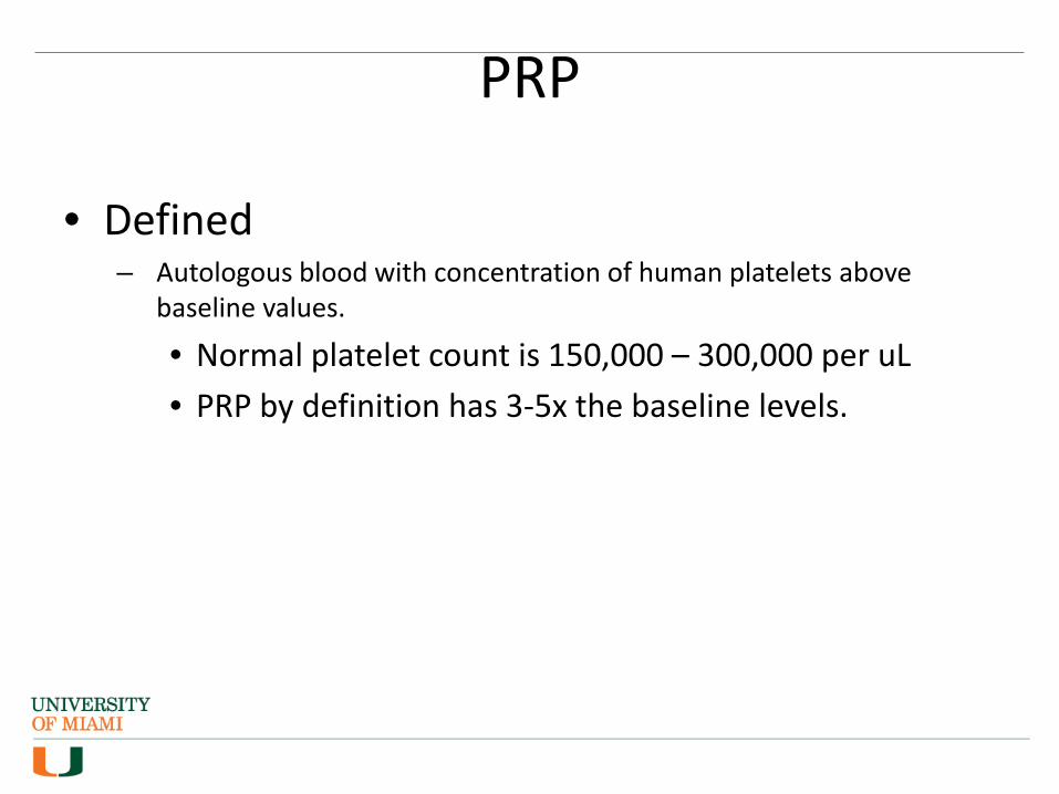
• Defined– Autologous blood with concentration of human platelets above
baseline values.
• Normal platelet count is 150,000 – 300,000 per uL• PRP by definition has 3-5x the baseline levels.

Platelets
• Blood cells without nucleus, principal role in coagulation
• 3 types of granules (a, d, and l),• Alpha granules contain growth factors• Release of this growth factors occur with exposure to
collagen, VW-f, thrombin or calcium chloride

PRP
PRP
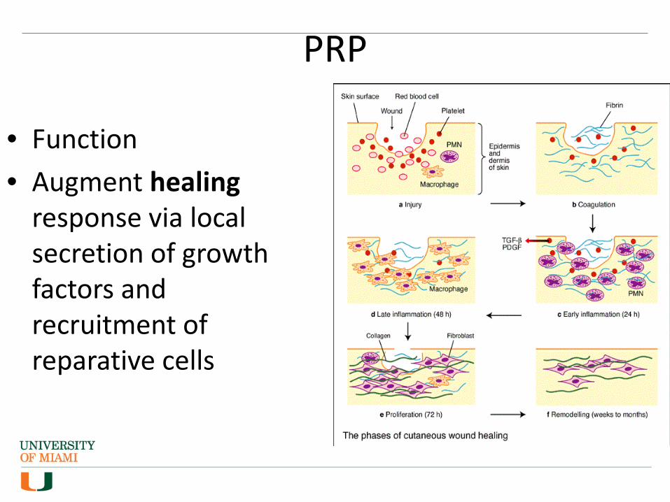
• Function• Augment healing
response via local secretion of growth factors and recruitment of reparative cells
PRP PREPARATION
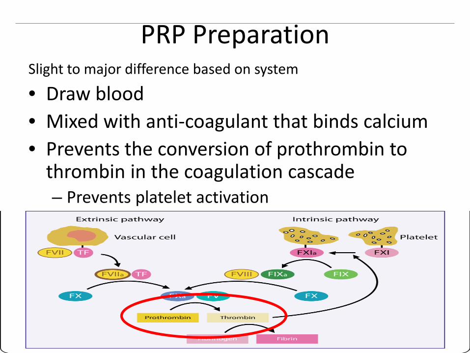
PRP Preparation Slight to major difference based on system
• Draw blood • Mixed with anti-coagulant that binds calcium• Prevents the conversion of prothrombin to
thrombin in the coagulation cascade– Prevents platelet activation

PRP Preparation Centrifugation # 1(“soft spin”) Separate whole blood into RBC fraction and plasma fraction (plts, wbc, clotting factors)
Centrifugation # 2 (“hard spin”) Platelet poor and PRP
ActivationAddition of coagulant releases growth factors (70% released within first 10 minutes)

PRP Preparation
No universal classificationPAW classification system• Absolute number of
platelets• Method of activation• Presence or absence of
WBCs• No evidence base behind
this classification

PRP Challenges
• Variability– More than 40 available commercial systems– Amount of whole blood used– Amount of plasma used to suspend concentrated platelets– Variability between patients blood samples using same
system• No consensus on appropriate number of platelets
– More is better?– Saturation effect ?– WBC
• Timing

Stem Cells

Stem CellsBasic characteristics• Self Renewal• Prolonged cell division• Unspecialized • Can differentiate into specialized cells when provided
appropriate environmentClassified in two ways• Plasticity• Source

Stem Cell Plasticity
Totipotent Pluripotent MultipotentUnipotent
Plasticity – ability to become different cell types

Stem CellsMesenchymal Stem Cells• Prime interest in orthopedics• Source: Bone marrow (also; fat, skin, tendon, periosteum)• Most common harvest site: Iliac crest• Multipotent - In appropriate environment can differentiate
into bone, tendon, muscle and adipose tissues

Stem Cell in Spots
• Purpose in Sports Medicine– Much of the soft tissue in sports medicine heals
poorly (tendons, cartilage etc.) via formation of scar tissue that is biomechanically inferior to the native tissue
– Delivery of regenerative cells may provide cells for improved healing and replacement of the native tissue

Isolation of Stem Cells

Stem Cells
Isolation of Stem Cells• No consensus regarding optimal number of cells
for healing in MSK tissues• Iliac crest BM aspirate yield a poor number of
MSCs• MSCs decrease with age• Success of bone marrow aspirate of MSCs may be
due to number progenitor cells (Hernigou et al. JBJS, 2005)

Stem Cells Isolation• Many Sources and Techniques• Example in Brief• Bone Marrow Obtained from Iliac crest in
Aspiration needle loaded with heparin
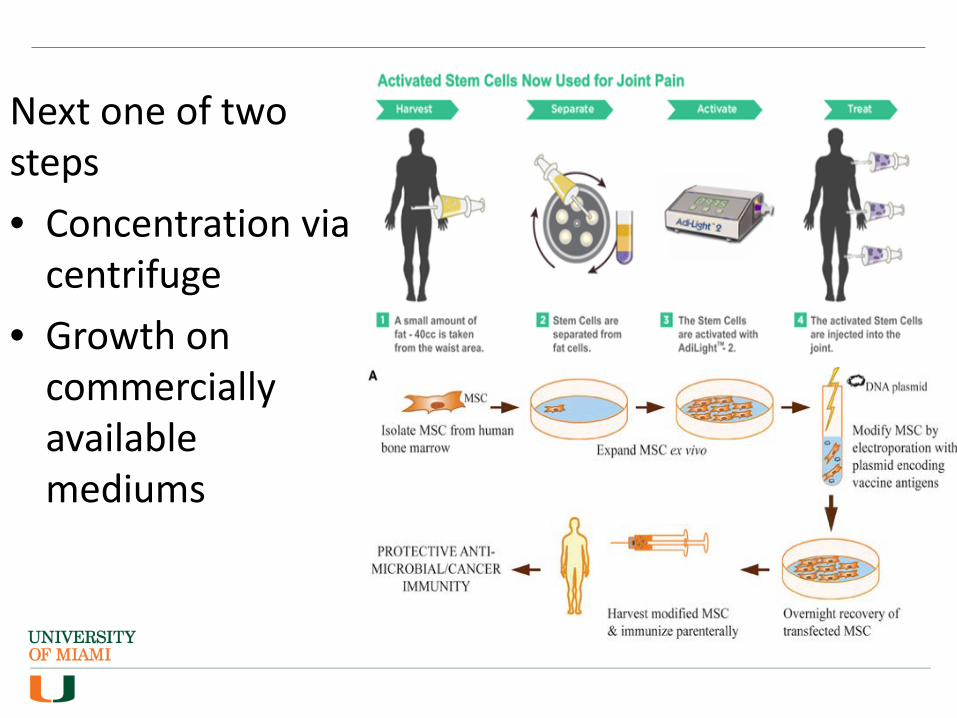
Next one of two steps• Concentration via
centrifuge• Growth on
commercially available mediums

Stem Cell Isolation• MSC Characteristics
– Adherence to plastic under normal culture conditions– Fibroblast-like morphology– Presence of surface CD73, CD90, CD105– Lack of surface CD11b, CD14, CD19, CD34, CD45, CD79a
and HLA-DR• Play an important role in isolation• In US centrifugation remains only FDA approved
method of concentration

Clinical Applications of BiologicsTendon Injury

Biologics and Tendon Injury
• Tendons– Damaged tendon tissues heals very slowly and rarely
attains the structural integrity and mechanical strength or normal, undamaged tendon
• Poor vascularity• Microtrauma
– Tendon tissue that is torn is often degenerative and prone to failure with surgery

PRP and Tendon Injury
• Complex healing process• Many of GF and Cytokines involved are
present in PRP• Additionally PRP can aid in angiogenesis• Anti-Inflammatory properties• Most studies Elbow, Knee and Ankle

PRP and Tendon Injury• Patellar Tendinopathy• Recent literature
– Comparison of PRP to ESWT and DN– Enhanced structural integrity at 3 mos
(Charousset et al. AJSM, 2014)

PRP and Tendon Injury• Lateral Epicondylitis• Recent Literature
–PRP vs GC vs Saline •(Krogh et al. AJSM 2013)•Improvement without significant difference
–PRP vs Autologous whole blood •(Raeissadat et al. BMC Sports 2014)•Improvement without significant difference

PRP and Tendon Injury
• Achilles Tendinopathy– No therapeutic effect
• Two level I studies comparing PRP vs Saline for Achilles tendinopathy
• No difference between the control groups• Achilles Tendon rupture
– Conflicting evidence on PRP for augmentation during Achilles tendon repair (Sanchez et al. AJSM 2007 vs Schepul et al AJSM 2010)
– Strong support in animal studies

PRP and Tendon Injury
• Limitations and future studies– Lack of placebo– Lack of non-treatment group– Larger studies, better controls
• Considerations– Cost – Placebo affect– Natural history of disease process

Stem Cells and Tendon Injury
• Limited small case series currently showing benefit
• Summary (Ahmad et al. Arthroscopy, 2012)– Animal studies
• Increase collagen fiber density, • Enhance tissue architecture
– Stem cells may regenerate tendon tissue and enhance tendon repair construct
Clinical Applications of BiologicsRotator Cuff Repair

Biologics and RTC Repair
• Increasing incidence with aging population
• Common cause of shoulder pain and occupational disability
• Improvements in arthroscopic instrumentation and suture anchor technology
• Tendon failure is very high in the literature
• Retear rate ~20%

PRP and Rotator Cuff
• Several level I (8), studies investigated PRP augmentation at time of RCR
• Summary (Chalal et al. Arthroscopy, 2013)– Retear rates same for Large to massive tears– Potential decrease in retear rate for small to
medium tears (7.9 vs 26.8%)– No improvement in clinical outcomes when
compared to control. – Future outcomes stratified by tear size

Stem Cells and Rotator CuffHernigou et al. Int. Orth. 2014• RCR w/ BMMSC vs RCR w/o
– 2 groups of 45 pts, tears 1.5-3.5cm, isolated supraspinatus. Treated from 2000-2005, avg size 2.2cm
– Control from historical data, matched to patient intervention group, previous to 2000
• US/MRI eval of tendon @ 3mo, 6mo, 1y, 2y, 10Y• Results at 6 mos – 100% of MSC group intact vs 67%• Results at 10 years – 87% of MSC group were intact, compared to
44% in the non-MSC group• 39 intact patients had avg # of MSCs of 54,000 injected• 6 patients with retear had avg # of MSCs of 19,000

Clinical Applications of BiologicsCartilage Repair

Biologics and Cartilage Repair
• Highly organized tissue • Complex biomechanical properties• Poor intrinsic capacity for healing • Large defects heal via fibrocartilage,
with inferior biomechanical properties
• Damage from Trauma or degeneration results in morbidity and functional impairment

Biologics and Cartilage Repair• PRP and Cartilage defect• Theory
– Promote cell survival and chondrocyteproliferation
• Growth Factors – IGF-1 – anabolic growth factor for
articular cartilage– TGF-B – stimulates chondrocyte
adhesion• Application
– With a collagen gel– With a scaffold (eg. Polyglycolic Acid)– In conjunction with Microfracture, ACI
etc.

PRP and Cartilage Defects
Limited Research currently• Animal Studies
– Rabbit studies– Larger animal studies (Goats, Sheep)
• Human studies – Very limited – May form “Hyaline-like” repair tissue
w/ microfracture (Scliari et al.)– Small # of patients , with short follow
up– Results are promising, but the paucity
of literature may be due to poor results in unpublished studies.
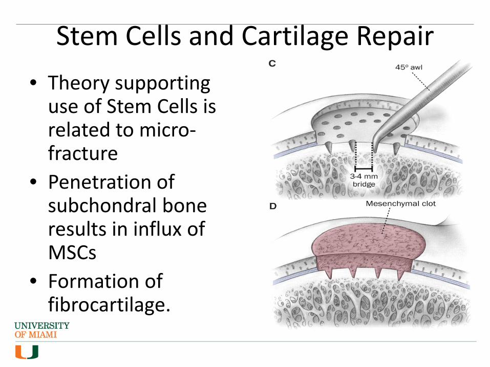
Stem Cells and Cartilage Repair• Theory supporting
use of Stem Cells is related to micro-fracture
• Penetration of subchondral bone results in influx of MSCs
• Formation of fibrocartilage.

Stem Cells and Cartilage Repair• Cartilage is secreted by
chondrocytes• Low mitotic activity• ACI - resection of healthy
cartilage and culture of Chondrocytes
• Replantation into patients• Good clinical results,
– Donor site morbidity– limited supply of chondrocytes,

Stem Cells and Cartilage Repair• MSCs versus ACI
– Multiple donor sites that won’t affect healthy cartilage, obviates need for staged procedure
– High proliferative capacity– Multipotency enabling it to differentiate into
chondrocytes
Stem Cells and Cartilage RepairThe Evidence – Clinical studies• Still very limited, good results in several animal studies • Nejadnik et al. AJSM, 2010 - ACI versus BMMSC • Gobbi et al. AJSM 2014
– 25 patients w/ symptomatic large chondral defects (ICRS grade 4)– Treated w/ MSCs and collagen matrix w/ 3 year follow up.– Results: MRIs showed complete filling of defect in 80% of patients,– Second look arthroscopy in 7 patients – smooth healthy, continuous cartilage
• Overall– Promising results based on clinical, MRI, histologic and arthroscopic evaluations– If results equivalent to ACI, valid replacement for this treatment– Still very limited, further studies to evaluate delivery and surgical methods– Patient selection important – OA, Malalignment, ligamentous laxity, BMI

Clinical Applications of BiologicsMeniscus Repair
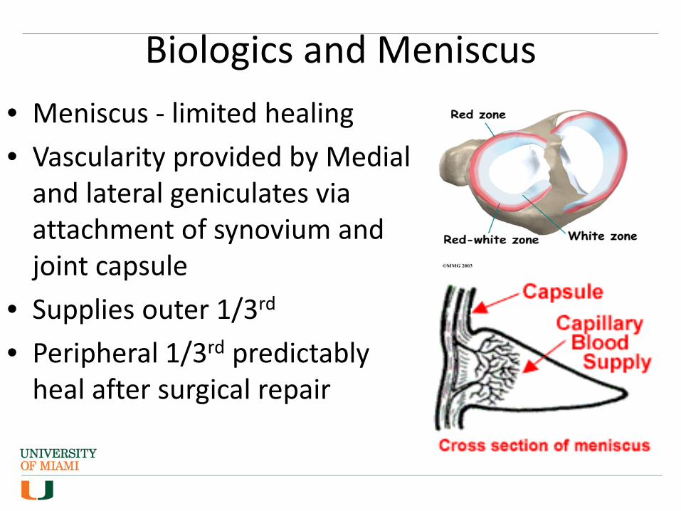
Biologics and Meniscus• Meniscus - limited healing • Vascularity provided by Medial
and lateral geniculates via attachment of synovium and joint capsule
• Supplies outer 1/3rd
• Peripheral 1/3rd predictably heal after surgical repair

PRP and Meniscus
• “Arthroscopic meniscal repair using an exogenous fibrin clot” CORR 1990– Meniscal tears augmented with fibrin clot,
showed 8% failure rate compared to 41% without

PRP and Meniscus
• Ishida et al Tissue Engineering 2007– 1.5mm full-thickness defect in avascular region of
rabbit meniscus– significantly greater scores for number of fibro-
chondrocytes and production of ECM in PRP group• Zellner et al J Biomed Mater Res 2010
– Similar study to Ishida using rabbits but 2cm punch defects in non-vascular zone
– No improvement with PRP compared to control• No Current Human studies

Stem Cells and Meniscus
• Meniscus repairs heal better when associated with concomitant ACL reconstruction – Clot formation and release of
MSCs from bone tunnels and notchplasty
• Argument for both PRP and MSCs

Stem Cells and Meniscus
Animal studies• BM, synovial and adipose derived MSCs have
been applied to tears in avascular zone of Rabbit an porcine menisci
• Apparent regeneration of meniscus tissue via Macroscopic, histologic and MRI evaluation

Stem Cells and Meniscus
Humans Studies• Very limited• Vangsness et al. JBJS 2014• RDBCT
– 3 groups of 20, patients were candidates for partial medial meniscectomy based on MRI and surgeon’s evaluation
– Group A – 50 million hMSCs, Group B 150 million hMSCs, Group C control of suspension w/o MSCs
– Intraarticular injection deliver 7-10 days after surgery• Results
– No adverse effects– 24% of patients in Group A had significant increase in meniscal volume
vs 0% in control (defined >15% increase in meniscal volume) @ 2 years

Summary• Safe• Good pre-clinical and lab background• Early clinical results for Stem Cells promising• Future
– Standardization– Delivery and Isolation techniques– Quantification – Larger studies, Better comparison groups