biofocus‘ molecular diagnostic panel - artoi · biofocus‘ molecular diagnostic panel ... immune...
TRANSCRIPT

Molecular detection of infectious diseases
Human & veterinary hereditary diseases / geneticpredisposition
Molecular Oncology
Biofocus‘ Molecular Diagnostic Panel
Dr. Lothar Prix
Biofocus GmbH, Recklinghausen, Germany
www.biofocus.de

Detection von Circulating Tumor Cells in blood
(CTCs) „residual disease“
Determination of „drug target“‐genes and
resistance‐markes
Goal: personalized therapy
Aims of Molecular Oncology

Overexpression / mutation of growth factor receptors
Typical Genetic Alterations in Tumor Cells
Erb‐b2, EGFR, c‐Kit, K‐ras
Genes associated with cell replicationTelomerase, c‐myc
Angiogenesis factors
VEGF, bFGF
Apoptosis‐Genes (= programed cell death)
Bcl‐2, p53, Survivin

Dissemination ‐Metastasis
CTCs:rare: 100 – 1000 per ml blood
sparely: 1 CTC in 10^6 to 10^7 WBCs
Isolation of CTCs is challenging

CTC isolationblood sample Molecularcharacterisation
Size differentiation
(eg filtration)
Isolation of CTCs from blood by positive selection
Immuno absorption
(eg. magnetic beads)
Wash

Molecular identification of captured cells as CTCs
Isolation of RNA from captured CTCs
Differential Gene expression CTCs Blood
Molecular Tumor markers by quantitative real‐time PCR:
• Cytokeratins (CK19, CK20)
• Cell cycle genes (c‐myc, erbb2, telomerase, survivin)
• tissue specific genes: PSA (prostate), G250 (renal), MART (melanoma)

Therapy Monitoring
Adjuvant therapy (after removal of primary tumor)
AdjuvantTherapy
Blood testCTCs
RESPONSE
NON‐RESPONSE
CTCs
CTCsResistance /Progress

Genetic Detection of CTCs from blood
Four‐Marker assay; Carcinoma (e.g. CK19, ERBB2, C‐MYC, Telomerase):
121/20060 %
1/701.4 %
≥ 2 Markerpositive
159/20080 %
Tumor CA Patientsn = 200
3/704.3 %
Normal-patientsn = 70
≥1 Markerpositive

Detection rate of CTCs
Average ca. 80 % in advanced tumors
0 10 20 30 40 50 60 70 80 90 100
breastovarian
endometrialuterine
cervicalcolorectal
prostatepancreatic
lungmesothelioma
gastricrenal
head&neckmelanoma
bladder
% CTC positiv e

Tumor cell dormancy hypothesis
Recurrence after long intervals of remission (decades)
Remaining tiny sources of tumor cells in the body („occult metastasis“)
More patients in remission have detectable CTCs than are at statistical risk for recurrence
Only a portion of patients with CTCs will develop recurrence
Limited life span of CTCs, process of constant cell death and replication
CTCs can be repeatedly detected in dormancy patients at steady low levels, (may at some point negative)
Increase of CTC‐markers may indicate relapse

Case report

Molecular characterization of CTCs
Gene expression analysis in CTCs for prediction of therapy resistance:
Drug Metabolizing Genes
Activation, Degradation, Detoxification
„Drug‐Target“ Genes
Cellular function inhibited by the drug
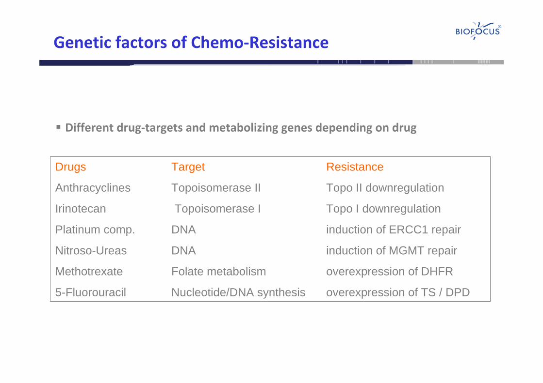
Genetic factors of Chemo‐Resistance
Different drug‐targets and metabolizing genes depending on drug
Drugs Target Resistance
Anthracyclines Topoisomerase II Topo II downregulation
Irinotecan Topoisomerase I Topo I downregulation
Platinum comp. DNA induction of ERCC1 repair
Nitroso-Ureas DNA induction of MGMT repair
Methotrexate Folate metabolism overexpression of DHFR
5-Fluorouracil Nucleotide/DNA synthesis overexpression of TS / DPD

Mechanims of Chemo‐Resistance
DRUG
DRUG
DRUGxLoss of drug receptors

Multi‐resistance in chemo‐treated vs. non‐treated patients
multidrug resistance factors: MDR1, MRP1, GST/GCS
no chemoth.
pretreated
0
20
40
60
80
100
% p
atie
nts≥1
res
ista
nce
fact
or

Involved multidrug‐resistance factors
MDR1: Taxane, Anthracycline, Mitomycin, Irinotecan, Vinca‐Alkaloides
MRP1: Anthracycline, Vinca‐Alkaloide, Methotrexat
GST/GCS: Anthracycline, Mitomycin, Vinca‐Alkaloides, Platin
no chemo
pretreated
0
50
100
150MDRMRPGST/GCS
resi
stan
ce fa
ctor
s: c
umm
ulat
ive

Impact of resistance factors on relapse‐free‐survival

Therapy: Prediction and Outcome
Partial responsesensitivHerceptinMamma
Complete responseMitomycin: sensitiv5-FU: sensitivEpirubicin: intermediate
Epirubicin + Mitomycin + 5-FUThymoma
Complete responsesensitivSorafenibMelanoma
Partial responseMTX: resistentCisplatin.: resistent
MTX + CisplatinMamma
Partial responseMTX: sensitivGemcitabin: sensitiv
MTX + GemcitabinMamma
Partial responseMTX: sensitiv5-FU: intermediate
MTX + 5-FUMamma
Partial responsesensitivHerceptinPancreas
Partial responsesensitivOxaliplatinColon
Progress / deathGemcitabine: resistentCyclophpos: sensitiv
Gemcitabine + Cyclophpos.Ovar
Progress / deathMitomycin: resistentCyclophpos.: resistent5-FU: intermediate
Mitomycin + 5-FU + Cyclophpos.Stomach
Progressresistent5-FUColon
ProgressresistentMitoxantronMamma
clinicaloutcome
Test-ResultTherapyTumor type

Limitations of the prediction model
Resistance Sensitivity:• Generally it is easier to predict resistance than response• Focus on major resistance pathways only
Alternative Therapies:Clinical response is observed despite positive resistance marker
Modulation of the resistance genes by alternative agents

Modulation of Resistance Factors
Alternative agents can modulate resistance genes:
MDR Curcumin, Acetogenin, Haelan
MRP Artemisinin, Haelan
GST Ellagic acid, Curcumin

Immune function testing by Cellular NK‐Test
Assay principle Tumor CellDye
Immune Cell
Tumor cells are stained by uptake of dye
Patients immune cells may attack tumor cells
Dye is released from destroyed tumor cells and quantified in supernatant
+/‐ stimulus

Cellular NK‐test
Testing of immune stimulative agents
upon requestInterleukin 2
upon requestCarnivora
upon requestFraxini mistletoe‐extract
upon requestEurixor mistletoe‐extract
upon requestHelixor mistletoe‐extract
routinelyLektinol mistletoe‐extract
routinelyIscador mistletoe‐extract
routinelyFactor AF2
routinelyThymus‐extract
Included in the assayAgent
basal
lektin
olisc
ador
helixo
rFak
tor AF2
Thymus
0
20
40
60
80w/o stimulationafter stimulation
% k
illin
g ra
te

Testing for alternative agents
Testing is possible for alternative agents with known genetic basis of action:
Curcumin
Haelan951
Acetogenin, Graviola
Dammarane sapogenins
C‐statin
IP6 (Inositol‐6‐Phosphate)
Quercetin
Agent
Ellagic Acid
Arglabin, Laetrile
Taurolidine
Indol‐3‐carbinol (I3C)
Vitamin C
Amygdalin B17
Artemsinin derivatives

Testing for Amygdalin B17
Measuring expression of Rhodanese and COX2expression in tumor cells aids in selection of
Amygdalin therapy

Testing for alternative agents e.g. Amygdalin B17
1. Suppressing expression of COX2 (Cyclooxigenase 2)
COX2:• Inflammation• Tumor promotion
CANCER CELLhigh levels of COX2 Amygdalin B17
suppresses COX2 expression
Amygdalin B17COX2
COX2

Testing for alternative agents e.g. Amygdalin B17
2. Detoxification by Rhodanese
beta‐glucosidasebeta‐glucuronidase
Rhodanese
Amygdalin
BenzaldehydeGlucoseCyanide
Poisening of cancer cells�
Detoxification of Cyanide
→ Resistance to B17

Rhodanese expression in CTCs
Overexpression of rhodanese in CTC is obviously rare
overexpressed
base‐level (equal to normal cells)
underexpressed
Rhodanese level
6 %
51 %
43 %
Observed in % of CTCs (n=45)