biochemistry of blood elements 624 vladimíra kvasnicová modified and reduced by eva samcová the...
TRANSCRIPT

Biochemistry of Blood Elements
624Vladimíra Kvasnicová
modified and reduced by Eva Samcová
The figure is found at http://www.biosbcc.net/doohan/sample/htm/Blood%20cells.htm (March 2007)

Blood Elements Count
erythrocytes 4 - 6 x 10⁶ / l
thrombocytes 150 - 400 x 103 / l
leukocytes 4 - 9 x 10³ / l
neutrophils 47 - 75 %eosinophils 1 - 4 %basophils 0 - 1 %lymphocytes 23 - 45 %monocytes 2 - 11 %
hematocrit
muži: 42-52 %
ženy: 37-47 %

2,000,000 erythrocytes / sec into circulation
• The lifetime of erythrocytes (red blood cells - RBC) is 120 days New erythrocytes – reticulocytes - contain more ribosomes and components of ER
• The life span of erythrocytes can be dramatically reduced in the case of a series of hemolytic anemia (in hemolytic anemia occurs increasingly hemolysis – destruction of red blood cells)
• The production of red blood cells is regulated by erythropoietin (EPO) - synthesized in the kidney
What to study
• Erythrocyte – structures• Erythrocyte - metabolism• Hemoglobin. Structure of hemoglobin• Saturation curve• Function of erythrocytes: Gas transport

Red Blood Cells(erythrocytes)
Structure large surface
(diffusion of gases)
cytoskeletal proteins (elasticity)
membrane as an osmometer
(Na+/K+-ATPase)
The figure is found at http://www.biosbcc.net/doohan/sample/htm/Blood%20cells.htm (March 2007)
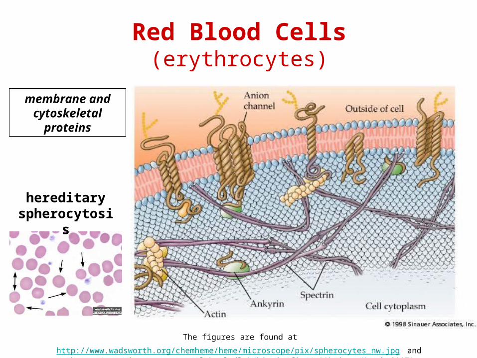
The figures are found at http://www.wadsworth.org/chemheme/heme/microscope/pix/spherocytes_nw.jpg and http://www.mie.utoronto.ca/labs/lcdlab/biopic/fig/4.23b.jpg (March 2007)
Red Blood Cells(erythrocytes)
membrane and cytoskeletal
proteins
hereditary spherocytos
is

Red Blood Cells(erythrocytes)
membrane transporters Na+/K+-ATPase (active transport)
GLUT-1 (insulin independent)
anion exchanger = band 3 protein (Cl-/HCO3-)
membrane antigens blood groups

The figure is found at http://www.life.umd.edu/classroom/bsci422/mosser/ABO.gif (March 2007)
Membrane antigens – example: ABO system
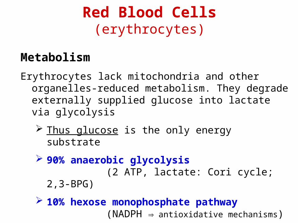
Red Blood Cells(erythrocytes)
Metabolism
Erythrocytes lack mitochondria and other organelles-reduced metabolism. They degrade externally supplied glucose into lactate via glycolysis
Thus glucose is the only energy substrate
90% anaerobic glycolysis(2 ATP, lactate: Cori cycle; 2,3-BPG)
10% hexose monophosphate pathway(NADPH antioxidative mechanisms)

Glucose 6-Phosphate DehydrogenaseGenetic Deficiency or Presence of Genetic Variants in
Erythrocytes
• Enzyme catalyzes the oxidation of G6P to 6-phosphogluconate and the reduction of NADP+ in major pathway of NADPH production – pentose cycle
• NADPH maintains glutathione in its reduced state• GSH is necessary for the integrity of the erythrocyte
membrane – cells more susceptible to oxidative damage by reactive oxygen species - to hemolysis.
• One of the most common enzymopathies.100 milion people suffer from this deficiency – particularly in the area Tropical Africa, Mediterranean region, some parts of Asia and the Black Population in America.
• Result is usually hemolytic anemia. 300 known genetic variants of this enzyme – wide range of symptoms.
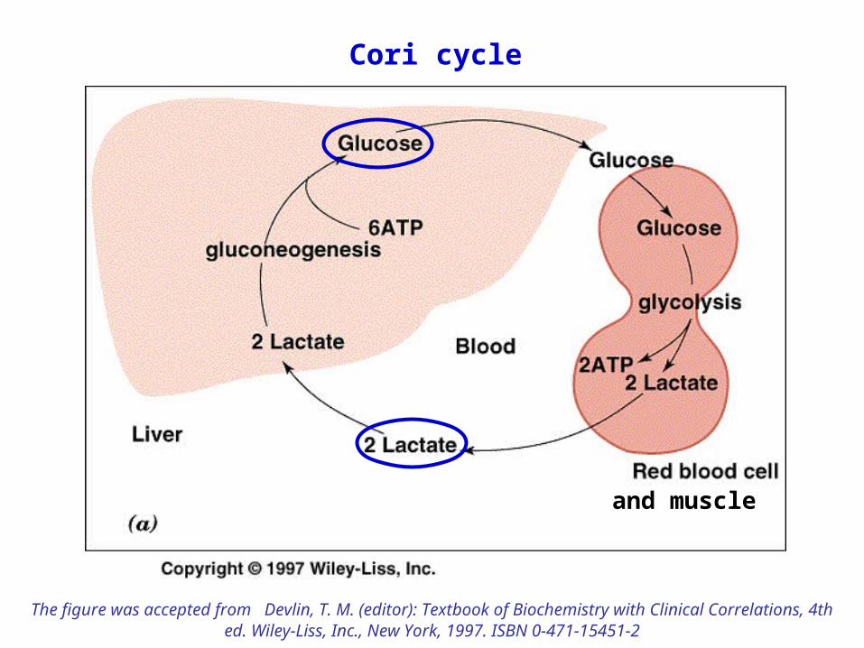
Cori cycle
and muscle
The figure was accepted from Devlin, T. M. (editor): Textbook of Biochemistry with Clinical Correlations, 4th ed. Wiley‑Liss, Inc., New York, 1997. ISBN 0‑471‑15451‑2

Red Blood CellsFunction
erythrocyte as a bag for hemoglobin
O2 → transport, reactive oxygen species (ROS)
CO2 → transport, formation of HCO3-
H+ → transport, maintaining pH(35% of blood buffering capacity)
• superoxide dismutase• catalase • glutathione peroxidase antioxidative system• glutathione reductase• methemoglobin reductase

antioxidative enzymes superoxide dismutase (SOD)
O2• + O2
• + 2 H+ H2O2 + O2
catalase (CAT)H2O2 + H2O2 2 H2O + O2
glutathione peroxidase (GPx)2 GSH + H2O2 GS-SG + 2 H2O2 GSH + R-O-OH GS-SG + H2O + ROH
glutathione reductaseGS-SG + NADPH+H+ 2 GSH + NADP+
methemoglobin reductase - in erythrocytesHb-Fe3+ + e- Hb-Fe2+ (coenzyme: NADH or NADPH)

The figure is found at http://www.med.unibs.it/~marchesi/ppp.html (March 2007)
Hexose Monophosphate
Pathway
glutathione reductase
GS-SG + NADPH+H+
2 GSH + NADP+
= „redox buffer“
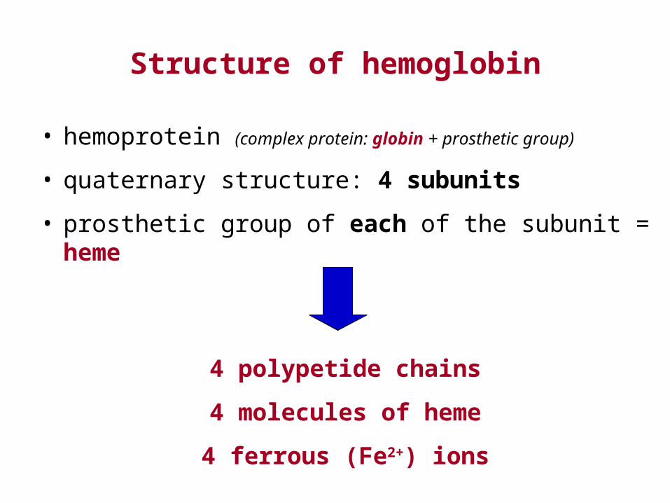
Structure of hemoglobin
• hemoprotein (complex protein: globin + prosthetic group)
• quaternary structure: 4 subunits
• prosthetic group of each of the subunit = heme
4 polypetide chains
4 molecules of heme
4 ferrous (Fe2+) ions

The figure is found at http://dtc.pima.edu/~biology/202alpha/lesson1/hemoglobin.jpg (March 2007)
Mr = 64 500

The figures are found at http://www.medical-definitions.net/images/hemoglobin.jpgand http://omlc.bme.ogi.edu/spectra/hemoglobin/hemestruct/heme-struct.gif (March 2007)
Pyrrole
hemoglobin
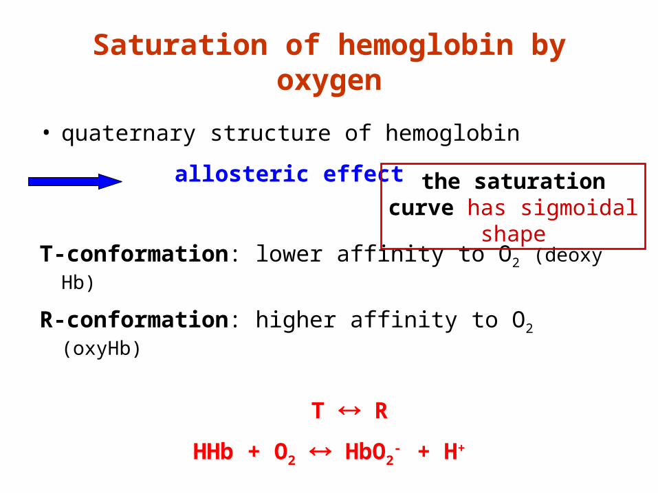
Saturation of hemoglobin by oxygen
• quaternary structure of hemoglobin
allosteric effect
T-conformation: lower affinity to O2 (deoxy Hb)
R-conformation: higher affinity to O2 (oxyHb)
T R
HHb + O2 HbO2- + H+
the saturation curve has sigmoidal
shape
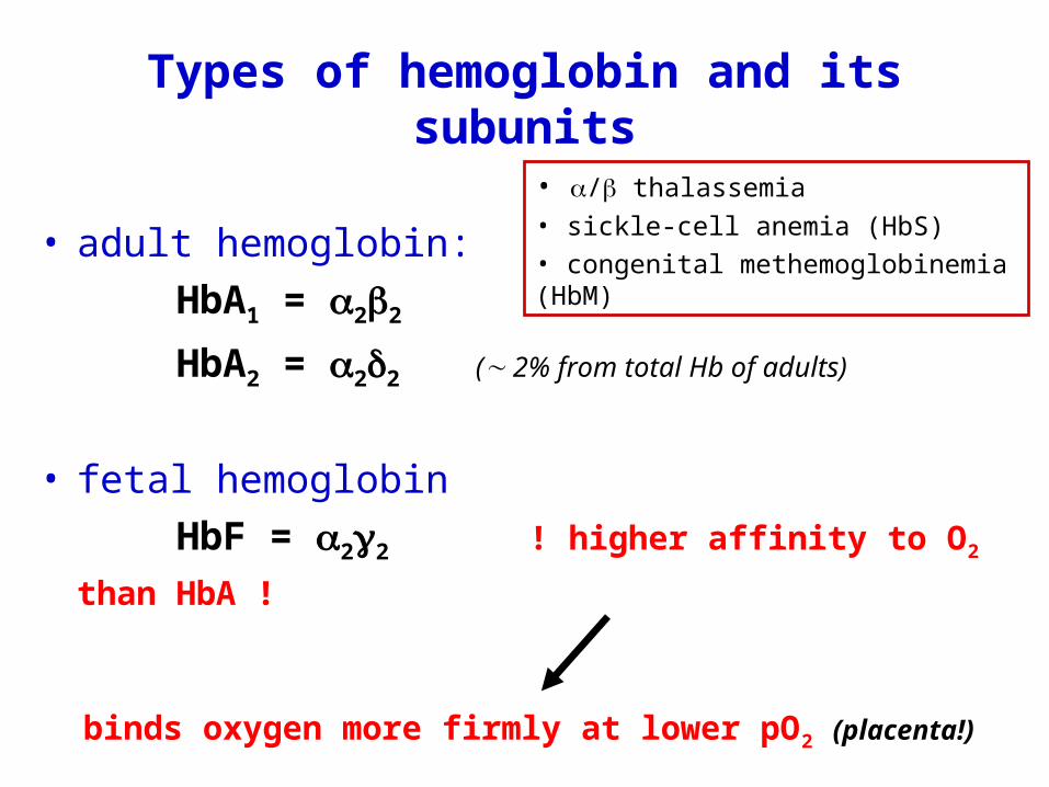
Types of hemoglobin and its subunits
• adult hemoglobin:
HbA1 = 22
HbA2 = 22 ( 2% from total Hb of adults)
• fetal hemoglobin
HbF = 22 ! higher affinity to O2 than
HbA !
binds oxygen more firmly at lower pO2 (placenta!)
• / thalassemia• sickle-cell anemia (HbS)• congenital methemoglobinemia (HbM)

The figure is found at http://www.labcorp.com/datasets/labcorp/html/img/fethgb.jpg (March 2007)

Synthesis of hemoglobin
• bone marrow
• in erytroblasts, not in erythrocytes
• 4 individual subunits are connected by noncovalent bonds to form tetramer of Hb
• hemoglobin is an intracellular protein: within ery
concentration of Hb in blood:
female 120 – 162 g/l
male 135 – 172 g/l

Synthesis of hemoglobinDisorders:
• THALASSEMIA = group of genetically determined disorders: absence or reduced synthesis of a globin chain ( or thalassemia)
• ANEMIA (= decreased oxygen-carrier capacity of blood)
• Hemolytic anemia is a condition in which red blood cells are destroyed and removed from the bloodstream before their normal lifespan is over.
sideropenic anemia – insufficient concentration of Fe
sickle cell anemia – point mutationin the -globin gene forms abnormalHbS (Glu → Val)

Transport of blood gases
Air composition:
78% N2 21% O2 1% water, inert gases, CO2 (0,04%)
Air pressure:
1 atm = 101 325 Pa (~ 101 kPa) = 760 Torr (= mmHg)
1 mmHg = 0,1333 kPa
1 kPa = 7,5 mmHg
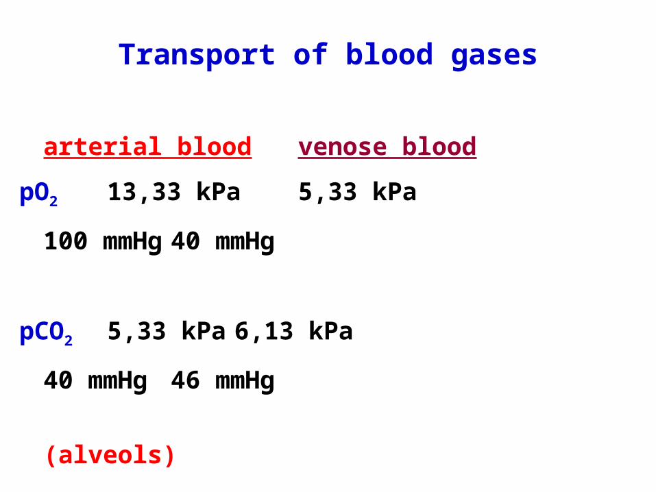
Transport of blood gases
arterial blood venose blood
pO2 13,33 kPa 5,33 kPa
100 mmHg 40 mmHg
pCO2 5,33 kPa 6,13 kPa
40 mmHg 46 mmHg
(alveols)

Transport of blood gases- function of hemoglobin -
• it transports O2 and part of CO2 (and CO)
• it binds H+ (reacts as a buffer)
• O2 and CO: bound to Fe2+ in heme → 4 O2 / 1 Hb
„oxyhemoglobin“ HbO2 /„carbonylhemoglobin“ COHb
• CO2 is bound to globin! (-NH2 of side chains of amino acids)
„carbaminohemoglobin“ HbCO2
• H+ is bound to residues of His„deoxyhemoglobin“ HHb

Transport of blood gases- transport of CO2 -
1. largely in a form of HCO3- (~ 70%)
CO2 + H2O H2CO3 HCO3- + H+
enzyme: carbonic anhydrase spontaneous dissociation
(in erytrocytes)
2. bound to hemoglobin (~ 23%)
3. freely dissolved (~ 7%)
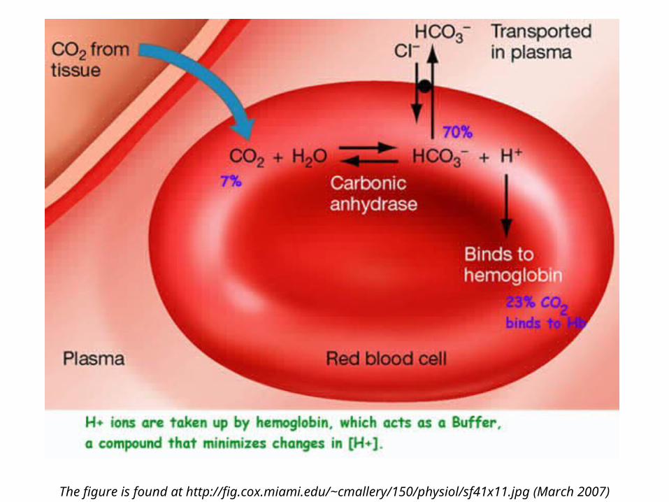
The figure is found at http://fig.cox.miami.edu/~cmallery/150/physiol/sf41x11.jpg (March 2007)

Transport of blood gases - reactions in erytrocytes -
tissues:
CO2 + H2O → H2CO3 → HCO3- + H+
H+ + HbO2- → HHb + O2 → aerobic metabolism
(HCO3- formed in the erythrocyte is then transported to plasma by an anion
exchanger in exchange with Cl-; this process is called Hamburger´s effect or „chloride shift“; in the lungs HCO3
- is transported back into the erythrocyte by the same exchange with Cl-)
lungs:
HHb + O2 → HbO2- + H+
H+ + HCO3- → H2CO3 → H2O + CO2 → excreted

The figure is from http://science.kennesaw.edu/~jdirnber/Bio2108/Lecture/LecPhysio/42-29-BloodCO2Transport-AL.gif (March 07)
O2
O2

Hemoglobin saturation curve- saturation with oxygen -
The figure is found at http://employees.csbsju.edu/hjakubowski/classes/ch331/bind/MbHbbindcurve.gif(March 2007)

The figure is found at http://dr-amy.com/rich/oxygen/fig1.gif (March 2007)
Right shifted = oxygen is more easily released from Hb but worse bound to it

Saturation of hemoglobin with oxygen
Factors affecting the saturation:
alkaline pH and pO2 stabilize R-conformation
(IN LUNGS)
acidic pH, pCO2, temperature and 2,3-BPG
stabilize T-conformation, i.e. deoxyHb
(IN PERIPHERY)
shift of the saturation curve toward right

The figure is found at http://employees.csbsju.edu/hjakubowski/classes/ch331/bind/MbHbbindcurve.gif(March 2007)
Bohr´s effect= the saturation of Hb by O2 lowers because lowering
pH(shift toward right)

Patological forms of hemoglobin
1. methemoglobin (over 3%)metHb
Fe3+ instad of Fe2+
unable to transport oxygen !!!
2. glycohemoglobin (over 6%)
HbA1c
after long term increased glycemia (Glc bound to Hb)
3. carbonylhemoglobin (over 2%) COHb
after CO poisoning
4. sulfhemoglobin, cyanhemoglobin
poisoning by H2S, HCN or by cyanides
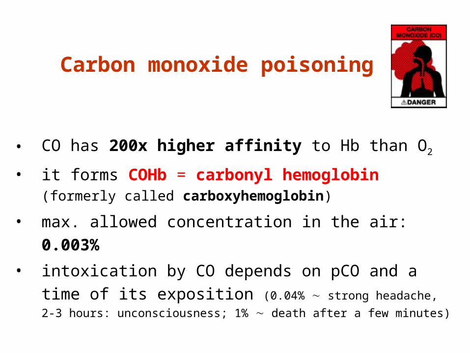
Carbon monoxide poisoning
• CO has 200x higher affinity to Hb than O2
• it forms COHb = carbonyl hemoglobin(formerly called carboxyhemoglobin)
• max. allowed concentration in the air: 0.003%
• intoxication by CO depends on pCO and a time of its exposition (0.04% strong headache, 2-3
hours: unconsciousness; 1% death after a few minutes)

The figure is found at http://dr-amy.com/rich/oxygen/fig1.gif (March 2007)

Carbon monoxide poisoning
may result due to:
• exposure to automobile exhaust
• smoke inhalation
• an improperly ventilated gas heater
• or other appliance
= incomplete burning(incomplete oxidation of organic material)

Saturation of
hemoglobin with CO
The figure is found at http://www.uhseast.com/134221.cfm
(March 2007)
COHb / total Hb (ratio in %)
physiological value:
2%
TREATEMENT
• fresh air
• exposure to high concentrations of oxygen (the 100% oxygen is administered by a face mask)
it is recommended in patients who have a history of loss of consciousness, carbonyl hemoglobin saturation greater than 25%, metabolic acidosis and cerebellar findings on neurologic exam
Carbon monoxide poisoning