biochemistry i 3 . stage
TRANSCRIPT

BIOCHEMISTRY I
3rd. Stage Lec.
ENZYMES
Biomedical Importance:
Enzymes, which catalyze the biochemical reactions, are essential for life. They
participate in the breakdown of nutrients to supply energy and chemical building
blocks; the assembly of those building blocks into proteins, DNA, membranes, cells,
and tissues; and the harnessing of energy to power cell motility, neural function, and
muscle contraction.
The vast majority of enzymes are proteins. Notable exceptions include ribosomal RNAs
and a handful of RNA molecules imbued with endonuclease or nucleotide ligase
activity known collectively as ribozymes.
The ability to detect and to quantify the activity of specific enzymes in blood, other
tissue fluids, or cell extracts provides information that complements the physician’s
ability to diagnose and predict the prognosis of many diseases. Further medical
applications include changes in the quantity or in the catalytic activity of key enzymes
that can result from genetic defects, nutritional deficits, tissue damage, toxins, or
infection by viral or bacterial pathogens (eg, Vibrio cholerae). Medical scientists
address imbalances in enzyme activity by using pharmacologic agents to inhibit
specific enzymes and are investigating gene therapy as a means to remedy deficits in
enzyme level or function.
In addition to serving as the catalysts for all metabolic processes, their impressive
catalytic activity, substrate specificity, and stereospecificity enable enzymes to fulfill
key roles in additional processes related to human health and well-being. Proteases and
amylases augment the capacity of detergents to remove dirt and stains, and enzymes
play important roles in producing or enhancing the nutrient value of food products for
both humans and animals. The protease rennin, for example, is utilized in the
production of cheeses while lactase is employed to remove lactose from milk for the
benefit of lactose-intolerant persons deficient in this hydrolytic enzyme. Finally,
stereospecific enzyme catalysts can be of particular value in the biosynthesis of complex
drugs or antibiotics.
The Nature of Enzymes:
Enzymes may be defined as biocatalysts synthesized by living cells. They are
protein in nature (exception: ribozymes which are RNA in nature), colloidal and
thermolabile in character, and specific in their action.
Most enzymes are globular proteins that exhibit at least tertiary structure with a
complex 3-D configuration, capable of binding substrate molecules to a part of their
surface. The enzymes that catalyze the conversion of one or more compounds
(substrates) into one or more different compounds (products) generally enhance the
rates of the corresponding noncatalyzed reaction by factors of 1016 or more. Like almost
all catalysts, enzymes are neither consumed nor permanently altered as a consequence
of their participation in a reaction.
Unlike most catalysts used in synthetic chemistry, enzymes are specific not simply for
the type of reaction catalyzed, but also for a single substrate or a small set of closely
related substrates.
Enzymes are also stereospecific catalysts that typically catalyze reactions of only one
stereoisomer of a given compound (for example, D- but not L-sugars, L- but not D-
amino acids).

Dr. Ali A. Kasim College of Pharmacy/ University of Baghdad
Enzymes Nomenclature and Classification:
The early discovered enzymes were designated by first appending the suffix –ase to
a descriptor for the type of reaction catalyzed. For example, enzymes that remove
hydrogen atoms are generally referred to as dehydrogenases, enzymes that hydrolyze
proteins as proteases, and enzymes that catalyze rearrangements in configuration as
isomerases, and so on. These general descriptors to be preceded with terms indicating
the substrate on which the enzyme acts (xanthine oxidase), its source (pancreatic
ribonuclease), its mode of regulation (hormone-sensitive lipase). Where needed,
alphanumeric designators are added to the end of the enzyme name to identify multiple
forms of an enzyme (eg, RNA polymerase III; protein kinase Cb).
While simple and straightforward, as more enzymes were discovered these early
naming conventions increasingly resulted in the appearance of multiple names for the
same enzyme and duplication in the naming of enzymes exhibiting similar catalytic
capabilities. To address these problems, the International Union of Biochemistry (IUB)
developed a definite system of enzyme nomenclature in which each enzyme has a
unique name and code number (the enzyme commission (E.C.) number) that identify
the type of reaction catalyzed by the enzyme and the substrates involved. E.C. number
is a unique identifier for each enzyme classified according to IUB system.
According to IUB system enzymes are grouped into the following six classes:
1. Oxidoreductases - enzymes that catalyze oxidations and reductions.
2. Transferases - enzymes that catalyze transfer of moieties such as glycosyl, methyl,
or phosphoryl groups.
3. Hydrolases - enzymes that catalyze hydrolytic cleavage of C-C, C-O, C-N, and other
covalent bonds.
4. Lyases - enzymes that catalyze non-hydrolytic cleavage of C-C, C-O, C-N, and other
covalent bonds by atom (functional group) elimination, generating double bonds.
5. Isomerases - enzymes that catalyze geometric or structural changes within a
molecule.
6. Ligases - enzymes that catalyze the joining together (ligation) of two molecules in
reactions coupled to the hydrolysis of ATP.
The IUB name of hexokinase is ATP:D-hexose 6-phosphotransferase E.C. 2.7.1.1.
This name identifies hexokinase as a member of class 2 (transferases), subclass 7
(transfer of a phosphoryl group), sub-subclass 1 (alcohol is the phosphoryl acceptor),
and sub-class 1 (alcohol phosphorylated is on carbon six of a hexose). Despite their
clarity, IUB names are lengthy and relatively cumbersome, so we generally continue to
refer to hexokinase and many other enzymes by their traditional, albeit sometimes
ambiguous names. On the other hand, E.C. numbers are particularly useful to
differentiate enzymes with similar functions or catalytic activities.
Dr. Ali A. Kasim College of Pharmacy/ University of Baghdad
Prosthetic Groups, Cofactors, and Coenzymes Role in Catalysis:
Many enzymes contain small organic molecules or metal ions that participate
directly in substrate binding or in catalysis. Termed prosthetic groups, cofactors, and
coenzymes. The term holoenzyme refers to the active enzyme with its non-protein
component, whereas the enzyme without its non-protein moiety is termed an
apoenzyme and is inactive.
Prosthetic groups are tightly and stably incorporated into a protein’s structure by
covalent or noncovalent forces. They are either small organic molecules or inorganic
metal ions. Examples include pyridoxal phosphate, flavin mononucleotide (FMN),
flavin adenine dinucleotide (FAD), thiamin pyrophosphate, and biotin. Metal ions
constitute the most common type of prosthetic group. The roughly one-third of all
enzymes that contain tightly bound Fe, Co, Cu, Mg, Mn, and Zn are termed
metalloenzymes. Prosthetic groups may facilitate the binding and orientation of substrates, or by acting
as Lewis acids or bases to render substrates more electrophilic (electron-poor) or
nucleophilic (electron-rich), and hence more reactive.
Cofactors can associate reversibly with the enzymes or with the substrates. While
cofactors serve functions similar to those of prosthetic groups, they bind in a transient,
dissociable manner. Therefore, unlike associated prosthetic groups, cofactors must be
present in the medium surrounding the enzyme for catalysis to occur. The most
common cofactors are metal ions. Enzymes that require a metal ion cofactor are termed
metal-activated enzymes to distinguish them from the metalloenzymes for which
bound metal ions serve as prosthetic groups.
Coenzymes are small organic molecules that serve as recyclable shuttles
(transporters) that transport many substrates from one point within the cell to another.
The function of these shuttles is twofold. First, they stabilize species such as hydrogen
atoms (FADH) or hydride ions (NADH) that are too reactive to persist for any
significant time in the presence of the water or organic molecules that permeate cells.
Second, they serve as an adaptor or handle that facilitates the recognition and binding
of small chemical groups, such as acetate (coenzyme A) or glucose (UDP), by their
target enzymes.
The Active Site:
An active site is a cleft or pocket on the surface of the enzyme. It is the recognition
site for binding substrates; the specificity of binding depends on the precisely defined
arrangement of atoms in an active site. Because the enzyme and the substrate interact
by means of short-range forces that require close contact, a substrate must have a
matching shape to fit into the site.
An active site also xc. Within the active site, substrates are brought into close proximity
to one another in optimal alignment with the cofactors, prosthetic groups, and amino
acid side chains that participate in catalyzing the transformation of substrates into
products. Catalysis is further enhanced by the capacity of the active site to shield
substrates from water and generate an environment whose polarity, hydrophobicity,
acidity, or alkalinity can differ markedly from that of the surrounding cytoplasm.
Dr. Ali A. Kasim College of Pharmacy/ University of Baghdad
Enzymes Mechanisms to Facilitate Catalysis:
Enzymes use combinations of four general mechanisms to achieve dramatic
enhancements of the rates of chemical reactions.
1. Catalysis by Proximity
For molecules to interact, they must come within bond-forming distance of one
another. The higher their concentration, the more frequently they will encounter one
another, and the greater will be the rate of their reaction. When an enzyme binds
substrate molecules at its active site, it creates a region of high local substrate
concentration in which the substrate molecules reactive groups are oriented in a position
ideal for them to chemically interact. This results in rate enhancements of at least a
thousand fold over the same non-enzyme-catalyzed reaction.
2. Acid-Base Catalysis
In addition to contributing to the ability of the active site to bind substrates, the
ionizable functional groups of aminoacyl side chains, and where present of prosthetic
groups, can contribute to catalysis by acting as acids or bases. We distinguish two types
of acid-base catalysis. Specific acid or base catalysis refers to reactions for which the
only participating acid or base are protons or hydroxide ions. The rate of reaction thus
is sensitive to changes in the concentration of protons or hydroxide ions, but is
independent of the concentrations of other acids (proton donors) or bases (proton
acceptors) present in the solution or at the active site. Reactions whose rates are
responsive to all the acids or bases present are said to be subject to general acid or
base catalysis. Enzymes of the aspartic protease family, which includes the digestive enzyme pepsin,
the lysosomal cathepsins, and the protease produced by the human immunodeficiency
virus (HIV) share a common mechanism that employs two conserved aspartyl residues
as acid-base catalysts. Catalysis by aspartic proteases involves the direct hydrolytic
attack of water on a peptide bond.
3. Catalysis by Strain
Enzymes that catalyze lytic reactions, chemical transformations that involve
breaking a covalent bond, typically bind their substrates in a conformation that is
somewhat unfavorable for the bond targeted for cleavage. This strained conformation
represents the transition state, or midway point, in the transformation of substrates to
products. The resulting strain selectively stretches or distorts the targeted bond,
weakening it and making it more vulnerable to cleavage.
4. Covalent Catalysis
The process of covalent catalysis involves the formation of a covalent bond between
the enzyme and one or more substrates. The modified enzyme thus becomes a reactant.
Covalent catalysis introduces a new reaction pathway whose activation energy is
lower—and the reaction therefore is faster—than the reaction pathway in homogeneous
solution. The chemically modified state of the enzyme is, however, transient. Covalent
catalysis often follows a “ping-pong” mechanism in which the first substrate is bound
and its product released prior to the binding of the second substrate. Completion of the
reaction returns the enzyme to its original, unmodified state. Its role thus remains
catalytic. Covalent catalysis is particularly common among enzymes that catalyze
group transfer reactions.

Dr. Ali A. Kasim College of Pharmacy/ University of Baghdad
Enzyme-Substrate Interaction Models:
Two models have been proposed to explain how an enzyme binds its substrate. The
old model "lock and key model" accounted for the specificity of enzyme-substrate
interactions, the implied rigidity of the enzyme’s active site failed to account for the
dynamic changes that accompany substrate binding and catalysis (Figure 1). The other
model "induced fit model", which states that when substrates approach and bind to an
enzyme they induce a conformational change that is analogous to placing a hand
(substrate) into a glove (enzyme). The enzyme in turn induces reciprocal changes in its
substrates, joining the energy of binding to facilitate the transformation of substrates
into products (Figure 1). The induced fit model has been fully confirmed by biophysical
studies of enzyme motion during substrate binding.
Figure 1: Enzyme-substrate binding models. (a) Lock and Key Model (b) Induced
Fit Model.
Isoenzymes:
Isoenzymes are physically distinct versions of a given enzyme, each of which
catalyzes the same reaction. They are sometimes referred to as isozymes. Isoenzymes
are produced by different genes and are not redundant despite their similar functions.
They occur in many tissues throughout the body and are important for different
developmental and metabolic processes.
Isoenzymes have different substrates, and they may also possess differences in
properties such as sensitivity to particular regulatory factors or substrate affinity (eg,
hexokinase and glucokinase) that adapt them to specific tissues or circumstances rather
than distinct substrate specificities.
Dr. Ali A. Kasim College of Pharmacy/ University of Baghdad
Detection of Enzymes:
The relatively small quantities of enzymes present in cells hinder determination of
their presence and concentration. However, enzymes ability to rapidly transform
thousands of molecules of a specific substrate into products had enabled their detection
and quantification. Under appropriate conditions, the rate of the catalytic reaction being
monitored is proportionate to the amount of enzyme present, which allows its
concentration to be calculated. Assays of the catalytic activity of enzymes are
frequently used in research and clinical laboratories.
Overview of amino acids metabolism - Amino acids serve as substrates for the synthesis of protein, - Amino acids provide nitrogen for the synthesis of other nitrogen-containing compounds, - Amino acids are catabolized as fuels.
Classification of amino acids:
1. Chemical classification:
According to the chemistry of the side chains.
According to polarity of side chains.
2. Nutritional classification:
Essential Non-essential
NOTE:
All of the 20 amino acids present in proteins are essential for health. Some clinical conditions are associated with amino acid deficiency states, such as
Kwashiorkor and Marasmus diseases.
Kwashiorkor is protein deficiency with adequate energy intake whereas Marasmus is inadequate energy intake in all forms, including protein.
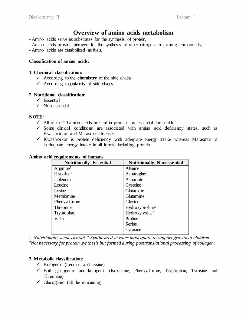
Amino acid requirements of humans
Nutritionally Essential Nutritionally Nonessential
Arginine1 Histidine1
Isoleucine Leucine
Lysine Methionine Phenylalanine
Threonine Tryptophan
Valine
Alanine Asparagine
Aspartate Cysteine
Glutamate Glutamine Glycine
Hydroxyproline2 Hydroxylysine2
Proline Serine Tyrosine
1“Nutritionally semiessential.” Synthesized at rates inadequate to support growth of children. 2Not necessary for protein synthesis but formed during posttranslational processing of collagen.
3. Metabolic classification:
Ketogenic (Leucine and Lysine)
Both glucogenic and ketogenic (Isoleucine, Phenylalanine, Tryptophan, Tyrosine and
Threonine)
Glucogenic (all the remaining)
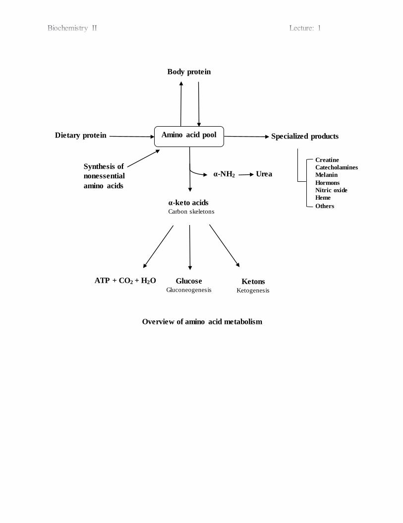
Overview of amino acid metabolism
Amino acid pool
Body protein
α-keto acids Carbon skeletons
Synthesis of
nonessential
amino acids
Dietary protein Specialized products
Glucose Gluconeogenesis
Ketons Ketogenesis
ATP + CO2 + H2O
α-NH2 Urea
Creatine
Catecholamines
Melanin
Hormons
Nitric oxide
Heme
Others

Amino acids pool
The amount of free amino acids distributed throughout the body is called amino acid pool.
Plasma level for most amino acids varies widely throughout the day. It ranges between 4–8 mg/dl. It tends to increase in the fed state and tends to decrease in the post absorptive state.
Sources of amino acid pool
1. Dietary protein.
2. Breakdown of tissue proteins. 3. Biosynthesis of nonessential amino acids.
NOTE:
In general, the rate of protein synthesis equals the rate of degradation (steady-state). However,
there is a constant need for dietary intake of protein because:
Some amino acids are also used for energy production and storage and for synthesis of
non-protein molecules.
There are situations where protein synthesis must exceed protein degradation, such as
during growth, pregnancy, and recovery from illness.
Digestion of dietary proteins
Protein digestion begins in the stomach.
The highly acidic environment of the stomach denatures proteins. Denatured proteins are susceptible to proteolytic digestion.
The primary enzyme involved in proteolytic digestion is pepsin, which catalyzes the nonspecific hydrolysis of peptide bonds at an optimal pH of 2.
In the lumen of the small intestine, the pancreas secretes zymogens of trypsin (Trypsin
cleaves peptide chains mainly at the carboxyl side of the amino acids lysine or arginine, except when either is followed by proline.), chymotrypsin (Chymotrypsin prefers large
hydrophobic residues. Chymotrypsin preferentially catalyzes the hydrolysis of peptide bonds involving tyrosine, phenylalanine, and tryptophan), elastase etc.
Proteolytic enzymes break the proteins down into free amino acids as well as dipeptides
and tripeptides, which in turn are absorbed by the intestinal mucosa cells and subsequently are released into the blood stream where they are absorbed by other tissues.
Turnover of cellular proteins
Turnover of cellular proteins (continuous degradation and synthesis) occur in all forms of
life. Each day, humans turn over 1% to 2% of their total body protein, principally muscle
protein. Approximately 75% of the amino acids liberated by protein degradation are reutilized, the
remaining excess free amino acids are not stored for future use (i.e. amino acids not
immediately incorporated into new protein are rapidly degraded). The relative susceptibility of a protein to degradation is expressed as its half-life (t1/2).
Half-lives of proteins may range from under 30 minutes to over 150 hours, or even the life time of an organ.

Cellular functions of protein degradation
1. The recycling of amino acids.
2. Elimination of misfolded and damaged proteins (due to environmental toxins, translation errors and genetic mutations) when cannot be repaired.
3. Regulation of cellular metabolism and cellular growth (increase or decrease the number of enzyme molecules and regulatory substances).
4. The generation of active proteins (the proteolytic cleavage of the precursor generates an
active enzyme). 5. The regulation of cell division ( degradation of cyclins)
Pathways of protein degradation
A. General “non-specific” protein degradation takes place in lysosomes (specialized
organelles that operate at low pH (to denature proteins) and contain proteases for proteins, lipases for lipids, and many other hydrolases (~ 50 total)). By this pathway extracellular,
membrane-associated, and long-lived intracellular proteins are degraded in lysosomes by ATP-independent processes.
Many normal and pathological processes involve increased lysosomal activity, including:
Disuse atrophy of muscles and regression of the uterus after childbirth (the muscular mass
of the uterus is reduced from about 2 kg to about 50 g in just nine days).
Chronic inflammatory diseases such as rheumatoid arthritis involve extracellular release
lysosomal enzymes, which attack surrounding tissues.
B. Controlled or programmed protein degradation involves the Ubiquitin-Proteasome
system. Degradation of regulatory proteins with short half-lives and of abnormal or misfo lded
proteins occurs in the cytosol within the proteasomes, and requires ATP and ubiquitin
(Ub) (a small polypeptide found in all eukaryotic cells, by which the cell distinguish between functional proteins and intracellular proteins that need to be degraded).
Three different enzymes add progressively more Ub molecules, in tandem chains, an
energy-requiring process (ATP). The more Ub molecules attached, the more rapid the
degradation.
Carboxyl terminal of Ub is attached to the ε-amino groups of lysyl residues in the target
protein (isopeptide bond). The residue present at its amino terminus affects whether a protein is ubiquitinated. Amino
terminal Met, or Ser residues retard, whereas Asp, or Arg accelerate ubiquitination. Subsequent degradation of Ub-tagged proteins takes place in the proteasome, a
macromolecule that also is ubiquitous in eukaryotic cells.
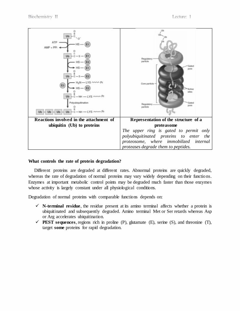
Reactions involved in the attachment of
ubiquitin (Ub) to proteins
Representation of the structure of a
proteasome
The upper ring is gated to permit only
polyubiquitinated proteins to enter the proteosome, where immobilized internal proteases degrade them to peptides.
What controls the rate of protein degradation?
Different proteins are degraded at different rates. Abnormal proteins are quickly degraded,
whereas the rate of degradation of normal proteins may vary widely depending on their functions.
Enzymes at important metabolic control points may be degraded much faster than those enzymes
whose activity is largely constant under all physiological conditions.
Degradation of normal proteins with comparable functions depends on:
N-terminal residue, the residue present at its amino terminal affects whether a protein is ubiquitinated and subsequently degraded. Amino terminal Met or Ser retards whereas Asp or Arg accelerates ubiquitination.
PEST sequences, regions rich in proline (P), glutamate (E), serine (S), and threonine (T), target some proteins for rapid degradation.

Interorgan exchange of amino acids
The net balance between release from endogenous protein stores and utilization by various
tissues keeps the steady-state concentrations of circulating plasma amino acids between meals.
In the postabsorptive state, free amino acids, particularly alanine and glutamine, are released from muscle into the circulation. Alanine is extracted primarily by the liver, and glutamine is extracted by the gut and the kidney,
both of which convert a significant portion to alanine. Alanine is a key gluconeogenic amino acid, and the rate of hepatic gluconeogenesis from alanine is far higher than from all other amino acids.
Glutamine also serves as a source of ammonia for excretion by the kidney. Branched-chain amino acids, particularly valine, are released by muscle and taken up predominantly by the brain.
The kidney provides a major source of serine for uptake by peripheral tissues, including liver and muscle.
Interorgan amino acid exchange in normal postabsorptive humans
In the fed state (following a protein-rich meal), the splanchnic tissues release amino acids while the peripheral muscles extract amino acids.
Glu or Asp + Pyruvate
α-ketoglutarate or
oxaloacetate
Transaminase
+ Alanine
Biosynthesis of Nonessential Amino Acids Humans do not have the ability to synthesize 10 of the necessary 20 amino acids and must
obtain them from the diet. These 10 are termed the nutritionally essential amino acids. The number of enzymes required by cells to synthesize the nutritionally essential amino acids
is large in relation to the number of enzymes required to synthesize the nutritionally nonessentia l amino acids. This suggests that there is a positive survival advantage in retaining the ability to manufacture ‘easy’ amino acids while losing the ability to make ‘difficult’ amino acids.
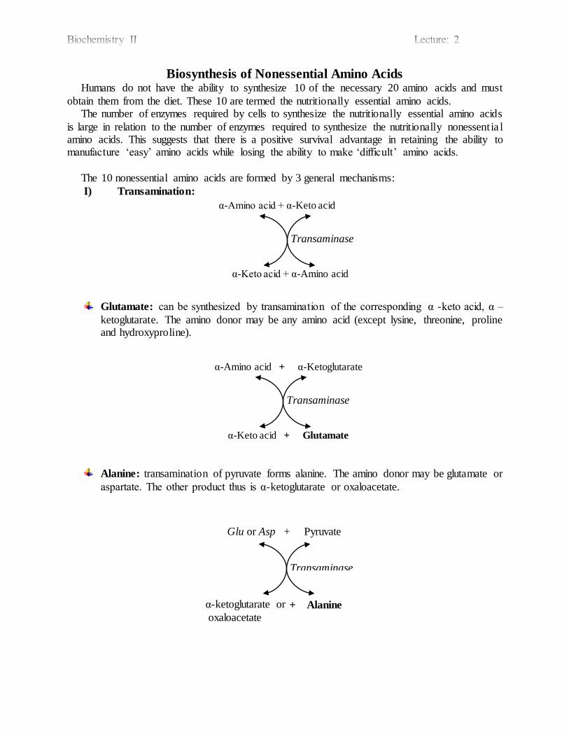
The 10 nonessential amino acids are formed by 3 general mechanisms:
I) Transamination:
Glutamate: can be synthesized by transamination of the corresponding α -keto acid, α –
ketoglutarate. The amino donor may be any amino acid (except lysine, threonine, proline and hydroxyproline).
Alanine: transamination of pyruvate forms alanine. The amino donor may be glutamate or
aspartate. The other product thus is α-ketoglutarate or oxaloacetate.
α-Amino acid + α-Keto acid
α-Keto acid + α-Amino acid
Transaminase
α-Amino acid + α-Ketoglutarate
α-Keto acid + Glutamate
Transaminase
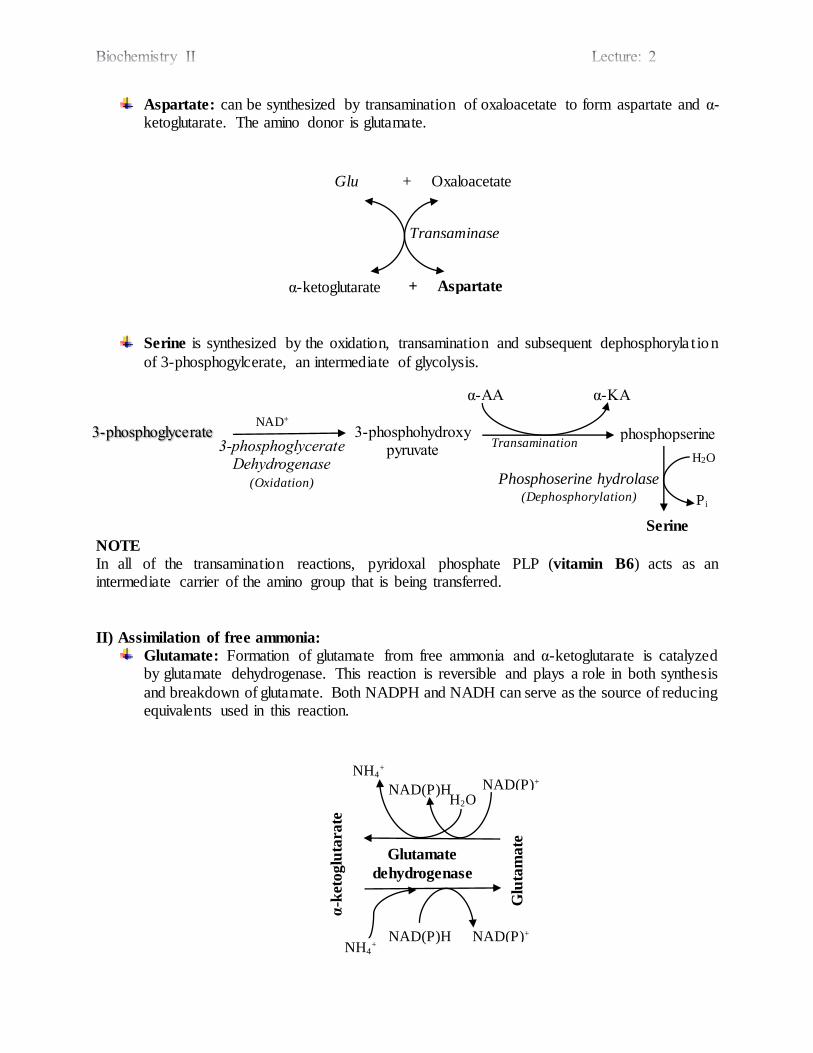
Aspartate: can be synthesized by transamination of oxaloacetate to form aspartate and α-ketoglutarate. The amino donor is glutamate.
Serine is synthesized by the oxidation, transamination and subsequent dephosphoryla t ion
of 3-phosphogylcerate, an intermediate of glycolysis.
NOTE
In all of the transamination reactions, pyridoxal phosphate PLP (vitamin B6) acts as an intermediate carrier of the amino group that is being transferred.
II) Assimilation of free ammonia:
Glutamate: Formation of glutamate from free ammonia and α-ketoglutarate is catalyzed by glutamate dehydrogenase. This reaction is reversible and plays a role in both synthesis
and breakdown of glutamate. Both NADPH and NADH can serve as the source of reducing equivalents used in this reaction.
Glutamate
dehydrogenase
NAD(P)+
H2O NAD(P)H
NH4+
NH4+
NAD(P)H NAD(P)+
Glu
tam
ate
α-k
etoglu
tara
te
Glu + Oxaloacetate
α-ketoglutarate
Transaminase
+ Aspartate
3-phosphoglycerate 3-phosphohydroxy pyruvate
NAD+
3-phosphoglycerate Dehydrogenase
(Oxidation)
Transamination phosphopserine
α-AA α-KA
Serine
Phosphoserine hydrolase (Dephosphorylation)
H2O
Pi
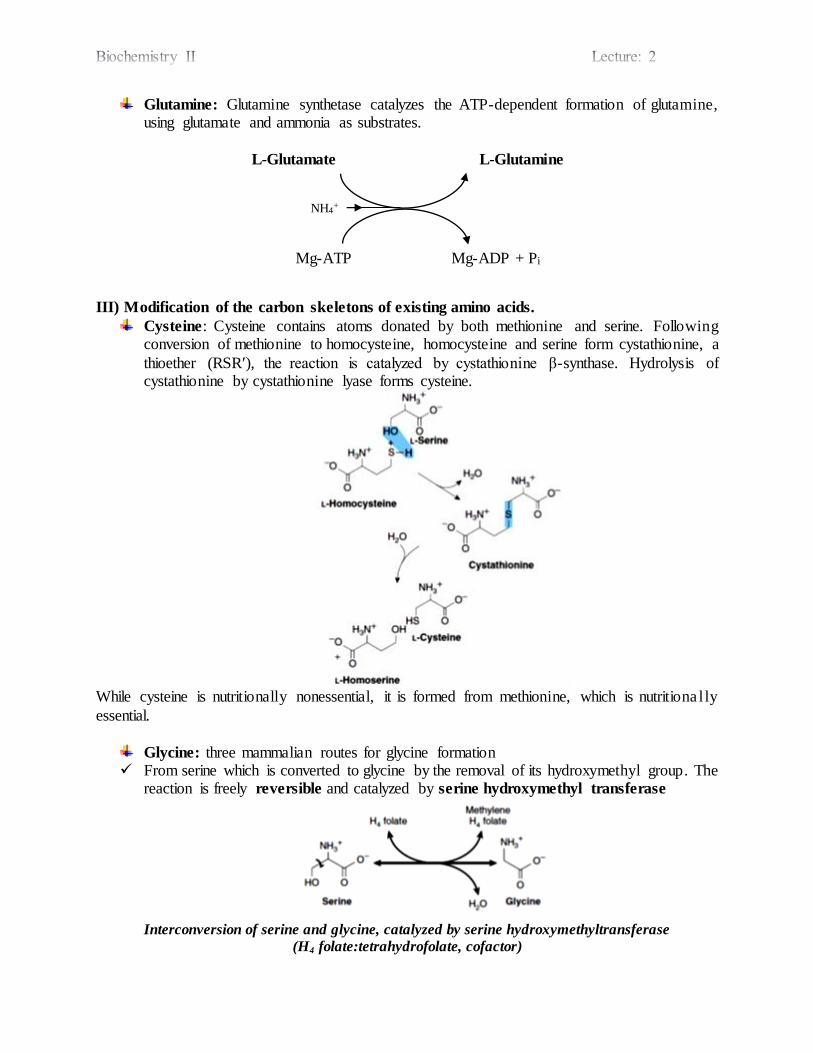
Glutamine: Glutamine synthetase catalyzes the ATP-dependent formation of glutamine, using glutamate and ammonia as substrates.
L-Glutamate L-Glutamine
Mg-ATP Mg-ADP + Pi
III) Modification of the carbon skeletons of existing amino acids.
Cysteine: Cysteine contains atoms donated by both methionine and serine. Following conversion of methionine to homocysteine, homocysteine and serine form cystathionine, a
thioether (RSRʹ), the reaction is catalyzed by cystathionine β-synthase. Hydrolysis of cystathionine by cystathionine lyase forms cysteine.
While cysteine is nutritionally nonessential, it is formed from methionine, which is nutritiona l ly
essential.
Glycine: three mammalian routes for glycine formation From serine which is converted to glycine by the removal of its hydroxymethyl group. The
reaction is freely reversible and catalyzed by serine hydroxymethyl transferase
Interconversion of serine and glycine, catalyzed by serine hydroxymethyltransferase
(H4 folate:tetrahydrofolate, cofactor)
NH4+

From glyoxylate and glutamate or alanine catalyzed by glycine aminotransferase (irreversible, unlike other transamination reactions)
From choline
Tyrosine: Phenylalanine is hydroxylated to form tyrosine a reaction catalyzed by phenylalanine hydroxylase.
NOTE
Provided that the diet contains adequate nutritionally essential phenylalanine, tyrosine is nutritionally nonessential.
Since the reaction is irreversible, dietary tyrosine cannot replace phenylalanine.
Proline: Glutamate is reduced and cyclized to form proline.
Asparagine: Asparagine is synthesized from aspartate catalyzed by asparagine synthetase. Coupled hydrolysis of PPi to Pi by pyrophosphatase ensures that the reaction is strongly favored. (Note similarities to and differences from the glutamine synthetase reaction)
L-Aspartate L-Asparagine
Mg-ATP Mg-AMP + PPi
H2O + Gln Glu

Hydroxyproline and Hydroxylysine
They are present principally in collagen. Since there is no tRNA for either hydroxylated amino acid, neither dietary hydroxypro line
nor hydroxylysine is incorporated into protein. They arise from proline and lysine, but only after these amino acids have been incorporated
into peptides. Hydroxylation of prolyl and lysyl residues is catalyzed by prolyl hydroxylase and lysyl
hydroxylase of tissues, including skin and skeletal muscle, and of granulating wounds.
These hydroxylases require ascorbate as a cofactor, thus, a deficiency of the vitamin C results in scurvy (impaired hydroxylation of peptidyl proline and peptidyl lysine results in
a failure to provide the substrates for cross-linking of maturing collagens).
Selenocysteine, the 21st amino acid
While the occurrence of selenocysteine in proteins is uncommon, at least 25 human selenoproteins are known.
Selenocysteine occurs at the active sites of several enzymes that catalyze redox reactions.
Examples include the human enzymes thioredoxin reductase, glutathione peroxidase, and the deiodinase that converts thyroxine to triiodothyronine.
The replacement of selenocysteine by cysteine in these enzymes can actually impair their catalytic activity.
Impairments in human selenoproteins have been implicated in tumorigenesis and
atherosclerosis, and are associated with selenium deficiency cardiomyopathy (Keshan disease).
Unlike hydroxyproline or hydroxylysine, selenocysteine arises co-translationally during its
incorporation into peptides.
Selenocysteine
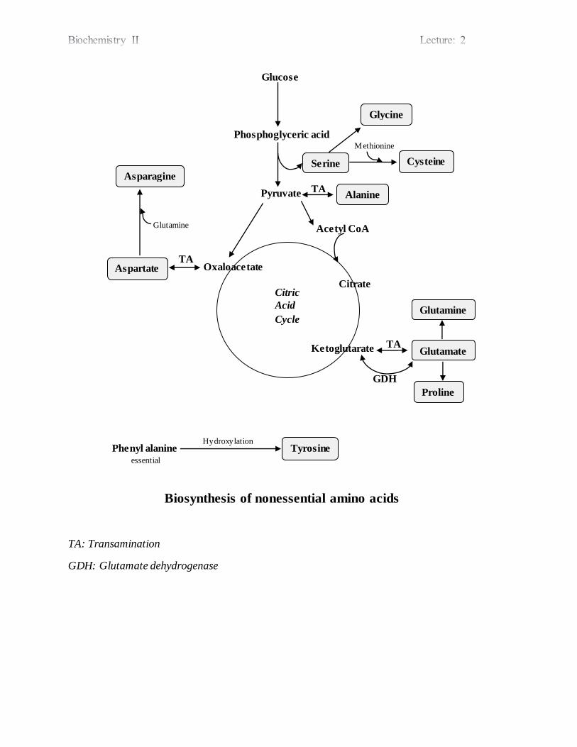
Biosynthesis of nonessential amino acids
TA: Transamination
GDH: Glutamate dehydrogenase
Phenyl alanine essential
Tyrosine Hydroxylation
TA
Proline
Glutamate
Glutamine
GDH
Citrate
Ketoglutarate
Citric
Acid
Cycle
TA Aspartate
Asparagine
Oxaloacetate
Acetyl CoA
Alanine Pyruvate
Glycine
Cysteine Serine
Glucose
Phosphoglyceric acid Methionine
TA
Glutamine
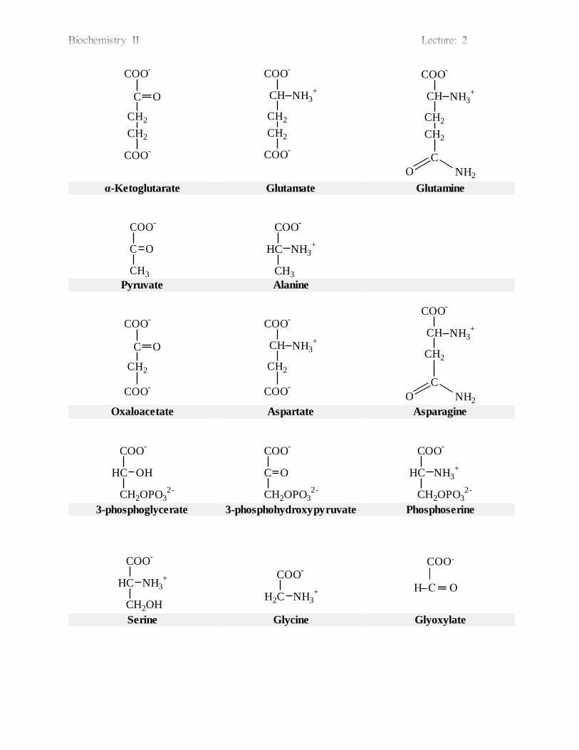
COO-
C O
CH2
CH2
COO-
COO-
CH NH3+
CH2
CH2
COO-
COO-
CH NH3+
CH2
CH2
C
O NH2 α-Ketoglutarate Glutamate Glutamine
COO-
C O
CH3
COO-
HC NH3+
CH3
Pyruvate Alanine
COO
-
C O
CH2
COO-
COO
-
CH NH3+
CH2
COO-
COO
-
CH NH3+
CH2
C
O NH2 Oxaloacetate Aspartate Asparagine
COO
-
HC OH
CH2OPO32-
COO
-
C O
CH2OPO32-
COO
-
HC NH3+
CH2OPO32-
3-phosphoglycerate 3-phosphohydroxypyruvate Phosphoserine
COO
-
HC NH3+
CH2OH
COO-
H2C NH3+
COO-
H C ═ O
Serine Glycine Glyoxylate
BIOCHEMISTRY I
3rd. Stage Lec.
Dr. Ali A. Kasim College of Pharmacy/ University of Baghdad
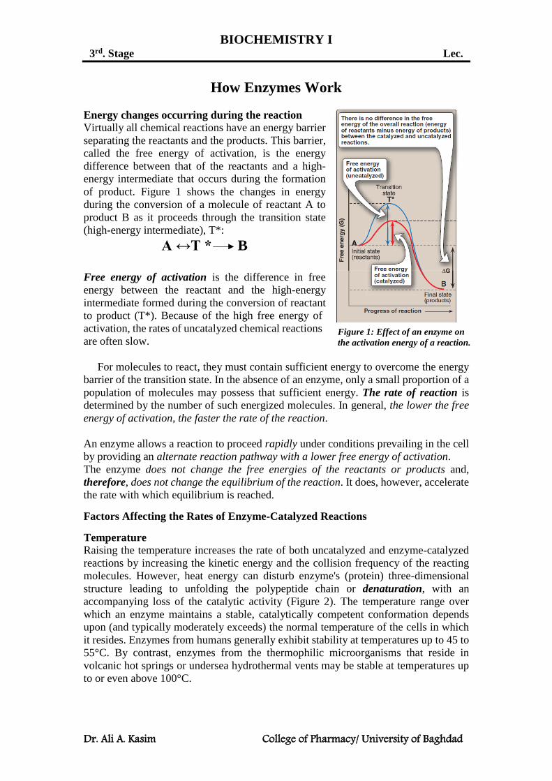
How Enzymes Work
Energy changes occurring during the reaction
Virtually all chemical reactions have an energy barrier
separating the reactants and the products. This barrier,
called the free energy of activation, is the energy
difference between that of the reactants and a high-
energy intermediate that occurs during the formation
of product. Figure 1 shows the changes in energy
during the conversion of a molecule of reactant A to
product B as it proceeds through the transition state
(high-energy intermediate), T*:
A ↔T * B
Free energy of activation is the difference in free
energy between the reactant and the high-energy
intermediate formed during the conversion of reactant
to product (T*). Because of the high free energy of
activation, the rates of uncatalyzed chemical reactions
are often slow.
For molecules to react, they must contain sufficient energy to overcome the energy
barrier of the transition state. In the absence of an enzyme, only a small proportion of a
population of molecules may possess that sufficient energy. The rate of reaction is
determined by the number of such energized molecules. In general, the lower the free
energy of activation, the faster the rate of the reaction.
An enzyme allows a reaction to proceed rapidly under conditions prevailing in the cell
by providing an alternate reaction pathway with a lower free energy of activation.
The enzyme does not change the free energies of the reactants or products and,
therefore, does not change the equilibrium of the reaction. It does, however, accelerate
the rate with which equilibrium is reached.
Factors Affecting the Rates of Enzyme-Catalyzed Reactions
Temperature
Raising the temperature increases the rate of both uncatalyzed and enzyme-catalyzed
reactions by increasing the kinetic energy and the collision frequency of the reacting
molecules. However, heat energy can disturb enzyme's (protein) three-dimensional
structure leading to unfolding the polypeptide chain or denaturation, with an
accompanying loss of the catalytic activity (Figure 2). The temperature range over
which an enzyme maintains a stable, catalytically competent conformation depends
upon (and typically moderately exceeds) the normal temperature of the cells in which
it resides. Enzymes from humans generally exhibit stability at temperatures up to 45 to
55°C. By contrast, enzymes from the thermophilic microorganisms that reside in
volcanic hot springs or undersea hydrothermal vents may be stable at temperatures up
to or even above 100°C.
Figure 1: Effect of an enzyme on
the activation energy of a reaction.

Dr. Ali A. Kasim College of Pharmacy/ University of Baghdad
The temperature coefficient (Q10) is the factor by which the rate of a biologic process
increases for a 10°C increase in temperature. For the temperatures over which enzymes
are stable, the rates of most biological processes typically double for a 10°C rise in
temperature (Q10 = 2). For mammals and other homeothermic organisms (organisms
that maintain a constant body temperature despite fluctuating environmental
temperatures), changes in enzyme reaction rates with temperature assume physiologic
importance only in circumstances such as fever or hypothermia.
Hydrogen Ion Concentration
The rate of almost all enzyme-catalyzed reactions exhibits a significant dependence on
hydrogen ion concentration. Most intracellular enzymes exhibit optimal activity at pH
values between 5 and 9. The relationship of activity to hydrogen ion concentration
(Figure 3) reflects the balance between enzyme denaturation at high or low pH and
effects on the charged state of the enzyme, the substrates, or both. For enzymes whose
mechanism involves acid-base catalysis, the residues involved must be in the
appropriate state of protonation for the reaction to proceed. The binding and recognition
of substrate molecules with dissociable groups also typically involves the formation of
salt bridges with the enzyme. The most common charged groups are carboxylate groups
(negative) and protonated amines (positive). Gain or loss of critical charged groups
adversely affects substrate binding and thus will retard or abolish catalysis.
Figure 3: Effect of pH on enzyme activity.
A negatively charged enzyme (E−) that binds a positively charged substrate (SH+). Shown is the
proportion (%) of SH+ [\\\] and of E− [///] as a function of pH. Only in the cross-hatched area do both
the enzyme and the substrate bear an appropriate charge.
Figure 2: Effect of temperature on
an enzyme-catalyzed reaction.
Dr. Ali A. Kasim College of Pharmacy/ University of Baghdad
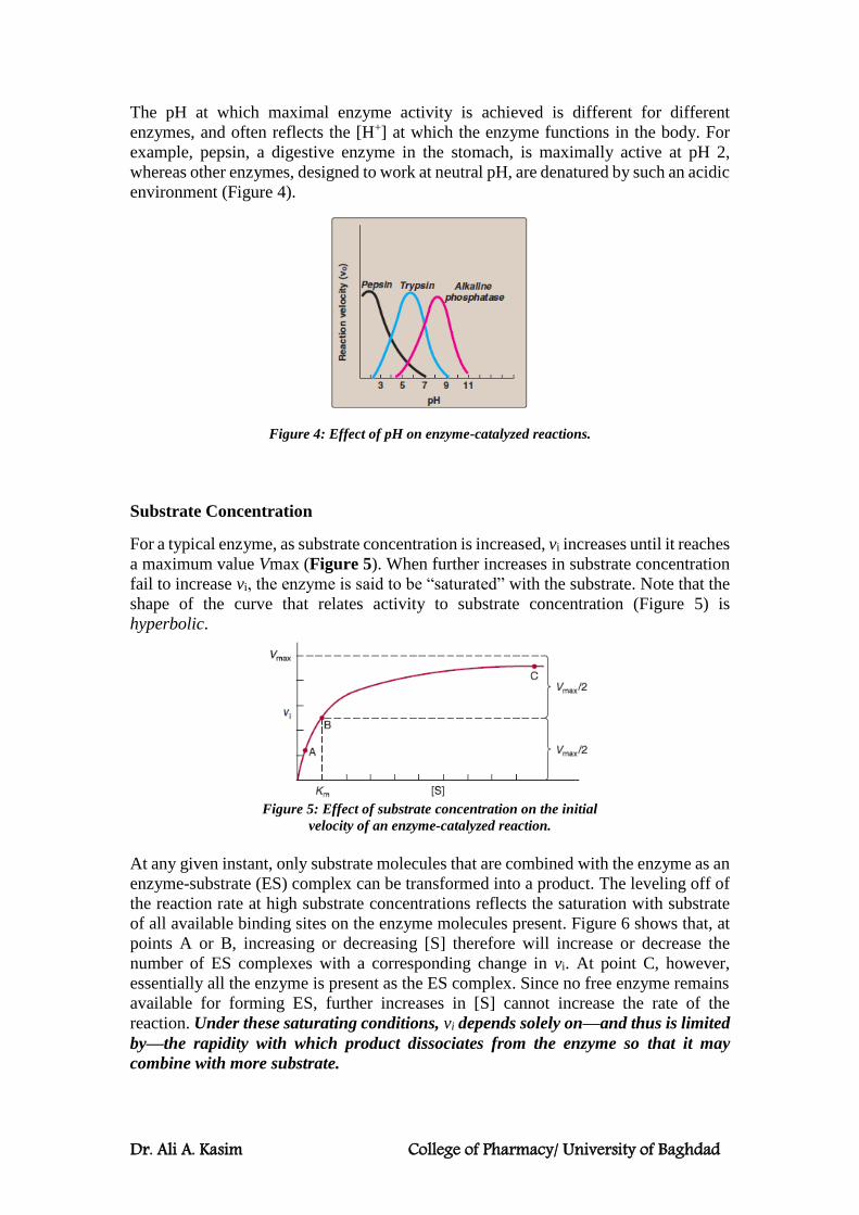
The pH at which maximal enzyme activity is achieved is different for different
enzymes, and often reflects the [H+] at which the enzyme functions in the body. For
example, pepsin, a digestive enzyme in the stomach, is maximally active at pH 2,
whereas other enzymes, designed to work at neutral pH, are denatured by such an acidic
environment (Figure 4).
Figure 4: Effect of pH on enzyme-catalyzed reactions.
Substrate Concentration
For a typical enzyme, as substrate concentration is increased, vi increases until it reaches
a maximum value Vmax (Figure 5). When further increases in substrate concentration
fail to increase vi, the enzyme is said to be “saturated” with the substrate. Note that the
shape of the curve that relates activity to substrate concentration (Figure 5) is
hyperbolic.
Figure 5: Effect of substrate concentration on the initial
velocity of an enzyme-catalyzed reaction.
At any given instant, only substrate molecules that are combined with the enzyme as an
enzyme-substrate (ES) complex can be transformed into a product. The leveling off of
the reaction rate at high substrate concentrations reflects the saturation with substrate
of all available binding sites on the enzyme molecules present. Figure 6 shows that, at
points A or B, increasing or decreasing [S] therefore will increase or decrease the
number of ES complexes with a corresponding change in vi. At point C, however,
essentially all the enzyme is present as the ES complex. Since no free enzyme remains
available for forming ES, further increases in [S] cannot increase the rate of the
reaction. Under these saturating conditions, vi depends solely on—and thus is limited
by—the rapidity with which product dissociates from the enzyme so that it may
combine with more substrate.

Dr. Ali A. Kasim College of Pharmacy/ University of Baghdad
Figure 6: Representation of an enzyme in the presence of different substrate concentration.
THE MICHAELIS-MENTEN EQUATION MODEL THE EFFECTS OF
SUBSTRATE CONCENTRATION
The Michaelis-Menten Equation
The Michaelis-Menten equation illustrates in mathematical terms the relationship
between initial reaction velocity vi and substrate concentration [S], shown graphically
in Figure 5:
vi = 𝑉max [𝑆]
𝐾m+[𝑆]
The Michaelis constant Km is the substrate concentration at which vi is half the
maximal velocity (Vmax/2) attainable at a particular concentration of the enzyme.
The dependence of initial reaction velocity on [S] and Km may be illustrated by
evaluating the Michaelis-Menten equation under three conditions.
1. When [S] is much less than Km (point A in Figures 5 and 6), the term Km + [S]
is essentially equal to Km. Replacing Km + [S] with Km reduces Michaelis-
Menten equation to:
vi ≈ 𝑉max [𝑆]
𝐾m ≈
𝑉max
𝐾m∗ [𝑆]
Since Vmax and Km are both constants, their ratio is a constant. In other words, when
[S] is considerably below Km, vi is proportionate to K[S]. The initial reaction velocity
therefore is directly proportional to [S].
2. When [S] is much greater than Km (point C in Figures 5 and 6), the term
Km + [S] is essentially equal to [S].
vi ≈ 𝑉max [𝑆]
[𝑆] ≈ 𝑉max

Dr. Ali A. Kasim College of Pharmacy/ University of Baghdad
Thus, when [S] greatly exceeds Km, the reaction velocity is maximal (Vmax) and
unaffected by further increases in the substrate concentration.
3. When [S] = Km (point B in Figures 5 and 6):
vi = 𝑉max [𝑆]
𝐾m+[𝑆] =
𝑉max [𝑆]
2[𝑆] =
𝑉max
2
Thus, when [S] equals Km, the initial velocity is half-maximal. Moreover, Km is (and
may be determined experimentally from) the substrate concentration at which the initial
velocity is half-maximal.
Important conclusions about Michaelis-Menten kinetics
1. Characteristics of Km: Km or the Michaelis constant is characteristic of an enzyme
and its particular substrate, and reflects the affinity of the enzyme for that substrate. Km
does not vary with the concentration of enzyme.
a. Small Km: A numerically small (low) Km reflects a high affinity of the enzyme for
substrate, because a low concentration of substrate is needed to half-saturate the
enzyme, that is, to reach a velocity that is 1⁄2Vmax.
b. Large Km: A numerically large (high) Km reflects a low affinity of enzyme for
substrate because a high concentration of substrate is needed to half-saturate the
enzyme.
2. Relationship of velocity to enzyme concentration: The rate of the reaction is
directly proportional to the enzyme concentration at all substrate concentrations. For
example, if the enzyme concentration is halved, the initial rate of the reaction (vi), as
well as that of Vmax, are reduced to half that of the original.
3. Order of reaction: When [S] is much less than Km, the velocity of the reaction is
approximately proportional to the substrate concentration. The rate of reaction is then
said to be first order with respect to substrate. When [S] is much greater than Km, the
velocity is constant and equal to Vmax. The rate of reaction is then independent of
substrate concentration, and is said to be zero order with respect to substrate.
Amino Acid Catabolism
Catabolism of Amino Acid Nitrogen
In normal adults, nitrogen intake matches nitrogen excreted (nitrogen equilibrium). Positive
nitrogen balance, means an excess of ingested over excreted nitrogen, and usually accompanies
growth and pregnancy. Negative nitrogen balance , where output exceeds intake, may follow surgery, advanced cancer, and the nutritional disorders kwashiorkor and marasmus.
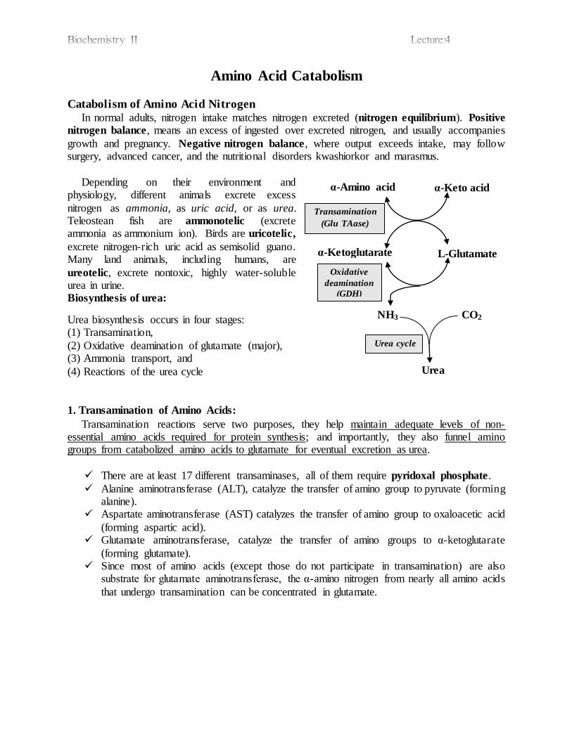
Depending on their environment and physiology, different animals excrete excess
nitrogen as ammonia, as uric acid, or as urea. Teleostean fish are ammonotelic (excrete ammonia as ammonium ion). Birds are uricotelic,
excrete nitrogen-rich uric acid as semisolid guano. Many land animals, including humans, are
ureotelic, excrete nontoxic, highly water-soluble urea in urine.
Biosynthesis of urea:
Urea biosynthesis occurs in four stages: (1) Transamination,
(2) Oxidative deamination of glutamate (major), (3) Ammonia transport, and
(4) Reactions of the urea cycle
1. Transamination of Amino Acids:
Transamination reactions serve two purposes, they help maintain adequate levels of non-essential amino acids required for protein synthesis; and importantly, they also funnel amino groups from catabolized amino acids to glutamate for eventual excretion as urea.
There are at least 17 different transaminases, all of them require pyridoxal phosphate.
Alanine aminotransferase (ALT), catalyze the transfer of amino group to pyruvate (forming alanine).
Aspartate aminotransferase (AST) catalyzes the transfer of amino group to oxaloacetic acid
(forming aspartic acid). Glutamate aminotransferase, catalyze the transfer of amino groups to α-ketoglutarate
(forming glutamate). Since most of amino acids (except those do not participate in transamination) are also
substrate for glutamate aminotransferase, the α-amino nitrogen from nearly all amino acids
that undergo transamination can be concentrated in glutamate.
α-Keto acid α-Amino acid
α-Ketoglutarate L-Glutamate
NH3 CO2
Urea
Transamination
(Glu TAase)
Oxidative
deamination
(GDH)
Urea cycle
Levels of ALT and AST in serum are of diagnostic value. Aminotransferases are normally
intracellular enzymes, with normal low levels in serum indicating release of cellular contents
during normal cell turnover. Elevated levels in serum indicate damage to tissues rich in a particular
enzyme.
ALT is present mainly in the cytoplasm of liver cells, while AST is present in both cytoplasm and mitochondria in liver, heart and skeletal muscles.
In liver diseases, there is an increase in both serum ALT and AST levels. In acute liver diseases, e.g. acute viral hepatitis, the increase is more in ALT. In chronic liver diseases,
e.g. liver cirrhosis the increase is more in AST. In heart diseases, e.g. myocardial infarction, there is an increase in AST only. In skeletal muscle diseases, e.g. myasthenia gravis, there is an increase in AST only.
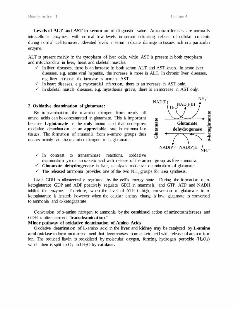
2. Oxidative deamination of glutamate:
By transamination the α-amino nitrogen from nearly all amino acids can be concentrated in glutamate. This is important
because L-glutamate is the only amino acid that undergoes oxidative deamination at an appreciable rate in mammalian tissues. The formation of ammonia from α-amino groups thus
occurs mainly via the α-amino nitrogen of L-glutamate.
In contrast to transaminase reactions, oxidative deamination yields an α-keto acid with release of the amino group as free ammonia.
Glutamate dehydrogenase in liver, catalyzes oxidative deamination of glutamate.
The released ammonia provides one of the two NH3
groups for urea synthesis.
Liver GDH is allosterically regulated by the cell’s energy state. During the formation of α-ketoglutarate GDP and ADP positively regulate GDH in mammals, and GTP, ATP and NADH
inhibit the enzyme. Therefore, when the level of ATP is high, conversion of glutamate to α-ketoglutarate is limited; however when the cellular energy charge is low, glutamate is converted to ammonia and α-ketoglutarate
Conversion of α-amino nitrogen to ammonia by the combined action of aminotransferases and
GDH is often termed “transdeamination.” Minor pathway of oxidative deamination of Amino Acids
Oxidative deamination of L-amino acid in the liver and kidney may be catalyzed by L-amino
acid oxidase to form an α-imino acid that decomposes to an α-keto acid with release of ammonium ion. The reduced flavin is reoxidized by molecular oxygen, forming hydrogen peroxide (H2O2),
which then is split to O2 and H2O by catalase.
Glutamate
dehydrogenase
NAD(P)+
H2O NAD(P)H
NH4+
NH4+
NAD(P)H NAD(P)+
Glu
tam
ate
α-k
etoglu
tara
te
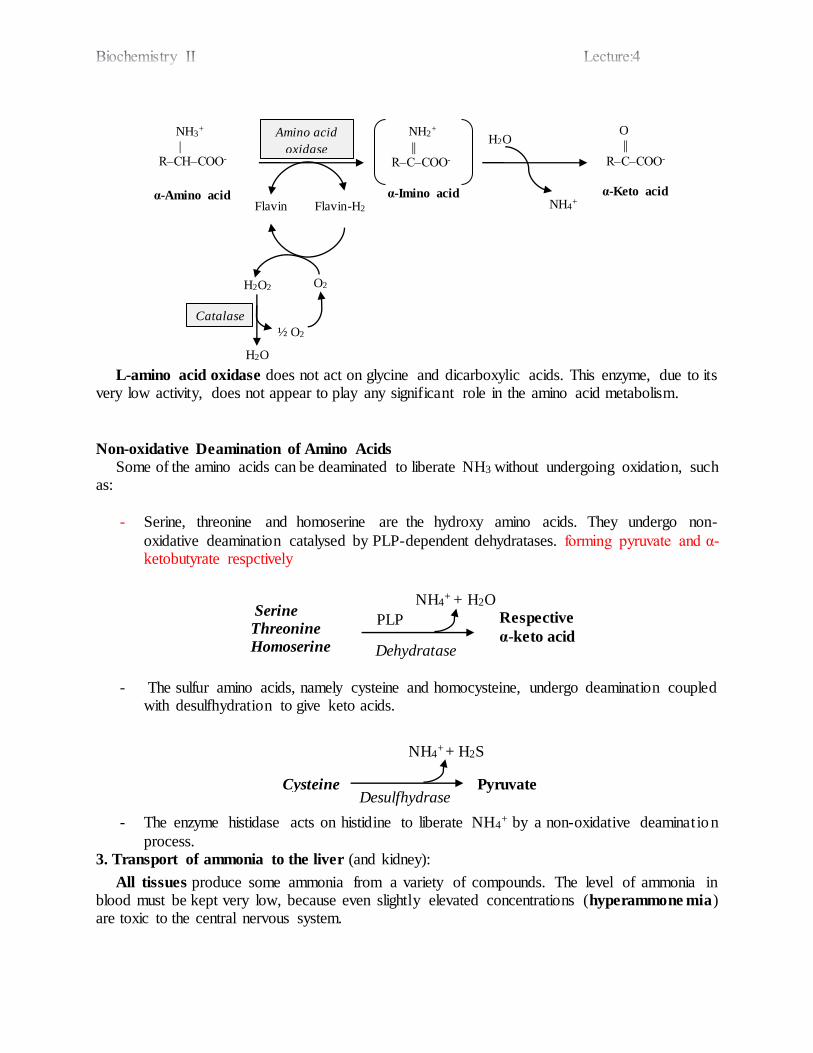
L-amino acid oxidase does not act on glycine and dicarboxylic acids. This enzyme, due to its very low activity, does not appear to play any significant role in the amino acid metabolism.
Non-oxidative Deamination of Amino Acids
Some of the amino acids can be deaminated to liberate NH3 without undergoing oxidation, such as:
- Serine, threonine and homoserine are the hydroxy amino acids. They undergo non-
oxidative deamination catalysed by PLP-dependent dehydratases. forming pyruvate and α-ketobutyrate respctively
- The sulfur amino acids, namely cysteine and homocysteine, undergo deamination coupled with desulfhydration to give keto acids.
- The enzyme histidase acts on histidine to liberate NH4+ by a non-oxidative deamination
process. 3. Transport of ammonia to the liver (and kidney):
All tissues produce some ammonia from a variety of compounds. The level of ammonia in blood must be kept very low, because even slightly elevated concentrations (hyperammone mia) are toxic to the central nervous system.
Serine
Threonine
Homoserine
PLP
Dehydratase
NH4+ + H2O
Respective
α-keto acid
Cysteine Desulfhydrase
NH4+
+ H2S
Pyruvate
α-Amino acid
H2O2 O2
Flavin-H2 Flavin α-Imino acid
NH3+
|
R‒CH‒COO-
O
||
R‒C‒COO-
NH2+
||
R‒C‒COO-
Amino acid
oxidase
Catalase
α-Keto acid
½ O2
H2O
H2O
NH4+
There are two major mechanisms to transport ammonia to liver for its conversion to urea and ultimate excretion in the urine.
Glutamine synthetase in many tissues assimilates free ammonia on glutamate to form glutamine, an energy requiring process. Glutamine acts as a non-toxic transport and storage form of ammonia
(carries 2 NH3, actually). Glutamine is freely diffusible in tissues, hence easily transported.
Glutamine travels in the blood to the liver, where L-glutaminase releases free ammonia which can enter the urea cycle. An analogous reaction is catalyzed by L-asparaginase that deaminates asparagine.
The kidneys can also form ammonia from glutamine by action of renal glutaminase. This
ammonia
is excreted into the urine as ammonium ion (NH4
+), an important mechanism for
maintaining whole-body acid-base balance.
Hepatic glutaminase levels rise in response to high protein intake while renal glutaminase
increases in metabolic acidosis.
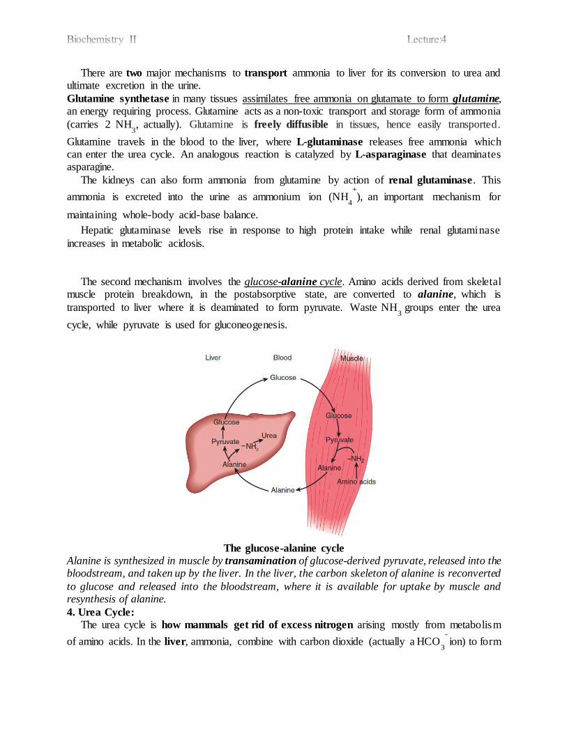
The second mechanism involves the glucose-alanine cycle. Amino acids derived from skeletal muscle protein breakdown, in the postabsorptive state, are converted to alanine, which is
transported to liver where it is deaminated to form pyruvate. Waste NH3
groups enter the urea
cycle, while pyruvate is used for gluconeogenesis.
The glucose-alanine cycle
Alanine is synthesized in muscle by transamination of glucose-derived pyruvate, released into the bloodstream, and taken up by the liver. In the liver, the carbon skeleton of alanine is reconverted
to glucose and released into the bloodstream, where it is available for uptake by muscle and resynthesis of alanine.
4. Urea Cycle: The urea cycle is how mammals get rid of excess nitrogen arising mostly from metabolism
of amino acids. In the liver, ammonia, combine with carbon dioxide (actually a HCO3
- ion) to form
urea. The urea is carried through the blood to the kidney, which isolates it for excretion in the urine. Urea accounts for about 90% of N-containing compounds in urine
The urea cycle is the first metabolic pathway to be elucidated, by Hans Krebs and Kurt Henseleit. Hence, the cycle is known as Krebs-Henseleit cycle. As ornithine is the first member
of the reaction, it is also called as Ornithine cycle. The two nitrogen atoms of urea are derived from two different sources, one from ammonia and the other directly from the alpha amino group of aspartic acid. Carbon atom is supplied by CO2.
Urea cycle occurs in five steps:
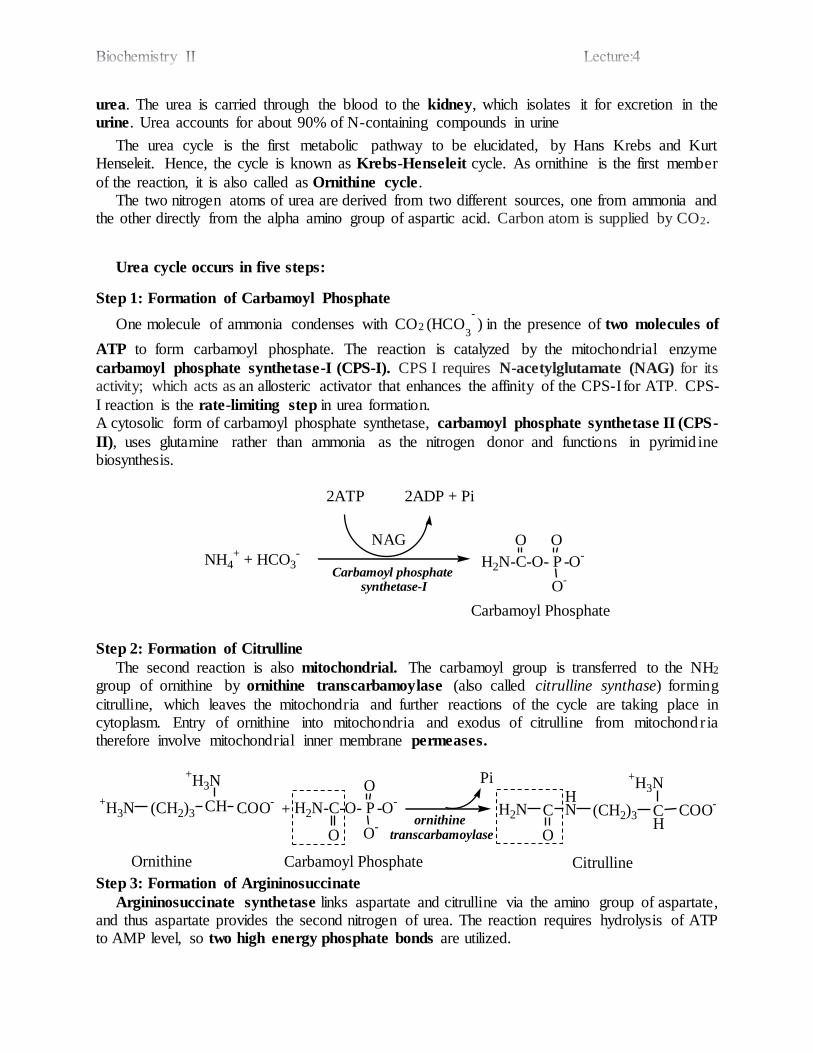
Step 1: Formation of Carbamoyl Phosphate
One molecule of ammonia condenses with CO2 (HCO3
- ) in the presence of two molecules of
ATP to form carbamoyl phosphate. The reaction is catalyzed by the mitochondrial enzyme
carbamoyl phosphate synthetase-I (CPS-I). CPS I requires N-acetylglutamate (NAG) for its activity; which acts as an allosteric activator that enhances the affinity of the CPS-I for ATP. CPS-
I reaction is the rate-limiting step in urea formation. A cytosolic form of carbamoyl phosphate synthetase, carbamoyl phosphate synthetase II (CPS-
II), uses glutamine rather than ammonia as the nitrogen donor and functions in pyrimid ine biosynthesis.
2ATP 2ADP + Pi
NH4+ + HCO3
-H2N-C-O- P -O
-
O
O-
O
Carbamoyl Phosphate
Carbamoyl phosphate synthetase-I
Step 2: Formation of Citrulline
The second reaction is also mitochondrial. The carbamoyl group is transferred to the NH2 group of ornithine by ornithine transcarbamoylase (also called citrulline synthase) forming
citrulline, which leaves the mitochondria and further reactions of the cycle are taking place in cytoplasm. Entry of ornithine into mitochondria and exodus of citrulline from mitochondria therefore involve mitochondrial inner membrane permeases.
H2N-C-O- P -O-
O O-
O
Carbamoyl Phosphate
COO-CH
+H3N
(CH2)3+H3N
Ornithine
+ COO-
CH
+H3N
(CH2)3
HNC
O
H2N
Citrulline
ornithine transcarbamoylase
Pi
Step 3: Formation of Argininosuccinate
Argininosuccinate synthetase links aspartate and citrulline via the amino group of aspartate, and thus aspartate provides the second nitrogen of urea. The reaction requires hydrolysis of ATP to AMP level, so two high energy phosphate bonds are utilized.
NAG

Step 4: Formation of Arginine
Argininosuccinate is cleaved by argininosuccinate lyase (argininosuccinase) to arginine and
fumarate. Subsequent addition of water to fumarate forms malate, whose subsequent oxidation forms oxaloacetate. Transamination of oxaloacetate by glutamate aminotransferase then re-forms
aspartate. The carbon skeleton of aspartate-fumarate thus acts as a carrier of the nitrogen of glutamate into a precursor of urea. Thus, the urea cycle is linked to citric acid cycle through fumarate, and so, it is called as "urea
bicycle"
.
COO-CH
NH3+
(CH2)3
HNC+
H2N
NH
CHCOO-
CH2
COO-
COO-
CH
NH3+
(CH2)3
HNC
NH2+
H2N
CH
COO-
CH
COO-
Fumarate
ArginineArgininosuccinate
Argininosuccinate lyase
Step 5: Formation of Urea
The final reaction of the cycle is the hydrolysis of arginine to urea and ornithine by arginase.
The ornithine returns to the mitochondria to react with another molecule of carbamoyl phosphate
COO-CH
NH3+
(CH2)3
HNC
O
H2N
Citrulline
+
CH NH3+
COO-
H2
C-OOC
Aspartate
COO-CH
NH3+
(CH2)3
HNC+
H2N
NH
CHCOO-
CH2
COO-
ATP AMP +PPi
Argininosuccinate synthetase
Argininosuccinate

so that the cycle will proceed. Ornithine and lysine are potent inhibitors of arginase, and compete with arginine.
COO-CH
NH3+
(CH2)3
HNC
NH2+
H2N
Arginine
COO-CH
NH3+
(CH2)3+H3N
Ornithine
Arginase
H2O
NH2CH2N
O
Urea
Arginase is mostly found in the liver, while the rest of the enzymes (four) of urea cycle are also present in other tissues. For this reason, arginine synthesis may occur to varying degrees in many
tissues. But only the liver can ultimately produce urea.
Overall Reaction and Energetics of Urea Cycle
Synthesis of 1 mol of urea requires 3 mol of ATP (utilized as 4 high energy phosphates), 1 mol
each of ammonium ion and of aspartate, and employs five enzymes. Urea synthesis is a cyclic process. While ammonium ion, CO2, ATP, and aspartate are consumed, the ornithine consumed in reaction 2 is regenerated in reaction 5. There thus is no net loss or gain of ornithine, citrulline,
argininosuccinate, or arginine.
NH4+ + CO2 + Aspartate + 3ATP Urea + Fumarate + 2 ADP + 2 Pi + AMP + PPi
Regulation of Urea Cycle
The first reaction catalysed by carbamoyl phosphate synthetase I (CPS I) is rate-limiting
reaction in urea synthesis. CPS I is allosterically activated by N-acetylglutamate (NAG). NAG is synthesized from glutamate and acetyl CoA by NAG synthase and degraded by a
NAG hydrolase.
High concentrations of arginine (an allosteric activator of NAG Synthase), and the
consumption of a protein-rich meal increases the level of NAG in liver, leading to enhanced urea synthesis.
During starvation the expression of all the enzymes of the urea cycle in liver increases
several fold, secondary to enhanced protein degradation to provide energy. The last four enzymes of urea cycle are mostly controlled by the concentration of their
respective substrates. Disposal of Urea
Urea produced in the liver is transported in blood to kidneys, and excreted in urine.

A small amount of urea synthesized in the liver enters, via circulation, the lumen of the intestine (primarily the small intestine), where urea is hydrolyzed by microbial urease into
ammonia and CO2. This ammonia is either lost in the feces or absorbed into the blood. In renal failure, the blood urea level is elevated (uremia), resulting in diffusion of more
urea into intestine and its breakdown to NH3. Hyperammonemia (increased blood NH3) is commonly seen in patients of kidney failure.
For these patients, oral administration of antibiotics (neomycin) to kill intestinal bacteria
is advised.
Metabolic Disorders of Urea Cycle
Defects in each enzyme of the urea cycle function or synthesis can lead to metabolic disorders. Urea cycle disorders are characterized by hyperammonemia, encephalopathy, and
respiratory alkalosis. Ammonia intoxication is most severe when the metabolic block occurs at reactions 1 or 2, for if citrulline can be synthesized, some ammonia has already
been removed by being covalently linked to an organic metabolite. Clinical symptoms common to all urea cycle disorders include vomiting, avoidance of
high-protein foods, intermittent ataxia, irritability, lethargy, and severe mental retardation.
The clinical features and treatment of all five disorders are similar. Significant improvement and minimization of brain damage can accompany a low-protein diet
ingested as frequent small meals to avoid sudden increases in blood ammonia levels. The goal of dietary therapy is to provide sufficient protein, arginine, and energy to promote growth and development while simultaneously minimizing the metabolic perturbations.
Urea cycle disorders with the corresponding defective enzyme or transporter
Disorder Defective Enzyme or Transporter
Hyper ammonemia Type I Carbamoyl Phosphate Synthetase I
Hyper ammonemia Type II Ornithine Transcarbamoylase
Citrullinemia Type I (Classic Citrullinemia)
Argininosuccinate synthetase
Argininosuccinic aciduria Argininosuccinate lyase
Hyperargininemia Arginase
Citrullinemia Type II Citrulline permease
Hyperammonemia Hyperornithinemia Homocitrullinuria (HHH) Syndrome
Ornithine permease
NOTE:

N-Acetyl Glutamate Synthase Deficiency: The sixth enzyme deficiency which lead to a urea
cycle disorder. The condition is almost similar to Hyperammonemia Type I. But, arginine, an
allosteric activator of NAG Synthase improves CPS-I defect as NAG activates CPS-I; while
arginine does not improve NAG deficiency, as the enzyme itself is defective.
Newborn Screening for Metabolic Diseases
Congenital metabolic diseases caused by the absence or functional impairment of metabolic enzymes have serious consequences.
Early dietary intervention can in many instances ameliorate the otherwise expected dire effects. The early detection of such metabolic diseases is thus is of primary importance.
Newborn screening using the powerful and sensitive technique of tandem mass
spectrometry (which can in a few minutes detect over 40 analytes), is of great importance in the early detection of metabolic disorders.
Ammonia
Ammonia is toxic and its accumulation in the body is fatal. Only traces (10-20 μg/dl) normally are present in peripheral blood.
It is removed by the liver that converts it to urea, which is nontoxic, water soluble and easily excreted in the urine.
Since urea is nontoxic to humans, high blood levels in renal disease are a consequence, not
a cause, of impaired renal function.
Sources of ammonia
Deamination and transamination of amino acids. Ammonia produced by the action of intestinal bacteria on the non-absorbed dietary
amino acids or from urea reaching the intestine via circulation which is hydrolyzed to NH3 and CO2.
Ammonia is released from monoamines (e.g. epinephrine, norepinephrine and dopamine)
by the action of monoamine oxidase enzyme. Ammonia is released during purine and pyrimidine catabolism.
Fate of ammonia
Biosynthesis of urea is the main fate of ammonia
Biosynthesis of nonessential amino acids Small amounts of ammonia are excreted in urine
Causes of hyperammonemia
1-Acquired hyperammonemia
Liver cell failure
Shunts between portal and systemic circulation 2-Congenital hyperammonemia
Defect in any one of the enzymes that catalyze urea cycle steps, or in ornithine and
citrulline permeases or NAG-synthase
Why is ammonia toxic?

GDH
α-ketoglutarate + NH3 + NADPH glutamate + NADP+
Accumulation of NH3 shifts the equilibrium to the right with more glutamate formation, hence
more utilization of α-ketoglutarate. α-Ketoglutarate is a key intermediate in citric acid cycle and its depleted levels impair the citric acid cycle. The net result is that production of energy (ATP) by the brain is reduced, leading to neurological manifestations (flapping tremors, slurred speech,
blurred vision, vomiting, coma, and death may occur if hyperammonemia not corrected).
Urea Cycle Reactions
BIOCHEMISTRY I
3rd. Stage Lec.
Enzymes Kinetics (Cont'd.)
A Linear Form of the Michaelis-Menten Equation Is Used to Determine Km &
Vmax
The direct measurement of the numeric value of Vmax, and therefore the calculation of
Km, often requires impractically high concentrations of substrate to achieve saturating
conditions. A linear form of the Michaelis-Menten equation circumvents this difficulty
and permits Vmax and Km to be extrapolated from initial velocity data obtained at less
than saturating concentrations of the substrate.
vi = 𝑉max [𝑆]
𝐾m+[𝑆]
invert:
1
𝑣𝑖 =
𝐾m+[𝑆]
𝑉max [𝑆]
= 𝐾m
𝑉max [𝑆] +
[𝑆]
𝑉max [𝑆]
1
𝑣𝑖 =
𝐾m
𝑉max *
1
[𝑆] +
1
𝑉max
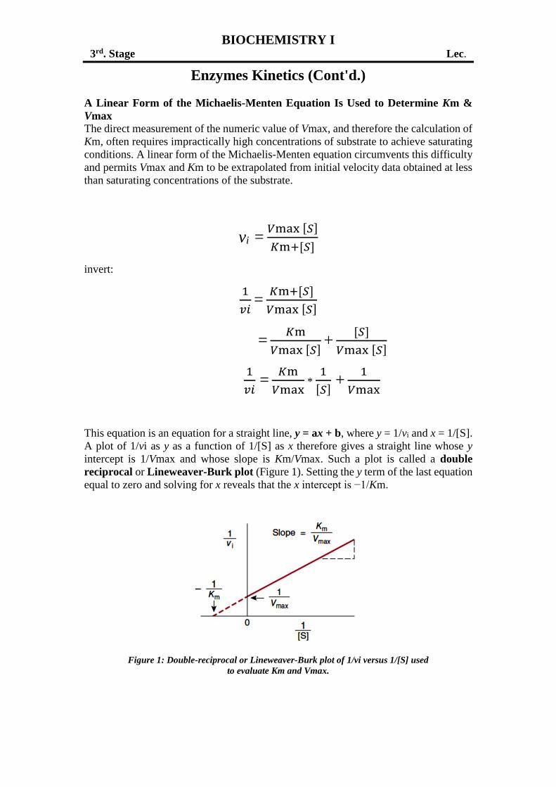
This equation is an equation for a straight line, y = ax + b, where y = 1/vi and x = 1/[S].
A plot of 1/vi as y as a function of 1/[S] as x therefore gives a straight line whose y
intercept is 1/Vmax and whose slope is Km/Vmax. Such a plot is called a double
reciprocal or Lineweaver-Burk plot (Figure 1). Setting the y term of the last equation
equal to zero and solving for x reveals that the x intercept is −1/Km.
Figure 1: Double-reciprocal or Lineweaver-Burk plot of 1/vi versus 1/[S] used
to evaluate Km and Vmax.
Dr. Ali A. Kasim College of Pharmacy/ University of Baghdad
The Catalytic Constant, kcat
Several parameters may be used to compare the relative activity of different enzymes
or of different preparations of the same enzyme. The activity of impure enzyme
preparations typically is expressed as a specific activity (Vmax divided by the protein
concentration). For a homogeneous enzyme, one may calculate its turnover number
(Vmax divided by the moles of enzyme present). But if the number of active sites
present is known, the catalytic activity of a homogeneous enzyme is best expressed as
its catalytic constant, kcat (Vmax divided by the number of active sites, St):
kcat = 𝑉max
𝑆t
The units of kcat are reciprocal time (sec-1).
Catalytic Efficiency, kcat/Km
While the maximum capacity of a given enzyme to convert substrate to product is
important, the benefits of a high kcat can only be realized if Km is sufficiently low.
Thus, catalytic efficiency of enzymes is best expressed in terms of the ratio of these two
kinetic constants, kcat/Km.
For certain enzymes, once substrate binds to the active site, it is converted to product
and released so rapidly as to render these events effectively instantaneous. For these
exceptionally efficient catalysts, the rate-limiting step in catalysis is the formation of
the ES complex. Such enzymes are said to be diffusion-limited, or catalytically perfect,
since the fastest possible rate of catalysis is determined by the rate at which molecules
move or diffuse through the solution.
The Hill Equation Describes the Behavior of Enzymes That Exhibit Cooperative
Binding of Substrate
While most enzymes display the simple saturation kinetics and are adequately
described by the Michaelis-Menten expression, some enzymes bind their substrates in
a cooperative fashion. Cooperative behavior is an exclusive property of multimeric
enzymes that bind substrate at multiple sites.
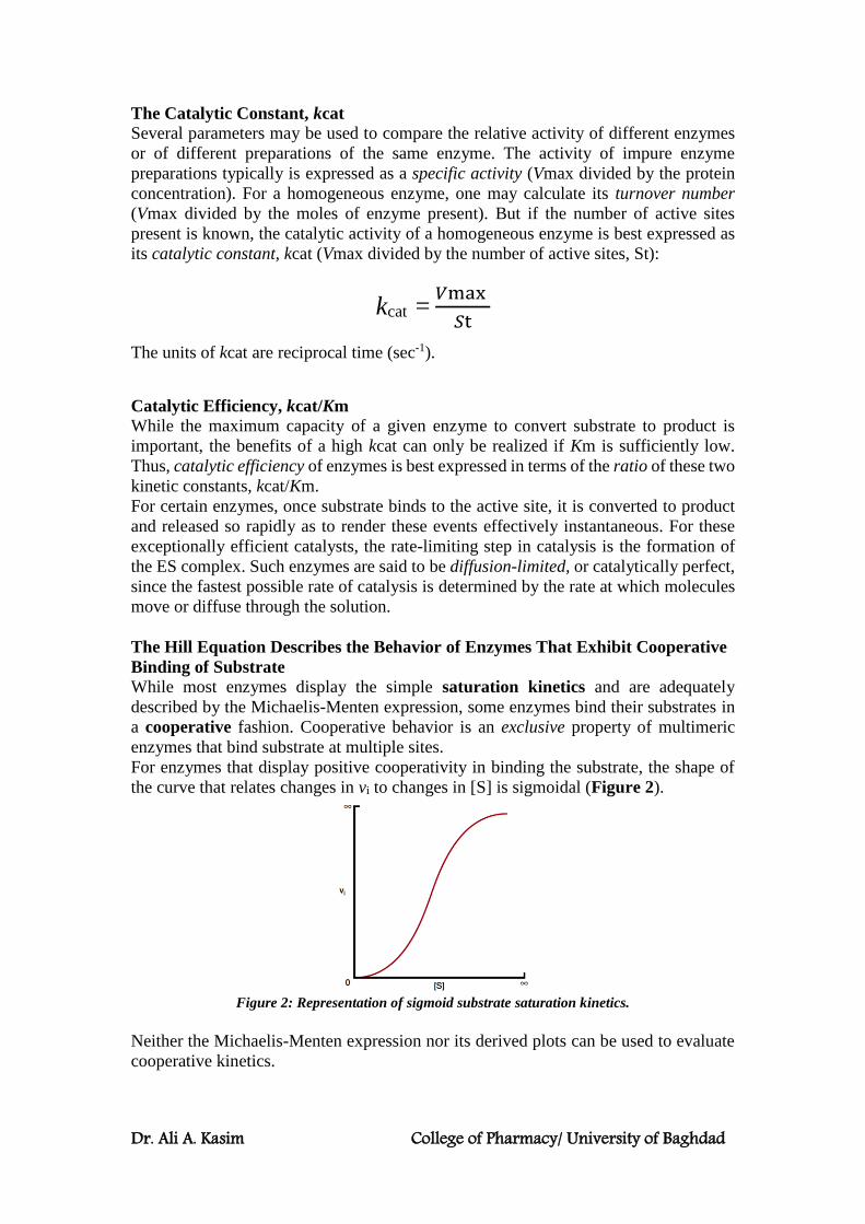
For enzymes that display positive cooperativity in binding the substrate, the shape of
the curve that relates changes in vi to changes in [S] is sigmoidal (Figure 2).
Figure 2: Representation of sigmoid substrate saturation kinetics.
Neither the Michaelis-Menten expression nor its derived plots can be used to evaluate
cooperative kinetics.
Dr. Ali A. Kasim College of Pharmacy/ University of Baghdad
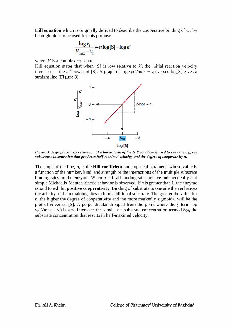
Hill equation which is originally derived to describe the cooperative binding of O2 by
hemoglobin can be used for this purpose.
where k′ is a complex constant.
Hill equation states that when [S] is low relative to k′, the initial reaction velocity
increases as the nth power of [S]. A graph of log vi/(Vmax − vi) versus log[S] gives a
straight line (Figure 3).
Figure 3: A graphical representation of a linear form of the Hill equation is used to evaluate S50, the
substrate concentration that produces half-maximal velocity, and the degree of cooperativity n.
The slope of the line, n, is the Hill coefficient, an empirical parameter whose value is
a function of the number, kind, and strength of the interactions of the multiple substrate
binding sites on the enzyme. When n = 1, all binding sites behave independently and
simple Michaelis-Menten kinetic behavior is observed. If n is greater than 1, the enzyme
is said to exhibit positive cooperativity. Binding of substrate to one site then enhances
the affinity of the remaining sites to bind additional substrate. The greater the value for
n, the higher the degree of cooperativity and the more markedly sigmoidal will be the
plot of vi versus [S]. A perpendicular dropped from the point where the y term log
vi/(Vmax − vi) is zero intersects the x-axis at a substrate concentration termed S50, the
substrate concentration that results in half-maximal velocity.

Amino Acid Catabolism
Catabolism of the carbon skeleton
Transamination typically initiates amino acids catabolism. Removal of α-amino nitrogen by transamination, catalyzed by an aminotransferase, is the first catabolic reaction of most of the
protein amino acids. The exceptions are proline, hydroxyproline, threonine, and lysine, whose α-amino groups do not participate in transamination. Then, oxidative deamination (major) and non-
oxidative deamination remove the nitrogen atom; and the resultant hydrocarbon skeletons are then degraded to metabolic intermediates. The carbon atoms of fat, carbohydrate, and protein are interconvertible. All or a portion of the
carbon skeleton of every amino acid is convertible either to carbohydrate, fat, or both fat and carbohydrate.
The amino acids may be grouped for discussion on the basis of the specific keto acid products of their deamination.
The 3-carbon α-keto acid pyruvate is produced from alanine, cysteine, glycine, serine,
and threonine.
Alanine deamination via transaminase directly yields pyruvate.
Serine is deaminated to form pyruvate via serine dehydratase.
Also, serine following conversion to glycine, is catalyzed by glycine hydroxymethyltransferase , then catabolism merges with that of glycine.
Threonine aldolase cleaves threonine to glycine and acetaldehyde. Oxidation of
acetaldehyde to acetate is followed by formation of acetyl-CoA.
Serine
PLP
Dehydratase
NH4+ + H2O
Pyruvate
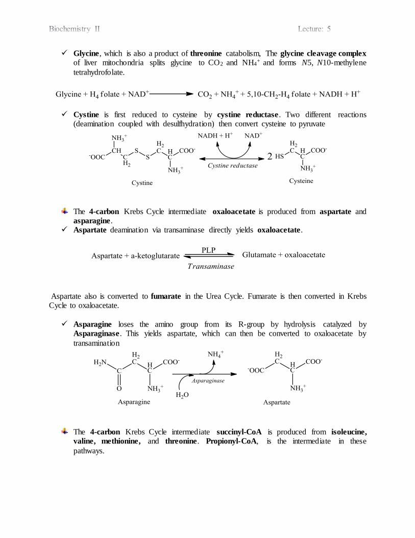
Glycine, which is also a product of threonine catabolism, The glycine cleavage complex
of liver mitochondria splits glycine to CO2 and NH4+ and forms N5, N10-methylene
tetrahydrofolate.
Cystine is first reduced to cysteine by cystine reductase. Two different reactions (deamination coupled with desulfhydration) then convert cysteine to pyruvate
The 4-carbon Krebs Cycle intermediate oxaloacetate is produced from aspartate and asparagine.
Aspartate deamination via transaminase directly yields oxaloacetate.
Aspartate also is converted to fumarate in the Urea Cycle. Fumarate is then converted in Krebs Cycle to oxaloacetate.
Asparagine loses the amino group from its R-group by hydrolysis catalyzed by Asparaginase. This yields aspartate, which can then be converted to oxaloacetate by
transamination
The 4-carbon Krebs Cycle intermediate succinyl-CoA is produced from isoleucine,
valine, methionine, and threonine. Propionyl-CoA, is the intermediate in these
pathways.

The branched chain amino acids, isoleucine and valine, initially share in part a common
pathway, catalyzed by a multi-subunit complex, branched chain α-keto acid
dehydrogenase.
Threonine undergoes deamination by threonine dehydratase to α-ketobutyrate which is
decarboxylated to propionyl-CoA.
Methionine is converted to S-Adenosylmethionine (SAM) by an ATP-dependent reaction. SAM serves as a methyl group donor in various synthetic reactions. The resulting
adenosylhomocysteine is hydrolyzed to homocysteine, which may be catabolized via a complex pathway to cysteine and homoserine (please refer to the biosynthetic reaction of cysteine in lecture 2); homoserine in turn is deaminated by dehydratase to α-ketobutyrate
(lecture 3) which is decarboxylated to propionyl-CoA, and thus, succinyl-CoA.
Or methionine may be regenerated from homocysteine by methyl transfer from N5-methyl-
tetrahydrofolate (H4 folate), via a methyltransferase enzyme that utilizes B12 as prosthetic group.
The methyl group is transferred from tetrahydrofolate to B12 to homocysteine. Another pathway converts homocysteine to glutathione.
H4 folate
N5-methyl-H4 folate
Threonine
PLP
Dehydratase
NH4+ + H2O
α-ketobutyrate
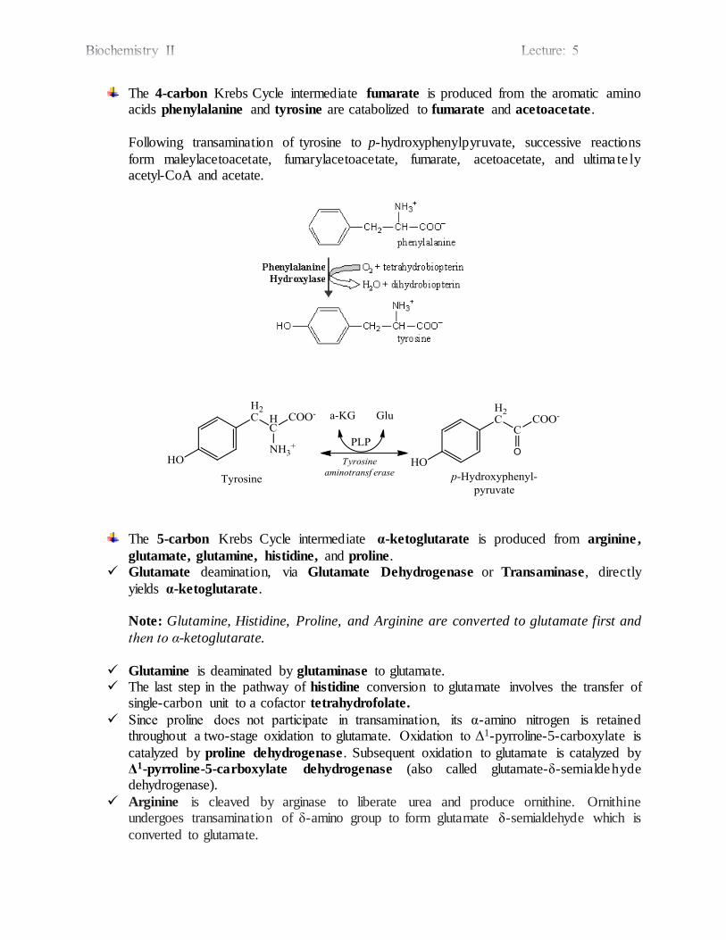
The 4-carbon Krebs Cycle intermediate fumarate is produced from the aromatic amino acids phenylalanine and tyrosine are catabolized to fumarate and acetoacetate.
Following transamination of tyrosine to p-hydroxyphenylpyruvate, successive reactions
form maleylacetoacetate, fumarylacetoacetate, fumarate, acetoacetate, and ultima te ly acetyl-CoA and acetate.
The 5-carbon Krebs Cycle intermediate α-ketoglutarate is produced from arginine ,
glutamate, glutamine, histidine, and proline. Glutamate deamination, via Glutamate Dehydrogenase or Transaminase, directly
yields α-ketoglutarate.
Note: Glutamine, Histidine, Proline, and Arginine are converted to glutamate first and
then to α-ketoglutarate.
Glutamine is deaminated by glutaminase to glutamate. The last step in the pathway of histidine conversion to glutamate involves the transfer of
single-carbon unit to a cofactor tetrahydrofolate.
Since proline does not participate in transamination, its α-amino nitrogen is retained throughout a two-stage oxidation to glutamate. Oxidation to Δ1-pyrroline-5-carboxylate is
catalyzed by proline dehydrogenase. Subsequent oxidation to glutamate is catalyzed by Δ1-pyrroline-5-carboxylate dehydrogenase (also called glutamate-δ-semialdehyde dehydrogenase).
Arginine is cleaved by arginase to liberate urea and produce ornithine. Ornithine undergoes transamination of δ-amino group to form glutamate δ-semialdehyde which is
converted to glutamate.


Overview of the metabolic intermediates that result from amino acid catabolism
Where:
The amino acids bold italic are ketogenic
Underlined amino acids are both glucogenic and ketogenic
The remaining amino acids are glucogenic only
Pyruvic Acid
Acetyl CoA
Succinyl CoA
Ketoglutaric
Acid
Fumaric Acid
Oxaloacetic Acid
Citric
Acid
Cycle Arg
Gln
His
Pro
Ala
Cys
Gly
Ser
Thr
Trp
Asn
Phe
Tyr
Ile
Met
Thr
Val
Glu
Asp
Ile
Leu
Lys
Phe
Thr
Trp
Tyr

BIOCHEMISTRY I
3rd. Stage Lec.
ENZYME INHIBITION
Any substance that can diminish the velocity of an enzyme-catalyzed reaction is
called an inhibitor. Inhibitors of the catalytic activities of enzymes provide both
pharmacologic agents and research tools for the study of the mechanism of enzyme
action.
Inhibitors can be classified on the basis of their site of action on the enzyme, on whether
they chemically modify the enzyme, or on the kinetic parameters they influence.
Compounds that mimic the transition state of an enzyme-catalyzed reaction (transition
state analogs) or that take advantage of the catalytic machinery of an enzyme
(mechanism-based inhibitors) can be particularly potent inhibitors. Kinetically, we
distinguish two classes of inhibitors based upon whether raising the substrate
concentration does or does not overcome the inhibition.
Competitive Inhibitors:
The effects of competitive inhibitors can be overcome by raising the concentration
of substrate. Most frequently, in competitive inhibition the inhibitor (I) binds to the
substrate-binding portion of the active site thereby blocking access by the substrate.
The structures of most classic competitive inhibitors therefore tend to resemble the
structure of a substrate, and thus are termed substrate analogs. Both substrate and its
structural analog (inhibitor) can bind to the active site of the enzyme, forming an ES or
an EI complex, respectively. However, EI complex will not proceed to form the
product.
For example, malonate, a structural analog of succinate, can competitively inhibit
succinate dehydrogenase. Succinate dehydrogenase catalyzes the removal of one
hydrogen atom from each of the two methylene carbons of succinate (−OOC-CH2-CH2-
COO−). Both succinate and malonate (−OOC-CH2-COO−) can bind to the active site of
succinate dehydrogenase, forming an ES or an EI complex, respectively. However,
since malonate contains only one methylene carbon, it cannot undergo
dehydrogenation.
Figure 1: Malonate as competitive inhibitor of succinate dehydrogenase
Dr. Ali A. Kasim College of Pharmacy/ University of Baghdad
The formation and dissociation of the EI complex is a dynamic process described
by:
for which the equilibrium constant 𝑲𝒊 is
𝑲𝒊 =[𝑬][𝑰]
[𝑬 − 𝑰]=
𝒌𝟏
𝒌−𝟏
In other words, a competitive inhibitor acts by decreasing the number of free enzyme
molecules available to bind substrate, that is, to form ES, and thus eventually to form
product.
Increasing the substrate concentration will increase the formation of ES complexes.
Since the formation of ES complexes removes free enzyme available to combine with
the inhibitor, thus, decreasing the concentration of the EI complex and raising the
reaction velocity. The extent to which [S] must be increased to completely overcome
the inhibition depends upon:
the concentration of the inhibitor present,
the affinity of the inhibitor (𝑲𝒊) for the enzyme, and
the affinity, Km, of the enzyme for its substrate.
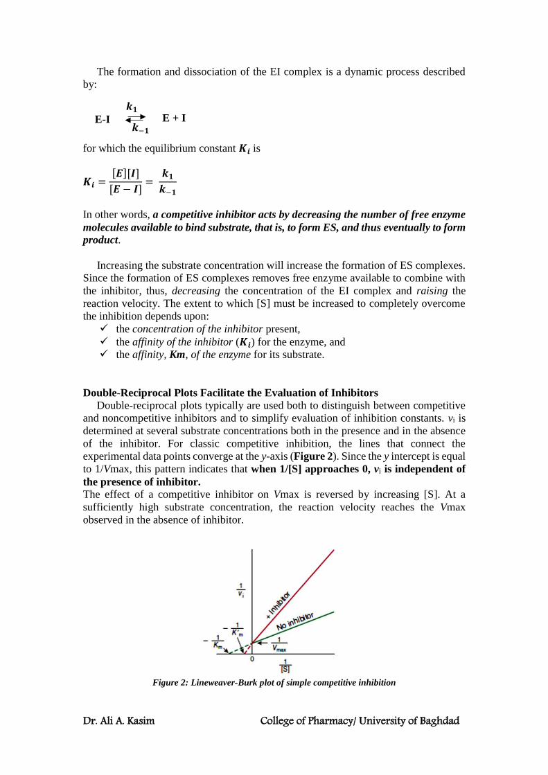
Double-Reciprocal Plots Facilitate the Evaluation of Inhibitors
Double-reciprocal plots typically are used both to distinguish between competitive
and noncompetitive inhibitors and to simplify evaluation of inhibition constants. vi is
determined at several substrate concentrations both in the presence and in the absence
of the inhibitor. For classic competitive inhibition, the lines that connect the
experimental data points converge at the y-axis (Figure 2). Since the y intercept is equal
to 1/Vmax, this pattern indicates that when 1/[S] approaches 0, vi is independent of
the presence of inhibitor.
The effect of a competitive inhibitor on Vmax is reversed by increasing [S]. At a
sufficiently high substrate concentration, the reaction velocity reaches the Vmax
observed in the absence of inhibitor.
Figure 2: Lineweaver-Burk plot of simple competitive inhibition
E-I
E + I 𝒌𝟏
𝒌−𝟏

Dr. Ali A. Kasim College of Pharmacy/ University of Baghdad
Note, however, that the intercept on the x-axis does vary with inhibitor concentration
and that, since −1/K′m is smaller than −1/Km, K′m (the “apparent Km”) becomes larger
in the presence of increasing concentrations of the inhibitor. Thus, a competitive
inhibitor has no effect on Vmax but raises K′m, the apparent Km for the substrate.
Ki values are used to compare different inhibitors of the same enzyme. The lower
the value for Ki, the more effective the inhibitor. For example, the statin drugs that act
as competitive inhibitors of HMG-CoA reductase have Ki values several orders of
magnitude lower than the Km for the substrate, HMG-CoA.
Simple Noncompetitive Inhibitors:
In noncompetitive inhibition, the inhibitor and substrate bind at different sites on the
enzyme. The noncompetitive inhibitor can bind either free enzyme (forming EI), or the
ES complex (forming EIS). However, while the enzyme-inhibitor complex can still
bind the substrate, its efficiency at transforming substrate to product, reflected by Vmax,
is decreased (Figure 3). Noncompetitive inhibitors bind enzymes at sites distinct from
the substrate-binding site and generally bear little or no structural resemblance to the
substrate.
Figure 3: Lineweaver-Burk plot of simple noncompetitive inhibition
An example of noncompetitive inhibitors is lead which noncompetitively forms
covalent bonds with the sulfhydryl side chains of cysteine in ferrochelatase, an enzyme
that catalyzes the insertion of Fe2+ into protoporphyrin (a precursor of heme).
Note:
Competitive Inhibitor
Noncompetitive
Inhibitor
Structure Resemble the structure of a
substrate (substrate analogs).
Little or no structural resemblance
to the substrate.
Binding Site
on Enzyme
Specifically at the catalytic
site, where it competes with
substrate for binding in a
dynamic equilibrium- like
process.
Inhibition is reversible by
substrate.
Binds E or ES complex other than at
the catalytic site. Substrate binding
unaltered, but EIS complex cannot
form products.
Inhibition cannot be reversed by
substrate.
Kinetic effect Vmax is unchanged;
Km is apparently increased.
Km appears unaltered;
Vmax is apparently decreased,
proportionately to inhibitor
concentration.

Dr. Ali A. Kasim College of Pharmacy/ University of Baghdad
IC50
A less rigorous alternative to Ki as a measure of inhibitory potency is the concentration
of inhibitor that produces 50% inhibition, IC50. Unlike the equilibrium dissociation
constant Ki, the numeric value of IC50 varies as a function of the specific circumstances
of substrate concentration, etc. under which it is determined.
Irreversible Inhibitors “Poison” Enzymes
In the above examples, the inhibitors form a dissociable, dynamic complex with the
enzyme. Fully active enzyme can therefore be recovered simply by removing the
inhibitor from the surrounding medium. However, a variety of other inhibitors act
irreversibly by chemically modifying the enzyme. These modifications generally
involve making or breaking covalent bonds with aminoacyl residues essential for
substrate binding, catalysis, or maintenance of the enzyme’s functional conformation.
Since these covalent changes are relatively stable, an enzyme that has been “poisoned”
by an irreversible inhibitor such as a heavy metal atom or an acylating reagent remains
inhibited even after the removal of the remaining inhibitor from the surrounding
medium.
Mechanism-Based Inhibition
It is an irreversible form of enzyme inhibition. “Mechanism-based” or “suicide”
inhibitors are specialized substrate analogs that contain a chemical group that can be
transformed by the catalytic machinery of the target enzyme (i.e. during the "normal"
catalysis reaction). After binding to the active site, catalysis by the enzyme generates a
highly reactive group that forms a covalent bond to and blocks the function of a
catalytically essential residue. The specificity and persistence of suicide inhibitors,
which are both enzyme-specific and unreactive outside the confines of the enzyme’s
active site, render them promising leads for the development of enzyme-specific drugs.
MOST ENZYME-CATALYZED REACTIONS INVOLVE TWO OR MORE
SUBSTRATES
While several enzymes have a single substrate, many others have two—and sometimes
more—substrates and products. The fundamental principles discussed earlier, while
illustrated for single-substrate enzymes, apply also to multisubstrate enzymes. The
mathematical expressions used to evaluate multisubstrate reactions are, however,
complex. The two-substrate, two-product reactions are termed “Bi-Bi” reactions.
Sequential or Single-Displacement Reactions
In sequential reactions, both substrates must combine with the enzyme to form a
ternary complex before catalysis can proceed. Sequential reactions are sometimes
referred to as single-displacement reactions because the group undergoing transfer is
usually passed directly, in a single step, from one substrate to the other. Sequential Bi-
Bi reactions can be further distinguished on the basis of whether the two substrates add
in a random or in a compulsory order. For random-order reactions, either substrate A
or substrate B may combine first with the enzyme to form an EA or an EB complex
(Figure 4, center). For compulsory-order reactions (Figure 4, top), A must first
combine with E before B can combine with the EA complex. One explanation for why
some enzymes follow a compulsory-order mechanism can be found in the induced fit
hypothesis: the addition of A induces a conformational change in the enzyme that
aligns residues that recognize and bind B.

Dr. Ali A. Kasim College of Pharmacy/ University of Baghdad
Ping-Pong Reactions
The term “ping-pong” applies to mechanisms in which one or more products are
released from the enzyme before all the substrates have been added. Ping-pong
reactions involve covalent catalysis and a transient, modified form of the enzyme. Ping-
pong Bi-Bi reactions are often referred to as double displacement reactions. The
group undergoing transfer is first displaced from substrate A by the enzyme to form
product P and a modified form of the enzyme (F). The subsequent group transfer from
F to the second substrate B, forming product Q and regenerating E, constitutes the
second displacement (Figure 4, bottom).
Most Bi-Bi Reactions Follow Michaelis-Menten Kinetics
Most Bi-Bi reactions conform to a somewhat more complex form of Michaelis-
Menten kinetics in which Vmax refers to the reaction rate attained when both substrates
are present at saturating levels. Each substrate has its own characteristic Km value,
which corresponds to the concentration that yields half-maximal velocity when the
second substrate is present at saturating levels. As for single-substrate reactions,
doublereciprocal plots can be used to determine Vmax and Km. vi is measured as a
function of the concentration of one substrate (the variable substrate) while the
concentration of the other substrate (the fixed substrate) is maintained constant. If the
lines obtained for several fixed-substrate concentrations are plotted on the same graph,
it is possible to distinguish a ping–pong mechanism, which yields parallel lines (Figure
5), from a sequential mechanism, which yields a pattern of intersecting lines.
Figure 4: Representations of three
classes of Bi-Bi reaction mechanisms.
Horizontal lines represent the enzyme.
Arrows indicate the addition of
substrates and departure of products.
Top: an ordered Bi-Bi reaction,
Center: a random Bi-Bi reaction,
Bottom: a ping-pong reaction
Figure 5: Lineweaver-Burk plot for a two-
substrate ping-pong reaction.
Increasing the concentration of one substrate
(S1) while maintaining that of the other
substrate (S2) constant alters both the x and
y intercepts, but not the slope.
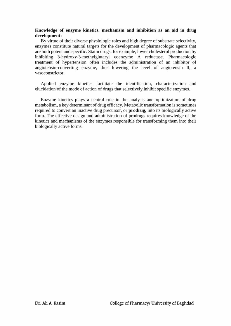
Dr. Ali A. Kasim College of Pharmacy/ University of Baghdad
Knowledge of enzyme kinetics, mechanism and inhibition as an aid in drug
development:
By virtue of their diverse physiologic roles and high degree of substrate selectivity,
enzymes constitute natural targets for the development of pharmacologic agents that
are both potent and specific. Statin drugs, for example, lower cholesterol production by
inhibiting 3-hydroxy-3-methylglutaryl coenzyme A reductase. Pharmacologic
treatment of hypertension often includes the administration of an inhibitor of
angiotensin-converting enzyme, thus lowering the level of angiotensin II, a
vasoconstrictor.
Applied enzyme kinetics facilitate the identification, characterization and
elucidation of the mode of action of drugs that selectively inhibit specific enzymes.
Enzyme kinetics plays a central role in the analysis and optimization of drug
metabolism, a key determinant of drug efficacy. Metabolic transformation is sometimes
required to convert an inactive drug precursor, or prodrug, into its biologically active
form. The effective design and administration of prodrugs requires knowledge of the
kinetics and mechanisms of the enzymes responsible for transforming them into their
biologically active forms.
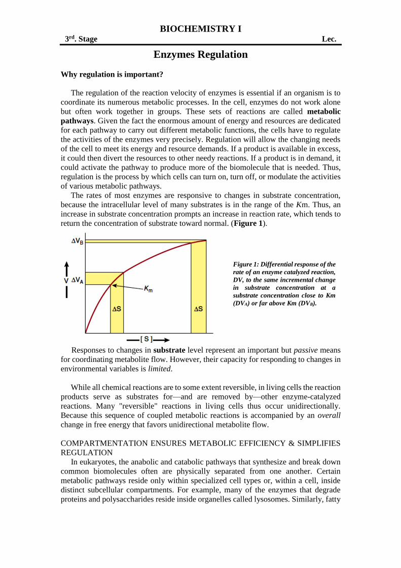
BIOCHEMISTRY I
3rd. Stage Lec.
Enzymes Regulation
Why regulation is important?
The regulation of the reaction velocity of enzymes is essential if an organism is to
coordinate its numerous metabolic processes. In the cell, enzymes do not work alone
but often work together in groups. These sets of reactions are called metabolic
pathways. Given the fact the enormous amount of energy and resources are dedicated
for each pathway to carry out different metabolic functions, the cells have to regulate
the activities of the enzymes very precisely. Regulation will allow the changing needs
of the cell to meet its energy and resource demands. If a product is available in excess,
it could then divert the resources to other needy reactions. If a product is in demand, it
could activate the pathway to produce more of the biomolecule that is needed. Thus,
regulation is the process by which cells can turn on, turn off, or modulate the activities
of various metabolic pathways.
The rates of most enzymes are responsive to changes in substrate concentration,
because the intracellular level of many substrates is in the range of the Km. Thus, an
increase in substrate concentration prompts an increase in reaction rate, which tends to
return the concentration of substrate toward normal. (Figure 1).
Responses to changes in substrate level represent an important but passive means
for coordinating metabolite flow. However, their capacity for responding to changes in
environmental variables is limited.
While all chemical reactions are to some extent reversible, in living cells the reaction
products serve as substrates for—and are removed by—other enzyme-catalyzed
reactions. Many "reversible" reactions in living cells thus occur unidirectionally.
Because this sequence of coupled metabolic reactions is accompanied by an overall
change in free energy that favors unidirectional metabolite flow.
COMPARTMENTATION ENSURES METABOLIC EFFICIENCY & SIMPLIFIES
REGULATION
In eukaryotes, the anabolic and catabolic pathways that synthesize and break down
common biomolecules often are physically separated from one another. Certain
metabolic pathways reside only within specialized cell types or, within a cell, inside
distinct subcellular compartments. For example, many of the enzymes that degrade
proteins and polysaccharides reside inside organelles called lysosomes. Similarly, fatty
Figure 1: Differential response of the
rate of an enzyme catalyzed reaction,
DV, to the same incremental change
in substrate concentration at a
substrate concentration close to Km
(DVA) or far above Km (DVB).
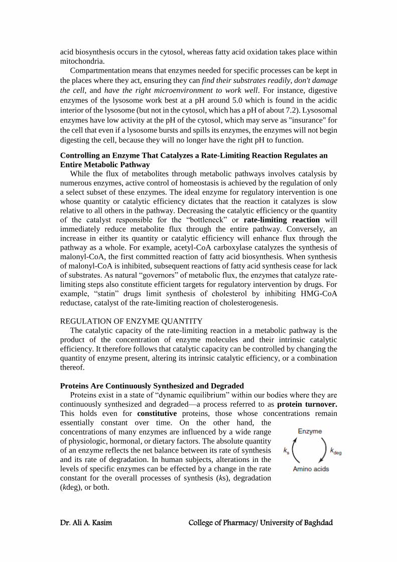
Dr. Ali A. Kasim College of Pharmacy/ University of Baghdad
acid biosynthesis occurs in the cytosol, whereas fatty acid oxidation takes place within
mitochondria.
Compartmentation means that enzymes needed for specific processes can be kept in
the places where they act, ensuring they can find their substrates readily, don't damage
the cell, and have the right microenvironment to work well. For instance, digestive
enzymes of the lysosome work best at a pH around 5.0 which is found in the acidic
interior of the lysosome (but not in the cytosol, which has a pH of about 7.2). Lysosomal
enzymes have low activity at the pH of the cytosol, which may serve as "insurance" for
the cell that even if a lysosome bursts and spills its enzymes, the enzymes will not begin
digesting the cell, because they will no longer have the right pH to function.
Controlling an Enzyme That Catalyzes a Rate-Limiting Reaction Regulates an
Entire Metabolic Pathway
While the flux of metabolites through metabolic pathways involves catalysis by
numerous enzymes, active control of homeostasis is achieved by the regulation of only
a select subset of these enzymes. The ideal enzyme for regulatory intervention is one
whose quantity or catalytic efficiency dictates that the reaction it catalyzes is slow
relative to all others in the pathway. Decreasing the catalytic efficiency or the quantity
of the catalyst responsible for the “bottleneck” or rate-limiting reaction will
immediately reduce metabolite flux through the entire pathway. Conversely, an
increase in either its quantity or catalytic efficiency will enhance flux through the
pathway as a whole. For example, acetyl-CoA carboxylase catalyzes the synthesis of
malonyl-CoA, the first committed reaction of fatty acid biosynthesis. When synthesis
of malonyl-CoA is inhibited, subsequent reactions of fatty acid synthesis cease for lack
of substrates. As natural “governors” of metabolic flux, the enzymes that catalyze rate-
limiting steps also constitute efficient targets for regulatory intervention by drugs. For
example, “statin” drugs limit synthesis of cholesterol by inhibiting HMG-CoA
reductase, catalyst of the rate-limiting reaction of cholesterogenesis.
REGULATION OF ENZYME QUANTITY
The catalytic capacity of the rate-limiting reaction in a metabolic pathway is the
product of the concentration of enzyme molecules and their intrinsic catalytic
efficiency. It therefore follows that catalytic capacity can be controlled by changing the
quantity of enzyme present, altering its intrinsic catalytic efficiency, or a combination
thereof.
Proteins Are Continuously Synthesized and Degraded
Proteins exist in a state of “dynamic equilibrium” within our bodies where they are
continuously synthesized and degraded—a process referred to as protein turnover.
This holds even for constitutive proteins, those whose concentrations remain
essentially constant over time. On the other hand, the
concentrations of many enzymes are influenced by a wide range
of physiologic, hormonal, or dietary factors. The absolute quantity
of an enzyme reflects the net balance between its rate of synthesis
and its rate of degradation. In human subjects, alterations in the
levels of specific enzymes can be effected by a change in the rate
constant for the overall processes of synthesis (ks), degradation
(kdeg), or both.
Dr. Ali A. Kasim College of Pharmacy/ University of Baghdad
Control of Enzyme Synthesis The synthesis of certain enzymes depends upon the presence of inducers, typically
substrates or structurally related compounds that stimulate the transcription of the gene
that encodes them. Escherichia coli grown on glucose will, for example, only catabolize
lactose after addition of a β-galactoside, an inducer that triggers synthesis of a β-
galactosidase and a galactoside permease. Inducible enzymes of humans include
tryptophan pyrrolase, threonine dehydratase, tyrosine-α-ketoglutarate
aminotransferase, enzymes of the urea cycle, HMG-CoA reductase, δ-aminolevulinate
synthase, and cytochrome P450. Conversely, an excess of a metabolite may limit
synthesis of its related enzyme via repression. Both induction and repression involve
cis elements, specific DNA sequences located upstream of regulated genes, and trans-
acting regulatory proteins.
Control of Enzyme Degradation
In animals many proteins are degraded by the ubiquitin proteasome pathway.
Degradation takes place in the proteasome, a large macromolecular complex made up
of more than 30 polypeptide subunits arranged in the form of a hollow cylinder. The
active sites of its proteolytic subunits face the interior of the cylinder, thus preventing
indiscriminate degradation of cellular proteins. Proteins are targeted to the interior of
the proteasome by “ubiquitination,” the covalent attachment of one or more ubiquitin
molecules. Ubiquitin is a small, approximately 8.5 kDa protein that is highly conserved
among eukaryotes. Ubiquitination is catalyzed by a large family of enzymes called E3
ligases, which attach ubiquitin to the sidechain amino group of lysyl residues.
The ubiquitin-proteasome pathway is responsible both for the regulated degradation of
selected cellular proteins, for example, cyclins, and for the removal of defective or
aberrant protein species. The key to the versatility and selectivity of the ubiquitin-
proteasome system resides in the variety of intracellular E3 ligases and their ability to
discriminate between the different physical or conformational states of target proteins.
Thus, the ubiquitin-proteasome pathway can selectively degrade proteins whose
physical integrity and functional competency have been compromised by the loss of or
damage to a prosthetic group, oxidation of cysteine or histidine residues, or deamidation
of asparagine or glutamine residues.
Recognition by proteolytic enzymes also can be regulated by covalent modifications
such as phosphorylation; binding of substrates or allosteric effectors; or association
with membranes, oligonucleotides, or other proteins. A growing body of evidence
suggests that dysfunctions of the ubiquitin-proteasome pathway contribute to the
accumulation of the misfolded proteins characteristic of several neurodegenerative
diseases.
MULTIPLE OPTIONS ARE AVAILABLE FOR REGULATING CATALYTIC
ACTIVITY
In humans the induction of protein synthesis is a complex multistep process that
typically requires hours to produce significant changes in overall enzyme level. By
contrast, changes in intrinsic catalytic efficiency effected by binding of dissociable
ligands (allosteric regulation) or by covalent modification achieve regulation of
enzymic activity within seconds. Consequently, changes in protein level generally
dominate when meeting long-term adaptive requirements, whereas changes in catalytic
efficiency are favored for rapid and transient alterations in metabolite flux.

Dr. Ali A. Kasim College of Pharmacy/ University of Baghdad
ALLOSTERIC REGULATION
Feedback inhibition refers to the process by which the end product of a multistep
biosynthetic pathway binds to and inhibits an enzyme catalyzing one of the early steps
in that pathway. In most cases, feedback inhibitors inhibit the enzyme that catalyzes the
first committed step in a particular biosynthetic sequence. In the following example, for
the biosynthesis of D from A is catalyzed by enzymes Enz1 through Enz3:
High concentrations of D inhibit the conversion of A to B. In this example, the feedback
inhibitor D acts as a negative allosteric effector of Enz1. Inhibition results, not from
the “backing up” of intermediates, but from the ability of D to bind to and inhibit Enz1.
Generally, D binds at an allosteric site, one spatially distinct from the catalytic site of
the target enzyme. Feedback inhibitors thus typically bear little or no structural
similarity to the substrates of the enzymes they inhibit. For example, NAD+ and 3-
phosphoglycerate, the substrates for 3-phosphoglycerate dehydrogenase, which
catalyzes the first committed step in serine biosynthesis, bear no resemblance to the
feedback inhibitor serine.
In branched biosynthetic pathways, such as those responsible for nucleotide
biosynthesis, the initial reactions supply intermediates required for the synthesis of
multiple end products. Figure 2 shows a hypothetical branched biosynthetic pathway
in which curved arrows lead from feedback inhibitors to the enzymes whose activity
they inhibit.
The sequences S3 → A, S4 → B, S4 → C, and S3 → → D each represent linear reaction
sequences that are feedback-inhibited by their end products. Branch point enzymes thus
can be targeted to direct later stages of metabolite flow.
The kinetics of feedback inhibition may be competitive, noncompetitive, partially
competitive, or mixed. Layering multiple feedback loops can provide additional fine
control. For example, as shown in Figure 3, the presence of excess product B decreases
the requirement for substrate S2. However, S2 is also required for synthesis of A, C, and
D. Therefore, for this pathway, excess B limits synthesis of all four end products,
regardless of the need for the other three. To circumvent this potential difficulty, each
end product may only partially inhibit catalytic activity. The effect of an excess of two
or more end products may be strictly additive or, alternatively, greater than their
individual effect (cooperative feedback inhibition).
A B C D
Enz.1 Enz.2 Enz.3
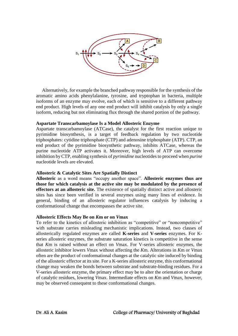
Dr. Ali A. Kasim College of Pharmacy/ University of Baghdad
Alternatively, for example the branched pathway responsible for the synthesis of the
aromatic amino acids phenylalanine, tyrosine, and tryptophan in bacteria, multiple
isoforms of an enzyme may evolve, each of which is sensitive to a different pathway
end product. High levels of any one end product will inhibit catalysis by only a single
isoform, reducing but not eliminating flux through the shared portion of the pathway.
Aspartate Transcarbamoylase Is a Model Allosteric Enzyme
Aspartate transcarbamoylase (ATCase), the catalyst for the first reaction unique to
pyrimidine biosynthesis, is a target of feedback regulation by two nucleotide
triphosphates: cytidine triphosphate (CTP) and adenosine triphosphate (ATP). CTP, an
end product of the pyrimidine biosynthetic pathway, inhibits ATCase, whereas the
purine nucleotide ATP activates it. Moreover, high levels of ATP can overcome
inhibition by CTP, enabling synthesis of pyrimidine nucleotides to proceed when purine
nucleotide levels are elevated.
Allosteric & Catalytic Sites Are Spatially Distinct
Allosteric as a word means “occupy another space”. Allosteric enzymes thus are
those for which catalysis at the active site may be modulated by the presence of
effectors at an allosteric site. The existence of spatially distinct active and allosteric
sites has since been verified in several enzymes using many lines of evidence. In
general, binding of an allosteric regulator influences catalysis by inducing a
conformational change that encompasses the active site.
Allosteric Effects May Be on Km or on Vmax
To refer to the kinetics of allosteric inhibition as “competitive” or “noncompetitive”
with substrate carries misleading mechanistic implications. Instead, two classes of
allosterically regulated enzymes are called K-series and V-series enzymes. For K-
series allosteric enzymes, the substrate saturation kinetics is competitive in the sense
that Km is raised without an effect on Vmax. For V-series allosteric enzymes, the
allosteric inhibitor lowers Vmax without affecting the Km. Alterations in Km or Vmax
often are the product of conformational changes at the catalytic site induced by binding
of the allosteric effector at its site. For a K-series allosteric enzyme, this conformational
change may weaken the bonds between substrate and substrate-binding residues. For a
V-series allosteric enzyme, the primary effect may be to alter the orientation or charge
of catalytic residues, lowering Vmax. Intermediate effects on Km and Vmax, however,
may be observed consequent to these conformational changes.
Dr. Ali A. Kasim College of Pharmacy/ University of Baghdad
Notes regarding Allosteric regulation:
Feedback regulation can be either stimulatory or inhibitory
In both mammalian and bacterial cells, some pathway end products “feedback” to
control their own synthesis, in many instances by feedback inhibition of an early
biosynthetic enzyme. We must, however, distinguish between feedback regulation, a
phenomenologic term lacking mechanistic implications, and feedback inhibition, a
mechanism for regulation of enzyme activity. For example, while dietary cholesterol
decreases hepatic synthesis of cholesterol, this feedback regulation does not involve
feedback inhibition. HMG-CoA reductase, the rate-limiting enzyme of
cholesterogenesis, is affected, but cholesterol does not inhibit its activity. Rather,
regulation in response to dietary cholesterol involves reduction of the expression of the
gene that encodes HMG-CoA reductase (enzyme repression); by cholesterol or a
cholesterol metabolite. As mentioned earlier, ATP, a product of the purine nucleotide
pathway, stimulates the synthesis of pyrimidine nucleotides by activating aspartate
transcarbamoylase, a process sometimes referred to as “feed forward” regulation.
Many hormones act via second messengers
Nerve impulses and the binding of many hormones to cell surface receptors elicit
changes in the rate of enzyme-catalyzed reactions within target cells by inducing the
release or synthesis of specialized allosteric effectors called second messengers. The
primary, or “first,” messenger is the hormone molecule or nerve impulse. Second
messengers include 3′, 5′-cAMP, synthesized from ATP by the enzyme adenylyl
cyclase in response to the hormone epinephrine. Membrane depolarization resulting
from a nerve impulse opens a membrane channel that releases calcium ions into the
cytoplasm, where they bind to and activate enzymes involved in the regulation of
muscle contraction and the mobilization of stored glucose from glycogen to supply the
increased energy demands of muscle contraction. Other second messengers include
3′,5′-cGMP, nitric oxide, and the polyphosphoinositols produced by the hydrolysis
of inositol phospholipids by hormone-regulated phospholipases.
REGULATORY COVALENT MODIFICATIONS: In mammalian cells, a wide range of regulatory covalent modifications occur. These
modifications can be either irreversible (partial proteolysis), or reversible
(phosphorylation, acetylation, methylation and ADP-ribosylation).
- Irreversible Covalent Modification: (proteolytic activation of proenzymes.)
Certain proteins are synthesized as inactive precursor proteins known as
proproteins. Selective, or “partial,” proteolysis of a proprotein by one or more
successive proteolytic “clips” converts it to a form that exhibits the characteristic
activity of the mature protein, for example, its catalytic activity.
The proprotein forms of enzymes are termed proenzymes or zymogens. Proteins
synthesized as proproteins include the hormone insulin (proprotein = proinsulin), the
digestive enzymes pepsin, trypsin, and chymotrypsin (proproteins = pepsinogen,
trypsinogen, and chymotrypsinogen, respectively), several factors of the blood clotting
and complement cascades, and the connective tissue protein collagen (proprotein =
procollagen).
Proteolytic activation of proproteins constitutes a physiologically irreversible
modification because reunification of the two portions of a protein produced by
hydrolysis of a peptide bond is entropically disfavored. Once a proprotein is activated,
it will continue to carry out its catalytic or other functions until it is removed by

Dr. Ali A. Kasim College of Pharmacy/ University of Baghdad
degradation or some other means. Zymogen activation thus represents a simple and
economical, although one way, mechanism for restraining the latent activity of a protein
until the appropriate circumstances are encountered. It is therefore not surprising that
partial proteolysis is employed frequently to regulate proteins that work in the
gastrointestinal tract or bloodstream rather than in the interior of cells.
Selective proteolysis involves one or more highly specific proteolytic clips that may
or may not be accompanied by separation of the resulting peptides. Most importantly,
selective proteolysis often results in conformational changes that properly configure an
enzyme’s active site. The conformational changes that accompany selective proteolysis
of prochymotrypsin (chymotrypsinogen) align three residues, forming the catalytic site.
Contact and catalytic residues can be located on different peptide chains but still be
within bond-forming distance of bound substrate.
Importance of existence of some enzymes as catalytically inactive proenzymes
The synthesis and secretion of proteases as catalytically inactive proenzymes
protect the tissue of origin (eg, the pancreas) from autodigestion, such as can occur in
pancreatitis. Certain physiologic processes such as digestion are intermittent but fairly
regular and predictable in frequency. Others such as blood clot formation, clot
dissolution, and tissue repair are brought “on line” only in response to pressing
pathophysiologic need. The processes of blood clot formation and dissolution clearly
must be temporally coordinated to achieve homeostasis. Enzymes needed intermittently
but rapidly often are secreted in an initially inactive form since new synthesis and
secretion of the required proteins might be insufficiently rapid to respond to a pressing
pathophysiologic demand such as the loss of blood.

Dr. Ali A. Kasim College of Pharmacy/ University of Baghdad
- Reversible Covalent Modification Acetylation, ADP-ribosylation, methylation, and phosphorylation are all examples of
“reversible” covalent modifications. In this context, reversible refers to the fact that the
modified protein can be restored to its original, modification-free state, not the
mechanism by which restoration takes place.
Mammalian proteins are the targets of a wide range of covalent modification
processes. Modifications such as prenylation, glycosylation, and hydroxylation
introduce unique structural features into newly synthesized proteins that tend to persist
for the lifetime of the protein. Among the covalent modifications that regulate protein
function, the most common by far are phosphorylation-dephosphorylation and
acetylation-deacetylation.
Phosphorylation
Protein kinases phosphorylate proteins by catalyzing transfer of the terminal
phosphoryl group of ATP to the hydroxyl groups of seryl, threonyl, or tyrosyl residues,
forming O-phosphoseryl, O-phosphothreonyl, or O-phosphotyrosyl residues,
respectively.
Some protein kinases target the side chains of histidyl, lysyl, arginyl, and aspartyl
residues. The unmodified form of the protein can be regenerated by hydrolytic removal
of phosphoryl groups, catalyzed by protein phosphatases.
A typical mammalian cell possesses thousands of phosphorylated proteins and
several hundred protein kinases and protein phosphatases that catalyze their
interconversion. The ease of interconversion of enzymes between their phospho- and
dephospho- forms accounts, in part, for the frequency with which phosphorylation-
dephosphorylation is utilized as a mechanism for regulatory control. Phosphorylation-
dephosphorylation permits the functional properties of the affected enzyme to be altered
only for as long as it serves a specific need. Once the need has passed, the enzyme can
be converted back to its original form, capable to respond to the next stimulatory event.
A second factor underlying the widespread use of protein phosphorylation-
dephosphorylation lies in the chemical properties of the phosphoryl group itself. In
order to alter an enzyme’s functional properties, any modification of its chemical
structure must influence the protein’s three-dimensional configuration. The high charge
density of protein-bound phosphoryl groups (generally –2 at physiologic pH) and their
tendency to form strong salt bridges with arginyl and lysyl residues renders them potent
agents for modifying protein structure and function. Phosphorylation generally
influences an enzyme’s intrinsic catalytic efficiency or other properties by inducing
conformational changes. Consequently, the amino acids targeted by phosphorylation
can be and typically are relatively distant from the catalytic site itself.
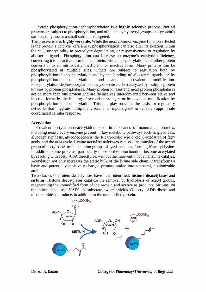
Dr. Ali A. Kasim College of Pharmacy/ University of Baghdad
Protein phosphorylation-dephosphorylation is a highly selective process. Not all
proteins are subject to phosphorylation, and of the many hydroxyl groups on a protein’s
surface, only one or a small subset are targeted.
The process is also highly versatile. While the most common enzyme function affected
is the protein’s catalytic efficiency, phosphorylation can also alter its location within
the cell, susceptibility to proteolytic degradation, or responsiveness to regulation by
allosteric ligands. Phosphorylation can increase an enzyme’s catalytic efficiency,
converting it to its active form in one protein, while phosphorylation of another protein
converts it to an intrinsically inefficient, or inactive form. Many proteins can be
phosphorylated at multiple sites. Others are subject to regulation both by
phosphorylation-dephosphorylation and by the binding of allosteric ligands, or by
phosphorylation-dephosphorylation and another covalent modification.
Phosphorylation-dephosphorylation at any one site can be catalyzed by multiple protein
kinases or protein phosphatases. Many protein kinases and most protein phosphatases
act on more than one protein and are themselves interconverted between active and
inactive forms by the binding of second messengers or by covalent modification by
phosphorylation-dephosphorylation. This interplay provides the basis for regulatory
networks that integrate multiple environmental input signals to evoke an appropriate
coordinated cellular response.
Acetylation
Covalent acetylation-deacetylation occur in thousands of mammalian proteins,
including nearly every enzyme present in key metabolic pathways such as glycolysis,
glycogen synthesis, gluconeogenesis, the tricarboxylic acid cycle, β-oxidation of fatty
acids, and the urea cycle. Lysine acetyltransferases catalyze the transfer of the acetyl
group of acetyl-CoA to the ε-amino groups of lysyl residues, forming N-acetyl lysine.
In addition, some proteins, particularly those in the mitochondria, become acetylated
by reacting with acetyl-CoA directly, ie, without the intervention of an enzyme catalyst.
Acetylation not only increases the steric bulk of the lysine side chain, it transforms a
basic and potentially positively charged primary amine into a neutral, nonionizable
amide.
Two classes of protein deacetylases have been identified: histone deacetylases and
sirtuins. Histone deacetylases catalyze the removal by hydrolysis of acetyl groups,
regenerating the unmodified form of the protein and acetate as products. Sirtuins, on
the other hand, use NAD+ as substrate, which yields O-acetyl ADP-ribose and
nicotinamide as products in addition to the unmodified protein.
Dr. Ali A. Kasim College of Pharmacy/ University of Baghdad
Reversible modifications of DNA-binding proteins
Histones and other DNA-binding proteins in chromatin are subject to extensive
modification by acetylation, methylation, phosphorylation, as well as ADP-
ribosylation. These modifications, modulate the manner in which the proteins within
chromatin interact with each other as well as the DNA itself, constitute the basis for the
“histone code.” The resulting changes in chromatin structure within the region affected
can render genes more accessible to the proteins responsible for their transcription,
thereby enhancing gene expression or, on a larger scale, facilitating replication of the
entire genome. On the other hand, changes in chromatin structure that restrict the
accessibility of genes to transcription factors, DNA-dependent RNA polymerases, etc,
thereby inhibiting transcription, are said to silence gene expression.
The Histone Code
The “histone code” represents a classic example of epigenetics, the hereditary
transmission of information by a means other than the sequence of nucleotides that
comprise the genome. In this instance, the pattern of gene expression within a newly
formed “daughter” cell will be determined, in part, by the particular set of histone
covalent modifications embodied in the chromatin proteins inherited from the
“parental” cell.

ONE-CARBON METABOLISM
The degradation pathways for several amino acids produce one-carbon units that are
transferred to tetrahydrofolate (H4folate or THF) as an intermediate carrier. Tetrahydrofolate, in turn, donates the single carbons to various biosynthet ic
intermediates. The following one-carbon units are encountered in the biological reactions, which constitute one-carbon pool
Methyl (−CH3)
Methylene (═CH2) Methenyl (−CH═) Hydroxymethyl (−CH2OH)
Formyl (−CH═O) Formimino (−CH═NH)
Folic acid is the vitamin as it is found in the diet. It consists of three distinct moietie s : a pteridine moiety, a p-aminobenzoate (PABA) moiety, and one or several glutamate
residues. Mammals can synthesize these components, but lack the enzymes necessary
for their conjugation. The number of glutamate residues contained in folic acid or THF changes at various stages of transport and utilization. Intestinal uptake and transport
through the blood occur with only one glutamate attached. After uptake into liver cells, several more glutamate residues are added.
The metabolically active form of folate is THF, which is formed from folate in two successive NADPH-dependent reductions, both catalyzed by the same enzyme, namely, dihydrofolate reductase. The hydrogen atoms are present at positions 5, 6, 7 and 8 of
THF. The one-carbon unit covalently binds with THF at position N5 or N10 or on both N5 and
N10 of pteroyl structure of folate.
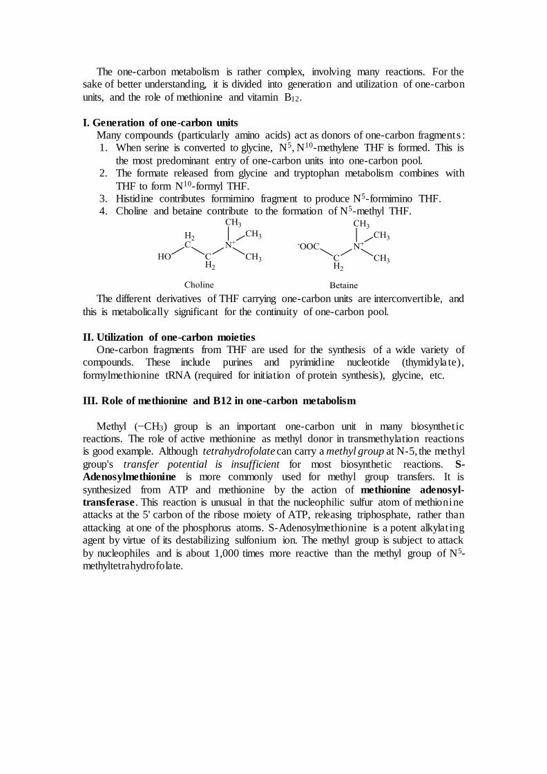
The one-carbon metabolism is rather complex, involving many reactions. For the sake of better understanding, it is divided into generation and utilization of one-carbon
units, and the role of methionine and vitamin B12.
I. Generation of one-carbon units
Many compounds (particularly amino acids) act as donors of one-carbon fragments : 1. When serine is converted to glycine, N5, N10-methylene THF is formed. This is
the most predominant entry of one-carbon units into one-carbon pool. 2. The formate released from glycine and tryptophan metabolism combines with
THF to form N10-formyl THF. 3. Histidine contributes formimino fragment to produce N5-formimino THF. 4. Choline and betaine contribute to the formation of N5-methyl THF.
The different derivatives of THF carrying one-carbon units are interconvertible, and
this is metabolically significant for the continuity of one-carbon pool.
II. Utilization of one-carbon moieties
One-carbon fragments from THF are used for the synthesis of a wide variety of compounds. These include purines and pyrimidine nucleotide (thymidyla te),
formylmethionine tRNA (required for initiation of protein synthesis), glycine, etc.
III. Role of methionine and B12 in one-carbon metabolism
Methyl (−CH3) group is an important one-carbon unit in many biosynthet ic reactions. The role of active methionine as methyl donor in transmethylation reactions is good example. Although tetrahydrofolate can carry a methyl group at N-5, the methyl
group's transfer potential is insufficient for most biosynthetic reactions. S-
Adenosylmethionine is more commonly used for methyl group transfers. It is
synthesized from ATP and methionine by the action of methionine adenosyl-
transferase. This reaction is unusual in that the nucleophilic sulfur atom of methionine attacks at the 5' carbon of the ribose moiety of ATP, releasing triphosphate, rather than
attacking at one of the phosphorus atoms. S-Adenosylmethionine is a potent alkylat ing agent by virtue of its destabilizing sulfonium ion. The methyl group is subject to attack
by nucleophiles and is about 1,000 times more reactive than the methyl group of N5-methyltetrahydrofolate.

After the release of methyl group, methionine is converted to homocysteine. For the regeneration of methionine, free homocysteine and N5-methyl THF are required and
this reaction is dependent on methylcobalamin (vitamin B12). The one-carbon pool, under the control of THF, is linked with methionine metabolism (transmethylat ion) through vitamin B12. Hence vitamin B12 is also involved in one-carbon metabolism.
Folate deficiency impairs DNA synthesis and cell division resulting in many
clinical conditions such as macrocytic anemia and neural tube defects in pregnancy (spina bifida or anencephaly).
Folic acid antagonists:
Methotrexate (structural analogue of folic acid), competitively inhibits dihydrofo la te
reductase, is used in the treatment of leukemia. Sulfonamides (structural analogues of PABA), competitively inhibit the enzyme
responsible for the conjugation of PABA in folic acid molecule, are antibacterial agents.
Note:
Some authors consider CO2 as a one-carbon unit, others do not agree. Carbon dioxide is involved in many biochemical reactions (carboxylation); biotin serves as a carrier of CO2 in these reactions. For instance, conversion of pyruvate to oxaloacetate
in gluconeogenesis.
Metabolic Diseases of Amino Acid Catabolism
Defective synthesis or decreased activity of enzymes involved in amino acid
catabolism is associated with many metabolic diseases. Metabolic defects in transaminases, which fulfill central metabolic functions, may be
incompatible with life. Consequently, no known metabolic defect is associated with this short catabolic pathway.
Proline
There are two metabolic disorders of proline catabolism.
The metabolic block in type I hyperprolinemia is at proline dehydrogenase. The metabolic block in type II hyperprolinemia is at Δ1-pyrroline-5-carboxylate
dehydrogenase, which also participates in the catabolism of arginine, ornithine, and
hydroxyproline. Since proline and hydroxyproline catabolism are affected, both Δ1-pyrroline-5-carboxylate and Δ1-pyrroline-3-hydroxy-5-carboxylate are excreted.
Hyperpolinemia is associated with seizures, intellectual disability, or other neurologica l
problems.
Arginine and Ornithine
Mutations in ornithine transaminase elevate plasma and urinary ornithine and are associated with gyrate atrophy of the choroid and retina. Treatment involves
restricting dietary arginine.
Note:
Defective arginase is associated with hyperargininemia In the hyperornithinemia–hyperhomocitrulinuria-hyperammonemia syndrome, a
defective mitochondrial ornithine permease impairs transport of ornithine into mitochondria where it participates as an intermediate in urea synthesis.
Histidine
Benign disorders of histidine catabolism include histidinemia and urocanic
aciduria associated with impaired histidase and urocanase, respectively.
In folic acid deficiency, transfer of the formimino group is impaired, and formiminoglutamate (Figlu) is excreted.
Note:
Excretion of Figlu following a dose of histidine thus can be used to detect folic acid deficiency.
Glycine
Nonketotic hyperglycinemia or Glycine encephalopathy, is a rare inborn error of
glycine degradation, due to defective glycine cleavage complex. Glycine accumula tes in all body tissues including the central nervous system. The defect in primary hyperoxaluria is the failure to catabolize glyoxylate formed by
the deamination of glycine. Subsequent oxidation of glyoxylate to oxalate results in urolithiasis, nephrocalcinosis, and early mortality from renal failure or hypertension.
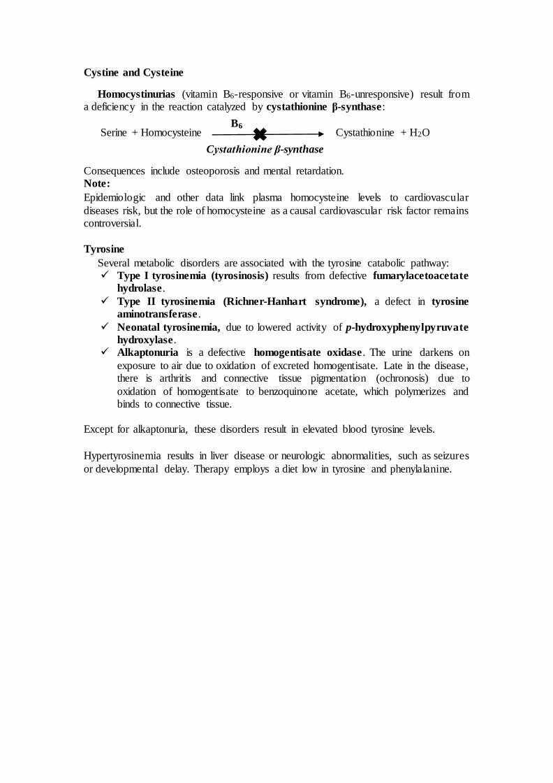
Cystine and Cysteine
Homocystinurias (vitamin B6-responsive or vitamin B6-unresponsive) result from a deficiency in the reaction catalyzed by cystathionine β-synthase:
Serine + Homocysteine Cystathionine + H2O
Consequences include osteoporosis and mental retardation. Note:
Epidemiologic and other data link plasma homocysteine levels to cardiovascular
diseases risk, but the role of homocysteine as a causal cardiovascular risk factor remains controversial.
Tyrosine
Several metabolic disorders are associated with the tyrosine catabolic pathway: Type I tyrosinemia (tyrosinosis) results from defective fumarylacetoacetate
hydrolase.
Type II tyrosinemia (Richner-Hanhart syndrome), a defect in tyrosine aminotransferase.
Neonatal tyrosinemia, due to lowered activity of p-hydroxyphenylpyruvate hydroxylase.
Alkaptonuria is a defective homogentisate oxidase. The urine darkens on
exposure to air due to oxidation of excreted homogentisate. Late in the disease, there is arthritis and connective tissue pigmentation (ochronosis) due to
oxidation of homogentisate to benzoquinone acetate, which polymerizes and binds to connective tissue.
Except for alkaptonuria, these disorders result in elevated blood tyrosine levels.
Hypertyrosinemia results in liver disease or neurologic abnormalities, such as seizures
or developmental delay. Therapy employs a diet low in tyrosine and phenylalanine.
6B
Cystathionine β-synthase

Phenylalanine
Hyperphenylalaninemias arise from defects in phenylalanine hydroxylase (type
I, classic phenylketonuria (PKU)), in dihydrobiopterin reductase (types II and III),
or in dihydrobiopterin biosynthesis (types IV and V).
There are two routes by which the excess phenylalanine can be metabolized :
oxidation to tyrosine (the normal and main route for degradation of phenylalanine, and
the normal route for biosynthesis of tyrosine), and transamination to phenylpyruva te
and subsequent further metabolism to phenyl lactate, phenylacetate, and phenylace tyl
glutamine (a minor route, which comes to the fore when the main route is blocked).
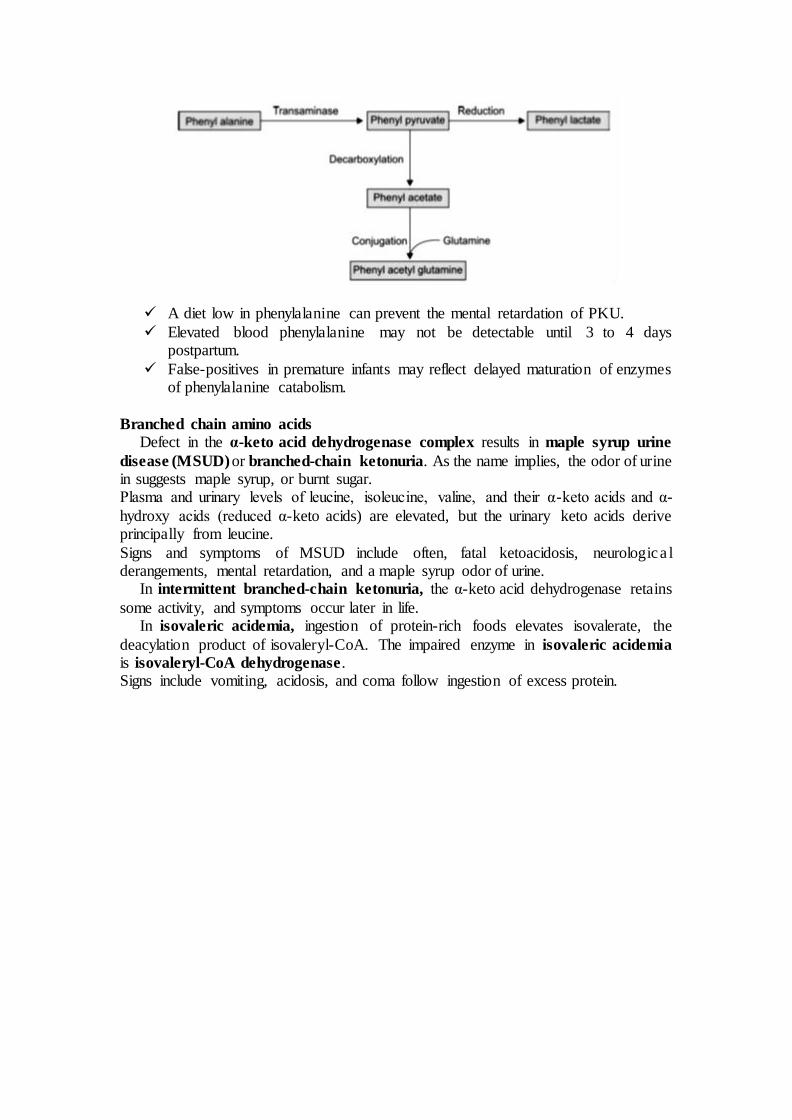
A diet low in phenylalanine can prevent the mental retardation of PKU.
Elevated blood phenylalanine may not be detectable until 3 to 4 days postpartum.
False-positives in premature infants may reflect delayed maturation of enzymes of phenylalanine catabolism.
Branched chain amino acids
Defect in the α-keto acid dehydrogenase complex results in maple syrup urine
disease (MSUD) or branched-chain ketonuria. As the name implies, the odor of urine in suggests maple syrup, or burnt sugar. Plasma and urinary levels of leucine, isoleucine, valine, and their α-keto acids and α-
hydroxy acids (reduced α-keto acids) are elevated, but the urinary keto acids derive principally from leucine.
Signs and symptoms of MSUD include often, fatal ketoacidosis, neurologica l derangements, mental retardation, and a maple syrup odor of urine. In intermittent branched-chain ketonuria, the α-keto acid dehydrogenase retains
some activity, and symptoms occur later in life. In isovaleric acidemia, ingestion of protein-rich foods elevates isovalerate, the
deacylation product of isovaleryl-CoA. The impaired enzyme in isovaleric acidemia
is isovaleryl-CoA dehydrogenase. Signs include vomiting, acidosis, and coma follow ingestion of excess protein.

BIOCHEMISTRY I
3rd. Stage Lec.
CLINICAL ENZYMOLOGY
In general, most enzymes are present in cells at much higher concentrations than in
plasma. Measurement of their levels in plasma indicates whether their tissue of origin
is damaged leading to the release of intracellular components into the blood. This forms
the basis of clinical enzymology. Thus, clinical enzymology refers to measurement of
enzyme activity for the diagnosis and treatment of diseases.
Since the tight control of enzyme activity is essential for homeostasis, any malfunction
of a single critical enzyme (mutation, overproduction, underproduction or deletion) can
lead to a genetic disease - commonly called inborn errors of metabolism. One
example is the most common type of phenylketonuria caused by a mutation of a single
amino acid in the enzyme phenylalanine hydroxylase, which catalyzes the first step in
the degradation of phenylalanine. The deficiency results in build-up of phenylalanine
and related un-physiological by-products. This can lead to mental retardation if the
disease is untreated early.
Detection of the plasma level of an enzyme immunologically (for its protein amount)
or colorimetrically (for its activity, preferred) have the following applications:
Diagnosis: detect and localize tissue cell damage or proliferation; e.g., high serum
creatine phosphokinase (CK) on the day of a suspected case of myocardial infarction
strengthen the diagnosis if ECG changes are doubtful.
Differential diagnosis: e.g., chest pain associates myocardial infarction and pulmonary
embolism. Elevated serum aspartate transaminase (AST) and lactate dehydrogenase
(LDH) characterizes myocardial infarction, whereas, elevated serum LDH only
characterizes pulmonary embolism.
Therapeutic follow up and/or early detection of a disease:
Chronic administration of several therapeutics - e.g., anti-depressant and anticancer
chemotherapies - elevates serum isocitrate dehydrogenase or ornithine carbamoyl-
transferase level when they induce minimal hepatotoxicity.
Serum alanine transaminase (ALT) level elevates in sub-clinical early viral hepatitis.
PLASMA ENZYMES: Enzymes present in plasma can be classified into 2 types, they are: Functional Plasma
enzymes and Non-functional plasma enzymes.
Plasma-derived enzymes (functional plasma enzymes):
They are normally occurring functional plasma enzymes.
Mostly synthesized by the liver.
Usually decreased in disease conditions.
Their field of activity is plasma components and their activity is higher in
plasma than in cells, e.g., coagulation and lipoprotein-metabolizing enzymes.
Their clinical importance is limited to diseases related to their own synthesis
and function; i.e., abnormalities of metabolism of plasma lipoproteins and blood
clotting, and the organ function of their synthesizing tissues, e.g.,
thromboplastin as a liver function test.
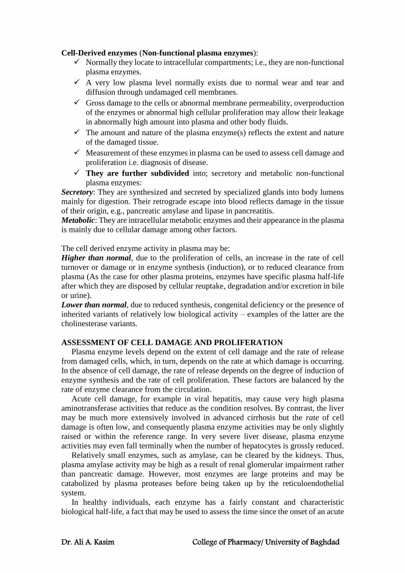
Dr. Ali A. Kasim College of Pharmacy/ University of Baghdad
Cell-Derived enzymes (Non-functional plasma enzymes):
Normally they locate to intracellular compartments; i.e., they are non-functional
plasma enzymes.
A very low plasma level normally exists due to normal wear and tear and
diffusion through undamaged cell membranes.
Gross damage to the cells or abnormal membrane permeability, overproduction
of the enzymes or abnormal high cellular proliferation may allow their leakage
in abnormally high amount into plasma and other body fluids.
The amount and nature of the plasma enzyme(s) reflects the extent and nature
of the damaged tissue.
Measurement of these enzymes in plasma can be used to assess cell damage and
proliferation i.e. diagnosis of disease.
They are further subdivided into; secretory and metabolic non-functional
plasma enzymes:
Secretory: They are synthesized and secreted by specialized glands into body lumens
mainly for digestion. Their retrograde escape into blood reflects damage in the tissue
of their origin, e.g., pancreatic amylase and lipase in pancreatitis.
Metabolic: They are intracellular metabolic enzymes and their appearance in the plasma
is mainly due to cellular damage among other factors.
The cell derived enzyme activity in plasma may be:
Higher than normal, due to the proliferation of cells, an increase in the rate of cell
turnover or damage or in enzyme synthesis (induction), or to reduced clearance from
plasma (As the case for other plasma proteins, enzymes have specific plasma half-life
after which they are disposed by cellular reuptake, degradation and/or excretion in bile
or urine).
Lower than normal, due to reduced synthesis, congenital deficiency or the presence of
inherited variants of relatively low biological activity – examples of the latter are the
cholinesterase variants.
ASSESSMENT OF CELL DAMAGE AND PROLIFERATION
Plasma enzyme levels depend on the extent of cell damage and the rate of release
from damaged cells, which, in turn, depends on the rate at which damage is occurring.
In the absence of cell damage, the rate of release depends on the degree of induction of
enzyme synthesis and the rate of cell proliferation. These factors are balanced by the
rate of enzyme clearance from the circulation.
Acute cell damage, for example in viral hepatitis, may cause very high plasma
aminotransferase activities that reduce as the condition resolves. By contrast, the liver
may be much more extensively involved in advanced cirrhosis but the rate of cell
damage is often low, and consequently plasma enzyme activities may be only slightly
raised or within the reference range. In very severe liver disease, plasma enzyme
activities may even fall terminally when the number of hepatocytes is grossly reduced.
Relatively small enzymes, such as amylase, can be cleared by the kidneys. Thus,
plasma amylase activity may be high as a result of renal glomerular impairment rather
than pancreatic damage. However, most enzymes are large proteins and may be
catabolized by plasma proteases before being taken up by the reticuloendothelial
system.
In healthy individuals, each enzyme has a fairly constant and characteristic
biological half-life, a fact that may be used to assess the time since the onset of an acute
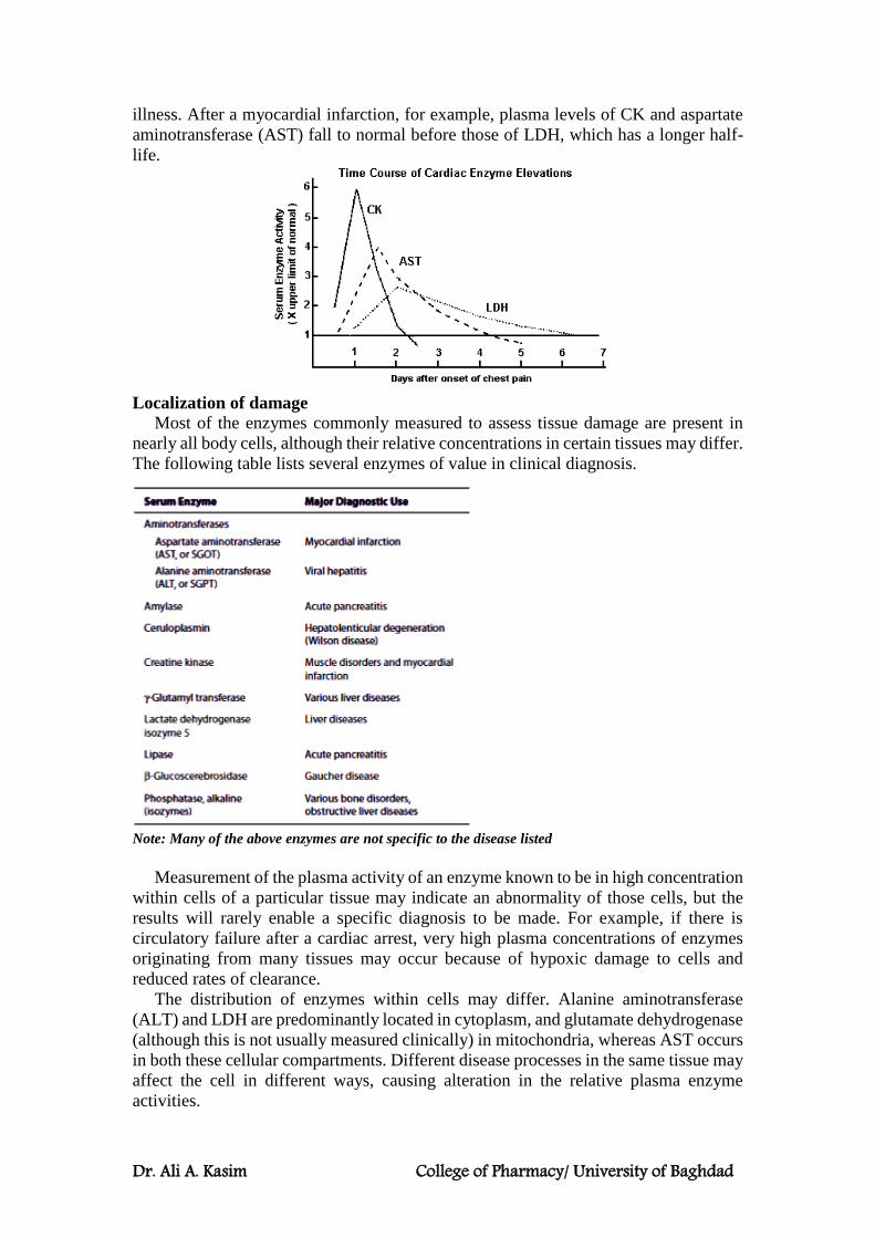
Dr. Ali A. Kasim College of Pharmacy/ University of Baghdad
illness. After a myocardial infarction, for example, plasma levels of CK and aspartate
aminotransferase (AST) fall to normal before those of LDH, which has a longer half-
life.
Localization of damage
Most of the enzymes commonly measured to assess tissue damage are present in
nearly all body cells, although their relative concentrations in certain tissues may differ.
The following table lists several enzymes of value in clinical diagnosis.
Note: Many of the above enzymes are not specific to the disease listed
Measurement of the plasma activity of an enzyme known to be in high concentration
within cells of a particular tissue may indicate an abnormality of those cells, but the
results will rarely enable a specific diagnosis to be made. For example, if there is
circulatory failure after a cardiac arrest, very high plasma concentrations of enzymes
originating from many tissues may occur because of hypoxic damage to cells and
reduced rates of clearance.
The distribution of enzymes within cells may differ. Alanine aminotransferase
(ALT) and LDH are predominantly located in cytoplasm, and glutamate dehydrogenase
(although this is not usually measured clinically) in mitochondria, whereas AST occurs
in both these cellular compartments. Different disease processes in the same tissue may
affect the cell in different ways, causing alteration in the relative plasma enzyme
activities.
Dr. Ali A. Kasim College of Pharmacy/ University of Baghdad
The diagnostic precision of plasma enzyme analysis may be improved by the
following:
Serial enzyme estimations. The rate of change of plasma enzyme activity is related to
a balance between the rate of entry and the rate of removal from the circulation. A
persistently raised plasma enzyme activity is suggestive of a chronic disorder or,
occasionally, impaired clearance.
Isoenzyme determination. Some enzymes exist in more than one form; these
isoenzymes may be separated by their different physical or chemical properties. If they
originate in different tissues, such identification will give more information than the
measurement of plasma total enzyme activity; for example, CK may be derived from
skeletal or cardiac muscle, but one of its isoenzymes is found predominantly in the
myocardium.
Estimation of more than one enzyme. Many enzymes are widely distributed, but their
relative concentrations may vary in different tissues. For example, although both ALT
and AST are abundant in the liver, the concentration of AST is much greater than that
of ALT in heart muscle.
Non-specific causes of raised plasma enzyme activities
Before attributing a change in plasma enzyme activity to a specific disease process,
it is important to exclude the presence of factitious or non-specific causes. Slight rises
in plasma ALT and AST activities are common, non-specific findings in many illnesses.
Moderate exercise, or a large intramuscular injection, may lead to a rise in plasma CK
activity; isoenzyme determination may identify skeletal muscle as the tissue of origin.
Some drugs, such as the anticonvulsants phenytoin and phenobarbital, may induce the
synthesis of the microsomal enzyme γ-glutamyl transferase (GGT), and so increase its
plasma activity in the absence of disease.
Plasma enzyme activities may be raised if the rate of clearance from the circulation
is reduced. In the absence of hepatic or renal disease, this may occur if, for example,
the plasma enzyme forms complexes with immunoglobulins, known as a macroenzyme.
Various enzymes can form clinically significant macroenzymes including amylase,
LDH, ALP and CK.
FACTORS AFFECTING RESULTS OF PLASMA ENZYME ASSAYS
Analytical factors
The total concentration of all plasma enzyme proteins is less than 1 g/L. The results of
enzyme assays are not usually expressed as concentrations, but as activities.
These activities are usually measured in terms of the activity unit (U) which is defined
as the amount which will catalyse the transformation of 1 micromole of the substrate
per minute.
Another unit of enzyme activity has been used. This is the katal (kat) which is defined
as the amount which will catalyse the transformation of one mole of substrate per
second. It is an impracticable unit and has not received widespread acceptance.
Changes in concentration may give rise to proportional changes in catalytic activity,
but the results of such measurements depend on many analytical factors, including:
substrate concentration, product concentration, enzyme concentration, reaction
temperature, reaction pH, and presence of activators or inhibitors.
The definition of ‘international units’ does not take these factors into account, and the
results from different laboratories, which are apparently expressed in the same units,
may not be directly comparable.
Dr. Ali A. Kasim College of Pharmacy/ University of Baghdad
Non-disease factors
Examples of non-disease factors affecting enzyme activities include the following.
Age
Plasma AST activity is moderately higher during the neonatal period than in adults.
Plasma ALP activity of bony origin is higher in children than in adults and peaks during
the pubertal bone growth spurt before falling to adult levels. A second peak occurs in
the elderly.
Sex
Plasma GGT activity is higher in men than in women. Plasma CK activity is also higher
in males, probably in part due to their increased muscle bulk.
Race/ethnicity
Plasma CK activity is higher in black people and Afro-Caribbeans than in white people.
Physiological conditions
Plasma ALP activity rises during the last trimester of pregnancy because of the presence
of the placental isoenzyme. Several enzymes, such as AST and CK, rise moderately in
plasma during and immediately after labour or strenuous exercise.
Plasma enzyme activities should therefore be interpreted in relation to the sex-, race-
ethnicity- and age-matched reference ranges of the issuing laboratory.
Isoenzymes and their Clinical Significance in Clinical Enzymology
Isoenzymes (also known as isozymes) are enzymes that differ in amino acid
sequence but catalyze the same chemical reaction. Isoenzymes are produced by
different genes and are not redundant despite their similar functions. They occur in
many tissues throughout the body and are important for different developmental and
metabolic processes.
Isoenzymes are useful biochemical markers and can be measured in the bloodstream
to diagnose medical conditions. Isoenzymes can be differentiated from one another
using gel electrophoresis. In gel electrophoresis, isoenzyme fragments are drawn
through a thick gel by an electric charge. Each isoenzyme has a distinct charge of its
own because of its unique amino acid sequence. This enables gel electrophoresis to
separate the fragments into bands for identification. Some clinically important
isoenzymes are as follows:
1) Creatine Kinase (CK) It is an enzyme found primarily in the heart and skeletal muscles, and to a lesser
extent in the brain. Small amounts are also found in lungs, liver, thyroid and adrenal
glands. Significant injury to any of these structures will lead to a measurable increase
in CK levels. It is not found in red blood cells and its level is not affected by hemolysis.
CK Isoenzymes There are three Isoenzymes. Measuring them is of value in the presence of elevated
levels of CK to determine the source of the elevation. Each isoenzyme is a dimer
composed of two protomers ‘M’ (for muscles) and ‘B’ (for Brain).
The three possible isoenzymes are;
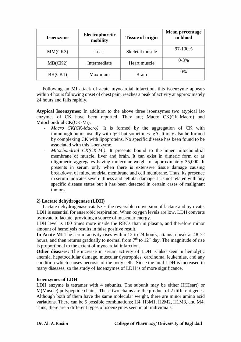
Dr. Ali A. Kasim College of Pharmacy/ University of Baghdad
Isoenzyme Electrophoretic
mobility Tissue of origin
Mean percentage
in blood
MM(CK3) Least Skeletal muscle 97-100%
MB(CK2) Intermediate Heart muscle 0-3%
BB(CK1) Maximum Brain 0%
Following an MI attack of acute myocardial infarction, this isoenzyme appears
within 4 hours following onset of chest pain, reaches a peak of activity at approximately
24 hours and falls rapidly.
Atypical Isoenzymes: In addition to the above three isoenzymes two atypical iso
enzymes of CK have been reported. They are; Macro CK(CK-Macro) and
Mitochondrial CK(CK-Mi).
- Macro CK(CK-Macro): It is formed by the aggregation of CK with
immunoglobulins usually with IgG but sometimes IgA. It may also be formed
by complexing CK with lipoproteins. No specific disease has been found to be
associated with this isoenzyme.
- Mitochondrial CK(CK-Mi): It presents bound to the inner mitochondrial
membrane of muscle, liver and brain. It can exist in dimeric form or as
oligomeric aggregates having molecular weight of approximately 35,000. It
presents in serum only when there is extensive tissue damage causing
breakdown of mitochondrial membrane and cell membrane. Thus, its presence
in serum indicates severe illness and cellular damage. It is not related with any
specific disease states but it has been detected in certain cases of malignant
tumors.
2) Lactate dehydrogenase (LDH)
Lactate dehydrogenase catalyzes the reversible conversion of lactate and pyruvate.
LDH is essential for anaerobic respiration. When oxygen levels are low, LDH converts
pyruvate to lactate, providing a source of muscular energy.
LDH level is 100 times more inside the RBCs than in plasma, and therefore minor
amount of hemolysis results in false positive result.
In Acute MI-The serum activity rises within 12 to 24 hours, attains a peak at 48-72
hours, and then returns gradually to normal from 7th to 12th day. The magnitude of rise
is proportional to the extent of myocardial infarction.
Other diseases: The increase in serum activity of LDH is also seen in hemolytic
anemia, hepatocellular damage, muscular dystrophies, carcinoma, leukemias, and any
condition which causes necrosis of the body cells. Since the total LDH is increased in
many diseases, so the study of Isoenzymes of LDH is of more significance.
Isoenzymes of LDH
LDH enzyme is tetramer with 4 subunits. The subunit may be either H(Heart) or
M(Muscle) polypeptide chains. These two chains are the product of 2 different genes.
Although both of them have the same molecular weight, there are minor amino acid
variations. There can be 5 possible combinations; H4, H3M1, H2M2, H1M3, and M4.
Thus, there are 5 different types of isoenzymes seen in all individuals.
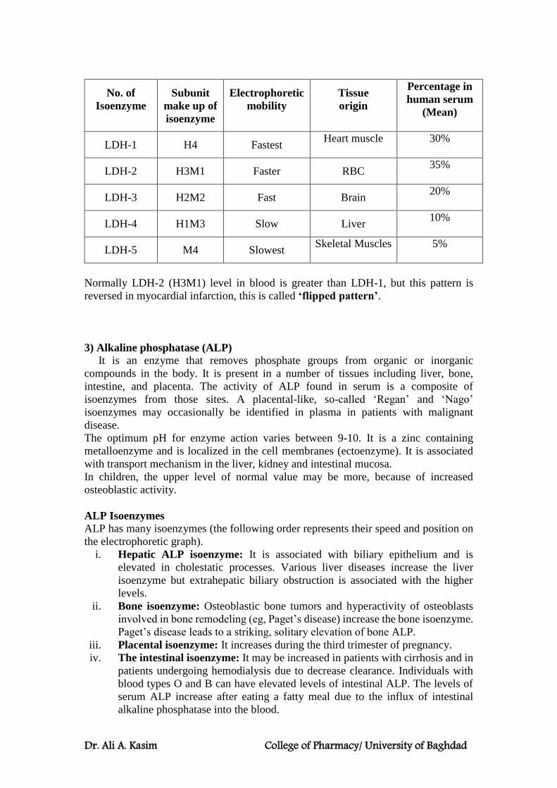
Dr. Ali A. Kasim College of Pharmacy/ University of Baghdad
No. of
Isoenzyme
Subunit
make up of
isoenzyme
Electrophoretic
mobility
Tissue
origin
Percentage in
human serum
(Mean)
LDH-1 H4 Fastest Heart muscle
30%
LDH-2 H3M1 Faster RBC 35%
LDH-3 H2M2 Fast Brain 20%
LDH-4 H1M3 Slow Liver 10%
LDH-5 M4 Slowest Skeletal Muscles
5%
Normally LDH-2 (H3M1) level in blood is greater than LDH-1, but this pattern is
reversed in myocardial infarction, this is called ‘flipped pattern’.
3) Alkaline phosphatase (ALP)
It is an enzyme that removes phosphate groups from organic or inorganic
compounds in the body. It is present in a number of tissues including liver, bone,
intestine, and placenta. The activity of ALP found in serum is a composite of
isoenzymes from those sites. A placental-like, so-called ‘Regan’ and ‘Nago’
isoenzymes may occasionally be identified in plasma in patients with malignant
disease.
The optimum pH for enzyme action varies between 9-10. It is a zinc containing
metalloenzyme and is localized in the cell membranes (ectoenzyme). It is associated
with transport mechanism in the liver, kidney and intestinal mucosa.
In children, the upper level of normal value may be more, because of increased
osteoblastic activity.
ALP Isoenzymes
ALP has many isoenzymes (the following order represents their speed and position on
the electrophoretic graph).
i. Hepatic ALP isoenzyme: It is associated with biliary epithelium and is
elevated in cholestatic processes. Various liver diseases increase the liver
isoenzyme but extrahepatic biliary obstruction is associated with the higher
levels.
ii. Bone isoenzyme: Osteoblastic bone tumors and hyperactivity of osteoblasts
involved in bone remodeling (eg, Paget’s disease) increase the bone isoenzyme.
Paget’s disease leads to a striking, solitary elevation of bone ALP.
iii. Placental isoenzyme: It increases during the third trimester of pregnancy.
iv. The intestinal isoenzyme: It may be increased in patients with cirrhosis and in
patients undergoing hemodialysis due to decrease clearance. Individuals with
blood types O and B can have elevated levels of intestinal ALP. The levels of
serum ALP increase after eating a fatty meal due to the influx of intestinal
alkaline phosphatase into the blood.

Dr. Ali A. Kasim College of Pharmacy/ University of Baghdad
Atypical ALP isoenzymes (Oncogenic markers)-In addition to 4 major isoenzymes,
2 more abnormal fractions are seen associated with tumors. These are Regan and Nagao
isoenzymes. They are also called “Carcino placental ALP”, as they resemble placental
isoenzymes.
Other Clinical Applications of Enzymes
1. Enzyme-Linked Immunoassays
The sensitivity of enzyme assays can be exploited to detect proteins that lack catalytic
activity. Enzyme-linked immunosorbent assays (ELISAs) use antibodies covalently
linked to a “reporter enzyme” such as alkaline phosphatase or horseradish peroxidase
whose products are readily detected, generally by the absorbance of light or by
fluorescence. Serum or other biologic samples to be tested are placed in plastic, multi-
well microtiter plates, where the proteins (the antigen) immobilized on the plastic
surface either non-specifically (via adsorption) or specifically (via capture by another
antibody specific to the same antigen). Any exposed plastic (or fixed antibodies) that
remains is subsequently “blocked” by adding a nonantigenic protein such as bovine
serum albumin. A solution of antibody covalently linked to a reporter enzyme
(detection antibodies) is then added. The antibodies adhere to the immobilized antigen
and are themselves immobilized. Excess free detection antibody molecules are then
removed by washing. The presence and quantity of bound antibody is then determined
by adding the substrate for the reporter enzyme.
Detection of Troponins by ELISA
Troponin, or the troponin complex, is a complex of three regulatory proteins (troponin
C, troponin I, and troponin T) that is integral to muscle contraction in skeletal muscle
and cardiac muscle, but not in smooth muscle. Immunological measurement of plasma
levels of cardiac troponins I and T provide sensitive and specific indicators of damage
to heart muscle. Cardiac troponin levels rise for 2 to 6 hours after an MI and remain
elevated for 4 to 10 days. In addition to MI, other heart muscle damage also elevates
serum troponin levels. Cardiac troponins thus serve as a marker of all heart muscle
damage.
Note: The genes that encode for the skeletal and cardiac isoforms of troponin C are
identical; thus, no structural difference exists between them. However, the skeletal and
cardiac subforms for troponin I and troponin T are distinct, and immunoassays have
been designed to differentiate between them.
2. Enzymes also can be employed in the clinical laboratory as tools for
determining the concentration of critical metabolites.
For example, glucose oxidase is frequently utilized to measure plasma glucose
concentration.
3. Enzymes facilitate diagnosis of genetic diseases.
Many diagnostic techniques take advantage of the specificity and efficiency of the
enzymes that act on oligonucleotides such as DNA. Enzymes known as restriction
endonucleases, for example, cleave double-stranded DNA at sites specified by a
sequence of four, six, or more base pairs called restriction sites. Cleavage of a sample
of DNA with a restriction enzyme produces a characteristic set of smaller DNA
fragments. Deviations in the normal product pattern, called restriction fragment
length polymorphisms (RFLPs), occur if a mutation renders a restriction site
unrecognizable to its cognate restriction endonuclease or, alternatively, generates a new
Dr. Ali A. Kasim College of Pharmacy/ University of Baghdad
recognition site. RFLPs are currently utilized to facilitate prenatal detection of a number
of hereditary disorders, including sickle cell trait, β-thalassemia, infant
phenylketonuria, and Huntington disease.
4. Medical applications of the Polymerase Chain Reaction (PCR)
Polymerase chain reaction (PCR) employs a thermostable DNA polymerase and
appropriate oligonucleotide primers to produce thousands of copies of a defined
segment of DNA from a minute quantity of starting material. PCR enables medical,
biological, and forensic scientists to detect and characterize DNA present initially at
levels too low for direct detection. In addition to screening for genetic mutations, PCR
can be used to detect and identify pathogens and parasites, through the selective
amplification of their DNA.
5. Enzymes are employed with increasing frequency as tools for the treatment of
injury and disease.
Tissue plasminogen activator (tPA) or streptokinase is used in the treatment of acute
MI, while trypsin has been used in the treatment of cystic fibrosis. Intravenous infusion
of recombinantly produced enzymes has been approved for the treatment of several
lysosomal storage diseases (are a group of approximately 50 rare inherited metabolic
disorders that result from defects in lysosomal function).
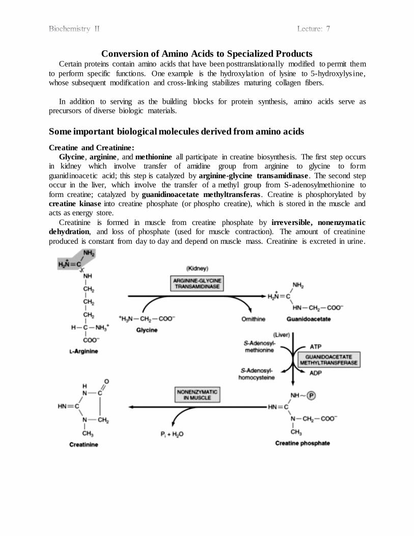
Conversion of Amino Acids to Specialized Products Certain proteins contain amino acids that have been posttranslationally modified to permit them
to perform specific functions. One example is the hydroxylation of lysine to 5-hydroxylys ine, whose subsequent modification and cross-linking stabilizes maturing collagen fibers.
In addition to serving as the building blocks for protein synthesis, amino acids serve as precursors of diverse biologic materials.
Some important biological molecules derived from amino acids
Creatine and Creatinine:
Glycine, arginine, and methionine all participate in creatine biosynthesis. The first step occurs in kidney which involve transfer of amidine group from arginine to glycine to form
guanidinoacetic acid; this step is catalyzed by arginine-glycine transamidinase. The second step occur in the liver, which involve the transfer of a methyl group from S-adenosylmethionine to
form creatine; catalyzed by guanidinoacetate methyltransferas . Creatine is phosphorylated by creatine kinase into creatine phosphate (or phospho creatine), which is stored in the muscle and acts as energy store.
Creatinine is formed in muscle from creatine phosphate by irreversible, nonenzymatic
dehydration, and loss of phosphate (used for muscle contraction). The amount of creatinine
produced is constant from day to day and depend on muscle mass. Creatinine is excreted in urine.
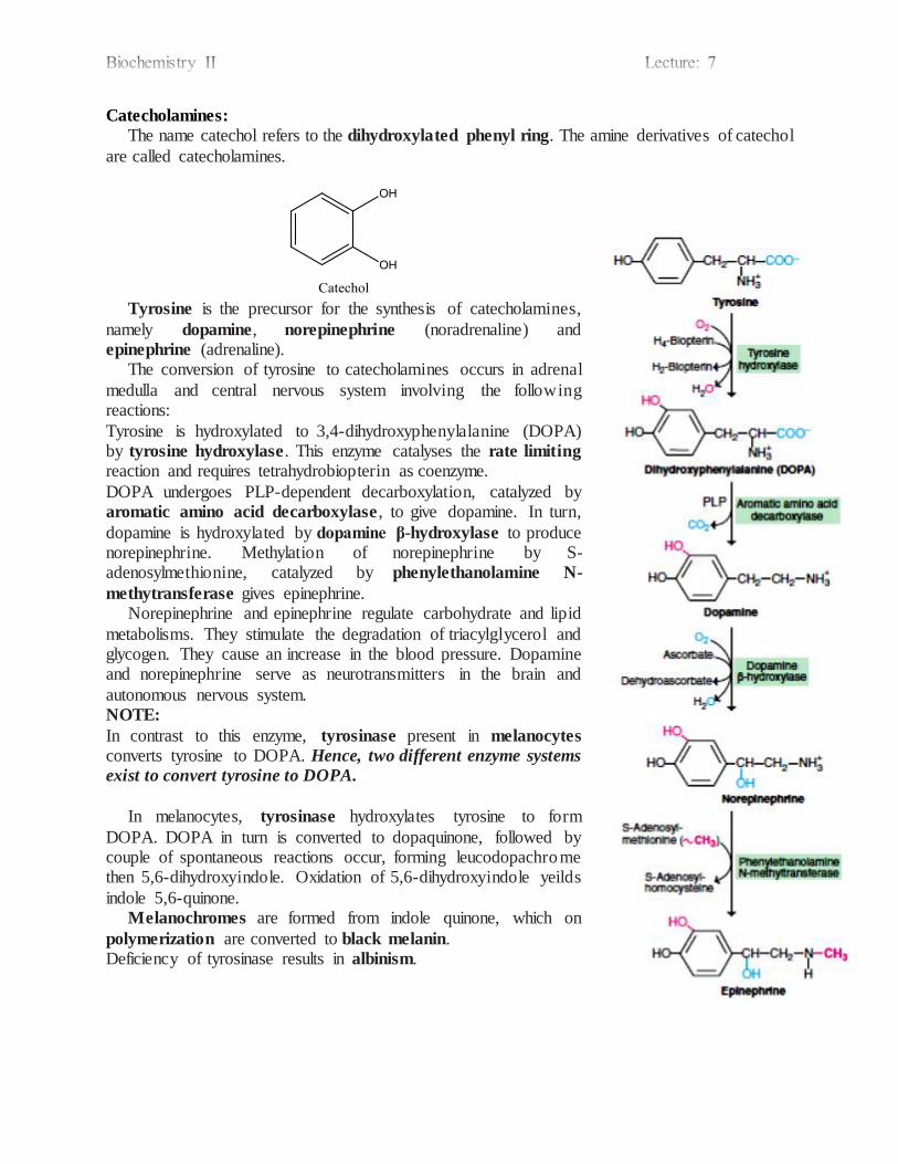
Catecholamines:
The name catechol refers to the dihydroxylated phenyl ring. The amine derivatives of catechol
are called catecholamines.
Tyrosine is the precursor for the synthesis of catecholamines,
namely dopamine, norepinephrine (noradrenaline) and epinephrine (adrenaline). The conversion of tyrosine to catecholamines occurs in adrenal
medulla and central nervous system involving the following reactions:
Tyrosine is hydroxylated to 3,4-dihydroxyphenylalanine (DOPA) by tyrosine hydroxylase. This enzyme catalyses the rate limiting
reaction and requires tetrahydrobiopterin as coenzyme.
DOPA undergoes PLP-dependent decarboxylation, catalyzed by aromatic amino acid decarboxylase , to give dopamine. In turn,
dopamine is hydroxylated by dopamine β-hydroxylase to produce norepinephrine. Methylation of norepinephrine by S-adenosylmethionine, catalyzed by phenylethanolamine N-
methytransferase gives epinephrine. Norepinephrine and epinephrine regulate carbohydrate and lipid
metabolisms. They stimulate the degradation of triacylglycerol and glycogen. They cause an increase in the blood pressure. Dopamine and norepinephrine serve as neurotransmitters in the brain and
autonomous nervous system. NOTE:
In contrast to this enzyme, tyrosinase present in melanocytes converts tyrosine to DOPA. Hence, two different enzyme systems
exist to convert tyrosine to DOPA.
In melanocytes, tyrosinase hydroxylates tyrosine to form
DOPA. DOPA in turn is converted to dopaquinone, followed by couple of spontaneous reactions occur, forming leucodopachrome then 5,6-dihydroxyindole. Oxidation of 5,6-dihydroxyindole yeilds
indole 5,6-quinone. Melanochromes are formed from indole quinone, which on
polymerization are converted to black melanin. Deficiency of tyrosinase results in albinism.
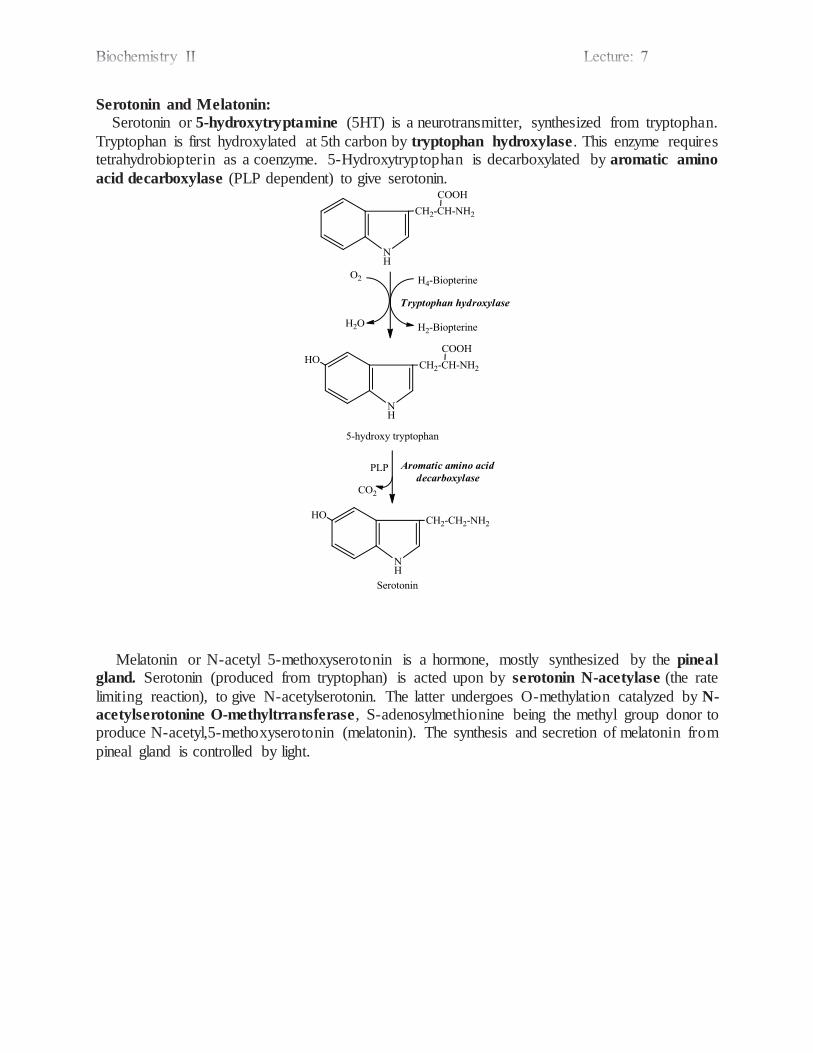
Serotonin and Melatonin:
Serotonin or 5-hydroxytryptamine (5HT) is a neurotransmitter, synthesized from tryptophan.
Tryptophan is first hydroxylated at 5th carbon by tryptophan hydroxylase. This enzyme requires tetrahydrobiopterin as a coenzyme. 5-Hydroxytryptophan is decarboxylated by aromatic amino
acid decarboxylase (PLP dependent) to give serotonin.
Melatonin or N-acetyl 5-methoxyserotonin is a hormone, mostly synthesized by the pineal
gland. Serotonin (produced from tryptophan) is acted upon by serotonin N-acetylase (the rate
limiting reaction), to give N-acetylserotonin. The latter undergoes O-methylation catalyzed by N-
acetylserotonine O-methyltrransferase, S-adenosylmethionine being the methyl group donor to produce N-acetyl,5-methoxyserotonin (melatonin). The synthesis and secretion of melatonin from
pineal gland is controlled by light.

Histamine:
A biogenic amine that functions in allergic reactions and gastric acid secretion, histamine is present in all tissues. Histamine is formed by decarboxylation of histidine; the reaction is catalyzed by the pyridoxal
phosphate-dependent enzyme histidine decarboxylase.
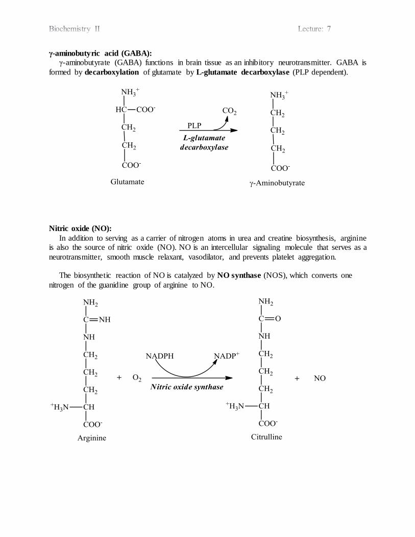
γ-aminobutyric acid (GABA):
γ-aminobutyrate (GABA) functions in brain tissue as an inhibitory neurotransmitter. GABA is
formed by decarboxylation of glutamate by L-glutamate decarboxylase (PLP dependent).
Nitric oxide (NO):
In addition to serving as a carrier of nitrogen atoms in urea and creatine biosynthesis, arginine is also the source of nitric oxide (NO). NO is an intercellular signaling molecule that serves as a
neurotransmitter, smooth muscle relaxant, vasodilator, and prevents platelet aggregation. The biosynthetic reaction of NO is catalyzed by NO synthase (NOS), which converts one
nitrogen of the guanidine group of arginine to NO.

Glutathione (GSH):
Glutatione is γ-Glutamylcysteinylglycine. γ-glutamate is attached via the γ-carbon instead of
the α-carbon. The active part is the -SH group of cysteine (sulfhydryl group). Biosynthetic reactions of GSH are catalyzed by γ-glutamyl cysteine synthetase and GSH
synthetase.
GSH in the reduced form has free -SH group. Two molecules can be bridged by a disulfide bond; which produces the oxidized form (GS-SG). Conversion reactions between the reduced and
the oxidized forms are catalyzed by glutathione peroxidase and glutathione reductase. By this mechanism GSH peroxidase and GSH reductase scavenge peroxide free radicals.
H2O2 2GSH NADP+
2H2O GSSG NADPH
Other functions of GSH include; conjugation of lipophilic drugs which converts them to hydrophilic molecules for excretion, and transport of amino acids especially in the renal epithelium.
Glutathione
peroxidase
Peroxidase
Glutathione
reductase
Peroxidase
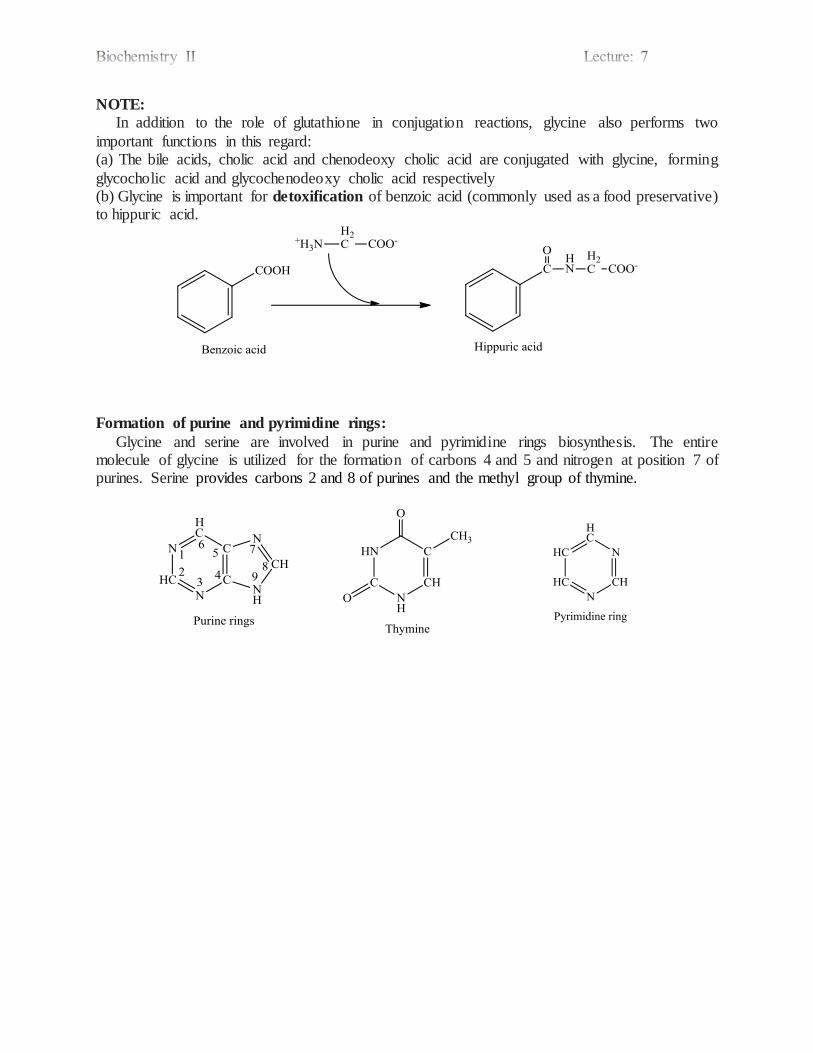
NOTE:
In addition to the role of glutathione in conjugation reactions, glycine also performs two
important functions in this regard: (a) The bile acids, cholic acid and chenodeoxy cholic acid are conjugated with glycine, forming
glycocholic acid and glycochenodeoxy cholic acid respectively (b) Glycine is important for detoxification of benzoic acid (commonly used as a food preservative) to hippuric acid.
Formation of purine and pyrimidine rings:
Glycine and serine are involved in purine and pyrimidine rings biosynthesis. The entire molecule of glycine is utilized for the formation of carbons 4 and 5 and nitrogen at position 7 of purines. Serine provides carbons 2 and 8 of purines and the methyl group of thymine.

Taurine:
Three enzyme catalyze reactions that convert cysteine to taurine, which can displace the
coenzyme A moiety of cholyl-CoA to form the bile acid taurocholic acid. The conversion of cysteine to taurine is initiated by its oxidation to cysteine sulfinate, catalyzed by the enzyme
cysteine dioxygenase. Decarboxylation of cysteine sulfinate by cysteine sulfinate
decarboxylase, forms hypotaurine, whose oxidation by hypotaurine dehydrogenase forms taurine.
Spermine and spermidine:
These polyamines function in cell proliferation and growth. Following decarboxylation of S-
adenosylmethionine by S-adenosylmethionine decarboxylase , three carbons and the α-amino group of methionine contribute to the biosynthesis of spermine and spermidine. Decarboxylated ornithine (by ornithine decarboxylase) which is called putrescine, react with
decarboxylated SAM to form spermidine. This reaction is catalyzed by spermidine synthase.
Another molecule of decarboxylated SAM react with spermidine to form spermine, this reaction
is catalyzed by spermine synthase.
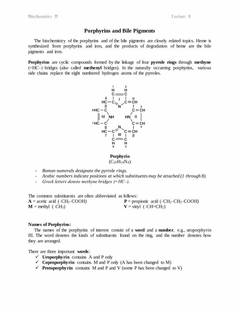
Porphyrins and Bile Pigments
The biochemistry of the porphyrins and of the bile pigments are closely related topics. Heme is synthesized from porphyrins and iron, and the products of degradation of heme are the bile
pigments and iron. Porphyrins are cyclic compounds formed by the linkage of four pyrrole rings through methyne
(=HC‒) bridges (also called methenyl bridges). In the naturally occurring porphyrins, various side chains replace the eight numbered hydrogen atoms of the pyrroles.
Porphyrin
(C20H14N4)
- Roman numerals designate the pyrrole rings. - Arabic numbers indicate positions at which substituents may be attached (1 through 8).
- Greek letters denote methyne bridges (=HC‒).
The common substituents are often abbreviated as follows: A = acetic acid (‒CH2‒COOH) P = propionic acid (‒CH2‒CH2‒COOH) M = methyl (‒CH3) V = vinyl (‒CH=CH2)
Names of Porphyrins:
The names of the porphyrins of interest consist of a word and a number, e.g., uroporphyrin III. The word denotes the kinds of substituents found on the ring, and the number denotes how
they are arranged.
There are three important words:
Uroporphyrin contains A and P only Coproporphyrin contains M and P only (A has been changed to M)
Protoporphyrin contains M and P and V (some P has been changed to V)
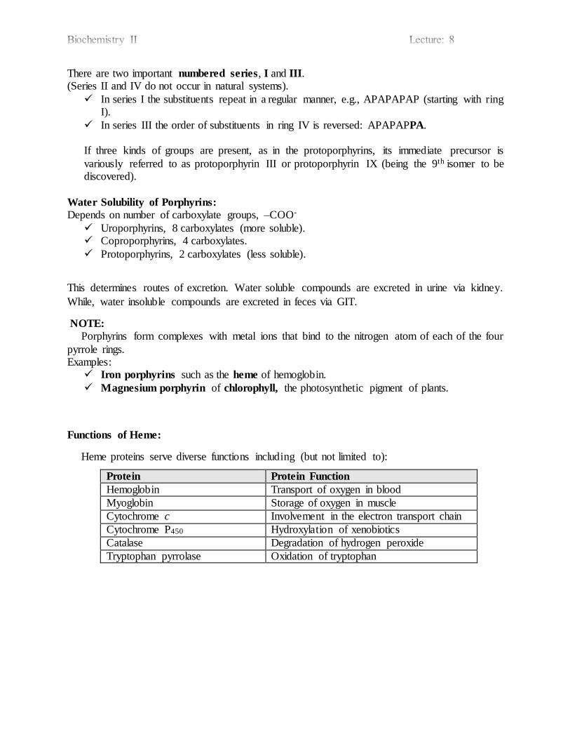
There are two important numbered series, I and III. (Series II and IV do not occur in natural systems).
In series I the substituents repeat in a regular manner, e.g., APAPAPAP (starting with ring I).
In series III the order of substituents in ring IV is reversed: APAPAPPA. If three kinds of groups are present, as in the protoporphyrins, its immediate precursor is
variously referred to as protoporphyrin III or protoporphyrin IX (being the 9th isomer to be discovered).
Water Solubility of Porphyrins:
Depends on number of carboxylate groups, ‒COO-
Uroporphyrins, 8 carboxylates (more soluble). Coproporphyrins, 4 carboxylates.
Protoporphyrins, 2 carboxylates (less soluble).
This determines routes of excretion. Water soluble compounds are excreted in urine via kidney.
While, water insoluble compounds are excreted in feces via GIT.
NOTE:
Porphyrins form complexes with metal ions that bind to the nitrogen atom of each of the four
pyrrole rings. Examples:
Iron porphyrins such as the heme of hemoglobin.
Magnesium porphyrin of chlorophyll, the photosynthetic pigment of plants.
Functions of Heme:
Heme proteins serve diverse functions including (but not limited to):
Protein Protein Function
Hemoglobin Transport of oxygen in blood
Myoglobin Storage of oxygen in muscle
Cytochrome c Involvement in the electron transport chain
Cytochrome P450 Hydroxylation of xenobiotics
Catalase Degradation of hydrogen peroxide
Tryptophan pyrrolase Oxidation of tryptophan
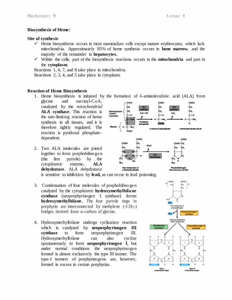
Biosynthesis of Heme:
Site of synthesis
Heme biosynthesis occurs in most mammalian cells except mature erythrocytes, which lack mitochondria. Approximately 85% of heme synthesis occurs in bone marrow, and the
majority of the remainder in hepatocytes.
Within the cells, part of the biosynthesis reactions occurs in the mitochondria and part in
the cytoplasm. Reactions 1, 6, 7, and 8 take place in mitochondria.
Reactions 2, 3, 4, and 5 take place in cytoplasm.
Reaction of Heme Biosynthesis
1. Heme biosynthesis is initiated by the formation of δ-aminolevulinic acid (ALA) from glycine and succinyl-CoA,
catalyzed by the mitochondrial ALA synthase. This reaction is the rate-limiting reaction of heme
synthesis in all tissues, and it is therefore tightly regulated. The
reaction is pyridoxal phosphate-dependent.
2. Two ALA molecules are joined together to form porphobilinogen
(the first pyrrole) by the cytoplasmic enzyme, ALA
dehydratase. ALA dehydratase
is sensitive to inhibition by lead, as can occur in lead poisoning.
3. Condensation of four molecules of porphobilinogen
catalyzed by the cytoplasmic hydroxymethylbilane
synthase (uroporphyrinogen I synthase) forms hydroxymethylbilane. The four pyrrole rings in
porphyrin are interconnected by methylene (-CH2-) bridges derived from α-carbon of glycine.
4. Hydroxymethylbilane undergo cyclization reaction
which is catalyzed by uroporphyrinogen III
synthase to form uroporphyrinogen III.
Hydroxymethylbilane can also cyclize spontaneously to form uroporphyrinogen I, but under normal conditions the uroporphyrinogen
formed is almost exclusively the type III isomer. The type-I isomers of porphyrinogens are, however,
formed in excess in certain porphyrias.
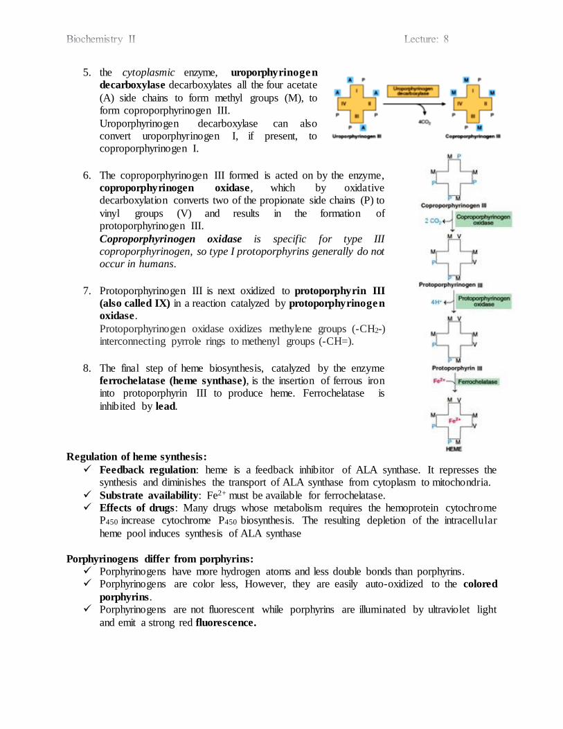
5. the cytoplasmic enzyme, uroporphyrinogen
decarboxylase decarboxylates all the four acetate
(A) side chains to form methyl groups (M), to form coproporphyrinogen III.
Uroporphyrinogen decarboxylase can also convert uroporphyrinogen I, if present, to coproporphyrinogen I.
6. The coproporphyrinogen III formed is acted on by the enzyme, coproporphyrinogen oxidase , which by oxidative decarboxylation converts two of the propionate side chains (P) to
vinyl groups (V) and results in the formation of protoporphyrinogen III.
Coproporphyrinogen oxidase is specific for type III coproporphyrinogen, so type I protoporphyrins generally do not occur in humans.
7. Protoporphyrinogen III is next oxidized to protoporphyrin III
(also called IX) in a reaction catalyzed by protoporphyrinogen
oxidase.
Protoporphyrinogen oxidase oxidizes methylene groups (-CH2-) interconnecting pyrrole rings to methenyl groups (-CH=).
8. The final step of heme biosynthesis, catalyzed by the enzyme ferrochelatase (heme synthase), is the insertion of ferrous iron into protoporphyrin III to produce heme. Ferrochelatase is
inhibited by lead.
Regulation of heme synthesis: Feedback regulation: heme is a feedback inhibitor of ALA synthase. It represses the
synthesis and diminishes the transport of ALA synthase from cytoplasm to mitochondria.
Substrate availability: Fe2+ must be available for ferrochelatase. Effects of drugs: Many drugs whose metabolism requires the hemoprotein cytochrome
P450 increase cytochrome P450 biosynthesis. The resulting depletion of the intracellular
heme pool induces synthesis of ALA synthase
Porphyrinogens differ from porphyrins:
Porphyrinogens have more hydrogen atoms and less double bonds than porphyrins. Porphyrinogens are color less, However, they are easily auto-oxidized to the colored
porphyrins. Porphyrinogens are not fluorescent while porphyrins are illuminated by ultraviolet light
and emit a strong red fluorescence.

Disorders of heme synthesis (porphyrias)
Porphyria can be classified depending on the etiology into: - Genetic (defective enzyme). - Acquired (e.g., lead poisoning).
Porphyrias may be divided into two major types based on the organs most affected:
Erythropoietic porphyria is a defect of porphyrin synthesis of bone marrow. Hepatic porphyria is a defect in porphyrin synthesis of the liver.
Example of acquired porphyrias
Lead toxicity:
Cause: inhibition of ALA dehydratase and Ferrochelatase by lead.
Results: elevated levels of protoporphyrin in erythrocytes and elevated urinary levels of ALA and coproporphyrin.
Examples of hereditary porphyria
Congenital erythropoietic porphyria:
Cause: Deficiency of uroporphyrinogen III synthase. Results: increase in type I porphyrin.
Acute intermittent porphyria:
Cause: Deficiency of hepatic uroporphyrinogen I synthase. Result: increase in ALA and porphobilinogen.
NOTE:
Certain drugs (enzyme inducers e.g, barbiturates, griseofulvin) induce the production of
cytochrome P450. In patients with porphyria, this can precipitate an attack of porphyria (drug-
induced porphyria) by depleting heme (the negative regulator of ALA synthase) levels. Thus, the
rate of synthesis of ALA synthase is increased with the subsequent increase in levels of potentially harmful heme precursors.
Catabolism of Heme
Most of the heme which is degraded comes from hemoglobin in red blood cells, which have a life span of about 120 days. Human adults normally destroy about 200 billion erythrocytes per
day. Thus, a 70-kg human turns over approximately 6 g of hemoglobin daily. Since 1 g of hemoglobin yields about 35 mg of bilirubin, human adults form 250 to 350 mg of bilirubin per
day. The globin is degraded to its constituent amino acids, the released iron enters the iron pool,
and all products are reused. The iron-free porphyrin portion of heme is also degraded.
1. Conversion of heme to bilirubin: Site:
- Mainly in the reticuloendothelial cells of the liver, spleen, and bone marrow. Enzymes:
- Heme oxygenase cleaves heme ring between the I and II pyrrole rings producing bilive rd in (green) and carbon is released as carbon monoxide (CO).
- Biliverdin reductase reduces the central (-CH=) bridge of biliverdin to (–CH2‒), producing bilirubin (red).
2. Transport of bilirubin in blood to the liver:
Bilirubin is only sparingly soluble in water. It must be transported in the blood by a carrier. The physiological carrier is serum albumin.
3. Uptake of bilirubin by hepatocytes:
Hepatocytes take up bilirubin from albumin by a large capacity, saturable facilitated transport system. Thus, transport does not appear to be rate-limiting for the metabolism of bilirubin.
The net uptake of bilirubin depends upon its removal by subsequent metabolism. Once internalized, bilirubin binds to cytosolic proteins such as glutathione S-transferase,
to prevent bilirubin from reentering the blood stream.
4. Conversion of bilirubin to bilirubin diglucuronide (Conjugation):
Bilirubin is nonpolar, and would persist in cells (e.g., bound to lipids) if not converted to a more water-soluble form.
Bilirubin is converted to a more polar molecule by conjugation with glucuronic acid. A bilirubin-specific UDP-glucuronyl transferase catalyzes transfer of two glucuronyl
moieties from UDP-glucuronate to bilirubin to form bilirubin diglucuronide in two steps.
5. Secretion of bilirubin diglucuronide into bile:
It is an active transport mechanism.
Probably is rate-limiting for the entire process of hepatic bilirubin metabolism. The protein involved is a multispecific organic anion transporter (MOAT) located in
the plasma membrane of the bile canaliculi.
6. Processing of bilirubin diglucuronide by intestinal bacteria:
In the intestine, the glucuronyl moieties of the conjugated bilirubin are removed by specific bacterial β-glucuronidases to reform bilirubin.
Bilirubin in the intestine is converted to urobilinogens compounds most of them are excreted into the stool as stercobilinogen (cause of brown color of stools) after oxidation.
A lesser amount (of urobilinogens) is recycled to the liver and either returned to bile or excreted in urine as urobilin (cause of yellow color of urine) after oxidation.
Bilirubin and its catabolic products are collectively known as the bile pigments, which
provide the yellow tinge in normal serum, the yellow-green hue in bile, the brown in stools, and the yellow in urine.
Hyperbilirubinemia causes Jaundice
- Hyperbilirubinemia, a blood level that exceeds 1 mg of bilirubin per dL (17 μmol/L),
may result from: Production of more bilirubin than the normal liver can conjugate, or
Failure of a damaged liver to conjugate normal amounts of bilirubin, or Obstruction of the excretory ducts of the liver prevents the excretion of bilirubin.
- When the blood concentration reaches 2 to 2.5 mg of bilirubin per dL, it diffuses into the tissues, which turn yellow, a condition termed jaundice (bilirubin deposits in the skin,
mucous membranes, and eyes), or icterus (bilirubin deposits in the blood).
NOTE:
- Hyperbilirubinemia may be classified depending on the type of bilirubin present in plasma,
as: Retention hyperbilirubinemia due to overproduction of bilirubin (unconjugated).
Regurgitation hyperbilirubinemia, due to reflux into the bloodstream because of biliary obstruction (conjugated).
- Only unconjugated bilirubin (due to its hydrophobicity), can cross the blood-brain barrier
into the central nervous system causing kernicterus (an encephalopathy due to hyperbilirubinemia), only in retention hyperbilirubinemia.
- Only conjugated bilirubin (due to its hydrophilicity), can appear in urine. Accordingly: Choluric jaundice (choluria is the presence of bile pigments in the urine) occurs only in
regurgitation hyperbilirubinemia. Acholuric jaundice occurs only in the presence of an excess of unconjugated bilirubin.
Causes of Hyperbilirubinemia (jaundice)
1. Pre-hepatic (hemolytic jaundice):
Hemolysis results in increased production of bilirubin. More bilirubin is conjugated and excreted than normally, but the conjugation
mechanism is overwhelmed, and an abnormally large amount of unconjugated
bilirubin is found in the blood.
2. Hepatic:
Toxic Hyperbilirubinemia
Can result from toxin induced liver dysfunction caused by chloroform, carbon
tetrachloride, acetaminophen, hepatitis virus, or cirrhosis. These acquired disorders involve hepatic parenchymal cell damage, which impairs
bilirubin conjugation, thus unconjugated bilirubin is retained in blood.
Gilbert's disease
A hereditary disorder in which there is decreased bilirubin uptake into the hepatocytes
As a result, unconjugated bilirubin accumulates.
Physiological jaundice and Crigler-Najjar syndrome
- Physiological jaundice results from inability of the immature liver of the newborn to
produce UDPG-transferase.
- Crigler-Najjar syndrome a congenital disease results from complete absence (type I) or
relative decrease (type II) in hepatic UDPG-transferase. Conjugation is impaired in these conditions thus unconjugated bilirubin is retained in the
body.
Dubin-Johnson syndrome
Caused by mutations in the gene encoding the protein involved in the secretion of
conjugated bilirubin after it has been formed.
Conjugated bilirubin returns to the blood.
3. Post-hepatic (biliary obstruction)
Results from blockage of the hepatic or common bile ducts, most often due to a gallstone
or to cancer of the head of the pancreas.
Bilirubin diglucuronide that cannot be excreted regurgitates into blood. Conjugated bilirubin appears in the blood and urine (choluric jaundice), and the stools
typically are a pale color.