biochemical and pathological changes ... - skeletal muscle
TRANSCRIPT
RESEARCH Open Access
Biochemical and pathological changesresult from mutated Caveolin-3 in muscleJosé Andrés González Coraspe1, Joachim Weis1, Mary E. Anderson2, Ute Münchberg3, Kristina Lorenz3,Stephan Buchkremer1, Stephanie Carr4, René Peiman Zahedi3,5,6, Eva Brauers1, Hannah Michels4,Yoshihide Sunada7, Hanns Lochmüller4,8,9,10, Kevin P. Campbell2, Erik Freier3, Denisa Hathazi3† and Andreas Roos3*†
Abstract
Background: Caveolin-3 (CAV3) is a muscle-specific protein localized to the sarcolemma. It was suggested thatCAV3 is involved in the connection between the extracellular matrix (ECM) and the cytoskeleton. Caveolinopathiesoften go along with increased CK levels indicative of sarcolemmal damage. So far, more than 40 dominantpathogenic mutations have been described leading to several phenotypes many of which are associated with amis-localization of the mutant protein to the Golgi. Golgi retention and endoplasmic reticulum (ER) stress has beendemonstrated for the CAV3 p.P104L mutation, but further downstream pathophysiological consequences remainedelusive so far.
Methods: We utilized a transgenic (p.P104L mutant) mouse model and performed proteomic profiling along withimmunoprecipitation, immunofluorescence and immunoblot examinations (including examination of α-dystroglycan glycosylation), and morphological studies (electron and coherent anti-Stokes Raman scattering (CARS)microscopy) in a systematic investigation of molecular and subcellular events in p.P104L caveolinopathy.
Results: Our electron and CARS microscopic as well as immunological studies revealed Golgi and ER proliferationsalong with a build-up of protein aggregates further characterized by immunoprecipitation and subsequent massspectrometry. Molecular characterization these aggregates showed affection of mitochondrial and cytoskeletalproteins which accords with our ultra-structural findings. Additional global proteomic profiling revealed vulnerabilityof 120 proteins in diseased quadriceps muscle supporting our previous findings and providing more generalinsights into the underlying pathophysiology. Moreover, our data suggested that further DGC components arealtered by the perturbed protein processing machinery but are not prone to form aggregates whereas othersarcolemmal proteins are ubiquitinated or bind to p62. Although the architecture of the ER and Golgi as organellesof protein glycosylation are altered, the glycosylation of α-dystroglycan presented unchanged.
Conclusions: Our combined data classify the p.P104 caveolinopathy as an ER-Golgi disorder impairing properprotein processing and leading to aggregate formation pertaining proteins important for mitochondrial function,cytoskeleton, ECM remodeling and sarcolemmal integrity. Glycosylation of sarcolemmal proteins seems to benormal. The new pathophysiological insights might be of relevance for the development of therapeutic strategiesfor caveolinopathy patients targeting improved protein folding capacity.
Keywords: Caveolin-3, Caveolinopathy, LGMD1C, Chaperonopathy, Protein aggregate, Skeletal muscle proteomics
* Correspondence: [email protected]†Denisa Hathazi and Andreas Roos contributed equally to this work.3Biomedical Research Department, Tissue Omics group, Leibniz-Institut fürAnalytische Wissenschaften - ISAS - e.V, Otto-Hahn-Str. 6b, 44227 Dortmund,GermanyFull list of author information is available at the end of the article
© The Author(s). 2018 Open Access This article is distributed under the terms of the Creative Commons Attribution 4.0International License (http://creativecommons.org/licenses/by/4.0/), which permits unrestricted use, distribution, andreproduction in any medium, provided you give appropriate credit to the original author(s) and the source, provide a link tothe Creative Commons license, and indicate if changes were made. The Creative Commons Public Domain Dedication waiver(http://creativecommons.org/publicdomain/zero/1.0/) applies to the data made available in this article, unless otherwise stated.
González Coraspe et al. Skeletal Muscle (2018) 8:28 https://doi.org/10.1186/s13395-018-0173-y
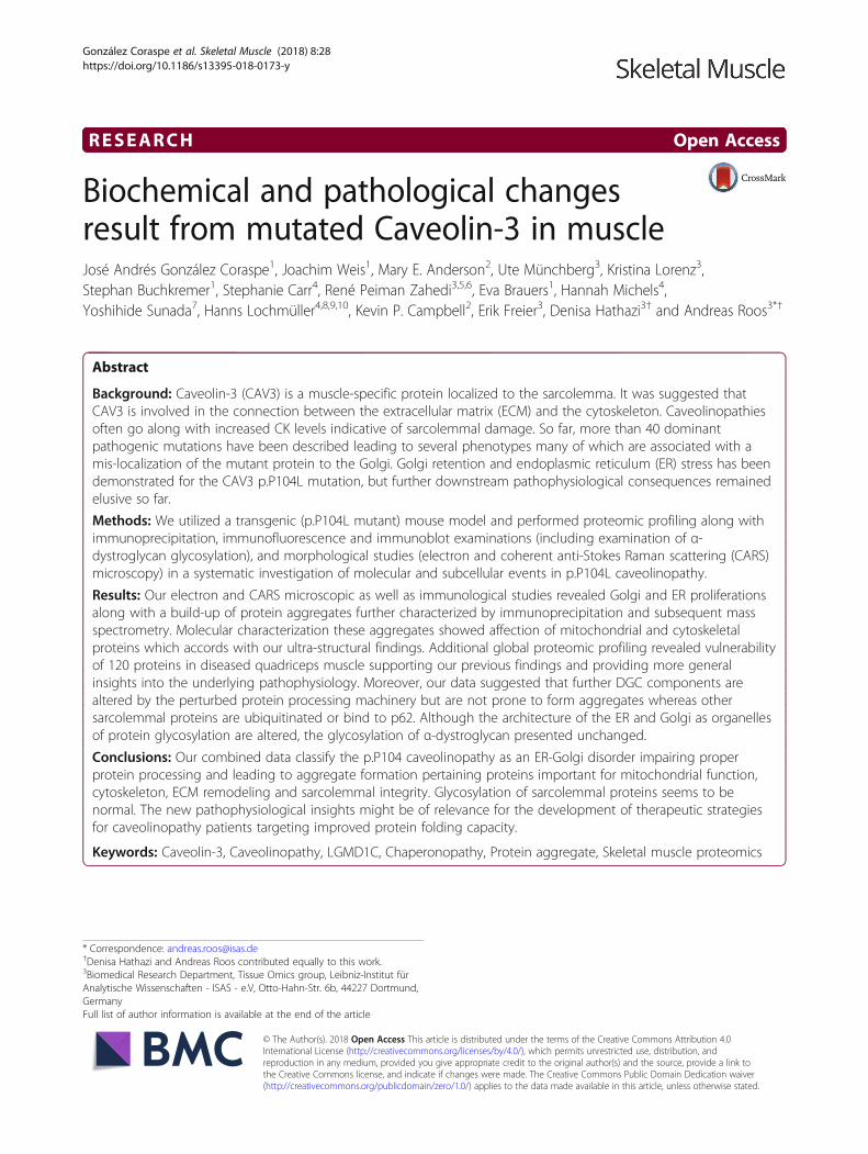
BackgroundCaveolin-3 (CAV3), a muscle-specific member of the cave-olin protein family, is a structural protein important forsignal transduction, lipid metabolism, cell growth,mechanoprotection, autophagy, maintenance of neuro-muscular junctions and apoptotic cell death [1–3]. CAV3first appears during myoblast differentiation and is local-ized to the sarcolemma within caveolae, 50–100 nmflask-shaped invaginations of the plasma membrane.There, it associates with the dystroglycan complex estab-lishing a connection between the extracellular matrix andthe cytoskeleton (Fig. 1a). Muscle diseases caused by mu-tations in the CAV3 gene are called caveolinopathies [1].So far, more than 40 pathogenic CAV3 mutations havebeen described which are localized within different proteindomains (Fig. 1b) and are leading to different disease phe-notypes including Limb Girdle Muscular dystrophy(LGMD), rippling muscle disease (RMD), distal myopathy(DM), hyperCKemia (HCK) and myalgia [1, 4]. A cleargenotype-phenotype correlation does not exist, and someof the phenotypes may present as a clinical continuum[5–7]. However, dominant mutants are commonly associ-ated with lowered sarcolemmal CAV3 levels. These are re-lated to dissociation of the hetero-oligomers at thesarcolemma, degradation by the ubiquitin-proteasomepathway, and abnormal accumulation of mutated andwild-type (wt) CAV3 within the Golgi causing ER-stressand thus activation of the unfolded protein response(UPR) [8–10] (Fig. 1c).A transgenic animal model expressing an additional
copy of CAV3 harbouring the p.P104L mutation waspublished in 2001 by Sunada and co-workers [11]. No-toriously, this missense mutation has been identified inLGMD patients [1]. Proline residues in proteins, basedon their biochemical properties, are important for struc-tural maintenance of integral membrane proteins [12]such as CAV3. Hence, a substitution of proline to leu-cine in an integral membrane protein should be ofpathogenic relevance. The phenotype of these mice hasbeen examined extensively and appears to phenocopythe human disorder [9, 11, 13].To better understand the consequences of CAV3 mu-
tations, we chose the paradigmatic p.P104L missensemutation and investigated protein stability and aggrega-tion, ER-Golgi pathology and the overall protein com-position as well as glycosylation of the α-dystroglycanprotein. For this purpose, quadriceps muscles of theabove-mentioned animal model have been utilized andimmunological, morphological and proteomic studieshave been applied. Electron as well as coherentanti-Stokes Raman scattering (CARS) microscopicstudies revealed presence of protein aggregates. Massspectrometry-based characterization of these aggregatessuggested mitochondrial, cytoskeletal, sarcolemmal and
ECM vulnerability in p.P104L CAV3 diseased muscle.This molecular observation especially accords with mito-chondrial perturbations detected on the ultra-structuraland global proteomic level. Moreover, data of our globalproteomic profiling revealed an increase of 77 and a de-crease of 43 proteins. Notably, further immunologicalstudies confirmed the proteomic findings. Interestingly,localization studies of sarcolemmal proteins—includingcomponents of the DGC—classified those as substratesof impaired ER-Golgi function. However, glycosylationof α-dystroglycan seems to be not impaired by thep.P104L CAV3 mutation caused altered ER-Golgimorphology.
MethodsAnimalsThe transgenic mouse model expressing p.P104L CAV3was kindly provided by co-author Professor YoshihideSunada (Department of Neurology, Kawasaki MedicalSchool, Okayama, Japan). Genotyping was performed asdescribed previously [9, 11, 13]. All procedures were ap-proved by the Uniklinik RWTH Aachen InstitutionalAnimal Care and Use authorities. To obtain animals car-rying the dominant p.P104L CAV3 mutation (Tg/+) aswell as respective wild-type littermates (+/+), breedingof Tg/+ with +/+ was carried out.
Light microscopy, immunohistochemistry andimmunofluorescenceFive-micrometre sections were cut from formalin-fixed-#paraffin-embedded (FFPE) muscle biopsy tissue. Thesewere used for hematoxylin and eosin (H&E) staining andfor immunohistochemistry (IHC) as well as for immuno-fluorescence (IF). For IHC and IF, sections were placedon silan-coated slides, treated with descending alcoholseries for re-hydration and unmasked by heat (steamoven, citrate buffer). Afterwards, sections were blockedwith 2% goat serum in PBS followed by incubation withprimary antibodies (Additional file 1: Table S2) overnight.For IHC, peroxidase-labeled secondary antiserum (1:200,DCS, Hamburg, Germany) and diaminobenzodine (DAKO,USA) were used to detect antibody binding and cellularstructures were counterstained with hematoxylin. ForIF, Alexa 488- and Alexa 555-conjugated secondaryantibodies (diluted 1:500, in 2% goat serum, respect-ively) were used to detect binding of primary anti-bodies and incubated at room temperature for 2 hfollowed by three washing steps in 1× PBS.To systematically study Golgi-dispersion, length to
width ratios have been calculated for 10 Golgi-structures(visualized utilizing a golgin-97 antibody) in type 1 fibresfrom three p.P104L CAV3 transgenic and wild-type ani-mals aged 6 months and 1 year, respectively.
González Coraspe et al. Skeletal Muscle (2018) 8:28 Page 2 of 19
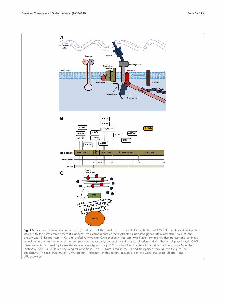
Fig. 1 Muscle caveolinopathies are caused by mutations of the CAV3 gene. a Subcellular localization of CAV3: the wild-type CAV3 proteinlocalizes to the sarcolemma where it associates with components of the dystrophin-associated glycoprotein complex. CAV3 interactsdirectly with β-dystroglycan, nNOS and dysferlin. Moreover, CAV3 indirectly interacts with F-actin, syntrophin, dystrobrevin and laminin-2as well as further components of the complex such as sarcoglycans and integrins. b Localization and distribution of paradigmatic CAV3missense mutations leading to skeletal muscle phenotypes. The p.P104L mutant CAV3 protein is causative for Limb Girdle MuscularDystrophy type 1 C. c Under physiological conditions, CAV3 is synthesized in the ER and transported through the Golgi to thesarcolemma. The missense mutant CAV3 proteins (hexagons in this carton) accumulate in the Golgi and cause ER stress andUPR activation
González Coraspe et al. Skeletal Muscle (2018) 8:28 Page 3 of 19

Electron microscopic studiesElectron microscopic using glutaraldehyde-fixed, resin-embedded quadriceps muscles of 26 weeks old wt andmutant mice were performed as described previously [14].
Coherent anti-Stokes Raman scattering microscopyTo obtain further information regarding the build-up ofprotein deposits, we employed—in addition to confocalfluorescence microscopy—coherent anti-Stokes Ramanscattering (CARS). CARS is a non-linear variant of spon-taneous Raman scattering and as such directly assessesthe molecular vibrations of the sample itself. Therefore,CARS is inherently label-free and not limited by theavailability of dyes or antibodies. A widespread use ofCARS is its application in lipid biology [15]. In this ap-plication, the localization of lipids is examined at a spe-cific wavenumber (2845 cm−1). By monitoring otherwave lengths (= molecular vibrations), other targets canbe investigated. Here, we focused on the spatial distribu-tion of proteins at 2932 cm−1 [16].CARS measurements were performed on a modified
Leica TCS SP 8 CARS microscope with an APE picoE-merald as laser source. The 1064 nm output of thepicoEmerald was used as Stokes, the OPO signal wastuned to 811 nm and provided both pump and probe.This combination results in a CARS signal at 655 nmand corresponds to a wavenumber of 2932 cm−1. Bothlasers were fixed at a power of 900 mW at the picoE-merald output port. Further subsequent laser attenu-ation in the Leica systems was varied between 30 and40%, depending on the sample. The laser beams were fo-cused onto the sample using a Leica HCL IRAPO 40×/1.1 water or Leica HC PL APO 20×/0.75 CS2 objective.The resulting CARS signal was collected via the sameobjective in EPI (backward) direction and subsequentlydetected in the spectral regime of 560–750 nm via aphoto multiplier tube (PMT) detector. The Second Har-monic Generation and Two-Photon fluorescence signalswere collected at the same time (380–560 nm). Further-more, all signals were also measured in a forward direc-tion via a NA 0.55 condenser and the same filter/detector arrangement as in the EPI direction.After the de-paraffinization step, the samples (quadri-
ceps muscles from 6 weeks and 26 weeks old wild-typeand p.P104L CAV3-transgenic animals, respectively)were thoroughly dried with a constant flow of dry air,measured in the CARS system. Afterwards, the stainingprotocol was pursued.
Protein lysate preparation and immunoblot studiesMuscle tissue was transferred to muscle lysis buffer(0.125 M Tris, pH 6.4, 4% SDS, 10% β-mercaptoethanol,10% glycerin, 0.001% bromophenol blue, 4 M urea andprotease inhibitor mix) and lysed by sonication. Samples
were chilled on ice and heated for 15 min at 56 °C. Pro-tein concentration was quantified by BCA proteinassay-reducing agent compatible kit (Pierce) accordingto the manufacturer’s instructions.For immunoblot studies, 10 μg protein was used in
each case, loaded on 10% polyacrylamide gels and sepa-rated for 120 min at 120 V. Following the separation,proteins were transferred to Immobilion-P PVDF mem-brane (0.45 μm, Millipore) over night at 10 V bytank-blot technique. Membranes were blocked with 1%casein buffer [1% casein; (Roche) in Tris-buffered saline(TBS) and 0.1% Tween20 (Sigma) as a 1:1 mix with ma-leic acid buffer composed of 100 mM maleic acid(Sigma) and 150 mM NaCl] for 2 h followed by fourwashing steps using TBS with 0.1% Tween20 (TBS-T).Membranes were incubated with several primary anti-bodies (Additional file 1: Table S2) at 4 °C (overnight)and then washed in TBS-T thrice. Horseradish peroxid-ase conjugated secondary goat anti-rabbit antibody(Sigma) or goat anti-mouse antibody (Sigma) was dilutedat 1:25,000 and added to membranes for 1 h. In the fol-lowing step, membranes were washed three times inTBS-T for 10 min. By using the enhanced chemiluminis-cence, horseradish peroxidase substrate (Super-SignalWest Pico and Super-Signal West Femto; Pierce) signalswere detected on CL-X Posure films (Thermo Scientific).For the study of α-dystroglycan glycosylation, wild-
type and p.P104L mutant muscles were minced on iceand homogenized in 1% Triton X-100 TBS and proteaseinhibitors using the Bullet Blender (Next Advance, Aver-ill Park, NY). Samples were enriched for glycoproteinsusing WGA agarose beads (Vector Labs) as describedpreviously [17]. Protein levels in samples were measuredusing the DC Protein Assay (Bio-Rad, Hercules, CA),and equal amounts were added to wheat-germ agglutinin(WGA) agarose beads (Vector Labs, Burlingame, CA).Samples were then run out on a 3–15% gradientSDS-PAGE gel, after which the protein was transferredto PVDF membranes. The latter were blotted with theantibodies monoclonal IIH6-antibody, which recognizesspecifically glycosylated DG, and the polyclonal antibodyAF6868, which recognizes both α- and β-dystroglycan,as published previously [18]. Images were captured usingthe Licor (Lincoln, NE) system and Odyssey software.
Global proteomic profiling studyMaterialsThe following materials were purchased from Sigma-Al-drich, Steinheim, Germany: ammonium hydrogen car-bonate (NH4HCO3), guanidine hydrochloride (GuHCl),iodoacetamide (IAA) and urea. Tris base was obtained fromApplichem Biochemica, Darmstadt, Germany. Sodiumdodecyl sulfate (SDS) was purchased from Carl Roth,Karlsruhe, Germany. Dithiothreitol (DTT), EDTA-free
González Coraspe et al. Skeletal Muscle (2018) 8:28 Page 4 of 19

protease inhibitor (Complete Mini) tablets were boughtfrom Roche Diagnostics, Mannheim, Germany. Sodiumchloride (NaCl) and calcium chloride (CaCl2) were fromMerck, Darmstadt. Sequencing grade modified trypsin wasfrom Promega, Madison, WI USA. Bicinchoninic acid assay(BCA) kit was acquired from Thermo Fisher Scientific,Dreieich, Germany. All chemicals for ultra-pure HPLC sol-vents such as formic acid (FA), trifluoroacetic acid (TFA)and acetonitrile (ACN) were obtained from Biosolve, Valk-enswaard, The Netherlands.
Tissue lysis and carbamidomethylationSix quadriceps muscles, i.e. three derived from 10 weeksold p.P104L CAV3-transgenic animals and three fromwild-type littermates were collected, snap-frozen in liquidnitrogen at − 80 °C and processed independently. Thewhole quadriceps muscle was lysed in 1.0 mL of 50 mMTris-HCl (pH 7.8) buffer containing 150 mM NaCl, 1%SDS, and Complete Mini. Tissue was homogenized bymechanical grinding followed by sonication with 1 to1 s pulses (10 s) followed by centrifugation at 4 °Cand 1600 g for 15 min. Protein concentration of thesupernatant was determined by BCA assay accordingto the manufacturer’s protocol. Disulfide bonds werereduced by addition of 10 mM DTT at 56 °C for30 min, and free sulfhydryl bonds were alkylated with30 mM IAA at room temperature (RT) in the darkfor 30 min.
Sample preparation and trypsin digestionAfter filter-aided sample preparation (FASP) [19, 20],cell lysates corresponding to 100 μg of protein were di-luted 10-fold with freshly prepared 8 M urea/100 mMTris-HCl (pH 8.5) buffer and placed on a Microcon cen-trifugal device (30 kDa cutoff ). The device was centri-fuged at 13,500 g at RT for 15 min. All subsequentcentrifugation steps were performed under the sameconditions. To eliminate residual SDS, three washingsteps were carried out with 100 μL of 8 M urea/100 mMTris-HCl (pH 8.5). To exchange the buffer, the filter waswashed thrice with 100 μL of 50 mM NH4HCO3
(pH 7.8). Then, proteins were incubated at 37 °C for14 h with 100 μL of proteolysis buffer: trypsin (Promega)(1:25 w/w, protease to substrate), 0.2 M GuHCl and2 mM CaCl2 in 50 mM NH4HCO3 (pH 7.8). The result-ing tryptic peptides were recovered by centrifugationwith 50 μL of 50 mM NH4HCO3 followed by 50 μL ofultra-pure water. Finally, peptides were acidified byaddition of 10% TFA (v/v) and digests were quality con-trolled as described previously [21].
LC-MS/MS analysisOne-microgramme peptides of three mutant and threewild-type quadriceps muscles were analysed using an
Orbitrap Elite mass spectrometer coupled with an Ul-timate 3000 nano RSLC system. In all measurements,samples were analysed in randomized order to minimizesystematic errors. Peptides were pre-concentrated on a100 μm× 2 cm C18 trapping column for 10 min using0.1% TFA (v/v) at a flow rate of 20 μL/min followed byseparation on a 75 μm× 50 cm C18 main column (bothPepmap, Thermo Scientific) with (1) a 210-min LC gra-dient ranging from 3 to 42% of 84% ACN, 0.1% FA (v/v)at a flow rate of 230 nL/min. All samples were measuredin a data-dependent acquisition.In the OrbiElite, MS survey scans were acquired in the
Orbitrap from m/z 300 to 1500 at a resolution of 60,000using the polysiloxane ion at m/z 371.101236 as lockmass [22]. The ten most intense signals were subjectedto collision-induced dissociation (CID) in the ion trap,taking into account a dynamic exclusion of 30 s. CIDspectra were acquired with normalized collision energyof 35%. AGC target values were set to 106 for MS1 and104 for ion trap MS2 scans, and maximum injectiontimes were set to 100 ms for both full MS and MS2scans.
Label-free data analysisData analysis of the acquired label-free quantitative MSwas performed using the Progenesis LC-MS softwarefrom Nonlinear Dynamics (Newcastle upon Tyne, UK).For all measurements, biological triplicates of quadricepsmuscle were compared to the corresponding controltriplicates separately.The MS raw data was aligned by Progenesis which
automatically selected one of the MS files as reference.After peak picking, only features within retention timeand m/z windows from 0 to 200 for OrbiElite data and300–1500 m/z, with charge states + 2, + 3, and + 4 wereconsidered for peptide statistics, analysis of variance(ANOVA) and principal component analysis (PCA). TheMS/MS spectra were exported as peak lists. The listswere then searched against a concatenated target/decoyversion of the mouse Uniprot database, (downloaded on11th of December 2013, containing 20,273 target se-quences) using Mascot 2.4 (Matrix Science), MS-GF+,and X!Tandem Jackhammer (2013.06.15) with the helpof searchGUI 1.24.0 [23]. As a proteolytic enzyme, tryp-sin was selected with a maximum of two missed cleav-ages. Carbamidomethylation of Cys was set as fixedmodification and oxidation of Met was selected as vari-able modification. MS and MS/MS tolerances were setto 10 ppm and 0.5 Da, respectively. Combined search re-sults were filtered at a false discovery rate (FDR) of 1%on the protein level and exported using the PeptideSha-ker [24] software 0.28.0 (http://compomics.github.io/projects/peptide-shaker.html) features which allows adirect re-import of the quality-controlled data into
González Coraspe et al. Skeletal Muscle (2018) 8:28 Page 5 of 19
Progenesis. Peptide sequences containing oxidized Metand pyro-Glu (derived from X!Tandem 2nd pass search)were excluded for further analysis. Only proteins thatwere quantified with unique peptides were exported.Then, for each protein, the average of the normalizedabundance (obtained from Progenesis) from the tripli-cate analyses was calculated to determine the ratios be-tween the controls and transgenic muscle samples. Onlyproteins matching the following criteria were consideredas regulated: they had to be (i) commonly quantified inall the replicates with (ii) an ANOVA p value of < 0.05(Progenesis) and (iii) an average ratio < 1.24 or > 0.84—all parameters depending on data distribution. Proteinsidentified with at least two unique peptides were classi-fied as confidential class I and proteins identified withsolely unique peptide were classified as confidential classII.
Immunoprecipitation of protein aggregates and massspectrometry-based analysisMaterialsSee above.
Sample preparation, cleanup and tryptic digestionTo further characterize the protein aggregates detected viaelectron and CARS microscopy as well as via immuno-fluorescence studies on a molecular level, utilizing anti-bodies targeting ubiquitin (ab7780; Abcam) and p62/SQSTM1 (ab109012; Abcam) (Additional file 1: Table S2)ubiquitinated as well as proteins binding to p62 were pre-cipitated from muscle (5 mg starting material) derivedfrom four 26 weeks old p.P104L CAV3-transgenic andwild-type animals respectively utilizing the “Pierce ProteinA/G Magnetic Beads” kit (Thermo Fisher Scientific).Precipitations were performed according to the manufac-turers’ instructions. To later exclude proteins non-specif-ically binding to the magnetic beads, for p.P104L CAV3mutant and wild-type samples (four respectively) wholeprotein extracts have been incubated with the beads with-out adding the antibodies. After elution, samples weresubjected to ethanol precipitation, which was used as acleanup method: protein solution was 10-fold diluted (ra-tio of 1:10) with ultrapure ice-cold ethanol (100%), andthen stored at − 40 °C for 60 min followed by centrifuga-tion at 4 °C for 30 min at 18,000 g. The pellet was driedunder a laminar flow hood and then solubilized with a so-lution containing 1 M GuHCl and 50 mM NH4HCO3
(pH 7.8). Proteins were reduced and alkylated as describedabove. Next, samples were diluted with 50 mM NH4HCO3
(pH 7.8) until a concentration of 0.2 M GuHCl wasattained. For the tryptic-digestion, the protein concentra-tion was roughly estimated as being 5 μg. Protein cleavagewas achieved by adding trypsin to the sample in a ratio of
1:20 (enzyme to substrate) followed by incubation for 12 hat 37 °C.Clean-up of tryptic peptides was performed with
commercially available C18 tips (Omix). The tips werefirstly activated by using 100% ACN followed byequilibration with 0.1% TFA. All volumes used wereassessed based on the size/capacity/type of tips. Afterequilibration of the resin, the peptide mixture wasloaded onto the tips and the flow through passed twotimes and was then discarded. Afterwards, the columnwas washed three times with 0.1 %TFA to ensureproper elimination of contaminants. Finally, peptideswere eluted from the resin with 60% ACN in 0.1%TFA. The eluates were dried in the SpeedVac andthen resuspended in 0.1% TFA.
LC-MS/MS analysisThe samples were analysed using a Velos ion trapmass spectrometer coupled with an Ultimate 3000RSLC system (both Thermo Scientific). Peptides wereseparated as described above using a 90-min LC gra-dient ranging from 3 to 42% of 84% ACN, 0.1% FA(v/v) at a flow rate of 300 nL/min. The full MS scanswere acquired in the m/z 300 to 2000 at a resolutionof 60,000 using the polysiloxane ion at m/z371.101236 as lock mass. The ten top most intenseions were subjected to collision induced dissociation(CID) with an NCE of 35% in the ion trap, consider-ing a dynamic exclusion of 30 s. AGC target valueswere set to 106 for MS1 and 104 for ion trap MS2scans, and maximum injection times were set to100 ms for both full MS and MS2 scans.
Data analysisRaw files were analysed in Proteome Discoverer 2.2.0388(Thermo Scientific). Searches were conducted in atarget/decoy manner against a concatenated proteinsequence database (Mus musculus, 16,966 target se-quences, downloaded from UniProt in October 2017and a common contaminant database comprising 247target sequences). Samples were searched using Mascot2.6.1 (Matrix Science) and the precursor mass toleranceand fragment mass tolerance were set to 10 ppm and0.02 Da, respectively. For all searches, enzyme specificitywas set to fully tryptic with an allowed maximum num-ber of two missed cleavages. Carbamidomethylation ofCys (+ 57.02146 Da) was defined as fixed, while oxida-tion of Met (+ 15.99491 Da) and acetylation of proteinN-termini (+ 42.01056 Da) were allowed as variablemodifications. For the ubiquitin IP samples diGly wasset as a variable modification as in this case the trypticdigest contains a diglycine-modified lysine, which servesas a signature for ubiquitination. False discovery rate(FDR) estimation was performed by the percolator node
González Coraspe et al. Skeletal Muscle (2018) 8:28 Page 6 of 19

and all data were filtered to meet a 1% FDR-level forPSMs as well as peptides. Resulting dataset were furtherfiltered for 1% FDR on the protein level, additionallyexcluding proteins without uniquely assigned peptidesand such not marked as ‘master’ proteins. All proteinsnon-specifically binding to the magnetic beads wereexcluded.
Transcript studiesRNA was extracted from quadriceps muscles derivedfrom 26-week-old p.P104L CAV3 transgenic and controlanimals (RNeasy®, Qiagen). Total RNA was reversetranscribed using Superscript II reverse transcriptase(Invitrogen). Quantitative real-time PCR was performedusing the C1000 Thermal Cycler CFX96 (BioRad). ForcDNA amplification of dysferlin (Dysf ), caveolin-3(Cav3), integrin-β-4 (Igtb4), δ-sarcoglycan (Scgd) andglyceraldehyde-3-phosphate dehydrogenase (Gapdh), areaction mix containing Sso Fast Eva Green Supermix(BioRad) was used in combination with the followingprimers: Gapdh forward primer 5′ TGGCAAAGTGGAGATTGTTG 3′; Gapdh reverse primer 5′ CATTATCGGCCTTGACTGTG 3′; Dysf forward primer 5′CTTACCACAGATGGACGATGC 3′; Dysf reverseprimer 5′ CATAGAGGGAAACATCGCAGGC 3′; Cav3forward primer 5′ GACATTGTGAAGGTAGATTTTG 3′;Cav3 reverse primer 5′ GGTAGCTCTTAATGCAGGGC3′; Igtb4 forward primer 5′ CTACTATGAGAAGCTCCATAAG 3′; Igtb4 reverse primer 5′ CATTGTATGTGCCCACTTCCC 3′; Scgd forward primer 5′ GCTGGTGACAGGTCCGAAGGC 3′; Scgd reverse primer 5′ GTGCCTTCAGCTCCTAAGACTC 3′. Data were normalizedwith glyceraldehyde-3-phosphate dehydrogenase (Gapdh) asinternal standard using the 2–ΔΔC T method [28].
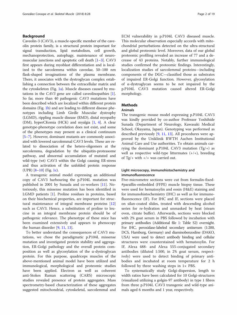
ResultsMutant Caveolin-3 (p.P104L) protein is less stable thanwild-typeIn caveolinopathy, mutant muscle fibres are known topresent with absence of sarcolemmal CAV3 (drasticallyreduced immunoreactivity). This pathological observa-tion is often associated with the presence of smalldot-like CAV3-immunoreactive deposits scattered in thesarcoplasm [11]. Prompted by these known pathophysio-logical findings, transcript and protein studies ofp.P104L CAV3 have been carried out utilizing quadri-ceps muscle derived from transgenic animals (Fig. 2a–c):results of our CAV3 transcript studies revealed increasedabundances in quadriceps muscle of p.P104L CAV3 mu-tant animals compared to wild-type littermates (both26 weeks of age; Fig. 2c). Immunoblot studies of CAV3quadriceps muscles from 6 and 26 weeks old transgenicanimals compared to the respective wild-type littermates
confirmed lowered abundance of the protein (Fig. 2d),suggesting a reduced stability of the mutant-protein (for-mation of irregular CAV3 wild-type-mutant proteincomplexes which undergo proteolysis). Interestingly,level of the CAV3 wild-type-mutant protein complexseem to decline with disease progression suggestingforced degradation (Fig. 2d). To exclude a direct impactof the p.P104L mutation on CAV3 antigene detection, arabbit polyclonal peptide antibody corresponding tomouse caveolin-3 amino acids 1 to 19 (ab2912; Abcam)was utilized. However, our combined results supportingthe concept of reduced protein stability rather than re-duced stability of the mutant transcript.
p.P104L Caveolin-3 causes Golgi/ER stressMis-localization of CAV3 aggregates (formed by mutantand wild-type protein) to the Golgi has already been de-scribed [8]. This pathological finding is also in line withthe previous description of UPR activation [9] which inturn suggests a more generalized stress burden of thesecretory apparatus comprised by discrete paired Golgistacks in close proximity to exit sites from the ER(Fig. 1c). Here, we further investigated the presence ofGolgi/ER stress by studying the structure of thesecretory apparatus on the morphological level andUPR-related as well as Golgi-ER-shape-related factors onthe protein level.EM of muscles derived from 26-week-old mutant ani-
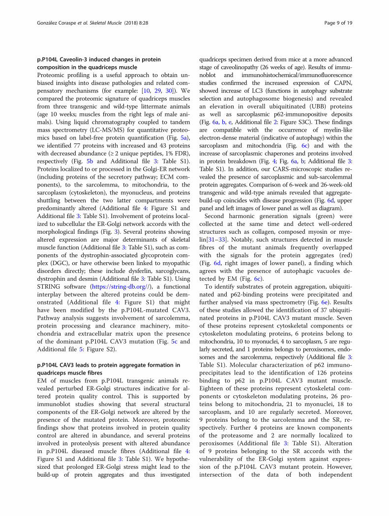
mals confirmed that membrane-bound vacuoles accu-mulate sub-sarcolemmally (Fig. 3 A.1, A.2). Thesevacuoles most likely correspond to abnormal caveolaeand/sarcoplasmic reticulum cisternae [10]. In addition,we found widened and proliferated ERGIC-Golgi struc-tures (Fig. 3 A.3–A.5). These ultrastructural findingscorrelate with increased and dispersed distribution ofGM130 (Fig. 3 B.1–B.4), a peripheral membrane compo-nent of the cis-Golgi stack. GM130 acts as a membraneskeleton maintaining the structure of the Golgi appar-atus, and as a vesicle tether that facilitates vesicle fusionto the Golgi membrane [25]. Golgin-97 immunoreactiv-ity was also analysed by fluorescence microscopy anddemonstrated pathological dispersion of Golgi struc-tures in muscle fibres of p.P104L CAV3 transgenicanimals (Fig. 3c). Decreased abundance of Atlastin-1(Fig. 4b) was in line with Golgi pathology as this pro-tein mediates homotypic fusion of ER membranes andfunctions in ER tubular network biogenesis and inGolgi biogenesis.Correspondingly, immunoblots and immunohistochemis-
try revealed forced phosphorylation of eIF2α and increasedabundance of the SR-resident SIL1-BiP chaperone complexand of HSP70 (sarcoplasmic chaperone stabilizing proteinsagainst aggregation) as well as decreased SEC61 levels inthe muscle fibres of p.P104L CAV3 transgenic animals
González Coraspe et al. Skeletal Muscle (2018) 8:28 Page 7 of 19

(Fig. 4a, b, Additional file 2: Figure S3C). The SEC61 pro-tein is the major component of a channel-forming translo-con complex that mediates co-translational translocation ofnascent (poly) peptides across the ER membrane; its down-regulation is in accordance with an attenuation of protein
synthesis to reduce the protein cargo through the secretoryapparatus [26]. Increased levels of RCN2, an ER-luminalprotein involved in Ca2+ and redox homeostasis [27], fur-ther suggests vulnerability of proper ER-Golgi functionbased on p.P104L Caveolin-3 expression.
A
D
B
C
Fig. 2 The CAV3 p.P104L mutation causes muscle pathology and influences protein stability. (a) Macroscopic comparison of hind limbs at 26weeks wildtype mouse and a CAV3 (p.P104L) transgenic mouse reveals a moderate decline in muscle mass in the transgenic mouse which morepronounced at 52 weeks. (b) H&E staining reveals a largely normal appearance of wildtype quadriceps muscle at age 26 weeks compared to thequadriceps muscle of a transgenic animal presenting with muscle fiber caliber variability and internalized muscle fiber nuclei (Paraffin sections) (c)Transcript studies of Cav3 in quadriceps muscle of 26 weeks old p.P104L CAV3 mutant animals and wildtype littermates revealed increased cDNAabundance p.P104L CAV3 mutants. (d) Immunoblot of quadriceps muscle protein extracts (8 and 26 weeks old animals) revealing a prominentstatistically significant CAV3 decrease in the mutant muscle. Coomassie blue staining was used as loading control
González Coraspe et al. Skeletal Muscle (2018) 8:28 Page 8 of 19
p.P104L Caveolin-3 induced changes in proteincomposition in the quadriceps muscleProteomic profiling is a useful approach to obtain un-biased insights into disease pathologies and related com-pensatory mechanisms (for example: [10, 29, 30]). Wecompared the proteomic signature of quadriceps musclesfrom three transgenic and wild-type littermate animals(age 10 weeks; muscles from the right legs of male ani-mals). Using liquid chromatography coupled to tandemmass spectrometry (LC-MS/MS) for quantitative proteo-mics based on label-free protein quantification (Fig. 5a),we identified 77 proteins with increased and 43 proteinswith decreased abundance (≥ 2 unique peptides, 1% FDR),respectively (Fig. 5b and Additional file 3: Table S1).Proteins localized to or processed in the Golgi-ER network(including proteins of the secretory pathway; ECM com-ponents), to the sarcolemma, to mitochondria, to thesarcoplasm (cytoskeleton), the myonucleus, and proteinsshuttling between the two latter compartments werepredominantly altered (Additional file 4: Figure S1 andAdditional file 3: Table S1). Involvement of proteins local-ized to subcellular the ER-Golgi network accords with themorphological findings (Fig. 3). Several proteins showingaltered expression are major determinants of skeletalmuscle function (Additional file 3: Table S1), such as com-ponents of the dystrophin-associated glycoprotein com-plex (DGC), or have otherwise been linked to myopathicdisorders directly; these include dysferlin, sarcoglycans,dystrophin and desmin (Additional file 3: Table S1). UsingSTRING software (https://string-db.org//), a functionalinterplay between the altered proteins could be dem-onstrated (Additional file 4: Figure S1) that mighthave been modified by the p.P104L-mutated CAV3.Pathway analysis suggests involvement of sarcolemma,protein processing and clearance machinery, mito-chondria and extracellular matrix upon the presenceof the dominant p.P104L CAV3 mutation (Fig. 5c andAdditional file 5: Figure S2).
p.P104L CAV3 leads to protein aggregate formation inquadriceps muscle fibresEM of muscles from p.P104L transgenic animals re-vealed perturbed ER-Golgi structures indicative for al-tered protein quality control. This is supported byimmunoblot studies showing that several structuralcomponents of the ER-Golgi network are altered by thepresence of the mutated protein. Moreover, proteomicfindings show that proteins involved in protein qualitycontrol are altered in abundance, and several proteinsinvolved in proteolysis present with altered abundancein p.P104L diseased muscle fibres (Additional file 4:Figure S1 and Additional file 3: Table S1). We hypothe-sized that prolonged ER-Golgi stress might lead to thebuild-up of protein aggregates and thus investigated
quadriceps specimen derived from mice at a more advancedstage of caveolinopathy (26 weeks of age). Results of immu-noblot and immunohistochemical/immunofluorescencestudies confirmed the increased expression of CAPN,showed increase of LC3 (functions in autophagy substrateselection and autophagosome biogenesis) and revealedan elevation in overall ubiquitinated (UBB) proteinsas well as sarcoplasmic p62-immunopositive deposits(Fig. 6a, b, e, Additional file 2: Figure S3C). These findingsare compatible with the occurrence of myelin-likeelectron-dense material (indicative of autophagy) within thesarcoplasm and mitochondria (Fig. 6c) and with theincrease of sarcoplasmic chaperones and proteins involvedin protein breakdown (Fig. 4; Fig. 6a, b; Additional file 3:Table S1). In addition, our CARS-microscopic studies re-vealed the presence of sarcoplasmic and sub-sarcolemmalprotein aggregates. Comparison of 6-week and 26-week-oldtransgenic and wild-type animals revealed that aggregate-build-up coincides with disease progression (Fig. 6d, upperpanel and left images of lower panel as well as diagram).Second harmonic generation signals (green) were
collected at the same time and detect well-orderedstructures such as collagen, composed myosin or mye-lin[31–33]. Notably, such structures detected in musclefibres of the mutant animals frequently overlappedwith the signals for the protein aggregates (red)(Fig. 6d, right images of lower panel), a finding whichagrees with the presence of autophagic vacuoles de-tected by EM (Fig. 6c).To identify substrates of protein aggregation, ubiquiti-
nated and p62-binding proteins were precipitated andfurther analysed via mass spectrometry (Fig. 6e). Resultsof these studies allowed the identification of 37 ubiquiti-nated proteins in p.P104L CAV3 mutant muscle. Sevenof these proteins represent cytoskeletal components orcytoskeleton modulating proteins, 6 proteins belong tomitochondria, 10 to myonuclei, 4 to sarcoplasm, 5 are regu-larly secreted, and 1 proteins belongs to peroxisomes, endo-somes and the sarcolemma, respectively (Additional file 3:Table S1). Molecular characterization of p62 immuno-precipitates lead to the identification of 126 proteinsbinding to p62 in p.P104L CAV3 mutant muscle.Eighteen of these proteins represent cytoskeletal com-ponents or cytoskeleton modulating proteins, 26 pro-teins belong to mitochondria, 21 to myonuclei, 18 tosarcoplasm, and 10 are regularly secreted. Moreover,9 proteins belong to the sarcolemma and the SR, re-spectively. Further 4 proteins are known componentsof the proteasome and 2 are normally localized toperoxisomes (Additional file 3: Table S1). Alterationof 9 proteins belonging to the SR accords with thevulnerability of the ER-Golgi system against expres-sion of the p.P104L CAV3 mutant protein. However,intersection of the data of both independent
González Coraspe et al. Skeletal Muscle (2018) 8:28 Page 9 of 19

A
B
C
Fig. 3 (See legend on next page.)
González Coraspe et al. Skeletal Muscle (2018) 8:28 Page 10 of 19

immunoprecipitation experiments revealed 15 proteinsubiquitinated and binding to p62 in muscle of thecaveolinopathy mouse model (Fig. 6e, Additional file 3:Table S1). Interestingly, 6 of those are mitochondrialproteins confirming the concept of mitochondrialvulnerability as already suggested by our electronmicroscopic and global proteomic findings. Further 3of these proteins are regularly secreted, 2 belong tocytoskeleton, 2 to myonuclei and 1 protein normallylocalizes to the sarcoplasm and to peroxisomes,respectively.
Components of the dystrophin-associated glycoproteincomplex are substrates of the impaired protein qualitycontrol in p.P104L caveolinopathyAs described above, our comparative proteome profilingrevealed altered abundance of several DGC components(Additional file 3: Table S1). Based on the results of thisunbiased screening, we hypothesized that proteins of theDGC might be affected indirectly by p.P104L mutantCAV3 mis-localization to the Golgi, ER-stress response ac-tivation and impaired protein quality control. Further im-munoblot and immunohistochemical studies focussing onparadigmatic components of the complex confirmedthe increase of the same proteins detected via shotgunproteomics (Fig. 7a). Deposit-like structures immunoreac-tive for DGC-components such as δ-sarcoglycan, dysferlinand integrin were identified (Fig. 7b, c and Additional file 2:Figure S3A) and focal sub-sarcolemmal enrichment ofδ-sarcoglycan and integrin as well as chaperones (HSP70)were observed (Fig. 4a, Fig. 7b and Additional file 2: FigureS3A). Prompted by this finding, next transcript level of dys-ferlin, δ-sarcoglycan and integrin-beta-4 has been studied.Results showed nearly equal level for dysferlinwhereas δ-sarcoglycan transcripts were ~ 50% increasedand integrin-beta-4 transcripts were ~ 60% decreased(Additional file 2: Figure S3B). The combined findings mightindicate that while some of the transcripts, such asδ-sarcoglycan, result in an elevated protein level for others,forced translation of stable or even decreased RNA leads toan increase in the corresponding proteins. Given that (i) theglobal proteomic signature of p.P104L CAV3 diseased quad-riceps muscle indicated affection of DGC components suchas dysferlin and (ii) UPR is also activated in diseased muscleas well as that (iii) further mass spectrometry studies didnot identify the DGC components as elements of the pro-teins aggregates (Additional file 3: Table S1), effective
targeting by UPR factors was hypothesized. Indeed, resultsof co-immunofluorescence studies revealed sarcoplasmicco-localization of GRP170 (a UPR-modulated chaperon)and dysferlin (Fig. 7c). However, although the architectureof the ER-Golgi network as organelles of protein glycosyl-ation are altered and certain DGC components displayvulnerability against p.P104L CAV3 protein expression,the myopathology does not affect proper glycosylation ofα-dystroglycan as a key protein of the DGC (Fig. 7d).
DiscussionMutant CAV3 has been shown to mis-localize to the Golgi[8, 10] and cause ER-stress accompanied by activation ofthe UPR as a cellular defence mechanism, but the molecu-lar consequences of the ER-Golgi disturbance as part of theCAV3 pathophysiology remained elusive. To systematicallyaddress this gap of knowledge and to further strengthen theunderstanding of pathophysiological processes in caveolino-pathies, we comprehensively analysed quadriceps musclesfrom a well-characterized mouse model harbouring thedominant p.P104L mutation [11, 13].
Vulnerability of the ER-Golgi system in p.P104LcaveolinopathyWe performed morphological investigations of the ER-Golgisystem, linked the structural perturbations to altered abun-dance of proteins involved in maintenance of organelle archi-tecture and showed that prolonged perturbation of theER-Golgi machinery—indicated by the altered proteinabundance of ASNA1, EF1B, RCN2, UGGT1, TMEM43and our immunohistochemical studies (Additional file 3:Table S1, Fig. 4 and Additional file 2: Figure S3)—resultsin the build-up of protein aggregates within diseasedquadriceps muscles fibres. This observation is not only inline with the increased expression of protease inhibitors(i.e. ANT3, ILEUA, SPB6, MUG1 and A1AT4) but alsowith the increased abundance of overall ubiquitinated pro-teins (Fig. 6a). Concomitantly, increased expression of(co-)chaperones (HSP70/ HSPB1, HSPB2, HSPB7, BiP,SIL1; Additional file 3: Table S1 and Fig. 4) and of proteinsinvolved in proteolysis such as CAPN/calpain (proteaseinvolved in the breakdown of misshaped proteins prevent-ing toxic aggregation) and PSMA5 most likely represent acellular defence mechanism antagonizing the build-up oftoxic protein aggregates (Additional file 3: Table S1).
(See figure on previous page.)Fig. 3 Expression of p.P104L missense mutant CAV3 disturbs ER-Golgi integrity. (a) EM findings in p.P104L CAV3 mutant quadriceps muscle:subsarcolemmal accumulation of vesicular structures (most likely corresponding to abnormal caveolae) (black arrows in 3A.1 and 3A.2) anddispersed ERGIC-Golgi structures (black arrows in 3A.3 – 3A.5). (b) Immunohistochemical studies focusing on ER-Golgi stress confirm GM130increase. (c) Immunofluorescence studies focusing on Golgi-structure in 6 month and 1 year old animals (visualization via golgin-97 staining)revealed a statistically significant dispersion on p.P104L CAV3 transgenic animals
González Coraspe et al. Skeletal Muscle (2018) 8:28 Page 11 of 19
A
B
Fig. 4 Study of ER-Golgi relevant proteins. (a) Immunohistochemical studies focusing on ER-Golgi stress confirm UPR activation (increasedabundance of the phosphorylated (activated) form of eIF2α, of HSP70 and RCN2) in p.P104L muscle fibres. Interestingly, the cytosolic chaperoneHSP70 shows enrichment at the sub-sarcolemmal sarcoplasm of muscle fibres expressing the mutant protein. RCN2 is accumulating in damagedmuscle fibres. (b) Pathomorphological findings highlighted in Figure 3 are accompanied by reduced expression of ATL1, a protein important forthe structural maintenance of the ER-Golgi system as well as altered abundance of proteins controlled by ER-stress and UPR (SIL1 and BiP, HSP70,RCN2 and SEC61). Coomassie brilliant blue staining: loading control
González Coraspe et al. Skeletal Muscle (2018) 8:28 Page 12 of 19

A
B
C
Fig. 5 Proteomic studies to unravel the molecular nature of p.P104L induced muscular dystrophy in mice. a Proteomic workflow applied in thisstudy. b Results of our label-free shotgun proteomic profiling are shown as a volcano plot. All points (each one represents a protein) over thehorizontal line have a statistically significant p-ANOVA of ≤ 0.05. In total, 43 proteins are decreased (red points) and 77 proteins are increased inabundance (green points) in mutant mouse muscle. c Results of an in silico pathway analysis of proteins altered in abundance confirm alterationsof the protein processing machinery, vulnerability of mitochondria as well as altered in ECM protein processing and cellular metabolism. Inaddition, there is evidence of oxidative stress and sarcolemmal vulnerability
González Coraspe et al. Skeletal Muscle (2018) 8:28 Page 13 of 19

Protein aggregation in p.P104L caveolinopathyAs elevated CAV3 level has been demonstrated to be bene-ficial for autophagic protein clearance [2], overall reductionof cellular CAV3 upon p.P104L CAV3 pathophysiology(Fig. 2) might influence the efficiency of cellular proteolysisvia the autophagic system, an assumption supported by thebuild-up of protein aggregates. Our electron and CARS mi-croscopy studies showed electron-dense myelin-like struc-tures (most likely corresponding to protein aggregates)within the sarcoplasm and the sub-sarcolemmal region thatcould be inadequately processed substrates of the perturbedER-Golgi machinery. Further molecular characterization ofthese aggregates revealed affection of a variety of proteinsbelonging to different subcellular compartments such as
the sarcolemma, cytoskeleton and mitochondria (Add-itional file 3: Table S1) suggesting that the p.P104L muta-tion has as widespread effect on overall organelle integrityand function in muscle. Presumably, impaired protein pro-cessing leading to the accumulation of aggregates seemsto be an upstream event in the pathological cascade ofcaveolinopathy. Therefore, rescuing or stabilizing the na-tive conformations of proteins is an obvious therapeuticstrategy. Over the last decade, small molecules known aschaperones have been shown to be effective in reducinglevels of misshaped proteins, thus minimizing the accu-mulation of aggregates with toxic potential leading and re-ducing negative downstream pathological consequences[34] (Additional file 6: Figure S4).
A
C
B D E
Fig. 6 Activation of proteolysis and protein aggregation in p.P104L caveolinopathy. A Immunoblot studies confirming the increasedabundance of CAPN2 identified in the proteome profile and showing increased abundance of ubiquitinated proteins as well as of LC3.Coomassie brilliant blue was used as loading control. B Increased abundance of CAPN2 was confirmed via immunohistochemical studieswhich also revealed elevated level of SERPINE (a protein functioning as a protease) at the sub-sarcolemmal region. C EM showingautophagic vacuoles containing electron-dense myelin-like structures (6C.1–6C.7) in quadriceps muscles of animals with advancedmyopathy as well as damaged, probably degenerating mitochondria (6C.8–6C.10). D CARS (two photon) microscopy confirms thepresence of sarcoplasmic and sub-sarcolemmal protein-dense structures in quadriceps muscle fibres of p.P104 CAV3 transgenic animals(red fluorescent dots) but not in control muscles from wild-type littermates. Concomitant second harmonic generation signals (greenfluorescent dots) corresponding to organized molecular structures such as composed myosin and myelin also indicate the presence ofsmall abnormal build-up of organized structures within the sarcoplasm of quadriceps muscle fibres of diseased animals frequentlyoverlapping with the protein aggregates identified by two photon microscopy. Comparative analyses and quantification of aggregates inyounger and older animals revealed that aggregate-build-up coincides with disease progression. E Immunoprecipitation of ubiquitinatedand p62-binding proteins with subsequent mass spectrometry-based identification towards the molecular characterization of the proteinaggregates detected via electron and CARS microscopy. Data intersection revealed similar alteration of 15 proteins
González Coraspe et al. Skeletal Muscle (2018) 8:28 Page 14 of 19

Vulnerability of further DGC components and thesarcolemma in p.P104L caveolinopathyConstant production and Golgi mis-localization of mutantCAV3 most likely perturbs proper ER-Golgi function,which should also subsequently affect the substrate proteinsprocessed within these compartments. Notably, DGC com-ponents are known to be processed within these compart-ments [35]. Indeed, our proteomic study revealed increasedabundance of DGC components such as dysferlin, sarcogly-cans, integrins, dystrophin and syntrophin (Additional file 3:Table S1, Fig. 7 and Additional file 2: Figure S3). Moreover,results of our immunological analyses indicated an abnor-mal localization of paradigmatic DGC components andshowed, apart from immunoreactivity with the sarcolemma,a patchy distribution within the sarcoplasm and focalenrichment within the sub-sarcolemmal region (Fig. 7 andAdditional file 2: Figure S3). However, DGC componentshave not been identified as constituents of the proteinaggregates (Additional file 3: Table S1) and co-immunofluorescence studies showed co-localization of dysferlinwith GRP170, a UPR-modulated chaperone suggestingthat sarcoplasmic enrichment of DGC components ismore likely based on prolonged processing/ chaperonemediated re-folding due to perturbed ER-Golgi functionrather than on formation of aggregates.Prompted by the altered architecture of the ER-Golgi
network as cellular place of protein glycosylation as wellas abnormal subcellular distribution of DGC compo-nents, glycosylation of α-dystroglycan as a paradigmaticDGC component has been studied but revealed no sig-nificant changes suggesting that p.P104L CAV3 patho-physiology targeting the ER-Golgi network does nothave an impact on glycosylation of certain DGC key pro-teins. This assumption agrees with the identification ofincreased abundance of UGGT1, an ER-resident proteinthat selectively re-glucosylates unfolded glycoproteins,thus providing quality control for protein transport outof the ER (https://www.ncbi.nlm.nih.gov/gene/56886).Hence, our combined data suggest that a vulnerability
of the sarcolemma in caveolinopathy is not only basedon the absence/ detrimental reduction of CAV3 [1]—moreover reflected by decrease of PTRF (Additional file 3:Table S1) but also on the aggregation of proteins importantfor sarcolemmal integrity and presumably strengthened byaltered/prolonged processing of DGC components. In thiscontext, direct functional relevance is indicated by aggrega-tion of MAP3K2 playing a role in “caveolae kiss-and-rundynamics” [36] as well as by MYOC, a regulator of musclehypertrophy through the components of dystrophin-associ-ated protein complex [37]. However, our global proteomicprofiling results also support the concept of sarcolemmalvulnerability, as a decrease of CAD13 and SLC41A3 wasdetected, proteins important for cell-cell contacts [38]. In-creased abundance of DERM, regulating cell-cell
adhesion by surface integrin binding seems to com-pensate affection of integrins (as part of the DGC)in the presence of mutated CAV3 (Additional file 2:Figure S3A). Also, the cellular decrease of CA3supports this assumption as CA3 is a known serumbiomarker for muscular dystrophy mirroring sarcolemmalfragility [39]. Compensatory mechanisms may counteractsarcolemmal vulnerability, which is supported by ourproteomic results revealing the increased abundance of sixplausible proteins (Additional file 3: Table S1). Amongthose proteins, PACN3 regulates internalization of sarco-lemmal proteins. PACN3 overexpression impairs internal-ization of proteins such as GLUT1 and TRPV4 andincreases the sarcolemmal level of those proteins. AsGLUT1 facilitates the transport of glucose across theplasma membranes of mammalian cells [40] our dataprovide a molecular explanation for the recently de-scribed perturbed glucose metabolism in muscle cellsupon p.P104L CAV3 expression [41]. TRPV4 contrib-utes to cell volume control and plays a role in themodulation of muscle fibre atrophy [42]. ATP1A1 isincreased (Additional file 3: Table S1) which controlsthe exchange of sodium and potassium ions acrossthe sarcolemma, a process perturbed in Duchennemuscular dystrophy [43]. This accords with the acti-vation of mechanisms antagonizing sarcolemmal vul-nerability and improving cellular fitness.
Vulnerability of cytoskeletal components in p.P104LcaveolinopathyAltered sarcolemmal-integrity as suggested (i) by ourmolecular characterization of the aggregates and (ii) byparadigmatic DGC components (linking the cytoskeletonto the basal lamina) showing partial mis-localizationindicate downstream effects on cytoskeletal integrity.Indeed, our combined proteomic findings in p.P104LCAV3 diseased muscle are also indicative for cytoskeletalvulnerability (Fig. 4, Additional file 4: Figure S1 andAdditional file 5: Figure S2 and Additional file 3: Table S1)linking the p.P104L CAV3 pathophysiology to impairedmuscle fibre contraction: a variety of cytoskeletal proteinshave been identified as constituents of the aggregatesidentified in muscle of diseased animals and results of ourglobal proteomic studies showed a decrease of NEXN,MYH8, MYPT2 and MURC, whereas the decrease inTMOD4 and increases in 14-3-3-eta, COBL, HSPB7,GELS, MARCS, TUBA4A, TUBA8, DPYL2, SYNM,MAP4 and PDLIM3 (Additional file 3: Table S1) levels aresuggestive of antagonizing mechanisms to avoid musclefibre break-down.
Vulnerability of mitochondria in p.P104L caveolinopathyCellular increase of CAV3 notoriously attenuates mito-chondrial damage and cell death in (cardio) myocytes
González Coraspe et al. Skeletal Muscle (2018) 8:28 Page 15 of 19

A
B
C
D
Fig. 7 DGC components are substrates of the impaired protein processing machinery in p.P104L caveolinopathy. A Immunoblots of paradigmaticcomponents of the DGC confirm the proteomic findings by showing increased protein abundance of dystrophin, dysferlin, α- and β-dystroglycan,α-1-syntrophin as well as α-, β- and δ-sarcoglycan. Protein phosphatase 2AAA, stable in our proteome profile, has been used as loading control,and shows stable levels, in line with our proteomic findings. B Increased abundance of DGC components was also confirmed byimmunohistochenistry focusing on δ-sarcoglycan as a paradigmatic example. Interestingly, apart from occasional sarcoplasmic deposits (blackarrows in 7B.4), an enrichment in the sub-sarcolemmal region could be identified (7B.2 and 7B.3). C Immunofluorescence-based co-localizationstudies showing (irregular) sarcoplasmic dots immunoreactive for GRP170 (a co-chaperon of the SIL1-BiP machinery) and dysferlin. D Immunoblotanalysis utilizing the monoclonal antibody IIH6, which recognizes specifically glycosylated α-dystroglycan (left panel) and the polyclonal antibodyAF6868, which recognizes both α- and β-dystroglycan (right panel)
González Coraspe et al. Skeletal Muscle (2018) 8:28 Page 16 of 19

[44] thus indicating a functional relevance of CAV3 levelfor mitochondrial function. Notably, altered mitochondrialfunction in caveolinopathy influencing energy productionand oxidative stress burden can be deduced from ourglobal proteomic findings (Additional file 3: Table S1) andmolecular characterization of aggregates identified inquadriceps muscles of p.P104L transgenic animals more-over revealed vulnerability of a total of 26 different mito-chondrial proteins involved in diverse mitochondrialprocesses as well as in structural integrity (Fig. 6e andAdditional file 3: Table S1). Vulnerability of mitochondrialproteins in p.P104L CAV3 pathophysiology accords withthe identified ultra-structural abnormalities (Fig. 6c).
Further pathophysiological insights into p.P104LcaveolinopathyThe proteomic response to p.P104L mutant CAV3 expres-sion moreover revealed an increased abundance of secretedproteins belonging to the extracellular matrix (ECM; in par-ticular collagens) (Additional file 3: Table S1). Althoughthese proteins are normally processed within the ER-Golgimachinery representing substrates, their increase might alsoarise from ECM accumulation/fibrosis, a well-knownpathophysiological hallmark of intermediate to late stagemuscular dystrophies [45]. However, mass spectrometry-based characterization of aggregates detected in p.P104LCAV3 diseased muscle identified several proteins regularlytargeted for secretion as molecular constituents thusstrengthening the hypothesis of impaired substrate process-ing and protein aggregate formation. Longer-term observa-tion would be required to fully clarify whether the increaseof ECM proteins exclusively reflects protein retention inthe Golgi/ER associated with protein aggregate formationor incipient fibrosis or a combination thereof.Additionally, results of our comparative proteome pro-
filing revealed changes in annexins: annexins represent aclass of dysferlin-interacting proteins [46] and modulatemyoblast cell differentiation by promoting migration ofsatellite cells and, consequently, skeletal muscle differen-tiation [47]. Annexins are also involved in the modula-tion of inflammatory processes and membrane repair ofhuman skeletal muscle cells [46, 48]. Thus, increasedabundance of annexins may contribute to the regener-ation of muscle fibres in caveolinopathies and may havetherapeutic implications with respect to the developmentof annexin mimetics. Similarly, increased abundance ofCD81, which plays an important role in the restitutionof normal muscle architecture during muscle regener-ation [49] might provide an attractive therapeutic target.Apart from altered GLUT1 (see above) there is further
indication for an altered glucose metabolism withchanges in the abundance of PGM1, HK2 and RTN2(Additional file 3: Table S1). Interestingly, regardingother pathways of potential importance, recent
publications revealed that CAV3 directly interacts withthe insulin receptor and that loss of CAV3 interfereswith downstream insulin signalling and lipid uptake, im-plicating CAV3 as a regulator of the insulin receptor andregulator of lipid uptake [50]. Our proteomic findingssupport a role of CAV3 in lipid homeostasis by demon-strating decreased levels of FITM1, CES1D and EDF1(Additional file 3: Table S1).Finally, decreased abundance of SMYD1, EDF1 and
PDLI5 might provide molecular insights into the natureof the dilatative cardiomyopathy described in this mousemodel [13].
ConclusionsResults of our combined morphological and biochemicalstudies confirm the previously reported ER-Golgi path-ology caused by mislocalization of missense mutantCAV3 permanently stressing subcellular compartments.The organelle pathology is linked to impaired proteinprocessing accompanied by the build-up of sarcoplasmicand sub-sarcolemmal protein aggregates. Thus, ourstudy adds caveolinopathies to the growing list of sec-ondary chaperonopathies [48]. Combined findings un-ravel substrates of the impaired ER-Golgi machinery(secondarily) disturbing sarcolemmal and mitochondrialintegrity, the cytoskeleton and the ECM. On a moregeneral note, our study represents a promising exampleof the suitability of applied proteomics to obtain deepinsights into the underlying pathophysiology of diseasessuch as neuromuscular disorders [51].
Additional files
Additional file 1: Table S2. List of antibodies used in this study.(DOC 53 kb)
Additional file 2: Figure S3. Further studies of substrates of thepathophysiology and quantification of immunoblot findings. (A)Immunohistochemistry of integrin α 5 and β 4 revealed a perturbedlocalization (similar to the result of δ-sarcoglycan staining depicted inFig. 6B in the quadriceps muscle fibres of p.P104L mutant animals. (B)mRNA expression of dysferlin (Dysf), integrin-β-4 (Igtb4) and δ-sarcoglycan(Scgd) normalized to glycerinaldehyde-3-phosphate dehydrogenase (Gapdh)in quadriceps muscle of 26 weeks old p.P104L CAV3 mutant animals andwild-type littermates (n = 4). N represents the number of independentsamples measured in triplicates (*P < 0.05 vs. wild-type). Statisticalsignificance between groups was analysed by an unpaired t-test usingGraphPad software (San Diego, USA). Differences were consideredsignificant with P < 0.05. All data are shown as means ±SEM. Thesestudies revealed almost equal mRNA abundance for Dysf but decreasedabundance for Itgb4 and increased abundance for Scgd in p.P104L CAV3mutants. (C) Quantification results of immunoblot findings (towardsverification of proteomic findings). (PPTX 9155 kb)
Additional file 3: Table S1. Overview of all proteomic findings.(XLSX 915 kb)
Additional file 4: Figure S1. Cytoscape network analysis of theregulated proteins and in silico data from different perspectives: geneinterrelations revealed by correlation weights, protein interactions as wellas functional context given by the GO enrichment analysis. The nodesare linked based on their kappa score level (≥0.3), where only the label of
González Coraspe et al. Skeletal Muscle (2018) 8:28 Page 17 of 19
the most significant term per group is shown. The node size representsthe term enrichment significance. Functionally related groups partiallyoverlap. (PPTX 559 kb)
Additional file 5: Figure S2. Further pathway analysis of alteredproteins. PANTHER-based pathway analysis was performed for increasedand decreased proteins separately and indicates vulnerability of cytoskel-eton in p.P104L diseased quadriceps muscle fibres. (PPTX 237 kb)
Additional file 6: Figure S4. Illustration showing the proposedmechanisms of pathogenesis in Caveolinopathy associated with thep.P104L mutation. (PPTX 38 kb)
AbbreviationsACN: Acetonitrile; BCA: Bicinchoninic acid assay; CaCl2: Calcium chloride;CARS: Coherent anti-Stokes Raman scattering; CID: Collision induceddissociation; CK: Creatine kinase; Cys: Cysteine; DGC: Dystroglycan complex;DM: Distal myopathy; DTT: Dithiothreitol; ECM: Extracellular matrix;EDTA: Ethylenediaminetetraacetic acid; EM: Electron microscopy;ER: Endoplasmic reticulum; ERGIC: Endoplasmic reticulum Golgi intermediatecompartment; FA: Formic acid; FDR: False discovery rate; Glu: Glutamic acid;GO: Gene ontology; GuHCl: Guanidinium chloride; H&E: Hematoxylin andeosin; HCK: HyperCKemia; HEK293: Human embryonic kidney cell line;HPLC: High-performance liquid chromatography; IAA: Iodoacetamide;IHC: Immunohistochemistry; LC-MS/MS: Liquid chromatography coupled totandem mass spectrometry; LGMD: Limb Girdle Muscular Dystrophy;Met: Methionine; NaCl: Sodium chloride; PBS: Phosphate-buffered saline;PCR: Polymerase chain reaction; PMT: Photo multiplier tube;PVDF: Polyvinylidene difluoride; RMD: Rippling muscle disease; SDS: Sodiumdodecyl sulfate; SR: Sarcoplasmic reticulum; TFA: Trifluoroacetic acid;TRIS: (Hydroxymethyl)aminomethane; UPR: Unfolded protein response;WT: Wild-type
AcknowledgementsWe thank Mrs. Claudia Krude, Mrs. Hannelore Mader and Mrs. AstridKnischewski for expert technical assistance.
FundingFinancial support by the Ministerium für Innovation, Wissenschaft undForschung des Landes Nordrhein-Westfalen, the Senatsverwaltung fürWirtschaft, Technologie und Forschung des Landes Berlin and the Bundesmi-nisterium für Bildung und Forschung is gratefully acknowledged. This workwas also supported by a grant from the Deutsche Gesellschaft für Muskelk-ranke (DGM to A. R.) and the IZKF Aachen (to J. W.; grant no. N5-3) as well asby a Paul D. Wellstone Muscular Dystrophy Cooperative Research CenterGrant (1U54NS053672). K.P.C. is an investigator of the Howard Hughes Med-ical Institute.
Availability of data and materialsThe datasets used and/or analysed during the current study are availablefrom the corresponding author on reasonable request.
Authors’ contributionsAR, HL and JW designed the study. YS provided the animals and SB, EB aswell as JAGC prepared the animals. JAGC, DH, SC, HM and MEA performedthe experiments. Data were revised by AR, RPZ, KPC and EF. JAGC draftedthe manuscript. All co-authors revised the manuscript prior submission. Allauthors read and approved the final manuscript.
Ethics approvalAll procedures were approved by the UK Aachen Institutional Animal Careand Use Committee and conducted in compliance with the Guide for theCare and Use of Laboratory Animals.
Consent for publicationNot applicable.
Competing interestsThe authors declare that they have no competing interests.
Publisher’s NoteSpringer Nature remains neutral with regard to jurisdictional claims inpublished maps and institutional affiliations.
Author details1Institute of Neuropathology, RWTH Aachen University Hospital, Pauwelsstr.30, 52074 Aachen, Germany. 2Howard Hughes Medical Institute, Departmentsof Molecular Physiology and Biophysics, of Neurology, University of Iowa,Iowa City, IA 52242, USA. 3Biomedical Research Department, Tissue Omicsgroup, Leibniz-Institut für Analytische Wissenschaften - ISAS - e.V,Otto-Hahn-Str. 6b, 44227 Dortmund, Germany. 4Institute of Genetic Medicine,International Centre for Life, Central Parkway, Newcastle upon Tyne, England,UK. 5Gerald Bronfman Department of Oncology, Jewish General Hospital,McGill University, Montreal, Quebec H4A 3T2, Canada. 6Segal CancerProteomics Centre, Lady Davis Institute, Jewish General Hospital, McGillUniversity, Montreal, Quebec H3T 1E2, Canada. 7Department of Neurology,Kawasaki Medical School, 577 Matsushima, Kurashiki, Okayama 701-0192,Japan. 8Department of Neuropediatrics and Muscle Disorders, Medical Center– University of Freiburg, Faculty of Medicine, Freiburg, Germany. 9CentroNacional de Análisis Genómico (CNAG-CRG), Center for Genomic Regulation,Barcelona Institute of Science and Technology (BIST), Barcelona, Catalonia,Spain. 10Children’s Hospital of Eastern Ontario Research Institute, University ofOttawa, Ottawa, Canada and Division of Neurology, Department of Medicine,The Ottawa Hospital, Ottawa, Canada.
Received: 18 February 2018 Accepted: 24 July 2018
References1. Gazzerro E, Sotgia F, Bruno C, Lisanti MP, Minetti C. Caveolinopathies: from
the biology of caveolin-3 to human diseases. Eur J Hum Genet. 2010;18:137–45.
2. Kassan A, Pham U, Nguyen Q, Reichelt ME, Cho E, Patel PM, Roth DM, HeadBP, Patel HH. Caveolin-3 plays a critical role in autophagy after ischemia-reperfusion. Am J Physiol Cell Physiol. 2016;311:C854–65.
3. Takamori M. Synaptic homeostasis and its immunological disturbance inneuromuscular junction disorders. Int J Mol Sci. 2017;18(4).
4. Macias A, Gambin T, Szafranski P, Jhangiani SN, Kolasa A, Obersztyn E, LupskiJR, Stankiewicz P, Kaminska A. CAV3 mutation in a patient with transienthyperCKemia and myalgia. Neurol Neurochir Pol. 2016;50:468–73.
5. Vorgerd M, Ricker K, Ziemssen F, Kress W, Goebel HH, Nix WA, Kubisch C,Schoser BG, Mortier W. A sporadic case of rippling muscle disease causedby a de novo caveolin-3 mutation. Neurology. 2001;57:2273–7.
6. Fischer D, Schroers A, Blumcke I, Urbach H, Zerres K, Mortier W, Vorgerd M,Schroder R. Consequences of a novel caveolin-3 mutation in a largeGerman family. Ann Neurol. 2003;53:233–41.
7. Traverso M, Gazzerro E, Assereto S, Sotgia F, Biancheri R, Stringara S, GibertiL, Pedemonte M, Wang X, Scapolan S, et al. Caveolin-3 T78M and T78Kmissense mutations lead to different phenotypes in vivo and in vitro. LabInvestig. 2008;88:275–83.
8. Brauers E, Dreier A, Roos A, Wormland B, Weis J, Kruttgen A. Differentialeffects of myopathy-associated caveolin-3 mutants on growth factorsignaling. Am J Pathol. 2010;177:261–70.
9. Kuga A, Ohsawa Y, Okada T, Kanda F, Kanagawa M, Toda T, Sunada Y.Endoplasmic reticulum stress response in P104L mutant caveolin-3transgenic mice. Hum Mol Genet. 2011;20:2975–83.
10. Brauers E, Roos A, Kollipara L, Zahedi RP, Beckmann A, Mohanadas N, BauerH, Hausler M, Thoma S, Kress W, et al. The Caveolin-3 G56S sequencevariant of unknown significance: muscle biopsy findings and functional cellbiological analysis. Proteomics Clin Appl. 2017;11:1-2.
11. Sunada Y, Ohi H, Hase A, Ohi H, Hosono T, Arata S, Higuchi S, Matsumura K,Shimizu T. Transgenic mice expressing mutant caveolin-3 show severemyopathy associated with increased nNOS activity. Hum Mol Genet.2001;10:173–8.
12. Pi J, Dogovski C, Pittard AJ. Functional consequences of changing prolineresidues in the phenylalanine-specific permease of Escherichia coli. JBacteriol. 1998;180:5515–9.
13. Ohsawa Y, Toko H, Katsura M, Morimoto K, Yamada H, Ichikawa Y, MurakamiT, Ohkuma S, Komuro I, Sunada Y. Overexpression of P104L mutantcaveolin-3 in mice develops hypertrophic cardiomyopathy with enhanced
González Coraspe et al. Skeletal Muscle (2018) 8:28 Page 18 of 19

contractility in association with increased endothelial nitric oxide synthaseactivity. Hum Mol Genet. 2004;13:151–7.
14. Roos A, Buchkremer S, Kollipara L, Labisch T, Gatz C, Zitzelsberger M, BrauersE, Nolte K, Schroder JM, Kirschner J, et al. Myopathy in Marinesco-Sjogrensyndrome links endoplasmic reticulum chaperone dysfunction to nuclearenvelope pathology. Acta Neuropathol. 2014;127:761–77.
15. Le TT, Yue S, Cheng JX. Shedding new light on lipid biology with coherentanti-Stokes Raman scattering microscopy. J Lipid Res. 2010;51:3091–102.
16. Cheng JX, Xie XS. Vibrational spectroscopic imaging of living systems: anemerging platform for biology and medicine. Science. 2015;350:aaa8870.
17. Michele DE, Barresi R, Kanagawa M, Saito F, Cohn RD, Satz JS, Dollar J,Nishino I, Kelley RI, Somer H, et al. Post-translational disruption ofdystroglycan-ligand interactions in congenital muscular dystrophies. Nature.2002;418:417–22.
18. Briggs DC, Yoshida-Moriguchi T, Zheng T, Venzke D, Anderson ME, Strazzulli A,Moracci M, Yu L, Hohenester E, Campbell KP. Structural basis of lamininbinding to the LARGE glycans on dystroglycan. Nat Chem Biol. 2016;12:810–4.
19. Manza LL, Stamer SL, Ham AJ, Codreanu SG, Liebler DC. Samplepreparation and digestion for proteomic analyses using spin filters.Proteomics. 2005;5:1742–5.
20. Wisniewski JR, Zougman A, Nagaraj N, Mann M. Universalsample preparation method for proteome analysis. Nat Methods.2009;6:359–62.
21. Kollipara L, Buchkremer S, Weis J, Brauers E, Hoss M, Rutten S, Caviedes P,Zahedi RP, Roos A. Proteome profiling and ultrastructural characterization ofthe human RCMH cell line: myoblastic properties and suitability formyopathological studies. J Proteome Res. 2016;15:945–55.
22. Olsen JV, de Godoy LM, Li G, Macek B, Mortensen P, Pesch R, Makarov A,Lange O, Horning S, Mann M. Parts per million mass accuracy on anOrbitrap mass spectrometer via lock mass injection into a C-trap. Mol CellProteomics. 2005;4:2010–21.
23. Vaudel M, Barsnes H, Berven FS, Sickmann A, Martens L. SearchGUI: anopen-source graphical user interface for simultaneous OMSSA andX!Tandem searches. Proteomics. 2011;11:996–9.
24. Vaudel M, Burkhart JM, Zahedi RP, Oveland E, Berven FS, Sickmann A,Martens L, Barsnes H. PeptideShaker enables reanalysis of MS-derivedproteomics data sets. Nat Biotechnol. 2015;33:22–4.
25. Seemann J, Jokitalo EJ, Warren G. The role of the tethering proteins p115and GM130 in transport through the Golgi apparatus in vivo. Mol Biol Cell.2000;11:635–45.
26. McKibbin C, Mares A, Piacenti M, Williams H, Roboti P, Puumalainen M,Callan AC, Lesiak-Mieczkowska K, Linder S, Harant H, et al. Inhibition ofprotein translocation at the endoplasmic reticulum promotes activation ofthe unfolded protein response. Biochem J. 2012;442:639–48.
27. Ludvigsen M, Jacobsen C, Maunsbach AB, Honore B. Identification andcharacterization of novel ERC-55 interacting proteins: evidence for theexistence of several ERC-55 splicing variants; including the cytosolic ERC-55-C. Proteomics. 2009;9:5267–87.
28. Livak KJ, Schmittgen TD. Analysis of relative gene expression data usingreal-time quantitative PCR and the 2(-Delta Delta C(T)) method. Methods.2001;25:402–8.
29. Roos A, Kollipara L, Buchkremer S, Labisch T, Brauers E, Gatz C, Lentz C,Gerardo-Nava J, Weis J, Zahedi RP. Cellular signature of SIL1 depletion:disease pathogenesis due to alterations in protein composition beyond theER machinery. Mol Neurobiol. 2016;53:5527–41.
30. Murphy S, Dowling P, Zweyer M, Mundegar RR, Henry M, Meleady P,Swandulla D, Ohlendieck K. Proteomic analysis of dystrophin deficiency andassociated changes in the aged mdx-4cv heart model of dystrophinopathy-related cardiomyopathy. J Proteome. 2016;145:24–36.
31. Chu SW, Chen IH, Liu TM, Sun CK, Lee SP, Lin BL, Cheng PC, Kuo MX, Lin DJ,Liu HL. Nonlinear bio-photonic crystal effects revealed with multimodalnonlinear microscopy. J Microsc. 2002;208:190–200.
32. Campagnola PJ, Loew LM. Second-harmonic imaging microscopy forvisualizing biomolecular arrays in cells, tissues and organisms. NatBiotechnol. 2003;21:1356–60.
33. Mohler W, Millard AC, Campagnola PJ. Second harmonic generationimaging of endogenous structural proteins. Methods. 2003;29:97–109.
34. Cortez L, Sim V. The therapeutic potential of chemical chaperones inprotein folding diseases. Prion. 2014;8.
35. Soheili T, Gicquel E, Poupiot J, N'Guyen L, Le Roy F, Bartoli M, Richard I.Rescue of sarcoglycan mutations by inhibition of endoplasmic reticulum
quality control is associated with minimal structural modifications. HumMutat. 2012;33:429–39.
36. Huang J, Tu Z, Lee FS. Mutations in protein kinase subdomain Xdifferentially affect MEKK2 and MEKK1 activity. Biochem Biophys ResCommun. 2003;303:532–40.
37. Joe MK, Kee C, Tomarev SI. Myocilin interacts with syntrophins and ismember of dystrophin-associated protein complex. J Biol Chem. 2012;287:13216–27.
38. de Baaij JH, Arjona FJ, van den Brand M, Lavrijsen M, Lameris AL, Bindels RJ,Hoenderop JG. Identification of SLC41A3 as a novel player in magnesiumhomeostasis. Sci Rep. 2016;6:28565.
39. Ayoglu B, Chaouch A, Lochmuller H, Politano L, Bertini E, Spitali P, Hiller M,Niks EH, Gualandi F, Ponten F, et al. Affinity proteomics within rare diseases:a BIO-NMD study for blood biomarkers of muscular dystrophies. EMBO MolMed. 2014;6:918–36.
40. Olson AL, Pessin JE. Structure, function, and regulation of the mammalianfacilitative glucose transporter gene family. Annu Rev Nutr. 1996;16:235–56.
41. Deng YF, Huang YY, Lu WS, Huang YH, Xian J, Wei HQ, Huang Q. The Caveolin-3 P104L mutation of LGMD-1C leads to disordered glucose metabolism inmuscle cells. Biochem Biophys Res Commun. 2017;486:218–23.
42. Ishido M, Nakamura T. Marked decrease of aquaporin-4 protein isindependent of the changes in alpha1-syntrophin and TRPV4 levels inresponse to denervation-induced muscle atrophy in vivo. J Muscle Res CellMotil. 2017;38:175-81.
43. Dunn JF, Burton KA, Dauncey MJ. Ouabain sensitive Na+/K(+)-ATPasecontent is elevated in mdx mice: implications for the regulation of ions indystrophic muscle. J Neurol Sci. 1995;133:11–5.
44. Deng F, Wang S, Zhang L, Xie X, Cai S, Li H, Xie GL, Miao HL, Yang C, Liu X,Xia Z. Propofol through upregulating Caveolin-3 attenuates post-hypoxicmitochondrial damage and cell death in H9C2 cardiomyocytes duringhyperglycemia. Cell Physiol Biochem. 2017;44:279–92.
45. Smith LR, Hammers DW, Sweeney HL, Barton ER. Increased collagencross-linking is a signature of dystrophin-deficient muscle. MuscleNerve. 2016;54:71–8.
46. Defour A, Medikayala S, Van der Meulen JH, Hogarth MW, Holdreith N,Malatras A, Duddy W, Boehler J, Nagaraju K, Jaiswal JK. Annexin A2 linkspoor myofiber repair with inflammation and adipogenic replacement of theinjured muscle. Hum Mol Genet. 2017;26:1979–91.
47. Bizzarro V, Petrella A, Parente L. Annexin A1: novel roles in skeletal musclebiology. J Cell Physiol. 2012;227:3007–15.
48. Carmeille R, Bouvet F, Tan S, Croissant C, Gounou C, Mamchaoui K, Mouly V,Brisson AR, Bouter A. Membrane repair of human skeletal muscle cellsrequires Annexin-A5. Biochim Biophys Acta. 1863;2016:2267–79.
49. Charrin S, Latil M, Soave S, Polesskaya A, Chretien F, Boucheix C, RubinsteinE. Normal muscle regeneration requires tight control of muscle cell fusionby tetraspanins CD9 and CD81. Nat Commun. 2013;4:1674.
50. Talukder MA, Preda M, Ryzhova L, Prudovsky I, Pinz IM. Heterozygouscaveolin-3 mice show increased susceptibility to palmitate-induced insulinresistance. Physiol Rep. 2016;4.
51. Roos A, Thompson R, Horvath R, Lochmuller H, Sickmann A. Intersection ofproteomics and genomics to “solve the unsolved” in rare disorders such asneurodegenerative and neuromuscular diseases. Proteomics Clin Appl. 2018;12(2).
González Coraspe et al. Skeletal Muscle (2018) 8:28 Page 19 of 19