bioblot htlv new product launching -...
TRANSCRIPT
BIOKIT S.A. Can Malé, s/n. 080186 Lliçà d’Amunt Barcelona, Spain Tel. +34 93 860 9000 Fax +34 93860 9009 www.biokit.com
Dear customer,
We announce the launching of a new member of the bioelisa/bioblot family: bioblot HTLV 18 tests Code 3000-1471 This assay is intended as a supplemental antibody assay for characterizing samples found repeatedly reactive to Human T-Lymphotropic Virus type I (HTLV-I) and type II (HTLV-II) by screening methods. Bioblot HTLV is useful for both the confirmation and differentiation of HTLV-I and HTLV-II seroreactivities. Bioblot HTLV is based on Western Blot technology. Proteins from HTLV-I viral lysate and HTLV-I and HTLV-II specific recombinant proteins are present in the nitrocellulose strips. Each strip also includes an internal sample-addition control to minimise the risk of false negatives due to operational errors as well as to ensure the addition of samples. Enclosed you will find a product information sheet that we hope will be helpful for introducing the new assay.
18.12.2012
BL/316
Bioblot HTLV New product launching
Page 2 of 7
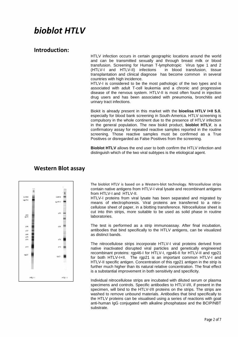
bioblot HTLV
Introduction: HTLV infection occurs in certain geographic locations around the world and can be transmitted sexually and through breast milk or blood transfusion. Screening for Human T-lymphotropic Virus type 1 and 2 (HTLV-I and HTLV-II) infections in blood transfusion, tissue transplantation and clinical diagnose has become common in several countries with high incidence. HTLV-I is considered to be the most pathologic of the two types and is associated with adult T-cell leukemia and a chronic and progressive disease of the nervous system. HTLV-II is most often found in injection drug users and has been associated with pneumonia, bronchitis and urinary tract infections. Biokit is already present in this market with the bioelisa HTLV I+II 5.0, especially for blood bank screening in South-America. HTLV screening is compulsory in the whole continent due to the presence of HTLV infection in the general population. The new biokit product, bioblot HTLV, is a confirmatory assay for repeated reactive samples reported in the routine screening. Those reactive samples must be confirmed as a True Positives or disregarded as False Positives from the screening. Bioblot HTLV allows the end user to both confirm the HTLV infection and distinguish which of the two viral subtypes is the etiological agent.
Western Blot assay
The bioblot HTLV is based on a Western-blot technology. Nitrocellulose strips contain native antigens from HTLV-I viral lysate and recombinant antigens from HTLV-I and HTLV-II. HTLV-I proteins from viral lysate has been separated and migrated by means of electrophoresis. Viral proteins are transferred to a nitro-cellulose sheet of paper, in a blotting transference. Nitrocellulose sheet is cut into thin strips, more suitable to be used as solid phase in routine laboratories. The test is performed as a strip immunoassay. After final incubation, antibodies that bind specifically to the HTLV antigens, can be visualized as distinct bands. The nitrocellulose strips incorporate HTLV-I viral proteins derived from native inactivated disrupted viral particles and genetically engineered recombinant proteins: rgp46-I for HTLV-I, rgp46-II for HTLV-II and rgp21 for both HTLV-I+II. The rgp21 is an important common HTLV-I and HTLV-II specific antigen. Concentration of this rgp21 antigen in the strip is further much higher than its natural relative concentration. The final effect is a substantial improvement in both sensitivity and specificity. Individual nitrocellulose strips are incubated with diluted serum or plasma specimens and controls. Specific antibodies to HTLV-I/II, if present in the specimen, will bind to the HTLV-I/II proteins on the strips. The strips are washed to remove unbound materials. Antibodies that bind specifically to the HTLV proteins can be visualised using a series of reactions with goat anti-human IgG conjugated with alkaline phosphatase and the BCIP/NBT substrate.

Page 3 of 7
This method has the sensitivity to detect marginal amounts of HTLV specific antibodies in serum or plasma.
Assay components: Bioblot HTLV includes all components to run the assay manually: Western-Blot Strips, Controls, Concentrated conjugate, Diluent, concentrated washing solution, blotting powder, ready to use substrate, forceps and a dedicated plastic tray. A rocking platform for incubations an aspiration device for washing are necessary but not provided. Bioblot HTLV has been validated to be performed in combination with the AUTOBLOT-20 instrument, an automated walk-away processor for Western Blot, also commercialized by Biokit. The protocol assay is common to the other two bioblot products: Bioblot HIV-1 Plus and Bioblot HCV. All three assay can be performed simultaneously in the same run.
Assay Protocol:
1.
Using forceps, carefully remove the required number of STRIPS from the tube and place numbered side up into each well. Include strips for Strong positive I, Strong positive II and Negative controls.
2. Add 2 ml of DILUTED WASHING SOLUTION to each well. 2 ml
3.
Incubate the strips for at least 5 minutes at room temperature (25 ± 3°C) on a rocking platform (speed of 12 to 16 oscillations per minute). Remove buffer by aspiration.
5 minutes
4. Add 2 ml of WORKING BLOTTING BUFFER to each well. 2 ml
5.
Add 20 μl each of patients’ sera or controls to appropriate wells. Care should be taken to ensure specimens are not added directly on the strips.
20 μl
6.
Cover the tray with the cover provided and incubate for 1 hour at room temperature (25 ± 3°C) on the rocking platform.
60 minutes
7.
Carefully uncover the tray to avoid splashing or mixing of samples. Tilt the tray to aspirate the mixture from the wells. Change aspirator tips between samples to avoid cross-contamination.
8.
Wash each strip 3 times with 2 ml of DILUTED WASHING SOLUTION allowing 5 minutes soak on the rocking platform between each wash.
3 x 2 ml
9. Add 2 ml of WORKING CONJUGATE SOLUTION to each well.
2 ml
10. Cover tray and incubate for 1 hour at room temperature (25 3°C) on the rocking platform.
60 minutes
11. Aspirate CONJUGATE from the wells. Wash as in step 8. 3 x 2 ml
12. Add 2 ml of SUBSTRATE SOLUTION to each well. 2 ml
13. Cover tray and incubate for 15 minutes on the rocking platform.
15 minutes

Page 4 of 7
Interpretation:
PROFILE INTERPRETATION
No detection of HTLV specific bands. NEGATIVE
Detection of GAG (p19 with or without p24), and two ENV (rgp46-I and rgp21) bands. HTLV-I POSITIVE
Detection of GAG (p24 with or without p19) and two ENV (rgp46-II and rgp21) bands. HTLV-II POSITIVE
Detection of GAG (p19 and p24) and ENV (rgp21).
HTLV-I SEROPOSITIVE indicated if p19 ≥ p24
HTLV-II SEROPOSITIVE indicated if p19 < p24
HTLV POSITIVE
HTLV specific bands present but pattern does not meet criteria for positive.
However, the following indeterminate banding may be interpreted as SERONEGATIVE:
- HTLV-I GAG indeterminate Western blot patterns (HGIP): Presence of p19, p26, p28, p32, p36, p53 but absence of p24 and any ENV proteins.
- Any combination of GAG proteins (p19, p26, p28, p32, p36, p53) but absence of p24 and any ENV proteins.
- Any single GAG proteins (p19, p24, p26, p28, p32 p36, p53).
INDETERMINATE
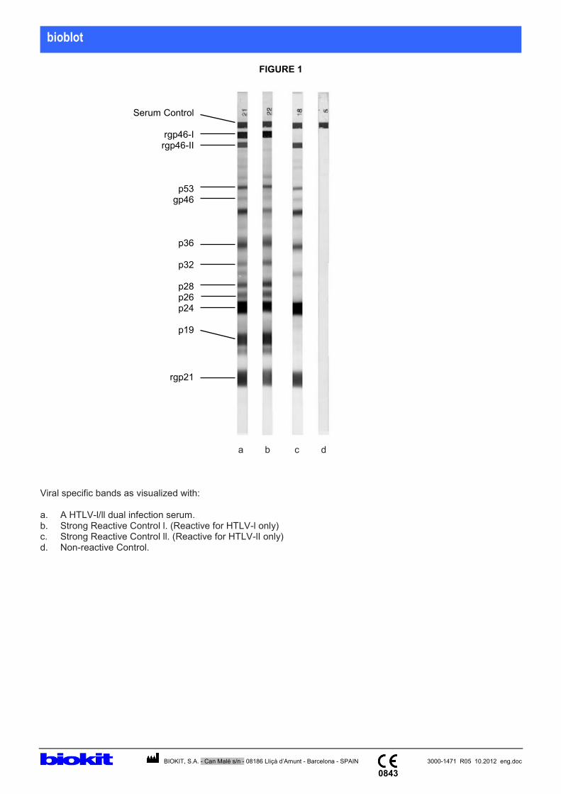
Example of Interpretation a. Dual HTLV I+II Infection b. Reactive for HTLV-l only c. Reactive for HTLV-II only d. Negative sample
Performance:
Sensitivity: Specimens which are established to be positive for HTLV-I or / and HTLV-II antibodies by commercial ELISAs were used to determine the sensitivity of bioblot HTLV. A. Comparison to Line Immunoassay 1 Blot results comparing bioblot HTLV and Line Immunoassay 1 (LI 1) for positive samples purchased from Boston Biomedica, Inc., USA (BBI), and ProMedDx were as follows:

Page 5 of 7
*bioblot HTLV gave 2 indeterminate and 1 negative result which was also detected negative by Line Immunoassay 1. Line immunoassay 1 gave 3 negative results. The two blots gave the following discriminations for the 102 HTLV positive samples:
**Both HTLV-I and HTLV-II specific markers appeared, indicating co-infection. ***Unable to type HTLV strains because of the absence of specific markers. Both bioblot HTLV and LI 1 gave similar results. The few discordant results are due to different antigens immobilised on the blots and the different methods used. bioblot HTLV showed a sensitivity of 97.1% which was equivalent to that obtained with Line Immunoassay 1. B. Comparison to Line Immunoassay 2 The French Society of Blood Transfusion Anti-HTLV-I/II Performance Panel, SFTS-94 consisting of 26 HTLV-I and 6 HTLV-II samples were studied. Results of bioblot HTLV on this panel were compared to Line Immunoassay 2 (LI 2) as follows:
bioblot HTLV correctly identifies HTLV positive samples, giving a sensitivity of >99.9% in this panel. Using the same panel, the comparative kit (LI 2) gave a sensitivity of 96.9%.
Specificity:
A total of 200 blood donor samples were tested resulting in a specificity of 92.5%. 15 samples were indeterminate and there were no false positive results. If 150 clinical specimens, 50 pregnancy specimens, 50 potentially interfering specimens (10 each of icteric, haemolysed, triglyceride, lipemic, total protein specimens), and 73 potentially cross-reactive specimens (TB, Helicobactor pylori, HEV, Dengue, HBV, HCV, HIV-1, HIV-2), are included, the overall specificity was 89.2% (461/517). 56 samples were indeterminate and there were no false positive results. 6 samples were true positives confirmed by another confirmatory test.
Competitors: There are very few competitors for HTLV confirmatory assays. The two most important are Innogenetics with the Innolia HTLV Score and Biorad with the New-Lav Blot HTLV. The Innolia HTLV Score is an immunoblot: Only HTLV Recombinant proteins are aligned in a Nylon plastic strip. WHO experts recommend the use of native antigens for confirmation as all the screening methods
Page 6 of 7
are also based on recombinant proteins. A confirmatory method should utilize “real” native antigens to determinate the reactivity of the patient antibodies to all the “real” possible viral antigens. Recombinant antibodies may be more specific that native antigens but they are not as sensitive. As an Innolia feature, the strips can be seen and interpreted easily because they have sharp borders and always in the same exact position on the strip. The New-Lav Blot HTLV assay is a Western Blot with only native antigens. However, there is only the HTLV-I viral antigen present and no additional recombinant antigens. The biorad assay is not able to distinguish between HTLV-I and HTLV-II infection. The presence of only native antigen may affect the specificity and make interpretation much more difficult . The bioblot HTLV is the only assay that has all the advantages: Western blot technology and the inclusion of recombinant proteins. Testing samples against real native antigens is the best way for confirmation of retrovirus like HIV and HTLV. The inclusion of specific recombinant antigens also helps in the differentiation of HTLV-I or HTLV-II infection. The high density of rgp21 in the strip provide an good level of both sensitivity and specificity.
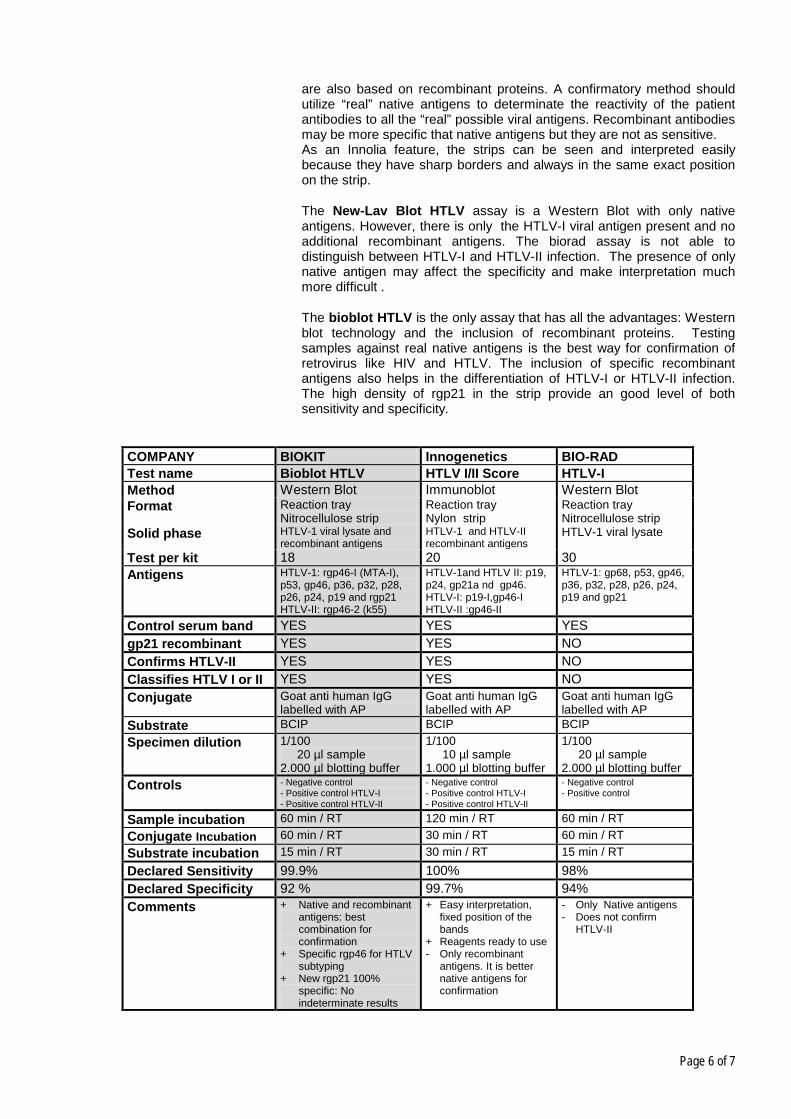
COMPANY BIOKIT Innogenetics BIO-RAD Test name Bioblot HTLV HTLV I/II Score HTLV-I Method Western Blot Immunoblot Western Blot Format Reaction tray
Nitrocellulose strip Reaction tray Nylon strip
Reaction tray Nitrocellulose strip
Solid phase HTLV-1 viral lysate and recombinant antigens
HTLV-1 and HTLV-II recombinant antigens
HTLV-1 viral lysate
Test per kit 18 20 30 Antigens HTLV-1: rgp46-I (MTA-I),
p53, gp46, p36, p32, p28, p26, p24, p19 and rgp21 HTLV-II: rgp46-2 (k55)
HTLV-1and HTLV II: p19, p24, gp21a nd gp46. HTLV-I: p19-I,gp46-I HTLV-II :gp46-II
HTLV-1: gp68, p53, gp46, p36, p32, p28, p26, p24, p19 and gp21
Control serum band YES YES YES gp21 recombinant YES YES NO Confirms HTLV-II YES YES NO Classifies HTLV I or II YES YES NO Conjugate Goat anti human IgG
labelled with AP Goat anti human IgG labelled with AP
Goat anti human IgG labelled with AP
Substrate BCIP BCIP BCIP Specimen dilution 1/100
20 µl sample 2.000 µl blotting buffer
1/100 10 µl sample 1.000 µl blotting buffer
1/100 20 µl sample 2.000 µl blotting buffer
Controls - Negative control - Positive control HTLV-I - Positive control HTLV-II
- Negative control - Positive control HTLV-I - Positive control HTLV-II
- Negative control - Positive control
Sample incubation 60 min / RT 120 min / RT 60 min / RT Conjugate Incubation 60 min / RT 30 min / RT 60 min / RT Substrate incubation 15 min / RT 30 min / RT 15 min / RT Declared Sensitivity 99.9% 100% 98% Declared Specificity 92 % 99.7% 94% Comments
+ Native and recombinant antigens: best combination for confirmation
+ Specific rgp46 for HTLV subtyping
+ New rgp21 100% specific: No indeterminate results
+ Easy interpretation, fixed position of the bands
+ Reagents ready to use - Only recombinant
antigens. It is better native antigens for confirmation
- Only Native antigens - Does not confirm
HTLV-II
Page 7 of 7
Conclusion:
Bioblot HTLV is a state of the art Western Blot assay for confirming repeated reactive HTLV screening samples. The Western-Blot strips include native HTLV-I antigens as well as specific recombinant antigens. The rgp46-I and rpg46-II are suitable for differentiation of HTLV-I and HTLV-II infections. The rgp21, a common antigen for both HTLV-I and HTLV-II, is present in the strip with higher density than in its natural relative concentration. The final rgp21 effect is an substantial improvement in both sensitivity and specificity. Bioblot HTLV combines the advantages of both Western blot and immunoblots assays:
- Use of necessary native antigens for confirmation versus screening methods that are all based on recombinant antigens.
- Use of recombinant antigens to differentiate HTLV-I from HTLV-II infections.
- Use of highly sensitive and specific recombinant rgp21 for improving the performance, the balance,sensitivity and specificity.
Bioblot HTLV has been validated to be performed in combination with the AUTOBLOT-20 instrument, an automated walk-away processor for Western Blot. The assay protocol is common to the other two bioblot products: Bioblot HIV-1 Plus and Bioblot HCV. All three assay can be performed simultaneously in the same run.
Annexes
- Package insert in English - Kit labels - CE Mark approval

bioblot
BIOKIT, S.A. - Can Malé s/n - 08186 Lliçà d’Amunt - Barcelona - SPAIN 3000-1471 R05 10.2012 eng.doc
0843
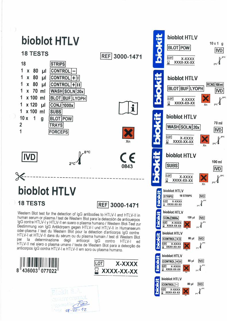
bioblot HTLV 3000-1471 18 tests
Qualitative enzyme immunoassay for the detection of antibodies to human T-Lymphotropic Virus type I (HTLV-I) and type II (HTLV-II) in human serum or plasma. It is intended for use as a more specific supplemental test on human serum or plasma specimens found repeatedly reactive using screening procedures such as ELISA.
Summary Recent epidemiology studies in the United States and Europe confirm the presence of a mixed prevalence of both HTLV-I and HTLV-II among different high risk populations such as intravenous drug users. Screening tests for HTLV-I/II are widely available. Repeatedly reactive specimens from screening tests require additional and more specific tests to confirm HTLV-I or HTLV-II seropositivity. Such supplemental tests must be capable of identifying antibodies to core (gag) and envelope (env) proteins of HTLV-I and HTLV-II. Western Blot strips incorporating HTLV-I native viral antigens is one such commonly used supplemental test. However, due to the lack of native envelope antigens on the classical HTLV-I Western Blot, it is often necessary to use radioimmunoprecipitation methods to further confirm for the presence of HTLV-I/II antibodies. Discrimination of HTLV-I and HTLV-II seropositives require supplemental assays (i.e. specific peptide, ELISAs, PCR). The bioblot HTLV has improved sensitivity and specificity for both the confirmation and differentiation of HTLV-I and HTLV-II seroreactivities. This is accomplished by incorporating MTA-1, a unique HTLV envelope recombinant protein (rgp46-I), combined with K55, a unique HTLV-II envelope recombinant protein (rgp46-II) and rgp21, a common yet specific HTLV-I and HTLV-II epitope recombinant envelope protein. Each strip also includes an internal sample addition control to minimise the risk of false negatives due to operational errors and to ensure the addition of samples. The bioblot HTLV is intended as a supplemental antibody assay for characterising samples found repeatedly reactive by screening methods. The possible serological profiles defined by the bioblot HTLV include the following: HTLV seropositive, HTLV-I seropositive, HTLV-II seropositive, seronegative and indeterminate. Principle The nitrocellulose strips are incorporated with HTLV-I viral proteins derived from native inactivated disrupted viral particles and genetically engineered proteins. Individual nitrocellulose strips are incubated with diluted serum or plasma specimens and controls. Specific antibodies to HTLV-I/II, if present in the specimen, will bind to the HTLV-I/II proteins on the strips. The strips are washed to remove unbound materials. Antibodies that bind specifically to the HTLV proteins can be visualised using a series of reactions with goat anti-human IgG conjugated with alkaline phosphatase and the substrate BCIP/NBT. This method has the sensitivity to detect marginal amounts of HTLV specific antibodies in serum or plasma. Components
1. STRIPS NITROCELLULOSE STRIPS:
18 strips of nitrocellulose incorporated with HTLV-I viral lysate, recombinant envelope antigens and a serum addition control (anti-human IgG) band. The strips are numbered consecutively from 1 to 18 or from 19 to 36. Keep dry and away from light.
2. CONJ 1000x CONCENTRATE CONJUGATE:
1 x 120 µl of goat IgG anti-human IgG antibodies conjugated with alkaline phosphatase. Contains sodium azide.
3. CONTROL – NEGATIVE CONTROL:
1 x 80 µl of inactivated human serum, negative for HTLV-I/II, HIV-1/2 and HCV antibodies and hepatitis B surface antigen (HBsAg). Contains sodium azide and thimerosal.
4. CONTROL + I STRONG POSITIVE CONTROL HTLV-I:
1 x 80 µl of inactivated human serum containing a high titre of HTLV-I IgG antibodies. Negative for hepatitis B surface antigen (HBsAg) and for antibodies to HIV-1/2 and HCV. Contains sodium azide and thimerosal.
5. CONTROL + II STRONG POSITIVE CONTROL HTLV-II:
1 x 80 µl of inactivated human serum containing a high titre of HTLV-II IgG antibodies. Negative for hepatitis B surface antigen (HBsAg) and for antibodies to HIV-1/2 and HCV. Contains sodium azide and thimerosal.
6. WASH SOLN 20x CONCENTRATE WASHING SOLUTION:
1 x 70 ml of concentrate Tris buffer (20x). Contains Tween 20 and thimerosal.
READ HIGHLIGHTED CHANGES
bioblot
BIOKIT, S.A. - Can Malé s/n - 08186 Lliçà d’Amunt - Barcelona - SPAIN 3000-1471 R05 10.2012 eng.doc
0843
7. BLOT BUF LYOPH LYOPHILISED BLOTTING BUFFER:
1 x 100 ml of lyophilised Tris buffer. To be reconstituted in reagent grade water. Contains heat inactivated animal and non-animal proteins and thimerosal.
8. SUBS SUBSTRATE:
1 x 100 ml of 5-bromo-4-chloro-3-indolyl-phosphate (BCIP) and nitroblue tetrazolium (NBT) solution. Ready to use.
9. BLOT POW BLOTTING POWDER:
10 x 1 g of powdered non-fat dry milk.
10. TRAYS INCUBATION TRAYS:
2 incubation trays of 9 wells.
11. FORCEPS FORCEPS:
1 pair. Precautions bioblot HTLV is intended for IN VITRO diagnostic use. For professional use only. Please refer to the product labelling for information on potentially hazardous components. HEALTH AND SAFETY INFORMATION CAUTION: This kit contains materials of human origin. No test method can offer complete assurance that human blood products will not transmit infection. HANDLE ASSAY SPECIMENS, STRONG POSITIVE I, STRONG POSITIVE II AND NEGATIVE CONTROLS AS POTENTIALLY INFECTIOUS AGENTS. It is recommended that the components and test specimens be handled using good laboratory working practices. They should be disposed of in accordance with established safety procedures. The Strong Positive Control I, Strong Positive Control II and Negative Control contain thimerosal and sodium azide while lyophilized Blotting Buffer and Concentrate Washing Solution contain thimerosal and Conjugate contains sodium azide. Sodium azide can react with copper and lead used in some plumbing systems to form explosive salts. The quantities used in this kit are small, nevertheless when disposing of azide-containing materials they should be flushed away with relatively large quantities of water to prevent metal azide build-up in plumbing system. The following are the appropriate risk (R) phrases.
R20/21/22 Harmful by inhalation, in contact with skin and if swallowed. The Substrate contains 5-bromo-4-chloro-3-indolyl phosphate and nitroblue tetrazolium which is classified pursuant to applicable European Economic Community (EEC) Directives as harmful (Xn). The following are the appropriate risk (R) phrases.
R20/21/22 Harmful by inhalation, in contact with skin and if swallowed. 1. Avoid microbial contamination of reagents when opening and removing aliquots from the original vials or
bottles. 2. Do not pipette by mouth. 3. Handle test specimens, nitrocellulose strips, Strong positive I, Strong positive II and Negative controls as
potentially infectious agents. 4. Wear laboratory coats and disposable gloves while performing the assay. Discard gloves in bio -hazard
waste-bags. Wash hands thoroughly afterwards. 5. It is highly recommended that this assay be performed in a biohazard cabinet. 6. Keep materials away from food and drink. 7. In case of accident or contact with eyes, rinse immediately with plenty of water and seek medical adv ice.
bioblot
BIOKIT, S.A. - Can Malé s/n - 08186 Lliçà d’Amunt - Barcelona - SPAIN 3000-1471 R05 10.2012 eng.doc
0843
8. Consult a physician immediately in the event that contaminated materials are ingested or come in contact
with open lacerations, or other breaks in the skin. 9. Wipe spills of potentially infectious materials immediately with absorbent paper and swab the
contaminated area with 1% sodium hypochlorite solution before work is resumed. Sodium hypochlorite should not be used on acid containing spills unless the area is wiped dry with absorbent paper first. Material used (including disposable gloves) should be disposed off as potentially biohazardous material. Do not autoclave material containing sodium hypochlorite.
10. Autoclave all used and contaminated materials at 121°C at 15 p.s.i. for 30 minutes before disposal.
Alternatively, decontaminate materials in 5% sodium hypochlorite solution for 30-60 minutes before disposal in biohazard waste-bags.
11. Decontaminate all used chemicals and reagents by adding sufficient volume of sodium hypochlorite to
make a final concentration of at least 1%. Leave for 30 minutes to ensure effective decontamination. 12. We do not recommend re-use of incubation trays. ANALYTICAL PRECAUTIONS 1. Optimal assay performance requires STRICT ADHERENCE to the assay procedure described in this
Instruction Manual. Deviations from the procedure may lead to aberrant results. 2. DO NOT MODIFY OR SUBSTITUTE REAGENTS FROM ONE KIT LOT TO ANOTHER. Controls, conjugate
and Western Blot strips are matched for optimal performance. Use only the reagents supplied with the kit. 3. Do not use kit components beyond the expiry date printed on the kit box. 4. Avoid microbial contamination of the reagents, when opening and removing aliquots from the original vials or
bottles. As this will prematurely reduce the shelf life of the kits and give erroneous results. Use aseptic techniques including pipettes or disposable pipette tips when drawing aliquots from vials.
5. The kit controls should be assayed concurrently with patients’ samples for each test run. 6. Use a new pipette tip for each specimen aliquot to prevent cross contamination.
7. For best results dispense all reagents while cold and return to 2°C to 8°C storage as soon as possible.
8. It is recommended that glassware to be used with the reagents should be washed with 2M hydrochloric
acid and rinsed thoroughly with distilled or deionised water prior to use. 9. Use only reagent grade quality, deionised or distilled water to dilute reagents. 10. All reagents must be mixed well before use. 11. Working Conjugate solution, Diluted Washing Solution and Blotting Buffer should be prepared fresh prior
to use. 12. The Working Conjugate solution should be prepared using a polypropylene container or beaker. 13. Do not expose reagents or perform test in an area containing a high level of chemical disinfectant fumes
(e.g. hypochlorite fumes) during storage or during incubation steps. Contact inhibits colour reaction. Also do not expose reagents to strong light.
14. The assay should preferably be performed at room temperature (25°C ± 3°C). 15. Make sure that the test strips are laid with the numbers on the strips facing upwards. 16. For Western Blot Assay, it is important to use a rocking platform shaker and not a rotary shaker.
Otherwise, performance of the kit will be compromised. The recommended speed and tilt angle of the shaker are 12 to 16 cycles per minute, and 5 to 10 degrees, respectively.

bioblot
BIOKIT, S.A. - Can Malé s/n - 08186 Lliçà d’Amunt - Barcelona - SPAIN 3000-1471 R05 10.2012 eng.doc
0843
17. Ensure that automated equipment if used is validated before use. 18. Ensure that the specimens are added away from the strip. Tray can be tilted and specimen added where the
buffer is collected at lower end. This prevents dark spot formation due to specimen addition on the strip. 19. Avoid the use of self-defrosting freezers for the storage of reagents and samples. Storage 1. Store bioblot HTLV kit and its components at 2-8°C when not in use. 2. All test reagents and strips when stored at 2°C to 8°C, are stable until the expiry date given on the kit. Do not
freeze reagents.
A. Antigen strips - Avoid unnecessary exposure of antigen strips to light. B. Reagents - Store reagents in their original vials or bottles, and they should be capped for storage. - Dispense all reagents while cold and return to 2°C to 8°C storage as soon as possible.
- Precipitates may form when the substrate is stored at 2°C to 8°C. This will not affect the performance of the kit.
CAUTION: Avoid unnecessary exposure of substrate to light. Collection of samples Serum or plasma samples collected in EDTA, heparin or sodium citrate may be used. Before storage, ensure that blood clot or blood cells have been separated by centrifugation. Samples should be stored at 2°C to 8°C if the test is to be run within 7 days of collection or frozen at -20°C or colder if the test is to be delayed for more than 7 days. Clear, non-haemolysed samples are preferred. Lipaemic, icteric or contaminated (particulate or bacterial) samples should be filtered (0.45 μm) or centrifuged before testing. Patients’ sera can be inactivated but this is not a requirement for optimal test performance. Inactivate as follows: 1. Loosen caps of serum containers. 2. Heat serum to 56°C for 30 minutes in a water bath. 3. Allow serum to cool before retightening caps. 4. Serum can be stored frozen until analysis. We recommend that the patients’ sera should not undergo repeated freeze-thaw cycles prior to testing. Material required not included in the kit - Deionised or distilled water. - Disposable gloves. - Rocking platform (designed with a rocking speed range of 12 to 16 oscillations per minute, and which moves
through a 5° to 10° tilt to wash strips evenly). - Pipettes and tips of appropriate volume. - Aspirator with sodium hypochlorite trap.
- 56°C water bath (optional).
- Sodium hypochlorite for decontamination. Previous operations 1. DILUTED WASHING SOLUTION
(a) DILUTED WASHING SOLUTION should be prepared fresh prior to use. (b) Dilute 1 volume of WASHING SOLUTION CONCENTRATE (20X) with 19 volumes of reagent grade
water. Mix well.
2. WORKING BLOTTING BUFFER (a) Reconstitute each bottle of LYOPHILIZED BLOTTING BUFFER with 100ml reagent grade water. Mix
well to dissolve. This RECONSTITUTED BLOTTING BUFFER is stable for 6 weeks if stored at 2-8°C. (b) The WORKING BLOTTING BUFFER should be prepared fresh prior to use.
bioblot
BIOKIT, S.A. - Can Malé s/n - 08186 Lliçà d’Amunt - Barcelona - SPAIN 3000-1471 R05 10.2012 eng.doc
0843
(c) Add 1 g of BLOTTING POWDER to every 20 ml of the reconstituted BLOTTING BUFFER prepared in
step 2(a) above. Stir to ensure powder dissolves completely. (d) Stir again before dispensing.
3. WORKING CONJUGATE SOLUTION NOTE: Prepare solution in polypropylene container / beaker. (a) WORKING CONJUGATE SOLUTION should be prepared fresh prior to use. (b) Prepare WORKING CONJUGATE SOLUTION by diluting CONJUGATE 1:1000 into WORKING
BLOTTING BUFFER, for example, 10 µl CONJUGATE to 10 ml WORKING BLOTTING BUFFER.
4. SUBSTRATE SOLUTION (ready to use) (a) Dispense directly the required volume from the bottle. Use a clean pipette. Cap tightly after use.
Assay procedure NOTE: a) Aspirate all used chemicals and reagents into a trap containing Sodium hypochlorite. b) All incubations are to be carried out on a rocking platform. CAUTION: Some samples cause dark patches on the spot of the strip where they are added. To avoid this problem, one should ensure the following: I. Sample should be added only after BLOTTING BUFFER is added. II. Tilt the tray slightly by elevating either the top or bottom end of the tray. The Blotting Buffer will flow to the
lower end of the tray. Add the sample where the Blotting Buffer is collected. When all the samples are added, return the tray back to its original flat position. Always ensure that the strips are kept wet during the process.
III. Alternatively, if tilting the tray is not desired, the samples may be added to the top or bottom end of the well.
This way if dark patches showed, the reading of the strip results will not be affected. Procedure: 1. Using forceps, carefully remove required number of STRIPS from the tube and place
numbered side up into each well. Include strips for Strong positive I, Strong positive II and Negative controls.
2. Add 2 ml of DILUTED WASHING SOLUTION to each well.
2 ml
3. Incubate the strips for at least 5 minutes at room temperature (25 3°C) on a rocking platform (speed of 12 to 16 oscillations per minute). Remove buffer by aspiration.
5 minutes
4. Add 2 ml of WORKING BLOTTING BUFFER to each well.
2 ml
5. Add 20 μl each of patients’ sera or controls to appropriate wells. Care should be taken to ensure specimens are not added directly on the strips.
20 μl
6. Cover the tray with the cover provided and incubate for 1 hour at room temperature (25 3°C) on the rocking platform.
60 minutes
7. Carefully uncover the tray to avoid splashing or mixing of samples. Tilt the tray to aspirate the mixture from the wells. Change aspirator tips between samples to avoid cross-contamination.
8. Wash each strip 3 times with 2 ml of DILUTED WASHING SOLUTION allowing 5 minutes soak on the rocking platform between each wash.
3 x 2 ml
9. Add 2 ml of WORKING CONJUGATE SOLUTION to each well.
2 ml
10. Cover tray and incubate for 1 hour at room temperature (25 3°C) on the rocking platform.
60 minutes
11. Aspirate CONJUGATE from the wells. Wash as in step 8.
3 x 2 ml
bioblot
BIOKIT, S.A. - Can Malé s/n - 08186 Lliçà d’Amunt - Barcelona - SPAIN 3000-1471 R05 10.2012 eng.doc
0843
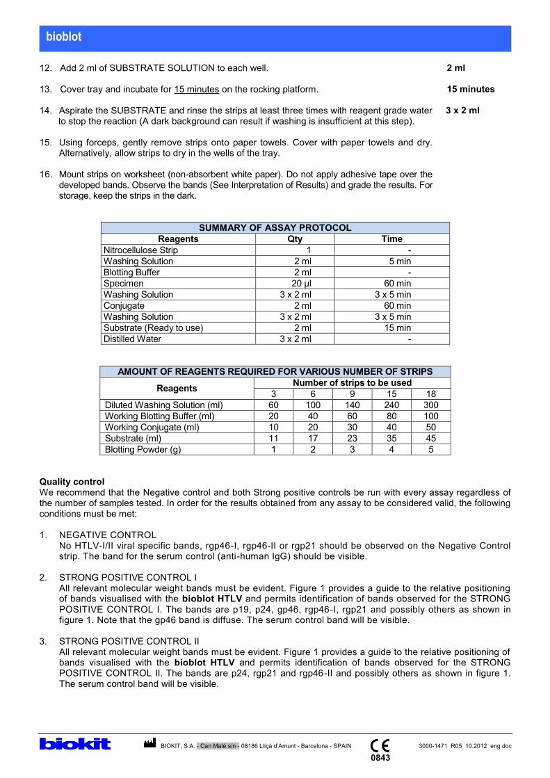
12. Add 2 ml of SUBSTRATE SOLUTION to each well.
2 ml
13. Cover tray and incubate for 15 minutes on the rocking platform.
15 minutes
14. Aspirate the SUBSTRATE and rinse the strips at least three times with reagent grade water to stop the reaction (A dark background can result if washing is insufficient at this step).
3 x 2 ml
15. Using forceps, gently remove strips onto paper towels. Cover with paper towels and dry.
Alternatively, allow strips to dry in the wells of the tray.
16. Mount strips on worksheet (non-absorbent white paper). Do not apply adhesive tape over the developed bands. Observe the bands (See Interpretation of Results) and grade the results. For storage, keep the strips in the dark.
SUMMARY OF ASSAY PROTOCOL
Reagents Qty Time
Nitrocellulose Strip 1 -
Washing Solution 2 ml 5 min
Blotting Buffer 2 ml -
Specimen 20 μl 60 min
Washing Solution 3 x 2 ml 3 x 5 min
Conjugate 2 ml 60 min
Washing Solution 3 x 2 ml 3 x 5 min
Substrate (Ready to use) 2 ml 15 min
Distilled Water 3 x 2 ml -
AMOUNT OF REAGENTS REQUIRED FOR VARIOUS NUMBER OF STRIPS
Reagents Number of strips to be used
3 6 9 15 18
Diluted Washing Solution (ml) 60 100 140 240 300
Working Blotting Buffer (ml) 20 40 60 80 100
Working Conjugate (ml) 10 20 30 40 50
Substrate (ml) 11 17 23 35 45
Blotting Powder (g) 1 2 3 4 5
Quality control We recommend that the Negative control and both Strong positive controls be run with every assay regardless of the number of samples tested. In order for the results obtained from any assay to be considered valid, the following conditions must be met: 1. NEGATIVE CONTROL No HTLV-I/II viral specific bands, rgp46-I, rgp46-II or rgp21 should be observed on the Negative Control
strip. The band for the serum control (anti-human IgG) should be visible. 2. STRONG POSITIVE CONTROL I All relevant molecular weight bands must be evident. Figure 1 provides a guide to the relative positioning
of bands visualised with the bioblot HTLV and permits identification of bands observed for the STRONG POSITIVE CONTROL I. The bands are p19, p24, gp46, rgp46-I, rgp21 and possibly others as shown in figure 1. Note that the gp46 band is diffuse. The serum control band will be visible.
3. STRONG POSITIVE CONTROL II All relevant molecular weight bands must be evident. Figure 1 provides a guide to the relative positioning of
bands visualised with the bioblot HTLV and permits identification of bands observed for the STRONG POSITIVE CONTROL II. The bands are p24, rgp21 and rgp46-II and possibly others as shown in figure 1. The serum control band will be visible.

bioblot
BIOKIT, S.A. - Can Malé s/n - 08186 Lliçà d’Amunt - Barcelona - SPAIN 3000-1471 R05 10.2012 eng.doc
0843
Interpretation of results NOTE: Developed strips must be completely dry to avoid misinterpretation. The presence or absence of antibodies to HTLV-I/II in the sample is determined by comparing each nitrocellulose strip to the assay control strips tested with the NEGATIVE and both STRONG POSITIVE controls. The Figure 1 (see last page of this brochure) is suggested as an aid to identify the various bands which develop on the strip reacted with the two STRONG POSITIVE controls. The interpretation process involves the following: 1. Validate that the serum control band is visible. If the control is negative, the results should be considered
invalid as this indicates a technical error such as not adding sample, conjugate or substrate. 2. Identify the molecular weight of each band of the test strip using the STRONG POSITIVE I control, the
STRONG POSITIVE II control strips or both as a guide. 3. Interpretation of the test strip is then based on the detection of specific band patterns as recommended by the
appropriate authorities (i.e. Health Ministry, World Health Organization, etc.). We recommend the following guidelines for the interpretation of the bioblot HTLV. Results should be recorded for each band detected and should be interpreted as NEGATIVE, HTLV-I POSITIVE, HTLV-II POSITIVE or INDETERMINATE.
PROFILE INTERPRETATION
No detection of HTLV specific bands. NEGATIVE
Detection of GAG (p19 with or without p24), and two ENV (rgp46-I and rgp21) bands.
HTLV-I POSITIVE
Detection of GAG (p24 with or without p19) and two ENV (rgp46-II and rgp21) bands.
HTLV-II POSITIVE
Detection of GAG (p19 and p24) and ENV (rgp21).
HTLV-I SEROPOSITIVE indicated if p19 p24 HTLV-II SEROPOSITIVE indicated if p19 < p24
HTLV POSITIVE
(1)
HTLV specific bands present but pattern does not meet criteria for positive. However, the following indeterminate banding may be interpreted as SERONEGATIVE: - HTLV-I GAG indeterminate Western blot patterns
(HGIP): Presence of p19, p26, p28, p32, p36, p53 but absence of p24 and any ENV proteins.
- Any combination of GAG proteins (p19, p26, p28, p32, p36, p53) but absence of p24 and any ENV proteins.
- Any single GAG proteins (p19, p24, p26, p28, p32 p36, p53).
INDETERMINATE(2)
(1)
Seropositive no-typeable HTLV samples can be further resolved by using Wiktor et al's algor ithm in absence of rgp46-I and rgp46-II. This algorithm, which uses the relative reactivity of p19 and p24 is shown to be effective in differentiating the two serotypes.
7,11,12,13,14
(2)Indeterminate interpretation is based on WHO's 1990, guideline.
15 However, various studies suggested that
certain indeterminate banding patterns (as listed) can be interpreted as seronegative especially with healthy blood donors.
16-25 For example, a study involving 37,724 healthy blood donors, confirmed that HGIP can be
safely interpreted as seronegative.26
However, caution should be exercised when indeterminate patterns are obtained with IVD users or blood donors from endemic areas, and neurological disease patients.
26,27

bioblot
BIOKIT, S.A. - Can Malé s/n - 08186 Lliçà d’Amunt - Barcelona - SPAIN 3000-1471 R05 10.2012 eng.doc
0843
Serum from individuals infected with both HTLV-I and HTLV-II although rare may occur. Serum from such individuals will have a banding pattern indicating HTLV-I and HTLV-II positive. Available data demonstrate that seroreactivity to rgp46-I is specific for HTLV-I and seroreactivity to rgp46-II is specific for HTLV-II. Therefore, sera reactive to rgp46-I, rgp46-II, rgp21 and p19 and p24 will provide a dual infection classification. Limitations of the method In order to obtain optimum results with this method, the steps described herein must be strictly followed. Any deviation may give rise to incorrect results. A NEGATIVE result does not exclude the possibility of exposure to or infection with HTLV-I or HTLV-II. INDETERMINATE blots should not be used as the basis for diagnosis of HTLV-I/II infection. It is reported that unspecific reactivity to both p19 and p24 has been observed in uninfected low risk populations although p24 indeterminates are relatively rare. The sensitivity of rgp46-I has been reported to be 95% in France, 100% of PCR confirmed samples in Jamaica and United States, and 98% of HTLV-I seropositive blood donors. The sensitivity of rgp46-II has been shown to be more than 98% among PCR confirmed samples from United States. The overall sensitivity of each type specific antigen, rpg46-I and rgp46-II, is estimated to be greater than 97%. The small percentage of HTLV-I and HTLV-II samples that are not reactive with either rpg46-I or rgp46-II are reactive with at least rgp21 and one or more GAG bands, p19 or p24, meeting the criteria of either HTLV Seropositive or indeterminate. There were no false negative interpretations reported. Supplemental tests such as PCR may be helpful in the discrimination of HTLV seropositives samples that can not be identified as either HTLV-I or HTLV-II with the bioblot HTLV. Performance characteristics The performance of bioblot HTLV for the detection of antibodies to HTLV-I and HTLV-II was evaluated using HTLV-I/II seropositive and seronegative specimens and was compared with two line immunoassays (immunoblots) which incorporate HTLV-I and HTLV-II antigens (recombinant proteins or peptides). Sensitivity: Specimens which are established to be positive for HTLV-I or / and HTLV-II antibodies by commercial ELISAs were used to determine the sensitivity of bioblot HTLV. A. Comparison to Line Immunoassay 1 Blot results comparing bioblot HTLV and Line Immunoassay 1 (LI 1) for positive samples purchased from Boston Biomedica, Inc., USA (BBI), and ProMedDx were as follows:
Method Line Immunoassay 1
Total NEG / IND POS
bioblot HTLV
NEG / IND 3* 0 3
POS 0 102 102
Total 3 102 105
*bioblot HTLV gave 2 indeterminate and 1 negative result which was also detected negative by Line Immunoassay 1. LIne immunoassay 1 gave 3 negative results. The two blots gave the following discriminations for the 102 HTLV positive samples:
Method
Interpretation
Total HTLV-I HTLV-II
HTLV-I & HTLV-II**
Non- typeable***
bioblot HTLV 45 53 4 0 102
Ll 1 48 51 0 3 102
**Both HTLV-I and HTLV-II specific markers appeared, indicating co-infection. ***Unable to type HTLV strains because of the absence of specific markers.

bioblot
BIOKIT, S.A. - Can Malé s/n - 08186 Lliçà d’Amunt - Barcelona - SPAIN 3000-1471 R05 10.2012 eng.doc
0843
Both bioblot HTLV and LI 1 gave similar results. The few discordant results are due to different antigens immobilised on the blots and the different methods used. bioblot HTLV showed a sensitivity of 97.1% which was equivalent to that obtained with Line Immunoassay 1. B. Comparison to Line Immunoassay 2 The French Society of Blood Transfusion Anti-HTLV-I/II Performance Panel, SFTS-94 consisting of 26 HTLV-I and 6 HTLV-II samples were studied. Results of bioblot HTLV on this panel were compared to Line Immunoassay 2 (LI 2) as follows:
Method Interpretation
Total HTLV-I HTLV-II Non-typeable False NEG
bioblot HTLV 26 6 0 0 32
Ll 2 21 6 4 1 32
bioblot HTLV correctly identifies HTLV positive samples, giving a sensitivity of >99.9% in this panel. Using the same panel, the comparative kit (LI 2) gave a sensitivity of 96.9%. Specificity: A total of 200 blood donor samples were tested resulting in a specificity of 92.5%. 15 samples were indeterminate and there were no false positive results. If 150 clinical specimens, 50 pregnancy specimens, 50 potentially interfering specimens (10 each of icteric, haemolysed, triglyceride, lipemic, total protein specimens), and 73 potentially cross-reactive specimens (TB, Helicobactor pylori, HEV, Dengue, HBV, HCV, HIV-1, HIV-2), are included, the overall specificity was 89.2% (461/517). 56 samples were indeterminate and there were no false positive results. 6 samples were true positives confirmed by another confirmatory test. Limited expressed warranty disclaimer The manufacturer makes no expressed warranty other than that the test kit will function as an in-vitro diagnostic assay within the specifications and limitations described in the Product Instruction Manual when used in accordance with the instructions contained therein. The manufacturer disclaims any warranty, expressed or implied, including such expressed or implied warranty with respect to merchantability, fitness for use or implied utility for any purpose. The manufacturer is limited to either replacement of the product or refund of the purchase price of the product. The manufacturer shall not be liable to the purchaser or third parties for any damage, injury or economic loss howsoever caused by the product in the use or in the application thereof. Technical problems Should there be a technical problem / complaint, please do the following: 1. Note the kit lot number, the expiry date and the strip lot number. 2. Retain the kits and the results that were obtained. 3. Contact your local distributor.

bioblot
BIOKIT, S.A. - Can Malé s/n - 08186 Lliçà d’Amunt - Barcelona - SPAIN 3000-1471 R05 10.2012 eng.doc
0843
bioblot: Troubleshooting guide
Problem Possible causes Problem Possible causes 1. Dark spots develop on strips
1a. Bacterial or fungal contamination of test sample.
4. Strong Background develops on strip in the absence or presence of positive bands.
4a. Overdeveloped strips (stop reaction sooner).
1b. Precipitation of immune complexes in aged test sample.
4b. Incomplete washing.
1c. Bacterial or fungal contamination on strip due to improper storage.
5. Absence of Serum Control Band
5a. Serum not added.
1d. Strips physically damaged, cracked or scratched.
5b. Strips flipped over during assay.
1e. Strips not properly washed between assay steps.
5c. Conjugate not added.
2. White patches develop on strips
2a. Strips flipped over during assay.
5d. Substrate not added.
2b. Trays not properly washed before use.
6. Strips are defective
6a. They are cracked.
2c. Poor dissolution of Blotting Powder.
6b. They contain air bubbles which cause the appearance of white spots in reactive zones big enough to prevent any detection.
2d. Electrotransblot interference during manufacturing
6c. They show dark spots due to fungal growth upon initial opening of the strip tubes. However, if dark spots develop sometime later after initial opening of the tube then the problem is due to improper strip storage conditions at the user’s site.
3. Bands other than the Serum Control band develops on negative control
3a. Tray wells or Control may have been crossed contaminated.

bioblot
BIOKIT, S.A. - Can Malé s/n - 08186 Lliçà d’Amunt - Barcelona - SPAIN 3000-1471 R05 10.2012 eng.doc
0843
bioblot: Troubleshooting guide
Problem Possible causes Problem Possible causes 7.1. Expected bands do
not develop or are of weak intensity.
7.1a. Reagents not properly prepared.
8. Non-specific bands and/or dark background develop on strips
8a. Wrong test sample or conjugate dilution.
And positive control weak
7.1b.Wrong conjugate dilution.
8b. Test sample/reagent incubation too long.
The problem is probably caused by the reagents.
7.1c. Unstable reagents due to improper temperature exposure.
8c. Incomplete washing during assay.
7.1d. Conjugate contaminated with human IgG.
8d. Incubation temperature greater than 30°C.
7.1e. Incorrect substrate pH due to exposure to strong UV light or reducing agent.
8e. Test sample reactive with non-viral proteins.
7.1f. Trays, reagent(s) or water having high phosphate concentration.
9. HTLV-specific bands detected on negative control and/or known negative samples
9a. Trays contaminated with positive test sample or positive control.
7.1g. Rotary platform used instead of Rocking platform.
9b. Negative control/test sample contaminated with positive control/ test sample.
7.2 Expected bands do
not develop or are of weak intensity.
And positive control
OK The problem is
probably caused by test sample.
7.2a. Wrong test sample dilution.
9c. Same pipette tip used for delivery and/or removal of, test samples/control.
7.2b. Test sample
contaminated with conjugate.
9d. Test sample may be an ELISA “false” negative.
7.2c. Test samples
severely immune-complexed.
10. Watery marks on developed strips
10a.Strips left to dry after pre-soaking step prior to adding Blotting Buffer.
7.2d. Test sample IgG
deteriorated or denatured due to repeated freeze- thaw or improper storage.
7.2e. Rotary platform used instead of Rocking platform.
7.2f.Test sample may be an ELISA “false” positive.
bioblot
BIOKIT, S.A. - Can Malé s/n - 08186 Lliçà d’Amunt - Barcelona - SPAIN 3000-1471 R05 10.2012 eng.doc
0843
FIGURE 1
Serum Control rgp46-I rgp46-II p53 gp46 p36 p32 p28 p26 p24 p19
rgp21
a b c d
Viral specific bands as visualized with: a. A HTLV-l/ll dual infection serum. b. Strong Reactive Control l. (Reactive for HTLV-l only) c. Strong Reactive Control ll. (Reactive for HTLV-lI only) d. Non-reactive Control.

Biokit SA Can Malé, 08186 Lliçà d’Amunt Barcelona, Spain
EC Design - Examination Certificate Annex IV, section 4 of Council Directive 98/79/EC on In Vitro Diagnostic Medical Devices
Device Descriptions: Bioblot HTLV Test Kit (18T)
Model: 3000-1471
File Number A12230 Cycle Start Date 20 November 2012
Certificate No. 683.121120 Effective Date 20 November 2012
Expiry Date 03 March 2014
Authorised by
Ivor Barrett
Certification Manager For and on Behalf of UL International (UK) Ltd
We hereby declare that a design examination has been carried out on the device(s) listed per report 12CA48048, following the requirements of the national legislation to which the undersigned is subject, transposing Annex IV section 4 of Council Directive 98/79/EC on In Vitro Diagnostic Medical Devices. We certify that the design of the device(s) listed conforms with the relevant provisions of Annex IV section 4 of the aforementioned directive as transposed into national legislation. This certificate is issued with 0 attachments listing model numbers.
Notified Body
0843
OBL 00-NB-F0410 1.0

Biokit SA Can Malé, 08186 Lliçà d’Amunt Barcelona, Spain
EC Certificate - Full Quality Assurance System Approval Certificate Annex IV, section 3 of Council Directive 98/79/EC on In Vitro Diagnostic Medical Devices
Scope of Certificate: Supply of Annex II List A IVD Devices
Device Classification: Annex II List A
Device Descriptions: Please refer to Attachment 1
Model: Please refer to Attachment 1
File Number A12230 Cycle Start Date 10 June 2009
Certificate No. 522.121120 Effective Date 20 November 2012
Expiry Date 20 November 2013
Authorised by
Ivor Barrett
Certification Manager For and on Behalf of UL International (UK) Ltd
We hereby declare that an examination of the full quality assurance system has been carried out per report 12CA48048, following the requirements of the national legislation to which the undersigned is subject, transposing Annex IV (with the exemption of sections 4 and 6) of Council Directive 98/79/EC on In Vitro Diagnostic Medical Devices. We certify that the full quality assurance system conforms with the relevant provisions of the aforementioned directive and is subject to periodic surveillance as required by 98/79/EC, Annex IV, Section 5. For Annex II, List A devices where they are covered by this certificate, an EC Design Examination certificate according to 98/79/EC, Annex IV, Section 4 is required. This certificate is issued with 1 attachment listing model numbers and 0 addendums listing additional locations covered by this certificate.
Notified Body
0843
OBL 00-NB-F0409 1.0
Biokit SA Can Malé, 08186 Lliçà d’Amunt Barcelona, Spain
Attachment 1 of 1
The products detailed below are covered under the scope of this certificate
Device Description Model Classification
Bioblot HIV-1 Plus Test Kit (18T) 3000-1467 Annex II List A
Bioblot HTLV Test Kit (18T) 3000-1471 Annex II List A
Bioblot HCV Test Kit (18T) 3000-1474 Annex II List A
Bioelisa HTLV-I+II 5.0 (192T) 3000-1165 Annex II List A
File Number A12230 Cycle Start Date 10 June 2009
Certificate No. 522.121120 Effective Date 20 November 2012
Expiry Date 20 November 2013
Authorised by
Ivor Barrett
Certification Manager For and on Behalf of UL International (UK) Ltd
Notified Body
0843
OBL 00-NB-F0409 1.0