bik1 interacts with peprs to mediate ethylene-induced immunity
TRANSCRIPT
BIK1 interacts with PEPRs to mediateethylene-induced immunityZixu Liua,b, Ying Wub, Fan Yangc, Yiyue Zhangb, She Chenc, Qi Xieb, Xingjun Tiana,1, and Jian-Min Zhoub,1
aSchool of Life Science, Nanjing University, Nanjing 210093, China; bState Key Laboratory of Plant Genomics and National Center for Plant Gene Research,Institute of Genetics and Developmental Biology, Chinese Academy of Sciences, Beijing 100101, China; and cNational Institute of Biological Sciences, Beijing102206, China
*Edited by Paul Schulze-Lefert, Max Planck Institute for Plant Breeding Research, Cologne, Germany, and approved January 23, 2013 (received for reviewSeptember 6, 2012)
Plants have evolved intricate immune mechanisms to combatpathogen infection. Upon perception of pathogen-derived signals,plants accumulate defense hormones such as ethylene (ET), jasmo-nate, salicylate, and damage-associated molecular patterns toamplify immune responses. In particular, the Arabidopsis peptidePep1 and its family members are thought to be damage-associatedmolecular patterns that trigger immunity through Pep1 receptorkinases PEPR1 and PEPR2. Here we show that PEPR1 specificallyinteracts with receptor-like cytoplasmic kinases botrytis-inducedkinase 1 (BIK1) and PBS1-like 1 (PBL1) to mediate Pep1-induceddefenses. In vitro and in vivo studies suggested that PEPR1, andlikely PEPR2, directly phosphorylates BIK1 in response to Pep1 treat-ment. Surprisingly, the pepr1/pepr2 double-mutant seedlings dis-played reduced in sensitivity to ET, as indicated by the elongatedhypocotyls. ET-induced expression of defense genes and resistanceto Botrytis cinerea were compromised in pepr1/pepr2 and bik1mutants, reenforcing an important role of PEPRs and BIK1 in ET-mediated defense signaling. Pep treatment partially mimicked ET-induced seedling growth inhibition in a PEPR- and BIK1-dependentmanner. Furthermore, both ET and Pep1 treatments induced BIK1phosphorylation in a PEPR-dependent manner. However, the Pep1-induced BIK1 phosphorylation, seedling growth inhibition, anddefense gene expressionwere independent of canonical ET signal-ing components. Together our results illustrate a mechanism bywhich ET and PEPR signaling pathways act in concert to amplifyimmune responses.
PAMPs | DAMPs | disease resistance | plant immunity | phytohormone
Plants have evolved a sophisticated innate immune system tocope with attacks by diverse pathogenic microbes. At the
core are cytoplasmic immune receptors and cell-surface immunereceptors detecting various danger signals during pathogen in-fection. Plant cell surface-localized immune receptors, also calledpattern-recognition receptors (PRRs), consist of a variety of re-ceptor-like kinases and receptor-like proteins. Many PRRs, in-cluding FLS2, EFR, Xa21, CERK1, CEBiP, LYM1, LYM3, LYP4,and LYP6 (1–8), directly sense pathogen/microbe-associatedmolecular patterns (PAMPs), such as flagellin, elongation factor,quorum-sensing protein, or peptidoglycans from bacteria orchitin from fungal cell wall. In addition to PAMPs, PRRs alsoperceive endogenous damage-associated molecular patterns(DAMPs), such as plant cell wall fragments released by patho-gen lytic enzymes or plant peptides synthesized de novo duringpathogen infection. The Arabidopsis Pep1, a 23-aa peptide pro-cessed from PROPEP1 (9, 10), is thought to be a DAMP per-ceived by two closely related LRR receptor kinases, PEPR1 andPEPR2, to trigger immune responses (11–13). Members of thePROPEP family are transcriptionally induced by defense hor-mones jasmonates (JA), ethylene (ET), and salicylate (SA), orwounding, and this is thought to amplify danger signals duringpathogen infection. Pep1 seems to be conserved in both dicotsand monocots, because ZmPep1 has also been shown to regulatedefense gene expression in maize (14).PRRs interact with other components in a highly dynamic
manner. The ligand-binding to FLS2 and EFR is known to recruit
BAK1, a receptor-like kinase, forming active receptor complexes(15–18). Downstream, the receptor-like cytoplasmic kinase BIK1and its closely related protein PBL1 interact directly with FLS2,EFR, and CERK1. The activation of these PRRs results in a rapidphosphorylation of BIK1 and PBL1, which then dissociate fromthe receptors to activate downstream signaling (19, 20).In addition to immune receptors, the delicate control of
plant innate immunity also involves plant hormones, amongwhich SA, ET, and JA play key roles in regulating defenseresponses (21). In particular, increasing evidence indicates thatET is intimately associated with PTI signaling pathways. Forexample, the activation of MPK6 by flg22 stabilizes 1-amino-cyclopropane-1-carboxylate (ACC) synthases ACS2 and ACS6,which are rate-limiting enzymes for ET biosynthesis (22). ETexerts its regulation on defense responses through EIN3 andEIL1, two closely related transcription factors (23, 24). Forexample, FLS2 transcription is positively regulated by EIN3and EIL1 (25, 26). EIN3/EIL1 also negatively regulate SA-dependent immunity by binding to the promoter of SID2,which controls SA biosynthesis (27). Thus, a linear intra-cellular signaling pathway from ER-localized ET receptors tothe nuclear-localized transcription factors is thought to directlyexecute defense gene regulation.Interestingly, a recent report shows that etiolated bik1 seed-
lings are partially insensitive to ET and display elongated hy-pocotyl (28). Furthermore, BIK1 is phosphorylated upon ETtreatment. However, the molecular mechanism by which BIK1regulates ET signaling remains unknown. Here we show thatBIK1 directly interacts with and is phosphorylated by PEPR1.Pep1-induced defenses were diminished in the bik1 mutantplants. Like bik1, the pepr1/pepr2 double mutant was partiallyinsensitive to seedling growth inhibition by ET. Pep peptides canmimic ET-induced seedling growth inhibition in a PEPR- andBIK1-dependent but EIN3/EIL1- and/or EIN2-independentmanner. PEPR1, and likely PEPR2, are required for ET- andPep1-induced phosphorylation of BIK1. Furthermore, the ET-induced expression of several defense genes and resistance toBotrytis cinerea were compromised in pepr1/pepr2 and bik1. Ourfindings indicate that Pep peptides, PEPRs, and BIK1 form anextension of the canonical ET signaling pathway to regulateplant immunity.
ResultsBIK1 Interacts with PEPR1 in Vitro and in Vivo. To understand mech-anisms by which BIK1 regulates ET signaling and plant immunity,
Author contributions: Z.L., X.T., and J.-M.Z. designed research; Z.L., F.Y., and Y.Z. per-formed research; Z.L., Y.W., S.C., and J.-M.Z. analyzed data; and Z.L., Q.X., X.T., and J.-M.Z.wrote the paper.
The authors declare no conflict of interest.
*This Direct Submission article had a prearranged editor.
See Commentary on page 5748.1To whom correspondence may be addressed. E-mail: [email protected] or [email protected].
This article contains supporting information online at www.pnas.org/lookup/suppl/doi:10.1073/pnas.1215543110/-/DCSupplemental.
www.pnas.org/cgi/doi/10.1073/pnas.1215543110 PNAS | April 9, 2013 | vol. 110 | no. 15 | 6205–6210
PLANTBIOLO
GY
SEECO
MMEN
TARY
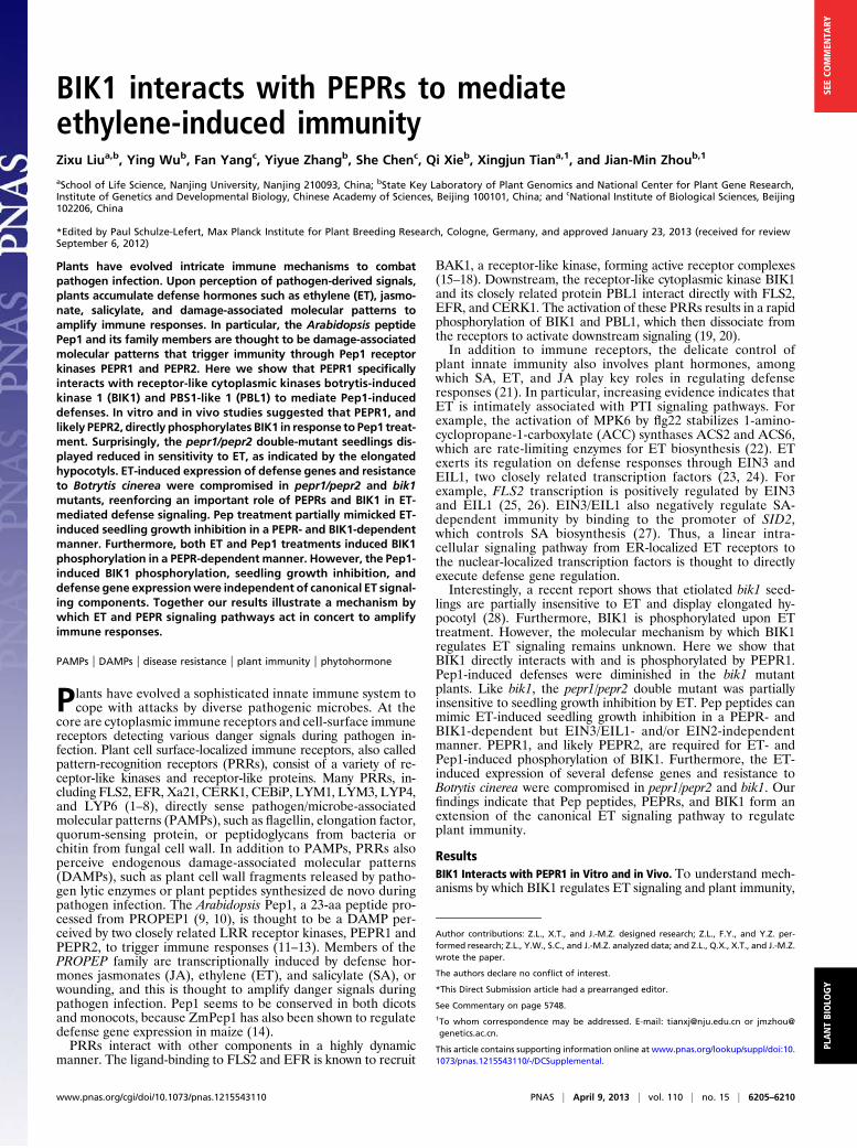
we performed a yeast two-hybrid screen by using BIK1 as bait.Two independent clones containing amino acids 1022–1123 and1025–1123 of the C terminus of PEPR1 kinase domain (KD)showed a strong interaction with BIK1 (Fig. 1A). GST pull-downassay showed that a GST-tagged BIK1 also interacted witha His-tagged PEPR1 KD (amino acids 827–1123) in vitro (Fig.1B). The BIK1S236A mutant carrying an amino acid substitutionat an autophosphorylation site (20) also interacted with thePEPR1 KD, suggesting that this interaction is independent of thephosphorylation on S236 (Fig. 1B). Coimmunoprecipitation assayindicated that the full-length PEPR1-FLAG, but not BAK1-FLAG,specifically interacted with BIK1-HA in Arabidopsis protoplasts(Fig. 1C). The lack of BIK1–BAK1 interaction is consistent withour previous report (20) but contradicts a report by Lu et al.(19). Together these data indicated that BIK1 can interact withPEPR1 KD.
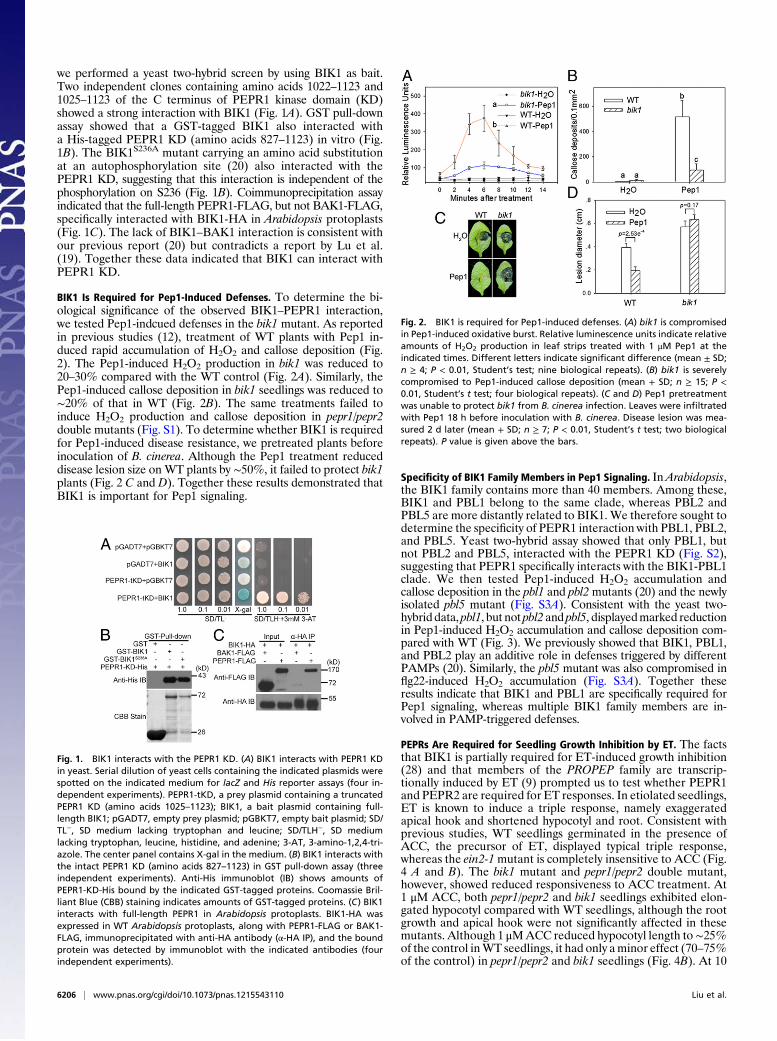
BIK1 Is Required for Pep1-Induced Defenses. To determine the bi-ological significance of the observed BIK1–PEPR1 interaction,we tested Pep1-indcued defenses in the bik1 mutant. As reportedin previous studies (12), treatment of WT plants with Pep1 in-duced rapid accumulation of H2O2 and callose deposition (Fig.2). The Pep1-induced H2O2 production in bik1 was reduced to20–30% compared with the WT control (Fig. 2A). Similarly, thePep1-induced callose deposition in bik1 seedlings was reduced to∼20% of that in WT (Fig. 2B). The same treatments failed toinduce H2O2 production and callose deposition in pepr1/pepr2double mutants (Fig. S1). To determine whether BIK1 is requiredfor Pep1-induced disease resistance, we pretreated plants beforeinoculation of B. cinerea. Although the Pep1 treatment reduceddisease lesion size on WT plants by ∼50%, it failed to protect bik1plants (Fig. 2 C and D). Together these results demonstrated thatBIK1 is important for Pep1 signaling.
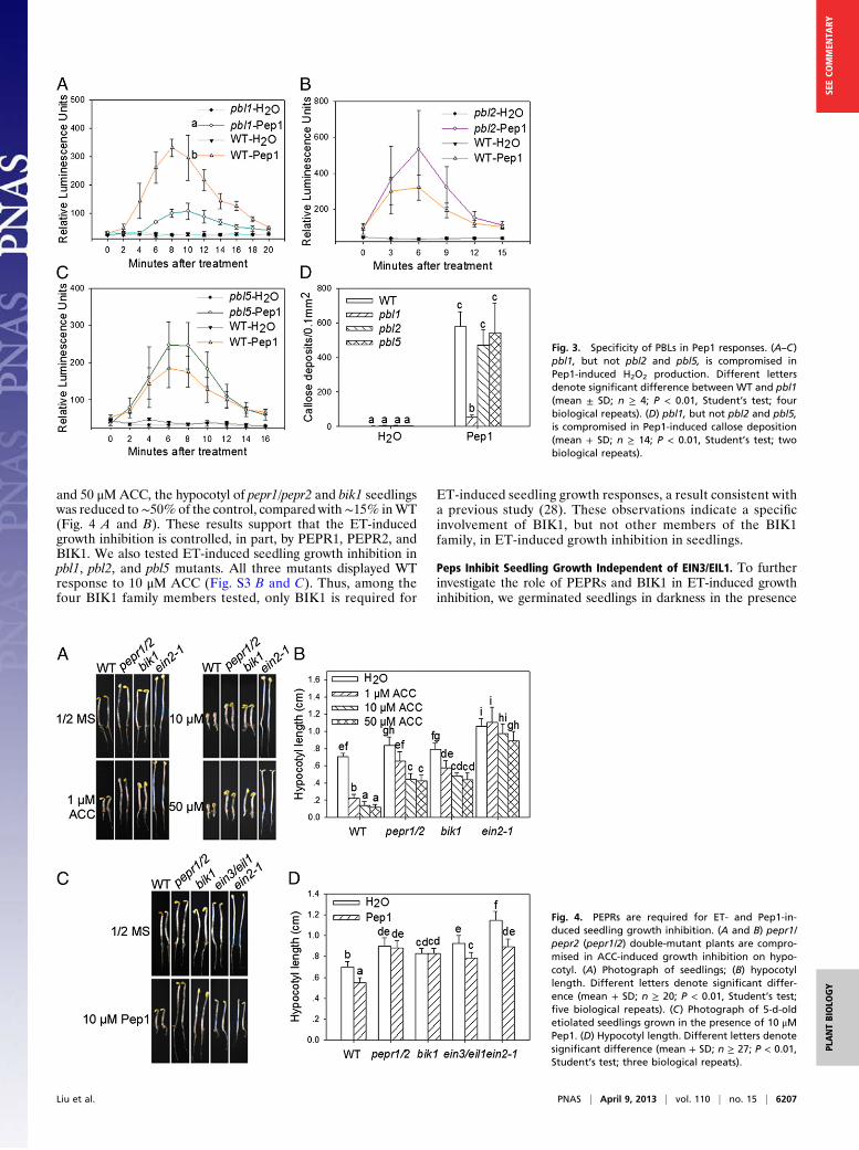
Specificity of BIK1 Family Members in Pep1 Signaling. InArabidopsis,the BIK1 family contains more than 40 members. Among these,BIK1 and PBL1 belong to the same clade, whereas PBL2 andPBL5 are more distantly related to BIK1. We therefore sought todetermine the specificity of PEPR1 interaction with PBL1, PBL2,and PBL5. Yeast two-hybrid assay showed that only PBL1, butnot PBL2 and PBL5, interacted with the PEPR1 KD (Fig. S2),suggesting that PEPR1 specifically interacts with the BIK1-PBL1clade. We then tested Pep1-induced H2O2 accumulation andcallose deposition in the pbl1 and pbl2mutants (20) and the newlyisolated pbl5 mutant (Fig. S3A). Consistent with the yeast two-hybrid data, pbl1, but not pbl2 and pbl5, displayedmarked reductionin Pep1-induced H2O2 accumulation and callose deposition com-pared with WT (Fig. 3). We previously showed that BIK1, PBL1,and PBL2 play an additive role in defenses triggered by differentPAMPs (20). Similarly, the pbl5 mutant was also compromised inflg22-induced H2O2 accumulation (Fig. S3A). Together theseresults indicate that BIK1 and PBL1 are specifically required forPep1 signaling, whereas multiple BIK1 family members are in-volved in PAMP-triggered defenses.
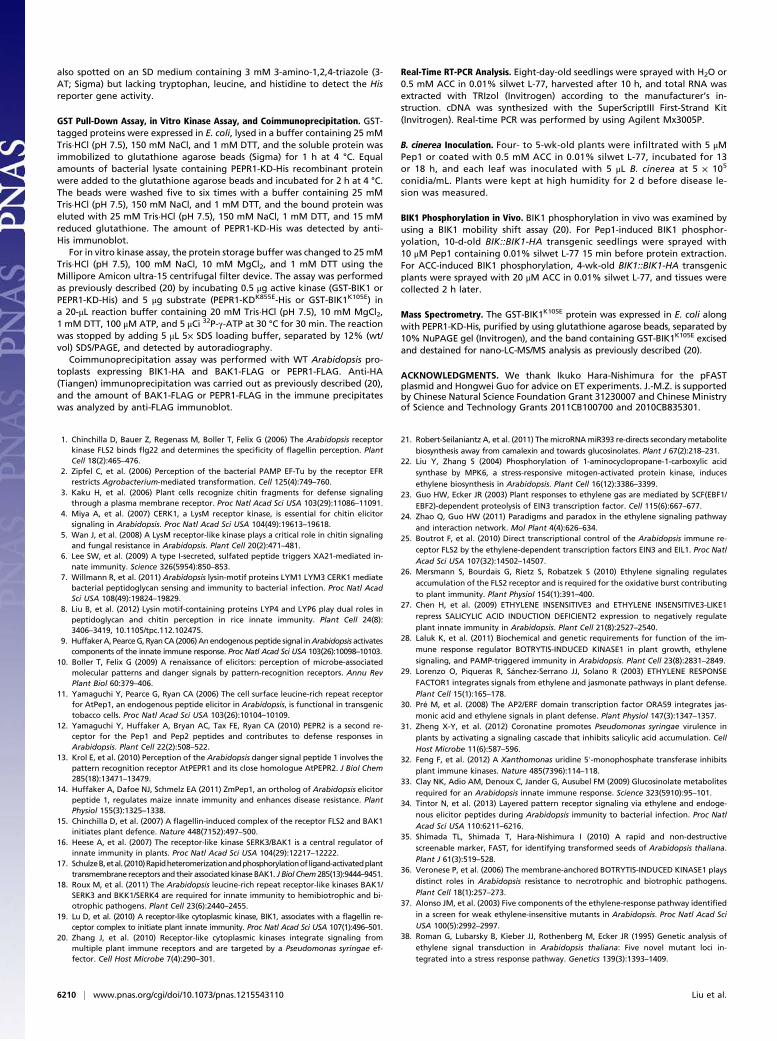
PEPRs Are Required for Seedling Growth Inhibition by ET. The factsthat BIK1 is partially required for ET-induced growth inhibition(28) and that members of the PROPEP family are transcrip-tionally induced by ET (9) prompted us to test whether PEPR1and PEPR2 are required for ET responses. In etiolated seedlings,ET is known to induce a triple response, namely exaggeratedapical hook and shortened hypocotyl and root. Consistent withprevious studies, WT seedlings germinated in the presence ofACC, the precursor of ET, displayed typical triple response,whereas the ein2-1mutant is completely insensitive to ACC (Fig.4 A and B). The bik1 mutant and pepr1/pepr2 double mutant,however, showed reduced responsiveness to ACC treatment. At1 μM ACC, both pepr1/pepr2 and bik1 seedlings exhibited elon-gated hypocotyl compared with WT seedlings, although the rootgrowth and apical hook were not significantly affected in thesemutants. Although 1 μMACC reduced hypocotyl length to∼25%of the control inWT seedlings, it had only aminor effect (70–75%of the control) in pepr1/pepr2 and bik1 seedlings (Fig. 4B). At 10
Fig. 1. BIK1 interacts with the PEPR1 KD. (A) BIK1 interacts with PEPR1 KDin yeast. Serial dilution of yeast cells containing the indicated plasmids werespotted on the indicated medium for lacZ and His reporter assays (four in-dependent experiments). PEPR1-tKD, a prey plasmid containing a truncatedPEPR1 KD (amino acids 1025–1123); BIK1, a bait plasmid containing full-length BIK1; pGADT7, empty prey plasmid; pGBKT7, empty bait plasmid; SD/TL−, SD medium lacking tryptophan and leucine; SD/TLH−, SD mediumlacking tryptophan, leucine, histidine, and adenine; 3-AT, 3-amino-1,2,4-tri-azole. The center panel contains X-gal in the medium. (B) BIK1 interacts withthe intact PEPR1 KD (amino acids 827–1123) in GST pull-down assay (threeindependent experiments). Anti-His immunoblot (IB) shows amounts ofPEPR1-KD-His bound by the indicated GST-tagged proteins. Coomassie Bril-liant Blue (CBB) staining indicates amounts of GST-tagged proteins. (C) BIK1interacts with full-length PEPR1 in Arabidopsis protoplasts. BIK1-HA wasexpressed in WT Arabidopsis protoplasts, along with PEPR1-FLAG or BAK1-FLAG, immunoprecipitated with anti-HA antibody (α-HA IP), and the boundprotein was detected by immunoblot with the indicated antibodies (fourindependent experiments).
Fig. 2. BIK1 is required for Pep1-induced defenses. (A) bik1 is compromisedin Pep1-induced oxidative burst. Relative luminescence units indicate relativeamounts of H2O2 production in leaf strips treated with 1 μM Pep1 at theindicated times. Different letters indicate significant difference (mean ± SD;n ≥ 4; P < 0.01, Student’s test; nine biological repeats). (B) bik1 is severelycompromised to Pep1-induced callose deposition (mean + SD; n ≥ 15; P <0.01, Student’s t test; four biological repeats). (C and D) Pep1 pretreatmentwas unable to protect bik1 from B. cinerea infection. Leaves were infiltratedwith Pep1 18 h before inoculation with B. cinerea. Disease lesion was mea-sured 2 d later (mean + SD; n ≥ 7; P < 0.01, Student’s t test; two biologicalrepeats). P value is given above the bars.
6206 | www.pnas.org/cgi/doi/10.1073/pnas.1215543110 Liu et al.
and 50 μMACC, the hypocotyl of pepr1/pepr2 and bik1 seedlingswas reduced to∼50%of the control, compared with∼15% inWT(Fig. 4 A and B). These results support that the ET-inducedgrowth inhibition is controlled, in part, by PEPR1, PEPR2, andBIK1. We also tested ET-induced seedling growth inhibition inpbl1, pbl2, and pbl5 mutants. All three mutants displayed WTresponse to 10 μM ACC (Fig. S3 B and C). Thus, among thefour BIK1 family members tested, only BIK1 is required for
ET-induced seedling growth responses, a result consistent witha previous study (28). These observations indicate a specificinvolvement of BIK1, but not other members of the BIK1family, in ET-induced growth inhibition in seedlings.
Peps Inhibit Seedling Growth Independent of EIN3/EIL1. To furtherinvestigate the role of PEPRs and BIK1 in ET-induced growthinhibition, we germinated seedlings in darkness in the presence
Fig. 3. Specificity of PBLs in Pep1 responses. (A–C)pbl1, but not pbl2 and pbl5, is compromised inPep1-induced H2O2 production. Different lettersdenote significant difference between WT and pbl1(mean ± SD; n ≥ 4; P < 0.01, Student’s test; fourbiological repeats). (D) pbl1, but not pbl2 and pbl5,is compromised in Pep1-induced callose deposition(mean + SD; n ≥ 14; P < 0.01, Student’s test; twobiological repeats).
Fig. 4. PEPRs are required for ET- and Pep1-in-duced seedling growth inhibition. (A and B) pepr1/pepr2 (pepr1/2) double-mutant plants are compro-mised in ACC-induced growth inhibition on hypo-cotyl. (A) Photograph of seedlings; (B) hypocotyllength. Different letters denote significant differ-ence (mean + SD; n ≥ 20; P < 0.01, Student’s test;five biological repeats). (C) Photograph of 5-d-oldetiolated seedlings grown in the presence of 10 μMPep1. (D) Hypocotyl length. Different letters denotesignificant difference (mean + SD; n ≥ 27; P < 0.01,Student’s test; three biological repeats).
Liu et al. PNAS | April 9, 2013 | vol. 110 | no. 15 | 6207
PLANTBIOLO
GY
SEECO
MMEN
TARY

of Pep1. The WT seedlings exhibited a significant inhibition ofhypocotyl and root elongation, whereas bik1 and pepr1/pepr2seedlings were completely insensitive to the Pep1 treatment (Fig.4 C and D). Pep1 did not seem to induce apical hook, an ob-servation consistent with the PEPR-independent induction ofapical hook by ET. Similar growth inhibition was observed withwhen seedlings were germinated in the presence of Pep2 and, toa lesser extent, Pep3 (Fig. S4). We reasoned that Peps, PEPRs,and BIK1 form a unique pathway inhibiting hypocotyl growthdownstream of the canonical ET signaling pathway. Indeed, Pep1and Pep2 were fully capable of inhibiting the hypocotyl elonga-tion in ein2-1 and/or ein3/eil1 seedlings (Fig. 4 C and D and Fig.S4). Together these results confirm that Peps act downstream ofthe canonical ET signaling pathway to inhibit hypocotyl growth. Itshould be noted, however, that the pepr1/pepr2 seedlings retainedroot growth inhibition by ET (Fig. 4 A and B), suggesting thatsome of the seedling growth inhibition occurs through a pathwayindependent of PEPRs.
PEPRs and BIK1 Mediate ET-Induced Defenses. To further assess therole of PEPRs in the ET signaling network, we examined ETresponse gene expression in the pepr1/pepr2 double mutant. Apreliminary RNA-Seq analysis suggested that 164 ET responsegenes were down-regulated in pepr1/pepr2. In particular, a num-ber of genes encoding transcription factors, including ANAC055,ORA59, ERF1, and ERF-1 (29–31), that are implicated indefenses were down-regulated in pepr1/pepr2 seedlings. Weconfirmed the expression of ANAC055, ORA59, ERF1, and ERF-1by real-time RT-PCR (Fig. 5 A–D). In ACC-treated pepr1/pepr2and bik1 seedlings, the levels of ANAC055 and ORA59 tran-scripts were reduced to less than half compared with WT seed-lings. The levels of ERF-1 and ERF1 transcripts were alsoreduced, albeit to a lesser extent. The effect of pepr1/pepr2 andbik1 mutations on the ET inducibility was more pronouncedwhen fold change of ACC vs. water treatment was compared.The ET-induced expression of all four genes was nearly abolishedin ein3/eil1 seedlings, indicating that these genes are controlled bythe canonical ET pathway. All four genes were strongly induced byboth Pep1 and Pep2 in a PEPR-dependent manner (Fig. S5), in-dicating that these genes are controlled by both ET- and PEPR-mediated signaling.To further investigate the role of PEPRs and BIK1 in ET-
induced defenses, we examined ET-induced resistance to B. cinerea.A pretreatment of plants with ACC reduced the lesion size to49% in WT plants but failed to protect pepr1/pepr2 and ein3/eil1plants (Fig. 5 E and F). In bik1, the ACC treatment reduced thelesion size to ∼70% of the H2O control, and the protection wassignificantly smaller than in WT. The fls2 mutant showed WTprotection by the ACC treatment (Fig. S6), suggesting thatPEPRs are specifically required for the ET-induced resistanceB. cinerea. Together these results further support an importantrole of PEPRs and BIK1 in ET-mediated immune regulation.
PEPR1 Kinase Domain Directly Phosphorylates BIK1. The direct in-teraction between PEPR1 KD and BIK1 raises the question ofwhether PEPR1 phosphorylates BIK1. The GST-BIK1K105E
recombinant protein, which carries a mutation in the ATP-bind-ing site rendering BIK1 unable to autophosphorylate, was in-cubated with PEPR1-KD-His in an in vitro kinase assay. GST-BIK1K105E, but not GST, was phosphorylated by the PEPR1 KD(Fig. 6A). GST-BIK1 was also able to phosphorylate the inactivePEPR1-KDK855E-His (Fig. S7), suggesting a reciprocal phosphor-ylation between PEPR1 and BIK1. Mass spectrometry identifiedphosphorylation on serine 233, serine 236, and threonine 237in BIK1K105E when it was coexpressed with PEPR1-KD-His inEscherichia coli, indicating that these residues are major sitesphosphorylated by PEPR1 KD (Fig. S8). Serine 236 and threo-nine 237 are known to be required for BIK1 function in PAMPsignaling and ET responses (19, 20, 28, 32). As expected, over-expression of the BIK1S236A/T237A mutant in protoplasts com-pletely blocked Pep1-induced FRK1 reporter expression (Fig.
S9A), indicating that these phosphorylation sites are necessaryfor Pep1 signaling. To determine whether Pep1 induces BIK1phosphorylation in vivo, we generated transgenic plants car-rying a BIK1-HA transgene under the control of the BIK1 na-tive promoter (BIK1::BIK1-HA) in various genetic backgroundsand examined BIK1-HA phosphorylation after Pep1 treatment.As expected, the Pep1 treatment of BIK1::BIK1-HA transgenicplants generated in the WT or ein3/eil1 background induceda protein phosphatase-reversible BIK1-HA mobility shift in-dicative of phosphorylation (Fig. 6 B and C), indicating thatPep1 induces BIK1 phosphorylation independent of EIN3/EIL1. Likewise, Pep1 also induced BIK1-HA phosphorylationwhen the latter was transiently expressed in WT protoplasts(Fig. S9B). However, Pep1 failed to induce BIK1-HA phos-phorylation in BIK1::BIK1-HA transgenic plants generated inthe pepr1/pepr2 background (Fig. 6B), indicating that PEPRsare essential for Pep1-induced BIK1 phosphorylation. Thesedata further support that BIK1 is a direct substrate of PEPR1,and likely PEPR2.We next sought to determine whether the ET-induced BIK1
phosphorylation is mediated by PEPRs. BIK1-HA was phos-phorylated upon ACC treatment in the WT background butcompletely abolished in the pepr1/pepr2 mutant background (Fig.6D), indicating that PEPR1, and likely PEPR2, is essential forthe ET-induced BIK1 phosphorylation. Together these resultssupport that Peps, PEPRs, and BIK1 constitute a pathway acting
Fig. 5. PEPRs and BIK1 are required for ET-induced defenses. (A–D) pepr1/pepr2 and bik1 are compromised in ET-induced defense gene expression.Real-time RT-PCR analyses of the indicated genes in plants treated with ACCand H2O. **Significant difference between H2O and ACC treatments. Dif-ferent letters denote significant difference of difference among differentlines (mean + SD; n = 3; P < 0.01; ANOVA; three biological repeats). (E andF ) pepr1/pepr2 and bik1 are compromised in ET-induced resistance to B.cinerea. Leaves were pretreated with ACC 13 h before inoculation withB. cinerea. (E) Photograph of disease symptoms taken 2 d after inoculation.(F ) Lesion size of B. cinerea-infected leaves. **Significant difference be-tween H2O and ACC treatments. Different letters denote significant differ-ence of difference among different lines (mean + SD; n ≥ 9; P < 0.01;ANOVA; four biological repeats).
6208 | www.pnas.org/cgi/doi/10.1073/pnas.1215543110 Liu et al.

downstream of the canonical ET signaling cascade to regulateET responses.
DiscussionIn this study, we show that BIK1 and PBL1, but not PBL2 andPBL5, specifically interact with the DAMP receptor PEPR1. BothBIK1 and PBL1, but not PBL2 and PBL5, are required for Pep1-inudced defenses, indicating that BIK1 and PBL1 play a specificrole in Pep signaling. We also show that PEPRs contribute to ET-induced seedling growth inhibition, defense gene expression, andB. cinerea resistance in plants, explaining the previous report thatBIK1 is required for complete ET-induced growth inhibition (28).Although both BIK1 and PBL1 are required for Pep1-inducedresponses, PBL1 does not seem to be required for ET responses.The canonical ET signaling components include the ER-localizedET receptors, theCTR1kinase,EIN2, theF-box proteinsEBF1 andEBF2, and transcription factorsEIN3 andEIL1 (24). The surprisingresults that cell surface-localized receptor kinases PEPR1 andPEPR2 play a profound role in ET responses indicate that the ETsignaling network is more complex than previously thought.Because PROPEP genes are known to be induced by ET, the
PEPR-mediated ET responses are likely a result of the accumula-tion of Pep peptides. Indeed, Pep1 and Pep2 treatment can at leastpartially mimic ET responses in seedlings. Pep1 and Pep2 are fullycapable of inhibiting seedling growth in ein2 and/or ein3/eil1mutants, suggesting that Pep peptides act downstream of EIN3/EIL1 to regulate ET responses, although it is not known whetherEIN3 and EIL1 directly bind to the promoter of PROPEP genes.PEPR1, and likely PEPR2, directly phosphorylatesBIK1on serine
236 and threonine 237 in vitro, suggesting that BIK1 is a substrate ofthe PEPR1 kinase. In support of this, BIK1 is phosphorylated inresponse to ET and Pep1 treatments in a PEPR-dependent man-ner. Overexpression of the BIK1S236A/T237A mutant rendered pro-toplasts insensitive to Pep1, as indicated by a lack of FRK1 reporterexpression. These two phosphorylation sites are also known to beimportant for ET responses, because the BIK1S236A/T237A mutantwas unable to restore ET responsiveness to the bik1mutant plants(28). Thus, the BIK1 phosphorylation by PEPRs is critical foramplifying the ET signal. BIK1 can also phosphorylate the PEPR1KD in vitro, raising a possibility that BIK1 and PEPR1 cross-phosphorylate during the activation by Peps.
ET is known to play an important role in regulating plantdefenses. ET accumulates upon PAMP-treatment through theMPK-mediated stabilization of ACS2 and ACS6 (22), and this isthought to regulate downstream defense responses. Much of theET-induced defense responses are considered to be directly con-trolled by EIN3/EIL1 or other transcription factors regulated byEIN3/EIL1. For example, EIN3 is known to transcriptionally ac-tivate ERF1, an important transcription activator that integratesboth JA and ET signaling to regulate defenses (29), such as thebiosynthesis of indole glucosinolate (33). However, emerging evi-dence indicates that ET can also enhance PAMP-triggered im-munity by inducing FLS2 transcription (25, 26). The companionarticle by Tintor et al. (34) shows that EIN2 and EIN3/EIL1 arerequired for elf18-induced signaling and that PEPR1/PEPR2contributes to EFR-triggered immunity. These results are consis-tent with our observation that PEPRs and BIK1 are requiredfor ET-triggered defense responses and disease resistance toB. cinerea. Together these results demonstrate that ET can amplifydefense signals by activating the PRR complex defined by PEPR1/PEPR2, and BIK1, illustrating a unique mechanism by which ETand an endogenous peptide signaling pathway act in concert toamplify plant innate immunity.
Materials and MethodsDNA Constructs. To generate the PEPR1-KD-His construct, cDNA encoding theC-terminal 297 amino acids (827–1123) of PEPR1-KD was PCR amplified andinserted between NdeI and XhoI sites of pET30a. The full-length PEPR1coding region was PCR amplified and cloned into PUC-35S-FLAG-RBS togenerate the PEPR1-FLAG construct. The previously described BIK1::BIK1-HA-RBS expression cassette (20) was PCR amplified, mobilized to PENTR/D-TOPOvector (Invitrogen), and subsequently recombined into the Gateway com-patible pFAST-G01, which contains a GFP marker specifically expressed inseed coat to facilitate selection of transgenic seeds (35). The resulting plas-mid pFAST-BIK1::BIK1-HA was used for plant transformation.
Plant Materials, Growth, and Pathogen Infection. Arabidopsis plant materialsused in this study include WT (Col-0), bik1, pbl1, pbl2 (20, 36), ein3-1/eil1-1(37), and ein2-1 (38). Plant materials newly made include pbl5 (Fig. S3),the pepr1/pepr2 double mutant generated by crossing pepr1 [ArabidopsisBiological Resource Center (ABRC) stock #CS800015] and pepr2 (ABRC stock#CS800008), and BIK1::BIK1-HA transgenic plants generated by transformingWT, ein3/eil1, and pepr1/pepr2 plants with pFAST-BIK1::BIK1-HA.
For seedling growth inhibition assays, seeds were germinated on 1/2 MSplates with or without different concentrations of ACC or 10 μMPep peptidesand kept in darkness at 23 °C for 5 d. For other assays, plants were grownunder 10 h daylight and 14 h night at 23 °C.
Oxidative Burst, Callose, and Dual-Luciferase Reporter Assays. These assayswere performed as previously reported (20). For oxidative burst assay, leafstrips of 4-wk-old plants were induced with 1 μM Pep1, and the relative lu-minescence units were detected by GLOMAX 96-well microplate luminometer(Promega). For callose deposition assay, 4-wk-old plants were infiltrated with1.5 μM Pep1 for 12 h. Leaves were then harvested for callose staining andmicroscopy. For reporter assay, protoplasts transfected with appropriateplasmids were treated with 1 μΜ Pep1 for 3 h, and Dual-Luciferase Reporterassay was performed according to manufacturer’s instruction (Promega).
Yeast Two-Hybrid Library Construction and Screening. To construct a yeasttwo-hybrid cDNA library, mRNAwas isolated from tissues at different growthstages of salt-treatedWT plants. cDNAwas synthesized using a commercial kit(Stratagene), ligated into EcoRI/XhoI digested pGADT7 vector (Clontech), andtransformed into E. coli XL1-Blue MRF’. The cDNA library contained ∼1.5 × 10⁶
primary transformants. The BIK1 coding region was cloned into pGBKT7(Clontech) and introduced into yeast cells carrying an Arabidopsis cDNA li-brary via a mating procedure (Clontech). Approximately 2 × 10⁷ yeast cloneswere screened with BIK1 as bait. To verify interactions between the PEPR1C terminus (amino acids 1025–1123) and various BIK1 family members, thepGADT7 plasmid containing the PEPR1 C-terminal fragment and pGBKT7plasmids containing BIK1, PBL1, PBL2, and PBL5 were cotransformed intoyeast cells. The resulting transformants were grown on a synthetic dropout(SD) medium (Difco Yeast Nitrogen Base without amino acid; BD) lackingtryptophan and leucine but supplemented with 80 μg/mL X-gal and 25 mMphosphate buffer to detect the lacZ reporter activity. Transformants were
Fig. 6. PEPRs mediate BIK1 phosphorylation in response to Pep1 and ET. (A)The PEPR1 KD phosphorylates BIK1 in vitro (four independent experiments).GST-BIK1K105E was incubated with PEPR1-KD-His in a kinase buffer containing32P-γ-ATP, and protein phosphorylation was detected by autoradiography.CBB indicates loading of the protein. (B) Pep1-induces BIK1 phosphorylationin plants (three independent experiments). BIK1::BIK1-HA transgenic plants(WT or pepr1/pepr2 background) were sprayed with H2O (-) or 10 μM Pep1,and tissues were harvested 15 min later. BIK1-HA phosphorylation was de-tected as a band-shift in an anti-HA immunoblot. PPase, protein phospha-tase. (C) Pep1 induces BIK1 phosphorylation in ein3/eil1 plants carrying theBIK1::BIK1-HA transgene (two independent experiments). (D) ACC inducesBIK1 phosphorylation in a PEPR1/PEPR2-dependent manner. BIK1::BIK1-HAtransgenic plants of pepr1/pepr2 and WT background were treated with ACCfor 2 h, and BIK1 phosphorylation was detected as by anti-HA immunoblot(three independent experiments).
Liu et al. PNAS | April 9, 2013 | vol. 110 | no. 15 | 6209
PLANTBIOLO
GY
SEECO
MMEN
TARY
also spotted on an SD medium containing 3 mM 3-amino-1,2,4-triazole (3-AT; Sigma) but lacking tryptophan, leucine, and histidine to detect the Hisreporter gene activity.
GST Pull-Down Assay, in Vitro Kinase Assay, and Coimmunoprecipitation. GST-tagged proteins were expressed in E. coli, lysed in a buffer containing 25 mMTris·HCl (pH 7.5), 150 mM NaCl, and 1 mM DTT, and the soluble protein wasimmobilized to glutathione agarose beads (Sigma) for 1 h at 4 °C. Equalamounts of bacterial lysate containing PEPR1-KD-His recombinant proteinwere added to the glutathione agarose beads and incubated for 2 h at 4 °C.The beads were washed five to six times with a buffer containing 25 mMTris·HCl (pH 7.5), 150 mM NaCl, and 1 mM DTT, and the bound protein waseluted with 25 mM Tris·HCl (pH 7.5), 150 mM NaCl, 1 mM DTT, and 15 mMreduced glutathione. The amount of PEPR1-KD-His was detected by anti-His immunoblot.
For in vitro kinase assay, the protein storage buffer was changed to 25mMTris·HCl (pH 7.5), 100 mM NaCl, 10 mM MgCl2, and 1 mM DTT using theMillipore Amicon ultra-15 centrifugal filter device. The assay was performedas previously described (20) by incubating 0.5 μg active kinase (GST-BIK1 orPEPR1-KD-His) and 5 μg substrate (PEPR1-KDK855E-His or GST-BIK1K105E) ina 20-μL reaction buffer containing 20 mM Tris·HCl (pH 7.5), 10 mM MgCl2,1 mM DTT, 100 μM ATP, and 5 μCi 32P-γ-ATP at 30 °C for 30 min. The reactionwas stopped by adding 5 μL 5× SDS loading buffer, separated by 12% (wt/vol) SDS/PAGE, and detected by autoradiography.
Coimmunoprecipitation assay was performed with WT Arabidopsis pro-toplasts expressing BIK1-HA and BAK1-FLAG or PEPR1-FLAG. Anti-HA(Tiangen) immunoprecipitation was carried out as previously described (20),and the amount of BAK1-FLAG or PEPR1-FLAG in the immune precipitateswas analyzed by anti-FLAG immunoblot.
Real-Time RT-PCR Analysis. Eight-day-old seedlings were sprayed with H2O or0.5 mM ACC in 0.01% silwet L-77, harvested after 10 h, and total RNA wasextracted with TRIzol (Invitrogen) according to the manufacturer’s in-struction. cDNA was synthesized with the SuperScriptIII First-Strand Kit(Invitrogen). Real-time PCR was performed by using Agilent Mx3005P.
B. cinerea Inoculation. Four- to 5-wk-old plants were infiltrated with 5 μMPep1 or coated with 0.5 mM ACC in 0.01% silwet L-77, incubated for 13or 18 h, and each leaf was inoculated with 5 μL B. cinerea at 5 × 10⁵
conidia/mL. Plants were kept at high humidity for 2 d before disease le-sion was measured.
BIK1 Phosphorylation in Vivo. BIK1 phosphorylation in vivo was examined byusing a BIK1 mobility shift assay (20). For Pep1-induced BIK1 phosphor-yolation, 10-d-old BIK::BIK1-HA transgenic seedlings were sprayed with10 μM Pep1 containing 0.01% silwet L-77 15 min before protein extraction.For ACC-induced BIK1 phosphorylation, 4-wk-old BIK1::BIK1-HA transgenicplants were sprayed with 20 μM ACC in 0.01% silwet L-77, and tissues werecollected 2 h later.
Mass Spectrometry. The GST-BIK1K105E protein was expressed in E. coli alongwith PEPR1-KD-His, purified by using glutathione agarose beads, separated by10% NuPAGE gel (Invitrogen), and the band containing GST-BIK1K105E excisedand destained for nano-LC-MS/MS analysis as previously described (20).
ACKNOWLEDGMENTS. We thank Ikuko Hara-Nishimura for the pFASTplasmid and Hongwei Guo for advice on ET experiments. J.-M.Z. is supportedby Chinese Natural Science Foundation Grant 31230007 and Chinese Ministryof Science and Technology Grants 2011CB100700 and 2010CB835301.
1. Chinchilla D, Bauer Z, Regenass M, Boller T, Felix G (2006) The Arabidopsis receptorkinase FLS2 binds flg22 and determines the specificity of flagellin perception. PlantCell 18(2):465–476.
2. Zipfel C, et al. (2006) Perception of the bacterial PAMP EF-Tu by the receptor EFRrestricts Agrobacterium-mediated transformation. Cell 125(4):749–760.
3. Kaku H, et al. (2006) Plant cells recognize chitin fragments for defense signalingthrough a plasma membrane receptor. Proc Natl Acad Sci USA 103(29):11086–11091.
4. Miya A, et al. (2007) CERK1, a LysM receptor kinase, is essential for chitin elicitorsignaling in Arabidopsis. Proc Natl Acad Sci USA 104(49):19613–19618.
5. Wan J, et al. (2008) A LysM receptor-like kinase plays a critical role in chitin signalingand fungal resistance in Arabidopsis. Plant Cell 20(2):471–481.
6. Lee SW, et al. (2009) A type I-secreted, sulfated peptide triggers XA21-mediated in-nate immunity. Science 326(5954):850–853.
7. Willmann R, et al. (2011) Arabidopsis lysin-motif proteins LYM1 LYM3 CERK1 mediatebacterial peptidoglycan sensing and immunity to bacterial infection. Proc Natl AcadSci USA 108(49):19824–19829.
8. Liu B, et al. (2012) Lysin motif-containing proteins LYP4 and LYP6 play dual roles inpeptidoglycan and chitin perception in rice innate immunity. Plant Cell 24(8):3406–3419, 10.1105/tpc.112.102475.
9. HuffakerA, PearceG, RyanCA (2006)Anendogenous peptide signal inArabidopsis activatescomponents of the innate immune response. Proc Natl Acad Sci USA 103(26):10098–10103.
10. Boller T, Felix G (2009) A renaissance of elicitors: perception of microbe-associatedmolecular patterns and danger signals by pattern-recognition receptors. Annu RevPlant Biol 60:379–406.
11. Yamaguchi Y, Pearce G, Ryan CA (2006) The cell surface leucine-rich repeat receptorfor AtPep1, an endogenous peptide elicitor in Arabidopsis, is functional in transgenictobacco cells. Proc Natl Acad Sci USA 103(26):10104–10109.
12. Yamaguchi Y, Huffaker A, Bryan AC, Tax FE, Ryan CA (2010) PEPR2 is a second re-ceptor for the Pep1 and Pep2 peptides and contributes to defense responses inArabidopsis. Plant Cell 22(2):508–522.
13. Krol E, et al. (2010) Perception of the Arabidopsis danger signal peptide 1 involves thepattern recognition receptor AtPEPR1 and its close homologue AtPEPR2. J Biol Chem285(18):13471–13479.
14. Huffaker A, Dafoe NJ, Schmelz EA (2011) ZmPep1, an ortholog of Arabidopsis elicitorpeptide 1, regulates maize innate immunity and enhances disease resistance. PlantPhysiol 155(3):1325–1338.
15. Chinchilla D, et al. (2007) A flagellin-induced complex of the receptor FLS2 and BAK1initiates plant defence. Nature 448(7152):497–500.
16. Heese A, et al. (2007) The receptor-like kinase SERK3/BAK1 is a central regulator ofinnate immunity in plants. Proc Natl Acad Sci USA 104(29):12217–12222.
17. SchulzeB,etal. (2010)Rapidheteromerizationandphosphorylationof ligand-activatedplanttransmembrane receptors and their associated kinase BAK1. J Biol Chem 285(13):9444–9451.
18. Roux M, et al. (2011) The Arabidopsis leucine-rich repeat receptor-like kinases BAK1/SERK3 and BKK1/SERK4 are required for innate immunity to hemibiotrophic and bi-otrophic pathogens. Plant Cell 23(6):2440–2455.
19. Lu D, et al. (2010) A receptor-like cytoplasmic kinase, BIK1, associates with a flagellin re-ceptor complex to initiate plant innate immunity. Proc Natl Acad Sci USA 107(1):496–501.
20. Zhang J, et al. (2010) Receptor-like cytoplasmic kinases integrate signaling frommultiple plant immune receptors and are targeted by a Pseudomonas syringae ef-fector. Cell Host Microbe 7(4):290–301.
21. Robert-Seilaniantz A, et al. (2011) ThemicroRNAmiR393 re-directs secondarymetabolitebiosynthesis away from camalexin and towards glucosinolates. Plant J 67(2):218–231.
22. Liu Y, Zhang S (2004) Phosphorylation of 1-aminocyclopropane-1-carboxylic acidsynthase by MPK6, a stress-responsive mitogen-activated protein kinase, inducesethylene biosynthesis in Arabidopsis. Plant Cell 16(12):3386–3399.
23. Guo HW, Ecker JR (2003) Plant responses to ethylene gas are mediated by SCF(EBF1/EBF2)-dependent proteolysis of EIN3 transcription factor. Cell 115(6):667–677.
24. Zhao Q, Guo HW (2011) Paradigms and paradox in the ethylene signaling pathwayand interaction network. Mol Plant 4(4):626–634.
25. Boutrot F, et al. (2010) Direct transcriptional control of the Arabidopsis immune re-ceptor FLS2 by the ethylene-dependent transcription factors EIN3 and EIL1. Proc NatlAcad Sci USA 107(32):14502–14507.
26. Mersmann S, Bourdais G, Rietz S, Robatzek S (2010) Ethylene signaling regulatesaccumulation of the FLS2 receptor and is required for the oxidative burst contributingto plant immunity. Plant Physiol 154(1):391–400.
27. Chen H, et al. (2009) ETHYLENE INSENSITIVE3 and ETHYLENE INSENSITIVE3-LIKE1repress SALICYLIC ACID INDUCTION DEFICIENT2 expression to negatively regulateplant innate immunity in Arabidopsis. Plant Cell 21(8):2527–2540.
28. Laluk K, et al. (2011) Biochemical and genetic requirements for function of the im-mune response regulator BOTRYTIS-INDUCED KINASE1 in plant growth, ethylenesignaling, and PAMP-triggered immunity in Arabidopsis. Plant Cell 23(8):2831–2849.
29. Lorenzo O, Piqueras R, Sánchez-Serrano JJ, Solano R (2003) ETHYLENE RESPONSEFACTOR1 integrates signals from ethylene and jasmonate pathways in plant defense.Plant Cell 15(1):165–178.
30. Pré M, et al. (2008) The AP2/ERF domain transcription factor ORA59 integrates jas-monic acid and ethylene signals in plant defense. Plant Physiol 147(3):1347–1357.
31. Zheng X-Y, et al. (2012) Coronatine promotes Pseudomonas syringae virulence inplants by activating a signaling cascade that inhibits salicylic acid accumulation. CellHost Microbe 11(6):587–596.
32. Feng F, et al. (2012) A Xanthomonas uridine 5′-monophosphate transferase inhibitsplant immune kinases. Nature 485(7396):114–118.
33. Clay NK, Adio AM, Denoux C, Jander G, Ausubel FM (2009) Glucosinolate metabolitesrequired for an Arabidopsis innate immune response. Science 323(5910):95–101.
34. Tintor N, et al. (2013) Layered pattern receptor signaling via ethylene and endoge-nous elicitor peptides during Arabidopsis immunity to bacterial infection. Proc NatlAcad Sci USA 110:6211–6216.
35. Shimada TL, Shimada T, Hara-Nishimura I (2010) A rapid and non-destructivescreenable marker, FAST, for identifying transformed seeds of Arabidopsis thaliana.Plant J 61(3):519–528.
36. Veronese P, et al. (2006) The membrane-anchored BOTRYTIS-INDUCED KINASE1 playsdistinct roles in Arabidopsis resistance to necrotrophic and biotrophic pathogens.Plant Cell 18(1):257–273.
37. Alonso JM, et al. (2003) Five components of the ethylene-response pathway identifiedin a screen for weak ethylene-insensitive mutants in Arabidopsis. Proc Natl Acad SciUSA 100(5):2992–2997.
38. Roman G, Lubarsky B, Kieber JJ, Rothenberg M, Ecker JR (1995) Genetic analysis ofethylene signal transduction in Arabidopsis thaliana: Five novel mutant loci in-tegrated into a stress response pathway. Genetics 139(3):1393–1409.
6210 | www.pnas.org/cgi/doi/10.1073/pnas.1215543110 Liu et al.