bene ts of linear conditioning with metadata for image
TRANSCRIPT

Proceedings of Machine Learning Research 143:416–430, 2021 MIDL 2021 – Full paper track
Benefits of Linear Conditioning with Metadata for ImageSegmentation
Andreanne Lemay 1,2 [email protected] NeuroPoly Lab, Institute of Biomedical Engineering, Polytechnique Montreal, Canada2 Mila, Quebec AI Institute, Canada
Charley Gros1,2 [email protected]
Olivier Vincent1,2 [email protected]
Yaou Liu3 [email protected] Beijing Tiantan Hospital, Capital Medical University, China
Joseph Paul Cohen∗2,4 [email protected] Stanford University Center for Artificial Intelligence in Medicine & Imaging
Julien Cohen-Adad∗1,2,5 [email protected] Functional Neuroimaging Unit, CRIUGM, University of Montreal, Montreal, Canada
Abstract
Medical images are often accompanied by metadata describing the image (vendor, acquisi-tion parameters) and the patient (disease type or severity, demographics, genomics). Thismetadata is usually disregarded by image segmentation methods. In this work, we adapt alinear conditioning method called FiLM (Feature-wise Linear Modulation) for image seg-mentation tasks. This FiLM adaptation enables integrating metadata into segmentationmodels for better performance. We observed an average Dice score increase of 5.1% onspinal cord tumor segmentation when incorporating the tumor type with FiLM. The meta-data modulates the segmentation process through low-cost affine transformations appliedon feature maps which can be included in any neural network’s architecture. Addition-ally, we assess the relevance of segmentation FiLM layers for tackling common challengesin medical imaging: multi-class training with missing segmentations, model adaptation tomultiple tasks, and training with a limited or unbalanced number of annotated data. Ourresults demonstrated the following benefits of FiLM for segmentation: FiLMed U-Net wasrobust to missing labels and reached higher Dice scores with few labels (up to 16.7%) com-pared to single-task U-Net. The code is open-source and available at www.ivadomed.org.
Keywords: Deep learning, linear conditioning, segmentation, metadata, task adaptation.
1. Introduction
Segmentation tasks in the medical domain are often associated with metadata: medicalcondition of the patients, demographic specifications, acquisition center, acquisition pa-rameters, etc. Depending on which structure is segmented, these metadata can help deeplearning models improve their performance, however, metadata is usually overlooked. In thiswork, we improve segmentation models using recent advances in visual question answeringcalled FiLM (Perez et al., 2018; de Vries et al., 2017) (Feature-wise Linear Modulation).Using FiLM to condition a segmentation model enables the integration of prior metadatainto neural networks through linear modulation layers. For instance, knowledge of the
∗ Contributed equally
© 2021 A.L. , C. Gros, O. Vincent, Y. Liu, J.P. Cohen & J. Cohen-Adad.

Benefits of Linear Conditioning with Metadata for Image Segmentation
tumor type could provide useful information to the model. (Rebsamen et al., 2019) demon-strated that by stratifying the learning by brain tumor type, high-grade glioma, or low-gradeglioma, segmentation could be improved. With FiLM, the tumor type information can beincluded without requiring multiple models as done in (Rebsamen et al., 2019). The inputmetadata generates feature-specific affine coefficients learned during training, enabling themodel to modulate the segmentation output to improve its performance.
The metadata could also be exploited for task adaptation. When training a multi-classsegmentation model, each class needs to be annotated on every image, as missing labels willhamper the learning (Zhou et al., 2019). Label availability often represents a bottleneck indeep learning (Minaee et al., 2020). Segmentation is costly in terms of time, money, andlogistics (Bhalgat et al., 2018). For instance, chest CT scans contain hundreds of 2D scans(up to 861 axial slices in the dataset used for this work) depending on the resolution. As areference, Google sets the price of image segmentation to 870 USD for 1000 images 1, whichtotals 435 USD for a single subject with 500 axial slices. For medical segmentation requiringexpert knowledge (e.g., tumor segmentation), this price could be higher considering thehourly wage of a radiologist. As for the time, (Ciga and Martel, 2021) reports that it takesbetween 15 minutes and two hours depending on the size and resolution to segment a singleimage of lymph nodes for breast cancer. An approach dealing with missing modalities andrequiring fewer labels can reduce the monetary and time-related costs.
We hypothesize that conditioning the model based on the organ to be segmented (e.g.,“kidney”, “liver”) will make it robust to missing segmentations. A multi-class model couldthen be trained on data from multiple datasets with a single class annotated in each. Sincethe different tasks share weights, fewer labels are required for a given class as the model canlearn from the other tasks. This enables the model to easily adapt a single segmentationmodel to several tasks requiring only a small amount of annotations for novel tasks.
1.1. Prior work
Conditional linear modulation was introduced in many deep learning fields: visual rea-soning (Perez et al., 2018; de Vries et al., 2017), style transfer (Dumoulin et al., 2017),speech recognition (Kim et al., 2017), domain adaptation (Li et al., 2018), few-shot learn-ing (Oreshkin et al., 2018), to name a few. In the medical image field, FiLM was leveragedfor learning when limited or no annotation is available for one modality (Chartsias et al.,2020). Image reconstruction was performed with FiLM to enable self-supervised learningof the anatomical and modality factors of an image. Modality factors were passed throughFiLM to modulate anatomical factors generating a reconstructed image of a given modality.While in (Chartsias et al., 2020) information extracted from the image is used for modula-tion, in this work, we want to assess the impact of integrating metadata that is not directlyencoded in the image.
The adaptation of FiLM (i.e., linear conditioning) for segmentation was experimented oncardiovascular magnetic resonance modulated by the distribution of class labels (Jacenkowet al., 2019), on ACDC with modulation on spatio-temporal information (Jacenkow et al.,2020) and on multiple sclerosis lesions with a FiLMed U-Net conditioned on the modality(T2-weighted or T2star-weighted) (Vincent et al., 2020). (Jacenkow et al., 2019) had con-
1. https://cloud.google.com/ai-platform/data-labeling/pricing
417

Benefits of Linear Conditioning with Metadata for Image Segmentation
sistent improvement by including the prior information on an encoder-decoder architecturebut mitigated results on the U-Net architecture. Results from (Vincent et al., 2020) wereinconclusive regarding the performance of FiLM compared to a regular U-Net. A possibleexplanation for this lack of improvement is that the modality-related features might alreadybe encoded in the regular U-Net, therefore the metadata added to FiLM is not informativeenough and thus does not translate to an increase in segmentation performance. In light ofthese results, in the present work, we generalized the modified-FiLM implementation to beable to modulate a model by inputting any type of discrete metadata data.
1.2. Contribution
The key contributions of this work are: (i) We introduce an adaptation of linear conditioning(Perez et al., 2018) based on metadata for segmentation tasks using the U-Net architecture.(ii) We demonstrate that including metadata can contribute to the model’s performance.As a proof of concept, we input the spinal cord tumor type (astrocytoma, ependymoma,hemangioblastoma), which is often associated with its size, composition, and anatomicallocation. The tumor type knowledge led to an average Dice score improvement of 5.1%.(iii) We show that robust learning with missing annotations can be achieved with FiLM.Moreover, we illustrate that linear modulation enables task adaptation with fewer labeleddata when jointly trained on multiple tasks. A Dice score improvement of up to 16.7% wasobserved when using our approach with a limited number of annotations compared to asingle class U-Net.
2. Methods
2.1. Architecture and Implementation
The core architecture is based on the 2D U-Net (Ronneberger et al., 2015) (Figure 1). Themodel has two inputs: the image and the one-hot encoded metadata (i.e., prior knowledge).FiLM layers and generator are responsible for conditioning the neural network with thegiven metadata. Two parameters, γ(i) and β(i), are required to linearly modulate the inputs
of the ith FiLM layer. The metadata is passed through a multi-layer perceptron (i.e., FiLMgenerator) with two hidden layers (64 and 16 neurons). The FiLM generator outputs onevalue of γ and β for each filter (i.e., feature extractor) which are respectively multipliedand added by the FiLM layers to each convolutional feature map. The computational costof FiLM is low and independent of the image resolution. The weights from the generatorare shared for a more efficient learning (Perez et al., 2018). Since the input of the FiLMgenerator is the same, the same features should be extracted from the metadata. The valuesare constrained between 0 and 1 due to the sigmoid activation. Preliminary experimentsfavored sigmoid over ReLU or tanh activation function for the FiLM parameters. γ(i)values near 0 silence some features, while γ(i) values near 1 output the key features. Sincethe linear modulation is computationally inexpensive, FiLM layers were placed after eachconvolutional unit to ensure the metadata is properly used by the network. The code isopen-source and available in the ivadomed toolbox (Gros et al., 2021).
418

Benefits of Linear Conditioning with Metadata for Image Segmentation
Figure 1: FiLMed U-Net architecture of depth 3. Depth describes the number of maximum poolingor up convolutions in the U-Net. γ and β values are generated using a multi-layer perceptron withshared weights across FiLM layers. γ and β have the same shape as the input. An element-wisemultiplication is applied between the input and γ while the β is added.
2.2. Experiment 1: Segmentation using relevant metadata
This experiment assessed the relevance of including metadata during the training.
2.2.1. Dataset: Spinal cord tumor
We used a spinal cord tumor segmentation dataset (Lemay et al., 2021). The datasetincluded 343 MRI scans, where each image was associated with one of the following tumortypes: astrocytoma (101), ependymoma (122), or hemangioblastoma (120). The tumortype can be informative for the model since each type has particular characteristics, e.g.,size, location, contrast intensity patterns, tissue constitution, (Kim et al., 2014; Baleriaux,1999). Two modalities, Gadolinium-enhanced T1-weighted (Gd-e T1w) and T2-weighted(T2w), are required to properly segment each component of the tumor: tumor core, edema,and liquid-filled cavity. Here, for simplicity, only the tumor core labels were used.
2.2.2. Training scheme
The first scenario used the FiLM architecture without any input metadata, while the secondscenario included the tumor type as metadata. To simulate the absence of metadata, thesame input vector was passed through FiLM, hence no informative data is seen by the model.The same architecture was used in both scenarios in order to isolate the specific effect of theinput metadata. Preliminary experiments gave similar results when using a regular U-Netarchitecture without the FiLM layers or a FiLMed U-Net with always the same input. A320x256 sagittal image of resolution 1mmx1mm associated with the tumor type constituted
419

Benefits of Linear Conditioning with Metadata for Image Segmentation
one training sample. The dataset was split per patient with the following proportions: 60%training, 20% validation, 20% testing. To compare the overall segmentation performance,10 models were trained with different random splits.
2.3. Experiment 2: FiLM for multiple tasks
Here, the ability of FiLM to modulate the network to adapt to different segmentation taskswas assessed. The FiLMed model was presented with labels from three classes that are allincluded in the scan, but only one segmentation was given at the time. The class of thepresented segmentation was input into the network to teach the model to properly segmenteach class. A similar experiment was performed with few segmentations and unbalanceddatasets.
2.3.1. Dataset: Spleen, kidneys, and liver
The organs selected for this task were the spleen, kidneys, and liver. The datasets werecollected from two different sources: Medical Segmentation Decathlon (Simpson et al.,2019) for spleen and liver scans, and KiTS19 (Heller et al., 2019) for kidney scans. Liverand kidney scans had tumor labeling which was ignored for the current experiments: organand tumor annotations were merged as a single segmentation. Due to the large size of thekidney and liver datasets, subdatasets were extracted. Since the spleen dataset contained 41scans with associated ground truths, only the first 41 kidney and liver scans were retained.
2.3.2. Training scheme
First, the FiLMed U-Net was trained on the spleen, kidney, and liver images with the wholedataset (41 images for each). A training example was a 2D axial slice of 512x512 pixelspaired with the available label (kidney, spleen, or liver). The dataset was split per patientwith the following proportions: 60% training, 20% validation, 20% testing.
Second, the performance on small and unbalanced datasets was assessed with an in-dependent sub-experiment: FiLMed U-Net was trained on subdatasets of the spleen andkidney datasets. For simplicity, only two classes were used. The experimental design ofthis sub-experiment is presented in appendix A. The subdatatsets were randomly chosenwith a size of 2, 4, 6, 8, and 12 for one class and 12 subjects of the other class (i.e., a totalof 10 models: {2, 4, 6, 8, 12} spleens with 12 kidneys each and {2, 4, 6, 8, 12} kidneyswith 12 spleens each). The size of the dataset included all the subjects for training andvalidation. The models were tested on 25 subjects of the class with the least subject. For amodel trained on 2 kidney subjects and 12 spleen subjects, the model would be tested on 25kidney subjects not included in the training or validation set. During the training process,the data was sampled to expose each class evenly to the model even when the number ofsubjects is unbalanced. All the trainings were repeated 10 times with varying random splits(100 trainings).
Regular 2D U-Nets trained on only one class at the time, spleen, kidney, or liver weretrained following the same training, validation, and test splits for comparison.
2.4. Training parameters
The tumor types or organ labels were evenly separated into three groups, training, valida-tion, and testing groups, and the data were sampled with a batch size of 8. The FiLMed
420

Benefits of Linear Conditioning with Metadata for Image Segmentation
U-Nets of depth 4 for the spinal cord tumor and 5 for the chest CT were trained with aDice loss function until the validation loss plateaued for 50 epochs (early stopping withε = 0.001). The depth was chosen according to the size of the input images. The initiallearning rate was 0.001 and was modulated according to a cosine annealing learning rate.
2.5. Evaluation
The Dice score was selected to compare the performance of each approach. All FiLMedapproaches were compared with the conventional approach: training without informativemetadata for spinal cord tumors and on a regular U-Net for the multi-organ segmentationtasks. To assess the statistical differences between groups, a one-sided Wilcoxon signed-ranktest with a p-value < 5% was considered to be a significant difference.
3. Results
3.1. Experiment 1: Segmentation using relevant metadata
Prior knowledge of the tumor type led to a significant Dice score improvement between theregular U-Net and the FiLMed U-Net: 10.5% for the hemangioblastomas (p-value=0.006),4.5% for the astrocytomas (p-value=0.003), and 5.1% for all tumors combined (p-value=0.003)(Table 1). Astrocytomas and hemangioblastomas showed the highest Dice score gain whenthe model was informed with the tumor type. Astrocytomas are typically large, have ill-defined boundaries, and present heterogeneous, moderate, or partial enhancement in theGd-e T1w contrast (Baleriaux, 1999). Conversely, hemangioblastomas are usually associ-ated with a small tumor core (Baleriaux, 1999) intensely enhanced on Gd-e T1w (Bakeret al., 2000). These distinctive characteristics can be learned by the model to perform amore informed segmentation (see appendix B to visualize segmentation differences).
Table 1: Spinal cord tumor core segmentation performance for regular and FiLMed U-Net (mean ±STD % for 10 random splits). The FiLMed U-Net was trained with the tumor type as input. **p-value < 0.05 for one-sided Wilcoxon signed-rank test.
Dice score [%]
Tumor type No prior info. Prior info.
Astrocytoma 53.3± 4.8 57.8± 4.9 **Ependymoma 57.2± 3.2 57.7± 2.4 **Hemangioblastoma 51.2± 4.0 61.7± 3.7 **
All 54.0± 2.2 59.1± 2.3 **
3.2. Experiment 2: FiLM for multiple tasks
Table 2 shows that the FiLMed multi-class model trained with missing labels (i.e., onlyone organ labeled per scan) was able to reach equivalent performance to single-class U-Nets(i.e., one model per class) trained without missing annotations. As a reference, a multi-class2D U-net without FiLM was trained with the same dataset containing missing labels. Poorperformance was reached with an average Dice score of 41.7 ± 16.0 for all classes combined:
421

Benefits of Linear Conditioning with Metadata for Image Segmentation
Table 2: Multiple-organ segmentation Dice score with multi-class, single-class and FiLMed U-Nets(mean ± STD %). The FiLMed U-Net was trained on spleen, kidney, and liver while regular U-Netswere trained on each class independently. A one-sided Wilcoxon signed-rank test was performed oncolumns 2 (2D U-Net) and 3 (FiLMed U-Net): no statistical difference was observed.
Our experiments Literature
TaskMulti-class2D U-Net
Single-class2D U-Net
Multi-classFiLMed U-Net
2D U-Net(On whole challenge dataset)
Liver 50.3± 18.3 95.1± 1.4 94.1± 1.6 94.37±N/A (Isensee et al., 2018)
Spleen 35.6± 14.2 91.7± 6.3 92.2± 5.3 94.2±N/A (Isensee et al., 2019)
Kidney 39.2± 13.1 90.4± 9.3 90.7± 8.1 93.0± 1.2 (Ahmed, 2020)
only partial segmentation of each organ was performed by the model. This result illustratesthe hindered learning caused by the missing annotations. Inputting the class label throughFiLM layers allowed the model to properly train with missing segmentations enabling theoption to have a single model adapted to multiple tasks even when all annotations are notavailable. For comparison, the Dice scores reached by other studies on the whole challengedatasets, 61 spleens, 300 kidneys, and 201 livers, with 2D U-Nets was included. While beingtrained on less data (41 images per dataset), our 2D FiLMed U-Net reached Dice scorescomparable with these published studies (see Table 2).
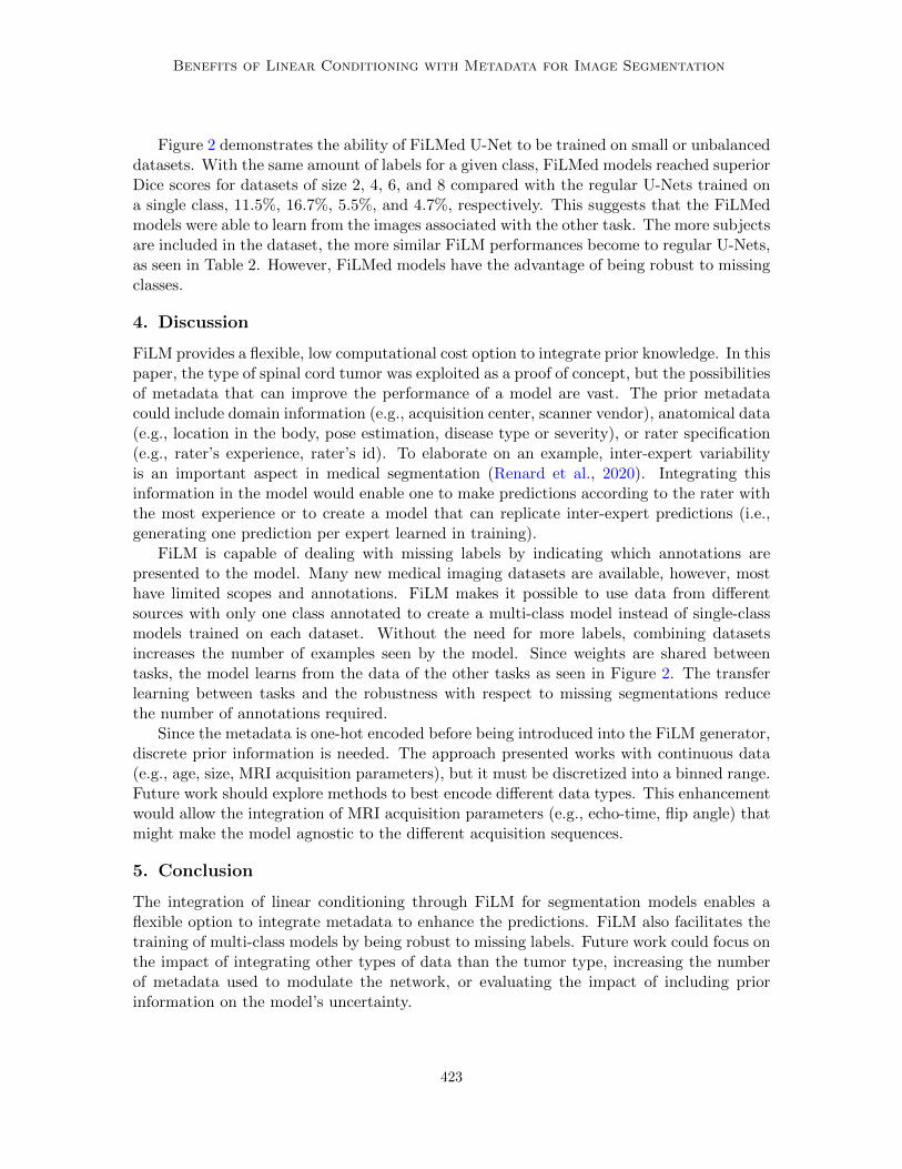
Figure 2: Spleen and kidney segmentation Dice scores for small and unbalanced datasets. Thenumber of subjects combines training and validation subjects. Dice scores for all experiments on thetest set (25 subjects) were averaged across the number of subjects and aggregated according to theapproach, FiLMed (red) or regular U-Net (blue). The error bars show the standard deviation. ∆indicates the difference of mean Dice scores between the two approaches. The data totals 10 modelstrained on different random splits. ** p-value < 5% with one-sided Wilcoxon signed-rank test.
422
Benefits of Linear Conditioning with Metadata for Image Segmentation
Figure 2 demonstrates the ability of FiLMed U-Net to be trained on small or unbalanceddatasets. With the same amount of labels for a given class, FiLMed models reached superiorDice scores for datasets of size 2, 4, 6, and 8 compared with the regular U-Nets trained ona single class, 11.5%, 16.7%, 5.5%, and 4.7%, respectively. This suggests that the FiLMedmodels were able to learn from the images associated with the other task. The more subjectsare included in the dataset, the more similar FiLM performances become to regular U-Nets,as seen in Table 2. However, FiLMed models have the advantage of being robust to missingclasses.
4. Discussion
FiLM provides a flexible, low computational cost option to integrate prior knowledge. In thispaper, the type of spinal cord tumor was exploited as a proof of concept, but the possibilitiesof metadata that can improve the performance of a model are vast. The prior metadatacould include domain information (e.g., acquisition center, scanner vendor), anatomical data(e.g., location in the body, pose estimation, disease type or severity), or rater specification(e.g., rater’s experience, rater’s id). To elaborate on an example, inter-expert variabilityis an important aspect in medical segmentation (Renard et al., 2020). Integrating thisinformation in the model would enable one to make predictions according to the rater withthe most experience or to create a model that can replicate inter-expert predictions (i.e.,generating one prediction per expert learned in training).
FiLM is capable of dealing with missing labels by indicating which annotations arepresented to the model. Many new medical imaging datasets are available, however, mosthave limited scopes and annotations. FiLM makes it possible to use data from differentsources with only one class annotated to create a multi-class model instead of single-classmodels trained on each dataset. Without the need for more labels, combining datasetsincreases the number of examples seen by the model. Since weights are shared betweentasks, the model learns from the data of the other tasks as seen in Figure 2. The transferlearning between tasks and the robustness with respect to missing segmentations reducethe number of annotations required.
Since the metadata is one-hot encoded before being introduced into the FiLM generator,discrete prior information is needed. The approach presented works with continuous data(e.g., age, size, MRI acquisition parameters), but it must be discretized into a binned range.Future work should explore methods to best encode different data types. This enhancementwould allow the integration of MRI acquisition parameters (e.g., echo-time, flip angle) thatmight make the model agnostic to the different acquisition sequences.
5. Conclusion
The integration of linear conditioning through FiLM for segmentation models enables aflexible option to integrate metadata to enhance the predictions. FiLM also facilitates thetraining of multi-class models by being robust to missing labels. Future work could focus onthe impact of integrating other types of data than the tumor type, increasing the numberof metadata used to modulate the network, or evaluating the impact of including priorinformation on the model’s uncertainty.
423

Benefits of Linear Conditioning with Metadata for Image Segmentation
Acknowledgments
We thank the contributors of the ivadomed project, Lucas Rouhier, Ainsleigh Hill, ValentineLouis-Lucas, and Christian Perone for fruitful discussions.
Funded by the Canada Research Chair in Quantitative Magnetic Resonance Imaging[950-230815], the Canadian Institute of Health Research [CIHR FDN-143263], the CanadaFoundation for Innovation [32454, 34824], the Fonds de Recherche du Quebec - Sante[28826], the Fonds de Recherche du Quebec - Nature et Technologies [2015-PR-182754],the Natural Sciences and Engineering Research Council of Canada [RGPIN-2019-07244],the Canada First Research Excellence Fund (IVADO and TransMedTech), the CourtoisNeuroMod project and the Quebec BioImaging Network. This research is based on workpartially supported by the CIFAR AI and COVID-19 Catalyst Grants. A.L. has a fellow-ship from NSERC, FRQNT, and UNIQUE, C.G. has a fellowship from IVADO [EX-2018-4],O.V. has a fellowship from NSERC, FRQNT, and UNIQUE.
References
Mohamed Ahmed. Medical image segmentation using attention-based deep neural networks,2020. URL https://www.diva-portal.org/smash/get/diva2:1477227/FULLTEXT01.
pdf.
Kim B Baker, Christopher J Moran, Franz J Wippold, James G Smirniotopoulos, Fabio JRodriguez, Steven P Meyers, and Todd L Siegal. Mr imaging of spinal hemangioblas-toma. American Journal of Roentgenology, 174(2):377–382, 2000. URL https://www.
ajronline.org/doi/full/10.2214/ajr.174.2.1740377.
Danielle Baleriaux. Spinal cord tumors. European radiology, 9(7):1252–1258, 1999. URLhttps://doi.org/10.1007/s003300050831.
Yash Bhalgat, Meet Shah, and Suyash Awate. Annotation-cost minimization for medicalimage segmentation using suggestive mixed supervision fully convolutional networks. InNeural Information Processing Systems (NeurIPS), 2018. URL https://arxiv.org/
pdf/1812.11302.pdf.
Agisilaos Chartsias, Giorgos Papanastasiou, Chengjia Wang, Colin Stirrat, Scott Semple,David Newby, Rohan Dharmakumar, and Sotirios A. Tsaftaris. Multimodal cardiac seg-mentation using disentangled representation learning. In Statistical Atlases and Com-putational Models of the Heart. Multi-Sequence CMR Segmentation, CRT-EPiggy andLV Full Quantification Challenges, pages 128–137, Cham, 2020. Springer InternationalPublishing. ISBN 978-3-030-39074-7.
Ozan Ciga and Anne L Martel. Learning to segment images with classification labels.Medical Image Analysis, 68:101912, 2021. URL https://doi.org/10.1016/j.media.
2020.101912.
Harm de Vries, Florian Strub, Jeremie Mary, Hugo Larochelle, Olivier Pietquin, andAaron C. Courville. Modulating early visual processing by language. In Neural In-
424

Benefits of Linear Conditioning with Metadata for Image Segmentation
formation Processing Systems (NIPS), pages 6597–6607, 2017. URL http://papers.
nips.cc/paper/7237-modulating-early-visual-processing-by-language.
Vincent Dumoulin, Jonathon Shlens, and Manjunath Kudlur. A learned representation forartistic style. In International Conference on Learning Representations (ICLR), 2017.URL https://arxiv.org/pdf/1610.07629.pdf.
Charley Gros, Andreanne Lemay, Olivier Vincent, Lucas Rouhier, Marie-Helene Bourget,Anthime Bucquet, Joseph Paul Cohen, and Julien Cohen-Adad. ivadomed: A medicalimaging deep learning toolbox. Journal of Open Source Software, 6(58):2868, 2021. URLhttps://doi.org/10.21105/joss.02868.
Nicholas Heller, Niranjan Sathianathen, Arveen Kalapara, Edward Walczak, Keenan Moore,Heather Kaluzniak, Joel Rosenberg, Paul Blake, Zachary Rengel, Makinna Oestreich,et al. The kits19 challenge data: 300 kidney tumor cases with clinical context, ct semanticsegmentations, and surgical outcomes. arXiv preprint arXiv:1904.00445, 2019. URLhttps://arxiv.org/pdf/1904.00445.pdf.
Fabian Isensee, Jens Petersen, Andre Klein, David Zimmerer, Paul F Jaeger, Simon Kohl,Jakob Wasserthal, Gregor Koehler, Tobias Norajitra, Sebastian Wirkert, et al. nnu-net:Self-adapting framework for u-net-based medical image segmentation. arXiv preprintarXiv:1809.10486, 2018.
Fabian Isensee, Paul F Jager, Simon AA Kohl, Jens Petersen, and Klaus H Maier-Hein.Automated design of deep learning methods for biomedical image segmentation. arXivpreprint arXiv:1904.08128, 2019. URL https://arxiv.org/pdf/1904.08128.pdf.
Grzegorz Jacenkow, Agisilaos Chartsias, Brian Mohr, and Sotirios A Tsaftaris. Condi-tioning convolutional segmentation architectures with non-imaging data. arXiv preprintarXiv:1907.12330, 2019.
Grzegorz Jacenkow, Alison Q O’Neil, Brian Mohr, and Sotirios A Tsaftaris. Inside: Steeringspatial attention with non-imaging information in cnns. In International Conference onMedical Image Computing and Computer-Assisted Intervention, pages 385–395. Springer,2020.
DH Kim, J-H Kim, Seung Hong Choi, C-H Sohn, Tae Jin Yun, Chi Heon Kim, and K-HChang. Differentiation between intramedullary spinal ependymoma and astrocytoma:comparative mri analysis. Clinical radiology, 69(1):29–35, 2014. URL https://doi.org/
10.1016/j.crad.2013.07.017.
Taesup Kim, Inchul Song, and Yoshua Bengio. Dynamic layer normalization for adaptiveneural acoustic modeling in speech recognition. In InterSpeech, 2017. URL https:
//arxiv.org/pdf/1707.06065.pdf.
Andreanne Lemay, Charley Gros, Zhizheng Zhuo, Jie Zhang, Yunyun Duan, Julien Cohen-Adad, and Yaou Liu. Multiclass spinal cord tumor segmentation on mri with deep learn-ing, 2021. URL https://arxiv.org/pdf/2012.12820.pdf.
425

Benefits of Linear Conditioning with Metadata for Image Segmentation
Yanghao Li, Naiyan Wang, Jianping Shi, Xiaodi Hou, and Jiaying Liu. Adaptive batchnormalization for practical domain adaptation. Pattern Recognition, 80:109–117, 2018.URL https://doi.org/10.1016/j.patcog.2018.03.005.
Shervin Minaee, Yuri Boykov, Fatih Porikli, Antonio Plaza, Nasser Kehtarnavaz, andDemetri Terzopoulos. Image segmentation using deep learning: A survey. arXiv preprintarXiv:2001.05566, 2020. URL https://arxiv.org/pdf/2001.05566.pdf.
Boris N Oreshkin, Pau Rodriguez, and Alexandre Lacoste. Tadam: Task dependent adaptivemetric for improved few-shot learning. arXiv preprint arXiv:1805.10123, 2018. URLhttps://arxiv.org/pdf/1805.10123.pdf.
Ethan Perez, Florian Strub, Harm De Vries, Vincent Dumoulin, and Aaron Courville. Film:Visual reasoning with a general conditioning layer. In Proceedings of the AAAI Conferenceon Artificial Intelligence, volume 32, 2018. URL https://arxiv.org/pdf/1709.07871.
pdf.
Michael Rebsamen, Urspeter Knecht, Mauricio Reyes, Roland Wiest, Raphael Meier, andRichard McKinley. Divide and conquer: Stratifying training data by tumor grade im-proves deep learning-based brain tumor segmentation. Frontiers in Neuroscience, 13:1182,2019. ISSN 1662-453X. doi: 10.3389/fnins.2019.01182. URL https://www.frontiersin.
org/article/10.3389/fnins.2019.01182.
Felix Renard, Soulaimane Guedria, Noel De Palma, and Nicolas Vuillerme. Variability andreproducibility in deep learning for medical image segmentation. Scientific Reports, 10(1):1–16, 2020. URL https://doi.org/10.1038/s41598-020-69920-0.
Olaf Ronneberger, Philipp Fischer, and Thomas Brox. U-net: Convolutional networks forbiomedical image segmentation. In Medical Image Computing and Computer-AssistedIntervention – MICCAI 2015, pages 234–241. Springer International Publishing, 2015.URL https://doi.org/10.1007/978-3-319-24574-4_28.
Amber L Simpson, Michela Antonelli, Spyridon Bakas, Michel Bilello, Keyvan Farahani,Bram Van Ginneken, Annette Kopp-Schneider, Bennett A Landman, Geert Litjens, Bjo-ern Menze, et al. A large annotated medical image dataset for the development andevaluation of segmentation algorithms. arXiv preprint arXiv:1902.09063, 2019. URLhttps://arxiv.org/pdf/1902.09063.pdf.
Olivier Vincent, Charley Gros, Joseph Paul Cohen, and Julien Cohen-Adad. Automaticsegmentation of spinal multiple sclerosis lesions: How to generalize across MRI contrasts?,2020. URL https://arxiv.org/pdf/2003.04377.pdf.
Yuyin Zhou, Zhe Li, Song Bai, Chong Wang, Xinlei Chen, Mei Han, Elliot Fishman, andAlan L Yuille. Prior-aware neural network for partially-supervised multi-organ segmen-tation. In Proceedings of the IEEE/CVF International Conference on Computer Vision,pages 10672–10681, 2019. URL https://arxiv.org/pdf/1904.06346.pdf.
426

Benefits of Linear Conditioning with Metadata for Image Segmentation
Appendix A. Experimental design of organ segmentation with limitedannotations
Figure 3: Experimental design of organ segmentation with limited annotations. The images associ-ated to each model represent the training and validation set. This experimental design was used togenerate Figure 2.
427

Benefits of Linear Conditioning with Metadata for Image Segmentation
Appendix B. Spinal cord tumor segmentation
Figure 4: Tumor segmentation prediction by FiLMed U-Net informed by the tumor type, “Withprior”, or not informed, “No prior”. A1 and A2 presents two subjects with astrocytomas. H1 andH2 presents two subjects with hemangioblastomas. GT: Ground truth.
Astrocytomas are typically large, have ill-defined boundaries, and present heterogeneous,moderate, or partial enhanced in the Gd-e T1w contrast (Baleriaux, 1999). Astrocytomasare usually extensive, expanding from 2 to 19 vertebral bodies in size (Baleriaux, 1999).In both A1 and A2 predictions from the model without prior information, the segmentedtumor size was one vertebral body or less and corresponded to the most enhanced tumorsignal on the Gd-e T1w (ignoring the rest of the lesion).
In counterpart, hemangioblastomas are usually associated with a small tumor core (Ba-leriaux, 1999) intensely enhanced on Gd-e T1w (Baker et al., 2000). Figure 4 H1 presentsa hemangioblastoma barely apparent in T2w and hidden by the cavity (hyperintense sig-nal). The small hyperintense signal on the Gd-e T1w contrast was overseen by the regularapproach. On H2, the model oversegmented the tumor and identified a second tumor on ahypointense signal. The false positive tumor identification does not present an intense Gd-eT1w enhancement which is usually the case for hemangioblastomas. This false positive isnot present for the model informed by the tumor type.
To assess the impact of inputting the tumor type, each prediction was modulated bythe different tumor types. Table 3 presents the quantitative results for each condition whileFigure 5 qualitatively illustrates the impact of changing the tumor type. The highest Dicescores are reached when the input label corresponds to the true label. The modulation
428

Benefits of Linear Conditioning with Metadata for Image Segmentation
Figure 5: Impact of inputting different tumor types with FiLMed U-Net on the model’s segmentation.True label represents the tumor type while input label is the tumor type input into the model throughFiLM. Astr.: Astrocytoma, Epen.:Ependymoma, Hema.: Hemangioblastoma.
429

Benefits of Linear Conditioning with Metadata for Image Segmentation
Table 3: Spinal cord tumor core segmentation Dice scores for FiLMed U-Net with the different tumortypes as input (mean ± STD % for 10 random splits). True label represents the tumor type whileinput label is the tumor type input into the model through FiLM. ** p-value < 0.05 for one-sidedWilcoxon signed-rank test compared to the highest value in each row.
Input label
True label Astrocytoma Ependymoma Hemangioblastoma
Astrocytoma 57.9± 4.9 57.3± 4.9 32.2± 5.1 **Ependymoma 57.6± 2.6 57.7± 2.4 35.9± 4.7 **Hemangioblastoma 41.5± 4.7 ** 41.8± 6.4 ** 61.7± 3.7
with FiLM successfully encoded knowledge about the tumor types and the predictions arein agreement with known characteristics of the different types. Astrocytoma and ependy-moma yield similar predictions. Both tumor types have overlapping characteristics (Kimet al., 2014): high intensity signals on T2w, comparable enhancement patterns, similarsize (astrocytoma: 2-19 vertebral bodies, ependymoma: 2-13 vertebral bodies (Baleriaux,1999)), etc. Predictions with hemangioblastoma as input diverge from the other tumortypes. Hemangioblastoma predictions reflect their characteristics: small tumor cores in-tensely enhanced in Gd-e T1w, as seen in Figure 4. When inputting the hemangioblastomalabel for the astrocytoma (first row of Figure 5) no prediction is given since the Gd-e T1wmodality has moderate enhancement. Similarly, for the ependymoma, only the most Gd-enhanced portion of the tumor is predicted when assigning the hemangioblastoma labelwith FiLM (second row of Figure 5). The results from Table 3 and Figure 4 - 5 confirmthat FiLM layers are able to learn characteristics from the metadata that are relevant forthe segmentation.
430