bassem et al, j tumor res 215, 1:1 l of t u m rr n au rj ... and thoracic surgery department, habib...
TRANSCRIPT

Volume 1 • Issue 1 • 1000104J Tumor ResISSN: JTR, an open access journal
Research Article Open Access
Bassem et al., J Tumor Res 2015, 1:1
Case Report Open Access
Journal of Tumor ResearchJour
nal of Tumor Research
Unusual Presentation of a Primary Pericardial Malignant Mesothelioma: Constrictive Pericarditis A Case Report and Review of the LiteratureRekik Bassem1, Ben Jmaa Hèla2*, Jerbi Bassem1, Tabebi Nada1, Cherif Taieb2, Elleuch Ahmed1, Souissi Iheb3, Kammoun Samir1, Masmoudi Sayda2 and Frikha Imed2
1Cardiology Department, Hédi Chaker University Hospital, Sfax University, Sfax Tunisia2Cardiovascular and Thoracic Surgery Department, Habib Bourguiba university Hospital, Sfax University, Sfax Tunisia3Anesthesiology department, Habib Bourguiba university Hospital, Sfax University, Sfax Tunisia
*Corresponding author: Ben Jmaà Hèla, Department of cardiovascular andthoracic surgery Habib Bourguiba Hospital Sfax, Tunisia, Tel: 0021696704740;E-mail: [email protected]
Received October 18, 2015; Accepted December 04, 2015; Published December 15, 2015
Citation: Bassem R, Hèla BJ, Bassem J, Nada T, Taieb C, et al. (2015) Unusual Presentation of a Primary Pericardial Malignant Mesothelioma: Constrictive Pericarditis A Case Report and Review of the Literature. J Tumor Res 1: 104.
Copyright: © 2015 Bassem R, et al. This is an open-access article distributed under the terms of the Creative Commons Attribution License, which permits unrestricted use, distribution, and reproduction in any medium, provided the original author and source are credited.
AbstractPrimary malignant pericardial mesothelioma is an extremely rare tumor with high lethality. We report the case
of a 30 year-old female without medical history, who was admitted in the department of cardiology for a dyspnea and chest pain due to a large pericardial effusion. A tamponade was confirmed by echocardiography, and emergent pericardial drainage was performed. Twelve months later, the patient developed a hemodynamically relevant pericardial constriction. She underwent a partial pericardectomy. The pericardium was thick and adherent. The histological examination revealed a pericardial infiltration by malignant mesothelioma. After surgery, five cycles of chemotherapy with carboplatine were beginned.
The treatment methods of primary pericardial mesothelioma are limited. Chemotherapy in addition to surgery seems to improve symptoms in case of constriction and non-response to pericardectomy.
Keywords: Pericardial constriction; Mesothelioma; Pericardectomy
IntroductionPrimary malignant pericardial mesothelioma is an extremely
rare tumor and seems to have a complex pathogenicity. Clinical presentation is variable. It can be revealed by a cardiac tamponade, and pericardial constriction. The outcome is fatal in the majority of cases despite surgery and chemotherapy.
Case PresentationA 30 year-old female without medical history, especially tuberculosis
contagion or asbestos exposure, was admitted in the department of cardiology for evectional dyspnea (NYHA III) and chest pain.
Transthoracic echocardiography illustrated a large pericardial effusion with compression of the cardiac chambers. A tamponade was confirmed, and an emergent pericardial drainage was performed. It removed 1500 ml of a sero-hematic liquid with predominance of neutrophil cells. Pericardial biopsy was normal.
Symptoms were markedly relieved. The patient was treated with non-steroidal anti-inflammatory, but the evolution was marked by the persistence of a low abundance effusion.
The patient was re-admitted twelve months later for lower limbs edema and dyspnea. Physical examination revealed signs of right heart failure. Electrocardiogram showed diffuse negative T waves. There was no biological inflammatory syndrome, but CA 15-3 and CA 125 tumors markers were slightly positive. Transthoracic echocardiography showed a recurrent large pericardial effusion. There were no signs of cardiac tamponade, but there was a thickened ventricular pericardium. The mitral flow combined to doppler tissue imaging showed signs of pericardial constriction (Figures 1 and 2). CT scan confirmed the pericardial effusion without any calcifications (Figure 3).
The patient developed a hemodynamically relevant pericardial constriction. Thus, right catheterization confirmed adistolia and showed high telediastolic pressure in the right ventricle and equal diastolic pressure between the right chambers and the pulmonary artery. So, a chronic constrictive pericarditis was suspected.
The patient underwent surgery for a total pericardectomy. The intraoperative examination showed a thickened pericardium with myocardial adherences (Figures 4 and 5). Great vessels seemed invaded by the tumoral pericardium. The patient underwent then a partial pericardectomy of the anterior surface of the heart because of the tumoral infiltration and adherences, and the risk of injury of the cardiac wall.
The histology examination confirmed the pericardial infiltration
Figure 1: Transthoracic echocardiography demonstrating a large pericardial effusion and a thickened pericardium.
Citation: Bassem R, Hèla BJ, Bassem J, Nada T, Taieb C, et al. (2015) Unusual Presentation of a Primary Pericardial Malignant Mesothelioma: Constrictive Pericarditis A Case Report and Review of the Literature. J Tumor Res 1: 104.
Page 2 of 3
Volume 1 • Issue 1 • 1000104J Tumor ResISSN: JTR, an open access journal
by malignant mesothelioma. Then a protocol combining five cycles of chemotherapy with carboplatine is beginned.
DiscussionPrimary pericardial tumors are rare. They can be begnin (teratoma,
fibroma, angioma, lipoma) or malignant (sarcoma, mesothelioma). In contrast, secondary tumors are more frequent especially lung cancer metastasis [1].
Primary pericardial mesothelioma is very rare. In post mortem cohort, the incidence was around 0.002% [1]. Nevertheless, it represents 50% of primary pericardial tumours [2].
Nilson et al. [3] reported 30 cases of pericardial mesothelioma which diagnosis was made in post-mortem [3].
It seems to have a complex pathogenicity combining environmental carcinogens (asbestos), radiations, virus and genetic factors [4]. Asbestos exposure is observed seldom in primitive pericardial localizations but it occurs if a pleural disease is coexisting [5]. During the last decade, the alleged role of SV40 virus has become predominant in the genesis of mesothelioma [6]. Our patient was not checked for SV40 infection.
Mesothelioma can be localized in the pericardium or it can diffusely encase the heart. It can infiltrate the myocardium, the atria, the coronary sinuses, the coronary arteries, and the conduction system of the heart. Commonly, the diagnosis is made at an advanced local stage [7].
The onset of symptoms is usually insidious. Clinical presentation is variable: It can be manifested by a pericardial effusion, a cardiac tamponade, a constrictive pericarditis and a heart failure caused by myocardial infiltration by tumor cells [8]. It can also simulate tuberculous pericarditis [9] or an atrial myxoma [10].
Metastases have also been described [11].
General symptoms are often associated with mesothelioma and they are often insidious, source of error and delay diagnosis. They include long-term fever, impaired general condition [9].
Thromboembolic complications are common in the pericardial mesothelioma [12]: the pericardial constriction can contribute to venous stasis promoting the formation of thrombi.
The main diagnostic imaging tools are echocardiogram, CT scan, and magnetic resonance imaging [2].
Transthoracic echocardiography contributes to the diagnosis of pericardial tumor without predicting its histological nature. The mesothelioma appears in ultrasound as a pericardial mass infiltrating the large vessels of the heart. It is rarely revealed by a pericardial effusion of great abundance causing a tamponade [5].
The magnetic resonance imaging and CT scan are useful to precise the degree of infiltration of the adjacent structures, the degree
Figure 2: The mitral flow combined to DTI showed signs of constriction.
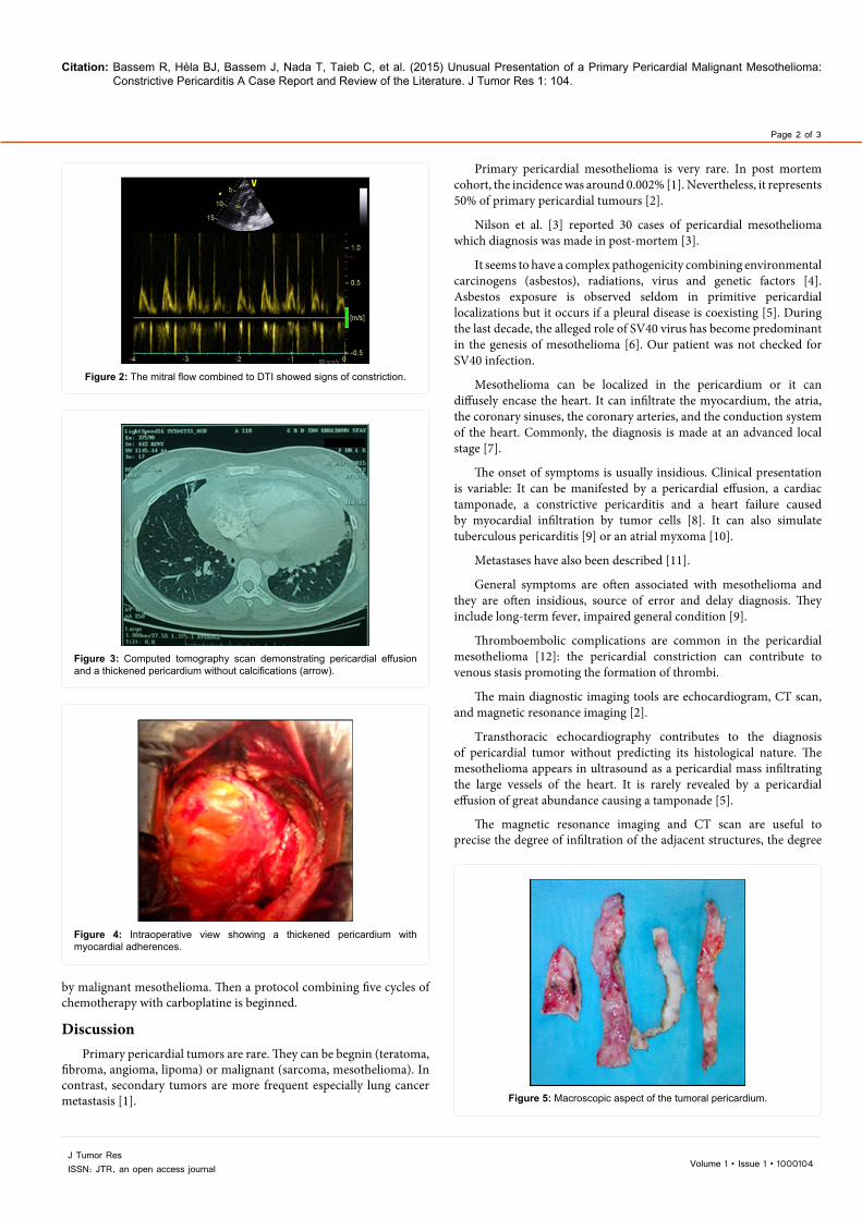
Figure 3: Computed tomography scan demonstrating pericardial effusion and a thickened pericardium without calcifications (arrow).
Figure 4: Intraoperative view showing a thickened pericardium with myocardial adherences.
Figure 5: Macroscopic aspect of the tumoral pericardium.

Citation: Bassem R, Hèla BJ, Bassem J, Nada T, Taieb C, et al. (2015) Unusual Presentation of a Primary Pericardial Malignant Mesothelioma: Constrictive Pericarditis A Case Report and Review of the Literature. J Tumor Res 1: 104.
Page 3 of 3
Volume 1 • Issue 1 • 1000104J Tumor ResISSN: JTR, an open access journal
of constriction, and to help determining its resectability [13]. In our observation, chest CT has not objectified mass or calcification.
The definitive diagnosis is often made after sternotomy or thoracotomy by analyzing the macroscopic aspect of the pericardium, and histological examination of a pericardial biopsy [14].
The cytological and biochemical examination of the liquid often gives negative results and may show a high content of hyaluronic acid [3].
Surgery is an effective treatment method. Surgical resection can be curative in localized cases, but owing to the frequently late presentation, surgical intervention is usually palliative [15]. Its main role is therefore to control symptoms, as is the case of partial pericardiectomy in cardiac tamponade or in cardiac constriction [16]. It is done to prevent the compression of the heart and obstruction of the vessels. Complete resection is impossible [17].
Pericardial mesothelioma responds poorly to radiotherapy. Currently, to reduce the size of the tumor after the excision, radiological therapy is performed as an adjuvant therapy [17]. Chemotherapy combination with doxorubicin, vincristine, and cyclophosphamide can reduce the tumor mass [18].
New approaches to battle against this cancer are promising. These include anti-angiogenesis drugs, photodynamic therapy, and gene therapy [19-21]. All these therapies are tested in clinical trials, which give some hope for the treatment of primary pericardial mesothelioma.
It’s a highly aggressive tumour with global survival of patients less than 6 months [2]. Furthermore, a case of survival of 3 years has been reported [22].
Conclusion Primary malignant pericardial mesothelioma is rare and can be
misdiagnosed. It can be rarely revealed by a constrictive pericarditis.
The treatment options of the tumor are limited, and the prognosis is poor. It should be appropriately managed with chemotherapy in addition to surgery in case of pericardial constriction and non-response to pericardectomy.
References
1. Mukai K, Shinkai T, Tominaga K, Shimosato Y (1988) The incidence ofsecondary tumors of the heart and pericardium: a 10-year study. Jpn J ClinOncol 18: 195-201.
2. Santos C, Montesinos J, Castañer E, Sole JM, Baga R (2008) Primarypericardial mesothelioma. Lung Cancer 60: 291-293.
3. Nilsson A, Rasmuson T (2009) Primary Pericardial Mesothelioma: Report of aPatient and Literature Review. Case Rep Oncol 2: 125-132.
4. Carbone M, Kratzke RA, Testa JR (2002) The pathogenesis of mesothelioma. Semin Oncol 29: 2-17.
5. Lingamfelter DC, Cavuoti D, Gruszecki AC (2009) Fatal hemopericardial tamponade due to primary pericardial mesothelioma: a case report. DiagnPathol 4: 44.
6. Rizzo P, Bocchetta M, Powers A, Foddis R, Stekala E, et al. (2001) SV40 andthe pathogenesis of mesothelioma. Semin Cancer Biol 11: 63-71.
7. Butz T, Faber L, Langer C, Körfer J, Lindner O, et al. (2009) Primary malignantpericardial mesothelioma - a rare cause of pericardial effusion and consecutive constrictive pericarditis: a case report. J Med Case Rep 3: 9256.
8. Reardon KA, Reardon MA, Moskaluk CA, Grosh WW, Read PW (2010) Primarypericardial malignant mesothelioma and response to radiation therapy. RareTumors 2: e51.
9. Rose DS, Vigneswaran WT, Bovill BA, Riordan JF, Sapsford RN, et al. (1992) Primary pericardial mesothelioma presenting as tuberculous pericarditis. Postgrad Med J 68: 137-139.
10. Lund O, Hansen OK, Ardest S, Baandrup U (1987) Primary malignant pericardial mesothelioma mimicking left atrial myxoma. Case report. Scand J Thorac Cardiovasc Surg 21: 273-275.
11. Eren NT, Akar AR (2002) Primary pericardial mesothelioma. Curr Treat Options Oncol 3: 369-373.
12. Gong W, Ye X, Shi K, Zhao Q (2014) Primary malignant pericardial mesothelioma-a rare cause of superior vena cava thrombosis and constrictive pericarditis. J Thorac Dis 6: E272-275.
13. Langer C, Butz T, Horstkotte D (2006) Multimodality in imaging calcific constrictive pericarditis. Heart 92: 1289.
14. Llewellyn MJ, Atkinson MW, Fabri B (1987) Pericardial constriction caused by primary mesothelioma. Br Heart J 57: 54-57.
15. Kayatta MO, Dineen SP, Sica G, Puskas JD, Pickens A (2013) Primary pericardial mesothelioma in a 19-year-old presenting as pericarditis. Ann Thorac Surg 96: 680-681.
16. Vigneswaran WT, Stefanacci PR (2000) Pericardial mesothelioma. Curr Treat Options Oncol 1: 299-302.
17. Bang JH, Roh MS, Hong SH, Choi PJ, Woo JS (2010) Surgical experience of pericardial mesothelioma presenting as constrictive pericarditis. Journal of Cardiology Cases 2: e96-e98.
18. Maruyama R, Sakai M, Nakamura T, Suemitsu R, Okamoto T, et al. (2006) Triplet chemotherapy for malignant pericardial mesothelioma: a case report. Jpn J Clin Oncol 36: 245-248.
19. Sardar MR, Kuntz C, Patel T, Saeed W, Gnall E, et al. (2012) Primary pericardial mesothelioma unique case and literature review. Tex Heart Inst J 39: 261-264.
20. Giroux Leprieur E1, Hirata T2, Mo M3, Chen Z4, Okamoto J2, et al. (2014) The homeobox gene EMX2 is a prognostic and predictive marker in malignantpleural mesothelioma. Lung Cancer 85: 465-471.
21. Morimoto C, Ohnuma K (2012) [Antibody therapy for malignant mesothelioma: humanized anti-cD26 mAb therapy]. Nihon Rinsho 70: 2177-2182.
22. Fujita K, Hata M, Sezai A, Minami K (2014) Three-year survival after surgery for primary malignant pericardial mesothelioma: report of a case. Surg Today44: 948-951.