b850 ring from photosynthetic complex lh2 – comparison of ... · oxidation–reduction process....
TRANSCRIPT

B850 Ring from Photosynthetic Complex LH2 –Comparison of Different Static Disorder Types
Pavel Herman, David Zapletal
Abstract— Properties of light–harvesting (LH) pigment–protein complexes are strongly influenced by their interactionswith environment. These interactions could be modeled bystatic and dynamic disorder. Influence of static disorder onB850 ring from LH2 complex of purple bacteria is investigatedin present paper. The nearest neighbour approximation modelof the ring is considered. Four types of uncorrelated Gaussianstatic disorder (fluctuations of transfer integrals, fluctuationsof radial positions of molecules on the ring, fluctuations ofangular positions of molecules on the ring and fluctuationsof directions of molecular dipole moments) are taking intoaccount. The most important statistical properties of thenearest neighbour transfer integral distributions for differentstrengths of static disorder are calculated. Results obtained forfour above mentioned types of static disorder are discussedand compared.
Keywords—LH2 complex, B850 ring, static disorder,Hamiltonian, transfer integral distributions
I. INTRODUCTION
PHOTOSYNTHESIS is the process by which greenplants and certain other organisms (bacteria, blue–
green algae) transform light energy into chemical en-ergy. During this process light energy is captured andused to convert water, carbon dioxide, and miner-als into oxygen and energy–rich organic compounds.In chemical terms, photosynthesis is a light–energizedoxidation–reduction process. Oxidation refers to the re-moval of electrons from a molecule; reduction refersto the gain of electrons by a molecule. These reac-tions occur in two stages: the light stage, consisting ofphotochemical (i.e., light–capturing) reactions; and thedark stage, comprising chemical reactions controlled byenzymes. During the first stage, the energy of light isabsorbed and used to drive a series of electron transfers,
Manuscript received October, 2016.This work was supported by the Faculty of Science, University of Hradec
Kralove (project of specific research No. 2105/2016 – P. Herman).P. Herman is with the Department of Physics, Faculty of Science, University
of Hradec Kralove, Rokitanskeho 62, 50003 Hradec Kralove, Czech Republic(e-mail: [email protected]).
D. Zapletal is with the Institute of Mathematics and Quantitative Methods,Faculty of Economics and Administration, University of Pardubice, Studentska95, 53210 Pardubice, Czech Republic (e-mail: [email protected]).
resulting in the synthesis of ATP and the electron–donor reduced nicotine adenine dinucleotide phosphate(NADPH). During the dark stage, the ATP and NADPHformed in the light–capturing reactions are used to reducecarbon dioxide to organic carbon compounds [1].
Our interest is mainly focused on first (light) stageof photosynthesis in purple bacteria. Solar photons areabsorbed by a complex system of membrane–associatedpigment–proteins (light–harvesting (LH) antenna) andthe electronic excited state is efficiently transferred to areaction center, where the light energy is converted intoa chemical energy [2]. The antenna systems of photo-synthetic units from purple bacteria are formed by ringunits LH1, LH2, LH3, and LH4. The geometric structureis known in great detail from X–ray crystallography. Thegeneral organization of above mentioned light–harvestingcomplexes is the same: identical subunits are repeatedcyclically in such a way that a ring–shaped structureis formed. However the symmetries of these rings aredifferent.
Crystal structure of LH2 complex contained in purplebacterium Rhodopseudomonas acidophila was first de-scribed in high resolution by McDermott et al. [3], thenfurther e.g. by Papiz et al. [4]. The bacteriochlorophyll(BChl) molecules are organized in two concentric rings.One ring (B800 ring) features a group of nine well–separated BChl molecules with absorption band at about800 nm. The second ring (B850 ring) consists of eighteenclosely packed BChl molecules (B850) absorbing around850 nm. Dipole moments in LH2 ring have tangentialarrangement. The whole LH2 complex is nonameric, itconsists of nine identical subunits. LH2 complexes fromother purple bacteria have analogous ring structure.
Some bacteria contain also other types of com-plexes such as the B800–820 LH3 complex (Rhodopseu-domonas acidophila strain 7050) or LH4 complex(Rhodopseudomonas palustris). LH3 complex like LH2one is usually nonameric but LH4 one is octameric (itconsists of eight identical subunits). They can also differin orientation of molecular dipole moments and strengthof mutual interactions between bacteriochorophylls. Forinstance, interactions between the nearest neighbour bac-teriochlorophylls in B–α/B–β ring from LH4 complexare approximately two times smaller in comparison withB850 ring from LH2 complex and they have oppositesign.
The intermolecular distances under 1 nm determine
INTERNATIONAL JOURNAL OF MATHEMATICS AND COMPUTERS IN SIMULATION Volume 10, 2016
ISSN: 1998-0159 361

strong exciton couplings between corresponding pig-ments. That is why an extended Frenkel exciton statesmodel could be used in theoretical approach. In spite ofextensive investigation, the role of the protein moiety ingoverning the dynamics of the excited states has not beentotally clear yet. At room temperature the solvent andprotein environment fluctuates with characteristic timescales ranging from femtoseconds to nanoseconds. Thesimplest approach is to substitute fast fluctuations bydynamic disorder and slow fluctuations by static disorder.
Static disorder effect on the anisotropy of fluores-cence for LH2 complexes was studied by Kumble andHochstrasser [6] and Nagarajan et al. [7], [8]. We ex-tended these investigations by consideration of dynamicdisorder. We studied this effect for simple model systems[9]–[11] and then for models of B850 ring (from LH2)[12], [13]. Various types of uncorrelated static disorder(in local excitation energies, in transfer integrals, etc.)and correlated one (e.g., elliptical deformation) wereused in the past [14]–[16] and also different arrangementsof optical dipole moments were compared [17]–[20].Recently we have focused on the modelling of absorptionand steady state fluorescence spectra of LH2 and LH4complexes within the nearest neighbour approximationmodel [21]–[25]. We have also extended our model tofull Hamiltonian model and published the results fordifferent types of static disorder [26]–[34].
Main goal of the present paper is the investigationof four types of static disorder (Gaussian fluctuationsin transfer integrals, in radial positions of moleculeson the ring, in angular positions of molecules on thering and in directions of dipole moments of molecules)and comparison of their influence on Hamiltonian ofB850 ring from LH2 complex. The rest of the paperis structured as follows. Section II introduces the ringmodel with different types of static disorder, used unitsand parameters could be found in Section III, resultsare presented and discussed in Section IV and someconclusions are drawn in Section V.
II. MODEL
We consider only one exciton on molecular ring whichcan model B850 ring from LH2 complex. The Hamilto-nian of an exciton on this ring reads
H = H0ex +Hph +Hex−ph +Hs. (1)
A. Ideal ringFirst term in Eq. (1),
H0ex =
N∑m=1
E0ma†mam +
N∑m,n=1(m6=n)
J0mna
†man, (2)
corresponds to an exciton, e.g. the system without anydisorder. The operator a†m (am) creates (annihilates) an
exciton at site m, E0m is the local excitation energy of
m–th molecule and J0mn (for m 6= n) is the so–called
transfer integral between sites m and n. Local excitationenergies E0
m are the same for all bacteriochlorophylls onunperturbed ring, i.e.
E0m = E0, m = 1, . . . , N.
Inside one ring the pure exciton Hamiltonian H0ex can
be diagonalized using the wave vector representationwith corresponding delocalized Bloch states α and ener-gies Eα. Using Fourier transformed excitonic operatorsaα, the Hamiltonian in α–representation reads
H0ex =
N∑α=1
Eαa†αaα. (3)
The interaction strengths between the nearest neighbourbacteriochlorophylls inside one subunit and betweensubunits are almost the same in B850 ring from LH2complex (see Figure 1 (B) in [5]). That is why such ringcan be modeled as homogeneous case. If we consider thenearest neighbour approximation model (only the nearestneighbour transfer matrix elements are nonzero), we have
J0mn = J0(δm,n+1 + δm,n−1). (4)
In that case the form of operators aα is
aα =N∑n=1
aneiαn, α =2π
Nl, l = 0, . . . ,±N
2, (5)
where N = 18 and the simplest exciton Hamiltonianfor B850 ring from LH2 complex in α–representation isgiven by Eq. (3) with
Eα = E0 − 2J0 cosα. (6)
In dipole–dipole approximation, transfer integrals Jmncan be written as
Jmn =~dm · ~dm|~rmn|3
− 3
(~dm · ~rmn
) (~dn · ~rmn
)|~rmn|5
=
= |~dm||~dn|cosϕmn − 3 cosϕm cosϕn
|~rmn|3. (7)
Here ~dm and ~dn are local dipole moments of m–th andn–th molecule respectively, ~rmn is the vector connectingm–th and n–th molecule and ϕm (ϕn) is the anglebetween ~dm (~dn) and ~rmn. The angle between m–thand n–th vector of local dipole moment (~dm, ~dn) isreferred to as ϕmn. Geometric arrangement of the ringhas to correspond with the interaction strengths betweenthe nearest neighbour bacteriochlorophylls. That is whydistances rm,m+1 of neighbouring molecules in B850ring from the LH2 complex have to be the same (withoutany disorder) and angles βm,m+1 have to be the same too(βm,m+1 = 2π/18, see Figure 1).
INTERNATIONAL JOURNAL OF MATHEMATICS AND COMPUTERS IN SIMULATION Volume 10, 2016
ISSN: 1998-0159 362

Fig. 1. Geometric arrangement of ideal B850 ring from LH2 complex(without any fluctuations)
B. Dynamic disorderThe second term in Eq. (1),
Hph =∑q
hωqb†qbq, (8)
represents phonon bath in harmonic approximation.Phonon creation and annihilation operators are denotedby b†q and bq, respectively.
The third term,
Hex−ph =1√N
∑m
∑q
Gmq hωqa†mam(b†q + bq), (9)
describes exciton–phonon interaction which is assumedto be site–diagonal and linear in bath coordinates (theterm Gmq denotes the exciton–phonon coupling constant).
C. Static disorderLast term in Eq. (1), Hs, corresponds to static dis-
order. Different types of static disorder can be takeninto account. Fluctuations in local excitation energies ofbacteriochlorophylls δεm,
Em = E0 + δεm, (10)
represent one of the most commonly used types of staticdisorder.
Consideration of fluctuations in transfer integrals δJmn(m 6= n),
Jmn = Jnm = J0mn + δJmn, (11)
is another way how the static disorder can be modeled.δJmn can be treated as uncorrelated Gaussian fluctua-tions (with the standard deviation ∆J ) or they can beconnected with deviation in geometric arrangement ofthe ring. In following, from various types of geometricdeviations we deal with three ones:
a) uncorrelated fluctuations of radial positions ofmolecules δrm on the ring (Gaussian distributionand standard deviation ∆r),
rm = r0 + δrm, (12)
where r0 is the radius of the ring without anydisorder (see Figure 2);
Fig. 2. B850 ring from LH2 complex – fluctuations in radialpositions of bacteriochlorophylls δrm
b) uncorrelated fluctuations of angular positions ofmolecules δνm on the ring (Gaussian distributionand standard deviation ∆ν),
νm = ν0m + δνm, (13)
where ν0m is the angular position of m–th batherio-chorophyll on the ring, directions of bacteriochloro-phyll dipole moments in new positions are againtangential to the ring (see Figure 3);
Fig. 3. B850 ring from LH2 complex – fluctuations in angularpositions of bacteriochlorophylls δνm
c) uncorrelated fluctuations of bacteriochlorophylldipole moment directions δϑm (Gaussian distribu-tion and standard deviation ∆ϑ)
ϑm = ϑ0m + δϑm, (14)
where ϑ0m determines dipole moment direction ofm–th bacteriochlorophyll molecule. Positions ofbacteriochlorophylls remain the same as in unper-turbed ring (see Figure 4).
Only fluctuations in ring plane are considered for allthree above mentioned types of static disorder connectedwith ring geometry.
INTERNATIONAL JOURNAL OF MATHEMATICS AND COMPUTERS IN SIMULATION Volume 10, 2016
ISSN: 1998-0159 363
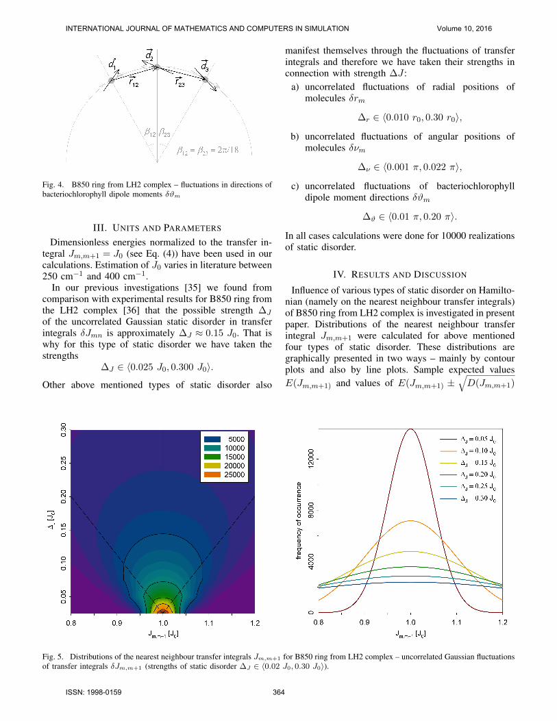
Fig. 4. B850 ring from LH2 complex – fluctuations in directions ofbacteriochlorophyll dipole moments δϑm
III. UNITS AND PARAMETERS
Dimensionless energies normalized to the transfer in-tegral Jm,m+1 = J0 (see Eq. (4)) have been used in ourcalculations. Estimation of J0 varies in literature between250 cm−1 and 400 cm−1.
In our previous investigations [35] we found fromcomparison with experimental results for B850 ring fromthe LH2 complex [36] that the possible strength ∆J
of the uncorrelated Gaussian static disorder in transferintegrals δJmn is approximately ∆J ≈ 0.15 J0. That iswhy for this type of static disorder we have taken thestrengths
∆J ∈ 〈0.025 J0, 0.300 J0〉.
Other above mentioned types of static disorder also
manifest themselves through the fluctuations of transferintegrals and therefore we have taken their strengths inconnection with strength ∆J :
a) uncorrelated fluctuations of radial positions ofmolecules δrm
∆r ∈ 〈0.010 r0, 0.30 r0〉,
b) uncorrelated fluctuations of angular positions ofmolecules δνm
∆ν ∈ 〈0.001 π, 0.022 π〉,
c) uncorrelated fluctuations of bacteriochlorophylldipole moment directions δϑm
∆ϑ ∈ 〈0.01 π, 0.20 π〉.
In all cases calculations were done for 10000 realizationsof static disorder.
IV. RESULTS AND DISCUSSION
Influence of various types of static disorder on Hamilto-nian (namely on the nearest neighbour transfer integrals)of B850 ring from LH2 complex is investigated in presentpaper. Distributions of the nearest neighbour transferintegral Jm,m+1 were calculated for above mentionedfour types of static disorder. These distributions aregraphically presented in two ways – mainly by contourplots and also by line plots. Sample expected valuesE(Jm,m+1) and values of E(Jm,m+1) ±
√D(Jm,m+1)
Fig. 5. Distributions of the nearest neighbour transfer integrals Jm,m+1 for B850 ring from LH2 complex – uncorrelated Gaussian fluctuationsof transfer integrals δJm,m+1 (strengths of static disorder ∆J ∈ 〈0.02 J0, 0.30 J0〉).
INTERNATIONAL JOURNAL OF MATHEMATICS AND COMPUTERS IN SIMULATION Volume 10, 2016
ISSN: 1998-0159 364
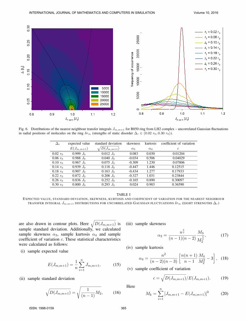
Fig. 6. Distributions of the nearest neighbour transfer integrals Jm,m+1 for B850 ring from LH2 complex – uncorrelated Gaussian fluctuationsin radial positions of molecules on the ring δrm (strengths of static disorder ∆r ∈ 〈0.02 r0, 0.30 r0〉).
∆r expected value standard deviation skewness kurtosis coefficient of variationE(Jm,m+1)
√D(Jm,m+1) α3 α4 c
0.02 r0 0.999 J0 0.012 J0 0.083 0.030 0.012040.06 r0 0.988 J0 0.040 J0 -0.034 0.506 0.040290.10 r0 0.967 J0 0.075 J0 -0.309 1.230 0.078060.14 r0 0.939 J0 0.118 J0 -0.447 1.446 0.125150.18 r0 0.907 J0 0.163 J0 -0.434 1.277 0.179330.22 r0 0.872 J0 0.208 J0 -0.327 1.031 0.238440.26 r0 0.836 J0 0.252 J0 -0.165 0.890 0.300970.30 r0 0.800 J0 0.293 J0 0.024 0.903 0.36590
TABLE IEXPECTED VALUE, STANDARD DEVIATION, SKEWNESS, KURTOSIS AND COEFFICIENT OF VARIATION FOR THE NEAREST NEIGHBOUR
TRANSFER INTEGRAL Jm,m+1 DISTRIBUTIONS FOR UNCORRELATED GAUSSIAN FLUCTUATIONS δrm (EIGHT STRENGTHS ∆r )
are also drawn in contour plots. Here√D(Jm,m+1) is
sample standard deviation. Additionally, we calculatedsample skewness α3, sample kurtosis α4 and samplecoefficient of variation c. These statistical characteristicswere calculated as follows:
(i) sample expected value
E(Jm,m+1) =1
n
n∑i=1
Jm,m+1, (15)
(ii) sample standard deviation
√D(Jm,m+1) =
√1
(n− 1)M2, (16)
(iii) sample skewness
α3 =n
5
2
(n− 1)(n− 2)
M3
M3
2
2
, (17)
(iv) sample kurtosis
α4 =n2
(n− 2)(n− 3)
[n(n+ 1)
n− 1
M4
M22
− 3
], (18)
(v) sample coefficient of variation
c =√D(Jm,m+1)/E(Jm,m+1). (19)
Here
Mk =n∑i=1
[Jm,m+1 − E(Jm,m+1)]k (20)
INTERNATIONAL JOURNAL OF MATHEMATICS AND COMPUTERS IN SIMULATION Volume 10, 2016
ISSN: 1998-0159 365

Fig. 7. Distributions of the nearest neighbour transfer integrals Jm,m+1 for B850 ring from LH2 complex – uncorrelated Gaussian fluctuationsin angular positions of molecules on the ring δνm (strengths of static disorder ∆ν ∈ 〈0.001 π, 0.022 π〉).
∆ν expected value standard deviation skewness kurtosis coefficient of variationE(Jm,m+1)
√D(Jm,m+1) α3 α4 c
0.001 π 1.000 J0 0.012 J0 0.042 0.004 0.012240.004 π 1.002 J0 0.049 J0 0.189 0.065 0.048720.007 π 1.005 J0 0.086 J0 0.339 0.211 0.085600.010 π 1.010 J0 0.124 J0 0.494 0.455 0.123100.013 π 1.017 J0 0.164 J0 0.657 0.815 0.161530.016 π 1.026 J0 0.206 J0 0.834 1.327 0.201220.019 π 1.037 J0 0.251 J0 1.018 1.959 0.242450.022π 1.050 J0 0.298 J0 1.164 2.348 0.28426
TABLE IIEXPECTED VALUE, STANDARD DEVIATION, SKEWNESS, KURTOSIS AND COEFFICIENT OF VARIATION FOR THE NEAREST NEIGHBOUR
TRANSFER INTEGRAL Jm,m+1 DISTRIBUTIONS FOR UNCORRELATED GAUSSIAN FLUCTUATIONS δνm (EIGHT STRENGTHS ∆ν )
and n is the number of cases in our samples(n = 180000). It corresponds with dimension of Hamil-tonian (N = 18) and number of static disorder realiza-tions (10000).
Uncorrelated Gaussian distributions of Jm,m+1 arepresented in Figure 5 for comparison to other types ofstatic disorder that are connected with deviations of ringgeometry. For this type of fluctuations (δJm,m+1), ofcourse, the expected value of the distribution of the near-est neighbour transfer integrals Jm,m+1 is independent ofthe strength of static disorder, i.e. E(Jm,m+1) = J0, andthe strength of static disorder ∆J equals the standarddeviation
√D(Jm,m+1).
Figure 6 shows the distributions of Jm,m+1 for Gaus-sian uncorrelated static disorder δrm in radial positions
of molecules on the ring. The distributions of Jm,m+1 forother two above mentioned types of static disorder canbe seen in Figure 7 (Gaussian uncorrelated fluctuationsof angular positions of molecules on the ring δνm)and in Figure 8 (Gaussian uncorrelated fluctuations ofmolecular dipole moment directions δϑm). For thesethree types of static disorder (connected with deviationsin ring geometry) expected value E(Jm,m+1) depends onstatic disorder strength. Dependencies of E(Jm,m+1) and√D(Jm,m+1) on corresponding static disorder strength
are presented in Figure 6 – left column (δrm), Figure 7– left column (δνm) and Figure 8 – left column (δϑm).Values of E(Jm,m+1),
√D(Jm,m+1), α3, α4 and c (see
Eq. (15) – Eq. (19)) for chosen static disorder strengths
INTERNATIONAL JOURNAL OF MATHEMATICS AND COMPUTERS IN SIMULATION Volume 10, 2016
ISSN: 1998-0159 366

Fig. 8. Distributions of the nearest neighbour transfer integrals Jm,m+1 for B850 ring from LH2 complex – uncorrelated Gaussian fluctuationsin directions of molecular dipole moments δϑm (strengths of static disorder ∆ϑ ∈ 〈0.02 π, 0.20 π〉).
∆ϑ expected value standard deviation skewness kurtosis coefficient of variationE(Jm,m+1)
√D(Jm,m+1) α3 α4 c
0.02 π 0.996 J0 0.009 J0 -0.760 0.951 0.009020.05 π 0.976 J0 0.033 J0 -1.676 4.585 0.033900.08 π 0.939 J0 0.073 J0 -1.952 5.819 0.077660.11 π 0.888 J0 0.126 J0 -1.941 5.350 0.141440.14 π 0.824 J0 0.186 J0 -1.811 4.260 0.225910.17 π 0.752 J0 0.250 J0 -1.629 3.056 0.332320.20 π 0.674 J0 0.312 J0 -1.428 1.964 0.46295
TABLE IIIEXPECTED VALUE, STANDARD DEVIATION, SKEWNESS, KURTOSIS AND COEFFICIENT OF VARIATION FOR THE NEAREST NEIGHBOUR
TRANSFER INTEGRAL Jm,m+1 DISTRIBUTIONS FOR UNCORRELATED GAUSSIAN FLUCTUATIONS δϑm (SEVEN STRENGTHS ∆ϑ)
are presented in Table I (δrm), Table II (δνm) and TableIII (δϑm).
In case of Gaussian distribution of transfer integralsJm,m+1 expected value E(Jm,m+1) is independent ofstatic disorder strength (E(Jm,m+1) = J0) and standarddeviation
√D(Jm,m+1) equals the strength of static
disorder√D(Jm,m+1) = ∆J . That is why, coefficient of
variation c corresponds to strength of static disorder ∆J ,i.e. c = ∆J/J0 (see Eq. (19)). In this case skewness α3
and kurtosis α4 equal zero, i.e. they are also independentof static disorder strength ∆J .
If we consider other types of static disorder (con-nected with deviations in ring geometry – δrm, δνm,δϑm), Gaussian distribution of molecular positions ordipole moment directions results in non–Gaussian dis-
tribution of transfer integrals Jm,m+1. That is why,expected value E(Jm,m+1), skewness α3 and kurtosisα4 are nonconstant and standard deviation
√D(Jm,m+1)
does not equal the strength of static disorder (see Figures6 – 8 and Tables I – III). As concerns expected valueE(Jm,m+1), we can see decrease of it for increasingstatic disorder strength in case of fluctuations in radialpositions of molecules on the ring δrm (see Figure 6 andTable I) and fluctuations in molecular dipole momentdirections δϑm (see Figure 8 and Table III). On theother hand, E(Jm,m+1) increases with growing strengthof static disorder in angular positions of molecules on thering δνm (see Figure 7 and Table II). In all these threecases dependence of standard deviation
√D(Jm,m+1) on
static disorder strength is nonlinear. The most important
INTERNATIONAL JOURNAL OF MATHEMATICS AND COMPUTERS IN SIMULATION Volume 10, 2016
ISSN: 1998-0159 367
change of expected value occurs in case of fluctua-tions of molecular dipole moment directions δϑm. Incontrast with this type of static disorder the changesof E(Jm,m+1) are very low for fluctuations in angularpositions of molecules δνm. Non–Gaussian distributionsof Jm,m+1 manifest themselves by nonzero skewness andkurtosis in all three cases of static disorder connectedwith deviations in ring geometry. Skewness is negativefor static disorder in radial positions of molecules δrmand static disorder in directions of molecular dipole mo-ments δϑm. Contrary, in case of static disorder in angularpositions of molecules δνm the distribution of Jm,m+1
is skewed to right hand side. Most significant skewnesscan be seen in case of static disorder in directions ofmolecular dipole moments δϑm (Figure 8). All thesethree distributions have higher kurtosis in comparisonwith Gaussian distribution of Jm,m+1.
Due to nonconstant expected value, influences ofdifferent types of fluctuations to distribution of Jm,m+1
can be compared using coefficient of variation. Ourprevious investigations [35] led to suitable strength ofstatic disorder in transfer integrals ∆J ≈ 0.15 J0 andconsequently c ≈ 0.15. As concerns other types od staticdisorder, approximately same value of coefficient ofvariation corresponds to the following disorder strengths:∆r ≈ 0.16 r0, ∆ν ≈ 0.012π and ∆ϑ ≈ 0.11π.
V. CONCLUSIONS
Comparison of the results obtained within differenttypes of static disorder can be summarized as follows.Expected value of the nearest neighbour transfer in-tegral distribution depends on static disorder strengthfor all presented types of fluctuations connected withring geometry. The most essential change appears incase of static disorder in dipole moment directions. Inthis case also the dependence of standard deviation ofthe distribution on the static disorder strength has thehighest nonlinearity. This is connected with the highestskewness of this distribution. Through the comparison ofcoefficient of variation we are able to estimate suitablestrength of static disorder types connected with ringgeometry fluctuations.
REFERENCES
[1] D. W. Lawlor, Photosynthesis, Spriger, New York 2001.[2] R. van Grondelle and V. I. Novoderezhkin, Energy transfer in photo-
synthesis: experimental insights and quantitative models, Phys. Chem.Chem. Phys. 8, 2003, pp. 793–807.
[3] G. McDermott, et al., Crystal structure of an integral membrane light-harvesting complex from photosynthetic bacteria, Nature 374, 1995, pp.517–521.
[4] M. Z. Papiz, et al., The structure and thermal motion of the B 800-B850LH2 complex from Rps. acidophila at 2.0 A resolution and 100 K: newstructural features and functionally relevant motions, J. Mol. Biol. 326,2003, pp. 1523–1538.
[5] W. P. F. de Ruijter, et al., Observation of the Energy–Level Structureof the Low–Light Adapted B800 LH4 Complex by Single–MoleculeSpectroscopy, Biophys. J. 87, 2004, pp. 3413–3420.
[6] R. Kumble and R. Hochstrasser, Disorder–induced exciton scat-tering in the light–harvesting systems of purple bacteria: Influ-ence on the anisotropy of emission and band → band transitions,J. Chem. Phys. 109, 1998, pp. 855–865.
[7] V. Nagarajan, et al., Femtosecond pump–probe spectroscopy of the B850antenna complex of Rhodobacter sphaeroides at room temperature,J. Phys. Chem. B 103, 1999, pp. 2297–2309.
[8] V. Nagarajan and W. W. Parson, Femtosecond fluorescence depletionanisotropy: Application to the B850 antenna complex of Rhodobactersphaeroides, J. Phys. Chem. B 104, 2000, pp. 4010–4013.
[9] V. Capek, I. Barvık and P. Herman, Towards proper parametrization inthe exciton transfer and relaxation problem: dimer, Chem. Phys. 270,2001, pp. 141–156.
[10] P. Herman and I. Barvık, Towards proper parametrization in the excitontransfer and relaxation problem. II. Trimer, Chem. Phys. 274, 2001, pp.199–217.
[11] P. Herman, I. Barvık and M. Urbanec, Energy relaxation and transferin excitonic trimer, J. Lumin. 108, 2004, pp. 85–89.
[12] P. Herman, et al., Exciton scattering in light–harvesting systems ofpurple bacteria, J. Lumin. 94–95, 2001, pp. 447–450.
[13] P. Herman and I. Barvık, Non–Markovian effects in the anisotropy ofemission in the ring antenna subunits of purple bacteria photosyntheticsystems, Czech. J. Phys. 53, 2003, pp. 579–605.
[14] P. Herman, et al., Influence of static and dynamic disorder on theanisotropy of emission in the ring antenna subunits of purple bacteriaphotosynthetic systems, Chem. Phys. 275, 2002, pp. 1–13.
[15] P. Herman and I. Barvık, Temperature dependence of the anisotropy offluorescence in ring molecular systems, J. Lumin. 122–123, 2007, pp.558–561.
[16] P. Herman, D. Zapletal and I. Barvık, Computer simulation of theanisotropy of fluorescence in ring molecular systems: Influence ofdisorder and ellipticity, Proc. IEEE 12th Int. Conf. on ComputationalScience and Engineering, Vancouver: IEEE Comp. Soc., 2009, pp. 437–442.
[17] P. Herman and I. Barvık, Coherence effects in ring molecular systems,Phys. Stat. Sol. C 3, 2006, 3408–3413.
[18] P. Herman, D. Zapletal and I. Barvık, The anisotropy of fluorescencein ring units III: Tangential versus radial dipole arrangement, J. Lu-min. 128, 2008, pp. 768–770.
[19] P. Herman, I. Barvık and D. Zapletal, Computer simulation of theanisotropy of fluorescence in ring molecular systems: Tangential vs.radial dipole arrangement, Lecture Notes in Computer Science 5101,2008, pp. 661–670.
[20] P. Herman, D. Zapletal and I. Barvık, Lost of coherence due to disorderin molecular rings, Phys. Stat. Sol. C 6, 2009, pp. 89–92.
[21] P. Herman, D. Zapletal and J. Slegr, Comparison of emission spectraof single LH2 complex for different types of disorder, Phys. Proc. 13,2011, pp. 14–17.
[22] D. Zapletal and P. Herman, Simulation of molecular ring emissionspectra: localization of exciton states and dynamics, Int. J. Math. Comp.Sim. 6, 2012, pp. 144–152.
[23] M. Horak, P. Herman and D. Zapletal, Simulation of molecular ringemission spectra – LH4 complex: localization of exciton states anddynamics, Int. J. Math. Comp. Sim. 7, 2013, pp. 85–93.
[24] P. Herman and D. Zapletal, Intermolecular coupling fluctuation effecton absorption and emission spectra for LH4 ring, Int. J. Math. Comp.Sim. 7, 2013, pp. 249–257.
[25] M. Horak, P. Herman and D. Zapletal, Modeling of emission spectrafor molecular rings – LH2, LH4 complexes, Phys. Proc. 44, 2013, pp.10–18.
[26] P. Herman, D. Zapletal and M. Horak, Emission spectra of LH2complex: full Hamiltonian model, Eur. Phys. J. B 86, 2013, art. no.215.
[27] P. Herman and D. Zapletal, Emission Spectra of LH4 Complex: FullHamiltonian Model, Int. J. Math. Comp. Sim. 7, 2013, pp. 448–455.
[28] P. Herman and D. Zapletal, Simulation of Emission Spectra for LH4Ring: Intermolecular Coupling Fluctuation Effect, Int. J. Math. Comp.Sim. 8, 2014, pp. 73–81.
[29] D. Zapletal and P. Herman, Photosynthetic complex LH2 – Absorptionand steady state fluorescence spectra, Energy 77, 2014, pp. 212–219.
[30] P. Herman and D. Zapletal, Simulations of emission spectra for LH4Ring – Fluctuations in radial positions of molecules, Int. J. Biol. Biomed.Eng. 9, 2015, pp. 65–74.
[31] P. Herman and D. Zapletal, Computer simulation of emission andabsorption spectra for LH2 ring, LNEE 343, 2015, pp. 221–234.
INTERNATIONAL JOURNAL OF MATHEMATICS AND COMPUTERS IN SIMULATION Volume 10, 2016
ISSN: 1998-0159 368

[32] P. Herman and D. Zapletal, Modeling of Absorption and Steady StateFluorescence Spectra of Full LH2 Complex (B850 – B800 Ring), Int.J. Math. Mod. Meth. Appl. Sci. 9, 2015, pp. 614–623.
[33] P. Herman and D. Zapletal, Modeling of Emission and AbsorptionSpectra of LH2 Complex (B850 and B800 Ring) – Full HamiltonianModel, Int. J. Math. Comp. Sim. 10, 2016, pp. 208–217.
[34] P. Herman and D. Zapletal, B–α/B–β Ring from Photosynthetic Com-plex LH4, Modeling of Absorption and Fluorescence Spectra, Int. J.Math. Comp. Sim. 10, 2016, pp. 332–344.
[35] P. Herman, I. Barvık and D. Zapletal, Energetic disorder and excitonstates of individual molecular rings, J. Lumin. 119–120, 2006, pp. 496–503.
[36] C. Hofmann, T. J. Aartsma and J. Kohler, Energetic disorder and theB850–exciton states of individual light–harvesting 2 complexes fromRhodopseudomonas acidophila, Chem. Phys. Lett. 395, 2004, pp. 373–378.
INTERNATIONAL JOURNAL OF MATHEMATICS AND COMPUTERS IN SIMULATION Volume 10, 2016
ISSN: 1998-0159 369