award number: w81xwh-14-1-0537 - dtic.mil · an achromatic wide angle keplerian telescope beam...
TRANSCRIPT

AWARD NUMBER: W81XWH-14-1-0537
TITLE: Mobile, Multimodal, Label-Free Imaging Probe Analysis of Choroidal Oximetry and Retinal Hypoxia
PRINCIPAL INVESTIGATOR: Dr. Stephen T.C. Wong
CONTRACTING ORGANIZATION: The Methodist Hospital Houston TX 77030
REPORT DATE: December 2017
TYPE OF REPORT: Final
PREPARED FOR: U.S. Army Medical Research and Materiel Command Fort Detrick, Maryland 21702-5012
DISTRIBUTION STATEMENT: Approved for Public Release; Distribution Unlimited
The views, opinions and/or findings contained in this report are those of the author(s) and should not be construed as an official Department of the Army position, policy or decision unless so designated by other documentation.

REPORT DOCUMENTATION PAGE Form Approved
OMB No. 0704-0188 Public reporting burden for this collection of information is estimated to average 1 hour per response, including the time for reviewing instructions, searching existing data sources, gathering and maintaining the data needed, and completing and reviewing this collection of information. Send comments regarding this burden estimate or any other aspect of this collection of information, including suggestions for reducing this burden to Department of Defense, Washington Headquarters Services, Directorate for Information Operations and Reports (0704-0188), 1215 Jefferson Davis Highway, Suite 1204, Arlington, VA 22202-4302. Respondents should be aware that notwithstanding any other provision of law, no person shall be subject to any penalty for failing to comply with a collection of information if it does not display a currently valid OMB control number. PLEASE DO NOT RETURN YOUR FORM TO THE ABOVE ADDRESS. 1. REPORT DATEDecember 2017
2. REPORT TYPEFinal
3. DATES COVERED30 Sep 2014 - 29 Sep 2017
4. TITLE AND SUBTITLEMobile, Multimodal, Label-Free Imaging Probe Analysis ofChoroidal
5a. CONTRACT NUMBER
Choroidal Oximetry and Retinal Hypoxia 5b. GRANT NUMBER W81XWH-14-1-0537 5c. PROGRAM ELEMENT NUMBER
6. AUTHOR(S)Stephen T.C. Wong, Jiasong Li
5d. PROJECT NUMBER
5e. TASK NUMBER
E-Mail: [email protected]
5f. WORK UNIT NUMBER
7. PERFORMING ORGANIZATION NAME(S) AND ADDRESS(ES)
AND ADDRESS(ES)
8. PERFORMING ORGANIZATION REPORTNUMBER
The Methodist Hospital Research Institute 6670 Bertner Ave. Houston, TX 77030
9. SPONSORING / MONITORING AGENCY NAME(S) AND ADDRESS(ES) 10. SPONSOR/MONITOR’S ACRONYM(S)
U.S. Army Medical Research and Materiel Command Fort Detrick, Maryland 21702-5012 11. SPONSOR/MONITOR’S REPORT
NUMBER(S)
12. DISTRIBUTION / AVAILABILITY STATEMENT
Approved for Public Release; Distribution Unlimited
13. SUPPLEMENTARY NOTES
14. ABSTRACTFocus Area: Mitigation and treatment of traumatic injuries, war-related injuries, and diseases to ocular structures and the visual system
Rationale: Ocular trauma occurs in 13% of the injuries sustained by wounded soldiers in active combat zones, with damage to the retina occurring in at least 50% of these same eye injuries. Primary blast-induced injury (PBI), which can occur in eyes that are not punctured or ruptured by the blast, is correlated with a high degree of permanent visual impairment. The cause of this deterioration of visual function is currently unknown. However, one aspect of injury that is thought to contribute to permanent visual loss is the combination of chronic ischemia and resulting neurotoxicity that greatly affect function of the retina. In retinas where these processes occur, their effects ultimately can lead to irreversible neurodegeneration of the delicate, light-sensing photoreceptor neurons, which are essential to proper visual function.
Retinal ischemia is caused by insufficient oxygen levels reaching the retinal neurons. Neurons such as the photoreceptors alter their cellular behavior under these hypoxic stress conditions, and eventually begin to die if oxygen levels are not restored to normal. One possible cause of ischemia/hypoxia in the retina is damage to the choroidal vasculature that supplies oxygen to the retina. The choroidal capillaries that supply the retina can become damaged following PBI injury, leading to oxygen starvation of the photoreceptors. Altered cellular behavior may be driven by TRPM7, which was is present in photoreceptor neurons. TRPM7 function is influenced by cellularoxygen supplies and may regulate hypoxia-induced cellular stress responses in photoreceptors themselves.
Objective: Our aim is to examine the potential link between trauma-induced hypoxic/ischemic conditions in the choroid and anoxic activation of TRPM7 leading to photoreceptor cell death. Using the mobile biophotonics device developed in this proposal to measureblood oxygen levels in the eye following an injury and the function of TRPM7 in regions of the retina underlying damaged choroidal blood vessels, we seek to understand how long-term, permanent vision loss results from traumatic eye injuries.
Applicability and Potential Impact: The described research seeks to stabilize and improve long-term vision in combat veterans who havesustained primary blast injuries (PBI) to the eye without physical rupture or puncture of the eye. Retinal injury results in over 50% ofsustained combat eye injuries, and likely contributes to long-term vision loss after the initial injury. By understanding the role TRPM7plays in hypoxic stress, it may be possible to halt vision loss by inhibiting abnormal TRPM7 function, thus aiming to return its function tonormal, or by developing oxygen-based therapies to mitigate the initial progression of hypoxia.
Military Benefit: Our research seeks to provide a mobile endoscope probe with the potential to be used by military clinicians to quickly
15. SUBJECT TERMS CARS, imaging, microscopy, blast-injury, retina, neuron
16. SECURITY CLASSIFICATION OF: 17. LIMITATION 18. NUMBER 19a. NAME OF RESPONSIBLE PERSON
OF ABSTRACT OF PAGES
a. REPORT
Unclassified
b. ABSTRACT
Unclassified
c. THIS PAGE
Unclassified Unclassified 16
19b. TELEPHONE NUMBER (include area code)
Standard Form 298 (Rev. 8-98) Prescribed by ANSI Std. Z39.18

Table of Contents
Page
1. Introduction………………………………………………………….5
2. Keywords…………………………………………………………….5
3. Accomplishments………..…………………………………………...5
4. Impact…………………………...……………………………………13
5. Changes/Problems...….………………………………………………14
6. Products…………………………………….……….….…………….14
7. Participants & Other Collaborating Organizations………………15
5
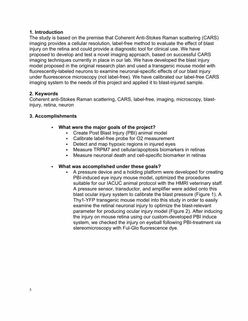
1. IntroductionThe study is based on the premise that Coherent Anti-Stokes Raman scattering (CARS)imaging provides a cellular resolution, label-free method to evaluate the effect of blastinjury on the retina and could provide a diagnostic tool for clinical use. We haveproposed to develop and test a novel imaging approach, based on successful CARSimaging techniques currently in place in our lab. We have developed the blast injurymodel proposed in the original research plan and used a transgenic mouse model withfluorescently-labeled neurons to examine neuronal-specific effects of our blast injuryunder fluorescence microscopy (not label-free). We have calibrated our label-free CARSimaging system to the needs of this project and applied it to blast-injured sample.
2. KeywordsCoherent anti-Stokes Raman scattering, CARS, label-free, imaging, microscopy, blast-injury, retina, neuron
3. Accomplishments
§ What were the major goals of the project?§ Create Post Blast Injury (PBI) animal model§ Calibrate label-free probe for O2 measurement§ Detect and map hypoxic regions in injured eyes§ Measure TRPM7 and cellular/apoptosis biomarkers in retinas§ Measure neuronal death and cell-specific biomarker in retinas
§ What was accomplished under these goals?§ A pressure device and a holding platform were developed for creating
PBI-induced eye injury mouse model, optimized the proceduressuitable for our IACUC animal protocol with the HMRI veterinary staff.A pressure sensor, transductor, and amplifier were added onto thisblast ocular injury system to calibrate the blast pressure (Figure 1). AThy1-YFP transgenic mouse model into this study in order to easilyexamine the retinal neuronal injury to optimize the blast-relevantparameter for producing ocular injury model (Figure 2). After inducingthe injury on mouse retina using our custom-developed PBI inducesystem, we checked the injury on eyeball following PBI-treatment viastereomicroscopy with Ful-Glo fluorescence dye.

6
Figure 1. PBI-device build up and optimization. (1A-F) The component of our PBI-devices, output pressure detection sensor, amplifier, and input pressure panel. (1G) Correlation between input-output pressures. (1H) Measurement of the pressure change at different distance from the barrel.
Figure 2. Optimization of output pressure by changing the setting of blast generator. (2A-B) Correlation between output pressure and blast time duration. (2C) After PBI-treatment, the eyes of dead animal were labeled by Ful-Glo fluorescence, observed under the close-global injury and morphology of eyeball with a stereomicroscope

7
with blue light. (2D-G) In the control and all three blasted groups, the overall globe and the cornea are intact while the dye appears smooth across the entire surface of the eye with treatment. Blast pressure at these levels did not cause open-globe injury.
§ Fresh hemoglobin solution (18mg/ml) was prepared by dissolving in PBS (0.1M, pH 7.2). The hemoglobin solution was filtered and aliquoted to three tubes, which were perfused with O2, air, or CO2 for 5 minutes. The oxidized hemoglobin (oxiHg) levels were examined with our label-free CARS imaging system. A ten microliter sample was transferred to a hemometer, covered with a glass coverslip, focused under 60 x10 magnitude, and detected with CARS system. We repeated the experiment 4 times. CARS signal was detected for each sample within 10 minutes following perfusion. As shown in Figures 3 and 4, CARS signal is exactly correlated with the oxygen level in the perfused Hg solution (R2=0.998). This reinforces our hypothesis that the label-free CARS imaging system is powerful to examine oxidized Hg level with high fidelity.
Figure 3. Calibration of CARS imaging system with hemoglobin solution.

8
Figure 4. Test the efficacy of CARS in detection of oxidized hemoglobin in vitro. Table 1. The value of CARS and oxygen level (n=2-4)
CO2 Air O2 CARS 479 1723 6264
Oxygen level 5 21 95 n 3 2 4
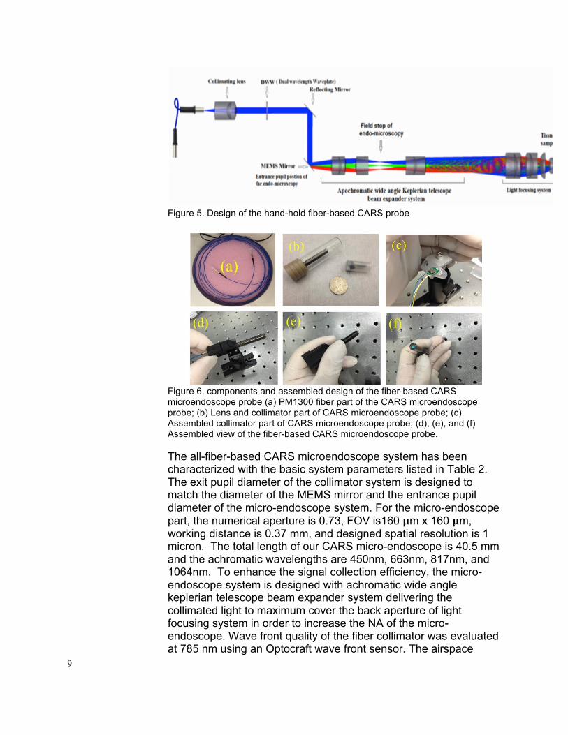
A fiber-based hand-held probe was designed (Figure 5), assembled, fabricated, and calibrated for easy application (Figure 6). The probe used a customized MEMS scanning mirror and miniature optical and mechanical components as originally proposed. Figure 3 shows the design of the probe, and Figure 4 shows the components and assembled design of the fiber-based CARS microendoscope probe. The CARS excitation laser emerging from the fiber is coupled into the collimator system with suitable numerical aperture match. The collimated light is scanned by the MEMS mirror and then projected to the micro-objective subsystem which is aimed to achieve a large NA (numerical aperture) and high light coupling efficiency. The micro-objective subsystem contains an achromatic wide angle keplerian telescope beam expander and light focusing subsystem. The achromatic wide angle keplerian telescope beam expander is used to amplify the entrance light to fill the back aperture in order to insure maximum NA. The field of view (FOV) of the collimator system is the same as the MEMS mirror effective reflecting area.
9
Figure 5. Design of the hand-hold fiber-based CARS probe
Figure 6. components and assembled design of the fiber-based CARS microendoscope probe (a) PM1300 fiber part of the CARS microendoscope probe; (b) Lens and collimator part of CARS microendoscope probe; (c) Assembled collimator part of CARS microendoscope probe; (d), (e), and (f) Assembled view of the fiber-based CARS microendoscope probe. The all-fiber-based CARS microendoscope system has been characterized with the basic system parameters listed in Table 2. The exit pupil diameter of the collimator system is designed to match the diameter of the MEMS mirror and the entrance pupil diameter of the micro-endoscope system. For the micro-endoscope part, the numerical aperture is 0.73, FOV is160 𝛍m x 160 𝛍m, working distance is 0.37 mm, and designed spatial resolution is 1 micron. The total length of our CARS micro-endoscope is 40.5 mm and the achromatic wavelengths are 450nm, 663nm, 817nm, and 1064nm. To enhance the signal collection efficiency, the micro-endoscope system is designed with achromatic wide angle keplerian telescope beam expander system delivering the collimated light to maximum cover the back aperture of light focusing system in order to increase the NA of the micro-endoscope. Wave front quality of the fiber collimator was evaluated at 785 nm using an Optocraft wave front sensor. The airspace

10
between the fiber and optics was adjusted to achieve wave front quality smaller than 0.05 λ RMS (root mean square) at 785 nm, which is within the design tolerance 0.5 λ at 785 nm. Lights of different wavelengths (575nm, 660nm, and 850nm) were used to test the resolution as shown in the figure 7. The USAF resolution target was placed at the focus position of the microendoscope barrel, the 20 mm objective lens and image receiver (Samsung phone) were used to acquire a high resolution targeted image. The smallest element, 3 in the 9th group, was resolved by the microendscope. It has a line spacing of 645 line pairs/mm, corresponding to a line width of approximately 0.78 µm. There is no distortion in the shape of the individual lines in the image except for a conical defocus deformation in the 660nm resolution image in which the transmission resolution image is not at the center of the objective. Table 2. Basic system parameters of CARS microendoscope probe Basic System Descriptions Parameters Excitation Wavelength range for delivery 817nm(pump), 1064nm(stokes)
Signal wavelength to be collected from tissue
663nm(CARS) 500nm(TPEF) 400nm(SHG)
Input aperture (diameter of exit pupil) Agree with the dimension of MEMS scanning mirror 1.12mm
Distance from the MEMS mirror to objective lens 4mm NA(after immersion in water) 0.75 Actual resolution 0.78 micron or smaller Field of view 160𝜇mx160𝜇m
Figure 7. Resolution measurement of microendoscope probe at (b) 575nm, (c) 660nm, and (d) 850nm respectively.
§ The whole retina was isolated for protein analysis and pathological
assays, and three different triple whole-mount staining was introduced to examine the morphology of neurite, cell body, and blood vessel network to determine the vulnerable retinal regions for future biochemical and pathological analysis. Using CARS imaging technology, we detected the hypoxic map on

11
whole mount retinal tissue from acutely ocular injured mice. Wild type C57B6/J mice (n= 3) were anesthetized following our protocol, and mounted in PBI eye injury device. The right eye was exposed to blast (21psi, 8.5 ms duration) generated from a paintball gun (Mini GS, Empire Inc), while the left eye was used as control. Mice were sacrificed and whole mount retinas were freshly isolated following our protocol. Fresh retinal tissues were incubated in D-Hanks solution in a mini chamber on glass slides for label free CARS imaging system (pump wavelength, 817nm, stoke wavelength, 1064nm), the de-oxidized hemoglobin (deoxy-Hb) provides high intensity in CARS images, as shown in (Figure 8).
Figure 8. Detection of hypoxic map in the whole mount retinas. Left: Home-developed femtosecond (fs) CARS imaging system used in this study. Right: Hypoxic map in the whole mount retinas. The bright pixels indicate the de-oxidized
§ Using fresh retinal tissues, we compared the total protein levels of TRPM7 in the control and lesion group via western blot. Part of retinal tissues were dissected and snapped frozen on dry ice, and thawed in 1x RIPA lysis buffer with protease inhibitor cocktail (1x, Roche Inc). Tissues were then briefly sonicated and spun to harvest protein. 60ug samples were loaded on 4-20% SDS PAGE gel, followed by regular western blot protocol. NC membrane with protein translots was incubated in primary antibodies (anti-TRPM7, 1:2000, ProSci Inc; or anti-beta actin, 1: 3000, Cell Signaling Lab) overnight at 4 degree, then followed by incubation with HRP-conjugated secondary antibodies (1: 8000, Thermo Scientific ). Enhanced ECL was used to generate luminescence signal and Image J software was used to detect signal intensity. After normalization with internal standard, a

12
1.25-fold increase was shown in the protein levels of TRPM7 in the lesion side retinas (Figure 9). In the future, we will use immunofluorescence imaging to detect the distribution of TRPM7 in different retinal zones to overlap our CARS imaging results and other apoptosis markers.
Figure 9. PBI induces a mild increase in TRMP7 protein in the lesion side retinas.
§ YFP mice were used to generate PBI ocular injury. Whole mount retinas were isolated to detect neuronal injury and cell-specific biomarker via morphological analysis. GFP-labeled neuron numbers and neurite morphology (intersection in 2 different zones-10µm or 45 µm distant to the soma) were examined. We did not find significant change in neuron numbers in lesion side retinas. However, the distant neurite number was decreased in the lesion side (Figure 10).
§
Figure 10. Measure neuronal death and cell-specific biomarker in retinas. (A) Whole mount retinal was dissected. Neurites near and distant to the soma were examined by counting the intersection number at 10µm and 45 µm distance (B) control, and (C) lesion side.

13
§ What opportunities for training and professional development has the
project provided? § One PhD student, Weng Sheng and three Postdocs, Jared Gilliam,
Xiaoyun Xu, and Jiasong Li, were supported by this funding to conduct their research.
§ § How were the results disseminated to communities of interest?
§ Nothing to Report.
§ What do you plan to do during the next reporting period to accomplish the goals?
§ Nothing to Report. 4. Impact
a. What was the impact on the development of the principal discipline(s) of the project?
i. We have created PBI animal model as proposed. ii. We have developed, fabricated, and calibrated the hand-hold, fiber-
based CARS microendoscopy probe to measure the oxy-hemoglobin content as proposed.
iii. We have detected, and map the hypoxic regions in injured PBI retinas using CARS imaging system as proposed, adopted probe into this measurement is currently in process.
iv. We have measured TPRM7 biomarker in mapped hypoxic eye regions as proposed.
v. We have analyzed and evaluated TPRM7 activity in both normal and injured retina as proposed, more analyzing is currently in process.
b. What was the impact on other disciplines? i. Our label-free, superior resolution, and real-time CARS imaging
system can improve the diagnosis in both speed and accuracy. c. What was the impact on technology transfer?
1. Our label-free, superior resolution, and real-time CARS imaging system can be used for many other clinical or biology applications. Since it targets specific chemical-bonds, it can image all biological material contains such chemical-bond. The cellular-level resolution image is comparable with labeled imaging technologies, such as fluorescence imaging, confocal imaging, two-photon image, etc. CARS imaging has high potential to be applied in live animals, even in clinical usage since no label agents is required.
2. Our miniaturized imaging platform (CARS endoscope) can potentially be integrated into the arms of a surgical robot such as da Vinci robotic surgery system due to the minimal cross sectional area of imaging probe, and will reduce the number of unnecessary biopsies and the risk of nerve impairment during

14
the surgical procedures by providing surgeons and urologists a precise picture of prostatic and peri-prostatic tissues.
3. The miniaturized imaging platform has a great potential to be integrated into the arms of surgical robots and revolutionize the ways of prostate surgeries being operated nowadays.
d. What was the impact on society beyond science and technology? i. Nothing to Report.
5. Changes/Problems The Project Director/Principal Investigator (PD/PI) is reminded that the recipient organization is required to obtain prior written approval from the awarding agency Grants Officer whenever there are significant changes in the project or its direction. If not previously reported in writing, provide the following additional information or state, "Nothing to Report," if applicable:
§ Changes in approach and reasons for change § Nothing to report
§ Actual or anticipated problems or delays and actions or plans to resolve them
§ The laser diode was replaced, and re-alignment of the laser, followed by the alignment of the whole imaging system was performed after the diode replacement. It took the laser manufacture more than 10 months to fabricate, deliver, install, align, and test the laser, and another one month for us to put the CARS imaging system to perfect working order.
§ Changes that had a significant impact on expenditures § Nothing to report.
§ Significant changes in use or care of human subjects, vertebrate animals,
biohazards, and/or select agents § Nothing to report.
6. Products
§ Publications, conference papers, and presentations A manuscript outlining our research accomplishments is currently under preparation.
§ Website(s) or other Internet site(s) Nothing to report.
§ Technologies or techniques Nothing to report.
§ Inventions, patent applications, and/or licenses
Nothing to Report.
§ Other Products

15
Nothing to report. 7. Participants & Other Collaborating Organizations
§ What individuals have worked on the project? § Provide the following information for: (1) PDs/PIs; and (2) each person who
has worked at least one person month per year on the project during the reporting period, regardless of the source of compensation (a person month equals approximately 160 hours of effort). If information is unchanged from a previous submission, provide the name only and indicate "no change."
Name: Sheng Weng Project Role: PhD Graduate Student Researcher Identifier (e.g. ORCID ID): Nearest person month worked: 6 Contribution to Project:
Dr. Weng has performed work in the area of biophotonics experimentations
Funding Support: Mr. Weng’s salary was supported by Rice University.
Name: Jared Gilliam Project Role: Postdoc Researcher Identifier (e.g. ORCID ID):
Nearest person month worked: 6 months 9/30/14-9/29/15
Contribution to Project: Dr. Gilliam has performed work in the design and experimentation.
Funding Support: N/A
Name: Dongbing Gao Project Role: Technician Researcher Identifier (e.g. ORCID ID):
Nearest person month worked: 6 months per funding year Contribution to Project: Mr. Gao performed basic laboratory research tasks as

16
needed. Funding Support: N/A
Name: Xiaoyun Xu Project Role: Postdoc Researcher Identifier (e.g. ORCID ID):
Nearest person month worked:
12 months – 9 months from Apr16-Apr17 and 3 months from Apr17-Sep17.
Contribution to Project: Dr. Xu has performed work in the validation experimentations
Funding Support: N/A
Name: Jiasong Li Project Role: Postdoc Researcher Identifier (e.g. ORCID ID):
Nearest person month worked: 8 months – Apr16-Sep17
Contribution to Project: Dr. Xu has performed work in the validation experimentation and intepretation of results
Funding Support: N/A
Name: Stephen Wong Project Role: PI Researcher Identifier (e.g. ORCID ID): 143285 Nearest person month worked: 1 month per funding year
Contribution to Project: Dr. Wong led the design and execution of the project, supervision postdocs and staff.
Funding Support: N/A
§ Has there been a change in the active other support of the PD/PI(s) or senior/key personnel since the last reporting period?

17
§ Nothing to report § What other organizations were involved as partners?
§ Nothing to report