autophagy in stress, development and disease gordon research conference 2003 chair: beth levine...
TRANSCRIPT

Autophagy in Stress, Development and Disease
Gordon Research Conference2003
Chair: Beth Levine (Columbia University)Vice-Chair: Dan Klionsky (University of Michigan)
Outline1. What is Autophagy?2. Macroautophagy in yeast and mammalian cells.3. Markers of autophagy.4. TOR signaling and growth control.5. Autophagy in development and aging.6. Autophagy in plant and mammalian cells.7. Autophagy, signaling, and oncogenesis.8. Autophagy in host response to microbial invasion.9. Autophagy deregulation in disease.10.Autophagy and apoptosis.11.Summary

What is autophagy?
There are several different types of autophagy: Microautophagy,chaperone-mediated autopahgy, and Macroautopahgy. In addition, thereare other routes for transporting cargo to the lysosomal compartmentfor degradation (e.g. macropexopahgy, micropexophagy, cytoplasm-to-vacuole (Cvt) pathway, multivesicular body (Mvb) pathway).
Macroautophagy is the main route for sequestration of cytoplasm to the lyticcompartment and is conserved in all nucleate cell types analyzed (yeast, plants,Animals). Genetic screens in yeast have identified some of the genes required.
These genes are called apg or aut, with some overlap with Cvt genes.New common nomenclature proposed at this meeting: Atg
Autophagy (American Heritage Dictionary)Syllabication: au-toph-a-gyPronunciation: o-tofe-jeNoun: The process of self-digestion by a cell through the action of enzymes originating within the same cell.

Role of Autophagy
Three main functions:
I. Adaptive response to nutrient starvation.
II. Housekeeping mechanism involved in cytoplasmic homeostasis.
III. Tissue-specific functions. e.g. intracellular biogenesis of surfactant, erythroid maturation

Macroautophagy in yeast
Marcoautophagy is a dynamic process involving the rearrangement of subcellular membranes to sequester cytoplasm and organelles for delivery to the lysosome where the sequestered cargo is degraded and recycled. Autophagy is responsible for degradation of organelles and long-lived proteins (half-life > 5 hrs).
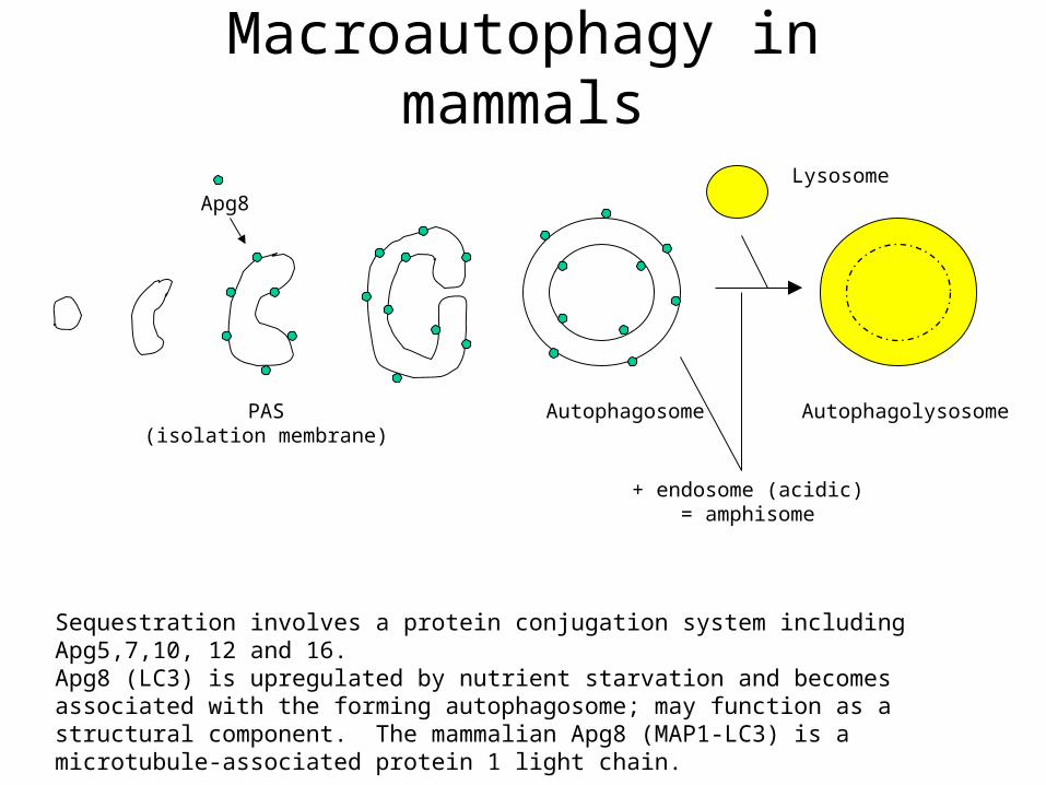
Macroautophagy in mammals
Sequestration involves a protein conjugation system including Apg5,7,10, 12 and 16.Apg8 (LC3) is upregulated by nutrient starvation and becomes associated with the forming autophagosome; may function as a structural component. The mammalian Apg8 (MAP1-LC3) is a microtubule-associated protein 1 light chain.
Lysosome
AutophagolysosomeAutophagosomePAS(isolation membrane)
Apg8
+ endosome (acidic)= amphisome
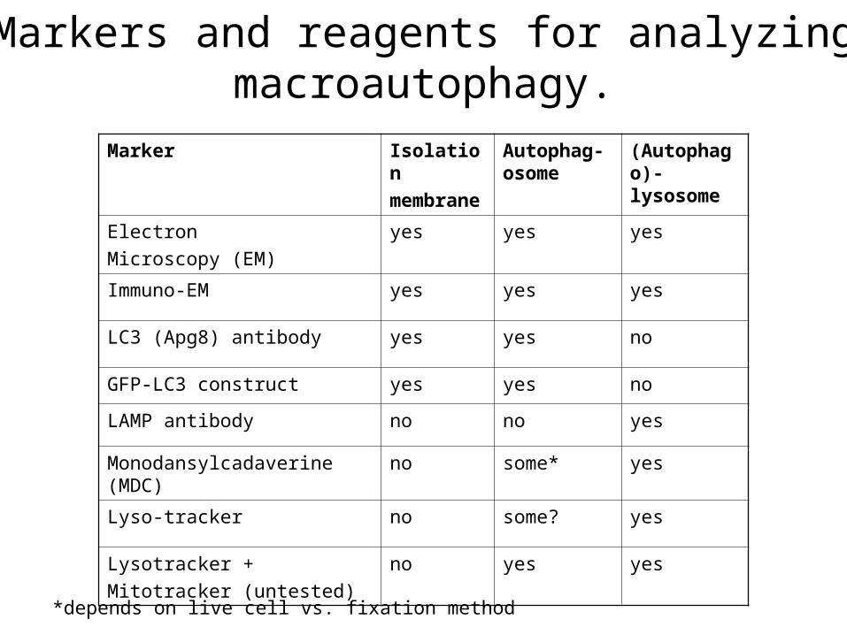
Markers and reagents for analyzing macroautophagy.
Marker Isolation membrane
Autophag-osome
(Autophago)-lysosome
Electron Microscopy (EM)
yes yes yes
Immuno-EM yes yes yes
LC3 (Apg8) antibody yes yes no
GFP-LC3 construct yes yes no
LAMP antibody no no yes
Monodansylcadaverine (MDC)
no some* yes
Lyso-tracker no some? yes
Lysotracker +Mitotracker (untested)
no yes yes
*depends on live cell vs. fixation method
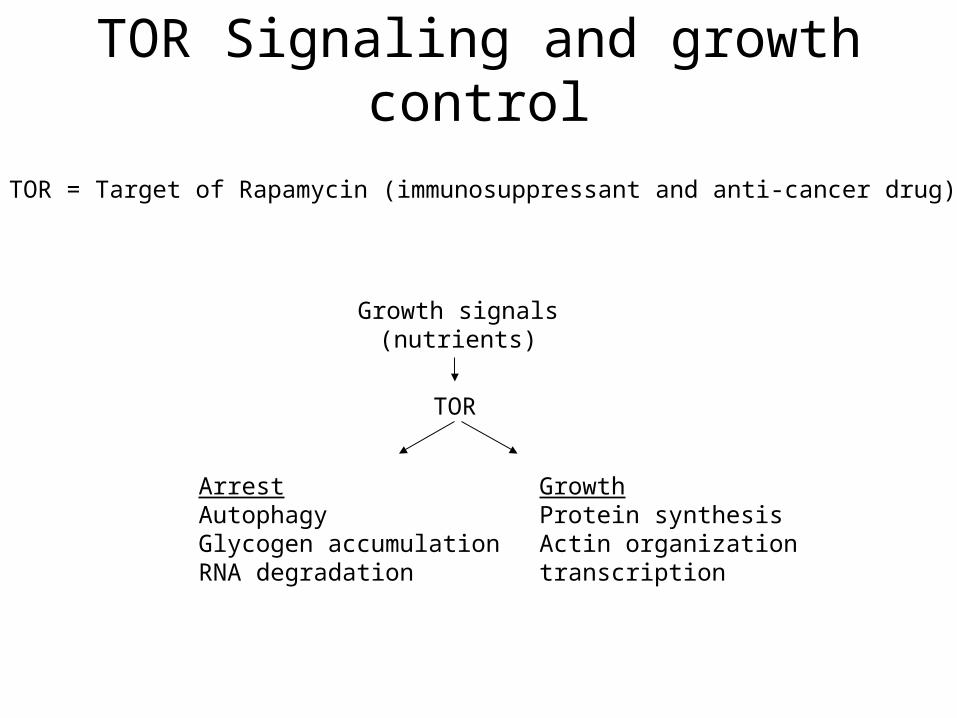
TOR Signaling and growth control
TOR = Target of Rapamycin (immunosuppressant and anti-cancer drug)
Growth signals(nutrients)
TOR
ArrestAutophagyGlycogen accumulationRNA degradation
GrowthProtein synthesisActin organizationtranscription

Mike Hall (U of Basel)TOR Signaling and Growth Control in Yeast
TOR1/2
Apg1 (S/T kinase)
Autophagy
Observation: When you treat cells with cAMP, you inhibit rap-induced autophagy.
Found that Tor is upstream of cAMP and PKA signaling; APG1 has no PKA phosphorylation sites so something must be acting in between.
Ras/cAMP/PKA

Tom Neufeld (U of Minnesota)Role of Autophagy in Tor-mediated Cellular
Growth in Drosophila
Model system = Drosophila fat body (equivalent of the liver);Cells are large and polyploid, and in a single layer; play a role in Detoxification, CHO metabolism and Energy storage.
Starvation induces inactivation of TOR, and a rapid decrease in fat body;Fat body cells shrink up to 90%; see an increase in autophagy
Assayed autophagy using lysotracker incorporation; also showed some GFP-Apg8localization.Drosophila apg1/aut3 mutant = pupal lethal
No lysotracker staining observed when induced starvation
Drosophila apg1,apg5,apg6, aut10, and S6K are required for starvation-induced autophagy (showed data for apg1 and S6K only; using RNAi lines from Liqun Luo at Stanford U, works on nervous system and autophagy)

Miklos Sass (Budapest, Hungary)Poster: Searching for genes regulating
autophagocytosis in insects
Model system: Drosophila fat body; Autophagy induced by 20-hydroxyecdysone at the L3 Feeding – L3 Wandering transition(I.e. if pick wandering L3s, you should see large #s of autophagosomes/lysosomes)
Did P element screen for L3 W mutants with reduced/absent autophagy in fat bodyAssays: Acridine orange staining, Neutral Red (like Lysotracker; stains acidic compartments), EM• also doing some cDNA microarray analyses (small home-made microarrays; CG7224)
Found mutants (approx 15/300) with reduced/absent staining; found mutant with no fusion between autophagosome and lysosomes
Caveats: some mutants weak; developmentally delayedSalivary glands examined? ( I have list of some of the mutants)
Note: Miklos commented that there do not appear to be many autophagosomes in the salivary gland (ie. by EM; timing?)
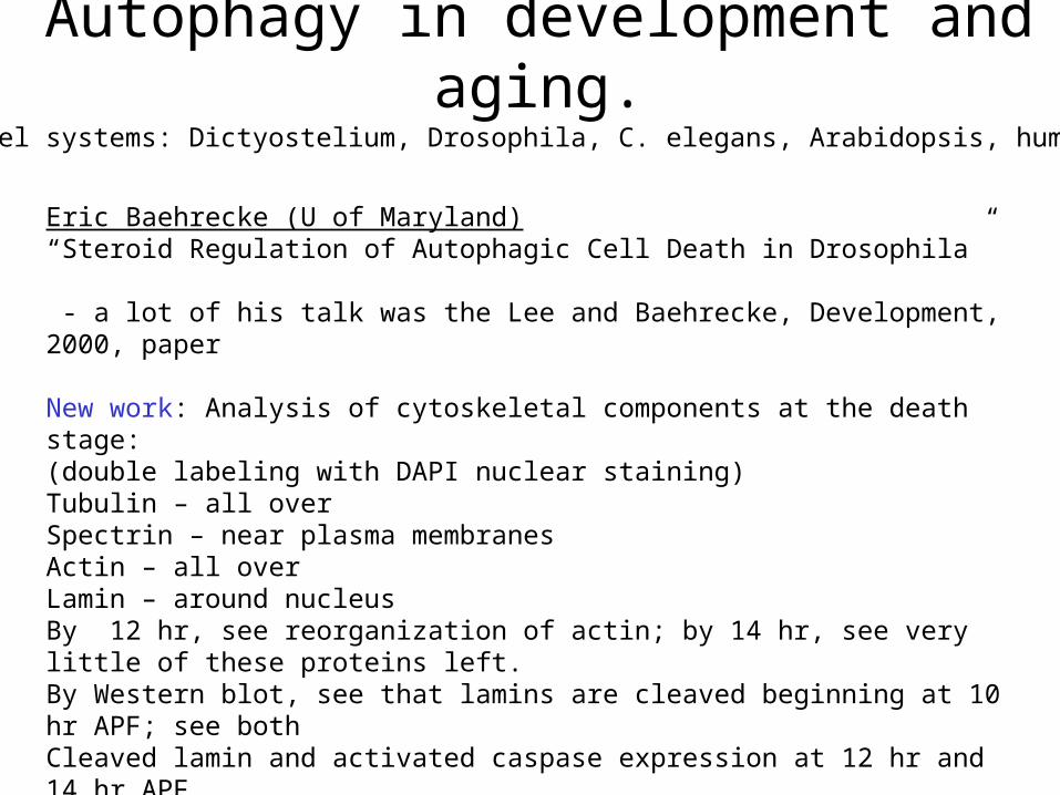
Autophagy in development and aging.
Model systems: Dictyostelium, Drosophila, C. elegans, Arabidopsis, humans
Eric Baehrecke (U of Maryland)“Steroid Regulation of Autophagic Cell Death in Drosophila”
- a lot of his talk was the Lee and Baehrecke, Development, 2000, paper
New work: Analysis of cytoskeletal components at the death stage: (double labeling with DAPI nuclear staining)Tubulin – all overSpectrin – near plasma membranesActin – all overLamin – around nucleusBy 12 hr, see reorganization of actin; by 14 hr, see very little of these proteins left.By Western blot, see that lamins are cleaved beginning at 10 hr APF; see bothCleaved lamin and activated caspase expression at 12 hr and 14 hr APF.
Effects of P35 and DN-dronc: nuclei intact but cytoplasm being degraded.

Baehrecke Lab continued……
• Notes: small vacuoles seen near plasma membrane; stained positive withLAMP-HRP, a lysosomal marker. (large eosin +ve vacuoles = ?)
• many images shown were from light microscope (paraffin sections); easier to do than plastic sections for EM
Microarray experimentQuestion: What other protein degradation systems are operative during salivary gland PCD?
Saw increased expression of:CG5505 (serine protease)CG3650 (serine protease)mmp1 (matrix metalloprotease)And decreased expression (?) of timp (inhibitor of matrix metalloproteases)
•mmp1 (RNA) is absent in BR-C and E93 mutants•Made UAS-TIMP x fkh-Gal4: 70% of sgs persist; 30% die; abnormal morphology
• also looking at apg genes in E93 and other mutants•mmp2, apg4 and apg7 may be direct targets of E93•mmp2 mutants – sgs “die” abnormally
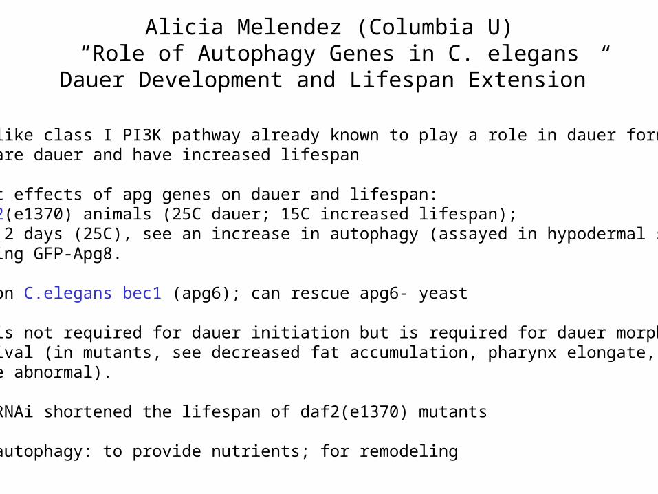
Alicia Melendez (Columbia U)“Role of Autophagy Genes in C. elegans Dauer
Development and Lifespan Extension”
Insulin-like class I PI3K pathway already known to play a role in dauer formation;Mutants are dauer and have increased lifespan
Looked at effects of apg genes on dauer and lifespan:Used daf2(e1370) animals (25C dauer; 15C increased lifespan); at dauer 2 days (25C), see an increase in autophagy (assayed in hypodermal seamcells using GFP-Apg8.
Focused on C.elegans bec1 (apg6); can rescue apg6- yeast
Ce-bec1 is not required for dauer initiation but is required for dauer morphogenesisand survival (in mutants, see decreased fat accumulation, pharynx elongate, cuticlestructure abnormal).
Ce-bec1 RNAi shortened the lifespan of daf2(e1370) mutants
Role of autophagy: to provide nutrients; for remodeling
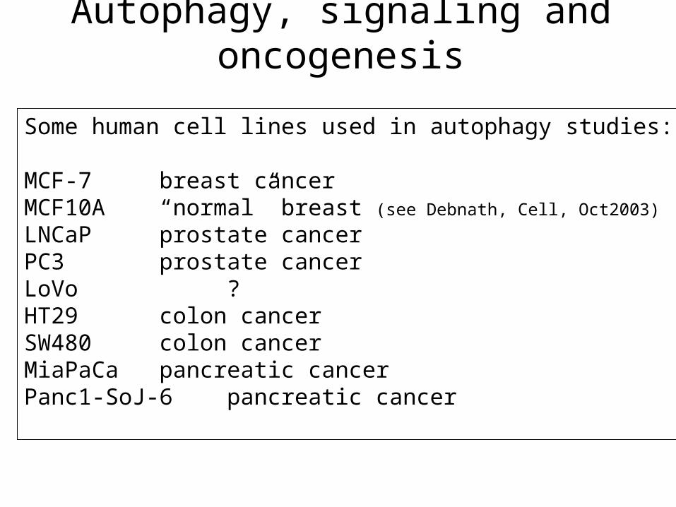
Autophagy, signaling and oncogenesis
Some human cell lines used in autophagy studies:
MCF-7 breast cancerMCF10A “normal” breast (see Debnath, Cell, Oct2003)
LNCaP prostate cancerPC3 prostate cancerLoVo ?HT29 colon cancerSW480 colon cancerMiaPaCa pancreatic cancerPanc1-SoJ-6pancreatic cancer
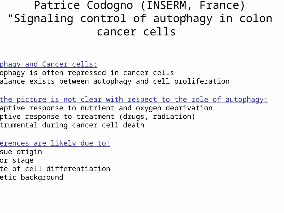
Patrice Codogno (INSERM, France)“Signaling control of autophagy in colon cancer
cells”
Autophagy and Cancer cells:•Autophagy is often repressed in cancer cells•A balance exists between autophagy and cell proliferation
BUT the picture is not clear with respect to the role of autophagy:• Adaptive response to nutrient and oxygen deprivation•Adaptive response to treatment (drugs, radiation)•Instrumental during cancer cell death
Differences are likely due to:•Tissue origin•Tumor stage•State of cell differentiation•Genetic background
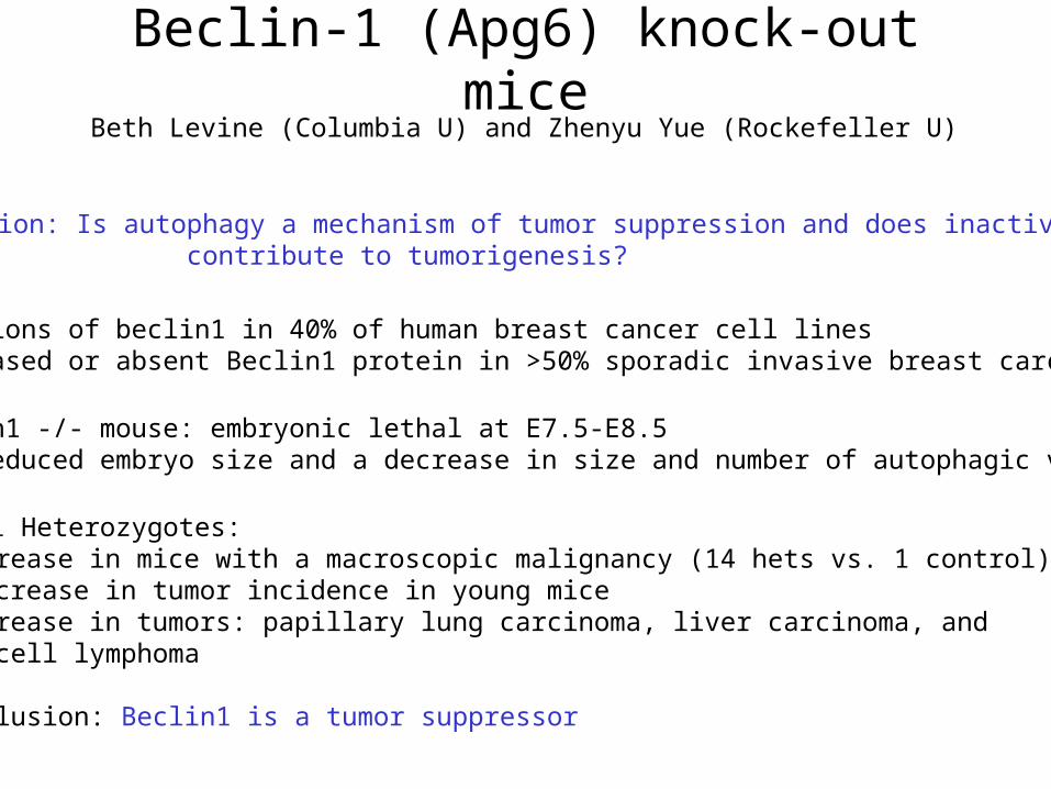
Beclin-1 (Apg6) knock-out miceBeth Levine (Columbia U) and Zhenyu Yue (Rockefeller U)
Question: Is autophagy a mechanism of tumor suppression and does inactivation contribute to tumorigenesis?
• deletions of beclin1 in 40% of human breast cancer cell lines• decreased or absent Beclin1 protein in >50% sporadic invasive breast carcinomas
• beclin1 -/- mouse: embryonic lethal at E7.5-E8.5• see reduced embryo size and a decrease in size and number of autophagic vacuoles
•Beclin1 Heterozygotes:•Increase in mice with a macroscopic malignancy (14 hets vs. 1 control)• increase in tumor incidence in young mice•Increase in tumors: papillary lung carcinoma, liver carcinoma, and B cell lymphoma
Conclusion: Beclin1 is a tumor suppressor

Beclin-1 continuedMechanism by which Beclin1 +/- promotes tumor formation is still not known:
• inefficient clearance of damaged organelles/cytoplasm?• decreased degradation of mitogenic signalling?• compromised autophagic PCD?• Bcl-2 interaction?
• analyzed 5 week old mice by BrdU incorporation and TUNEL saw no difference in cell death but marked increase in cell proliferation
More questions:•Tissue-specific effects (why tumors in lung, liver and B cell?)
Other Apg knock-out mice?Apg5-/- is homozygous lethal; +/- hets not looked at yet.
“Good genomics project” -what apg genes are mutated in which cancers? (use NCBI database?)
Note: See Lurcher knock-out mouse paper; autophagy and neuronal PCD

Autophagy in the host response to microbial
invasionSome viruses and bacteria Induce autophagy in the host cell upon infection.
e.g. Herpesvirus, Sindbis virus, RNA viruses including coronavirus; Legionella
A herpesvirus virulence protein (34.5) can interact with human Beclin1 and inhibit autophagy.
Hypothesis (herpesvirus): Autophagic degradation of viral protein providesPeptides - via the TAP receptor – for acquired immunity.
Hypothesis (RNA viruses): RNA viruses induce autophagosome formation toStimulate their own growth; RNA viruses require a membrane for their replication (I.e. use the autophagosomal membrane).

Autophagy Deregulation in Disease
1. Alpha-1-antitrypsin deficiency• Causes liver disease in children with variable effects• Most common genetic cause of emphysema in adults
2. Alzheimer’s Disease
3. Huntington’s Disease
4. Hereditary Myopathy (Autophagiv Vacuolar Myopathy)
Diseases associated with an induction of autophagy:

Autophagy and Apoptosis
Moderator: Questions:1. What is the relationship between autophagy and apoptosis?
2. Under what circumstances does autophagy induce cell death?
3. What are the therapeutic implications?
Note: 3-MA is used as an inhibitor of autophagy (by blocking PI3K)BUT 3-MA shown to inhibit several kinases including JNK; also blocks the mitochondrial membrane permeability transition
N.B. can also block some apoptosis-inducing pathways!

Autophagy and Apoptosis contd.
John LeMasters (U of North Carolina)Mitochondrial Inner Membrane Permeabilization in Autophagy and Cell Death
Hypothesis:Low levels of mitochondrial dysfunction leads to autophagy.Moderate levels of mitochondrial dysfunction leads to apoptosis.High levels of mitochondrial dysfunction leads to necrosis.
MPT – leads to sequestration and formation of the autophagosome; This prevents the release of cyto C and the induction of apoptosis; Therefore autophagy is playing a protective role.
Once the autophagosome capacity is exceeded, cyto C is released and apoptosis is induced.

Craig Thompson (U of Pennsylvania)“Autophagy is required for growth factor-independent cell survival”
Model system: IL-3 dependent hematopoietic cell line; dies after 48 hrsfollowing IL-3 withdrawal
• cells use whatever they can to produce ATP; eventually lose the ability tomaintain themselves, mitochondria degrade, release cyto C and cell death occurs.
•Cells expressing DN-Rab7 display prolonged GF-independent survival; Rab7 is asmall GTPase that functions to regulate the targeting of endocytic vesiclesto the lysosome• endocytosis eliminates nutrient transporters on the cell surface (thus get decreased survival)
Hypothesis: Bcl-2 family proteins (negative/positive regulators of death) function to regulate ATP release/ADP uptake into the mitochondria• they give the cells time to adapt and put into place the machinery to utilize theavailable nutrientse.g. Day 14 minus IL-3: cells are filled with Lysotracker-positive vacuoles; Bax -/-, Bak -/- cells remain viable but cell size decreases over time; autodegradetheir cellular contents
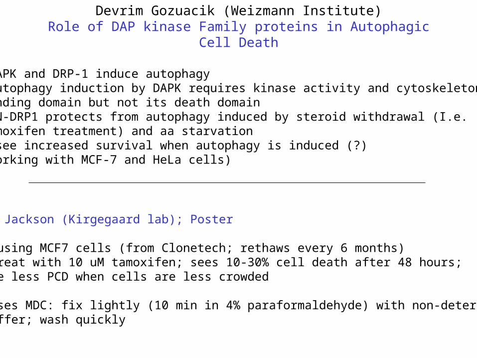
Devrim Gozuacik (Weizmann Institute)Role of DAP kinase Family proteins in Autophagic Cell
Death
•DAPK and DRP-1 induce autophagy•Autophagy induction by DAPK requires kinase activity and cytoskeleton binding domain but not its death domain•DN-DRP1 protects from autophagy induced by steroid withdrawal (I.e. tamoxifen treatment) and aa starvation• see increased survival when autophagy is induced (?)(working with MCF-7 and HeLa cells)
W. Jackson (Kirgegaard lab); Poster
• using MCF7 cells (from Clonetech; rethaws every 6 months)•Treat with 10 uM tamoxifen; sees 10-30% cell death after 48 hours; see less PCD when cells are less crowded
•Uses MDC: fix lightly (10 min in 4% paraformaldehyde) with non-detergentBuffer; wash quickly

MDC Label Mature Autophagic Vacuoles on Salivary Gland

26hr APF OreR
Autophagic vacuoles
Nucleus
Monodansylcadaverine MDC
• A lysosomotropic agent, it is concentrated into acid compartments by an ion trapping mechanism.
• A solvent polarity probe, it increases its relative fluorescence intensity by interacting with membrane lipids.
• MDC only accumulates in mature autophagic vscuoles, such as late autophagosome and autolysosome.