autophagy promotes degradation of internalized collagen ... · autophagy (neill et al., 2014). in a...
TRANSCRIPT

RESEARCH ARTICLE
Autophagy promotes degradation of internalized collagen andregulates distribution of focal adhesions to suppress cell adhesionShinichi Kawano, Takehiro Torisu*, Motohiro Esaki, Kumiko Torisu, Yuichi Matsuno and Takanari Kitazono
ABSTRACTAdhesion of cells to the extracellular matrix (ECM) via focal adhesions(FAs) is crucial for cell survival, migration, and differentiation.Although the regulation of FAs, including by integrins and the ECM,is important to cell behavior, how FAs are regulated is not well known.Autophagy is induced by both cell adhesion and cell detachment.Here, we showed that autophagosomes are located close tointernalized collagen and paxillin, which is a well-known marker ofFAs. Autophagy-deficient cells showed increased levels ofinternalized collagen compared with control cells. Moreover, paxillinexhibited a more peripheral distribution and the area of paxillin wasincreased, and adhesion-induced focal adhesion kinase signalingwas impaired and adhesion was enhanced, in autophagy-deficientcells. These results suggest that autophagy suppressed celladhesion by regulating internalized ECM and FAs.
KEY WORDS: Atg5, Atg7, Extracellular matrix
INTRODUCTIONAdhesion of cells to the extracellular matrix (ECM) plays animportant role in the regulation of cellular morphology, migration,proliferation, survival, and differentiation (Huveneers and Danen,2009). Cell–matrix adhesion is crucial during development, and fortissue maintenance and induction of tissue repair. Focal adhesions(FAs) are sites where a cell engages with the ECM; they not onlyfunction as scaffolds but also contribute to signaling that regulatescell structure, dynamics, and fate. FAs are enriched in integrins andin cytoskeletal and signal proteins such as paxillin, vinculin, andfocal adhesion kinase (FAK) (Winograd-Katz et al., 2014).Integrins are the principle cell-surface receptors involved in celladhesion for integration with the ECM. Integrins are activated bybinding of the ECM to cells at FAs in the process of cell–ECMadhesion (Berrier and Yamada, 2007). Active integrins andintegrin ligands undergo endocytic tracking and induce FAKautophosphorylation at tyrosine 397, which creates a binding site forthe Src-homology (SH) 2 domain of Src (Mitra et al., 2005).Phosphorylation of FAK at tyrosine 397 plays an important role inFA disassembly and is an established marker of activated FAK atFAs (Ezratty et al., 2005). The activated FAK–Src complexstimulates downstream signaling and regulates the activity of
several members of the Rho family of small GTPases. As a result,stimulation of FAs by the ECM enhances cell adhesion and resultsin cell spreading (Nagano et al., 2012).
Macroautophagy, hereafter termed autophagy, plays anindispensable role in the intracellular degradation of proteins andorganelles. Although starvation is the most extensively studiedcondition that induces autophagy, the process can be induced inresponse to several physiological and pathological conditions(Torisu et al., 2013). The ECM can modulate autophagicsignaling pathways, as several ECM constituents induceautophagy (Neill et al., 2014). In a study of cell migration,autophagy was reduced at the leading edge of cells compared withthe rear edge (Tuloup-Minguez et al., 2013). Mice lacking thecollagen VI gene exhibited muscular dystrophies caused bydefective autophagy (Grumati et al., 2010). Cell detachment fromthe matrix has been demonstrated as a trigger of autophagy, andautophagy protected cells from anoikis (Avivar-Valderas et al.,2011; Fung et al., 2008). Conversely, some reports suggest thatattachment induces autophagy. Blocking integrin with antibodiesreduced the extent of starvation-induced autophagy (Edick et al.,2007). The integrin ligand has been shown to stimulate autophagythrough integrins in a process mediated by p38-MAPK (Zhenget al., 2012); however the role of attachment-induced autophagy isnot well understood.
Internalized integrins and integrin ligands are trafficked to theendosome. Some of the integrins are degraded, but the majority ofinternalized integrins are recycled back to the plasma membrane(Bridgewater et al., 2012). Although collagen has been shown to belocalized in lysosomes in cells treated with a lysosome inhibitor(Everts et al., 1996), it is not well understood whether internalizedintegrin ligands are also degraded via autophagy.
To address this issue, we demonstrate here that autophagosomesare located close to the internalized ECM and internalizedcomplexes of FAs in the cell. Collagens are the main constituentof ECM. Atg5 and Atg7 are essential genes for autophagosomeformation (Kuma et al., 2004; Komatsu et al., 2005). Here, we showusing Atg5- and Atg7-deficient cells that autophagy regulates thedistribution of FAs and FA signaling, and thus regulates celladhesion and cell spreading.
RESULTSAutophagosomes colocalized with internalized ECMWe cultured fibroblasts for 24 h on culture dishes coated withfluorescein isothiocyanate (FITC)-labeled collagen. The fibroblaststook up the FITC-labeled collagen. The internalized collagenlocalized close to microtubule-associated protein 1-light chain 3(LC3), an established autophagosome marker (Fig. 1A). Analysis of13 random fields revealed that FITC-labeled collagen localizedclose to LC3 in 14% (24/171) of cells. To examine the distributionof collagen, cells were cultured on FITC-collagen and stained with aCellTracker probe. FITC-collagen was observed as dots in anReceived 12 June 2017; Accepted 26 September 2017
Department of Medicine and Clinical Science, Graduate School of MedicalSciences, Kyushu University, Fukuoka 812-8582, Japan.
*Author for correspondence ([email protected])
T.T., 0000-0001-8256-2549
This is an Open Access article distributed under the terms of the Creative Commons AttributionLicense (http://creativecommons.org/licenses/by/3.0), which permits unrestricted use,distribution and reproduction in any medium provided that the original work is properly attributed.
1644
© 2017. Published by The Company of Biologists Ltd | Biology Open (2017) 6, 1644-1653 doi:10.1242/bio.027458
BiologyOpen
by guest on July 5, 2020http://bio.biologists.org/Downloaded from

intracellular distribution (Fig. 1B). To examine involvement of theautophagy–lysosome pathway in the degradation of internalizedcollagen, we treated the fibroblasts with hydroxychloroquine(HCQ), an inhibitor of autophagic flux (Fig. 1C). Collagendeposition was increased after HCQ treatment (Fig. 1D).
Internalized collagen accumulated in autophagy-deficientfibroblastsAtg5 and Atg7 are essential molecules for the induction ofautophagy (Kuma et al., 2004; Komatsu et al., 2005). Wetherefore used Atg5- and Atg7-deficient murine embryonicfibroblasts (MEFs) to examine whether autophagy deficiencyaffected the amount of internalized collagen. We initiallyconfirmed the absence of Atg5 (Atg5–Atg12 conjugate) inAtg5−/− cells and the absence of Atg7 in Atg7−/− cells by westernblotting (Fig. 2A). In both Atg5−/− cells and Atg7−/− cells, the ratio
of LC3-I to LC3-II was consistent with impaired autophagy(Fig. 2A). In Atg5−/− cells, the amount of internalized FITC-labeledcollagen was greater than that in control cells (Fig. 2B,C). Thisincreased internalized collagen in autophagy-deficient cells wasalso reproduced using Atg7−/− cells (Fig. 2D,E). To test whetherthis increased collagen level was due to increased uptake ordecreased degradation in Atg5−/− cells, fibroblasts were pre-cultured with HCQ on a FITC-labeled collagen-coated dish for24 h for collagen uptake, and were then transferred to a new dishwithout FITC-labeled collagen to avoid further uptake (Fig. 3A).The amount of internalized FITC-labeled collagen was significantlygreater in autophagy-deficient cells than in control cells, both withand without HCQ treatment (Fig. 3B). Moreover, the percentage ofthe internalized FITC-labeled collagen that remained after washoutof HCQ was higher in Atg5−/− cells than in control cells (Fig. 3C).We reproduced similar findings using Atg7−/− cells, in which the
Fig. 1. Autophagosomes associated with collagen. (A) Representative confocal images of collagen (left), LC3 (middle), and merged images (right). Lowerpanels show enlarged images of the boxed regions in the upper panels. The fibroblasts in these images were cultured on FITC-labeled collagen and stained withanti-LC3 antibody. Scale bars: 5 µm (upper) and 1 µm (lower). (B) Representative image of orthographic project. Fibroblasts were cultured on FITC-labeledcollagen and stained with a CellTracker orange probe. (C) Collagen deposition with (right) and without (left) HCQ treatment. Fibroblasts were cultured on FITC-labeled collagen for 120 min. Scale bar: 20 µm. (D) Quantification of the area of FITC-labeled collagen per cell. Data are mean and s.e.m. in control and HCQ-treated cells (n=3 fields of control; five fields for HCQ-treated cells). *P<0.001, Student’s t-test. Three independent similar experiments are shown.
1645
RESEARCH ARTICLE Biology Open (2017) 6, 1644-1653 doi:10.1242/bio.027458
BiologyOpen
by guest on July 5, 2020http://bio.biologists.org/Downloaded from

amount of collagen was greater in Atg7−/− cells compared withcontrol cells, especially without HCQ treatment (Fig. 3D). Theseresults suggest that the difference in the amount of FITC-labeledcollagen between control and autophagy-deficient cells was moredependent on impaired degradation than on elevated uptake inautophagy-deficient cells.
Autophagy regulated focal adhesion and subsequentsignalingThe ECM controls cell adhesion through FAs (Turner, 2000). Weanalyzed the colocalization of FAs and autophagosomes. The
expression of the paxillin-EGFP fusion protein was engulfed in LC3(Fig. 4A). This result was confirmed by immunocytochemicalstaining of endogenous paxillin and LC3. This revealed that paxillinwas surrounded by puncta of LC3 (Fig. 4B). Similarly,immunocytochemistry showed that FAK-pY397 was surroundedby LC3 (Fig. 4C). To address whether this colocalization hadfunctional relevance, we analyzed paxillin localization inautophagy-deficient cells. In control cells, paxillin was distributedin punctate form in the cytoplasm and at the cell periphery, whereasin Atg5−/− cells it was distributed more at the cell periphery(Fig. 5A). The total area of FAs per cell was larger in Atg5−/− cells
Fig. 2. Internalized collagen levels were higher in autophagy-deficient cells than in control cells. (A) Left panels show western blot analysis of Atg5(Atg5–Atg12 conjugate), LC3-I and LC3-II in control and Atg5−/−MEFs. Right panels show western blot analysis of Atg7, LC3-I, and LC3-II in control and Atg7−/−
MEFs. NS, non-specific. GAPDH was used as an internal control. (B) Internalized FITC-labeled collagen in control (left) and Atg5−/− (right) MEFs. The cellswere cultured on FITC-labeled collagen for 120 min. Scale bar: 20 µm. (C) Quantification of the FITC-labeled collagen area per cell. Data presented aremean ands.e.m. in control andAtg5−/− cells. *P<0.05, Student’s t-test (n=5 fields of control; seven fields forAtg5−/− cells). Four independent similar experiments are shown.(D) Internalized FITC-labeled collagen in control (left) and Atg7−/− (right) MEFs. The cells were cultured on FITC-labeled collagen for 120 min. Scale bar: 20 µm.(E) Quantification of the FITC-labeled collagen area per cell. Data presented are mean and s.e.m. in control and Atg7−/− cells. *P<0.05, Student’s t-test (n=10).
1646
RESEARCH ARTICLE Biology Open (2017) 6, 1644-1653 doi:10.1242/bio.027458
BiologyOpen
by guest on July 5, 2020http://bio.biologists.org/Downloaded from

than in control cells, although the non-FA area per cell wascomparable in control and Atg5−/− cells (Fig. 5B). The number ofFAs was significantly larger in Atg5−/− cells than in control cells(Fig. 5C). Similarly, in Atg7−/− cells, paxillin was distributed moreat the cell periphery than in control cells (Fig. 5D). These resultsindicate that more FAs are retained in autophagy-deficient cells.Next, we investigated FA signaling. FAK and Src protein levels
did not differ markedly between control and Atg5−/− cells, evenafter adhesion. FAK phosphorylation at tyrosine 397 is a marker of
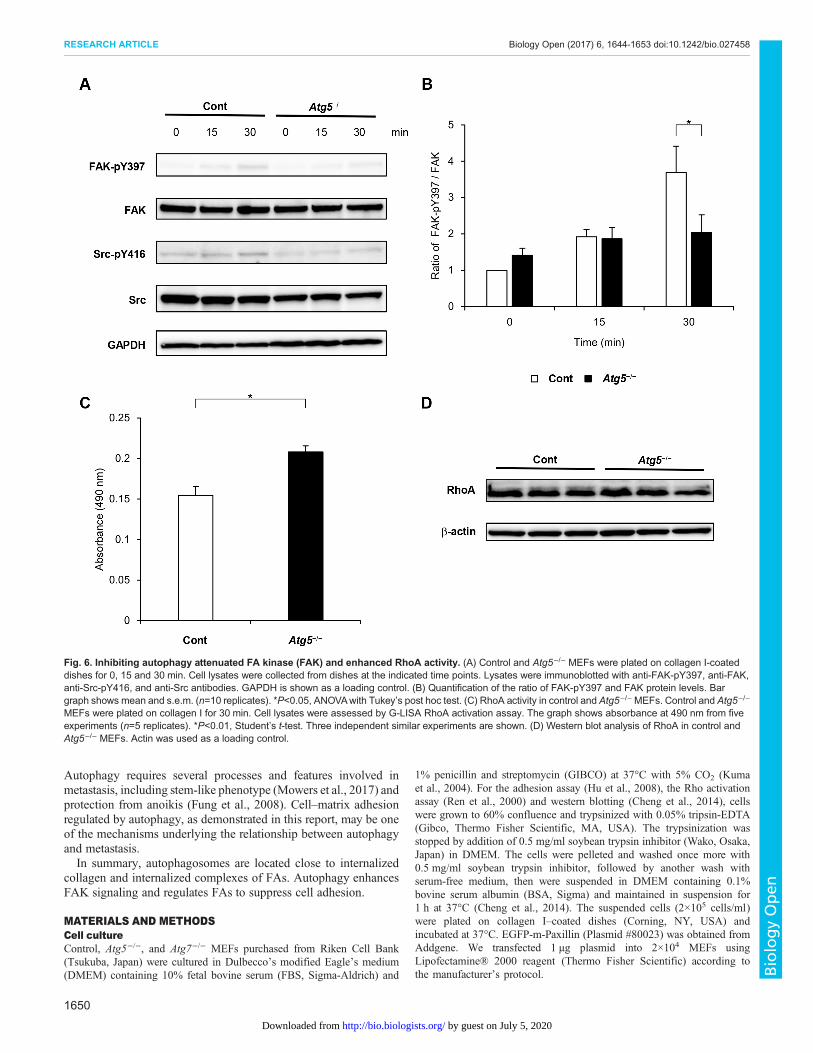
activated FAK in cell adhesion (Ezratty et al., 2005), andphosphorylation of Src at tyrosine 416 in the activation loop ofthe kinase domain upregulates its enzyme activity (Mitra et al.,2005). FAK phosphorylation at tyrosine 397 and Srcphosphorylation at tyrosine 416 were increased after cell adhesionto collagen in control cells (Fig. 6A). Although Src phosphorylationwas comparable between control and Atg5−/− cells, FAKphosphorylation was significantly attenuated in Atg5−/− cells at30 min after cell adhesion (Fig. 6B).
Fig. 3. Degradation of internalized collagen was decreased in autophagy-deficient cells compared with control cells. (A) Representative confocalimages of internalized collagen in control and Atg5−/− MEFs. These MEFs were pre-cultured on FITC-labeled collagen in the presence of HCQ (40 µM) for 16 h,then were transferred to new chamber slides without FITC-labeled collagen. Subsequently, the cells were cultured for 24 h with (upper panel) or without (lowerpanel) HCQ. Scale bar: 20 µm. (B) Quantification of the area of FITC-labeled collagen per cell in cells treated with HCQ (upper graph) and after washout of HCQ(lower graph). The bar graph for HCQ shows mean±s.e.m. in control and Atg5−/− cells and the graph for washout shows mean±s.e.m. in control and Atg5−/− cells(n=7 fields of control; six fields for Atg5−/− cells). Three independent similar experiments are shown. *P<0.05, ** P<0.001, Student’s t-test. (C) Internalizedcollagen remaining after washout of HCQ, as a percentage of internalized collagen in HCQ-treated cells. *P<0.01, Student’s t-test; mean±s.e.m.(D) Representative confocal images of internalized collagen in control and Atg7−/− MEFs. After pre-culture on FITC-labeled collagen in the presence of HCQ(40 µM) for 16 h, cells were cultured for 24 h with (upper panel) or without (lower panel) HCQ on new chamber slides without FITC collagen. Scale bar: 20 µm.
1647
RESEARCH ARTICLE Biology Open (2017) 6, 1644-1653 doi:10.1242/bio.027458
BiologyOpen
by guest on July 5, 2020http://bio.biologists.org/Downloaded from

FAK and RhoA regulate each other (Ren et al., 2000), and wetherefore analyzed RhoA activity. RhoA activity was higher inAtg5−/− cells than in control cells plated on collagen-coated dishesfor 30 min (Fig. 6C), although total RhoA protein levels did notdiffer between Atg5−/− and control cells (Fig. 6D).
Autophagy suppressed cell attachmentFinally, to address whether the molecular differences observedbetween control and Atg5−/− cells influenced cell behavior, we
conducted an adhesion assay. Thirty minutes after plating oncollagen-coated dishes, more autophagy-deficient cells than controlcells had adhered to the collagen (Fig. 7A). This result wasconsistent with a previous study using atg7 or atg12 knockdowncells (Kenific et al., 2016). When we examined cell morphology, weobserved more cell spreading among the Atg5−/− cells thanthe control cells (Fig. 7B). We reproduced similar findings usingAtg7−/− cells. We observed more cell spreading among the Atg7−/−
cells than the control cells (Fig. 7C,D). Consistent with this finding,F-actin filaments in the Atg5−/− cells were longer than those in thecontrol cells (Fig. 7E).
DISCUSSIONOur data suggest that autophagy is involved in the degradation ofinternalized collagen and the distribution of FAs to suppress celladhesion in fibroblast cells. Degradation of collagen in fibroblasts isa fundamental process in tissues under both physiological andpathological conditions (Everts et al., 1996). There are twopathways for collagen degradation: an extracellular pathway andan intracellular pathway. The extracellular pathway involvescleavage of collagen fibrils by matrix metalloprotease enzymes(McKleroy et al., 2013). In the intracellular pathway in fibroblasts,the engulfment of collagen fibrils is mediated by integrins (Dupuyand Caron, 2008). Collagen internalized from the ECM by fibrilphagocytosis is degraded in the lysosomal network (Everts et al.,1996). Our data demonstrate that autophagosomes were locatedclose to intracellular collagen, and that inhibiting autophagyincreased levels of internalized collagen. These results suggestthat autophagy is involved in the process of internalizing collagendegradation in the lysosome.
We showed that autophagy regulated FAK phosphorylation uponcell adhesion to collagen. It has been reported that autophagysuppresses FAK signaling under specific conditions, such as whenFAK is deleted (Sandilands et al., 2011). Recently, it wasdemonstrated that FAK activation and subsequent downstreamsignaling were dependent on the endocytosis of active integrins andintegrin ligands (Alanko and Ivaska, 2016; Alanko et al., 2015).Autophagy has been shown to maintain the endosome membrane(Kreibich et al., 2015), and an overlap between the autophagic andendocytotic pathways was demonstrated (Tooze et al., 2014). Inautophagy-deficient cells, endosomal dysfunction may beassociated with attenuated FAK activation. Furthermore, FAK andRhoA regulate each other. FAK has been shown to suppress RhoAactivity (Ren et al., 2000), and RhoA was found to induce FAKactivation (Del Re et al., 2008). Impaired degradation of activeRhoA has been reported in lysosomal v-ATPase-deficient cells inwhich autophagosome degradation was inhibited, accompanied byenhanced RhoA activity (Belaid et al., 2013). Consistent withprevious reports, our data show that although total RhoA proteinlevels were comparable in Atg5−/− and control cells, RhoA activitywas elevated in Atg5−/− cells compared with control cells. Ourresults suggest that impaired FAK signaling in autophagy-deficientcells resulted in enhanced RhoA activation. In FAK-deficient cells,FA turnover has been shown to be suppressed (Ren et al., 2000;Webb et al., 2004). In RhoA-overexpressing cells, the FA markersurface-associated vinculin was enriched (Cáceres et al., 2005). Wedemonstrated that paxillin and active form of FAK was engulfed inautophagosomes, and that the morphology of engulfed paxillin wasmostly of a round form. We demonstrated that endogenous paxillinexhibited more of a cell surface-associated distribution pattern inautophagy-deficient cells compared with control cells. We alsoobserved that an autophagy deficiency promoted adhesion and
Fig. 4. Autophagosomes associated with the internalized complexes offocal adhesion (FA) complex. (A) Representative confocal image of GFP-paxillin (left) and endogenous LC3 (middle). Lower panels show enlargedimages of the boxed regions in the upper images. Paxillin was surrounded bypuncta of LC3. Whole-cell merged image with DAPI counterstaining (right).Scale bar: 10 µm. (B) Fibroblasts were plated, and after 90 min were stainedwith antibodies against paxillin (left), LC3 (middle) and whole-cell mergedimage with DAPI counterstaining (right). The lower panels show enlargedimages of the boxed regions in the upper images. Scale bar: 10 µm.(C) Representative confocal image of FAK-pY397 (left), LC3 (middle), andwhole-cell merged image with DAPI counterstaining (right). Scale bar: 1 µm.
1648
RESEARCH ARTICLE Biology Open (2017) 6, 1644-1653 doi:10.1242/bio.027458
BiologyOpen
by guest on July 5, 2020http://bio.biologists.org/Downloaded from

resulted in more cell spreading during the adhesion period. Theseresults suggest that autophagy regulates cell motility via FAsignaling. One elegant study showed that FA lifetime was increasedin Atg7 and Atg12 knockdown cells compared with control cellsusing fluorescence-labeled paxillin overexpression (Kenific et al.,2016). Another study showed that autophagy interacted with and
then degraded paxillin to promote FA disassembly (Sharifi et al.,2016). These reports are consistent with our results.
In metastasis, cell–matrix adhesion is key to allowing cells toescape from their primary sites, and is required for them to be able tocolonize secondary sites. Many studies have shown an associationbetween autophagy and cancer metastasis (Mowers et al., 2017).
Fig. 5. FA complex distribution in autophagy-deficient cells and control cells. (A) Control and Atg5−/− MEFs were cultured for 120 min and stained withanti-paxillin antibody and with DAPI. Scale bar: 20 µm. (B) Area of paxillin, presented as mean and s.e.m, in control and Atg5−/− cells (n=8 fields of control;nine fields for Atg5−/− cells). *P<0.01, Student’s t-test. Four independent similar experiments are shown. (C) The number of FAs in control and Atg5−/− cells,presented as mean and s.e.m, in control and Atg5−/− cells. *P<0.01, Student’s t-test. (D) Representative confocal image of control and Atg7−/− MEFs, whichwere cultured for 120 min and stained with anti-paxillin antibody and DAPI. Scale bar: 20 µm.
1649
RESEARCH ARTICLE Biology Open (2017) 6, 1644-1653 doi:10.1242/bio.027458
BiologyOpen
by guest on July 5, 2020http://bio.biologists.org/Downloaded from
Autophagy requires several processes and features involved inmetastasis, including stem-like phenotype (Mowers et al., 2017) andprotection from anoikis (Fung et al., 2008). Cell–matrix adhesionregulated by autophagy, as demonstrated in this report, may be oneof the mechanisms underlying the relationship between autophagyand metastasis.In summary, autophagosomes are located close to internalized
collagen and internalized complexes of FAs. Autophagy enhancesFAK signaling and regulates FAs to suppress cell adhesion.
MATERIALS AND METHODSCell cultureControl, Atg5−/−, and Atg7−/− MEFs purchased from Riken Cell Bank(Tsukuba, Japan) were cultured in Dulbecco’s modified Eagle’s medium(DMEM) containing 10% fetal bovine serum (FBS, Sigma-Aldrich) and
1% penicillin and streptomycin (GIBCO) at 37°C with 5% CO2 (Kumaet al., 2004). For the adhesion assay (Hu et al., 2008), the Rho activationassay (Ren et al., 2000) and western blotting (Cheng et al., 2014), cellswere grown to 60% confluence and trypsinized with 0.05% tripsin-EDTA(Gibco, Thermo Fisher Scientific, MA, USA). The trypsinization wasstopped by addition of 0.5 mg/ml soybean trypsin inhibitor (Wako, Osaka,Japan) in DMEM. The cells were pelleted and washed once more with0.5 mg/ml soybean trypsin inhibitor, followed by another wash withserum-free medium, then were suspended in DMEM containing 0.1%bovine serum albumin (BSA, Sigma) and maintained in suspension for1 h at 37°C (Cheng et al., 2014). The suspended cells (2×10⁵ cells/ml)were plated on collagen I–coated dishes (Corning, NY, USA) andincubated at 37°C. EGFP-m-Paxillin (Plasmid #80023) was obtained fromAddgene. We transfected 1 µg plasmid into 2×10⁴ MEFs usingLipofectamine® 2000 reagent (Thermo Fisher Scientific) according tothe manufacturer’s protocol.
Fig. 6. Inhibiting autophagy attenuated FA kinase (FAK) and enhanced RhoA activity. (A) Control and Atg5−/− MEFs were plated on collagen I-coateddishes for 0, 15 and 30 min. Cell lysates were collected from dishes at the indicated time points. Lysates were immunoblotted with anti-FAK-pY397, anti-FAK,anti-Src-pY416, and anti-Src antibodies. GAPDH is shown as a loading control. (B) Quantification of the ratio of FAK-pY397 and FAK protein levels. Bargraph shows mean and s.e.m. (n=10 replicates). *P<0.05, ANOVAwith Tukey’s post hoc test. (C) RhoA activity in control and Atg5−/−MEFs. Control and Atg5−/−
MEFs were plated on collagen I for 30 min. Cell lysates were assessed by G-LISA RhoA activation assay. The graph shows absorbance at 490 nm from fiveexperiments (n=5 replicates). *P<0.01, Student’s t-test. Three independent similar experiments are shown. (D) Western blot analysis of RhoA in control andAtg5−/− MEFs. Actin was used as a loading control.
1650
RESEARCH ARTICLE Biology Open (2017) 6, 1644-1653 doi:10.1242/bio.027458
BiologyOpen
by guest on July 5, 2020http://bio.biologists.org/Downloaded from

AntibodiesThe following antibodies were used for immunoblotting: anti-LC3 (L8918,Sigma-Aldrich), anti-paxillin (ab32084, Abcam), anti-Atg5 (#12994, Cell
Signaling), anti-Atg7 (A2856, Sigma-Aldrich), anti-glyceraldehyde 3-phosphate dehydrogenase (GAPDH; ab181602, Abcam), anti-FAK-pY397 (44-624G, ThermoFisher Scientific), anti-FAK (610087, BD
Fig. 7. Autophagy-deficient cells exhibited more adherence than control cells. (A) Control and Atg5−/− MEFs were cultured on collagen for 30 min andstained with 0.5% crystal violet. Scale bar: 50 µm. (B) Adhesion assay in control and Atg5−/− MEFs. Crystal violet staining in these cells was eluted andabsorbance of the resulting solution at 550 nm was examined. Data presented are from three experiments. *P<0.01, Student’s t-test. (C) Representative crystalviolet staining images of control and Atg7−/− MEFs. Scale bar: 50 µm. (D) Adhesion assay in control and Atg7−/− MEFs. Data presented are from threeexperiments. *P<0.01, Student’s t-test. (E) Representative images of phalloidin staining for F-actin and DAPI staining of control andAtg5−/−MEFs. The cells werecultured on collagen for 120 min. Scale bar: 10 µm.
1651
RESEARCH ARTICLE Biology Open (2017) 6, 1644-1653 doi:10.1242/bio.027458
BiologyOpen
by guest on July 5, 2020http://bio.biologists.org/Downloaded from

Transduction Laboratories), anti-Src-pY416 (#2101, Cell Signaling), anti-Src (#2109, Cell Signaling), anti-RhoA (ab187027, Abcam), and anti-β-actin (ab8227, Abcam).
Western blottingAdherent cells cultured for 24 to 48 h were harvested on ice with a cellscraper. An additional culture of serum-starved cells was trypsinized andkept in suspension for 1 h, then collected in tubes. The cells were theincubated at 37°C for 15 and 30 min, and harvested (Cheng et al., 2014).The cell suspensions were centrifuged at 300 ×g at 4°C for 5 min and thesupernatant was discarded. The cell pellets were washed with chilledphosphate-buffered saline (PBS) and lysed in radioimmunoprecipitationassay lysis buffer (Nacalai Tesque, Kyoto, Japan) or NP40 lysisbuffer (Wako, Osaka, Japan) containing protease inhibitor (NacalaiTesque) and phosphatase inhibitor (Nacalai Tesque) cocktails for 5 minon ice. The cell lysate was centrifuged (16000 ×g at 4°C for 15 min). Thelysate [10–20 µg protein, as measured with a BCA Protein Assay kit(Thermo Fisher Scientific)] was then mixed with SDS sample buffer(Nacalai Tesque), separated by SDS-PAGE using pre-made 7.5% or 5–20%polyacrylamide gel plates (e-PAGEL, Atto, Tokyo, Japan), transferred toiBlot® 2 Transfer Stacks PVDF mini membranes using an iBlot® 2 DryBlotting system (Thermo Fisher Scientific), and immunoblotted withspecific antibodies at 1:1000 to 1:5000 dilution (Alanko et al., 2015; Torisuet al., 2013).
Immunofluorescence microscopyFor immunofluorescence microscopy, cells were grown on 4-well chamberslides (Lab-Tek, Thermo Fisher Scientific) that were pre-coated with1 µg/cm2 of fibronectin (Sigma-Aldrich, Germany), 1 µg/cm2 of collagen(Sigma), or 1 µg/cm2 of FITC-conjugated collagen I (4001, Chondrex, WA,USA) per well (Torisu et al., 2013, 2016). To label whole cells, they wereincubated with 1 µM of CellTracker (Thermo Fischer Science) orangefluorescent probe according to the manufacturer’s protocol. The cells werethen fixed with 4% paraformaldehyde in PBS (pH 7.4) for 10 min at roomtemperature, and permeabilized for 5 min with PBS containing 0.1% TritonX-100. Cells were incubated with Blocking One (Nacalai Tesque) for30 min and incubated with specific antibodies at 1:50 to 1:250 dilutionovernight at 4°C. We visualized F-actin polymerization via phalloidinstaining (A34055, Thermo Fisher Scientific). The cells were then incubatedwith secondary antibody for 30 min, and mounted in VECTASHIELDMounting Medium with DAPI (Vector Laboratories, Burlingame, CA,USA). Immunofluorescence samples were examined by confocalmicroscopy using a Zeiss LSM 700 microscope (Carl ZeissMicroImaging, Germany) (Torisu et al., 2013).
FA size analysisWe used endogenous paxillin as an FA marker (Sandilands et al., 2011;Sharifi et al., 2016). Image analysis was performed using ImageJ software(Wayne Rasband; the Research Services Branch, National Institute ofMental Health, Bethesda, MD, USA) after appropriate thresholding, aspreviously described (Sharifi et al., 2016).
Rho-activation assayRho-activation was assayed using a RhoA G-LISA kit (Cytoskeleton,Denver, CO, USA). Starved cells were trypsinized and kept in suspensionfor 1 h then incubated in a dish at 37°C for 30 min, and harvested (Chenget al., 2014). The RhoA G-LISA assay was performed according to themanufacturer’s protocol.
Adhesion assayThe adhesion assay was performed as previously described (Hu et al., 2008).Briefly, serum-starved cells were trypsinized and kept in suspension for 1 h,then incubated on collagen I-coated dishes at 37°C for 30 min. The cellswere fixed with 4% paraformaldehyde, then stained with 0.5% crystal violetin 20% ethanol (Sigma-Aldrich, Germany) for 10 min, washed with ddH2Oand dried completely. The cells were observed using a Nikon Eclipse Ti-Umicroscope (Nikon, Tokyo, Japan). Acetic acid (33%) was then added to the
dish to dissolve the crystal violet, and absorbance of the resulting solution at550 nm was examined.
Statistical analysisAll statistical analyses were performed using JMP Pro 11 (SAS InstituteInc., NC, USA). The statistical significance of differences between groupswas evaluated by an unpaired two-tailed Student’s t-test with Welch’scorrection, a Mann–Whitney U test, F test repeated-measures analysis ofvariance (ANOVA), or ANOVA with Tukey’s post hoc test. P<0.05 wasconsidered statistically significant in all experiments.
AcknowledgementsWe appreciate the technical assistance of the Research Support Center of theResearch Center for Human Disease Modeling, Kyushu University Graduate Schoolof Medical Sciences. We also thank Professor Mizushima (Tokyo University) forproviding Atg5−/− cells, and Professor Komatsu (Niigata University) for providingAtg7−/− cells.
Competing interestsThe authors declare no competing or financial interests.
Author contributionsConceptualization: T.T.; Validation: Y.M.; Investigation: S.K., T.T., K.T., Y.M.; Writing- original draft: S.K., T.T., M.E., K.T., T.K.; Writing - review & editing: S.K., T.T., M.E.,K.T., T.K.; Supervision: M.E., T.K.
FundingThis work was supported by grants from AstraZeneca (grant no. 201500247) andMitsubishi-Tanabe Pharma (grant no. MTPS20160414003).
ReferencesAlanko, J. and Ivaska, J. (2016). Endosomes: emerging platforms for integrin-
mediated FAK signalling. Trends Cell Biol. 26, 391-398.Alanko, J., Mai, A., Jacquemet, G., Schauer, K., Kaukonen, R., Saari, M., Goud,
B. and Ivaska, J. (2015). Integrin endosomal signalling suppresses anoikis. Nat.Cell Biol. 17, 1412-1421.
Avivar-Valderas, A., Salas, E., Bobrovnikova-Marjon, E., Diehl, J. A., Nagi, C.,Debnath, J. and Aguirre-Ghiso, J. A. (2011). PERK integrates autophagy andoxidative stress responses to promote survival during extracellular matrixdetachment. Mol. Cell. Biol. 31, 3616-3629.
Belaid, A., Cerezo, M., Chargui, A., Corcelle-Termeau, E., Pedeutour, F.,Giuliano, S., Ilie, M., Rubera, I., Tauc, M., Barale, S. et al. (2013). Autophagyplays a critical role in the degradation of active RHOA, the control of cellcytokinesis, and genomic stability. Cancer Res. 73, 4311-4322.
Berrier, A. L. and Yamada, K. M. (2007). Cell-matrix adhesion. J. Cell. Physiol. 213,565-573.
Bridgewater, R. E., Norman, J. C. andCaswell, P. T. (2012). Integrin trafficking at aglance. J. Cell Sci. 125, 3695-3701.
Caceres, M., Guerrero, J. and Martinez, J. (2005). Overexpression of RhoA-GTPinduces activation of the Epidermal Growth Factor Receptor, dephosphorylation offocal adhesion kinase and increased motility in breast cancer cells. Exp. Cell Res.309, 229-238.
Cheng, S. Y. S., Sun, G., Schlaepfer, D. D. and Pallen, C. J. (2014). Grb2promotes integrin-induced focal adhesion kinase (FAK) autophosphorylation anddirects the phosphorylation of protein tyrosine phosphatase alpha by the Src-FAKkinase complex. Mol. Cell. Biol. 34, 348-361.
Del Re, D. P., Miyamoto, S. and Brown, J. H. (2008). Focal adhesion kinase as aRhoA-activable signaling scaffold mediating Akt activation and cardiomyocyteprotection. J. Biol. Chem. 283, 35622-35629.
Dupuy, A. G. and Caron, E. (2008). Integrin-dependent phagocytosis - spreadingfrom microadhesion to new concepts. J. Cell Sci. 121, 1773-1783.
Edick, M. J., Tesfay, L., Lamb, L. E., Knudsen, B. S. and Miranti, C. K. (2007).Inhibition of integrin-mediated crosstalk with epidermal growth factor receptor/Erkor Src signaling pathways in autophagic prostate epithelial cells induces caspase-independent death. Mol. Biol. Cell 18, 2481-2890.
Everts, V., van der Zee, E., Creemers, L. and Beertsen, W. (1996). Phagocytosisand intracellular digestion of collagen, its role in turnover and remodelling.Histochem. J. 28, 229-245.
Ezratty, E. J., Partridge, M. A. and Gundersen, G. G. (2005). Microtubule-inducedfocal adhesion disassembly is mediated by dynamin and focal adhesion kinase.Nat. Cell Biol. 7, 581-590.
Fung, C., Lock, R., Gao, S., Salas, E. and Debnath, J. (2008). Induction ofautophagy during extracellular matrix detachment promotes cell survival. Mol.Biol. Cell 19, 797-806.
Grumati, P., Coletto, L., Sabatelli, P., Cescon, M., Angelin, A., Bertaggia, E.,Blaauw, B., Urciuolo, A., Tiepolo, T., Merlini, L. et al. (2010). Autophagy is
1652
RESEARCH ARTICLE Biology Open (2017) 6, 1644-1653 doi:10.1242/bio.027458
BiologyOpen
by guest on July 5, 2020http://bio.biologists.org/Downloaded from

defective in collagen VI muscular dystrophies, and its reactivation rescuesmyofiber degeneration. Nat. Med. 16, 1313-1320.
Hu, B., Kong, L. L., Matthews, R. T. and Viapiano, M. S. (2008). The proteoglycanbrevican binds to fibronectin after proteolytic cleavage and promotes glioma cellmotility. J. Biol. Chem. 283, 24848-24859.
Huveneers, S. and Danen, E. H. (2009). Adhesion signaling - crosstalk betweenintegrins, Src and Rho. J. Cell Sci. 122, 1059-1069.
Kenific, C. M., Stehbens, S. J., Goldsmith, J., Leidal, A. M., Faure, N., Ye, J.,Wittmann, T. and Debnath, J. (2016). NBR1 enables autophagy-dependentfocal adhesion turnover. J. Cell Biol. 212, 577-590.
Komatsu, M., Waguri, S., Ueno, T., Iwata, J., Murata, S., Tanida, I., Ezaki, J.,Mizushima, N., Ohsumi, Y., Uchiyama, Y. et al. (2005). Impairment of starvation-induced and constitutive autophagy in Atg7-deficient mice. J. Cell Biol. 169,425-434.
Kreibich, S., Emmenlauer, M., Fredlund, J., Ramo, P., Munz, C., Dehio, C.,Enninga, J. and Hardt, W.-D. (2015). Autophagy proteins promote repair ofendosomal membranes damaged by the salmonella type three secretion system1. Cell Host Microbe. 18, 527-537.
Kuma, A., Hatano, M., Matsui, M., Yamamoto, A., Nakaya, H., Yoshimori, T.,Ohsumi, Y., Tokuhisa, T. and Mizushima, N. (2004). The role of autophagyduring the early neonatal starvation period. Nature 432, 1032-1036.
McKleroy, W., Lee, T.-H. and Atabai, K. (2013). Always cleave up your mess:targeting collagen degradation to treat tissue fibrosis. Am. J. Physiol. Lung Cell.Mol. Physiol. 304, L709-L721.
Mitra, S. K., Hanson, D. A. and Schlaepfer, D. D. (2005). Focal adhesion kinase: incommand and control of cell motility. Nat. Rev. Mol. Cell Biol. 6, 56-68.
Mowers, E. E., Sharifi, M. N. and Macleod, K. F. (2017). Autophagy in cancermetastasis. Oncogene 36, 1619-1630.
Nagano, M., Hoshino, D., Koshikawa, N., Akizawa, T. and Seiki, M. (2012).Turnover of focal adhesions and cancer cell migration. Int. J. Cell Biol. 2012,310616.
Neill, T., Schaefer, L. and Iozzo, R. V. (2014). Instructive roles of extracellularmatrix on autophagy. Am. J. Pathol. 184, 2146-2153.
Ren, X. D., Kiosses, W. B., Sieg, D. J., Otey, C. A., Schlaepfer, D. D. andSchwartz, M. A. (2000). Focal adhesion kinase suppresses Rho activity topromote focal adhesion turnover. J. Cell Sci. 113, 3673-3678.
Sandilands, E., Serrels, B., McEwan, D. G., Morton, J. P., Macagno, J. P.,McLeod, K., Stevens, C., Brunton, V. G., Langdon, W. Y., Vidal, M. et al.(2011). Autophagic targeting of Src promotes cancer cell survival followingreduced FAK signalling. Nat. Cell Biol. 14, 51-60.
Sharifi, M. N., Mowers, E. E., Drake, L. E., Collier, C., Chen, H., Zamora, M., Mui,S. and Macleod, K. F. (2016). Autophagy promotes focal adhesion disassemblyand cell motility of metastatic tumor cells through the direct interaction of paxillinwith LC3. Cell Rep. 15, 1660-1672.
Tooze, S. A., Abada, A. and Elazar, Z. (2014). Endocytosis and autophagy:exploitation or cooperation? Cold Spring Harb. Perspect Biol. 6, a018358.
Torisu, T., Torisu, K., Lee, I. H., Liu, J., Malide, D., Combs, C. A., Wu, X. S.,Rovira, I. I., Fergusson, M. M., Weigert, R. et al. (2013). Autophagy regulatesendothelial cell processing, maturation and secretion of von Willebrand factor.Nat. Med. 19, 1281-1287.
Torisu, K., Singh, K. K., Torisu, T., Lovren, F., Liu, J., Pan, Y., Quan, A.,Ramadan, A., Al-Omran, M., Pankova, N. et al. (2016). Intact endothelialautophagy is required to maintain vascular lipid homeostasis. Aging Cell 15,187-191.
Tuloup-Minguez, V., Hamai, A., Greffard, A., Nicolas, V., Codogno, P. and Botti,J. (2013). Autophagy modulates cell migration and beta1 integrin membranerecycling. Cell Cycle 12, 3317-3328.
Turner, C. E. (2000). Paxillin and focal adhesion signalling. Nat. Cell Biol. 2,E231-E236.
Webb, D. J., Donais, K., Whitmore, L. A., Thomas, S. M., Turner, C. E., Parsons,J. T. and Horwitz, A. F. (2004). FAK-Src signalling through paxillin, ERK andMLCK regulates adhesion disassembly. Nat. Cell Biol. 6, 154-161.
Winograd-Katz, S. E., Fassler, R., Geiger, B. and Legate, K. R. (2014). Theintegrin adhesome: from genes and proteins to human disease. Nat. Rev. Mol.Cell Biol. 15, 273-288.
Zheng, Y.-H., Tian, C., Meng, Y., Qin, Y.-W., Du, Y.-H., Du, J. and Li, H.-H. (2012).Osteopontin stimulates autophagy via integrin/CD44 and p38 MAPK signalingpathways in vascular smooth muscle cells. J. Cell. Physiol. 227, 127-135.
1653
RESEARCH ARTICLE Biology Open (2017) 6, 1644-1653 doi:10.1242/bio.027458
BiologyOpen
by guest on July 5, 2020http://bio.biologists.org/Downloaded from