autoimmune lymphoproliferative …lib.ugent.be/fulltxt/rug01/002/061/627/rug01-002061627...academic...
TRANSCRIPT

Academic Year 2012 - 2013
AUTOIMMUNE LYMPHOPROLIFERATIVE SYNDROME:
NOVEL INSIGHTS INTO PATHOPHYSIOLOGY AND TREATMENT
Laura COREMANS
Promotor: Prof. Dr. Geneviève Laureys Co-promotor: Dr. Victoria Bordon Cueto de Braem
Dissertation presented in the 2nd Master year in the programme of
Master of Medicine in Medicine


TABLE OF CONTENTS
ABSTRACT ........................................................................................................ 1
SAMENVATTING .............................................................................................. 3
INTRODUCTION ............................................................................................... 7
1. Background ............................................................................................. 7
2. Pathophysiology ...................................................................................... 9
2.1.Genetics ............................................................................................... 9
2.2.Perforin/granzym apoptotic pathway ..................................................... 12
3. Clinical manifestations .......................................................................... 13
3.1.Lymphoproliferation ............................................................................ 13
3.2.Autoimmunity ..................................................................................... 13
3.3.Malignancy ......................................................................................... 14
4. Laboratory findings .............................................................................. 15
4.1.TCR αβ+ DNTs, IL-10 and B cell lymphocytosis .................................... 16
5. Diagnostic criteria ................................................................................. 17
5.1.Classification ...................................................................................... 19
5.2.Imaging .............................................................................................. 19
5.3.Histopathology .................................................................................... 19
5.4.Differential diagnosis........................................................................... 20
6. Treatment .............................................................................................. 22
6.1.Corticosteroids and immunosuppresants ................................................ 22
6.2.Mycophenolate mofetil (MMF) ........................................................... 22
6.3.mTOR inhibitor (Rapamycin, Sirolimus) ............................................... 23
6.4.Fansidar (phyrimethamine/ sulphadoxine) ............................................. 23
6.5.Anti-CD20 monoclonal antibodies (Rituximab) ..................................... 24
6.6.Splenectomy ....................................................................................... 25
6.7.IV immunoglobulin G .......................................................................... 25
6.8.Pentostatin .......................................................................................... 25

6.9.Hematopoietic stem cell transplantation (HSCT) .............................. 26
6.10.Arsenic trioxide (As2O3) .................................................................... 26
6.11.Valproic Acid (VPA) ......................................................................... 26
AIM ................................................................................................................ 27
METHODS AND MATERIALS ......................................................................... 28
1. Study population ................................................................................... 28
1.1.Background of study population and recruitment.................................... 28
1.2.Inclusion and exclusion criteria and sample size ..................................... 28
2. Data collection and study conduct ....................................................... 29
RESULTS ......................................................................................................... 31
1. Genetics ................................................................................................. 32
2. Clinical manifestations ......................................................................... 33
2.1.Lymphoproliferation ........................................................................... 33
2.2.Autoimmunity..................................................................................... 33
2.3.Malignancy......................................................................................... 35
3. Laboratory findings .............................................................................. 36
4. Imaging and histopathology ................................................................. 37
5. Treatment .............................................................................................. 38
DISCUSSION .................................................................................................... 43
CONCLUSION ................................................................................................. 48
REFERENCES ................................................................................................. 52
APPENDICES ................................................................................................ 56
Appendix 1 ....................................................................................................... i
Appendix 2 .................................................................................................... vii
Appendix 3 ................................................................................................... xiii


1
ABSTRACT
Background Autoimmune lymphoproliferative syndrome (ALPS) is an inherited disorder
of abnormal lymphocyte survival due to defective Fas-mediated apoptosis. This causes
autoreactive “double negative TCR αβ+ CD3
+ CD4ˉ CD8ˉ T cells” (DNTs) to accumulate in
the circulation and in lymphoid tissues. The majority of patients with ALPS carry an
underlying genetic mutation, located in genes associated with the Fas apoptotic pathway.
ALPS usually arises in early childhood. Clinically patients present with chronic
lymphadenopathies, hepatosplenomegaly and/or autoimmune cytopenias. Patients also have
an increased risk of malignancy.
Over the last decades significant progress has been made in understanding the
pathophysiology of ALPS and in developing new diagnostic criteria and therapeutic agents.
Objective ALPS is a relatively rare disorder. Worldwide, estimated cases of ALPS exceed
500, but that number has not reliably been confirmed. However, ALPS could be more
common than originally thought because of low awareness in clinical practice or
misdiagnosis. Diagnosis of ALPS can be a challenge partially because of its variable
phenotype and laboratory findings which are unspecific and can fluctuate over time.
Furthermore, recent studies have shown that some therapeutic agents commonly used for
other refractory non-ALPS autoimmune cytopenias are associated with significant morbidity
and mortality in ALPS. Therefore, making the correct diagnosis is important.
The aim of this study was to describe the variable phenotype in a group of patients
diagnosed with ALPS in order to raise clinical awareness. Moreover, the study aims to
contribute to making the correct diagnosis and choosing the most appropriate treatment
option.
Additionally, the effect of treatment with Fansidar (25 mg phyrimethamine/ 500 mg
sulphadoxine) on clinical outcome and laboratory results in the patient cohort is described
retrospectively. In 1998, Van der Werff et al. first reported normalisation of clinical and
laboratory features upon the administration of Fansidar. Since then, Fansidar has been
consistently used as a first-line treatment for lymphoproliferative and autoimmune
manifestations associated with ALPS in some Belgian centres.
Materials and methods The study population consisted of 12 patients diagnosed with
ALPS and followed at UZ Ghent and QFCUH in Brussels. Informed consent was obtained
for all patients or their legal guardians.

2
UZ Ghent’s Ethics Committee approval was obtained for the retrospective data collection in
medical records ranging from 1998 until present. Data on patients’ clinical, immunological
and laboratory features, relevant for the diagnosis and characterisation of ALPS was
collected. Furthermore, patients’ clinical outcome upon treatment was analyzed.
Existing literature in patients with ALPS was reviewed. These results were compared
retrospectively with our patient cohort for similarities and differences.
Results All patients included in the study (n=12) displayed chronic non-malignant non-
infectious lymphoproliferation, autoimmune manifestations and elevated TCR αβ+ DNT
levels (≥ 1.5%of total lymphocytes or ≥ 2.5% of CD3+ lymphocytes) at initial presentation.
No patient has developed malignancy to date. Family history was positive in 8 out of 12
patients.
IL-10, IL-18, sFasL and vitamin B12 are biomarkers for ALPS which were recently
incorporated in the diagnostic criteria. These biomarkers were inconsistently assessed in the
study population described herein.
Eleven out of 12 patients were treated with Fansidar as a first-line treatment or in the course
of the disease. Treatment with Fansidar led to a complete or incomplete remission of
autoimmune cytopenias in one-third of all patients (3/11). Upon treatment with Fansidar,
TCR αβ+ DNT levels improved completely or significantly in 5 out of 11 patients.
Conclusion Clinical awareness is needed in any child with a combination of unexplained
lymphoproliferation and/or autoimmune manifestations and positive family history of
autoimmune cytopenias with or without non-malignant non-infectious lymphoproliferation
dating back to infancy. A clinical practice algorithm is proposed providing the subsequent
laboratory tests to diagnose and confirm ALPS. It is recommended that in the future IL-10,
IL-18, sFasL and vitamin B12 are included in the standard diagnostic work-up for ALPS.
Larger prospective cohorts or RCTs are needed to determine whether the effects seen upon
treatment with Fansidar are statistically significant. These studies should preferably include
data on sFasL, IL-10, IL-18 and/or vitamin B12. Since Fansidar has been effective in the
treatment of autoimmune cytopenias in some patients and is not associated with severe
toxicities, it could be attempted as a first-line treatment in patients with ALPS.
Furthermore, close clinical follow-up, genetic counselling for the recurrence risk in
descendants and patient education of constitutional symptoms for the life-long risk of
malignancy are important in patients with ALPS.

3
SAMENVATTING
Achtergrond Auto-immuun lymfoproliferatief syndroom (ALPS) is een erfelijke
aandoening met een abnormale overleving van lymfocyten als gevolg van een defect in Fas-
gemedieerde apoptose. Hierdoor accumuleren “dubbel negatieve TCR αβ+ CD3
+ CD4ˉ CD8ˉ
T cellen” (DNTs) in de perifere circulatie en in de lymfoïde weefsels. Het merendeel van de
patiënten met ALPS heeft een onderliggende genetische mutatie, gelegen in genen die
coderen voor Fas-gemedieerde apoptose.
ALPS presenteert zich meestal op kinderleeftijd. Het klinisch fenotype omvat chronische
lymfadenopathieën, hepatosplenomegalie en/of auto-immune cytopenieën. Patiënten hebben
ook een verhoogd risico op maligniteiten.
In de laatste decennia is er significante vooruitgang geboekt in inzichten in de
pathofysiologie van ALPS en in het ontwikkelen van nieuwe diagnostische criteria en
behandelingsmogelijkheden.
Doelstelling ALPS is een relatief zeldzame aandoening. Wereldwijd wordt het aantal
patiënten op meer dan 500 geschat, maar dit aantal is nooit betrouwbaar bevestigd. ALPS
zou echter meer prevalent kunnen zijn dan oorspronkelijk gedacht, door een gebrek aan
kennis in de klinische praktijk of een gemiste diagnose. ALPS heeft een variabel klinisch
fenotype en laboratoriumparameters die niet specifiek zijn en kunnen veranderen na verloop
van tijd. Diagnose kan dus moeilijk zijn. Daarnaast hebben recente studies aangetoond dat
sommige geneesmiddelen die frequent gebruikt worden in de behandeling van refractaire
auto-immune cytopenieën bij andere ziektebeelden, geassocieerd zijn met een significante
morbiditeit en mortaliteit in ALPS. Daarom is het belangrijk om de correcte diagnose te
stellen.
Het doel van deze studie is het beschrijven van het variabel fenotype in een groep patiënten
met ALPS om te zorgen voor meer klinische bewustmaking. Daarnaast tracht de studie bij te
dragen tot het stellen van de juiste diagnose en het kiezen van de meest geschikte
behandelingsoptie.

4
Bijkomend wordt het effect van behandeling met Fansidar (25 mg phyrimethamine/ 500 mg
sulphadoxine) op de klinische outcome en laboratoriumresultaten van de patiëntencohorte
retrospectief beschreven. Van der Werff et al.. beschreven als eerste in 1998 de normalisatie
van klinische en laboratorium parameters na behandeling met Fansidar. Fansidar werd
sindsdien systematisch gebruikt in sommige Belgische centra als eerstelijnsbehandeling voor
lymfoproliferatieve en auto-immune manifestaties in ALPS.
Materialen en methoden De studiepopulatie bestond uit 12 patiënten, die gevolgd werden
in het UZ Gent en in het UKZKF Brussel. Informed consent werd verkregen van alle
patiënten of van hun wettelijke voogd. Het Ethisch Comité van het UZ Gent gaf
toestemming voor het retrospectief verzamelen van data uit medische dossiers daterend van
1998 tot nu. Gegevens over klinische, immunologische en laboratorium parameters, relevant
voor de diagnose en beschrijving van ALPS werden verzameld. Daarnaast werd het klinisch
resultaat van behandeling geanalyseerd.
Bestaande literatuur over ALPS werd geraadpleegd. Deze literatuurgegevens werden
retrospectief vergeleken op gelijkenissen en verschillen met de patiëntencohorte.
Resultaten Alle patiënten (n=12) in deze studiepopulatie vertoonden chronische non-
maligne, non-infectieuze lymfeproliferatie, auto-immune manifestaties en een verhoogd
aantal TCR αβ+ DNTs (≥ 1.5%van het totale aantal lymfocyten of ≥ 2.5% van het aantal
CD3+ lymfocyten) bij de eerste presentatie. Maligniteiten werden in geen enkele patiënt
vastgesteld. Familiale anamnese was positief in 8 van de 12 patiënten.
IL-10, IL-18, sFasL en vitamine B12 zijn biomarkers voor ALPS die recent opgenomen zijn
in de diagnostische criteria. Deze biomarkers zijn niet consistent bepaald in de
studiepopulatie die hierin beschreven is.
Fansidar werd toegediend als eerstelijnsbehandeling of in de loop van het ziekteproces in 11
van de 12 patiënten. Behandeling met Fansidar leidde tot een complete of incomplete
regressie van auto-immune cytopenieën in een derde van alle patiënten (3/11). TCR αβ+
DNT waarden normaliseerden of verbeterden significant in 5 van de 11 patiënten na
behandeling met Fansidar.

5
Conclusie Klinische alertheid is nodig in elk kind met een combinatie van onverklaarbare
lymfeproliferatie en/of auto-immune manifestaties en een positieve familiale anamnese van
auto-immune cytopenieën met of zonder non-maligne non-infectieuze lymfeproliferatie
teruggaand tot de kindertijd. Een algoritme voor klinische praktijk wordt voorgesteld waarin
de bijhorende laboratorium testen voor de diagnose en bevestiging van ALPS beschreven
worden. In de toekomst is het aanbevolen dat IL-10, IL-18, sFasL en vitamine B12
opgenomen worden in het standaard diagnostisch algoritme van ALPS.
Grotere prospectieve cohorten of RCTs zijn nodig om te bepalen of de effecten van
behandeling met Fansidar statistisch significant zijn. Deze studies bevatten bij voorkeur
gegevens over sFasL, IL-10, IL-18 en/of vitamine B12. Aangezien behandeling met
Fansidar effectief is gebleken in de behandeling van auto-immune cytopenieën in patiënten
met ALPS en geen ernstige bijwerkingen beschreven zijn, kan geprobeerd worden Fansidar
als eerstelijnsbehandeling in patiënten met ALPS te gebruiken.
Tot slot is nauwgezette klinische opvolging, genetisch advies voor het herhalingsrisico in
nakomelingen en patiënteneducatie over constitutionele symptomen gezien het levenslange
risico op maligniteiten belangrijk in patiënten met ALPS.

6

7
INTRODUCTION
1. Background
Autoimmune lymphoproliferative syndrome (ALPS) is an inherited autoimmune disorder
that usually arises in early childhood. It is a disorder of defective Fas-mediated T-
lymphocyte apoptosis, causing autoreactive “double negative TCR αβ+ CD3
+ CD4ˉ CD8ˉ
T cells” (DNTs) to accumulate in the circulation and in lymphoid tissues. In healthy
individuals, these TCR αβ+ DNTs usually constitute of less than 1% of total lymphocytes.
The clinical features are consistent with normal lymphocyte production but abnormal
survival and accumulation of lymphocytes. This results in a chronic, nonmalignant
lymphadenopathy, hepatosplenomegaly and autoimmune manifestations. ALPS is
currently defined as chronic, nonmalignant lymphoproliferation in patients with an
elevated percentage of TCR αβ+ DNTs (Table 2).
ALPS usually first manifests in childhood but clinical symptoms and lymphadenopathy
in particular disappear with age (1). However, elevated levels of TCR αβ+ DNTs and
hypergammaglobulinemia persist and autoimmune cytopenias follow a relapsing course
(2). The major determinants of morbidity and mortality in ALPS are the severe
autoimmune disease, postsplenectomy sepsis and malignancy (3). Patients have a life-
long risk of developing autoimmune disease and malignancies and require life-long
follow-up (3).
The syndrome was first described by Canale and Smith in 1967 as a symptom-complex
simulating malignant lymphoma (4). It is sometimes referred to as the Canale-Smith
syndrome. Several patients displaying lymphadenopathy, splenomegaly and autoimmune
cytopenia were described. The same features were also found in several relatives,
supporting the hypothesis that the disorder was caused by a gene mutation.
The essential role of Fas in apoptosis was clarified by studies in MRL lpr/gld mice. In
1992 Watanebe-Fukunga et al. found that these mice failed to express a lymphocyte
surface antigen, Fas (also called CD95 or Apo1) (5). This antigen is necessary for
triggering apoptosis on lymphocytes, as reported by Trauth et al. in 1989 (6). Genetic
studies have shown that the MRL/lpr phenotype results from homozygous mutations in
the Fas gene (5). The MRL/gld phenotype is caused by mutations in the gene encoding
Fas ligand (7). Clinically the mice present with elevated levels of peripheral TCR αβ+
DNTs, lymphoproliferation, hypergammaglobulinemia, glomerulonephritis and
autoantibody production (5).

8
Various authors later described several patients as the human equivalent of these murine
models with clinical features, elevated TCR αβ+ DNTs, defective apoptosis and specific
Fas mutations (8, 9). This clinical syndrome was subsequently termed the autoimmune
lymphoproliferative syndrome (ALPS).
In healthy individuals, Fas-induced apoptosis of activated peripheral T cells maintains T
cell homeostasis by limiting lymphocyte accumulation during the termination phase of an
immune response (3). Moreover apoptosis is critical to minimize lymphocyte
autoimmune reactions against self-antigens and maintain peripheral immune tolerance
(10).
Apoptosis is triggered by the interaction between the Fas receptor, a cell surface antigen
and Fas ligand (FasL) (Figure 1). Upon activation and clonal expansion, T and B cells
increase Fas expression and T cells increase expression of FasL (11).
The interaction between the extracellular domain of Fas and FasL causes a trimerization
of Fas. This leads to the interaction the Fas-death domain (DD), which is a portion of the
intracellular domain of Fas with the Fas-associated death domain protein (FADD). FADD
then binds a cysteine protease caspase-8 (or sometimes caspase-10) (12). Together they
form the death-inducing signalling complex (DISC). This initiates a downstream cleaving
and activation of proteins, leading to proteolysis, DNA degradation and apoptosis.
There are three, partly interconnected pathways through which the caspase cascade can
lead to apoptosis. The extrinsic or death receptor pathway is directly activated by a death
receptor, such as Fas and leads to caspase-8 receptor-ligand interactions, which requires
aggregation of the DISC.
The intrinsic or mitochondrial pathway is activated by several non-receptor mediated
stress stimuli (e.g. radiation, toxins, hypoxia). Subsequent mitochondrial-initiated release
of cytochrome C leads to activation of caspase-9 (13). Both pathways converge into the
same effector caspases, such as caspase-3. Evidence exists that molecules in one pathway
can influence the other and that the two pathways are linked (14). Moreover, The
perforin/granzym pathway is an additional pathway, which can activate caspase-10
through Granzyme B or trigger an apoptose-independent pathway through Granzyme A
(14). Interestingly, activation of the intrinsic pathway can be amplified by the cleavage of
Bid by caspase-8 and further activation of T-Bid by Granzyme B.

9
Figure 1: Schematic representation of apoptotic signalling pathways. The intrinsic pathway is triggered by
several stress stimuli. Subsequent mitochondrial-initiated release of cytochrome C leads to activation of
caspase-9. The extrinsic pathway is triggered by the interaction of FasL and Fas and leads to the
activation of caspase-8. The perforin/granzym pathway is an additional pathway, which can activate
caspase-10 through Granzyme B or trigger an apoptose-independent pathway through Granzyme A.
Activation of the intrinsic pathway can be amplified by the cleavage of Bid by caspase-8 and further
activation of T-Bid by Granzyme B.
Each pathway activates its own initiator caspase (8, 9 and 10), which in turn will activate the effector
caspase-3 and trigger apoptosis.
2. Pathophysiology
2.1. Genetics
The majority of patients with ALPS carry an identifiable genetic mutation, located in
genes associated with the Fas apoptotic pathway. Based on these findings 5 different
ALPS types have been identified (Table 3). Patients with ALPS-FAS have germline
mutations in the Tumor Necrosis Factor Receptor SuperFamily member 6 (TNFRSF6)
gene, encoding for Fas. This gene is located on chromosome 10. The TNFRSF6 gene
consists of 9 exons (15). Exons 1-5 encode the extracellular domain, responsible for
binding FasL. Exon 6 encodes the transmembrane domain of Fas. Exons 7-9 encode the
intracellular portion, which includes the Fas-death domain (DD), encoded by exon 9.
Over 80% of patients with ALPS have mutations in the intracellular portion and the DD
in particular (16).

10
Mutations can be either homozygous or heterozygous, presenting with a variable
clinical penetrance. Patients with heterozygous TFNRSF6 mutations represent the
majority of patients with ALPS. Homozygous TNFRSF6 mutations are far less frequent,
but the phenotype in these patients is more severe, with an early-onset (9).
Patients with ALPS-sFAS carry somatic TNFRSF6 mutations in the TCR αβ+ DNTs
and represent the second most common genetic mutations found in patients with ALPS
(17). Patients with ALPS-FASLG and FAS-CASP10 carry germline mutations in genes
encoding Fas ligand and caspase 10 respectively. A relatively large proportion of
patients (20-30%) have an unknown genetic defect and are classified as ALPS-U (18).
Penetrance
The relationship between genotype, phenotype and penetrance is complex. A high
degree of variability in clinical phenotype is seen in patients with ALPS and their
relatives. The expression greatly depends on the genetic background. In patients
carrying a heterozygous TNFRSF6 gene mutation, 70% develop clinical features (2).
The specific location of mutations on the TNFRSF6 gene is associated with
differences in severity and penetrance (19). Mutations affecting the intracellular Fas-
death domain show a higher penetrance of ALPS clinical phenotype. Significant
ALPS-related morbidity (splenectomy, autoimmune disease that requires treatment,
lymphoma) is more frequently seen in relatives with intracellular mutations (19).
Recent findings suggest different molecular disease mechanisms of mutations in the
intra- and extracellular domain (20). Heterozygous, missense mutations in the
intracellular portion and the Fas-death domain exhibit dominant-negative
interference of the signalling pathway (9, 19). The mutated Fas allele inhibits the
function of wild-type allele causing an absence of Fas function, inability to bind
FADD and form the DISC. Because Fas and FasL form homotrimers, the
combination of a mutant and a non-affected allele results in only one out of eight
configurations in a normal Fas trimer.
Mutations in the extracellular portion are thought to impair apoptosis through
haploinsufficiency (20, 21). Haploinsufficiency was found to prevent the surface
expression of Fas below a threshold that is needed for formation of the DISC and
subsequently trigger effective apoptosis (21).

11
Another group of missense and frame-shift mutations retain normal Fas surface
expression but are unable to bind FasL. Apoptosis is impaired to a lesser extent
because the wild-type allele will allow for expression of normal Fas on the cell
surface and is not inhibited by the mutated allele (20). This could account for the
lower disease severity and penetrance seen in families with extracellular mutations
(19, 22). Furthermore these patients were found to be significantly older at disease
onset than other ALPS-FAS or ALPS-sFAS patients (1).
Moreover, in 7 patient carrying a germline TNFRSF6 mutation, a complementary
somatic mutation in the second TNFRSF6 allele was found (23). These somatic
mutations were all found in patients carrying germline heterozygous mutations
affecting the extracellular portion of Fas. In patients carrying mutations in the
intracellular portion complementary somatic mutations were not found.
Complementary somatic mutations are therefore not compulsory for developing
clinical disease. However, the additional effect of the somatic mutation may account
for the higher clinical penetrance seen in patients carrying both mutations in the
extracellular portion, as opposed to healthy relatives carrying only the germline
mutation.
These findings support the “2-hit” hypothesis, with the second hit being acquired
later in life and may validate for the prolonged time to clinical manifestations seen in
patients with extracellular mutations.
However, a mutation in the TNFRSF6 gene cannot alone predict the degree of
penetrance, other complementary genetic or environmental factors are be required
(19).
Somatic mutations
Somatic TNFRSF6 mutations are suggested to be the second largest group of known
mutations in ALPS, affecting approximately 10% of patients (17). Patients have been
described with clinical features identical to ALPS and elevated levels of TCR αβ+
DNTs but with normal levels of Fas-mediated apoptosis and no family history of
ALPS (24). These patients were found to have heterozygous, somatic mutations in
the TNFRSF6 gene limited to the TCR αβ+ DNT compartment.
Similar to germline TNFRSF6 mutations, somatic TNFRSF6 mutations exert a
dominant negative effect on the Fas signalling pathway (24).

12
Since somatic TNFRSF6 mutations give rise to clinical and laboratory features which
are indistinguishable from germline TNFRSF6 mutations, sequencing of the
TNFRSF6 gene in DNA obtained from TCR αβ+ DNT cells and control cells could
help set the diagnosis (17, 25). Furthermore, some patients with ALPS-U could
possibly be identified as carrying somatic TNFRSF6 mutations.
2.2. Perforin/granzym apoptotic pathway
The predominant method through which cytotoxic T cells (CTLs) induce apoptosis is
the Fas/FasL interaction (14). However, an additional cytotoxic T cell-mediated
pathway exists that involves secretion of perforin, a pore-forming molecule that is
stored in granules of cytotoxic T cells. Perforin forms pores in the target-cell
membrane through which granzymes are introduced and apoptosis is induced in the
target cell (26-28). Granzyme A induces apoptosis through a caspase-independent
apoptotic pathway, but Granzyme B can activate the effector caspase-3 through
activation of caspase-10.
It seems that he role of the perforin/granzym pathway and the Fas/FasL pathway in
regulation of peripheral tolerance is overlapping but distinct (29).
Mateo et al. proved that despite resistance to Fas-mediated cell death, Fas-deficient T
cells could still undergo apoptosis after repeated T cell receptor (TCR) stimulation
(30). Peripheral activated T cells were found to significantly overexpress Granzyme A
and Granzyme B in comparison to matched controls.
These findings indicate that the perforin/granzym pathway may partially compensate
for Fas deficiency in T lymphocytes in ALPS patients.
However, apoptosis was equivalently detected in both symptomatic patients and their
asymptomatic relatives carrying the TNFRSF6 mutations. Compensation by the
perforin/granzyme pathway could therefore not account for the variable penetrance
seen in patients with ALPS (30).
Nevertheless, other studies found patients with an additional heterozygous mutation in
the Prf1gene together with a heterozygous mutation in the TNFRSF6 gene (26, 31).
Both genes are located on chromosome 10 (9).
This combination may lead to more severe and accelerated autoimmune disease,
increase susceptibility for ALPS and thus influence disease expression. However it
may not be sufficient for the development of ALPS, since healthy relatives carrying
both of these mutations have been described (32).

13
3. Clinical manifestations
Typically ALPS presents in 3 phases: massive lymphoproliferation, autoimmune
manifestations and malignancy (33).
3.1. Lymphoproliferation
All patients with ALPS present with chronic nonmalignant lymphadenopathy and/or
splenomegaly. This is defined as an enlargement of the lymph nodes and/or spleen for
more than 6 months. Some patients also present with hepatomegaly.
The degree of lymphadenopathy is variable. Most patients present with multiple lymph
nodes larger than 2 cm. Lymhadenopathy often involves the cervical and axillary chains
but other anatomic locations can also be involved. Lymphadenopathy usually regresses
with age. This is less often the case for splenomegaly.
3.2. Autoimmunity
Autoimmune manifestations or circulating autoantibodies are found in the majority of
patients with ALPS (3). Autoimmunity in patients with ALPS displays a typical
pattern of exacerbations and remissions or tends to become more severe with age.
Autoimmune haemolytic anaemia (AIHA) (<2 years of age: Hb <9.8 g/dl and >2 years
of age: Hb <11 g/dl) and autoimmune thrombocytopenia (platelet count < 150 x
103/µL) are most frequently found. Neutropenia (Absolute neutrophil count < 1500 /
µL) is also seen in ALPS but less frequently. Cytopenias in ALPS can be severe,
difficult to treat and even life threatening.
The most commonly found autoantibodies are directed against red blood cells and are
positive on the direct antiglobulin test (DAT or direct Coombs test). Moreover, the
majority of unaffected relatives do not have a positive direct Coombs test, indicating
that RBC autoantibodies can be a useful serologic marker in diagnosing ALPS (34).
Platelet antibodies (PA-Ab), human neutrophil antibodies (HNA), and antinucleair
antibodies (ANA) are also commonly positive (35, 36). However no association
between the detection of neutrophil or platelet antibodies and a clinical history of
neutropenia or thrombocytopenia respectively was found (36). This indicates that
other, additional factors contribute to the development of cytopenias in ALPS. For
instance, cytopenias can be exacerbated by splenic sequestration and hypersplenism
(37).

14
Furthermore, patients can theoretically but infrequently develop autoimmune
manifestations in any organ system, such as recurrent urticarial rashes consistent with
autoimmune vasculitis, arthritis or uveitis. Patients with glomerulonephritis and
Guillain-Barré syndrome have also been described (38). These clinical features could
be due to an immune hyperreactivity and deposition of antigen-autoantibody
complexes (9).
3.3. Malignancy
It is estimated that 10 to 20% of patients with germline TNFRSF6 mutations develop
malignancies, most commonly lymphoma (39). Moreover, an increased risk of
malignancy has been reported in asymptomatic relatives of patients with ALPS (40).
The average age of lymphoma diagnosis was 28 , whereas the average age of ALPS
onset was 5 years (40). Long-term surveillance for malignancy in patients with ALPS
and their relatives is therefore important.
A significant feature of lymphoma in ALPS is its diversity: B cell and T cell
lymphomas of diverse types have been found in multiple ALPS kindreds (40).
Mutations affecting the intracellular Fas-death domain (DD) are associated with the
highest risk of developing lymphomas (40, 41).
Abnormal and prolonged lymphocyte survival could allow additional oncogenic
mutations to accumulate and lead to secondary malignancies (42).
Furthermore, Gronbaek et al. found somatic TNFRSF6 gene mutations in 11%
(16/150) of sporadic B cell and T cell Non-Hodgkin lymphomas (42). These findings
suggest that the TNFRSF6 gene may function as a tumour-suppressor gene (43). Both
germline and somatic TNFRSF6 mutations and subsequent loss of tumour-suppressor
function may therefore predispose to the development of lymphomas in ALPS (41).

15
4. Laboratory findings
The most common laboratory findings in ALPS are increased levels of circulating TCR αβ+
DNTs and polyclonal hypergammaglobulinemia (IgG, sometimes associated with IgA).
Elevated CD3+CD4ˉ CD8ˉ T cells that express the αβ
+ T-cell receptor (TCR) (≥ 1.5% of
total lymphocytes or ≥ 2.5% of CD3+ lymphocytes) in the setting of normal or elevated
lymphocyte levels are a required diagnostic criterion for ALPS. Elevations above 3% of the
total lymphocytes (or ≥ 5% of CD3+ lymphocytes) are pathognomonic for ALPS (44-46).
DNTs are found to express the α/β TCR rather than the γ/δ TCR. However, ALPS can
infrequently lead to the proliferation of TCR γ/δ DNTs in the affected lymph node and in the
circulation ((9, 47, 48). It may be that the nature of the initial trigger defines which TCR will
dominate (48). It is hypothesized that development at young age of lymphadenopathy in
combination with an unidentified bacterial agent may contribute to the accumulation of TCR
γ/δ DNTs (48).
Furthermore, T cells display an in vitro defective Fas-mediated lymphocyte apoptosis (9).
The in vitro lymphocyte apoptosis assay measures the percentage of activated lymphocytes
undergoing apoptosis upon activation with Fas, which is subsequently impaired in patients
with defects in the Fas apoptotic pathway. However, several problems exist with the
diagnostic value and execution of this assay. It is therefore no longer considered as a
required criterion for the diagnosis of ALPS (Table 1 and Table 2).
Consistently high levels of vitamin B12 are found in patients with ALPS and were recently
incorporated in the diagnostic criteria of ALPS (49). The mechanism for this elevation is
unclear.
ALPS is also associated with several cytokine abnormalities, such as increased serum levels
of IL-10, IL-18 and soluble FAS ligand (sFASL) (49). Caminha et al. found that sFASL is a
sensitive biomarker for ruling out TNFRSF6 mutation, as healthy controls and patients with
ALPS-U or ALPS-related disorders showed only modest elevations of sFASL (49). In
contrast, patients with ALPS-FAS or ALPS-sFAS were found to have significantly higher
levels sFASL (> 200 pg/ml). These biomarkers are also currently included in the diagnostic
criteria of ALPS.
Overall low levels of IL-18 were found to protect against development of autoimmune
diseases in MRL/lpr mice (50). Therefore, it is hypothesized that IL-18 could contribute to
the pathogenesis of ALPS and reducing IL-18 levels can be a therapeutic strategy in
ALPS(50).

16
4.1. TCR αβ+ DNTs, IL-10 and B cell lymphocytosis
The origin and function of TCR αβ+ DNTs in ALPS is poorly understood, partly
because these cells do not grow in vitro and are therefore difficult to study. It is
unknown whether TCR αβ+ DNTs drive the disease or are merely an
epiphenomenon.
It has been postulated that TCR αβ+ DNTs are derived from cytotoxic CD8+ T-cells
that have lost their CD8+ expression in a process regulated at the transcriptional level
(47, 51-53).
TCR αβ+ DNTs were found to have a distinct cytokine production profile, producing
significantly more IL-10 than CD8+ T cells (53). Upon in vitro stimulation, a
fraction of the CD8+ T cells were found to acquire a cytokine production profile
similar to TCR αβ+ DNTs, supporting the hypothesis that CD8+ T cells can
differentiate into IL-10 producing TCR αβ+ DNTs. High levels of IL-10 were found
to correlate with greater disease expression (54, 55). As a result, TCR αβ+ DNTs may
play a role in ALPS pathogenesis by producing IL-10 which is thought to directly
stimulate B-cell proliferation, leading to the selective accumulation of autoimmune B
cells and subsequent autoimmunity (56). Moreover, high levels of IL-10 induce the
upregulation of the antiapoptotic proto-oncogene Bcl-2 in B and T cells (57).
Together this may exacerbate the apoptotic defects already inherent to ALPS and
predispose to malignancies.
In patients with ALPS, B cell lymphocytosis (CD19) is commonly found (58). These
autoreactive B cells may account for the polyclonal hypergammaglobulinemia,
autoantibodies and autoimmunity found in patients with ALPS (58).
This expansion might also be related to an abnormal activation state of B cells.
Autoreactive B cells can be eliminated by expressing Fas and therefore become
sensitive to killing by Fas ligand-expressing T cells (38, 59). Thus, defective Fas-
mediated apoptosis may lead to the accumulation of autoreactive B cells.
Other recent data suggests that TCR αβ+ DNTs may be dysregulated regulatory T
cells (60). Regulatory T cells are involved in downregulating the immune system and
tolerance to self-antigens. Dysregulated TCR αβ+ DNTs may be unable to
downregulate the immune responses and consequently lead to the development of
autoimmune disease (61, 62). It has been shown that TCR αβ+ DNTs display low
copies of TREC, which is considered an accurate marker for newly produced thymic

17
cells (61). Moreover, TCR αβ+ DNTs were found to lack some DNA rearrangement
events, crucial in the differentiation of CD8+ or CD4
+ T cells. This contradicts the
findings that TCR αβ+ DNTs are an expansion of CD4
+ or CD8
+ cells. TCR αβ
+
DNTs may result from reduced thymic activity, in addition to the apoptotic defect in
peripheral lymphocytes.
5. Diagnostic criteria
Diagnosis of this uncommon syndrome can be a challenge. Clinical features and laboratory
findings are unspecific and fluctuate over time.
In 1999 criteria were suggested for the diagnosis of ALPS by the National Institute of Health
(NIH) (Table 1). Since then there have been significant advances in the understanding of
ALPS. This has led to revisions to the existing diagnostic criteria and classification in 2009
(Table 2) (63).
Several problems with the diagnostic criteria from 1999 were detected. The lymphocyte
apoptosis assay is only available in selected centres, difficult to perform and not sensitive for
patients with somatic Fas or germline FasL mutations. TCR αβ+ DNTs do not survive in
vitro, therefore only Fas function and the ability to undergo apoptosis in non-TCR αβ+
DNTs is measured. Patients with somatic mutations, limited to de double-negative
compartment will therefore have a false negative lymphocyte apoptosis assay. Therefore,
this test is no longer considered mandatory for the diagnosis of ALPS. If the in vitro
lymphocyte apoptosis assay is performed, it is considered abnormal if the patients cells
show consistently (≥ 2 assays) 50% or less of the apoptosis observed in healthy controls.
Furthermore, genetic information and recently identified biomarkers for ALPS were
incorporated in the criteria.
In accordance with the revised diagnostic criteria, for an absolute ALPS diagnosis a patient
has to meet both required criteria and one of the primary accessory criteria. A probable
ALPS diagnosis requires both the required criteria and one of the secondary accessory
criteria (63).

18
Table 1: Diagnostic criteria for ALPS in 1999
Required Criteria
1. Chronic nonmalignant lymphadenopathy and/or splenomegaly
2. Increased peripheral TCR αβ+ DNTs
3. Defective lymphocyte apoptosis assay
Supporting Criteria
1. Family history of ALPS
2. Characteristic histopathology
3. Autoimmune manifestations
Note. Diagnostic criteria for ALPS as defined in 1999. Adapted from “Revised diagnostic criteria and
classification for the autoimmune lymphoproliferative syndrome (ALPS): report from the 2009 NIH
International Workshop” by Joao B. Oliveira, 2010, Blood, 116(14):e35-40. Copyright 2011 by The
American Society of Hematology. (63)
Table 2: Revised diagnostic criteria for ALPS in 2009
Required Criteria
1. Chronic (> 6 months), non-malignant, non-infectious lymphadenopathy and/or
splenomegaly
2. Elevated TCR αβ+ CD3
+ CD4ˉ CD8ˉ DNTs (≥ 1.5%of total lymphocytes or (≥
2.5% of CD3+ lymphocytes) in the setting of normal or elevated lymphocyte
counts
Accessory criteria
Primary
1. Defective lymphocyte apoptosis assay ( in 2 separate assays)
2. Somatic or germline pathogenic mutations in Fas, FasL or caspase-10
Secondary
1. Elevated plasma sFasL levels (> 200 pg/ml) OR elevated plasma IL-10 levels (>
20 pg/ml) OR elevated serum or plasma vitamin B12 levels (> 1500 ng/ml) OR
elevated plasma IL-18 levels (> 500 pg/ml)
2. Typical immunohistological findings as reviewed by an experienced
haematopathologist
3. Autoimmune cytopenias (haemolytic anaemia, thrombocytopenia or
neutropenia) AND elevated immunoglobulin G levels (polyclonal
hypergammaglobulinemia)
4. Family history of a non-malignant/non-infectious lymphoproliferation with or
without autoimmunity
Note. Revised diagnostic criteria for ALPS. Adapted from “Revised diagnostic criteria and classification
for the autoimmune lymphoproliferative syndrome (ALPS): report from the 2009 NIH International
Workshop” by Joao B. Oliveira, 2010, Blood, 116(14):e35-40. Copyright 2011 by The American Society
of Hematology. (63)

19
5.1. Classification
In the 2009 NIH conference recommendations about the molecular classification of ALPS
have also been introduced (63). Patients carrying both heterozygous and homozygous
TNFRSF6 mutations were unified under ALPS-FAS. Patients carrying somatic Fas
mutations should be classified as ALPS-sFAS, patients with FasL mutations as ALPS-
FASLG and patients with caspase-10 mutations as ALPS-CASP10.
It is likely that new genetic defects will be discovered in the group of patients with ALPS-U.
Table 3: Revised Classification of ALPS
Previous
Nomenclature
Revised
Nomenclature
Gene Definition
ALPS type 0 ALPS-FAS Fas Germline homozygous Fas
mutations
ALPS type Ia ALPS-FAS Fas Germline heterozygous Fas
mutations
ALPS type Is ALPS-sFAS Fas Somatic Fas mutations
ALPS type Ib ALPS-FASLG FasL Germline FasL mutations
ALPS type IIa ALPS-CASP10 Caspase-10 Germline caspase-10 mutations
ALPS type III ALPS-U Unknown Genetic defect is undetermined
Note. Revised classification of ALPS. Adapted from “Revised diagnostic criteria and classification for the
autoimmune lymphoproliferative syndrome (ALPS): report from the 2009 NIH International Workshop”
by Joao B. Oliveira, 2010, Blood, 116(14):e35-40. Copyright 2011 by The American Society of
Hematology. (63)
5.2. Imaging
Periodic ultrasounds are commonly used in the diagnostic work-up and follow-up of
lymphadenopathy and splenomegaly in patients with ALPS. Periodic CT scans of the chest,
abdomen and pelvis can also be useful but caution is warranted with the use of radiation,
especially in children.
5.3. Histopathology
Typically, marked paracortical expansion is seen in lymph node biopsies of patients with
ALPS (47). Increased numbers of TCR αβ+ DNTs are present in these paracortical regions.
Other features include follicular hyperplasia, polyclonal plasmocytosis and prominent
vascularity of the interfollicular areas (47).

20
The combination of follicular hyperplasia and paracortical expansion by an infiltrate
containing TCR αβ+ DNTs is pathognomonic for ALPS. Together with an overall intact
architecture of the lymph nodes, these features can help in the differential diagnosis with
other lymphoproliferative diseases and malignant lymphomas (3, 41).
5.4. Differential diagnosis
Patients with ALPS present with clinical and laboratory findings, such as elevated TCR αβ+
DNTs and defective in vitro lymphocyte assay, that have an important overlap with many
other paediatric haematological diseases.
These include T-cell lymphoma, common variable immunodeficiency (CVID), systemic
lupus erythematosus (SLE), X-linked lymphoproliferative disease (XLP), familial
hemophagocytic lymphohistiocytosis (FHLH) and Rosai-Dorfman disease or sinus
histiocytosis with massive lymphadenopathy (SHML) (64-66). As some patients with ALPS
display comorbidity with CVID, differential diagnosis can be even more difficult. Moreover,
some patients with ALPS develop lymphoma, imposing additional challenges for the
differential diagnosis between lymphoid hyperplasia and lymphoma. Clinical awareness of
constitutional symptoms (e.g. weight loss, fever, malaise) is necessary in making the
decision to investigate for malignancy.
However, only patients with ALPS have markedly elevated levels of TCR αβ+ DNTs above
3% of the total lymphocytes ( or ≥ 5% of CD3+ lymphocytes) and TCR αβ
+ DNT expansion
usually remains polyclonal and non-malignant (44, 45). Furthermore, histopathology can
help differentiate as it is often specific for ALPS.
ALPS-related apoptosis disorders (ALD)
This group of disorders is currently classified separately from ALPS. It consists of
patients with Caspase-8 deficiency syndrome (CEDS), patients with somatic mutations in
NRAS or KRAS, patients with Dianzani autoimmune lymphoproliferative disease
(DALD) and patients with X-linked lymphoproliferative syndrome (XLP1) (37, 63).
TNFRSF6 gene mutations are not seen in ALD.
Patients with germline caspase-8 mutations present with chronic lymphadenopathy,
splenomegaly and defective Fas-mediated apoptosis (67). However, unlike patients with
ALPS, patients with CEDS also display defects in the activation of T and B cells and NK
cells. Moreover, they suffer from recurrent Herpes Simplex and bacterial sinopulmonary
infections.

21
NRAS and KRAS genes encode for the Ras superfamily, which are small GTPases that
transmit intracellular signals and can regulate apoptosis (68). Patients with somatic
mutations in NRAS or KRAS present with autoimmunity and lymphoproliferation but
normal levels of TCR αβ+ DNTs and lymph node histopathology different from patients
with ALPS (69, 70). In addition, these patients display aberrations in the myeloid
compartment. These patients are now classified as RAS-associated autoimmune
leukoproliferative disorder (RALD).
Dianzani et al. first described patients with autoimmune manifestations,
lymphoproliferation, defective in vitro lymphocyte apoptosis assay but without elevated
TCR αβ+ DNTs (71). Patients display defects downstream in the Fas signalling pathway.
The genetic defect is unknown, but relatives of these patients display defective Fas
function, so a germline component is assumed (72). These patients are now classified as
Dianzani autoimmune lymphoproliferative disease (DALD).
X-linked lymphoproliferative syndrome (XLP1) is a rare immunodeficiency associated
with mutations in the SH2D1A gene. Upon infection with Epstein - Barr virus, patients
display dysregulated immune responses with severe infectious mononucleosis, acquired
hypogammaglobulinemia and/or malignant lymphoma (73).
Evans syndrome
Evans syndrome is a haematological disorder defined by autoimmune destruction of at
least 2 peripheral haematological cell types, after exclusion of other diagnoses (74). In
addition to autoimmune manifestations, patients with Evans syndrome may present with
lymphadenopathy and hepatosplenomegaly (75).
The underlying pathophysiology is unclear but is thought to be secondary to generalized
immune dysregulation (45). Based on these clinical similarities, it has been postulated
that a subset of patients may be misdiagnosed with Evans syndrome and may actually
have ALPS (44, 45). Elevated TCR αβ+ DNTs, in vitro defective lymphocyte apoptosis
assay and hypergammaglobulinaemia, which are highly suggestive for ALPS, were
indeed found in a subset of patients enrolled in the studies.
These findings suggest that ALPS may be more common than previously thought.
Furthermore it is recommended that patients suspected with Evans syndrome should be
screened for ALPS by evaluation of TCR αβ+ DNT levels as a part of their diagnostic
evaluation (44, 45).

22
6. Treatment
Lymphoproliferative symptoms (e.g. lymphadenopathy and splenomegaly) do not warrant
the consistent use of immunosuppressive drugs, unless they are associated with obstructive
symptoms or hypersplenism. This is due to observations that lymphoproliferation tends to
reoccur after termination of the immunosuppressive drugs and usually regresses with age
(38). Patients with lymphadenopathy but no cytopenias usually only require close clinical
and imaging follow-up. Furthermore, genetic counselling and advice concerning splenic
rupture is important.
Treatment for ALPS is mainly administered for autoimmune manifestations and
malignancies. However, only a few effective and tolerable drugs are available for patients
who do require treatment. Corticosteroids remain first-choice treatment although newer
drugs are being developed and tested. Recent studies have shown that therapies which are
commonly used in other refractory autoimmune cytopenias, such as rituximab and
splenectomy are relatively contraindicated in ALPS.
6.1. Corticosteroids and immunosuppresants
Short high-dosed courses of corticosteroids remain the treatment of first choice for
autoimmune cytopenias in ALPS and are usually highly effective (37). Other
immunosuppressant, such as azathioprine and mercaptopurine, may also be effective in the
treatment of autoimmune cytopenias.
However, prolonged use of corticosteroids is not recommended because of the high
prevalence of adverse effects and patients’ young age. Moreover, chronic exposure to
immunosuppressive agents bears a theoretical risk for development of secondary
malignancies, which would lead to an additional malignancy risk in patients with ALPS
(75).
6.2. Mycophenolate mofetil (MMF)
Mycophenolate mofetil is an inhibitor of the de novo synthesis of purine. It specifically
inhibits proliferating T and B cell and is currently used to prevent acute rejection of
transplanted organs (76). Furthermore, it is used as a second-line immunosuppressive
treatment for many autoimmune diseases.
MMF was found to be effective in the treatment of chronic, refractory cytopenias in patients
with ALPS (77). Moreover, a reduction in the required dose of other immunosuppressive
agents was observed.

23
However, patients did not have a normalisation of TCR αβ+ DNT levels or improvement of
lymphoproliferation. Furthermore, many patients have shown only partial response.
Therefore, MMF may be used to reduce the usage of corticosteroids in patients with ALPS.
However, it is recommended that MMF is not used as a first-line treatment and only as a
steroid-sparing agent (37).
6.3. mTOR inhibitor (Rapamycin, Sirolimus)
Mammalian target of Rapamycin (mTOR) is a protein kinase that regulates cell growth,
proliferation and survival. Targeting the mTOR pathway induces apoptosis in activated
lymphocytes through activation of the intrinsic or mitochondrial pathway and additionally
increases peripheral blood regulatory T cells (78).
A significant decrease in TCR αβ+ DNTs, lymphoproliferation and autoantibodies was seen
in MRL/lpr mice after treatment with Rapamycin (79, 80). In comparison to MMF,
Rapamycin showed a greater reduction of lymphoproliferation.
Teachey et al. have shown that Sirolimus is effective in reducing lymphoproliferation,
autoimmune cytopenias, non-haematological autoimmune manifestations and normalising
TCR αβ+ DNT levels refractory to corticosteroids (78). Moreover, the use of Sirolimus
could allow for a decrease in the required dose of corticosteroids or even a complete stop.
However, haematological side effects such as thrombocytopenia, leucopenia anaemia have
been described (81). Close therapeutic monitoring to maximize the therapeutic effect and
avoid toxicity is therefore required, given the haematological abnormalities already inherent
to ALPS.
6.4. Fansidar (phyrimethamine/ sulphadoxine)
Fansidar consists of two components; pyrimethamine and sulphadoxine. Each component
inhibits a different enzyme in the de novo synthesis of folic acid (82). In vitro apoptosis was
detectable for each component alone in stimulated T lymphocytes from both MRL/lpr mice
and patients with ALPS. However, pyrimethamine was found to be far more potent than
sulfadoxine in inducing apoptosis (82, 83).
Van der Werff et al. reported a marked shrinkage of lymphadenopathy, a decrease in TCR
αβ+ DNTs and IL-10 and reduction of autoimmune cytopenias upon treatment with Fansidar
(82, 84). These features reappeared after cessation of Fansidar. Possible mechanisms for the
observed improvement of autoimmune manifestations could be associated with the observed
decrease in IL-10 levels.

24
As previously noted, a decrease in IL-10 may lead to both a decrease in autoreactive B cells
and induction of apoptosis through downregulation of Bcl-2 (86). However, two out of the 7
patients enrolled in this study do not meet the strict criteria for the diagnosis of ALPS,
having normal TCR αβ+ DNT levels.
Nevertheless, in another clinical trial both pyrimethamine and Fansidar failed to show a
significant reduction of lymphoproliferation in MRL/lpr mice (83). Moreover,
lymphoproliferation did not improve significantly in seven patients with ALPS upon
treatment with pyrimethamine alone (83). Furthermore, no significant changes in TCR αβ+
DNTs, plasma IL-10 or serum IgG were found. In vitro apoptosis showed no difference in
Fas-mediated apoptosis defects in patients.
However, primary endpoints in this study were the safety of pyrimethamine in patients and
the ability to diminish lymphoproliferation. Autoimmune cytopenias was not a reason for
treatment initiation. Two out of 7 patients displayed decreased levels of platelet counts
and/or absolute neutrophil counts prior to treatment. Upon treatment with pyrimethamine,
both platelet counts and absolute neutrophil counts normalised. Three other patients had
received concomitant treatment with corticosteroids within 3 months prior to treatment with
pyrimethamine. Therefore, the efficacy of treatment with Fansidar is still a matter of debate.
Severe hypersensitivity reactions have been associated with pyrimethamine/ sulphadoxine
(85, 86). However, in the former studies pyrimethamine and Fansidar were found to be well
tolerated (82, 83). Observed adverse effects consisted only of mild to generalised rashes.
6.5. Anti-CD20 monoclonal antibodies (Rituximab)
Rituximab depletes the CD20+ B cells, which would subsequently reduce the autoantibodies
found in patients with autoimmune cytopenias. Rituximab is currently used in difficult-to-
control B-cell expansions, autoimmunity and B-cell lymphomas.
In ALPS, Rituximab has been successfully used in the management of refractory
autoimmune thrombocytopenia (87, 88). However, none of the patients with autoimmune
haemolytic anaemia responded to treatment with Rituximab (87). Furthermore, the use of
Rituximab did not produce clinically significant shrinkage of lymph nodes or spleen.
Observed adverse effects include prolonged neutropenia, profound and prolonged
hypogammaglobulinemia and absent antibody response to polysaccharide vaccines up to 4
years (87). These toxicities predispose to an additional infection risk, especially in asplenic
patients.

25
Moreover, 5-10% of patients develop common variable immunodeficiency (CVID) (87).
Caution is warranted since patients with ALPS are already predisposed to developing CVID.
Empirical use of Rituximab is therefore not recommended. It should be reserved for patients
who fail other therapeutic agents.
6.6. Splenectomy
Nearly 50% of patients with ALPS have undergone splenectomy as a successful treatment
for severe, refractory cytopenias and/or hypersplenism. Furthermore, a splenectomy is
sometimes performed because of discomfort and traumatic splenic rupture.
However, in ALPS splenectomy is associated with a long-term risk of recurrent cytopenias
and pneumococcal sepsis with significant morbidity and mortality (3). This complication
cannot be avoided by vaccination and treatment with penicillin, as opposed to patients with
non-ALPS autoimmune disease. Splenectomy should therefore be avoided in patients with
ALPS whenever possible. For patients with chronic, refractory life-threatening cytopenias,
partial splenectomy or splenic embolisation should be considered (37).
6.7. IV immunoglobulin G
In patients with ALPS, autoimmune thrombocytopenia usually does not respond sufficiently
to IV immunoglobulin G infusions, unlike many other non-ALPS autoimmune
manifestations (37).
6.8. Pentostatin
Pentostatin activates the intrinsic apoptotic pathway through accumulation of
deoxyadenosine-5’-triphosphate (dATP), which becomes cytotoxic at high concentrations.
Bajwa et al. reported the use of pentostatin in a patient with ALPS with cytopenias
refractory to splenectomy and immunosuppressive agents (89). Lymphocytosis was found to
decrease upon the administration of pentostatin. Pentostatin was well tolerated and effective
in keeping the patient in remission until a hematopoietic stem cell transplant (HSCT) was
performed.
These findings suggest that pentostatin may be useful in other patients with ALPS who are
refractory to MMF and/or Sirolimus.

26
6.9. Hematopoietic stem cell transplantation (HSCT)
HSCT has been successfully performed in 2 patients with ALPS (90, 91).
Sleight et al. described a patient carrying a homozygous TNFRSF6 mutation, resulting in an
extreme phenotype of ALPS (90). The patient was diagnosed with non-Hodgkin lymphoma
and showed no response to several courses of corticosteroids, G-CSF, α-interferon and a
splenectomy. Two years after HSCT was performed, the patient remained free of disease, no
signs or symptoms of ALPS were evident. In the second patient, control of
lymphoproliferation and autoimmune thrombocytopenia was achieved (91).
However, since the role of stem cell transplantation in ALPS is unclear, there is no need for
the routine use of HSCT and it should be reserved for patients with highly refractory
disease. Furthermore most patients with ALPS respond well to other therapies (37).
6.10. Arsenic trioxide (As2O3)
It has been postulated that arsenic trioxide eliminates autoreactive cells through induction of
the intrinsic or mitochondrial pathway.
Arsenic trioxide was found to significantly reduce autoimmune and lymphoproliferative
manifestations and decrease TCR αβ+ DNT levels in MRL/lpr mice (92). Arsenic trioxide is
thought to specifically activate caspases in the abnormal TCR αβ+ DNTs. This leads to the
elimination of TCR αβ+ DNTs but leaves the total number of T cells unchanged.
Furthermore, markedly reduced levels of anti-DNA autoantibodies, FasL, IL-10 and IL-18
were observed. As previously noted, reducing IL-18 levels could account for the positive
effects seen after treatment with arsenic trioxide. The reduction in IL-10 levels paralleled the
decrease in TCR αβ+ DNT levels.
6.11. Valproic Acid (VPA)
Valproic acid (VPA) is a histone deacetylase (HDAC) inhibitor, which is thought to induce
apoptosis through increased expression of Fas and FasL, increased caspase-3 activation and
increased release of cytochrome C (93).
A significant reduction in lymphoproliferative symptoms and TCR αβ+ DNTs was found
after treatment with VPA both in vitro and in MRL/lpr mice (94). VPA is currently being
studied in a clinical trial as a treatment for ALPS.
27
AIM
ALPS might be more common than previously thought. The clinical and laboratory
manifestations are highly variable and often difficult to differentiate from other
haematological paediatric diseases. The aim of this study was to describe the clinical,
immunological and laboratory findings in a group of patients diagnosed with ALPS in order
to raise clinical awareness. Furthermore, patients’ clinical outcome upon treatment was
analyzed. Existing literature in patients with ALPS was reviewed. These results were
compared retrospectively with our patient cohort for similarities and differences.
Moreover, a second patient cohort who underwent TCR αβ+ DNT assessment based on
clinical suspicion of ALPS for the period 2009-2012 at UZ Ghent was identified. The purpose
of this additional inquiry was to estimate in how many patients ALPS was included in the
differential diagnosis from 2009 to 2012 in UZ Ghent, how often TCR αβ+ DNT levels were
subsequently elevated and further diagnostic evaluation was warranted.
Additionally, the effect of treatment with Fansidar (25 mg phyrimethamine/ 500 mg
sulphadoxine) on clinical outcome and laboratory results in the patient cohort is
retrospectively described. In 1998, Van der Werff et al. first reported normalisation of clinical
and laboratory features upon the administration of Fansidar in a patient with ALPS (84). The
anti-malaria drug was initially used as chemoprophylaxis for Pneumocystis carinii during
lymphopenia. Since then, Fansidar has been consistently used as a first-line treatment for
lymphoproliferative and autoimmune manifestations associated with ALPS in some Belgian
centres.
Finally, a clinical practice algorithm will be provided describing the clinical and laboratory
features when ALPS should be considered, the diagnostic tests that should be performed and
corresponding sequence, suggestions for the management of autoimmune cytopenias and
long-term follow-up care. Consequently, the study aims to contribute to making the correct
diagnosis and choosing the most appropriate treatment option.

28
METHODS AND MATERIALS
1. Study population
1.1. Background of study population and recruitment
Nine patients were receiving or had received follow-up care at the department of
Paediatric Haematology-Oncology at UZ Ghent. Through their medical records, two
relatives of the initial 8 patients with phenotypic expression were identified and included
in the study. Their medical records were retrieved from the adult department of
haematology at UZ Ghent. Another patient, also receiving follow-up care at the adult
department and diagnosed with ALPS at infancy, was included.
Other medical centres in Belgium were contacted and their cooperation was asked for the
inclusion of more patients with ALPS, followed at these centres. This led to the inclusion
of one more patient followed at QFCUH in Brussels, bringing the total initial population
size to 13 patients (10 male and 3 female).
Informed consent was obtained for all patients or their legal guardians (Appendix 2). UZ
Ghent’s Ethics Committee approval was obtained for the retrospective data collection in
medical records (Appendix 3).
1.2. Inclusion and exclusion criteria and sample size
In one patient TCR αβ+ DNT levels were not assessed, this patient was therefore not
included in the study. As a result, the total study population consisted of 12 patients.
All patients included in the study (n=12) presented with chronic (> 6 months) non-
malignant non-infectious lymphoproliferation and elevated TCR αβ+ DNT levels (≥
1.5%of total lymphocytes or ≥ 2.5% of CD3+ lymphocytes) fulfilling the required
diagnostic criteria of ALPS (Table 4).
Six out of 12 patients met one or more of the primary accessory criteria and have an
absolute ALPS diagnosis. Six patients have a probable ALPS diagnosis, meeting one or
more of the secondary accessory criteria.

29
2. Data collection and study conduct
Existing literature on ALPS was reviewed, starting with five relevant and recent articles on
ALPS (1, 37, 63, 95, 96). Novel insights into pathophysiology, genetics, penetrance,
diagnostic criteria and treatment are described and reviewed herein.
Background information on the immune system and the physiological role of Fas in
apoptosis was found in the following medical textbooks: the 8th edition of Clinical Medicine
by Kumar and Clark, Review of Medical Physiology by Ganong and The Immune System
by Parham.
Furthermore, a website based-search was performed in the online database of biomedical
literature by the NCBI. The search string "Autoimmune Lymphoproliferative
Syndrome"[Mesh] was used. At the time, this query produced 46 articles. Articles were
selected based on relevancy of the abstract, full text availability and study design and
purpose. Related citations were reviewed and retained if they contained additional
information. More specific searches were performed on: haploinsufficiency and penetrance,
the origin and function of TCR αβ+ DNTs, the perforin/granzym pathway and effect on
disease penetrance, newly discovered biomarkers, differential diagnosis with Evans
syndrome and histopathologic features. The final literature review referred to 96 articles.
For the experimental section, information on patients’ clinical and laboratory features,
relevant for the diagnosis and characterisation of ALPS was collected. The data was
collected by reviewing medical charts and reports, ranging from 1998 until present.
The data was summarised and subdivided into 4 spreadsheets: Population Characteristics,
Clinical Manifestations, Laboratory Results and Therapy (Appendix 1).
On population characteristics and clinical manifestations, the following data was collected:
age at presentation and diagnosis, family history of ALPS, presence and duration of
lymphoproliferation and presence of malignancies.
The following laboratory features were evaluated: autoimmune manifestations (autoimmune
cytopenias and autoantibodies), TCR αβ+ DNT levels, in vitro lymphocyte apoptosis assay
(if performed), soluble FasL levels and biomarker levels (vitamin B12, IL-10 and IL-18).
Furthermore, imaging (ultrasound, CT, PET-CT) and histopathology results were reviewed.
If patients had undergone genetic testing, these results were also retained.

30
For patients undergoing treatment, reasons for treatment initiation with corresponding date
and dosage were collected. If applicable, any intermediate augmentation or treatment
cessation was also assessed. The efficacy results of the treatment over time was evaluated by
the degree of lymphoproliferation, platelet count, haemoglobin levels, absolute neutrophil
count and TCR αβ+ DNT levels as a percentage of CD3
+ lymphocytes.
Test results for all patients who underwent assessment of TCR αβ+ DNT levels for the
period 2009-2012 were collected by entering the search query “TCRABB AND OPCDBL”
in UZ Ghent’s clinical laboratory database. TCR αβ+ CD3
+ CD4ˉ CD8ˉ DNTs were
analysed by flow cytometry and expressed as a percentage of CD3+ lymphocytes. Additional
data on TCR αβ+ DNT levels of the initial study population (n=12) was incorporated in the
spreadsheets.
Descriptive and comparative analyses (mean values, range, standard deviations) were
performed on the collected patient data. The population size (n=12) was too small to
perform statistical analyses.

31
RESULTS
Table 4: Demographic and clinical features of study population
Patient
No.
Gender
Age at
presentation
(yrs)*
Age at
diagnosis
(yrs)**
Lymphoproliferation
Family history
Lymph-
adenopathy†
Spleno-
megaly‡
Hepato-
megaly
1
M
12
13
+ +
+ + + +
(S:14 yrs)
+
-
2
M
4
6
+
+ + + +
-
Son of patient no.10
3
M
12
13
+
+ +
-
-
4
M
7
7
+ +
+
+
Father: autoimmune
cytopenias
Nephew of
patient no. 5
5
M
39
43
+
+ +
-
Nephew of
patient no. 4
6
M
12
12
+
+ + + +
(S: 12 yrs)
+
-
7
M
6
8
+ +
+ +
-
Brother of
patient no. 8
8
M
6
6
+ +
+
-
Brother of patient no. 7
9
M
2
4
+
+ +
-
-
10
F
9
34
+
+
-
Mother of
patient no. 2
11
F
7
14
+ +
+ +
-
Father: germline TNFRSF6 mutation,
defective in vitro
apoptosis assay
12
F
2
2
+
+ + + +
+
Mother: germline
TNFRSF6 mutation,
no clinical symptoms
Maternal uncle: cytopenias,
lymphoproliferation
and non-Hodgkin
lymphoma
*Median age at presentation was 7 years.
** Median age at diagnosis was 10 years.
† Lymphadenopathy: + 1-2 cm nodes; ++ 2-4 cm nodes.
‡ Splenomegaly: +, 1-2 cm; ++, 3-4 cm; +++ 5-6 cm; ++++, >6cm below costal margin.
S: splenectomy (years).

32
1. Genetics
Three out of 12 patients underwent genetic testing. Patient no. 5 was found to carry a
germline TNFRSF6 nonsense mutation in exon 5, encoding the extracellular domain.
Patient no. 12 was found to carry a heterozygous missense TNFRSF6 mutation in exon 9,
encoding the Fas-death domain (DD). In patient no. 11, a germline TNFRSF6 nonsense
mutation in exon 7 was found. Exon 7 is known to encode the intracellular domain of Fas.
Patient 11 displayed defective in vitro lymphocyte apoptosis assay, whereas patient no. 5
did not. No other differences in presence of lymphoproliferation, autoimmune
manifestations or other ALPS related morbidity could be found between these 3 patients.
However, age at disease onset was markedly later in patient no.5 carrying the extracellular
TNFRSF6 mutation as opposed to patient no.11 and patient no. 12 carrying intracellular
TNFRSF6 mutation.
Family history was positive in 8 out of 12 patients. Patient no. 10 was diagnosed at the age
of 34 after the diagnosis of ALPS was suspected and confirmed in her son, patient no 3.
Patient no. 5 and patient no. 6 are nephews on the paternal side. Moreover, the finding that
the father of patient no. 5 displayed autoimmune cytopenias at infancy additionally
supports the hypothesis of paternal inheritance.
Patient no. 7 and patient no. 8 are siblings.
The father of patient no. 11 was found to carry the same germline TNFRSF6 nonsense
mutation and displayed defective in vitro lymphocyte apoptosis assay in 2 separate assays.
However, he has not displayed any lymphoproliferative or autoimmune manifestations.
The mother of patient no. 12 was found to carry the same heterozygous TNFRSF6
missense mutation. To date, she has not displayed any lymphoproliferative or autoimmune
features. However, a maternal uncle of patient no. 12 displayed thrombocytopenia and
lymphoproliferative symptoms in early childhood. A lymph node biopsy revealed sinus
histiocytosis, suggestive for Rosai-Dorfman disease (SHML). He was splenectomised at
the age of three At the age of 29, he developed Non-Hodgkin lymphoma, for which he was
treated with localised radiotherapy. He is currently in remission.

33
2. Clinical manifestations
All patients (n=12) presented with lymphoproliferative symptoms and one or more
autoimmune cytopenias. The median age of clinical onset was 7 years (range: 2-39). The
median age at diagnosis was 10 years (range: 2-43). A median diagnostic delay of 1.5
years is observed in the study population (range: 0-25 years, SD: 7.0). At present, patients’
median age is 20 years (range: 11-48) and median follow-up time is 7 years.
2.1. Lymphoproliferation
All patients (n=12) displayed chronic lymphadenopathy, present from over 6 months up
to 24 years. Multiple enlarged (1-4 cm) lymph nodes were observed, most frequently
affecting the cervical (8/12) and axillary (7/12) lymphatic chains. Other affected locations
were the inguinal (6/12), submandibular (4/12) and mesenteric (4/12) chains.
At presentation, splenomegaly was observed in all patients with an average spleen span
of 15.2 cm on ultrasound (range: 10 - 30 cm) or 6.5 cm below costal margin (range: 2.5-
20 cm). Hepatomegaly was present in 4 out of 12 patients (2-3 cm below costal margin).
2.2. Autoimmunity
All patients (n=12) presented with autoimmune manifestations and/or
hypergammaglobulinemia at first presentation (Table 5).
Ten out of 12 patients displayed one or more autoimmune cytopenias at initial
presentation. Nine patients presented with mild to severe thrombocytopenia (mean
platelet count ± SD: 4 x 10³
± 35 x 103/µL; range: 5-101 x 10
3/µL). Additionally, 6 of
these 9 patients also displayed mild to severe neutropenia (mean neutrophil count ± SD:
750 ± 388.2/µL; range: 247-1280/µL). Three out of 9 patients presented with haemolytic
anaemia in addition to thrombocytopenia.
One patient displayed only haemolytic anaemia at initial presentation. No patient
displayed only neutropenia or pancytopenia at initial presentation.
Seven out of 8 remaining patients without haemolytic anaemia at initial presentation
eventually developed haemolytic anaemia after a median duration of 3 years (SD: 2.5;
range: 1-8).
In summary, 9 patients displayed thrombocytopenia, 6 patients displayed neutropenia and
11 patients (n=12) displayed haemolytic anaemia at initial presentation or in the course of
the disease.

34
Table 5: Autoimmune manifestations and laboratory results of study population Patient
no
Autoimmunity TCR
αβ+
DNT†
Serum Ig‡ In vitro
apoptosis assay§
Autoimmune features Autoantibodies IgG IgA
1
Thrombocytopenia* +
Neutropeniaº + + +
Haemolytic anaemia
PA-Ab -
HNA -
DAT - ANA -
18%
+ +
+ +
↓
2
Haemolytic anaemia
DAT+
4%
+
N
↓
3
Thrombocytopenia + + +
Neutropenia + +
HNA -
ANA -
2,7%
N
N
NE
4
Thrombocytopenia + +
Neutropenia + + Haemolytic anaemia
DAT +
21,4%
+ +
-
NE
5
Thrombocytopenia + + +
Haemolytic anaemia
ITP
NE
4%
N
N
NE
6
Thrombocytopenia + + +
Haemolytic anaemia
DAT -
ANA -
3,7%
N
-
0
7
Thrombocytopenia + + +
Neutropenia + +
Haemolytic anaemia
ITP
HNA -
DAT +
ANA -
5,4%
N
-
0
8
Thrombocytopenia +/ + +
Neutropenia +
Haemolytic anaemia
DAT +
3,4%
+
-
NE
9
Thrombocytopenia + +
Neutropenia + +
Haemolytic anaemia
Polyarthritis
DAT -
6%
+ +
N
↓
10
Thrombocytopenia 0
Haemolytic anaemia Glomerulonephritis
Urticarial rash
NE
7%
+
N
NE
11
Haemolytic anaemia
DAT -
ANA -
4%
+
N
↓
12
Haemolytic anaemia
DAT -
7%
+
N
NE
Abbreviations: ITP, idiopathic thrombocytopenic purpura; DAT, direct antiglobulin test (Coombs); PA-Ab,
platelet antibodies; HNA, human neutrophil antibodies; ANF, antinuclear antibodies; NE, not evaluated.
Other antibodies not evaluated if not mentioned.
* Thrombocytopenia: +, mild (platelet count 100-150 x 103/µL); ++, moderate (50-100 x 103/µL); +++,
severe (<50 x 103/µL); 0, platelet count not registered in medical record.

35
ºNeutropenia: +, mild (absolute neutrophil count: 1000-1500 /µL); ++, moderate (500-1000 /µL), +++, severe
(<500 /µL).
†TCR αβ+ CD3+ CD4ˉ CD8ˉ DNTs expressed as a percentage of total CD3+ lymphocytes. TCR αβ+ DNT
levels (mean ± SD) were 7.2% ± 6.0%.
‡ Ig serum level: normal ranges vary according to age. N, in normal range; +, >+ 2-4 SD; ++, > + 4 SD; -, <2
SD.
§In vitro lymphocyte apoptosis assay: 0, normal >50%; ↓, defective.
Out of 11 patients who displayed haemolytic anaemia, 4 patients had a positive direct
antiglobulin test (DAT) and 5 patients did not. For the remaining 2 patients, the
antiglobulin test was not performed. Platelet antibody titers (PA-Ab) were only assessed
in one patient with thrombocytopenia and were negative.
Out of 5 patients with neutropenia, human neutrophil antibodies (HNA) were assessed in
3 patients and were all negative. Antinuclear antibody titers (ANA) were negative in all 4
patients in which they were evaluated.
Two out of 12 patients displayed autoimmune manifestations in other organs, such as
glomerulonephritis and urticarial rash at initial presentation and arthritis after 3 years.
2.3. Malignancy
No patient has developed malignancy. A maternal uncle of patient no. 12 developed Non-
Hodgkin lymphoma at the age of 29. A germline TNFRSF6 mutation has not been
confirmed but is suspected because of the patients’ clinical history. Furthermore, a
heterozygous TNFRSF6 missense mutation was found in his sister (mother of patient no.
12) and niece (patient no. 12). Upon local treatment with radiotherapy, the patient is in
remission.

36
3. Laboratory findings
All patients presented with elevated TCR αβ+ CD3
+ CD4ˉ CD8ˉ DNT levels. Mean TCR
αβ+ DNT levels as a percentage of total CD3
+ lymphocytes were 7.2% (SD: 6.0%, range:
2.7-21.4%).
Eight out of 12 patients displayed polyclonal hypergammaglobulinemia (IgG). Gamma
globulin levels vary according to age but IgG levels of 5 out of 8 patients were 2 to 4 times
higher than the standard deviation of normal age distribution. Immunoglobulin G levels of
3 out of 8 patients were more than 4 times higher than the standard deviation of normal age
distribution. Immunoglobulin G levels of one out of 4 patients who did not display
hypergammaglobulinemia only just remained in the normal age-dependent reference range
(11.5 g/dl, reference range: 3.9-11.7 g/dl).
Hypergammaglobulinemia was associated with hyperIgA in one patient and hypoIgA in 4
out of 12 patients.
The in vitro lymphocyte apoptosis assay was performed in 6 out 12 patients and defective
in 2 separate assays in 4 out of these 6 patients. Soluble FasL levels were elevated (264
pg/ml) in one patient with a proven TNFRSF6 germline mutation. In the other patients,
sFasL levels were not evaluated.
One out of 12 patients (patient no. 6) displayed elevated levels of vitamin B12 (>2000
ng/ml). Vitamin B12 levels were normal in 4 patients and not assessed in 7 other patients.
IL-10 and IL-18 levels were not determined in any patient.
Two out of 12 patients displayed B cell lymphocytosis. Normal ranges vary according to
age, but both percentages were 2 times higher than the standard deviation of normal age
distribution.
From 2009 to 2012, TCR αβ+ DNT levels were assessed and expressed as a percentage of
CD3+ lymphocytes in 124 patients at UZ Ghent on clinical suspicion of ALPS. Median age
at assessment was 10.25 years. Of all 124 patients, 46.8% were female and 53.2% were
male.
In 10 cases, TCR αβ+ DNT levels were elevated above 5% of total CD3
+ lymphocytes,
which is pathognomonic for ALPS. Two of these 10 patients are included in the study
population described herein.
In 18 cases, TCR αβ+ DNTs were elevated above 2.5% of total CD3
+ lymphocytes,
fulfilling one of the required criteria for ALPS. One of these 18 patients is included in the
study population described herein.

37
In total 22.6% of the requested TCR αβ+ DNT level assessments were positive from 2009
to 2012 at UZ Ghent. Out of 28 patients, 25% were female and 75% were male.
In 8 cases, TCR αβ+ DNT levels did not meet the strict criteria (range: 2-2.4 %). However,
results were suggestive of ALPS so further diagnostic evaluation is needed. In 88 cases
TCR αβ+ DNTs were not elevated above 2.5% of total CD3
+ lymphocytes. Therefore, no
immunophenotypic arguments for ALPS were found.
4. Imaging and histopathology
Ultrasound was consistently used in all patients (n=12) for confirming the degree of
lymphoproliferation at diagnosis and in follow-up care.
A CT scan was performed in 7 out of 12 patients. Multiple lymphadenopathies and
splenomegaly were present in all 7 patients. Hepatomegaly was present in 3 out of 12
patients. Accessory PET-CT scans were performed in 6 out of these 7 patients. In 5
patients elevated FDG-avidity was seen in the enlarged lymph nodes with moderate to
intense captation. However no arguments for malignancy were found, all
lymphadenopathies were classified as benign.
Lymph node biopsies were performed in 4 out of 12 patients. In only one patient typical
histopathologic features, such as paracortical expansion, follicular hyperplasia and
polyclonal plasmocytosis were found. Follicular hyperplasia was present in another patient.
Finally, the last two biopsies revealed a non-specific lymphadenitis.

38
5. Treatment
All 12 patients required treatment for autoimmune cytopenias and/or lymphoproliferative
symptoms (Table 6).
Table 6: Most recent treatment and corresponding outcome in study population
Patient
no
Treatment Outcome
Reason for initiation Most recent
agent
Dose Median
treatment
duration
(yrs)
Lympho-
proliferation
Autoimmuni
ty
TCR αβ+
DNTs/
CD3+
lymphocytes
1 Lymphoproliferation,
Pancytopenia
Fansidar 2 tab
weekly
? Incomplete
regression
? Normalisation
2 Lymphoproliferation Fansidar 2 tab
weekly
7.2 Complete
regression
n/a Normalisation
3
Lymphoproliferation,
Thrombocytopenia,
Neutropenia
Fansidar 3 tab
weekly
1.3 Incomplete
regression*
Incomplete
regression†
Normalisation
4 Lymphoproliferation,
Pancytopenia
MMF 2x250mg
daily
5.25 Complete
regression
Complete
regression
No effect
5 Thrombocytopenia,
Neutropenia
Corticosteroids 64 mg
daily
7 days n/a Complete
regression
No effect
6 Lymphoproliferation,
Haemolytic anaemia
Fansidar 2 tab
weekly
1.7 Complete
regression**
Complete
regression
No effect
7 Lymphoproliferation,
Thrombocytopenia,
Neutropenia
Fansidar 2 tab
weekly
4.8 No effect Complete
regression
Incomplete
regression
8 Lymphoproliferation,
Thrombocytopenia,
Neutropenia
Rapamycin 3mg daily 1.4 Incomplete
regression‡
Complete
regression
No effect
9 Pancytopenia MMF 2x250mg
daily
7.1 Complete
regression
Complete
regression
No effect
10 Lymphoproliferation Fansidar 1 tab
weekly
8.0 Complete
regression
n/a Normalisation
11 Lymphoproliferation Fansidar 2 tab
weekly
3.4 Incomplete
regression*
n/a No effect
12 Lymphoproliferation,
Haemolytic anaemia
Immuno-
suppresants
? 1.3 Incomplete
regression‡
Complete
regression
No effect
MMF: mycophenolate mofetil
? : no additional data on median treatment duration, lymphoproliferation and/or autoimmunity was available in
medical records.
*Spleen size was no longer palpable below costal margin. Lymphadenopathies regressed but remained palpable.
**Patient no. 6 was splenectomised prior to treatment with Fansidar.
‡ Lymphadenopathies were no longer palpable at clinical examination. Spleen size regressed but remained
palpable below costal margin.
†Platelet count normalised but absolute neutrophil count remained decreased.

39
Fansidar (Table 7)
Eleven out of 12 patients described herein were treated with Fansidar, containing 25 mg
pyrimethamine and 500 mg sulfadoxin, as a first-line treatment or in the course of the
disease. In 9 out of 11 patients, Fansidar was administered as a first-line treatment. In one
out of 11 patients, Fansidar was initiated for cytopenias refractory to IV Immunoglobulin
G administration and splenectomy. In another patient Fansidar was added to corticosteroids
because of persistent haemolytic anaemia and splenomegaly. Doses ranged from once to
twice weekly. Median treatment duration for all patients (n=11) is 2.2 years (range: 1
week-7 years).
In these 11 patients, reasons for treatment initiation were obstructive lymphoproliferative
symptoms in 3 patients, autoimmune cytopenias in 1 patient and both autoimmune
cytopenias and lymphoproliferative symptoms in 7 other patients.
Table 7: Clinical and laboratory outcome upon treatment with Fansidar
Lymphoproliferation
Autoimmune cytopenias TCR αβ+ DNTs/
CD3+ lymphocytes
Lymph
adenopathy
Splenomegaly‡ Platelet
Count
ANC
Hb
level
Complete
Regression/
Normalisation
4 / 10*
4 / 9
3 / 6
2 / 6
1/ 4
4 / 11§
Incomplete Regression
2 / 10**
2 / 9°
n/a
n/a
n/a
1 / 11†
No effect/ Exacerbation
4 / 10
3 / 9
3 / 6
4 / 6
3 / 4
6 / 11
Abbreviations: ANC: absolute neutrophil count
Median treatment duration for all patients is 2.2 years (range: 1 week-7 years). Lymphoproliferation: 11 patients were treated for lymphadenopathies and/ or splenomegaly.
Autoimmune cytopenias: out of 9 patients treated for autoimmune cytopenias: 2 were treated only for
haemolytic anaemia, 4 for both thrombocytopenia and neutropenia and 2 for pancytopenia.
*No lymphadenopathies palpable at clinical examination.
** Lymphadenopathies regressed (median: 60.3%) but remained palpable.
‡One out 10 patients has undergone a splenectomy.
° Spleen size decreased (median: 37.5%) but remained palpable below costal margin. § TCR αβ
+ DNTs/ CD3
+ lymphocytes decreased with a median of 57% (range: 20-66.7%).
† TCR αβ+ DNTs/ CD3
+ lymphocytes remained slightly elevated (2.9%).

40
After treatment with Fansidar was interrupted, autoimmune cytopenias and/or
lymphoproliferation reappeared in 4 out of 11 patients after a median medication-free
period of 18.5 weeks (range: 3 weeks-3.5 years). Median treatment duration when
Fansidar was discontinued was 32 weeks (range: 4.9 weeks-4.6 years). After Fansidar
was restarted once to twice weekly, autoimmune cytopenias and/or lymphoproliferation
completely regressed again.
In summary, complete remission (lymphoproliferation and autoimmune cytopenias) was
seen in 4 out of 11 patients upon the use of Fansidar after a median treatment duration of
3.5 years (range: 20 months-7 years). Median response duration after therapy is 2.4 years
(range: 6 months-12 years). However clinical and ultrasound follow-up is still required.
Significant improvement of lymphoproliferation and/or autommune cytopenias was seen
in two out of 11 patient, who are still receiving treatment with Fansidar to date (median
treatment duration: 4.7 years). One out of 11 patients is still undergoing treatment with
Fansidar but no additional data on laboratory markers or lymphoproliferation was
available.
In one out of 11 patients, a generalised urticarial rash appeared one week after the
initiation of Fansidar and treatment was discontinued. In 3 out of the remaining 10
patients clinical and laboratory markers did not or insufficiently improve upon treatment
with Fansidar and treatment was discontinued (median treatment duration: 11.5 months,
range: 7 months-3.8 years). Three of these 4 patients were subsequently treated with
mycophenolate mofetil, immunosuppressants and/or Rituximab. One out of 4 patients
was treated with an mTOR inhibitor.

41
Corticosteroids
Short high-dosed (64mg daily during 7 days) courses of corticosteroid therapy
(Methylprednisolone) were administered in 4 out of 12 patients for autoimmune
cytopenias, concomitantly with Fansidar or as monotherapy. In 3 out of these 4 patients,
haemolytic anaemia or autoimmune thrombocytopenia were treated with a combination
of Fansidar and short courses of corticosteroids. Autoimmune cytopenias regressed upon
treatment but reappeared when corticosteroids were tapered in 2 out of 3 patients.
Treatment with corticosteroids concomitantly with Fansidar was successful in one out of
3 patients. Monotherapy with corticosteroids was also successful in one out of 4 patients.
To date, both patients are medication-free and in remission.
Although it was not the reason for treatment initiation, lymphoproliferation completely
regressed in one out of 4 patients upon treatment with corticosteroids.
Lymphoproliferation persisted in the 3 other patients.
Splenectomy
Two out of 12 patients have undergone a splenectomy due to hypersplenism or refractory
cytopenias. The first patient was given Erythromycin 500mg twice weekly, as post-
splenectomy prophylaxis. However, recurrent post-splenectomy cytopenias
(thrombocytopenia and neutropenia) were seen. The patient was subsequently treated
successfully with Fansidar. No additional data on laboratory markers or
lymphoproliferation were available for the second patient who has undergone a
splenectomy due to cytopenias refractory to Fansidar. No pneumococcal sepsis was
observed in these 2 patients.
Other
Mycophenolate mofetil (Cellcept) was administered in 3 out of 12 patients. Reasons for
treatment initiation were autoimmune cytopenias refractory to Fansidar in 2 out of 3
patients (median treatment duration: 11.5 months). In one out of 3 patients, MMF was
initiated because of an allergic reaction to Fansidar within the first week of treatment.
MMF was administered at a dose of 2 x 250 mg daily. Platelet counts and absolute
neutrophil counts normalised upon treatment with MMF in 2 out of 3 patients.
Lymphoproliferation also regressed upon MMF. To date, both patients are still
undergoing treatment with MMF (median duration time: 6 years).

42
In one patient, insufficient increase in haemoglobin levels was seen (Hb level: 9.7 mg/dl).
The patient was subsequently treated with the immunosuppressants azathioprine (Imuran)
and mercaptopurine (Purinethol). Haemolytic anaemia regressed (Hb level 12.4 g/dl) and
immunosuppressive agents were discontinued after 3.25 years. To date the patient has
been in remission and medication-free for 16 months.
One out of 12 patients was treated with an mTOR inhibitor (Rapamycin) because of
autoimmune cytopenias refractory to both Fansidar and short courses of corticosteroids
(Methylprednisolone). Rapamycin was started at a dose of 2mg daily and increased to 3
mg daily under therapeutic monitoring. Platelet count and absolute neutrophil count
normalised and lymphoproliferation significantly improved. TCR αβ+ DNT levels
remained elevated upon treatment with Rapamycin. No haematological adverse effects
were observed in this patient.
Rituximab was administered in one out of 12 patients for haemolytic anaemia refractory
to Fansidar. However, Hb levels did not normalise upon treatment. To date, no adverse
effects of treatment with Rituximab have been observed.
One out of 12 patients was treated with immunoglobulin G IV because of
hypergammaglobulinemia. A small but insufficient decrease in spleen size (21.4%) was
observed. Platelet counts remained reduced (100 x 103/µL).

43
DISCUSSION
Genetics
In Belgium, genetic testing for ALPS is not performed, samples have to be sent abroad.
These costs are not refundable. Therefore, many patients hesitate to undergo the test.
Nevertheless, identifying a mutation can indisputably confirm the diagnosis of ALPS.
Furthermore, knowledge of the specific location of the mutation on the TNFRSF6 gene can
be useful in predicting future penetrance, severity of clinical phenotype and risk of
lymphoma. A minority of patients (3/12) described herein have undergone genetic testing.
Two patients were found to carry heterozygous, germline TNFRSF6 mutations affecting
the intracellular portion of Fas. The third patient was found to carry a heterozygous,
germline TNFRSF6 mutation affecting the extracellular portion. However, with the
exception of a normal in vitro lymphocyte apoptosis assays in the patient with the
extracellular mutation, no difference in clinical or laboratory features was observed
between these 3 patients. However, as described in literature, age of clinical onset was
markedly later in the patient carrying an extracellular TNFRSF6 mutation. In accordance
with the findings of Magerus-Chatinet, a complementary somatic mutation in the second
TNFRSF6 allele next to the germline TNFRSF6 extracellular mutation found in our
patient, might account for the similarity in clinical phenotype (23).
Family history of autoimmune cytopenias with or without non-malignant non-infectious
lymphoproliferation was positive in 2 out of 12 patients. Furthermore, 2 first-degree
relatives were found to carry the same germline TNFRSF6 mutation, but did not display
any clinical or immunologic features. These asymptomatic carriers are not considered as
patients with ALPS but do require life-long surveillance because of the increased risk of
malignancy.
Moreover, 6 out 12 patients described herein are related (first- and second-degree).
Clinical features
All patients presented with chronic, non-malignant non-infectious lymphoproliferation
(lymphadenopathy and/or splenomegaly). A median diagnostic delay of 1.5 years was
observed. However, this median value comprises of a wide range in delay. A longer delay
to diagnosis was more frequently seen in the oldest medical records, when the
pathophysiology and characteristics of ALPS were just being discovered.

44
As described in previous studies, the degree of lymphoproliferation was variable, ranging
from lymph nodes of 1 cm up to 4 cm and spleen length from 2.5 to 20 cm below costal
margin. However, no correlation was observed between the degree of lymphadenopathy
and the degree of splenomegaly. In one patient, splenomegaly was the predominant
manifestation of lymphoproliferation. Moreover, splenomegaly regressed in the same
degree as lymphadenopathy, independent of therapy.
Over the course of the disease, haemolytic anaemia was most frequently observed of all
autoimmune cytopenias (11/12). However, at initial presentation thrombocytopenia was
most frequent (9/12). Neutropenia was seen least frequently (5/12). Interestingly,
neutropenia was never present alone, but always in association with thrombocytopenia at
initial presentation.
Autoantibodies were not consistently assessed in every patient. Any conclusion is therefore
to be considered with caution. The DAT test was most frequently performed (9/12). In
accordance with other literature data, patients with haemolytic anaemia did not have
consistent positive results on the DAT (4/9). Furthermore, if other antibodies were assessed
(PA-Ab, HNA, ANA), no association with a clinical history of thrombocytopenia or
neutropenia was found.
Autoimmune manifestation in other organs were infrequently seen (2/12).
No patient described herein (n=12) has developed malignancy. In one patient, family
history was positive for Non-Hodgkin lymphoma. However, median follow-up time is 7
years to date and 9 out 12 patients are less than 25 years old (median age: 20 years, range:
11-48 years). Malignancy is known to develop later in life. It is important to communicate
this life-long risk to the patient and close clinical and imaging follow-up is required.
Furthermore, differential diagnosis between newly appearing lymphadenopathies and
lymphoma can be a challenge. Patient education of constitutional symptoms (e.g. weight
loss, fever, malaise) can be important for an early diagnosis.

45
Laboratory findings
All patients presented with elevated TCR αβ+
DNTs as a percentage of CD3+ lymphocytes
(mean 7.2%). However standard deviation was relatively high (SD: 6.0%) indicating that
TCR αβ+ DNT levels were spread out over a large range.
Eight out of 12 patients displayed polyclonal hypergammaglobulinemia. It is important to
assess IgG levels, as presence of both polyclonal hypergammaglobulinemia and
autoimmune cytopenias is required to fulfil the secondary accessory criteria.
The in vitro lymphocyte assay was performed in 6 out of 12 patients and was found to be
defective in 4 out of 6 patients. According to the criteria proposed in 1999, the 2 patients in
which the assay was negative in 2 separate assays, would not fulfil the criteria for a
diagnosis with ALPS. However, both patients meet one of more of the secondary accessory
criteria as proposed in the revised diagnostic criteria of 2009.
IL-10 and IL-18 were not determined in any patient although both are included in the
diagnostic criteria and were found to be sensitive biomarkers for ALPS. Furthermore, other
biomarkers such as sFasL and vitamin B12 were not consistently assessed in every patient
(1/12 and 5/12 respectively). However, diagnostic criteria were only recently revised,
whereas medical records ranging from 1998 to present were reviewed.
It is recommended that in the future IL-10, IL-18, sFasL and vitamin B12 are included in
the standard diagnostic work-up for ALPS. Additional studies to determine whether IL-10,
IL-18 and/or vitamin B12 can be used in evaluating therapeutic efficacy need to be
considered.
From 2009 to 2012, in 124 cases ALPS was part of the differential diagnosis. TCR αβ+
DNT levels were found to be elevated in 22.6% (28/124) of all patients. Interestingly, only
three out of 28 patients who were found to have elevated TCR αβ+ DNT levels from 2009
to 2012 at UZ Ghent are included in the initial study population (n=12) described herein.
This finding supports the hypothesis that ALPS might be more common than previously
thought. A subsequent study on the clinical phenotype, other laboratory features and final
diagnosis in the remaining 25 patients is recommended.
A male predominance is seen in this descriptive case report study (9/12). However, this
finding could be due to small sample size. Interestingly in the larger cohort of 28 patients
who were found to have elevated TCR αβ+ DNT levels at UZ Ghent from 2009 to 2012, a
male predominance was also seen (75%).

46
Imaging and histopathology
Periodic ultrasounds are a safe and reliable tool for evaluating the degree of
lymphoproliferation. It is therefore recommended for the diagnosis and follow-up in all
patients with ALPS. Because ALPS usually first manifests in early childhood, caution is
warranted with the use of radiation and repeated CT scans.
At UZ Ghent, a PET-CT scan is performed if an isolated lymph node enlarges, which is
suspicious for malignancy. It is not performed for generalised lymphadenopathy.
In a maternal uncle of one patient a lymph node biopsy revealed sinus histiocytosis
suggestive for Rosai-Dorfman disease (SHML). However, SHML is included in the
differential diagnosis of ALPS. Therefore, clinical awareness is important if clinical and
laboratory features are suggestive for ALPS.
Treatment
All patients (n=12) described herein required treatment. Eleven out of 12 patients were
treated with Fansidar as a first-line treatment or in the course of the disease. One out of 12
patients required only treatment with a high-dosed course of corticosteroids
(Methylprednisolone).
Contradictory to recommendations found in literature, lymphoproliferative symptoms were
the main reason for treatment initiation with Fansidar in 3 out of 11 patients. Fansidar was
well tolerated. The only toxicity observed was a generalised urticarial rash in one out of 11
patients.
In 4 out of 11 patients, lymphoproliferation and/or autoimmune cytopenias reappeared
after cessation of Fansidar and completely regressed again after treatment was restarted.
Median treatment duration when Fansidar was discontinued was 32 weeks (range: 4.9
weeks-4.6 years). In other clinical studies on the efficacy of treatment with Fansidar in
ALPS, total treatment duration was 12 weeks.
It is possible that the positive evolution of lymphoproliferative symptoms (complete or
significant regression) seen upon treatment with Fansidar in 5 out of 11 patients is biased
by the natural regression of lymphoproliferation with age. Furthermore, 3 out of 11
patients were receiving concomitant treatment with corticosteroids. It is therefore difficult
to determine the individual effect of both treatments on clinical and laboratory features.
Treatment with Fansidar did lead to a complete or incomplete remission of autoimmune
cytopenias in one-third of all patients (3/11). Upon treatment with Fansidar, TCR αβ+ DNT

47
levels improved completely or significantly in 5 out of 11 patients. However, larger
prospective cohorts or RCTs are needed to determine whether these effects are statistically
significant. These studies should preferably include data on sFasL, IL-10, IL-18 and/or
vitamin B12.
The observation that corticosteroids are effective in treating autoimmune cytopenias but
relapses occur upon tapering of corticosteroids, emphasize the need for effective and well-
tolerated therapeutic agents with a long-term effect.
Recent studies have shown that splenectomy and Rituximab, which are commonly used in
other refractory autoimmune cytopenias, are relatively contraindicated in ALPS. In the
study population described herein, recurrent post-splenectomy cytopenias were seen in one
out 2 patients who underwent a splenectomy. Consistent with other literature data,
autoimmune cytopenias in 2 out of 12 patients did not respond to treatment with Rituximab
or IV immunoglobulin G.
In 3 out of 4 patients with autoimmune cytopenias refractory to Fansidar, clinical and
laboratory features normalised upon the use of mycophenolate mofetil or Rapamycin. In
the last patient, third-line treatment with the immunosuppressants azathioprine and
mercaptopurine led to the regression of autoimmune cytopenias.

48
CONCLUSION
ALPS is an inherited disorder of abnormal lymphocyte survival that could be more common
than originally thought. Patients present with variable clinical phenotype and laboratory
findings which can fluctuate over time. Clinical features include lymphadenopathies,
hepatosplenomegaly and/or autoimmune cytopenias. Patients also have an increased risk of
malignancy.
Over the last decades significant progress has been made in understanding the
pathophysiology of ALPS and in developing new diagnostic criteria and therapeutic agents.
In the future, more novel insights into the effects of location of the TNFRSF6 gene mutation
and the perforin/granzyme pathway on disease penetrance, the origin and function of TCR
αβ+ DNTs and the utility of biomarkers in diagnosis and therapeutic effect are expected.
ALPS is often misdiagnosed, partially because of its variable phenotype and low awareness
in clinical practice. Differential diagnosis with other paediatric haematological disease can
also be a challenge. However, making the correct diagnosis of ALPS is important because
recent studies have shown that some therapeutic agents commonly used for other refractory
non-ALPS autoimmune cytopenias are associated with significant morbidity and mortality
in ALPS. Moreover, ALPS is associated with a life-long risk of malignancy. In the
development of future therapeutic agents, the long-term effect, positive or negative, on the
risk of malignancy should always be taken into account.
Clinical awareness is needed in any child with a combination of unexplained
lymphoproliferation and/or autoimmune manifestations and positive family history of
autoimmune cytopenias with or without non-malignant non-infectious lymphoproliferation
dating back to infancy. Furthermore, ALPS should also be considered as part of the
differential diagnosis of T-cell lymphoma, common variable immunodeficiency (CVID),
familial hemophagocytic lymphohistiocytosis (FHLH), X-linked lymphoproliferative
disease (XLP), systemic lupus erythematosus (SLE), Rosai-Dorfman disease or sinus
histiocytosis with massive lymphadenopathy (SHML) and Evans syndrome.
In Figure 2, an algorithm is proposed providing the clinical phenotype suspicious for ALPS
and subsequent laboratory tests to diagnose and confirm ALPS.
Assessment of TCR αβ+ DNT levels is recommended for initial screening. Elevations of
TCR αβ+ DNT levels as a percentage of CD3
+ lymphocytes above 5% are considered
pathognomonic for ALPS. In any case where TCR αβ+ DNT levels as a percentage of CD3+
lymphocytes are elevated above 2.5% further diagnostic evaluation is recommended.

49
Polyclonal hypergammaglobulinemia is necessary to fulfil the secondary accessory criteria,
in addition to autoimmune cytopenias.
Figure 2: Proposed diagnostic algorithm. Assessment of TCR αβ+ DNT levels is recommended for
initial screening. Assessment of IgG is necessary to fulfil the secondary accessory criteria, in
addition to autoimmune cytopenias. Subsequently, assessing biomarkers is needed to confirm a probable ALPS diagnosis. Additional tests can be performed to confirm an absolute diagnosis but
are not required.
Further diagnostic evaluation should consist of assessing sFasL levels, IL-10, IL-18 and/or
vitamin B12 levels. Out of these biomarkers, vitamin B12 levels is the easiest and most
inexpensive test to perform. The in vitro lymphocyte apoptosis assay may be performed
because a positive result may confirm an absolute diagnosis of ALPS. However, the test is
not mandatory for a probable diagnosis of ALPS.

50
Since the costs of genetic testing are not refundable in Belgium and confirmation of a
germline or somatic mutation is not mandatory for a probable diagnosis of ALPS, genetic
testing is not likely to be performed in all patients. However, genetic testing should always
be proposed because it can confirm an absolute diagnosis of ALPS. Furthermore, knowledge
of the specific location of the mutation on the TNFRSF6 gene can be useful in predicting
future penetrance, severity of clinical phenotype and risk of lymphoma.
Histopathologic features are usually specific for ALPS. However, lymph node biopsy is an
invasive procedure and should not be performed merely on clinical suspicion.
Furthermore, periodic ultrasound follow-up is recommended for all patients with ALPS.
Because ALPS usually first manifests in early childhood, caution is warranted with the use
of radiation and repeated CT scans. Furthermore, a PET-CT scan should only be performed
when an isolated lymph nodes becomes suspicious for malignancy (enlargement, painful).
Fansidar (25 mg phyrimethamine/ 500 mg sulphadoxine) is an anti-malaria drug first used as
a treatment for ALPS by Van der Werff et al. (84). In this descriptive case report study a
retrospective analysis of effect of treatment with Fansidar on clinical outcome and
laboratory results was made. However, several problems are associated with this study
design: no additional data on laboratory markers or lymphoproliferation was available for
some patients and the effect of time and concomitant use of other therapeutic agents cannot
be predicted. Moreover, the sample size was too small to conduct statistical analyses.
Nevertheless, complete remission (lymphoproliferation and autoimmune cytopenias) and
normalisation of TCR αβ+ DNT levels was seen in approximately one-third of all patients
(3/11). Larger prospective cohorts or RCTs are needed to determine whether these effects
are statistically significant. These studies should preferably include data on sFasL, IL-10,
IL-18 and/or vitamin B12.
Based on the findings described herein, for mild to moderate autoimmune cytopenias and
lymphoproliferation with obstructive symptoms treatment with Fansidar could be attempted
as a first-line treatment. Fansidar has been effective in the treatment of autoimmune
cytopenias in some patients. Furthermore, no severe toxicities were described in this
descriptive case report study or in other studies investigating the use of Fansidar as a
treatment for ALPS. The required dose varies according to weight: for children weighing
less than 20 kg, Fansidar should be initiated at a dose of 1 tab per week. For children
weighing more than 20 kg, the initial dose should be 2 tab per week. If adverse effects
occur, treatment should be discontinued. If autoimmune cytopenias do not improve within

51
the first 2 weeks of treatment, the dose should be increased with 1 tab. weekly. If
autoimmune cytopenias do not regress after one month of treatment with Fansidar, treatment
should be discontinued.
Subsequently MMF can be administered, which has also proven to be effective in the
treatment of autoimmune cytopenias in ALPS. However, patients were found to have only
partial response. If autoimmune cytopenias persist upon treatment with MMF, Rapamycin
could be attempted as a third-line treatment. Rapamycin is associated with haematological
side effects so caution is warranted with the use of Rapamycin in the treatment of ALPS.
In patients presenting with severe autoimmune cytopenias, the administration of a high-
dosed course of corticosteroids for 1 week is recommended because autoimmune cytopenias
can be life-threatening. Concomitantly, treatment with Fansidar could also be attempted.
Corticosteroids should be tapered very slowly over 8 to 12 weeks. If autoimmune cytopenias
reappear during treatment with Fansdiar and/or upon tapering of corticosteroids,
corticosteroids should be increased. Furthermore, Fansidar should be discontinued and
replaced by Mycophenolate mofetil. Slow tapering of corticosteroids should be reattempted.
If autoimmune cytopenias persist upon treatment with Mycophenolate mofetil or upon
tapering of corticosteroids, Rapamycin could be attempted as a third-line treatment.
Rituximab and splenectomy should only be considered as a fourth-line treatment for highly
refractory autoimmune cytopenias.
Furthermore, close clinical follow-up for the sometimes life-threatening autoimmune
cytopenias, genetic counselling for the recurrence risk in descendants and patient education
of constitutional symptoms for the life-long risk of malignancy are important in patients
with ALPS.
52
REFERENCES
1. Neven B, Magerus-Chatinet A, Florkin B, Gobert D, Lambotte O, De Somer L, et al. A survey of 90
patients with autoimmune lymphoproliferative syndrome related to TNFRSF6 mutation. Blood.
2011;118(18):4798-807. Epub 2011/09/03.
2. Rieux-Laucat F, Blachere S, Danielan S, De Villartay JP, Oleastro M, Solary E, et al.
Lymphoproliferative syndrome with autoimmunity: A possible genetic basis for dominant expression of the
clinical manifestations. Blood. 1999;94(8):2575-82. Epub 1999/10/09.
3. Sneller MC, Dale JK, Straus SE. Autoimmune lymphoproliferative syndrome. Current opinion in rheumatology. 2003;15(4):417-21. Epub 2003/06/24.
4. Canale VC, Smith CH. Chronic lymphadenopathy simulating malignant lymphoma. The Journal of
pediatrics. 1967;70(6):891-9. Epub 1967/06/01.
5. Watanabe-Fukunaga R, Brannan CI, Copeland NG, Jenkins NA, Nagata S. Lymphoproliferation
disorder in mice explained by defects in Fas antigen that mediates apoptosis. Nature. 1992;356(6367):314-7.
6. Trauth BC, Klas C, Peters AM, Matzku S, Moller P, Falk W, et al. Monoclonal antibody-mediated
tumor regression by induction of apoptosis. Science (New York, NY). 1989;245(4915):301-5. Epub 1989/07/21.
7. Takahashi T, Tanaka M, Brannan CI, Jenkins NA, Copeland NG, Suda T, et al. Generalized
lymphoproliferative disease in mice, caused by a point mutation in the Fas ligand. Cell. 1994;76(6):969-76. Epub
1994/03/25.
8. Sneller MC, Straus SE, Jaffe ES, Jaffe JS, Fleisher TA, Stetler-Stevenson M, et al. A novel lymphoproliferative/autoimmune syndrome resembling murine lpr/gld disease. The Journal of clinical
investigation. 1992;90(2):334-41. Epub 1992/08/01.
9. Fisher GH, Rosenberg FJ, Straus SE, Dale JK, Middleton LA, Lin AY, et al. Dominant interfering Fas
gene mutations impair apoptosis in a human autoimmune lymphoproliferative syndrome. Cell. 1995;81(6):935-
46. Epub 1995/06/16.
10. Krueger A, Fas SC, Baumann S, Krammer PH. The role of CD95 in the regulation of peripheral T-cell
apoptosis. Immunological reviews. 2003;193:58-69. Epub 2003/05/20.
11. Lenardo M, Chan KM, Hornung F, McFarland H, Siegel R, Wang J, et al. Mature T lymphocyte
apoptosis--immune regulation in a dynamic and unpredictable antigenic environment. Annual review of
immunology. 1999;17:221-53. Epub 1999/06/08.
12. Wang J, Zheng L, Lobito A, Chan FK, Dale J, Sneller M, et al. Inherited human Caspase 10 mutations underlie defective lymphocyte and dendritic cell apoptosis in autoimmune lymphoproliferative syndrome type II.
Cell. 1999;98(1):47-58. Epub 1999/07/21.
13. Marchetti P, Hirsch T, Zamzami N, Castedo M, Decaudin D, Susin SA, et al. Mitochondrial
permeability transition triggers lymphocyte apoptosis. Journal of immunology (Baltimore, Md : 1950).
1996;157(11):4830-6.
14. Elmore S. Apoptosis: a review of programmed cell death. Toxicologic pathology. 2007;35(4):495-516.
Epub 2007/06/15.
15. Cheng J, Liu C, Koopman WJ, Mountz JD. Characterization of human Fas gene. Exon/intron
organization and promoter region. Journal of immunology (Baltimore, Md : 1950). 1995;154(3):1239-45. Epub
1995/02/01.
16. Jackson CE, Puck JM. Autoimmune lymphoproliferative syndrome, a disorder of apoptosis. Current
opinion in pediatrics. 1999;11(6):521-7. Epub 1999/12/11. 17. Dowdell KC, Niemela JE, Price S, Davis J, Hornung RL, Oliveira JB, et al. Somatic FAS mutations are
common in patients with genetically undefined autoimmune lymphoproliferative syndrome. Blood.
2010;115(25):5164-9. Epub 2010/04/03.
18. Rieux-Laucat F, Le Deist F, Fischer A. Autoimmune lymphoproliferative syndromes: genetic defects of
apoptosis pathways. Cell death and differentiation. 2003;10(1):124-33. Epub 2003/03/26.
19. Jackson CE, Fischer RE, Hsu AP, Anderson SM, Choi Y, Wang J, et al. Autoimmune
lymphoproliferative syndrome with defective Fas: genotype influences penetrance. American journal of human
genetics. 1999;64(4):1002-14. Epub 1999/03/26.
20. Kuehn HS, Caminha I, Niemela JE, Rao VK, Davis J, Fleisher TA, et al. FAS haploinsufficiency is a
common disease mechanism in the human autoimmune lymphoproliferative syndrome. Journal of immunology
(Baltimore, Md : 1950). 2011;186(10):6035-43. Epub 2011/04/15. 21. Hsu AP, Dowdell KC, Davis J, Niemela JE, Anderson SM, Shaw PA, et al. Autoimmune
lymphoproliferative syndrome due to FAS mutations outside the signal-transducing death domain: molecular
mechanisms and clinical penetrance. Genetics in medicine : official journal of the American College of Medical
Genetics. 2012;14(1):81-9. Epub 2012/01/13.

53
22. Roesler J, Izquierdo JM, Ryser M, Rosen-Wolff A, Gahr M, Valcarcel J, et al. Haploinsufficiency,
rather than the effect of an excessive production of soluble CD95 (CD95{Delta}TM), is the basis for ALPS Ia in
a family with duplicated 3' splice site AG in CD95 intron 5 on one allele. Blood. 2005;106(5):1652-9. Epub
2005/05/05.
23. Magerus-Chatinet A, Neven B, Stolzenberg MC, Daussy C, Arkwright PD, Lanzarotti N, et al. Onset of
autoimmune lymphoproliferative syndrome (ALPS) in humans as a consequence of genetic defect accumulation.
The Journal of clinical investigation. 2011;121(1):106-12. Epub 2010/12/25.
24. Holzelova E, Vonarbourg C, Stolzenberg MC, Arkwright PD, Selz F, Prieur AM, et al. Autoimmune
lymphoproliferative syndrome with somatic Fas mutations. The New England journal of medicine.
2004;351(14):1409-18. Epub 2004/10/02.
25. Rossler J, Enders A, Lahr G, Heitger A, Winkler K, Fuchs H, et al. Identical phenotype in patients with somatic and germline CD95 mutations requires a new diagnostic approach to autoimmune lymphoproliferative
syndrome. The Journal of pediatrics. 2005;147(5):691-4. Epub 2005/11/18.
26. Clementi R, Dagna L, Dianzani U, Dupre L, Dianzani I, Ponzoni M, et al. Inherited perforin and Fas
mutations in a patient with autoimmune lymphoproliferative syndrome and lymphoma. The New England
journal of medicine. 2004;351(14):1419-24. Epub 2004/10/02.
27. Nagata S. Apoptosis by death factor. Cell. 1997;88(3):355-65. Epub 1997/02/07.
28. Stepp SE, Dufourcq-Lagelouse R, Le Deist F, Bhawan S, Certain S, Mathew PA, et al. Perforin gene
defects in familial hemophagocytic lymphohistiocytosis. Science (New York, NY). 1999;286(5446):1957-9.
Epub 1999/12/03.
29. Stepp SE, Mathew PA, Bennett M, de Saint Basile G, Kumar V. Perforin: more than just an effector
molecule. Immunology today. 2000;21(6):254-6. Epub 2000/05/29. 30. Mateo V, Menager M, de Saint-Basile G, Stolzenberg MC, Roquelaure B, Andre N, et al. Perforin-
dependent apoptosis functionally compensates Fas deficiency in activation-induced cell death of human T
lymphocytes. Blood. 2007;110(13):4285-92. Epub 2007/08/29.
31. Clementi R, Chiocchetti A, Cappellano G, Cerutti E, Ferretti M, Orilieri E, et al. Variations of the
perforin gene in patients with autoimmunity/lymphoproliferation and defective Fas function. Blood.
2006;108(9):3079-84. Epub 2006/05/25.
32. Rieux-Laucat F, Le Deist F, De Saint Basile G. Autoimmune lymphoproliferative syndrome and
perforin. The New England journal of medicine. 2005;352(3):306-7; author reply -7. Epub 2005/01/22.
33. Teachey DT. Autoimmune lymphoproliferative syndrome: new approaches to diagnosis and
management. Clinical advances in hematology & oncology : H&O. 2011;9(3):233-5. Epub 2011/04/09.
34. Stroncek DF, Carter LB, Procter JL, Dale JK, Straus SE. RBC autoantibodies in autoimmune
lymphoproliferative syndrome. Transfusion. 2001;41(1):18-23. Epub 2001/02/13. 35. Carter LB, Procter JL, Dale JK, Straus SE, Cantilena CC. Description of serologic features in
autoimmune lymphoproliferative syndrome. Transfusion. 2000;40(8):943-8. Epub 2000/08/29.
36. Kwon SW, Procter J, Dale JK, Straus SE, Stroncek DF. Neutrophil and platelet antibodies in
autoimmune lymphoproliferative syndrome. Vox sanguinis. 2003;85(4):307-12. Epub 2003/11/25.
37. Rao VK, Oliveira JB. How I treat autoimmune lymphoproliferative syndrome. Blood.
2011;118(22):5741-51. Epub 2011/09/03.
38. Sneller MC, Wang J, Dale JK, Strober W, Middelton LA, Choi Y, et al. Clincal, immunologic, and
genetic features of an autoimmune lymphoproliferative syndrome associated with abnormal lymphocyte
apoptosis. Blood. 1997;89(4):1341-8. Epub 1997/02/15.
39. Straus SE, Sneller M, Lenardo MJ, Puck JM, Strober W. An inherited disorder of lymphocyte
apoptosis: the autoimmune lymphoproliferative syndrome. Annals of internal medicine. 1999;130(7):591-601. Epub 1999/04/03.
40. Straus SE, Jaffe ES, Puck JM, Dale JK, Elkon KB, Rosen-Wolff A, et al. The development of
lymphomas in families with autoimmune lymphoproliferative syndrome with germline Fas mutations and
defective lymphocyte apoptosis. Blood. 2001;98(1):194-200. Epub 2001/06/22.
41. Poppema S, Maggio E, van den Berg A. Development of lymphoma in Autoimmune
Lymphoproliferative Syndrome (ALPS) and its relationship to Fas gene mutations. Leukemia & lymphoma.
2004;45(3):423-31. Epub 2004/05/27.
42. Gronbaek K, Straten PT, Ralfkiaer E, Ahrenkiel V, Andersen MK, Hansen NE, et al. Somatic Fas
mutations in non-Hodgkin's lymphoma: association with extranodal disease and autoimmunity. Blood.
1998;92(9):3018-24. Epub 1998/10/27.
43. Tamiya S, Etoh K, Suzushima H, Takatsuki K, Matsuoka M. Mutation of CD95 (Fas/Apo-1) gene in
adult T-cell leukemia cells. Blood. 1998;91(10):3935-42. Epub 1998/06/20. 44. Seif AE, Manno CS, Sheen C, Grupp SA, Teachey DT. Identifying autoimmune lymphoproliferative
syndrome in children with Evans syndrome: a multi-institutional study. Blood. 2010;115(11):2142-5. Epub
2010/01/14.

54
45. Teachey DT, Manno CS, Axsom KM, Andrews T, Choi JK, Greenbaum BH, et al. Unmasking Evans
syndrome: T-cell phenotype and apoptotic response reveal autoimmune lymphoproliferative syndrome (ALPS).
Blood. 2005;105(6):2443-8. Epub 2004/11/16.
46. Magerus-Chatinet A, Stolzenberg MC, Loffredo MS, Neven B, Schaffner C, Ducrot N, et al. FAS-L,
IL-10, and double-negative CD4- CD8- TCR alpha/beta+ T cells are reliable markers of autoimmune
lymphoproliferative syndrome (ALPS) associated with FAS loss of function. Blood. 2009;113(13):3027-30.
47. Lim MS, Straus SE, Dale JK, Fleisher TA, Stetler-Stevenson M, Strober W, et al. Pathological findings
in human autoimmune lymphoproliferative syndrome. The American journal of pathology. 1998;153(5):1541-
50. Epub 1998/11/12.
48. van den Berg A, Tamminga R, de Jong D, Maggio E, Kamps W, Poppema S. FAS gene mutation in a
case of autoimmune lymphoproliferative syndrome type IA with accumulation of gammadelta+ T cells. The American journal of surgical pathology. 2003;27(4):546-53. Epub 2003/03/27.
49. Caminha I, Fleisher TA, Hornung RL, Dale JK, Niemela JE, Price S, et al. Using biomarkers to predict
the presence of FAS mutations in patients with features of the autoimmune lymphoproliferative syndrome. The
Journal of allergy and clinical immunology. 2010;125(4):946-9 e6. Epub 2010/03/17.
50. Bossu P Fau - Neumann D, Neumann D Fau - Del Giudice E, Del Giudice E Fau - Ciaramella A,
Ciaramella A Fau - Gloaguen I, Gloaguen I Fau - Fantuzzi G, Fantuzzi G Fau - Dinarello CA, et al. IL-18 cDNA
vaccination protects mice from spontaneous lupus-like autoimmune disease. (0027-8424 (Print)).
51. Goldman FD, Vibhakar R, Puck JM, Straus SE, Ballas ZK, Hollenback C, et al. Aberrant T-cell antigen
receptor-mediated responses in autoimmune lymphoproliferative syndrome. Clinical immunology (Orlando,
Fla). 2002;104(1):31-9. Epub 2002/07/26.
52. Bleesing JJ, Brown MR, Novicio C, Guarraia D, Dale JK, Straus SE, et al. A composite picture of TcR alpha/beta(+) CD4(-)CD8(-) T Cells (alpha/beta-DNTCs) in humans with autoimmune lymphoproliferative
syndrome. Clinical immunology (Orlando, Fla). 2002;104(1):21-30. Epub 2002/07/26.
53. Crispin JC, Tsokos GC. Human TCR-alpha beta+ CD4- CD8- T cells can derive from CD8+ T cells and
display an inflammatory effector phenotype. Journal of immunology (Baltimore, Md : 1950). 2009;183(7):4675-
81. Epub 2009/09/08.
54. Lopatin U, Yao X, Williams RK, Bleesing JJ, Dale JK, Wong D, et al. Increases in circulating and
lymphoid tissue interleukin-10 in autoimmune lymphoproliferative syndrome are associated with disease
expression. Blood. 2001;97(10):3161-70. Epub 2001/05/09.
55. Fuss IJ, Strober W, Dale JK, Fritz S, Pearlstein GR, Puck JM, et al. Characteristic T helper 2 T cell
cytokine abnormalities in autoimmune lymphoproliferative syndrome, a syndrome marked by defective
apoptosis and humoral autoimmunity. Journal of immunology (Baltimore, Md : 1950). 1997;158(4):1912-8.
Epub 1997/02/15. 56. Ohga S, Nomura A, Takahata Y, Ihara K, Takada H, Wakiguchi H, et al. Dominant expression of
interleukin 10 but not interferon gamma in CD4(-)CD8(-)alphabetaT cells of autoimmune lymphoproliferative
syndrome. British journal of haematology. 2002;119(2):535-8. Epub 2002/10/31.
57. Levy Y, Brouet JC. Interleukin-10 prevents spontaneous death of germinal center B cells by induction
of the bcl-2 protein. The Journal of clinical investigation. 1994;93(1):424-8. Epub 1994/01/01.
58. Bleesing JJH. Autoimmune lymphoproliferative syndrome: A genetic disorder of abnormal lymphocyte
apoptosis. Immunology and allergy clinics of North America. 2002;22(2):339-55.
59. Garrone P, Neidhardt EM, Garcia E, Galibert L, van Kooten C, Banchereau J. Fas ligation induces
apoptosis of CD40-activated human B lymphocytes. The Journal of experimental medicine. 1995;182(5):1265-
73. Epub 1995/11/01.
60. Fischer K, Voelkl S, Heymann J, Przybylski GK, Mondal K, Laumer M, et al. Isolation and characterization of human antigen-specific TCR alpha beta+ CD4(-)CD8- double-negative regulatory T cells.
Blood. 2005;105(7):2828-35. Epub 2004/12/02.
61. Lev A, Simon AJ, Amariglio N, Rechavi G, Somech R. Thymic functions and gene expression profile
distinct double-negative cells from single positive cells in the autoimmune lymphoproliferative syndrome.
Autoimmunity reviews. 2012;11(10):723-30. Epub 2012/01/26.
62. Shevach EM. Regulatory T cells in autoimmmunity*. Annual review of immunology. 2000;18:423-49.
Epub 2000/06/03.
63. Oliveira JB, Bleesing JJ, Dianzani U, Fleisher TA, Jaffe ES, Lenardo MJ, et al. Revised diagnostic
criteria and classification for the autoimmune lymphoproliferative syndrome (ALPS): report from the 2009 NIH
International Workshop. Blood. 2010;116(14):e35-40. Epub 2010/06/12.
64. Lenardo MJ, Oliveira JB, Zheng L, Rao VK. ALPS-ten lessons from an international workshop on a
genetic disease of apoptosis. Immunity. 2010;32(3):291-5. Epub 2010/03/30. 65. Maric I, Pittaluga S, Dale JK, Niemela JE, Delsol G, Diment J, et al. Histologic features of sinus
histiocytosis with massive lymphadenopathy in patients with autoimmune lymphoproliferative syndrome. The
American journal of surgical pathology. 2005;29(7):903-11. Epub 2005/06/17.

55
66. Rensing-Ehl A, Warnatz K, Fuchs S, Schlesier M, Salzer U, Draeger R, et al. Clinical and
immunological overlap between autoimmune lymphoproliferative syndrome and common variable
immunodeficiency. Clinical immunology (Orlando, Fla). 2010;137(3):357-65. Epub 2010/09/14.
67. Chun HJ, Zheng L, Ahmad M, Wang J, Speirs CK, Siegel RM, et al. Pleiotropic defects in lymphocyte
activation caused by caspase-8 mutations lead to human immunodeficiency. Nature. 2002;419(6905):395-9.
Epub 2002/09/28.
68. Downward J. Ras signalling and apoptosis. Current opinion in genetics & development. 1998;8(1):49-
54. Epub 1998/04/08.
69. Niemela JE, Lu L, Fleisher TA, Davis J, Caminha I, Natter M, et al. Somatic KRAS mutations
associated with a human nonmalignant syndrome of autoimmunity and abnormal leukocyte homeostasis. Blood.
2011;117(10):2883-6. Epub 2010/11/17. 70. Takagi M, Shinoda K, Piao J, Mitsuiki N, Matsuda K, Muramatsu H, et al. Autoimmune
lymphoproliferative syndrome-like disease with somatic KRAS mutation. Blood. 2011;117(10):2887-90. Epub
2010/11/11.
71. Dianzani U, Bragardo M, DiFranco D, Alliaudi C, Scagni P, Buonfiglio D, et al. Deficiency of the Fas
apoptosis pathway without Fas gene mutations in pediatric patients with autoimmunity/lymphoproliferation.
Blood. 1997;89(8):2871-9. Epub 1997/04/15.
72. Ramenghi U, Bonissoni S, Migliaretti G, DeFranco S, Bottarel F, Gambaruto C, et al. Deficiency of the
Fas apoptosis pathway without Fas gene mutations is a familial trait predisposing to development of autoimmune
diseases and cancer. Blood. 2000;95(10):3176-82. Epub 2000/05/16.
73. Snow AL, Marsh RA, Krummey SM, Roehrs P, Young LR, Zhang K, et al. Restimulation-induced
apoptosis of T cells is impaired in patients with X-linked lymphoproliferative disease caused by SAP deficiency. The Journal of clinical investigation. 2009;119(10):2976-89. Epub 2009/09/18.
74. Wang WC. Evans syndrome in childhood: pathophysiology, clinical course, and treatment. The
American journal of pediatric hematology/oncology. 1988;10(4):330-8. Epub 1988/01/01.
75. Savasan S, Warrier I, Ravindranath Y. The spectrum of Evans' syndrome. Archives of disease in
childhood. 1997;77(3):245-8. Epub 1997/11/26.
76. Jacqz-Aigrain E, Khan Shaghaghi E, Baudouin V, Popon M, Zhang D, Maisin A, et al.
Pharmacokinetics and tolerance of mycophenolate mofetil in renal transplant children. Pediatric nephrology.
2000;14(2):95-9. Epub 2000/02/23.
77. Kossiva L, Theodoridou M, Mostrou G, Vrachnou E, Le Deist F, Rieux-Laucat F, et al. Mycophenolate
mofetil as an alternate immunosuppressor for autoimmune lymphoproliferative syndrome. Journal of pediatric
hematology/oncology. 2006;28(12):824-6. Epub 2006/12/14.
78. Teachey DT, Greiner R, Seif A, Attiyeh E, Bleesing J, Choi J, et al. Treatment with sirolimus results in complete responses in patients with autoimmune lymphoproliferative syndrome. British journal of haematology.
2009;145(1):101-6. Epub 2009/02/12.
79. Teachey DT, Obzut DA, Axsom K, Choi JK, Goldsmith KC, Hall J, et al. Rapamycin improves
lymphoproliferative disease in murine autoimmune lymphoproliferative syndrome (ALPS). Blood.
2006;108(6):1965-71. Epub 2006/06/08.
80. Janic MD, Brasanac CD, Jankovic JS, Dokmanovic BL, Krstovski RN, Kraguljac Kurtovic JN. Rapid
regression of lymphadenopathy upon rapamycin treatment in a child with autoimmune lymphoproliferative
syndrome. Pediatric blood & cancer. 2009;53(6):1117-9. Epub 2009/07/10.
81. Hartford CM, Ratain MJ. Rapamycin: something old, something new, sometimes borrowed and now
renewed. Clinical pharmacology and therapeutics. 2007;82(4):381-8. Epub 2007/08/31.
82. van der Werff Ten Bosch J, Schotte P, Ferster A, Azzi N, Boehler T, Laurey G, et al. Reversion of autoimmune lymphoproliferative syndrome with an antimalarial drug: preliminary results of a clinical cohort
study and molecular observations. British journal of haematology. 2002;117(1):176-88. Epub 2002/03/29.
83. Rao VK, Dowdell KC, Dale JK, Dugan F, Pesnicak L, Bi LL, et al. Pyrimethamine treatment does not
ameliorate lymphoproliferation or autoimmune disease in MRL/lpr-/- mice or in patients with autoimmune
lymphoproliferative syndrome. American journal of hematology. 2007;82(12):1049-55. Epub 2007/08/04.
84. van der Werff ten Bosch JE, Demanet C, Balduck N, Bakkus MH, De Raeve H, Desprechins B, et al.
The use of the anti-malaria drug Fansidar (pyrimethamine and sulphadoxine) in the treatment of a patient with
autoimmune lymphoproliferative syndrome and Fas deficiency. British journal of haematology.
1998;102(2):578-81. Epub 1998/08/08.
85. Zitelli BJ, Alexander J, Taylor S, Miller KD, Howrie DL, Kuritsky JN, et al. Fatal hepatic necrosis due
to pyrimethamine-sulfadoxine (Fansidar). Annals of internal medicine. 1987;106(3):393-5. Epub 1987/03/01.
86. Miller KD, Lobel HO, Satriale RF, Kuritsky JN, Stern R, Campbell CC. Severe cutaneous reactions among American travelers using pyrimethamine-sulfadoxine (Fansidar) for malaria prophylaxis. The American
journal of tropical medicine and hygiene. 1986;35(3):451-8. Epub 1986/05/01.
56
87. Rao VK, Price S, Perkins K, Aldridge P, Tretler J, Davis J, et al. Use of rituximab for refractory
cytopenias associated with autoimmune lymphoproliferative syndrome (ALPS). Pediatric blood & cancer.
2009;52(7):847-52. Epub 2009/02/14.
88. Wei A, Cowie T. Rituximab responsive immune thrombocytopenic purpura in an adult with underlying
autoimmune lymphoproliferative syndrome due to a splice-site mutation (IVS7+2 T>C) affecting the Fas gene.
European journal of haematology. 2007;79(4):363-6. Epub 2007/08/30.
89. Bajwa R, Savelli S, Gross T. Pentostatin for treatment of refractory autoimmune lymphoproliferative
syndrome. Pediatric blood & cancer. 2011;57(2):336-7. Epub 2011/04/16.
90. Sleight BJ, Prasad VS, DeLaat C, Steele P, Ballard E, Arceci RJ, et al. Correction of autoimmune
lymphoproliferative syndrome by bone marrow transplantation. Bone marrow transplantation. 1998;22(4):375-
80. Epub 1998/08/29. 91. Benkerrou M, Le Deist F, de Villartay JP, Caillat-Zucman S, Rieux-Laucat F, Jabado N, et al.
Correction of Fas (CD95) deficiency by haploidentical bone marrow transplantation. European journal of
immunology. 1997;27(8):2043-7.
92. Bobe P, Bonardelle D, Benihoud K, Opolon P, Chelbi-Alix MK. Arsenic trioxide: A promising novel
therapeutic agent for lymphoproliferative and autoimmune syndromes in MRL/lpr mice. Blood.
2006;108(13):3967-75. Epub 2006/08/24.
93. Kawagoe R Fau - Kawagoe H, Kawagoe H Fau - Sano K, Sano K. Valproic acid induces apoptosis in
human leukemia cells by stimulating both caspase-dependent and -independent apoptotic signaling pathways.
(0145-2126 (Print)).
94. Dowdell KC, Pesnicak L, Hoffmann V, Steadman K, Remaley AT, Cohen JI, et al. Valproic acid
(VPA), a histone deacetylase (HDAC) inhibitor, diminishes lymphoproliferation in the Fas -deficient MRL/lpr(-/-) murine model of autoimmune lymphoproliferative syndrome (ALPS). Experimental hematology.
2009;37(4):487-94. Epub 2009/02/17.
95. Turbyville JC, Rao VK. The autoimmune lymphoproliferative syndrome: A rare disorder providing
clues about normal tolerance. Autoimmunity reviews. 2010;9(7):488-93. Epub 2010/02/23.
96. Teachey DT, Seif AE, Grupp SA. Advances in the management and understanding of autoimmune
lymphoproliferative syndrome (ALPS). British journal of haematology. 2010;148(2):205-16. Epub 2009/11/26.

i
APPENDICES
Appendix 1
Patient Data
1. Population Characteristics

ii
2. Clinical Manifestations

iii
3. Laboratory Results
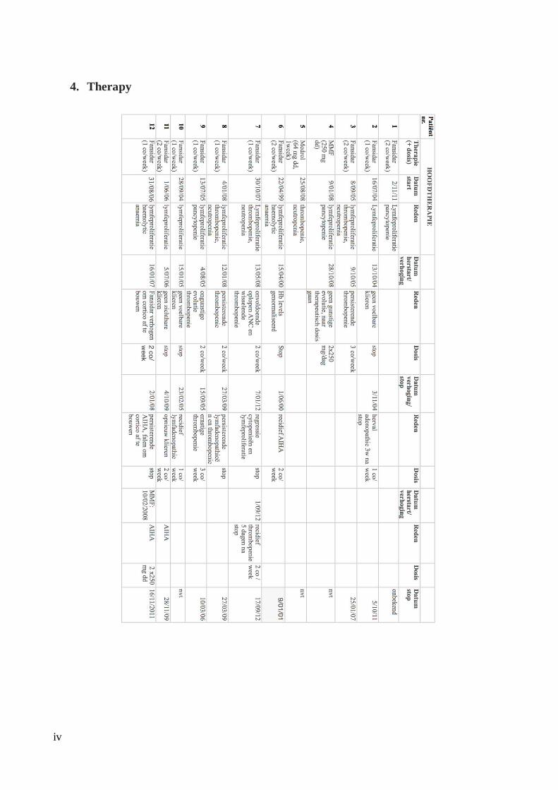
iv
4. Therapy
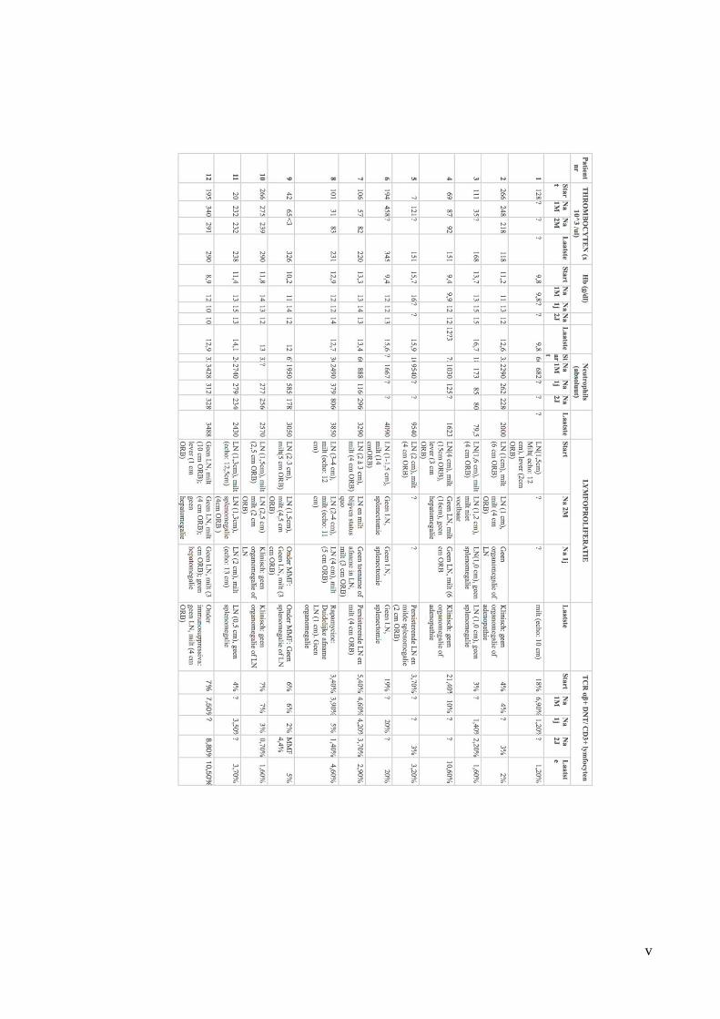
v

vi
vii
Appendix 2
Informed consent

viii

ix

x

xi

xii

xiii
Appendix 3
UZ Ghent’s Ethics Committee approval