atlas of light-ct images
TRANSCRIPT

7292019 Atlas of Light-CT images
httpslidepdfcomreaderfullatlas-of-light-ct-images 1118
copy LLTech 2012 ndash wwwlltechimagingcom - No copy without authorization 1
LLTech
Light-CT Scanner
Atlas of images2012
7292019 Atlas of Light-CT images
httpslidepdfcomreaderfullatlas-of-light-ct-images 2118
copy LLTech 2012 ndash wwwlltechimagingcom - No copy without authorization 2
bull BREAST Page 6
bull BRAIN Page 24
bull SKIN Page 34
bull LUNG Page 58
bull GI Page 64
bull URO GENITAL Page 69
bull NEEDLE CORE Page 78
bull THYROID Page 83
TABLE OF CONTENTS
bull FAT CELLS Page 86
bull CORNEA AND RETINA Page 88
bull DEVELOPMENT BIOLOGY Page 99
bull PLANT IMAGING Page 112
bull CONTACTS Page 118
Table of contents
7292019 Atlas of Light-CT images
httpslidepdfcomreaderfullatlas-of-light-ct-images 3118
copy LLTech 2012 ndash wwwlltechimagingcom - No copy without authorization 3
bull Optical in-depth biopsies of gross tissue within minutes
bull 1 microm 2D and 3D histopathological resolution
bull Easy exploration acquisition and rendering in DICOM format
bull Safe non-invasive and non-destructive process
Light-CTtrade key benefits
Fast and non-invasive 3D in-depth structural and cellular imaging
7292019 Atlas of Light-CT images
httpslidepdfcomreaderfullatlas-of-light-ct-images 4118
copy LLTech 2012 ndash wwwlltechimagingcom - No copy without authorization 4
Based on Full-Field Optical Coherence Tomography (FFOCT)
Combines microscope resolution with interferometry
High resolution in-depth C scans
Commercial device specs
bull Excellent resolution 15microm transverse 1microm axial
bull 70Hz max tomographic frame rate ndash 08 mm x 08 mm
bull Penetration depth 200microm ndash 1mm depending on tissue scattering
bull 25 mm diameter sample size
bull Small footprint Scanner and light source fit on 70 cm x 35 cm
Light Computed Tomography technology
7292019 Atlas of Light-CT images
httpslidepdfcomreaderfullatlas-of-light-ct-images 5118
copy LLTech 2012 ndash wwwlltechimagingcom - No copy without authorization 5
Optical
acquisition unit
moving
vertically
bull User friendly
acquisitionsoftware
bull DICOM 2D
and 3D
Viewer
Movable tray
with sample
holder
XY moving stage
Joystick for easy
control of XY Z
movements
White Light
Source
Integrated wide field camera to
take sample picture beforeimaging
Light-CTtrade Scanner for ex-vivo cellular imaging
7292019 Atlas of Light-CT images
httpslidepdfcomreaderfullatlas-of-light-ct-images 6118
copy LLTech 2012 ndash wwwlltechimagingcom - No copy without authorization 6
BREAST IMAGING
BREAST IMAGING
Back to table of contents
7292019 Atlas of Light-CT images
httpslidepdfcomreaderfullatlas-of-light-ct-images 7118copy LLTech 2012 ndash wwwlltechimagingcom - No copy without authorization 7
Grainy aspect of normal
fibrous tissue
Duct with
calcification
Lobule Adipocytes Vessel
Courtesy of Hocircpital Tenon Paris France
Healthy breast tissue
7292019 Atlas of Light-CT images
httpslidepdfcomreaderfullatlas-of-light-ct-images 8118copy LLTech 2012 ndash wwwlltechimagingcom - No copy without authorization 8 copy LLTech 2012
Breast tissue diagnosis decision tree
7292019 Atlas of Light-CT images
httpslidepdfcomreaderfullatlas-of-light-ct-images 9118copy LLTech 2012 ndash wwwlltechimagingcom - No copy without authorization 9
Breast HistologyDCIS ndash Ductal Carcinoma in situ
Breast Ductal Carinoma HampE vs LightCT on Fresh Tissue
Hopital Tenon France August 2010
Lobules
Fat cells
1 c m
7292019 Atlas of Light-CT images
httpslidepdfcomreaderfullatlas-of-light-ct-images 10118copy LLTech 2012 ndash wwwlltechimagingcom - No copy without authorization 10
Breast HistologyDCIS ndash Ductal Carcinoma in situ
Lobules
Duct withnecrosiss
7292019 Atlas of Light-CT images
httpslidepdfcomreaderfullatlas-of-light-ct-images 11118
7292019 Atlas of Light-CT images
httpslidepdfcomreaderfullatlas-of-light-ct-images 12118
7292019 Atlas of Light-CT images
httpslidepdfcomreaderfullatlas-of-light-ct-images 13118copy LLTech 2012 ndash wwwlltechimagingcom - No copy without authorization 13
Breast HistologyIdentification of the milk duct structures
Milk Duct End
Fat Cells
7292019 Atlas of Light-CT images
httpslidepdfcomreaderfullatlas-of-light-ct-images 14118copy LLTech 2012 ndash wwwlltechimagingcom - No copy without authorization 14
Healthy breast tissue
Lobule
Galactophorous duct cut longitudinally
Honeycomb configuration of adipocytes
copy LLTech 2012 Courtesy of Hocircpital Tenon Paris France
7292019 Atlas of Light-CT images
httpslidepdfcomreaderfullatlas-of-light-ct-images 15118
7292019 Atlas of Light-CT images
httpslidepdfcomreaderfullatlas-of-light-ct-images 16118copy LLTech 2012 ndash wwwlltechimagingcom - No copy without authorization 16
Invasive adenocarcinoma with in Situ Component
Enlarged ducts filled with cells
proliferation or necrosis
Highly scattering thin trabeculae
aspect of fibrous tissue
surrounding grey cellular zones
copy LLTech 2012 Courtesy of Hocircpital Tenon Paris France
7292019 Atlas of Light-CT images
httpslidepdfcomreaderfullatlas-of-light-ct-images 17118copy LLTech 2012 ndash wwwlltechimagingcom - No copy without authorization 17
Elongated compressed
ductules with slit-like lumen
Well delimited nodule withlobulated appearence
Fibroadenoma
copy LLTech 2012 Courtesy of Hocircpital Tenon Paris France
7292019 Atlas of Light-CT images
httpslidepdfcomreaderfullatlas-of-light-ct-images 18118copy LLTech 2012 ndash wwwlltechimagingcom - No copy without authorization 18
Fibroadenoma
Enlarged ductulescharacteristic of the lesion
copy LLTech 2012 Courtesy of Hocircpital Tenon Paris France
7292019 Atlas of Light-CT images
httpslidepdfcomreaderfullatlas-of-light-ct-images 19118copy LLTech 2012 ndash wwwlltechimagingcom - No copy without authorization 19
Lymphoid zone of the node
Vessel
Adipocytes in the center
of the node
Normal Sentinel Node
copy LLTech 2012 Courtesy of Hocircpital Tenon Paris France
7292019 Atlas of Light-CT images
httpslidepdfcomreaderfullatlas-of-light-ct-images 20118
7292019 Atlas of Light-CT images
httpslidepdfcomreaderfullatlas-of-light-ct-images 21118
copy LLTech 2012 ndash wwwlltechimagingcom - No copy without authorization 21
Normal lymphoid
zone with follicles
(Dark grey)
Metastatsis
(Light grey)
Hypervascularization
due to metastasis
Fibrous enveloppe of the
node
Invaded Sentinel Node
copy LLTech 2012 Courtesy of Hocircpital Tenon Paris France
7292019 Atlas of Light-CT images
httpslidepdfcomreaderfullatlas-of-light-ct-images 22118
copy LLTech 2012 ndash wwwlltechimagingcom - No copy without authorization 22
The Light-CT scanner allows fast tissue processing andpathology examination It completes the toolset available topathologists
When Chemical Fixation is needed
(Mostly Diagnostic)
When Frozen Section is needed
(Mostly Intra-operative)
Biopsy3D
Digital Image
2D
Digital Image
Slide
15-12 mins
Artefacts
Destructive
12-24 hours
Expensive
Destructive
5-8 minutes
Non-Destructive
Tissue Scanner
Light-CT could help to reduce the number of re-excisions in breast
7292019 Atlas of Light-CT images
httpslidepdfcomreaderfullatlas-of-light-ct-images 23118
copy LLTech 2012 ndash wwwlltechimagingcom - No copy without authorization 23
Light CT could help to reduce the number of re excisions in breastcancer surgery whilst preserving the current routines ofpathologists
Per surgery
LLTechrsquos
analysis
Histology Andor
Immunohistochemistry
Further
treatment
Post surgery
Surgical margin analysis
Metastasis analysis (gt 2mm) et
micro metastaisi
PN1mi gt 02 ndash 2 mm lt
SurgeryOKOK
Per surgery Post Surgery
C u r r e n t t e c h n i q u e s
U p t o 4 0 o
f d o u b l e p r o
c e d u r e s
L L T e c h L i g h t - C
T
S i g n i f i c a n t d e c r e a s e o f
d o u b l e p r o c e d u
r e s
Visual
analysis
cryostat
Further
treatmentSurgery
OK
Cancerouscancerous
OK
Poor identification of
metastasis
Up to 40 of double surgical
procedures
Several day analysisFew minutes analysis
Surgeon Pathologist Pathologist
Decrease of double surgical
procedures CancerousCancerous
No surgical
margins analysis
Histology Andor
Immuno
histochemistry
Node
Margin
Several day analysisFew minutes analysisSurgeon Pathologist Pathologist
7292019 Atlas of Light-CT images
httpslidepdfcomreaderfullatlas-of-light-ct-images 24118
copy LLTech 2012 ndash wwwlltechimagingcom - No copy without authorization 24
BRAIN IMAGING
Back to table of contents
7292019 Atlas of Light-CT images
httpslidepdfcomreaderfullatlas-of-light-ct-images 25118
copy LLTech 2012 ndash wwwlltechimagingcom - No copy without authorization 25
Rat brain
Architectural and cellular information seen at various scales
Myelinated fibers visible
1 mm
500 microm
100 microm
7292019 Atlas of Light-CT images
httpslidepdfcomreaderfullatlas-of-light-ct-images 26118
7292019 Atlas of Light-CT images
httpslidepdfcomreaderfullatlas-of-light-ct-images 27118
7292019 Atlas of Light-CT images
httpslidepdfcomreaderfullatlas-of-light-ct-images 28118
copy LLTech 2012 ndash wwwlltechimagingcom - No copy without authorization 28 copy LLTECH 2011
Collagen-rich matrixWide fascicles of
tumour cells
Large capillary
Fibroblastic meningioma grade I
500microm
100microm 200microm200microm
7292019 Atlas of Light-CT images
httpslidepdfcomreaderfullatlas-of-light-ct-images 29118
copy LLTech 2012 ndash wwwlltechimagingcom - No copy without authorization 29
Transitional meningioma grade I
Psammoma surrounding a calcification
Whorls formation
Collagen-rich matrix
500microm
100microm
50microm
50microm
7292019 Atlas of Light-CT images
httpslidepdfcomreaderfullatlas-of-light-ct-images 30118
7292019 Atlas of Light-CT images
httpslidepdfcomreaderfullatlas-of-light-ct-images 31118
copy LLTech 2012 ndash wwwlltechimagingcom - No copy without authorization 31 copy LLTECH 2011
Arrows Fibers transverse cut
Spinal cord normal tissue
500 microm
100 microm
100 microm
7292019 Atlas of Light-CT images
httpslidepdfcomreaderfullatlas-of-light-ct-images 32118
7292019 Atlas of Light-CT images
httpslidepdfcomreaderfullatlas-of-light-ct-images 33118
7292019 Atlas of Light-CT images
httpslidepdfcomreaderfullatlas-of-light-ct-images 34118
copy LLTech 2012 ndash wwwlltechimagingcom - No copy without authorization 34
Back to table of contents
SKIN IMAGING
N l ki h l
7292019 Atlas of Light-CT images
httpslidepdfcomreaderfullatlas-of-light-ct-images 35118
copy LLTech 2012 ndash wwwlltechimagingcom - No copy without authorization 35
Normal skin morphology
Epidermis
Collagen
Pilosebaceous
unit
Sweat gland
Adipocytes
b
a
c
e
d
500 microm
copy LLTech 2012 Courtesy of Hopitaux Universitaires de Genegraveve Genegraveve Switzerland
7292019 Atlas of Light-CT images
httpslidepdfcomreaderfullatlas-of-light-ct-images 36118
copy LLTech 2012 ndash wwwlltechimagingcom - No copy without authorization 36
Hypodermis
Skin morphology layers are well discriminated
5 mm x 8 mm mosaic of native field image
Ageing patient (solar elastosis)
En-face imaging of vertical excision
Epidermis
Dermis
1
3
2
4
7292019 Atlas of Light-CT images
httpslidepdfcomreaderfullatlas-of-light-ct-images 37118
En face skin slicing shows structural and cellular details
7292019 Atlas of Light-CT images
httpslidepdfcomreaderfullatlas-of-light-ct-images 38118
copy LLTech 2012 ndash wwwlltechimagingcom - No copy without authorization 38
Stratum corneum Stratum spinosum
Stratum basale Reticular region
Epidermis
Dermis
En-face skin slicing shows structural and cellular details
Melanin
papillary caps
Keratinocyte
nuclei
Blood vessels
Collagen
Corneocytes
100 microm 100 microm
100 microm100 microm
copy LLTech 2012 Courtesy of Hopitaux Universitaires de Genegraveve Genegraveve Switzerland
7292019 Atlas of Light-CT images
httpslidepdfcomreaderfullatlas-of-light-ct-images 39118
7292019 Atlas of Light-CT images
httpslidepdfcomreaderfullatlas-of-light-ct-images 40118
Vertical reconstruction of skin model
7292019 Atlas of Light-CT images
httpslidepdfcomreaderfullatlas-of-light-ct-images 41118
copy LLTech 2012 ndash wwwlltechimagingcom - No copy without authorization 41
Vertical reconstruction of skin model
50 microm
Stratum corneum
Stratum spinosum
Dermis
Keratinocyte nuclei
copy LLTech 2012
7292019 Atlas of Light-CT images
httpslidepdfcomreaderfullatlas-of-light-ct-images 42118
Normal
7292019 Atlas of Light-CT images
httpslidepdfcomreaderfullatlas-of-light-ct-images 43118
copy LLTech 2012 ndash wwwlltechimagingcom - No copy without authorization 43
skin
b
a
e
c
f
d
500 microm
7292019 Atlas of Light-CT images
httpslidepdfcomreaderfullatlas-of-light-ct-images 44118
7292019 Atlas of Light-CT images
httpslidepdfcomreaderfullatlas-of-light-ct-images 45118
copy LLTech 2012 ndash wwwlltechimagingcom - No copy without authorization 45
Adipocytes
9um depth 27um depth 60um depth
Reconstructed
depth slice
200 microm
50 microm
7292019 Atlas of Light-CT images
httpslidepdfcomreaderfullatlas-of-light-ct-images 46118
Skin pathologies basal cell carcinoma discrimination at
7292019 Atlas of Light-CT images
httpslidepdfcomreaderfullatlas-of-light-ct-images 47118
copy LLTech 2012 ndash wwwlltechimagingcom - No copy without authorization 47
Epidermis
Dermis
Hypodermis
A
AA
C
BCC
PSU
PSU
BCC Basal cell carcinoma
A Adipocytes
C Collagen
PSU Pilosebaceous unit
structural level
1 mm
copy LLTech 2012 Courtesy of Hopitaux Universitaires de Genegraveve Genegraveve Switzerland
C
Zoom on basal cell carcinoma discrimination at cellular level
7292019 Atlas of Light-CT images
httpslidepdfcomreaderfullatlas-of-light-ct-images 48118
copy LLTech 2012 ndash wwwlltechimagingcom - No copy without authorization 48
Zoom on basal cell carcinoma discrimination at cellular level
200 microm
100 microm
Peritumoral stroma
High cell density
copy LLTech 2012 Courtesy of Hopitaux Universitaires de Genegraveve Genegraveve Switzerland
Skin pathologies discrimination of actinic keratosis
7292019 Atlas of Light-CT images
httpslidepdfcomreaderfullatlas-of-light-ct-images 49118
copy LLTech 2012 ndash wwwlltechimagingcom - No copy without authorization 49
Skin pathologies discrimination of actinic keratosis
Enlarged epidermis
Extended epidermal ridges
1 mm
copy LLTech 2012 Courtesy of Hopitaux Universitaires de Genegraveve Genegraveve Switzerland
7292019 Atlas of Light-CT images
httpslidepdfcomreaderfullatlas-of-light-ct-images 50118
copy LLTech 2012 ndash wwwlltechimagingcom - No copy without authorization 50
Basal cell carcinoma in depth imaging
Z = 10 microns 7 x 42 mm FOV Z = 35 microns 7 x 42 mm FOV
BCC
SG Sebaceous glands
Z = 55 microns 7 x 42 mm FOV Z = 75 microns 7 x 42 mm FOV
SG
Application to Mohs surgerycopy LLTech 2012 Courtesy of Hopitaux Universitaires de Genegraveve Genegraveve Switzerland
7292019 Atlas of Light-CT images
httpslidepdfcomreaderfullatlas-of-light-ct-images 51118
copy LLTech 2012 ndash wwwlltechimagingcom - No copy without authorization 51 copy LLTECH 2011
Healthy vs Cheloid scar tissue
Healthy skin tissueFOV 75x5mm
Cheloid scar tissueFOV 65x65mm
copy LLTECH 2011
7292019 Atlas of Light-CT images
httpslidepdfcomreaderfullatlas-of-light-ct-images 52118
copy LLTech 2012 ndash wwwlltechimagingcom - No copy without authorization 52
Dermo-epidermaljunction
Cheloid scar tissue in detail
copy LLTECH 2011
Nodular structureThickcollagenfiberbundles
1mm
600 microm
500 microm
h l d d f
7292019 Atlas of Light-CT images
httpslidepdfcomreaderfullatlas-of-light-ct-images 53118
copy LLTech 2012 ndash wwwlltechimagingcom - No copy without authorization 53
Cheloid scar tissue viewed en face
20microm depth 120microm depth
7292019 Atlas of Light-CT images
httpslidepdfcomreaderfullatlas-of-light-ct-images 54118
Skin in-vivo sweat duct imaging in 3D
7292019 Atlas of Light-CT images
httpslidepdfcomreaderfullatlas-of-light-ct-images 55118
copy LLTech 2012 ndash wwwlltechimagingcom - No copy without authorization 55
g g
Vertical reconstruction from en-face images
Sweat duct 3D reconstruction
Stratum corneum
Stratum spinosum
SD sweat ducts
SD SD
100 microm
7292019 Atlas of Light-CT images
httpslidepdfcomreaderfullatlas-of-light-ct-images 56118
7292019 Atlas of Light-CT images
httpslidepdfcomreaderfullatlas-of-light-ct-images 57118
7292019 Atlas of Light-CT images
httpslidepdfcomreaderfullatlas-of-light-ct-images 58118
copy LLTech 2012 ndash wwwlltechimagingcom - No copy without authorization 58
LUNG IMAGING
Back to table of contents
H L
7292019 Atlas of Light-CT images
httpslidepdfcomreaderfullatlas-of-light-ct-images 59118
copy LLTech 2012 ndash wwwlltechimagingcom - No copy without authorization 59
Zone 1
Zone 2
Zone 3
Zone 4
Zone with
cancerous tissue
Limit of cancerous
stroma
alveoli with
macrophages
Alveoles
Human Lung
500microm
copy LLTech 2012 Courtesy of Hocircpital Hotel-Dieu Paris France
H L Z 1 Z t
7292019 Atlas of Light-CT images
httpslidepdfcomreaderfullatlas-of-light-ct-images 60118
copy LLTech 2012 ndash wwwlltechimagingcom - No copy without authorization 60
Stroma
Human Lung ndash Zone 1 ndash Zoom on cancerous stroma
Cancerous
area
200microm
copy LLTech 2012 Courtesy of Hocircpital Hotel-Dieu Paris France
Human Lung ndash Zone 2 ndash Limit cancerous stroma alveoli
7292019 Atlas of Light-CT images
httpslidepdfcomreaderfullatlas-of-light-ct-images 61118
copy LLTech 2012 ndash wwwlltechimagingcom - No copy without authorization 61
g
with visible macrophages and vessel
Alveoli
Macrophages
Vessel
copy LLTech 2012 Courtesy of Hocircpital Hotel-Dieu Paris France
Human Lung ndash Zone 3 ndash Limit cancerous stroma alveoli
7292019 Atlas of Light-CT images
httpslidepdfcomreaderfullatlas-of-light-ct-images 62118
copy LLTech 2012 ndash wwwlltechimagingcom - No copy without authorization 62
g
with visible macrophages
Macrophages
100microm
copy LLTech 2012 Courtesy of Hocircpital Hotel-Dieu Paris France
Human Lung ndash Zone 4 ndash Alveoli
7292019 Atlas of Light-CT images
httpslidepdfcomreaderfullatlas-of-light-ct-images 63118
copy LLTech 2012 ndash wwwlltechimagingcom - No copy without authorization 63
Human Lung Zone 4 Alveoli
Alveoli
200microm
copy LLTech 2012 Courtesy of Hocircpital Hotel-Dieu Paris France
7292019 Atlas of Light-CT images
httpslidepdfcomreaderfullatlas-of-light-ct-images 64118
copy LLTech 2012 ndash wwwlltechimagingcom - No copy without authorization 64
GI IMAGING
Back to table of contents
7292019 Atlas of Light-CT images
httpslidepdfcomreaderfullatlas-of-light-ct-images 65118
Colonic carcinoma
7292019 Atlas of Light-CT images
httpslidepdfcomreaderfullatlas-of-light-ct-images 66118
copy LLTech 2012 ndash wwwlltechimagingcom - No copy without authorization 66
Colonic Carcinoma
Colonic carcinoma
copy LLTech 2012 Courtesy of MD Anderson Houston USA
Intestines ndash Crohnrsquos disease
7292019 Atlas of Light-CT images
httpslidepdfcomreaderfullatlas-of-light-ct-images 67118
copy LLTech 2012 ndash wwwlltechimagingcom - No copy without authorization 67
Crohnrsquos disease structure not
visible from the surface
Fat cells
200 microm
Intestines Crohn s disease
copy LLTech 2012 Courtesy of Institut Curie Paris France
Rat stomach
7292019 Atlas of Light-CT images
httpslidepdfcomreaderfullatlas-of-light-ct-images 68118
copy LLTech 2012 ndash wwwlltechimagingcom - No copy without authorization 68
Rat stomach
Mucosa withgastric pits
Submucosa with
bright collagen
fibers
Muscularis
propria with
muscle bundles
copy LLTech 2012 Courtesy of Weill Cornell Medical College NY USA
200 microm
7292019 Atlas of Light-CT images
httpslidepdfcomreaderfullatlas-of-light-ct-images 69118
copy LLTech 2012 ndash wwwlltechimagingcom - No copy without authorization 69 copy LLTECH 2011
UROGENITAL IMAGING
Back to table of contents
Rat bladder
7292019 Atlas of Light-CT images
httpslidepdfcomreaderfullatlas-of-light-ct-images 70118
copy LLTech 2012 ndash wwwlltechimagingcom - No copy without authorization 70
Rat bladder
Stratifiedurothelium
Lamina propria
500 microm
Muscular
bundles of the
lamina propria
Serosa
copy LLTech 2012 Courtesy of Weill Cornell Medical College NY USA
100 microm
100 microm
Human bladder low grade papillary urothelial carcinoma
7292019 Atlas of Light-CT images
httpslidepdfcomreaderfullatlas-of-light-ct-images 71118
copy LLTech 2012 ndash wwwlltechimagingcom - No copy without authorization 71
Lesion with fibrovascular cores (arrow) and superficial umbrella cells (arrowheads)
Human bladder low grade papillary urothelial carcinoma
copy LLTech 2012 Courtesy of Weill Cornell Medical College NY USA
100 microm
7292019 Atlas of Light-CT images
httpslidepdfcomreaderfullatlas-of-light-ct-images 72118
Rat prostate
7292019 Atlas of Light-CT images
httpslidepdfcomreaderfullatlas-of-light-ct-images 73118
copy LLTech 2012 ndash wwwlltechimagingcom - No copy without authorization 73
p
copy LLTech 2012 Courtesy of Weill Cornell Medical College NY USA
Papillary folds of the
acinar glands
Prostatic acini
Fibromuscular stromaPeriprostatic fat Blood vessel
1 mm
200 microm
Mouse kidney
7292019 Atlas of Light-CT images
httpslidepdfcomreaderfullatlas-of-light-ct-images 74118
copy LLTech 2012 ndash wwwlltechimagingcom - No copy without authorization 74
1 mm
y
copy LLTech 2012 Courtesy of Institut Biomeacutedical de Bicecirctre Paris France
Renal cortex
Renal medulla
Renal sinus
7292019 Atlas of Light-CT images
httpslidepdfcomreaderfullatlas-of-light-ct-images 75118
copy LLTech 2012 ndash wwwlltechimagingcom - No copy without authorization 75
Mouse kidney ndash zoom on highlighted section of previous slide
800microm
copy LLTech 2012 Courtesy of Hocircpital Kremlin Bicecirctre Paris France
7292019 Atlas of Light-CT images
httpslidepdfcomreaderfullatlas-of-light-ct-images 76118
7292019 Atlas of Light-CT images
httpslidepdfcomreaderfullatlas-of-light-ct-images 77118
7292019 Atlas of Light-CT images
httpslidepdfcomreaderfullatlas-of-light-ct-images 78118
copy LLTech 2012 ndash wwwlltechimagingcom - No copy without authorization 78 copy LLTECH 2011
CORE NEEDLE BIOPSIES
Back to table of contents
Kidney
7292019 Atlas of Light-CT images
httpslidepdfcomreaderfullatlas-of-light-ct-images 79118
copy LLTech 2012 ndash wwwlltechimagingcom - No copy without authorization 79
100microm
50microm
Renal tubules
Vessel
500microm
copy LLTech 2012 Courtesy of Institut Curie Paris France
Breast Invasive lobular carcinoma
7292019 Atlas of Light-CT images
httpslidepdfcomreaderfullatlas-of-light-ct-images 80118
copy LLTech 2012 ndash wwwlltechimagingcom - No copy without authorization 80
500microm
stroma
adipocytes
copy LLTech 2012 Courtesy of Institut Curie Paris France
7292019 Atlas of Light-CT images
httpslidepdfcomreaderfullatlas-of-light-ct-images 81118
Breast Ductal carcinoma in situ
7292019 Atlas of Light-CT images
httpslidepdfcomreaderfullatlas-of-light-ct-images 82118
copy LLTech 2012 ndash wwwlltechimagingcom - No copy without authorization 82
500microm
copy LLTech 2012 Courtesy of Institut Curie Paris France
7292019 Atlas of Light-CT images
httpslidepdfcomreaderfullatlas-of-light-ct-images 83118
copy LLTech 2012 ndash wwwlltechimagingcom - No copy without authorization 83 copy LLTECH 2011
THYROID IMAGING
Back to table of contents
7292019 Atlas of Light-CT images
httpslidepdfcomreaderfullatlas-of-light-ct-images 84118
copy LLTech 2012 ndash wwwlltechimagingcom - No copy without authorization 84
Thyroid
800 microm
200 microm
copy LLTech 2012 Courtesy of Hocircpital Tenon Paris France
7292019 Atlas of Light-CT images
httpslidepdfcomreaderfullatlas-of-light-ct-images 85118
copy LLTech 2012 ndash wwwlltechimagingcom - No copy without authorization 85
Thyroid
800 microm
200 microm
copy LLTech 2012 Courtesy of Hocircpital Tenon Paris France
7292019 Atlas of Light-CT images
httpslidepdfcomreaderfullatlas-of-light-ct-images 86118
copy LLTech 2012 ndash wwwlltechimagingcom - No copy without authorization 86 copy LLTECH 2011
FAT CELLS IMAGING
Back to table of contents
7292019 Atlas of Light-CT images
httpslidepdfcomreaderfullatlas-of-light-ct-images 87118
copy LLTech 2012 ndash wwwlltechimagingcom - No copy without authorization 87
Brown fat cells - adipocytes
100 microm 100 microm
200 microm
10um depth
Adipocytes
45um depth
En face
slices
Reconstructed
depth slice
copy LLTech 2012 Courtesy of Centre de Recherche des Cordeliers Paris France
7292019 Atlas of Light-CT images
httpslidepdfcomreaderfullatlas-of-light-ct-images 88118
copy LLTech 2012 ndash wwwlltechimagingcom - No copy without authorization 88 copy LLTECH 2011
CORNEA AND RETINA
Back to table of contents
Human cornea reconstructed depth slice
7292019 Atlas of Light-CT images
httpslidepdfcomreaderfullatlas-of-light-ct-images 89118
copy LLTech 2012 ndash wwwlltechimagingcom - No copy without authorization 89
50 um
wwwlltechimagingcom
7292019 Atlas of Light-CT images
httpslidepdfcomreaderfullatlas-of-light-ct-images 90118
Large field en face view in stroma
7292019 Atlas of Light-CT images
httpslidepdfcomreaderfullatlas-of-light-ct-images 91118
copy LLTech 2012 ndash wwwlltechimagingcom - No copy without authorization 91
200 um
wwwlltechimagingcom
7292019 Atlas of Light-CT images
httpslidepdfcomreaderfullatlas-of-light-ct-images 92118
Rabbit cornea after LASIK reflectivity profiles reveal
i d tt i d thi k d ith li l l
7292019 Atlas of Light-CT images
httpslidepdfcomreaderfullatlas-of-light-ct-images 93118
copy LLTech 2012 ndash wwwlltechimagingcom - No copy without authorization 93
increased scattering and thickened epithelial layer
LASIK LASIK CONTROL CONTROL
copy LLTech 2012
Newborn mouse anterior segment
7292019 Atlas of Light-CT images
httpslidepdfcomreaderfullatlas-of-light-ct-images 94118
copy LLTech 2012 ndash wwwlltechimagingcom - No copy without authorization 94
Corneal epithelial cells
Basal membrane
Stroma
Descemetrsquos membrane
Aqueous humor
Lens capsule
Lens epithelial cells
Cortical fibers
copy LLTech 2012 Courtesy of ESPCI Paris Francecopy LLTech 2012
Rat retina comparison with histology
7292019 Atlas of Light-CT images
httpslidepdfcomreaderfullatlas-of-light-ct-images 95118
copy LLTech 2012 ndash wwwlltechimagingcom - No copy without authorization 95
Figure 1 Right Ex vivo rat retinaimaged using the f ull field OCT
system of ESPCI Left Histology
for comparison
Figure 1 Right Ex vivo rat retinaimaged using the f ull field OCT
system of ESPCI Left Histology
for comparison
Figure 1 Right Ex vivo rat retinaimaged using the f ull field OCT
system of ESPCI Left Histology
for comparisonField
230x300microm
RPE
PRL
ONL
OPL
INL
IPL
GCL
NFL
Invest Ophthalmol Vis Sci 2004 Nov45(11)4126-31
Grieve K Paques M Dubois A Sahel J Boccara C Le Gargasson JF copy LLTech 2012
Rat retina
7292019 Atlas of Light-CT images
httpslidepdfcomreaderfullatlas-of-light-ct-images 96118
copy LLTech 2012 ndash wwwlltechimagingcom - No copy without authorization 96 copy LLTech 2012 Courtesy of ESPCI Paris France
Nerve fiber layer
Retina
Invest Ophthalmol Vis Sci 2004 Nov45(11)4126-31
Grieve K Paques M Dubois A Sahel J Boccara C Le Gargasson JF
Transscleral image
7292019 Atlas of Light-CT images
httpslidepdfcomreaderfullatlas-of-light-ct-images 97118
7292019 Atlas of Light-CT images
httpslidepdfcomreaderfullatlas-of-light-ct-images 98118
7292019 Atlas of Light-CT images
httpslidepdfcomreaderfullatlas-of-light-ct-images 99118
In vivo imaging of drosophila melanogaster
7292019 Atlas of Light-CT images
httpslidepdfcomreaderfullatlas-of-light-ct-images 100118
copy LLTech 2012 ndash wwwlltechimagingcom - No copy without authorization 100 copy LLTech 2012 Courtesy of ESPCI Paris France
In vivo imaging of drosophila melanogaster ndash four stages of pupation over 72 hours
7292019 Atlas of Light-CT images
httpslidepdfcomreaderfullatlas-of-light-ct-images 101118
copy LLTech 2012 ndash wwwlltechimagingcom - No copy without authorization 101
Prepupa (0-2 h) Transition topupal stage (24 h)
Pupal stage (48 h) Advanced pupal phase
before eclosion (72 h)
copy LLTech 2012 Courtesy of ESPCI Paris France
Pupa 118 4 days 30um depth
100
7292019 Atlas of Light-CT images
httpslidepdfcomreaderfullatlas-of-light-ct-images 102118
copy LLTech 2012 ndash wwwlltechimagingcom - No copy without authorization 102
100 um
Drosophila
7292019 Atlas of Light-CT images
httpslidepdfcomreaderfullatlas-of-light-ct-images 103118
copy LLTech 2012 ndash wwwlltechimagingcom - No copy without authorization 103
200 microm
copy LLTech 2012 Courtesy of ESPCI Paris France
C Elegans ndash Fast identification of anatomy
7292019 Atlas of Light-CT images
httpslidepdfcomreaderfullatlas-of-light-ct-images 104118
copy LLTech 2012 ndash wwwlltechimagingcom - No copy without authorization 104
Body wall - cuticle
gonad arm
30 microm depth
uterus
ovocytes
intestine
100 microm 100 microm
15 microm depth
copy LLTech 2012 Courtesy of ENS Paris France
C elegans ndash Images at higher magnification 40X
7292019 Atlas of Light-CT images
httpslidepdfcomreaderfullatlas-of-light-ct-images 105118
copy LLTech 2012 ndash wwwlltechimagingcom - No copy without authorization 105
3D reconstruction 50 microm
100 microm
copy LLTech 2012 Courtesy of ENS Paris France
Zebra fish eye
7292019 Atlas of Light-CT images
httpslidepdfcomreaderfullatlas-of-light-ct-images 106118
copy LLTech 2012 ndash wwwlltechimagingcom - No copy without authorization 106
Zebra fish eye
100 microm
100 microm
En face slices
Reconstructed depth slice
copy LLTech 2012 Courtesy of Inserm Paris France
10um depth 20um depth 45um depth
7292019 Atlas of Light-CT images
httpslidepdfcomreaderfullatlas-of-light-ct-images 107118
Xenopus Laevis backtail
7292019 Atlas of Light-CT images
httpslidepdfcomreaderfullatlas-of-light-ct-images 108118
copy LLTech 2012 ndash wwwlltechimagingcom - No copy without authorization 108
100 um30um depth 60um depth
7292019 Atlas of Light-CT images
httpslidepdfcomreaderfullatlas-of-light-ct-images 109118
Mouse embryo in vitro 6 5 days old ndash
7292019 Atlas of Light-CT images
httpslidepdfcomreaderfullatlas-of-light-ct-images 110118
copy LLTech 2012 ndash wwwlltechimagingcom - No copy without authorization 110
Mouse embryo in vitro 65 days old
copy LLTech 2012 Courtesy of Ellvire Guiot and nstitut Jacques Monod Paris France
3D imaging of internal cavity
Sabellaria alveolata mouth palps
7292019 Atlas of Light-CT images
httpslidepdfcomreaderfullatlas-of-light-ct-images 111118
copy LLTech 2012 ndash wwwlltechimagingcom - No copy without authorization 111
500 microm
100 microm
Mouth palps
copy LLTech 2012 Courtesy of Museacutee National drsquoHistoire Naturelle Paris France
7292019 Atlas of Light-CT images
httpslidepdfcomreaderfullatlas-of-light-ct-images 112118
copy LLTech 2012 ndash wwwlltechimagingcom - No copy without authorization 112 copy LLTECH 2011
PLANT IMAGING
Back to table of contents
Mouse ear leaf epidermis
7292019 Atlas of Light-CT images
httpslidepdfcomreaderfullatlas-of-light-ct-images 113118
copy LLTech 2012 ndash wwwlltechimagingcom - No copy without authorization 113
100 microm
Epidermal cell
copy LLTech 2012
Stoma
Leaf veins
7292019 Atlas of Light-CT images
httpslidepdfcomreaderfullatlas-of-light-ct-images 114118
copy LLTech 2012 ndash wwwlltechimagingcom - No copy without authorization 114
114copy LLTech 2012
Fibers
Veins
100 microm
500 microm
Apple
7292019 Atlas of Light-CT images
httpslidepdfcomreaderfullatlas-of-light-ct-images 115118
copy LLTech 2012 ndash wwwlltechimagingcom - No copy without authorization 115
115copy LLTech 2012
100 microm
Wax
Skin
Flesh
500 microm
Water
Effect of harpin infiltration on tobacco leaf
7292019 Atlas of Light-CT images
httpslidepdfcomreaderfullatlas-of-light-ct-images 116118
copy LLTech 2012 ndash wwwlltechimagingcom - No copy without authorization 116
116copy LLTech 2012 Courtesy of UPMC Paris France
50 microm
50 microm
50 microm
50 microm
Upper epidermis
10 min after harpin infiltration 30 min after harpin infiltration
Palisade parenchyma
Epidermis cell
Chloroplast
7292019 Atlas of Light-CT images
httpslidepdfcomreaderfullatlas-of-light-ct-images 117118
copy LLTech 2012 ndash wwwlltechimagingcom - No copy without authorization 117
bull Light-CTtrade does not require tissue preparation nor staining of any kind
bull Creates images within minutes using a non-destructive process
bull Offers a 1 microm cellular resolution in 3D
bull Reveals structural and cellular information
bull Mosaicing allows fast visualisation at various scale
bull En-face high-resolution imaging allows vertical and 3D reconstruction
bull In-vivo images show good promise for in-vivo system
bull Clinical and research applications in cancer detection cosmetics biobanking small animal
developmental biology botanyhellip
Contact Details
7292019 Atlas of Light-CT images
httpslidepdfcomreaderfullatlas-of-light-ct-images 118118
USA
LLTech Inc
103 Carnegie Center DriveSuite 300
Princeton NJ 08540
USA
wwwlltechimagingcom
contactlltechfr
Phone + 1 609 955 3506
LLTech SAS
29 rue du Faubourg St-JacquesPeacutepiniegravere Paris Santeacute Cochin
75014
Paris - France
wwwlltechimagingcom
contactlltechfr
Phone +33 1 82 72 61 25
Europe
7292019 Atlas of Light-CT images
httpslidepdfcomreaderfullatlas-of-light-ct-images 2118
copy LLTech 2012 ndash wwwlltechimagingcom - No copy without authorization 2
bull BREAST Page 6
bull BRAIN Page 24
bull SKIN Page 34
bull LUNG Page 58
bull GI Page 64
bull URO GENITAL Page 69
bull NEEDLE CORE Page 78
bull THYROID Page 83
TABLE OF CONTENTS
bull FAT CELLS Page 86
bull CORNEA AND RETINA Page 88
bull DEVELOPMENT BIOLOGY Page 99
bull PLANT IMAGING Page 112
bull CONTACTS Page 118
Table of contents
7292019 Atlas of Light-CT images
httpslidepdfcomreaderfullatlas-of-light-ct-images 3118
copy LLTech 2012 ndash wwwlltechimagingcom - No copy without authorization 3
bull Optical in-depth biopsies of gross tissue within minutes
bull 1 microm 2D and 3D histopathological resolution
bull Easy exploration acquisition and rendering in DICOM format
bull Safe non-invasive and non-destructive process
Light-CTtrade key benefits
Fast and non-invasive 3D in-depth structural and cellular imaging
7292019 Atlas of Light-CT images
httpslidepdfcomreaderfullatlas-of-light-ct-images 4118
copy LLTech 2012 ndash wwwlltechimagingcom - No copy without authorization 4
Based on Full-Field Optical Coherence Tomography (FFOCT)
Combines microscope resolution with interferometry
High resolution in-depth C scans
Commercial device specs
bull Excellent resolution 15microm transverse 1microm axial
bull 70Hz max tomographic frame rate ndash 08 mm x 08 mm
bull Penetration depth 200microm ndash 1mm depending on tissue scattering
bull 25 mm diameter sample size
bull Small footprint Scanner and light source fit on 70 cm x 35 cm
Light Computed Tomography technology
7292019 Atlas of Light-CT images
httpslidepdfcomreaderfullatlas-of-light-ct-images 5118
copy LLTech 2012 ndash wwwlltechimagingcom - No copy without authorization 5
Optical
acquisition unit
moving
vertically
bull User friendly
acquisitionsoftware
bull DICOM 2D
and 3D
Viewer
Movable tray
with sample
holder
XY moving stage
Joystick for easy
control of XY Z
movements
White Light
Source
Integrated wide field camera to
take sample picture beforeimaging
Light-CTtrade Scanner for ex-vivo cellular imaging
7292019 Atlas of Light-CT images
httpslidepdfcomreaderfullatlas-of-light-ct-images 6118
copy LLTech 2012 ndash wwwlltechimagingcom - No copy without authorization 6
BREAST IMAGING
BREAST IMAGING
Back to table of contents
7292019 Atlas of Light-CT images
httpslidepdfcomreaderfullatlas-of-light-ct-images 7118copy LLTech 2012 ndash wwwlltechimagingcom - No copy without authorization 7
Grainy aspect of normal
fibrous tissue
Duct with
calcification
Lobule Adipocytes Vessel
Courtesy of Hocircpital Tenon Paris France
Healthy breast tissue
7292019 Atlas of Light-CT images
httpslidepdfcomreaderfullatlas-of-light-ct-images 8118copy LLTech 2012 ndash wwwlltechimagingcom - No copy without authorization 8 copy LLTech 2012
Breast tissue diagnosis decision tree
7292019 Atlas of Light-CT images
httpslidepdfcomreaderfullatlas-of-light-ct-images 9118copy LLTech 2012 ndash wwwlltechimagingcom - No copy without authorization 9
Breast HistologyDCIS ndash Ductal Carcinoma in situ
Breast Ductal Carinoma HampE vs LightCT on Fresh Tissue
Hopital Tenon France August 2010
Lobules
Fat cells
1 c m
7292019 Atlas of Light-CT images
httpslidepdfcomreaderfullatlas-of-light-ct-images 10118copy LLTech 2012 ndash wwwlltechimagingcom - No copy without authorization 10
Breast HistologyDCIS ndash Ductal Carcinoma in situ
Lobules
Duct withnecrosiss
7292019 Atlas of Light-CT images
httpslidepdfcomreaderfullatlas-of-light-ct-images 11118
7292019 Atlas of Light-CT images
httpslidepdfcomreaderfullatlas-of-light-ct-images 12118
7292019 Atlas of Light-CT images
httpslidepdfcomreaderfullatlas-of-light-ct-images 13118copy LLTech 2012 ndash wwwlltechimagingcom - No copy without authorization 13
Breast HistologyIdentification of the milk duct structures
Milk Duct End
Fat Cells
7292019 Atlas of Light-CT images
httpslidepdfcomreaderfullatlas-of-light-ct-images 14118copy LLTech 2012 ndash wwwlltechimagingcom - No copy without authorization 14
Healthy breast tissue
Lobule
Galactophorous duct cut longitudinally
Honeycomb configuration of adipocytes
copy LLTech 2012 Courtesy of Hocircpital Tenon Paris France
7292019 Atlas of Light-CT images
httpslidepdfcomreaderfullatlas-of-light-ct-images 15118
7292019 Atlas of Light-CT images
httpslidepdfcomreaderfullatlas-of-light-ct-images 16118copy LLTech 2012 ndash wwwlltechimagingcom - No copy without authorization 16
Invasive adenocarcinoma with in Situ Component
Enlarged ducts filled with cells
proliferation or necrosis
Highly scattering thin trabeculae
aspect of fibrous tissue
surrounding grey cellular zones
copy LLTech 2012 Courtesy of Hocircpital Tenon Paris France
7292019 Atlas of Light-CT images
httpslidepdfcomreaderfullatlas-of-light-ct-images 17118copy LLTech 2012 ndash wwwlltechimagingcom - No copy without authorization 17
Elongated compressed
ductules with slit-like lumen
Well delimited nodule withlobulated appearence
Fibroadenoma
copy LLTech 2012 Courtesy of Hocircpital Tenon Paris France
7292019 Atlas of Light-CT images
httpslidepdfcomreaderfullatlas-of-light-ct-images 18118copy LLTech 2012 ndash wwwlltechimagingcom - No copy without authorization 18
Fibroadenoma
Enlarged ductulescharacteristic of the lesion
copy LLTech 2012 Courtesy of Hocircpital Tenon Paris France
7292019 Atlas of Light-CT images
httpslidepdfcomreaderfullatlas-of-light-ct-images 19118copy LLTech 2012 ndash wwwlltechimagingcom - No copy without authorization 19
Lymphoid zone of the node
Vessel
Adipocytes in the center
of the node
Normal Sentinel Node
copy LLTech 2012 Courtesy of Hocircpital Tenon Paris France
7292019 Atlas of Light-CT images
httpslidepdfcomreaderfullatlas-of-light-ct-images 20118
7292019 Atlas of Light-CT images
httpslidepdfcomreaderfullatlas-of-light-ct-images 21118
copy LLTech 2012 ndash wwwlltechimagingcom - No copy without authorization 21
Normal lymphoid
zone with follicles
(Dark grey)
Metastatsis
(Light grey)
Hypervascularization
due to metastasis
Fibrous enveloppe of the
node
Invaded Sentinel Node
copy LLTech 2012 Courtesy of Hocircpital Tenon Paris France
7292019 Atlas of Light-CT images
httpslidepdfcomreaderfullatlas-of-light-ct-images 22118
copy LLTech 2012 ndash wwwlltechimagingcom - No copy without authorization 22
The Light-CT scanner allows fast tissue processing andpathology examination It completes the toolset available topathologists
When Chemical Fixation is needed
(Mostly Diagnostic)
When Frozen Section is needed
(Mostly Intra-operative)
Biopsy3D
Digital Image
2D
Digital Image
Slide
15-12 mins
Artefacts
Destructive
12-24 hours
Expensive
Destructive
5-8 minutes
Non-Destructive
Tissue Scanner
Light-CT could help to reduce the number of re-excisions in breast
7292019 Atlas of Light-CT images
httpslidepdfcomreaderfullatlas-of-light-ct-images 23118
copy LLTech 2012 ndash wwwlltechimagingcom - No copy without authorization 23
Light CT could help to reduce the number of re excisions in breastcancer surgery whilst preserving the current routines ofpathologists
Per surgery
LLTechrsquos
analysis
Histology Andor
Immunohistochemistry
Further
treatment
Post surgery
Surgical margin analysis
Metastasis analysis (gt 2mm) et
micro metastaisi
PN1mi gt 02 ndash 2 mm lt
SurgeryOKOK
Per surgery Post Surgery
C u r r e n t t e c h n i q u e s
U p t o 4 0 o
f d o u b l e p r o
c e d u r e s
L L T e c h L i g h t - C
T
S i g n i f i c a n t d e c r e a s e o f
d o u b l e p r o c e d u
r e s
Visual
analysis
cryostat
Further
treatmentSurgery
OK
Cancerouscancerous
OK
Poor identification of
metastasis
Up to 40 of double surgical
procedures
Several day analysisFew minutes analysis
Surgeon Pathologist Pathologist
Decrease of double surgical
procedures CancerousCancerous
No surgical
margins analysis
Histology Andor
Immuno
histochemistry
Node
Margin
Several day analysisFew minutes analysisSurgeon Pathologist Pathologist
7292019 Atlas of Light-CT images
httpslidepdfcomreaderfullatlas-of-light-ct-images 24118
copy LLTech 2012 ndash wwwlltechimagingcom - No copy without authorization 24
BRAIN IMAGING
Back to table of contents
7292019 Atlas of Light-CT images
httpslidepdfcomreaderfullatlas-of-light-ct-images 25118
copy LLTech 2012 ndash wwwlltechimagingcom - No copy without authorization 25
Rat brain
Architectural and cellular information seen at various scales
Myelinated fibers visible
1 mm
500 microm
100 microm
7292019 Atlas of Light-CT images
httpslidepdfcomreaderfullatlas-of-light-ct-images 26118
7292019 Atlas of Light-CT images
httpslidepdfcomreaderfullatlas-of-light-ct-images 27118
7292019 Atlas of Light-CT images
httpslidepdfcomreaderfullatlas-of-light-ct-images 28118
copy LLTech 2012 ndash wwwlltechimagingcom - No copy without authorization 28 copy LLTECH 2011
Collagen-rich matrixWide fascicles of
tumour cells
Large capillary
Fibroblastic meningioma grade I
500microm
100microm 200microm200microm
7292019 Atlas of Light-CT images
httpslidepdfcomreaderfullatlas-of-light-ct-images 29118
copy LLTech 2012 ndash wwwlltechimagingcom - No copy without authorization 29
Transitional meningioma grade I
Psammoma surrounding a calcification
Whorls formation
Collagen-rich matrix
500microm
100microm
50microm
50microm
7292019 Atlas of Light-CT images
httpslidepdfcomreaderfullatlas-of-light-ct-images 30118
7292019 Atlas of Light-CT images
httpslidepdfcomreaderfullatlas-of-light-ct-images 31118
copy LLTech 2012 ndash wwwlltechimagingcom - No copy without authorization 31 copy LLTECH 2011
Arrows Fibers transverse cut
Spinal cord normal tissue
500 microm
100 microm
100 microm
7292019 Atlas of Light-CT images
httpslidepdfcomreaderfullatlas-of-light-ct-images 32118
7292019 Atlas of Light-CT images
httpslidepdfcomreaderfullatlas-of-light-ct-images 33118
7292019 Atlas of Light-CT images
httpslidepdfcomreaderfullatlas-of-light-ct-images 34118
copy LLTech 2012 ndash wwwlltechimagingcom - No copy without authorization 34
Back to table of contents
SKIN IMAGING
N l ki h l
7292019 Atlas of Light-CT images
httpslidepdfcomreaderfullatlas-of-light-ct-images 35118
copy LLTech 2012 ndash wwwlltechimagingcom - No copy without authorization 35
Normal skin morphology
Epidermis
Collagen
Pilosebaceous
unit
Sweat gland
Adipocytes
b
a
c
e
d
500 microm
copy LLTech 2012 Courtesy of Hopitaux Universitaires de Genegraveve Genegraveve Switzerland
7292019 Atlas of Light-CT images
httpslidepdfcomreaderfullatlas-of-light-ct-images 36118
copy LLTech 2012 ndash wwwlltechimagingcom - No copy without authorization 36
Hypodermis
Skin morphology layers are well discriminated
5 mm x 8 mm mosaic of native field image
Ageing patient (solar elastosis)
En-face imaging of vertical excision
Epidermis
Dermis
1
3
2
4
7292019 Atlas of Light-CT images
httpslidepdfcomreaderfullatlas-of-light-ct-images 37118
En face skin slicing shows structural and cellular details
7292019 Atlas of Light-CT images
httpslidepdfcomreaderfullatlas-of-light-ct-images 38118
copy LLTech 2012 ndash wwwlltechimagingcom - No copy without authorization 38
Stratum corneum Stratum spinosum
Stratum basale Reticular region
Epidermis
Dermis
En-face skin slicing shows structural and cellular details
Melanin
papillary caps
Keratinocyte
nuclei
Blood vessels
Collagen
Corneocytes
100 microm 100 microm
100 microm100 microm
copy LLTech 2012 Courtesy of Hopitaux Universitaires de Genegraveve Genegraveve Switzerland
7292019 Atlas of Light-CT images
httpslidepdfcomreaderfullatlas-of-light-ct-images 39118
7292019 Atlas of Light-CT images
httpslidepdfcomreaderfullatlas-of-light-ct-images 40118
Vertical reconstruction of skin model
7292019 Atlas of Light-CT images
httpslidepdfcomreaderfullatlas-of-light-ct-images 41118
copy LLTech 2012 ndash wwwlltechimagingcom - No copy without authorization 41
Vertical reconstruction of skin model
50 microm
Stratum corneum
Stratum spinosum
Dermis
Keratinocyte nuclei
copy LLTech 2012
7292019 Atlas of Light-CT images
httpslidepdfcomreaderfullatlas-of-light-ct-images 42118
Normal
7292019 Atlas of Light-CT images
httpslidepdfcomreaderfullatlas-of-light-ct-images 43118
copy LLTech 2012 ndash wwwlltechimagingcom - No copy without authorization 43
skin
b
a
e
c
f
d
500 microm
7292019 Atlas of Light-CT images
httpslidepdfcomreaderfullatlas-of-light-ct-images 44118
7292019 Atlas of Light-CT images
httpslidepdfcomreaderfullatlas-of-light-ct-images 45118
copy LLTech 2012 ndash wwwlltechimagingcom - No copy without authorization 45
Adipocytes
9um depth 27um depth 60um depth
Reconstructed
depth slice
200 microm
50 microm
7292019 Atlas of Light-CT images
httpslidepdfcomreaderfullatlas-of-light-ct-images 46118
Skin pathologies basal cell carcinoma discrimination at
7292019 Atlas of Light-CT images
httpslidepdfcomreaderfullatlas-of-light-ct-images 47118
copy LLTech 2012 ndash wwwlltechimagingcom - No copy without authorization 47
Epidermis
Dermis
Hypodermis
A
AA
C
BCC
PSU
PSU
BCC Basal cell carcinoma
A Adipocytes
C Collagen
PSU Pilosebaceous unit
structural level
1 mm
copy LLTech 2012 Courtesy of Hopitaux Universitaires de Genegraveve Genegraveve Switzerland
C
Zoom on basal cell carcinoma discrimination at cellular level
7292019 Atlas of Light-CT images
httpslidepdfcomreaderfullatlas-of-light-ct-images 48118
copy LLTech 2012 ndash wwwlltechimagingcom - No copy without authorization 48
Zoom on basal cell carcinoma discrimination at cellular level
200 microm
100 microm
Peritumoral stroma
High cell density
copy LLTech 2012 Courtesy of Hopitaux Universitaires de Genegraveve Genegraveve Switzerland
Skin pathologies discrimination of actinic keratosis
7292019 Atlas of Light-CT images
httpslidepdfcomreaderfullatlas-of-light-ct-images 49118
copy LLTech 2012 ndash wwwlltechimagingcom - No copy without authorization 49
Skin pathologies discrimination of actinic keratosis
Enlarged epidermis
Extended epidermal ridges
1 mm
copy LLTech 2012 Courtesy of Hopitaux Universitaires de Genegraveve Genegraveve Switzerland
7292019 Atlas of Light-CT images
httpslidepdfcomreaderfullatlas-of-light-ct-images 50118
copy LLTech 2012 ndash wwwlltechimagingcom - No copy without authorization 50
Basal cell carcinoma in depth imaging
Z = 10 microns 7 x 42 mm FOV Z = 35 microns 7 x 42 mm FOV
BCC
SG Sebaceous glands
Z = 55 microns 7 x 42 mm FOV Z = 75 microns 7 x 42 mm FOV
SG
Application to Mohs surgerycopy LLTech 2012 Courtesy of Hopitaux Universitaires de Genegraveve Genegraveve Switzerland
7292019 Atlas of Light-CT images
httpslidepdfcomreaderfullatlas-of-light-ct-images 51118
copy LLTech 2012 ndash wwwlltechimagingcom - No copy without authorization 51 copy LLTECH 2011
Healthy vs Cheloid scar tissue
Healthy skin tissueFOV 75x5mm
Cheloid scar tissueFOV 65x65mm
copy LLTECH 2011
7292019 Atlas of Light-CT images
httpslidepdfcomreaderfullatlas-of-light-ct-images 52118
copy LLTech 2012 ndash wwwlltechimagingcom - No copy without authorization 52
Dermo-epidermaljunction
Cheloid scar tissue in detail
copy LLTECH 2011
Nodular structureThickcollagenfiberbundles
1mm
600 microm
500 microm
h l d d f
7292019 Atlas of Light-CT images
httpslidepdfcomreaderfullatlas-of-light-ct-images 53118
copy LLTech 2012 ndash wwwlltechimagingcom - No copy without authorization 53
Cheloid scar tissue viewed en face
20microm depth 120microm depth
7292019 Atlas of Light-CT images
httpslidepdfcomreaderfullatlas-of-light-ct-images 54118
Skin in-vivo sweat duct imaging in 3D
7292019 Atlas of Light-CT images
httpslidepdfcomreaderfullatlas-of-light-ct-images 55118
copy LLTech 2012 ndash wwwlltechimagingcom - No copy without authorization 55
g g
Vertical reconstruction from en-face images
Sweat duct 3D reconstruction
Stratum corneum
Stratum spinosum
SD sweat ducts
SD SD
100 microm
7292019 Atlas of Light-CT images
httpslidepdfcomreaderfullatlas-of-light-ct-images 56118
7292019 Atlas of Light-CT images
httpslidepdfcomreaderfullatlas-of-light-ct-images 57118
7292019 Atlas of Light-CT images
httpslidepdfcomreaderfullatlas-of-light-ct-images 58118
copy LLTech 2012 ndash wwwlltechimagingcom - No copy without authorization 58
LUNG IMAGING
Back to table of contents
H L
7292019 Atlas of Light-CT images
httpslidepdfcomreaderfullatlas-of-light-ct-images 59118
copy LLTech 2012 ndash wwwlltechimagingcom - No copy without authorization 59
Zone 1
Zone 2
Zone 3
Zone 4
Zone with
cancerous tissue
Limit of cancerous
stroma
alveoli with
macrophages
Alveoles
Human Lung
500microm
copy LLTech 2012 Courtesy of Hocircpital Hotel-Dieu Paris France
H L Z 1 Z t
7292019 Atlas of Light-CT images
httpslidepdfcomreaderfullatlas-of-light-ct-images 60118
copy LLTech 2012 ndash wwwlltechimagingcom - No copy without authorization 60
Stroma
Human Lung ndash Zone 1 ndash Zoom on cancerous stroma
Cancerous
area
200microm
copy LLTech 2012 Courtesy of Hocircpital Hotel-Dieu Paris France
Human Lung ndash Zone 2 ndash Limit cancerous stroma alveoli
7292019 Atlas of Light-CT images
httpslidepdfcomreaderfullatlas-of-light-ct-images 61118
copy LLTech 2012 ndash wwwlltechimagingcom - No copy without authorization 61
g
with visible macrophages and vessel
Alveoli
Macrophages
Vessel
copy LLTech 2012 Courtesy of Hocircpital Hotel-Dieu Paris France
Human Lung ndash Zone 3 ndash Limit cancerous stroma alveoli
7292019 Atlas of Light-CT images
httpslidepdfcomreaderfullatlas-of-light-ct-images 62118
copy LLTech 2012 ndash wwwlltechimagingcom - No copy without authorization 62
g
with visible macrophages
Macrophages
100microm
copy LLTech 2012 Courtesy of Hocircpital Hotel-Dieu Paris France
Human Lung ndash Zone 4 ndash Alveoli
7292019 Atlas of Light-CT images
httpslidepdfcomreaderfullatlas-of-light-ct-images 63118
copy LLTech 2012 ndash wwwlltechimagingcom - No copy without authorization 63
Human Lung Zone 4 Alveoli
Alveoli
200microm
copy LLTech 2012 Courtesy of Hocircpital Hotel-Dieu Paris France
7292019 Atlas of Light-CT images
httpslidepdfcomreaderfullatlas-of-light-ct-images 64118
copy LLTech 2012 ndash wwwlltechimagingcom - No copy without authorization 64
GI IMAGING
Back to table of contents
7292019 Atlas of Light-CT images
httpslidepdfcomreaderfullatlas-of-light-ct-images 65118
Colonic carcinoma
7292019 Atlas of Light-CT images
httpslidepdfcomreaderfullatlas-of-light-ct-images 66118
copy LLTech 2012 ndash wwwlltechimagingcom - No copy without authorization 66
Colonic Carcinoma
Colonic carcinoma
copy LLTech 2012 Courtesy of MD Anderson Houston USA
Intestines ndash Crohnrsquos disease
7292019 Atlas of Light-CT images
httpslidepdfcomreaderfullatlas-of-light-ct-images 67118
copy LLTech 2012 ndash wwwlltechimagingcom - No copy without authorization 67
Crohnrsquos disease structure not
visible from the surface
Fat cells
200 microm
Intestines Crohn s disease
copy LLTech 2012 Courtesy of Institut Curie Paris France
Rat stomach
7292019 Atlas of Light-CT images
httpslidepdfcomreaderfullatlas-of-light-ct-images 68118
copy LLTech 2012 ndash wwwlltechimagingcom - No copy without authorization 68
Rat stomach
Mucosa withgastric pits
Submucosa with
bright collagen
fibers
Muscularis
propria with
muscle bundles
copy LLTech 2012 Courtesy of Weill Cornell Medical College NY USA
200 microm
7292019 Atlas of Light-CT images
httpslidepdfcomreaderfullatlas-of-light-ct-images 69118
copy LLTech 2012 ndash wwwlltechimagingcom - No copy without authorization 69 copy LLTECH 2011
UROGENITAL IMAGING
Back to table of contents
Rat bladder
7292019 Atlas of Light-CT images
httpslidepdfcomreaderfullatlas-of-light-ct-images 70118
copy LLTech 2012 ndash wwwlltechimagingcom - No copy without authorization 70
Rat bladder
Stratifiedurothelium
Lamina propria
500 microm
Muscular
bundles of the
lamina propria
Serosa
copy LLTech 2012 Courtesy of Weill Cornell Medical College NY USA
100 microm
100 microm
Human bladder low grade papillary urothelial carcinoma
7292019 Atlas of Light-CT images
httpslidepdfcomreaderfullatlas-of-light-ct-images 71118
copy LLTech 2012 ndash wwwlltechimagingcom - No copy without authorization 71
Lesion with fibrovascular cores (arrow) and superficial umbrella cells (arrowheads)
Human bladder low grade papillary urothelial carcinoma
copy LLTech 2012 Courtesy of Weill Cornell Medical College NY USA
100 microm
7292019 Atlas of Light-CT images
httpslidepdfcomreaderfullatlas-of-light-ct-images 72118
Rat prostate
7292019 Atlas of Light-CT images
httpslidepdfcomreaderfullatlas-of-light-ct-images 73118
copy LLTech 2012 ndash wwwlltechimagingcom - No copy without authorization 73
p
copy LLTech 2012 Courtesy of Weill Cornell Medical College NY USA
Papillary folds of the
acinar glands
Prostatic acini
Fibromuscular stromaPeriprostatic fat Blood vessel
1 mm
200 microm
Mouse kidney
7292019 Atlas of Light-CT images
httpslidepdfcomreaderfullatlas-of-light-ct-images 74118
copy LLTech 2012 ndash wwwlltechimagingcom - No copy without authorization 74
1 mm
y
copy LLTech 2012 Courtesy of Institut Biomeacutedical de Bicecirctre Paris France
Renal cortex
Renal medulla
Renal sinus
7292019 Atlas of Light-CT images
httpslidepdfcomreaderfullatlas-of-light-ct-images 75118
copy LLTech 2012 ndash wwwlltechimagingcom - No copy without authorization 75
Mouse kidney ndash zoom on highlighted section of previous slide
800microm
copy LLTech 2012 Courtesy of Hocircpital Kremlin Bicecirctre Paris France
7292019 Atlas of Light-CT images
httpslidepdfcomreaderfullatlas-of-light-ct-images 76118
7292019 Atlas of Light-CT images
httpslidepdfcomreaderfullatlas-of-light-ct-images 77118
7292019 Atlas of Light-CT images
httpslidepdfcomreaderfullatlas-of-light-ct-images 78118
copy LLTech 2012 ndash wwwlltechimagingcom - No copy without authorization 78 copy LLTECH 2011
CORE NEEDLE BIOPSIES
Back to table of contents
Kidney
7292019 Atlas of Light-CT images
httpslidepdfcomreaderfullatlas-of-light-ct-images 79118
copy LLTech 2012 ndash wwwlltechimagingcom - No copy without authorization 79
100microm
50microm
Renal tubules
Vessel
500microm
copy LLTech 2012 Courtesy of Institut Curie Paris France
Breast Invasive lobular carcinoma
7292019 Atlas of Light-CT images
httpslidepdfcomreaderfullatlas-of-light-ct-images 80118
copy LLTech 2012 ndash wwwlltechimagingcom - No copy without authorization 80
500microm
stroma
adipocytes
copy LLTech 2012 Courtesy of Institut Curie Paris France
7292019 Atlas of Light-CT images
httpslidepdfcomreaderfullatlas-of-light-ct-images 81118
Breast Ductal carcinoma in situ
7292019 Atlas of Light-CT images
httpslidepdfcomreaderfullatlas-of-light-ct-images 82118
copy LLTech 2012 ndash wwwlltechimagingcom - No copy without authorization 82
500microm
copy LLTech 2012 Courtesy of Institut Curie Paris France
7292019 Atlas of Light-CT images
httpslidepdfcomreaderfullatlas-of-light-ct-images 83118
copy LLTech 2012 ndash wwwlltechimagingcom - No copy without authorization 83 copy LLTECH 2011
THYROID IMAGING
Back to table of contents
7292019 Atlas of Light-CT images
httpslidepdfcomreaderfullatlas-of-light-ct-images 84118
copy LLTech 2012 ndash wwwlltechimagingcom - No copy without authorization 84
Thyroid
800 microm
200 microm
copy LLTech 2012 Courtesy of Hocircpital Tenon Paris France
7292019 Atlas of Light-CT images
httpslidepdfcomreaderfullatlas-of-light-ct-images 85118
copy LLTech 2012 ndash wwwlltechimagingcom - No copy without authorization 85
Thyroid
800 microm
200 microm
copy LLTech 2012 Courtesy of Hocircpital Tenon Paris France
7292019 Atlas of Light-CT images
httpslidepdfcomreaderfullatlas-of-light-ct-images 86118
copy LLTech 2012 ndash wwwlltechimagingcom - No copy without authorization 86 copy LLTECH 2011
FAT CELLS IMAGING
Back to table of contents
7292019 Atlas of Light-CT images
httpslidepdfcomreaderfullatlas-of-light-ct-images 87118
copy LLTech 2012 ndash wwwlltechimagingcom - No copy without authorization 87
Brown fat cells - adipocytes
100 microm 100 microm
200 microm
10um depth
Adipocytes
45um depth
En face
slices
Reconstructed
depth slice
copy LLTech 2012 Courtesy of Centre de Recherche des Cordeliers Paris France
7292019 Atlas of Light-CT images
httpslidepdfcomreaderfullatlas-of-light-ct-images 88118
copy LLTech 2012 ndash wwwlltechimagingcom - No copy without authorization 88 copy LLTECH 2011
CORNEA AND RETINA
Back to table of contents
Human cornea reconstructed depth slice
7292019 Atlas of Light-CT images
httpslidepdfcomreaderfullatlas-of-light-ct-images 89118
copy LLTech 2012 ndash wwwlltechimagingcom - No copy without authorization 89
50 um
wwwlltechimagingcom
7292019 Atlas of Light-CT images
httpslidepdfcomreaderfullatlas-of-light-ct-images 90118
Large field en face view in stroma
7292019 Atlas of Light-CT images
httpslidepdfcomreaderfullatlas-of-light-ct-images 91118
copy LLTech 2012 ndash wwwlltechimagingcom - No copy without authorization 91
200 um
wwwlltechimagingcom
7292019 Atlas of Light-CT images
httpslidepdfcomreaderfullatlas-of-light-ct-images 92118
Rabbit cornea after LASIK reflectivity profiles reveal
i d tt i d thi k d ith li l l
7292019 Atlas of Light-CT images
httpslidepdfcomreaderfullatlas-of-light-ct-images 93118
copy LLTech 2012 ndash wwwlltechimagingcom - No copy without authorization 93
increased scattering and thickened epithelial layer
LASIK LASIK CONTROL CONTROL
copy LLTech 2012
Newborn mouse anterior segment
7292019 Atlas of Light-CT images
httpslidepdfcomreaderfullatlas-of-light-ct-images 94118
copy LLTech 2012 ndash wwwlltechimagingcom - No copy without authorization 94
Corneal epithelial cells
Basal membrane
Stroma
Descemetrsquos membrane
Aqueous humor
Lens capsule
Lens epithelial cells
Cortical fibers
copy LLTech 2012 Courtesy of ESPCI Paris Francecopy LLTech 2012
Rat retina comparison with histology
7292019 Atlas of Light-CT images
httpslidepdfcomreaderfullatlas-of-light-ct-images 95118
copy LLTech 2012 ndash wwwlltechimagingcom - No copy without authorization 95
Figure 1 Right Ex vivo rat retinaimaged using the f ull field OCT
system of ESPCI Left Histology
for comparison
Figure 1 Right Ex vivo rat retinaimaged using the f ull field OCT
system of ESPCI Left Histology
for comparison
Figure 1 Right Ex vivo rat retinaimaged using the f ull field OCT
system of ESPCI Left Histology
for comparisonField
230x300microm
RPE
PRL
ONL
OPL
INL
IPL
GCL
NFL
Invest Ophthalmol Vis Sci 2004 Nov45(11)4126-31
Grieve K Paques M Dubois A Sahel J Boccara C Le Gargasson JF copy LLTech 2012
Rat retina
7292019 Atlas of Light-CT images
httpslidepdfcomreaderfullatlas-of-light-ct-images 96118
copy LLTech 2012 ndash wwwlltechimagingcom - No copy without authorization 96 copy LLTech 2012 Courtesy of ESPCI Paris France
Nerve fiber layer
Retina
Invest Ophthalmol Vis Sci 2004 Nov45(11)4126-31
Grieve K Paques M Dubois A Sahel J Boccara C Le Gargasson JF
Transscleral image
7292019 Atlas of Light-CT images
httpslidepdfcomreaderfullatlas-of-light-ct-images 97118
7292019 Atlas of Light-CT images
httpslidepdfcomreaderfullatlas-of-light-ct-images 98118
7292019 Atlas of Light-CT images
httpslidepdfcomreaderfullatlas-of-light-ct-images 99118
In vivo imaging of drosophila melanogaster
7292019 Atlas of Light-CT images
httpslidepdfcomreaderfullatlas-of-light-ct-images 100118
copy LLTech 2012 ndash wwwlltechimagingcom - No copy without authorization 100 copy LLTech 2012 Courtesy of ESPCI Paris France
In vivo imaging of drosophila melanogaster ndash four stages of pupation over 72 hours
7292019 Atlas of Light-CT images
httpslidepdfcomreaderfullatlas-of-light-ct-images 101118
copy LLTech 2012 ndash wwwlltechimagingcom - No copy without authorization 101
Prepupa (0-2 h) Transition topupal stage (24 h)
Pupal stage (48 h) Advanced pupal phase
before eclosion (72 h)
copy LLTech 2012 Courtesy of ESPCI Paris France
Pupa 118 4 days 30um depth
100
7292019 Atlas of Light-CT images
httpslidepdfcomreaderfullatlas-of-light-ct-images 102118
copy LLTech 2012 ndash wwwlltechimagingcom - No copy without authorization 102
100 um
Drosophila
7292019 Atlas of Light-CT images
httpslidepdfcomreaderfullatlas-of-light-ct-images 103118
copy LLTech 2012 ndash wwwlltechimagingcom - No copy without authorization 103
200 microm
copy LLTech 2012 Courtesy of ESPCI Paris France
C Elegans ndash Fast identification of anatomy
7292019 Atlas of Light-CT images
httpslidepdfcomreaderfullatlas-of-light-ct-images 104118
copy LLTech 2012 ndash wwwlltechimagingcom - No copy without authorization 104
Body wall - cuticle
gonad arm
30 microm depth
uterus
ovocytes
intestine
100 microm 100 microm
15 microm depth
copy LLTech 2012 Courtesy of ENS Paris France
C elegans ndash Images at higher magnification 40X
7292019 Atlas of Light-CT images
httpslidepdfcomreaderfullatlas-of-light-ct-images 105118
copy LLTech 2012 ndash wwwlltechimagingcom - No copy without authorization 105
3D reconstruction 50 microm
100 microm
copy LLTech 2012 Courtesy of ENS Paris France
Zebra fish eye
7292019 Atlas of Light-CT images
httpslidepdfcomreaderfullatlas-of-light-ct-images 106118
copy LLTech 2012 ndash wwwlltechimagingcom - No copy without authorization 106
Zebra fish eye
100 microm
100 microm
En face slices
Reconstructed depth slice
copy LLTech 2012 Courtesy of Inserm Paris France
10um depth 20um depth 45um depth
7292019 Atlas of Light-CT images
httpslidepdfcomreaderfullatlas-of-light-ct-images 107118
Xenopus Laevis backtail
7292019 Atlas of Light-CT images
httpslidepdfcomreaderfullatlas-of-light-ct-images 108118
copy LLTech 2012 ndash wwwlltechimagingcom - No copy without authorization 108
100 um30um depth 60um depth
7292019 Atlas of Light-CT images
httpslidepdfcomreaderfullatlas-of-light-ct-images 109118
Mouse embryo in vitro 6 5 days old ndash
7292019 Atlas of Light-CT images
httpslidepdfcomreaderfullatlas-of-light-ct-images 110118
copy LLTech 2012 ndash wwwlltechimagingcom - No copy without authorization 110
Mouse embryo in vitro 65 days old
copy LLTech 2012 Courtesy of Ellvire Guiot and nstitut Jacques Monod Paris France
3D imaging of internal cavity
Sabellaria alveolata mouth palps
7292019 Atlas of Light-CT images
httpslidepdfcomreaderfullatlas-of-light-ct-images 111118
copy LLTech 2012 ndash wwwlltechimagingcom - No copy without authorization 111
500 microm
100 microm
Mouth palps
copy LLTech 2012 Courtesy of Museacutee National drsquoHistoire Naturelle Paris France
7292019 Atlas of Light-CT images
httpslidepdfcomreaderfullatlas-of-light-ct-images 112118
copy LLTech 2012 ndash wwwlltechimagingcom - No copy without authorization 112 copy LLTECH 2011
PLANT IMAGING
Back to table of contents
Mouse ear leaf epidermis
7292019 Atlas of Light-CT images
httpslidepdfcomreaderfullatlas-of-light-ct-images 113118
copy LLTech 2012 ndash wwwlltechimagingcom - No copy without authorization 113
100 microm
Epidermal cell
copy LLTech 2012
Stoma
Leaf veins
7292019 Atlas of Light-CT images
httpslidepdfcomreaderfullatlas-of-light-ct-images 114118
copy LLTech 2012 ndash wwwlltechimagingcom - No copy without authorization 114
114copy LLTech 2012
Fibers
Veins
100 microm
500 microm
Apple
7292019 Atlas of Light-CT images
httpslidepdfcomreaderfullatlas-of-light-ct-images 115118
copy LLTech 2012 ndash wwwlltechimagingcom - No copy without authorization 115
115copy LLTech 2012
100 microm
Wax
Skin
Flesh
500 microm
Water
Effect of harpin infiltration on tobacco leaf
7292019 Atlas of Light-CT images
httpslidepdfcomreaderfullatlas-of-light-ct-images 116118
copy LLTech 2012 ndash wwwlltechimagingcom - No copy without authorization 116
116copy LLTech 2012 Courtesy of UPMC Paris France
50 microm
50 microm
50 microm
50 microm
Upper epidermis
10 min after harpin infiltration 30 min after harpin infiltration
Palisade parenchyma
Epidermis cell
Chloroplast
7292019 Atlas of Light-CT images
httpslidepdfcomreaderfullatlas-of-light-ct-images 117118
copy LLTech 2012 ndash wwwlltechimagingcom - No copy without authorization 117
bull Light-CTtrade does not require tissue preparation nor staining of any kind
bull Creates images within minutes using a non-destructive process
bull Offers a 1 microm cellular resolution in 3D
bull Reveals structural and cellular information
bull Mosaicing allows fast visualisation at various scale
bull En-face high-resolution imaging allows vertical and 3D reconstruction
bull In-vivo images show good promise for in-vivo system
bull Clinical and research applications in cancer detection cosmetics biobanking small animal
developmental biology botanyhellip
Contact Details
7292019 Atlas of Light-CT images
httpslidepdfcomreaderfullatlas-of-light-ct-images 118118
USA
LLTech Inc
103 Carnegie Center DriveSuite 300
Princeton NJ 08540
USA
wwwlltechimagingcom
contactlltechfr
Phone + 1 609 955 3506
LLTech SAS
29 rue du Faubourg St-JacquesPeacutepiniegravere Paris Santeacute Cochin
75014
Paris - France
wwwlltechimagingcom
contactlltechfr
Phone +33 1 82 72 61 25
Europe

7292019 Atlas of Light-CT images
httpslidepdfcomreaderfullatlas-of-light-ct-images 4118
copy LLTech 2012 ndash wwwlltechimagingcom - No copy without authorization 4
Based on Full-Field Optical Coherence Tomography (FFOCT)
Combines microscope resolution with interferometry
High resolution in-depth C scans
Commercial device specs
bull Excellent resolution 15microm transverse 1microm axial
bull 70Hz max tomographic frame rate ndash 08 mm x 08 mm
bull Penetration depth 200microm ndash 1mm depending on tissue scattering
bull 25 mm diameter sample size
bull Small footprint Scanner and light source fit on 70 cm x 35 cm
Light Computed Tomography technology
7292019 Atlas of Light-CT images
httpslidepdfcomreaderfullatlas-of-light-ct-images 5118
copy LLTech 2012 ndash wwwlltechimagingcom - No copy without authorization 5
Optical
acquisition unit
moving
vertically
bull User friendly
acquisitionsoftware
bull DICOM 2D
and 3D
Viewer
Movable tray
with sample
holder
XY moving stage
Joystick for easy
control of XY Z
movements
White Light
Source
Integrated wide field camera to
take sample picture beforeimaging
Light-CTtrade Scanner for ex-vivo cellular imaging
7292019 Atlas of Light-CT images
httpslidepdfcomreaderfullatlas-of-light-ct-images 6118
copy LLTech 2012 ndash wwwlltechimagingcom - No copy without authorization 6
BREAST IMAGING
BREAST IMAGING
Back to table of contents
7292019 Atlas of Light-CT images
httpslidepdfcomreaderfullatlas-of-light-ct-images 7118copy LLTech 2012 ndash wwwlltechimagingcom - No copy without authorization 7
Grainy aspect of normal
fibrous tissue
Duct with
calcification
Lobule Adipocytes Vessel
Courtesy of Hocircpital Tenon Paris France
Healthy breast tissue
7292019 Atlas of Light-CT images
httpslidepdfcomreaderfullatlas-of-light-ct-images 8118copy LLTech 2012 ndash wwwlltechimagingcom - No copy without authorization 8 copy LLTech 2012
Breast tissue diagnosis decision tree
7292019 Atlas of Light-CT images
httpslidepdfcomreaderfullatlas-of-light-ct-images 9118copy LLTech 2012 ndash wwwlltechimagingcom - No copy without authorization 9
Breast HistologyDCIS ndash Ductal Carcinoma in situ
Breast Ductal Carinoma HampE vs LightCT on Fresh Tissue
Hopital Tenon France August 2010
Lobules
Fat cells
1 c m
7292019 Atlas of Light-CT images
httpslidepdfcomreaderfullatlas-of-light-ct-images 10118copy LLTech 2012 ndash wwwlltechimagingcom - No copy without authorization 10
Breast HistologyDCIS ndash Ductal Carcinoma in situ
Lobules
Duct withnecrosiss
7292019 Atlas of Light-CT images
httpslidepdfcomreaderfullatlas-of-light-ct-images 11118
7292019 Atlas of Light-CT images
httpslidepdfcomreaderfullatlas-of-light-ct-images 12118
7292019 Atlas of Light-CT images
httpslidepdfcomreaderfullatlas-of-light-ct-images 13118copy LLTech 2012 ndash wwwlltechimagingcom - No copy without authorization 13
Breast HistologyIdentification of the milk duct structures
Milk Duct End
Fat Cells
7292019 Atlas of Light-CT images
httpslidepdfcomreaderfullatlas-of-light-ct-images 14118copy LLTech 2012 ndash wwwlltechimagingcom - No copy without authorization 14
Healthy breast tissue
Lobule
Galactophorous duct cut longitudinally
Honeycomb configuration of adipocytes
copy LLTech 2012 Courtesy of Hocircpital Tenon Paris France
7292019 Atlas of Light-CT images
httpslidepdfcomreaderfullatlas-of-light-ct-images 15118
7292019 Atlas of Light-CT images
httpslidepdfcomreaderfullatlas-of-light-ct-images 16118copy LLTech 2012 ndash wwwlltechimagingcom - No copy without authorization 16
Invasive adenocarcinoma with in Situ Component
Enlarged ducts filled with cells
proliferation or necrosis
Highly scattering thin trabeculae
aspect of fibrous tissue
surrounding grey cellular zones
copy LLTech 2012 Courtesy of Hocircpital Tenon Paris France
7292019 Atlas of Light-CT images
httpslidepdfcomreaderfullatlas-of-light-ct-images 17118copy LLTech 2012 ndash wwwlltechimagingcom - No copy without authorization 17
Elongated compressed
ductules with slit-like lumen
Well delimited nodule withlobulated appearence
Fibroadenoma
copy LLTech 2012 Courtesy of Hocircpital Tenon Paris France
7292019 Atlas of Light-CT images
httpslidepdfcomreaderfullatlas-of-light-ct-images 18118copy LLTech 2012 ndash wwwlltechimagingcom - No copy without authorization 18
Fibroadenoma
Enlarged ductulescharacteristic of the lesion
copy LLTech 2012 Courtesy of Hocircpital Tenon Paris France
7292019 Atlas of Light-CT images
httpslidepdfcomreaderfullatlas-of-light-ct-images 19118copy LLTech 2012 ndash wwwlltechimagingcom - No copy without authorization 19
Lymphoid zone of the node
Vessel
Adipocytes in the center
of the node
Normal Sentinel Node
copy LLTech 2012 Courtesy of Hocircpital Tenon Paris France
7292019 Atlas of Light-CT images
httpslidepdfcomreaderfullatlas-of-light-ct-images 20118
7292019 Atlas of Light-CT images
httpslidepdfcomreaderfullatlas-of-light-ct-images 21118
copy LLTech 2012 ndash wwwlltechimagingcom - No copy without authorization 21
Normal lymphoid
zone with follicles
(Dark grey)
Metastatsis
(Light grey)
Hypervascularization
due to metastasis
Fibrous enveloppe of the
node
Invaded Sentinel Node
copy LLTech 2012 Courtesy of Hocircpital Tenon Paris France
7292019 Atlas of Light-CT images
httpslidepdfcomreaderfullatlas-of-light-ct-images 22118
copy LLTech 2012 ndash wwwlltechimagingcom - No copy without authorization 22
The Light-CT scanner allows fast tissue processing andpathology examination It completes the toolset available topathologists
When Chemical Fixation is needed
(Mostly Diagnostic)
When Frozen Section is needed
(Mostly Intra-operative)
Biopsy3D
Digital Image
2D
Digital Image
Slide
15-12 mins
Artefacts
Destructive
12-24 hours
Expensive
Destructive
5-8 minutes
Non-Destructive
Tissue Scanner
Light-CT could help to reduce the number of re-excisions in breast
7292019 Atlas of Light-CT images
httpslidepdfcomreaderfullatlas-of-light-ct-images 23118
copy LLTech 2012 ndash wwwlltechimagingcom - No copy without authorization 23
Light CT could help to reduce the number of re excisions in breastcancer surgery whilst preserving the current routines ofpathologists
Per surgery
LLTechrsquos
analysis
Histology Andor
Immunohistochemistry
Further
treatment
Post surgery
Surgical margin analysis
Metastasis analysis (gt 2mm) et
micro metastaisi
PN1mi gt 02 ndash 2 mm lt
SurgeryOKOK
Per surgery Post Surgery
C u r r e n t t e c h n i q u e s
U p t o 4 0 o
f d o u b l e p r o
c e d u r e s
L L T e c h L i g h t - C
T
S i g n i f i c a n t d e c r e a s e o f
d o u b l e p r o c e d u
r e s
Visual
analysis
cryostat
Further
treatmentSurgery
OK
Cancerouscancerous
OK
Poor identification of
metastasis
Up to 40 of double surgical
procedures
Several day analysisFew minutes analysis
Surgeon Pathologist Pathologist
Decrease of double surgical
procedures CancerousCancerous
No surgical
margins analysis
Histology Andor
Immuno
histochemistry
Node
Margin
Several day analysisFew minutes analysisSurgeon Pathologist Pathologist
7292019 Atlas of Light-CT images
httpslidepdfcomreaderfullatlas-of-light-ct-images 24118
copy LLTech 2012 ndash wwwlltechimagingcom - No copy without authorization 24
BRAIN IMAGING
Back to table of contents
7292019 Atlas of Light-CT images
httpslidepdfcomreaderfullatlas-of-light-ct-images 25118
copy LLTech 2012 ndash wwwlltechimagingcom - No copy without authorization 25
Rat brain
Architectural and cellular information seen at various scales
Myelinated fibers visible
1 mm
500 microm
100 microm
7292019 Atlas of Light-CT images
httpslidepdfcomreaderfullatlas-of-light-ct-images 26118
7292019 Atlas of Light-CT images
httpslidepdfcomreaderfullatlas-of-light-ct-images 27118
7292019 Atlas of Light-CT images
httpslidepdfcomreaderfullatlas-of-light-ct-images 28118
copy LLTech 2012 ndash wwwlltechimagingcom - No copy without authorization 28 copy LLTECH 2011
Collagen-rich matrixWide fascicles of
tumour cells
Large capillary
Fibroblastic meningioma grade I
500microm
100microm 200microm200microm
7292019 Atlas of Light-CT images
httpslidepdfcomreaderfullatlas-of-light-ct-images 29118
copy LLTech 2012 ndash wwwlltechimagingcom - No copy without authorization 29
Transitional meningioma grade I
Psammoma surrounding a calcification
Whorls formation
Collagen-rich matrix
500microm
100microm
50microm
50microm
7292019 Atlas of Light-CT images
httpslidepdfcomreaderfullatlas-of-light-ct-images 30118
7292019 Atlas of Light-CT images
httpslidepdfcomreaderfullatlas-of-light-ct-images 31118
copy LLTech 2012 ndash wwwlltechimagingcom - No copy without authorization 31 copy LLTECH 2011
Arrows Fibers transverse cut
Spinal cord normal tissue
500 microm
100 microm
100 microm
7292019 Atlas of Light-CT images
httpslidepdfcomreaderfullatlas-of-light-ct-images 32118
7292019 Atlas of Light-CT images
httpslidepdfcomreaderfullatlas-of-light-ct-images 33118
7292019 Atlas of Light-CT images
httpslidepdfcomreaderfullatlas-of-light-ct-images 34118
copy LLTech 2012 ndash wwwlltechimagingcom - No copy without authorization 34
Back to table of contents
SKIN IMAGING
N l ki h l
7292019 Atlas of Light-CT images
httpslidepdfcomreaderfullatlas-of-light-ct-images 35118
copy LLTech 2012 ndash wwwlltechimagingcom - No copy without authorization 35
Normal skin morphology
Epidermis
Collagen
Pilosebaceous
unit
Sweat gland
Adipocytes
b
a
c
e
d
500 microm
copy LLTech 2012 Courtesy of Hopitaux Universitaires de Genegraveve Genegraveve Switzerland
7292019 Atlas of Light-CT images
httpslidepdfcomreaderfullatlas-of-light-ct-images 36118
copy LLTech 2012 ndash wwwlltechimagingcom - No copy without authorization 36
Hypodermis
Skin morphology layers are well discriminated
5 mm x 8 mm mosaic of native field image
Ageing patient (solar elastosis)
En-face imaging of vertical excision
Epidermis
Dermis
1
3
2
4
7292019 Atlas of Light-CT images
httpslidepdfcomreaderfullatlas-of-light-ct-images 37118
En face skin slicing shows structural and cellular details
7292019 Atlas of Light-CT images
httpslidepdfcomreaderfullatlas-of-light-ct-images 38118
copy LLTech 2012 ndash wwwlltechimagingcom - No copy without authorization 38
Stratum corneum Stratum spinosum
Stratum basale Reticular region
Epidermis
Dermis
En-face skin slicing shows structural and cellular details
Melanin
papillary caps
Keratinocyte
nuclei
Blood vessels
Collagen
Corneocytes
100 microm 100 microm
100 microm100 microm
copy LLTech 2012 Courtesy of Hopitaux Universitaires de Genegraveve Genegraveve Switzerland
7292019 Atlas of Light-CT images
httpslidepdfcomreaderfullatlas-of-light-ct-images 39118
7292019 Atlas of Light-CT images
httpslidepdfcomreaderfullatlas-of-light-ct-images 40118
Vertical reconstruction of skin model
7292019 Atlas of Light-CT images
httpslidepdfcomreaderfullatlas-of-light-ct-images 41118
copy LLTech 2012 ndash wwwlltechimagingcom - No copy without authorization 41
Vertical reconstruction of skin model
50 microm
Stratum corneum
Stratum spinosum
Dermis
Keratinocyte nuclei
copy LLTech 2012
7292019 Atlas of Light-CT images
httpslidepdfcomreaderfullatlas-of-light-ct-images 42118
Normal
7292019 Atlas of Light-CT images
httpslidepdfcomreaderfullatlas-of-light-ct-images 43118
copy LLTech 2012 ndash wwwlltechimagingcom - No copy without authorization 43
skin
b
a
e
c
f
d
500 microm
7292019 Atlas of Light-CT images
httpslidepdfcomreaderfullatlas-of-light-ct-images 44118
7292019 Atlas of Light-CT images
httpslidepdfcomreaderfullatlas-of-light-ct-images 45118
copy LLTech 2012 ndash wwwlltechimagingcom - No copy without authorization 45
Adipocytes
9um depth 27um depth 60um depth
Reconstructed
depth slice
200 microm
50 microm
7292019 Atlas of Light-CT images
httpslidepdfcomreaderfullatlas-of-light-ct-images 46118
Skin pathologies basal cell carcinoma discrimination at
7292019 Atlas of Light-CT images
httpslidepdfcomreaderfullatlas-of-light-ct-images 47118
copy LLTech 2012 ndash wwwlltechimagingcom - No copy without authorization 47
Epidermis
Dermis
Hypodermis
A
AA
C
BCC
PSU
PSU
BCC Basal cell carcinoma
A Adipocytes
C Collagen
PSU Pilosebaceous unit
structural level
1 mm
copy LLTech 2012 Courtesy of Hopitaux Universitaires de Genegraveve Genegraveve Switzerland
C
Zoom on basal cell carcinoma discrimination at cellular level
7292019 Atlas of Light-CT images
httpslidepdfcomreaderfullatlas-of-light-ct-images 48118
copy LLTech 2012 ndash wwwlltechimagingcom - No copy without authorization 48
Zoom on basal cell carcinoma discrimination at cellular level
200 microm
100 microm
Peritumoral stroma
High cell density
copy LLTech 2012 Courtesy of Hopitaux Universitaires de Genegraveve Genegraveve Switzerland
Skin pathologies discrimination of actinic keratosis
7292019 Atlas of Light-CT images
httpslidepdfcomreaderfullatlas-of-light-ct-images 49118
copy LLTech 2012 ndash wwwlltechimagingcom - No copy without authorization 49
Skin pathologies discrimination of actinic keratosis
Enlarged epidermis
Extended epidermal ridges
1 mm
copy LLTech 2012 Courtesy of Hopitaux Universitaires de Genegraveve Genegraveve Switzerland
7292019 Atlas of Light-CT images
httpslidepdfcomreaderfullatlas-of-light-ct-images 50118
copy LLTech 2012 ndash wwwlltechimagingcom - No copy without authorization 50
Basal cell carcinoma in depth imaging
Z = 10 microns 7 x 42 mm FOV Z = 35 microns 7 x 42 mm FOV
BCC
SG Sebaceous glands
Z = 55 microns 7 x 42 mm FOV Z = 75 microns 7 x 42 mm FOV
SG
Application to Mohs surgerycopy LLTech 2012 Courtesy of Hopitaux Universitaires de Genegraveve Genegraveve Switzerland
7292019 Atlas of Light-CT images
httpslidepdfcomreaderfullatlas-of-light-ct-images 51118
copy LLTech 2012 ndash wwwlltechimagingcom - No copy without authorization 51 copy LLTECH 2011
Healthy vs Cheloid scar tissue
Healthy skin tissueFOV 75x5mm
Cheloid scar tissueFOV 65x65mm
copy LLTECH 2011
7292019 Atlas of Light-CT images
httpslidepdfcomreaderfullatlas-of-light-ct-images 52118
copy LLTech 2012 ndash wwwlltechimagingcom - No copy without authorization 52
Dermo-epidermaljunction
Cheloid scar tissue in detail
copy LLTECH 2011
Nodular structureThickcollagenfiberbundles
1mm
600 microm
500 microm
h l d d f
7292019 Atlas of Light-CT images
httpslidepdfcomreaderfullatlas-of-light-ct-images 53118
copy LLTech 2012 ndash wwwlltechimagingcom - No copy without authorization 53
Cheloid scar tissue viewed en face
20microm depth 120microm depth
7292019 Atlas of Light-CT images
httpslidepdfcomreaderfullatlas-of-light-ct-images 54118
Skin in-vivo sweat duct imaging in 3D
7292019 Atlas of Light-CT images
httpslidepdfcomreaderfullatlas-of-light-ct-images 55118
copy LLTech 2012 ndash wwwlltechimagingcom - No copy without authorization 55
g g
Vertical reconstruction from en-face images
Sweat duct 3D reconstruction
Stratum corneum
Stratum spinosum
SD sweat ducts
SD SD
100 microm
7292019 Atlas of Light-CT images
httpslidepdfcomreaderfullatlas-of-light-ct-images 56118
7292019 Atlas of Light-CT images
httpslidepdfcomreaderfullatlas-of-light-ct-images 57118
7292019 Atlas of Light-CT images
httpslidepdfcomreaderfullatlas-of-light-ct-images 58118
copy LLTech 2012 ndash wwwlltechimagingcom - No copy without authorization 58
LUNG IMAGING
Back to table of contents
H L
7292019 Atlas of Light-CT images
httpslidepdfcomreaderfullatlas-of-light-ct-images 59118
copy LLTech 2012 ndash wwwlltechimagingcom - No copy without authorization 59
Zone 1
Zone 2
Zone 3
Zone 4
Zone with
cancerous tissue
Limit of cancerous
stroma
alveoli with
macrophages
Alveoles
Human Lung
500microm
copy LLTech 2012 Courtesy of Hocircpital Hotel-Dieu Paris France
H L Z 1 Z t
7292019 Atlas of Light-CT images
httpslidepdfcomreaderfullatlas-of-light-ct-images 60118
copy LLTech 2012 ndash wwwlltechimagingcom - No copy without authorization 60
Stroma
Human Lung ndash Zone 1 ndash Zoom on cancerous stroma
Cancerous
area
200microm
copy LLTech 2012 Courtesy of Hocircpital Hotel-Dieu Paris France
Human Lung ndash Zone 2 ndash Limit cancerous stroma alveoli
7292019 Atlas of Light-CT images
httpslidepdfcomreaderfullatlas-of-light-ct-images 61118
copy LLTech 2012 ndash wwwlltechimagingcom - No copy without authorization 61
g
with visible macrophages and vessel
Alveoli
Macrophages
Vessel
copy LLTech 2012 Courtesy of Hocircpital Hotel-Dieu Paris France
Human Lung ndash Zone 3 ndash Limit cancerous stroma alveoli
7292019 Atlas of Light-CT images
httpslidepdfcomreaderfullatlas-of-light-ct-images 62118
copy LLTech 2012 ndash wwwlltechimagingcom - No copy without authorization 62
g
with visible macrophages
Macrophages
100microm
copy LLTech 2012 Courtesy of Hocircpital Hotel-Dieu Paris France
Human Lung ndash Zone 4 ndash Alveoli
7292019 Atlas of Light-CT images
httpslidepdfcomreaderfullatlas-of-light-ct-images 63118
copy LLTech 2012 ndash wwwlltechimagingcom - No copy without authorization 63
Human Lung Zone 4 Alveoli
Alveoli
200microm
copy LLTech 2012 Courtesy of Hocircpital Hotel-Dieu Paris France
7292019 Atlas of Light-CT images
httpslidepdfcomreaderfullatlas-of-light-ct-images 64118
copy LLTech 2012 ndash wwwlltechimagingcom - No copy without authorization 64
GI IMAGING
Back to table of contents
7292019 Atlas of Light-CT images
httpslidepdfcomreaderfullatlas-of-light-ct-images 65118
Colonic carcinoma
7292019 Atlas of Light-CT images
httpslidepdfcomreaderfullatlas-of-light-ct-images 66118
copy LLTech 2012 ndash wwwlltechimagingcom - No copy without authorization 66
Colonic Carcinoma
Colonic carcinoma
copy LLTech 2012 Courtesy of MD Anderson Houston USA
Intestines ndash Crohnrsquos disease
7292019 Atlas of Light-CT images
httpslidepdfcomreaderfullatlas-of-light-ct-images 67118
copy LLTech 2012 ndash wwwlltechimagingcom - No copy without authorization 67
Crohnrsquos disease structure not
visible from the surface
Fat cells
200 microm
Intestines Crohn s disease
copy LLTech 2012 Courtesy of Institut Curie Paris France
Rat stomach
7292019 Atlas of Light-CT images
httpslidepdfcomreaderfullatlas-of-light-ct-images 68118
copy LLTech 2012 ndash wwwlltechimagingcom - No copy without authorization 68
Rat stomach
Mucosa withgastric pits
Submucosa with
bright collagen
fibers
Muscularis
propria with
muscle bundles
copy LLTech 2012 Courtesy of Weill Cornell Medical College NY USA
200 microm
7292019 Atlas of Light-CT images
httpslidepdfcomreaderfullatlas-of-light-ct-images 69118
copy LLTech 2012 ndash wwwlltechimagingcom - No copy without authorization 69 copy LLTECH 2011
UROGENITAL IMAGING
Back to table of contents
Rat bladder
7292019 Atlas of Light-CT images
httpslidepdfcomreaderfullatlas-of-light-ct-images 70118
copy LLTech 2012 ndash wwwlltechimagingcom - No copy without authorization 70
Rat bladder
Stratifiedurothelium
Lamina propria
500 microm
Muscular
bundles of the
lamina propria
Serosa
copy LLTech 2012 Courtesy of Weill Cornell Medical College NY USA
100 microm
100 microm
Human bladder low grade papillary urothelial carcinoma
7292019 Atlas of Light-CT images
httpslidepdfcomreaderfullatlas-of-light-ct-images 71118
copy LLTech 2012 ndash wwwlltechimagingcom - No copy without authorization 71
Lesion with fibrovascular cores (arrow) and superficial umbrella cells (arrowheads)
Human bladder low grade papillary urothelial carcinoma
copy LLTech 2012 Courtesy of Weill Cornell Medical College NY USA
100 microm
7292019 Atlas of Light-CT images
httpslidepdfcomreaderfullatlas-of-light-ct-images 72118
Rat prostate
7292019 Atlas of Light-CT images
httpslidepdfcomreaderfullatlas-of-light-ct-images 73118
copy LLTech 2012 ndash wwwlltechimagingcom - No copy without authorization 73
p
copy LLTech 2012 Courtesy of Weill Cornell Medical College NY USA
Papillary folds of the
acinar glands
Prostatic acini
Fibromuscular stromaPeriprostatic fat Blood vessel
1 mm
200 microm
Mouse kidney
7292019 Atlas of Light-CT images
httpslidepdfcomreaderfullatlas-of-light-ct-images 74118
copy LLTech 2012 ndash wwwlltechimagingcom - No copy without authorization 74
1 mm
y
copy LLTech 2012 Courtesy of Institut Biomeacutedical de Bicecirctre Paris France
Renal cortex
Renal medulla
Renal sinus
7292019 Atlas of Light-CT images
httpslidepdfcomreaderfullatlas-of-light-ct-images 75118
copy LLTech 2012 ndash wwwlltechimagingcom - No copy without authorization 75
Mouse kidney ndash zoom on highlighted section of previous slide
800microm
copy LLTech 2012 Courtesy of Hocircpital Kremlin Bicecirctre Paris France
7292019 Atlas of Light-CT images
httpslidepdfcomreaderfullatlas-of-light-ct-images 76118
7292019 Atlas of Light-CT images
httpslidepdfcomreaderfullatlas-of-light-ct-images 77118
7292019 Atlas of Light-CT images
httpslidepdfcomreaderfullatlas-of-light-ct-images 78118
copy LLTech 2012 ndash wwwlltechimagingcom - No copy without authorization 78 copy LLTECH 2011
CORE NEEDLE BIOPSIES
Back to table of contents
Kidney
7292019 Atlas of Light-CT images
httpslidepdfcomreaderfullatlas-of-light-ct-images 79118
copy LLTech 2012 ndash wwwlltechimagingcom - No copy without authorization 79
100microm
50microm
Renal tubules
Vessel
500microm
copy LLTech 2012 Courtesy of Institut Curie Paris France
Breast Invasive lobular carcinoma
7292019 Atlas of Light-CT images
httpslidepdfcomreaderfullatlas-of-light-ct-images 80118
copy LLTech 2012 ndash wwwlltechimagingcom - No copy without authorization 80
500microm
stroma
adipocytes
copy LLTech 2012 Courtesy of Institut Curie Paris France
7292019 Atlas of Light-CT images
httpslidepdfcomreaderfullatlas-of-light-ct-images 81118
Breast Ductal carcinoma in situ
7292019 Atlas of Light-CT images
httpslidepdfcomreaderfullatlas-of-light-ct-images 82118
copy LLTech 2012 ndash wwwlltechimagingcom - No copy without authorization 82
500microm
copy LLTech 2012 Courtesy of Institut Curie Paris France
7292019 Atlas of Light-CT images
httpslidepdfcomreaderfullatlas-of-light-ct-images 83118
copy LLTech 2012 ndash wwwlltechimagingcom - No copy without authorization 83 copy LLTECH 2011
THYROID IMAGING
Back to table of contents
7292019 Atlas of Light-CT images
httpslidepdfcomreaderfullatlas-of-light-ct-images 84118
copy LLTech 2012 ndash wwwlltechimagingcom - No copy without authorization 84
Thyroid
800 microm
200 microm
copy LLTech 2012 Courtesy of Hocircpital Tenon Paris France
7292019 Atlas of Light-CT images
httpslidepdfcomreaderfullatlas-of-light-ct-images 85118
copy LLTech 2012 ndash wwwlltechimagingcom - No copy without authorization 85
Thyroid
800 microm
200 microm
copy LLTech 2012 Courtesy of Hocircpital Tenon Paris France
7292019 Atlas of Light-CT images
httpslidepdfcomreaderfullatlas-of-light-ct-images 86118
copy LLTech 2012 ndash wwwlltechimagingcom - No copy without authorization 86 copy LLTECH 2011
FAT CELLS IMAGING
Back to table of contents
7292019 Atlas of Light-CT images
httpslidepdfcomreaderfullatlas-of-light-ct-images 87118
copy LLTech 2012 ndash wwwlltechimagingcom - No copy without authorization 87
Brown fat cells - adipocytes
100 microm 100 microm
200 microm
10um depth
Adipocytes
45um depth
En face
slices
Reconstructed
depth slice
copy LLTech 2012 Courtesy of Centre de Recherche des Cordeliers Paris France
7292019 Atlas of Light-CT images
httpslidepdfcomreaderfullatlas-of-light-ct-images 88118
copy LLTech 2012 ndash wwwlltechimagingcom - No copy without authorization 88 copy LLTECH 2011
CORNEA AND RETINA
Back to table of contents
Human cornea reconstructed depth slice
7292019 Atlas of Light-CT images
httpslidepdfcomreaderfullatlas-of-light-ct-images 89118
copy LLTech 2012 ndash wwwlltechimagingcom - No copy without authorization 89
50 um
wwwlltechimagingcom
7292019 Atlas of Light-CT images
httpslidepdfcomreaderfullatlas-of-light-ct-images 90118
Large field en face view in stroma
7292019 Atlas of Light-CT images
httpslidepdfcomreaderfullatlas-of-light-ct-images 91118
copy LLTech 2012 ndash wwwlltechimagingcom - No copy without authorization 91
200 um
wwwlltechimagingcom
7292019 Atlas of Light-CT images
httpslidepdfcomreaderfullatlas-of-light-ct-images 92118
Rabbit cornea after LASIK reflectivity profiles reveal
i d tt i d thi k d ith li l l
7292019 Atlas of Light-CT images
httpslidepdfcomreaderfullatlas-of-light-ct-images 93118
copy LLTech 2012 ndash wwwlltechimagingcom - No copy without authorization 93
increased scattering and thickened epithelial layer
LASIK LASIK CONTROL CONTROL
copy LLTech 2012
Newborn mouse anterior segment
7292019 Atlas of Light-CT images
httpslidepdfcomreaderfullatlas-of-light-ct-images 94118
copy LLTech 2012 ndash wwwlltechimagingcom - No copy without authorization 94
Corneal epithelial cells
Basal membrane
Stroma
Descemetrsquos membrane
Aqueous humor
Lens capsule
Lens epithelial cells
Cortical fibers
copy LLTech 2012 Courtesy of ESPCI Paris Francecopy LLTech 2012
Rat retina comparison with histology
7292019 Atlas of Light-CT images
httpslidepdfcomreaderfullatlas-of-light-ct-images 95118
copy LLTech 2012 ndash wwwlltechimagingcom - No copy without authorization 95
Figure 1 Right Ex vivo rat retinaimaged using the f ull field OCT
system of ESPCI Left Histology
for comparison
Figure 1 Right Ex vivo rat retinaimaged using the f ull field OCT
system of ESPCI Left Histology
for comparison
Figure 1 Right Ex vivo rat retinaimaged using the f ull field OCT
system of ESPCI Left Histology
for comparisonField
230x300microm
RPE
PRL
ONL
OPL
INL
IPL
GCL
NFL
Invest Ophthalmol Vis Sci 2004 Nov45(11)4126-31
Grieve K Paques M Dubois A Sahel J Boccara C Le Gargasson JF copy LLTech 2012
Rat retina
7292019 Atlas of Light-CT images
httpslidepdfcomreaderfullatlas-of-light-ct-images 96118
copy LLTech 2012 ndash wwwlltechimagingcom - No copy without authorization 96 copy LLTech 2012 Courtesy of ESPCI Paris France
Nerve fiber layer
Retina
Invest Ophthalmol Vis Sci 2004 Nov45(11)4126-31
Grieve K Paques M Dubois A Sahel J Boccara C Le Gargasson JF
Transscleral image
7292019 Atlas of Light-CT images
httpslidepdfcomreaderfullatlas-of-light-ct-images 97118
7292019 Atlas of Light-CT images
httpslidepdfcomreaderfullatlas-of-light-ct-images 98118
7292019 Atlas of Light-CT images
httpslidepdfcomreaderfullatlas-of-light-ct-images 99118
In vivo imaging of drosophila melanogaster
7292019 Atlas of Light-CT images
httpslidepdfcomreaderfullatlas-of-light-ct-images 100118
copy LLTech 2012 ndash wwwlltechimagingcom - No copy without authorization 100 copy LLTech 2012 Courtesy of ESPCI Paris France
In vivo imaging of drosophila melanogaster ndash four stages of pupation over 72 hours
7292019 Atlas of Light-CT images
httpslidepdfcomreaderfullatlas-of-light-ct-images 101118
copy LLTech 2012 ndash wwwlltechimagingcom - No copy without authorization 101
Prepupa (0-2 h) Transition topupal stage (24 h)
Pupal stage (48 h) Advanced pupal phase
before eclosion (72 h)
copy LLTech 2012 Courtesy of ESPCI Paris France
Pupa 118 4 days 30um depth
100
7292019 Atlas of Light-CT images
httpslidepdfcomreaderfullatlas-of-light-ct-images 102118
copy LLTech 2012 ndash wwwlltechimagingcom - No copy without authorization 102
100 um
Drosophila
7292019 Atlas of Light-CT images
httpslidepdfcomreaderfullatlas-of-light-ct-images 103118
copy LLTech 2012 ndash wwwlltechimagingcom - No copy without authorization 103
200 microm
copy LLTech 2012 Courtesy of ESPCI Paris France
C Elegans ndash Fast identification of anatomy
7292019 Atlas of Light-CT images
httpslidepdfcomreaderfullatlas-of-light-ct-images 104118
copy LLTech 2012 ndash wwwlltechimagingcom - No copy without authorization 104
Body wall - cuticle
gonad arm
30 microm depth
uterus
ovocytes
intestine
100 microm 100 microm
15 microm depth
copy LLTech 2012 Courtesy of ENS Paris France
C elegans ndash Images at higher magnification 40X
7292019 Atlas of Light-CT images
httpslidepdfcomreaderfullatlas-of-light-ct-images 105118
copy LLTech 2012 ndash wwwlltechimagingcom - No copy without authorization 105
3D reconstruction 50 microm
100 microm
copy LLTech 2012 Courtesy of ENS Paris France
Zebra fish eye
7292019 Atlas of Light-CT images
httpslidepdfcomreaderfullatlas-of-light-ct-images 106118
copy LLTech 2012 ndash wwwlltechimagingcom - No copy without authorization 106
Zebra fish eye
100 microm
100 microm
En face slices
Reconstructed depth slice
copy LLTech 2012 Courtesy of Inserm Paris France
10um depth 20um depth 45um depth
7292019 Atlas of Light-CT images
httpslidepdfcomreaderfullatlas-of-light-ct-images 107118
Xenopus Laevis backtail
7292019 Atlas of Light-CT images
httpslidepdfcomreaderfullatlas-of-light-ct-images 108118
copy LLTech 2012 ndash wwwlltechimagingcom - No copy without authorization 108
100 um30um depth 60um depth
7292019 Atlas of Light-CT images
httpslidepdfcomreaderfullatlas-of-light-ct-images 109118
Mouse embryo in vitro 6 5 days old ndash
7292019 Atlas of Light-CT images
httpslidepdfcomreaderfullatlas-of-light-ct-images 110118
copy LLTech 2012 ndash wwwlltechimagingcom - No copy without authorization 110
Mouse embryo in vitro 65 days old
copy LLTech 2012 Courtesy of Ellvire Guiot and nstitut Jacques Monod Paris France
3D imaging of internal cavity
Sabellaria alveolata mouth palps
7292019 Atlas of Light-CT images
httpslidepdfcomreaderfullatlas-of-light-ct-images 111118
copy LLTech 2012 ndash wwwlltechimagingcom - No copy without authorization 111
500 microm
100 microm
Mouth palps
copy LLTech 2012 Courtesy of Museacutee National drsquoHistoire Naturelle Paris France
7292019 Atlas of Light-CT images
httpslidepdfcomreaderfullatlas-of-light-ct-images 112118
copy LLTech 2012 ndash wwwlltechimagingcom - No copy without authorization 112 copy LLTECH 2011
PLANT IMAGING
Back to table of contents
Mouse ear leaf epidermis
7292019 Atlas of Light-CT images
httpslidepdfcomreaderfullatlas-of-light-ct-images 113118
copy LLTech 2012 ndash wwwlltechimagingcom - No copy without authorization 113
100 microm
Epidermal cell
copy LLTech 2012
Stoma
Leaf veins
7292019 Atlas of Light-CT images
httpslidepdfcomreaderfullatlas-of-light-ct-images 114118
copy LLTech 2012 ndash wwwlltechimagingcom - No copy without authorization 114
114copy LLTech 2012
Fibers
Veins
100 microm
500 microm
Apple
7292019 Atlas of Light-CT images
httpslidepdfcomreaderfullatlas-of-light-ct-images 115118
copy LLTech 2012 ndash wwwlltechimagingcom - No copy without authorization 115
115copy LLTech 2012
100 microm
Wax
Skin
Flesh
500 microm
Water
Effect of harpin infiltration on tobacco leaf
7292019 Atlas of Light-CT images
httpslidepdfcomreaderfullatlas-of-light-ct-images 116118
copy LLTech 2012 ndash wwwlltechimagingcom - No copy without authorization 116
116copy LLTech 2012 Courtesy of UPMC Paris France
50 microm
50 microm
50 microm
50 microm
Upper epidermis
10 min after harpin infiltration 30 min after harpin infiltration
Palisade parenchyma
Epidermis cell
Chloroplast
7292019 Atlas of Light-CT images
httpslidepdfcomreaderfullatlas-of-light-ct-images 117118
copy LLTech 2012 ndash wwwlltechimagingcom - No copy without authorization 117
bull Light-CTtrade does not require tissue preparation nor staining of any kind
bull Creates images within minutes using a non-destructive process
bull Offers a 1 microm cellular resolution in 3D
bull Reveals structural and cellular information
bull Mosaicing allows fast visualisation at various scale
bull En-face high-resolution imaging allows vertical and 3D reconstruction
bull In-vivo images show good promise for in-vivo system
bull Clinical and research applications in cancer detection cosmetics biobanking small animal
developmental biology botanyhellip
Contact Details
7292019 Atlas of Light-CT images
httpslidepdfcomreaderfullatlas-of-light-ct-images 118118
USA
LLTech Inc
103 Carnegie Center DriveSuite 300
Princeton NJ 08540
USA
wwwlltechimagingcom
contactlltechfr
Phone + 1 609 955 3506
LLTech SAS
29 rue du Faubourg St-JacquesPeacutepiniegravere Paris Santeacute Cochin
75014
Paris - France
wwwlltechimagingcom
contactlltechfr
Phone +33 1 82 72 61 25
Europe
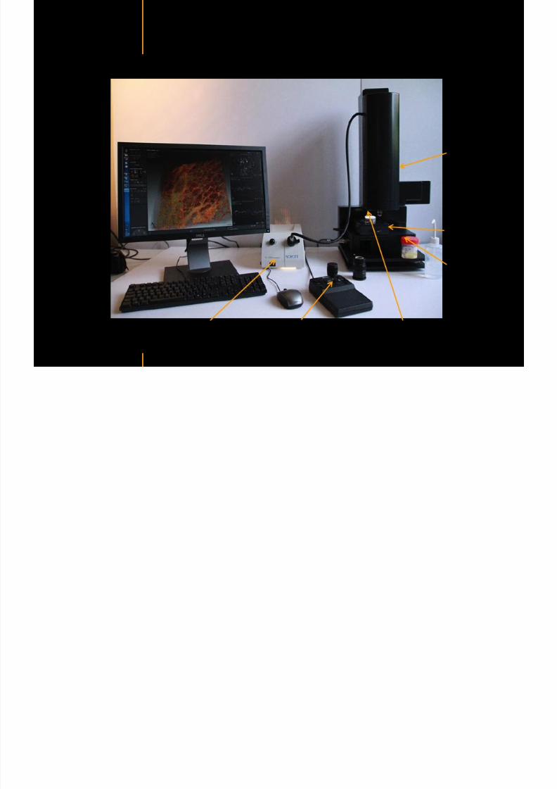
7292019 Atlas of Light-CT images
httpslidepdfcomreaderfullatlas-of-light-ct-images 8118copy LLTech 2012 ndash wwwlltechimagingcom - No copy without authorization 8 copy LLTech 2012
Breast tissue diagnosis decision tree
7292019 Atlas of Light-CT images
httpslidepdfcomreaderfullatlas-of-light-ct-images 9118copy LLTech 2012 ndash wwwlltechimagingcom - No copy without authorization 9
Breast HistologyDCIS ndash Ductal Carcinoma in situ
Breast Ductal Carinoma HampE vs LightCT on Fresh Tissue
Hopital Tenon France August 2010
Lobules
Fat cells
1 c m
7292019 Atlas of Light-CT images
httpslidepdfcomreaderfullatlas-of-light-ct-images 10118copy LLTech 2012 ndash wwwlltechimagingcom - No copy without authorization 10
Breast HistologyDCIS ndash Ductal Carcinoma in situ
Lobules
Duct withnecrosiss
7292019 Atlas of Light-CT images
httpslidepdfcomreaderfullatlas-of-light-ct-images 11118
7292019 Atlas of Light-CT images
httpslidepdfcomreaderfullatlas-of-light-ct-images 12118
7292019 Atlas of Light-CT images
httpslidepdfcomreaderfullatlas-of-light-ct-images 13118copy LLTech 2012 ndash wwwlltechimagingcom - No copy without authorization 13
Breast HistologyIdentification of the milk duct structures
Milk Duct End
Fat Cells
7292019 Atlas of Light-CT images
httpslidepdfcomreaderfullatlas-of-light-ct-images 14118copy LLTech 2012 ndash wwwlltechimagingcom - No copy without authorization 14
Healthy breast tissue
Lobule
Galactophorous duct cut longitudinally
Honeycomb configuration of adipocytes
copy LLTech 2012 Courtesy of Hocircpital Tenon Paris France
7292019 Atlas of Light-CT images
httpslidepdfcomreaderfullatlas-of-light-ct-images 15118
7292019 Atlas of Light-CT images
httpslidepdfcomreaderfullatlas-of-light-ct-images 16118copy LLTech 2012 ndash wwwlltechimagingcom - No copy without authorization 16
Invasive adenocarcinoma with in Situ Component
Enlarged ducts filled with cells
proliferation or necrosis
Highly scattering thin trabeculae
aspect of fibrous tissue
surrounding grey cellular zones
copy LLTech 2012 Courtesy of Hocircpital Tenon Paris France
7292019 Atlas of Light-CT images
httpslidepdfcomreaderfullatlas-of-light-ct-images 17118copy LLTech 2012 ndash wwwlltechimagingcom - No copy without authorization 17
Elongated compressed
ductules with slit-like lumen
Well delimited nodule withlobulated appearence
Fibroadenoma
copy LLTech 2012 Courtesy of Hocircpital Tenon Paris France
7292019 Atlas of Light-CT images
httpslidepdfcomreaderfullatlas-of-light-ct-images 18118copy LLTech 2012 ndash wwwlltechimagingcom - No copy without authorization 18
Fibroadenoma
Enlarged ductulescharacteristic of the lesion
copy LLTech 2012 Courtesy of Hocircpital Tenon Paris France
7292019 Atlas of Light-CT images
httpslidepdfcomreaderfullatlas-of-light-ct-images 19118copy LLTech 2012 ndash wwwlltechimagingcom - No copy without authorization 19
Lymphoid zone of the node
Vessel
Adipocytes in the center
of the node
Normal Sentinel Node
copy LLTech 2012 Courtesy of Hocircpital Tenon Paris France
7292019 Atlas of Light-CT images
httpslidepdfcomreaderfullatlas-of-light-ct-images 20118
7292019 Atlas of Light-CT images
httpslidepdfcomreaderfullatlas-of-light-ct-images 21118
copy LLTech 2012 ndash wwwlltechimagingcom - No copy without authorization 21
Normal lymphoid
zone with follicles
(Dark grey)
Metastatsis
(Light grey)
Hypervascularization
due to metastasis
Fibrous enveloppe of the
node
Invaded Sentinel Node
copy LLTech 2012 Courtesy of Hocircpital Tenon Paris France
7292019 Atlas of Light-CT images
httpslidepdfcomreaderfullatlas-of-light-ct-images 22118
copy LLTech 2012 ndash wwwlltechimagingcom - No copy without authorization 22
The Light-CT scanner allows fast tissue processing andpathology examination It completes the toolset available topathologists
When Chemical Fixation is needed
(Mostly Diagnostic)
When Frozen Section is needed
(Mostly Intra-operative)
Biopsy3D
Digital Image
2D
Digital Image
Slide
15-12 mins
Artefacts
Destructive
12-24 hours
Expensive
Destructive
5-8 minutes
Non-Destructive
Tissue Scanner
Light-CT could help to reduce the number of re-excisions in breast
7292019 Atlas of Light-CT images
httpslidepdfcomreaderfullatlas-of-light-ct-images 23118
copy LLTech 2012 ndash wwwlltechimagingcom - No copy without authorization 23
Light CT could help to reduce the number of re excisions in breastcancer surgery whilst preserving the current routines ofpathologists
Per surgery
LLTechrsquos
analysis
Histology Andor
Immunohistochemistry
Further
treatment
Post surgery
Surgical margin analysis
Metastasis analysis (gt 2mm) et
micro metastaisi
PN1mi gt 02 ndash 2 mm lt
SurgeryOKOK
Per surgery Post Surgery
C u r r e n t t e c h n i q u e s
U p t o 4 0 o
f d o u b l e p r o
c e d u r e s
L L T e c h L i g h t - C
T
S i g n i f i c a n t d e c r e a s e o f
d o u b l e p r o c e d u
r e s
Visual
analysis
cryostat
Further
treatmentSurgery
OK
Cancerouscancerous
OK
Poor identification of
metastasis
Up to 40 of double surgical
procedures
Several day analysisFew minutes analysis
Surgeon Pathologist Pathologist
Decrease of double surgical
procedures CancerousCancerous
No surgical
margins analysis
Histology Andor
Immuno
histochemistry
Node
Margin
Several day analysisFew minutes analysisSurgeon Pathologist Pathologist
7292019 Atlas of Light-CT images
httpslidepdfcomreaderfullatlas-of-light-ct-images 24118
copy LLTech 2012 ndash wwwlltechimagingcom - No copy without authorization 24
BRAIN IMAGING
Back to table of contents
7292019 Atlas of Light-CT images
httpslidepdfcomreaderfullatlas-of-light-ct-images 25118
copy LLTech 2012 ndash wwwlltechimagingcom - No copy without authorization 25
Rat brain
Architectural and cellular information seen at various scales
Myelinated fibers visible
1 mm
500 microm
100 microm
7292019 Atlas of Light-CT images
httpslidepdfcomreaderfullatlas-of-light-ct-images 26118
7292019 Atlas of Light-CT images
httpslidepdfcomreaderfullatlas-of-light-ct-images 27118
7292019 Atlas of Light-CT images
httpslidepdfcomreaderfullatlas-of-light-ct-images 28118
copy LLTech 2012 ndash wwwlltechimagingcom - No copy without authorization 28 copy LLTECH 2011
Collagen-rich matrixWide fascicles of
tumour cells
Large capillary
Fibroblastic meningioma grade I
500microm
100microm 200microm200microm
7292019 Atlas of Light-CT images
httpslidepdfcomreaderfullatlas-of-light-ct-images 29118
copy LLTech 2012 ndash wwwlltechimagingcom - No copy without authorization 29
Transitional meningioma grade I
Psammoma surrounding a calcification
Whorls formation
Collagen-rich matrix
500microm
100microm
50microm
50microm
7292019 Atlas of Light-CT images
httpslidepdfcomreaderfullatlas-of-light-ct-images 30118
7292019 Atlas of Light-CT images
httpslidepdfcomreaderfullatlas-of-light-ct-images 31118
copy LLTech 2012 ndash wwwlltechimagingcom - No copy without authorization 31 copy LLTECH 2011
Arrows Fibers transverse cut
Spinal cord normal tissue
500 microm
100 microm
100 microm
7292019 Atlas of Light-CT images
httpslidepdfcomreaderfullatlas-of-light-ct-images 32118
7292019 Atlas of Light-CT images
httpslidepdfcomreaderfullatlas-of-light-ct-images 33118
7292019 Atlas of Light-CT images
httpslidepdfcomreaderfullatlas-of-light-ct-images 34118
copy LLTech 2012 ndash wwwlltechimagingcom - No copy without authorization 34
Back to table of contents
SKIN IMAGING
N l ki h l
7292019 Atlas of Light-CT images
httpslidepdfcomreaderfullatlas-of-light-ct-images 35118
copy LLTech 2012 ndash wwwlltechimagingcom - No copy without authorization 35
Normal skin morphology
Epidermis
Collagen
Pilosebaceous
unit
Sweat gland
Adipocytes
b
a
c
e
d
500 microm
copy LLTech 2012 Courtesy of Hopitaux Universitaires de Genegraveve Genegraveve Switzerland
7292019 Atlas of Light-CT images
httpslidepdfcomreaderfullatlas-of-light-ct-images 36118
copy LLTech 2012 ndash wwwlltechimagingcom - No copy without authorization 36
Hypodermis
Skin morphology layers are well discriminated
5 mm x 8 mm mosaic of native field image
Ageing patient (solar elastosis)
En-face imaging of vertical excision
Epidermis
Dermis
1
3
2
4
7292019 Atlas of Light-CT images
httpslidepdfcomreaderfullatlas-of-light-ct-images 37118
En face skin slicing shows structural and cellular details
7292019 Atlas of Light-CT images
httpslidepdfcomreaderfullatlas-of-light-ct-images 38118
copy LLTech 2012 ndash wwwlltechimagingcom - No copy without authorization 38
Stratum corneum Stratum spinosum
Stratum basale Reticular region
Epidermis
Dermis
En-face skin slicing shows structural and cellular details
Melanin
papillary caps
Keratinocyte
nuclei
Blood vessels
Collagen
Corneocytes
100 microm 100 microm
100 microm100 microm
copy LLTech 2012 Courtesy of Hopitaux Universitaires de Genegraveve Genegraveve Switzerland
7292019 Atlas of Light-CT images
httpslidepdfcomreaderfullatlas-of-light-ct-images 39118
7292019 Atlas of Light-CT images
httpslidepdfcomreaderfullatlas-of-light-ct-images 40118
Vertical reconstruction of skin model
7292019 Atlas of Light-CT images
httpslidepdfcomreaderfullatlas-of-light-ct-images 41118
copy LLTech 2012 ndash wwwlltechimagingcom - No copy without authorization 41
Vertical reconstruction of skin model
50 microm
Stratum corneum
Stratum spinosum
Dermis
Keratinocyte nuclei
copy LLTech 2012
7292019 Atlas of Light-CT images
httpslidepdfcomreaderfullatlas-of-light-ct-images 42118
Normal
7292019 Atlas of Light-CT images
httpslidepdfcomreaderfullatlas-of-light-ct-images 43118
copy LLTech 2012 ndash wwwlltechimagingcom - No copy without authorization 43
skin
b
a
e
c
f
d
500 microm
7292019 Atlas of Light-CT images
httpslidepdfcomreaderfullatlas-of-light-ct-images 44118
7292019 Atlas of Light-CT images
httpslidepdfcomreaderfullatlas-of-light-ct-images 45118
copy LLTech 2012 ndash wwwlltechimagingcom - No copy without authorization 45
Adipocytes
9um depth 27um depth 60um depth
Reconstructed
depth slice
200 microm
50 microm
7292019 Atlas of Light-CT images
httpslidepdfcomreaderfullatlas-of-light-ct-images 46118
Skin pathologies basal cell carcinoma discrimination at
7292019 Atlas of Light-CT images
httpslidepdfcomreaderfullatlas-of-light-ct-images 47118
copy LLTech 2012 ndash wwwlltechimagingcom - No copy without authorization 47
Epidermis
Dermis
Hypodermis
A
AA
C
BCC
PSU
PSU
BCC Basal cell carcinoma
A Adipocytes
C Collagen
PSU Pilosebaceous unit
structural level
1 mm
copy LLTech 2012 Courtesy of Hopitaux Universitaires de Genegraveve Genegraveve Switzerland
C
Zoom on basal cell carcinoma discrimination at cellular level
7292019 Atlas of Light-CT images
httpslidepdfcomreaderfullatlas-of-light-ct-images 48118
copy LLTech 2012 ndash wwwlltechimagingcom - No copy without authorization 48
Zoom on basal cell carcinoma discrimination at cellular level
200 microm
100 microm
Peritumoral stroma
High cell density
copy LLTech 2012 Courtesy of Hopitaux Universitaires de Genegraveve Genegraveve Switzerland
Skin pathologies discrimination of actinic keratosis
7292019 Atlas of Light-CT images
httpslidepdfcomreaderfullatlas-of-light-ct-images 49118
copy LLTech 2012 ndash wwwlltechimagingcom - No copy without authorization 49
Skin pathologies discrimination of actinic keratosis
Enlarged epidermis
Extended epidermal ridges
1 mm
copy LLTech 2012 Courtesy of Hopitaux Universitaires de Genegraveve Genegraveve Switzerland
7292019 Atlas of Light-CT images
httpslidepdfcomreaderfullatlas-of-light-ct-images 50118
copy LLTech 2012 ndash wwwlltechimagingcom - No copy without authorization 50
Basal cell carcinoma in depth imaging
Z = 10 microns 7 x 42 mm FOV Z = 35 microns 7 x 42 mm FOV
BCC
SG Sebaceous glands
Z = 55 microns 7 x 42 mm FOV Z = 75 microns 7 x 42 mm FOV
SG
Application to Mohs surgerycopy LLTech 2012 Courtesy of Hopitaux Universitaires de Genegraveve Genegraveve Switzerland
7292019 Atlas of Light-CT images
httpslidepdfcomreaderfullatlas-of-light-ct-images 51118
copy LLTech 2012 ndash wwwlltechimagingcom - No copy without authorization 51 copy LLTECH 2011
Healthy vs Cheloid scar tissue
Healthy skin tissueFOV 75x5mm
Cheloid scar tissueFOV 65x65mm
copy LLTECH 2011
7292019 Atlas of Light-CT images
httpslidepdfcomreaderfullatlas-of-light-ct-images 52118
copy LLTech 2012 ndash wwwlltechimagingcom - No copy without authorization 52
Dermo-epidermaljunction
Cheloid scar tissue in detail
copy LLTECH 2011
Nodular structureThickcollagenfiberbundles
1mm
600 microm
500 microm
h l d d f
7292019 Atlas of Light-CT images
httpslidepdfcomreaderfullatlas-of-light-ct-images 53118
copy LLTech 2012 ndash wwwlltechimagingcom - No copy without authorization 53
Cheloid scar tissue viewed en face
20microm depth 120microm depth
7292019 Atlas of Light-CT images
httpslidepdfcomreaderfullatlas-of-light-ct-images 54118
Skin in-vivo sweat duct imaging in 3D
7292019 Atlas of Light-CT images
httpslidepdfcomreaderfullatlas-of-light-ct-images 55118
copy LLTech 2012 ndash wwwlltechimagingcom - No copy without authorization 55
g g
Vertical reconstruction from en-face images
Sweat duct 3D reconstruction
Stratum corneum
Stratum spinosum
SD sweat ducts
SD SD
100 microm
7292019 Atlas of Light-CT images
httpslidepdfcomreaderfullatlas-of-light-ct-images 56118
7292019 Atlas of Light-CT images
httpslidepdfcomreaderfullatlas-of-light-ct-images 57118
7292019 Atlas of Light-CT images
httpslidepdfcomreaderfullatlas-of-light-ct-images 58118
copy LLTech 2012 ndash wwwlltechimagingcom - No copy without authorization 58
LUNG IMAGING
Back to table of contents
H L
7292019 Atlas of Light-CT images
httpslidepdfcomreaderfullatlas-of-light-ct-images 59118
copy LLTech 2012 ndash wwwlltechimagingcom - No copy without authorization 59
Zone 1
Zone 2
Zone 3
Zone 4
Zone with
cancerous tissue
Limit of cancerous
stroma
alveoli with
macrophages
Alveoles
Human Lung
500microm
copy LLTech 2012 Courtesy of Hocircpital Hotel-Dieu Paris France
H L Z 1 Z t
7292019 Atlas of Light-CT images
httpslidepdfcomreaderfullatlas-of-light-ct-images 60118
copy LLTech 2012 ndash wwwlltechimagingcom - No copy without authorization 60
Stroma
Human Lung ndash Zone 1 ndash Zoom on cancerous stroma
Cancerous
area
200microm
copy LLTech 2012 Courtesy of Hocircpital Hotel-Dieu Paris France
Human Lung ndash Zone 2 ndash Limit cancerous stroma alveoli
7292019 Atlas of Light-CT images
httpslidepdfcomreaderfullatlas-of-light-ct-images 61118
copy LLTech 2012 ndash wwwlltechimagingcom - No copy without authorization 61
g
with visible macrophages and vessel
Alveoli
Macrophages
Vessel
copy LLTech 2012 Courtesy of Hocircpital Hotel-Dieu Paris France
Human Lung ndash Zone 3 ndash Limit cancerous stroma alveoli
7292019 Atlas of Light-CT images
httpslidepdfcomreaderfullatlas-of-light-ct-images 62118
copy LLTech 2012 ndash wwwlltechimagingcom - No copy without authorization 62
g
with visible macrophages
Macrophages
100microm
copy LLTech 2012 Courtesy of Hocircpital Hotel-Dieu Paris France
Human Lung ndash Zone 4 ndash Alveoli
7292019 Atlas of Light-CT images
httpslidepdfcomreaderfullatlas-of-light-ct-images 63118
copy LLTech 2012 ndash wwwlltechimagingcom - No copy without authorization 63
Human Lung Zone 4 Alveoli
Alveoli
200microm
copy LLTech 2012 Courtesy of Hocircpital Hotel-Dieu Paris France
7292019 Atlas of Light-CT images
httpslidepdfcomreaderfullatlas-of-light-ct-images 64118
copy LLTech 2012 ndash wwwlltechimagingcom - No copy without authorization 64
GI IMAGING
Back to table of contents
7292019 Atlas of Light-CT images
httpslidepdfcomreaderfullatlas-of-light-ct-images 65118
Colonic carcinoma
7292019 Atlas of Light-CT images
httpslidepdfcomreaderfullatlas-of-light-ct-images 66118
copy LLTech 2012 ndash wwwlltechimagingcom - No copy without authorization 66
Colonic Carcinoma
Colonic carcinoma
copy LLTech 2012 Courtesy of MD Anderson Houston USA
Intestines ndash Crohnrsquos disease
7292019 Atlas of Light-CT images
httpslidepdfcomreaderfullatlas-of-light-ct-images 67118
copy LLTech 2012 ndash wwwlltechimagingcom - No copy without authorization 67
Crohnrsquos disease structure not
visible from the surface
Fat cells
200 microm
Intestines Crohn s disease
copy LLTech 2012 Courtesy of Institut Curie Paris France
Rat stomach
7292019 Atlas of Light-CT images
httpslidepdfcomreaderfullatlas-of-light-ct-images 68118
copy LLTech 2012 ndash wwwlltechimagingcom - No copy without authorization 68
Rat stomach
Mucosa withgastric pits
Submucosa with
bright collagen
fibers
Muscularis
propria with
muscle bundles
copy LLTech 2012 Courtesy of Weill Cornell Medical College NY USA
200 microm
7292019 Atlas of Light-CT images
httpslidepdfcomreaderfullatlas-of-light-ct-images 69118
copy LLTech 2012 ndash wwwlltechimagingcom - No copy without authorization 69 copy LLTECH 2011
UROGENITAL IMAGING
Back to table of contents
Rat bladder
7292019 Atlas of Light-CT images
httpslidepdfcomreaderfullatlas-of-light-ct-images 70118
copy LLTech 2012 ndash wwwlltechimagingcom - No copy without authorization 70
Rat bladder
Stratifiedurothelium
Lamina propria
500 microm
Muscular
bundles of the
lamina propria
Serosa
copy LLTech 2012 Courtesy of Weill Cornell Medical College NY USA
100 microm
100 microm
Human bladder low grade papillary urothelial carcinoma
7292019 Atlas of Light-CT images
httpslidepdfcomreaderfullatlas-of-light-ct-images 71118
copy LLTech 2012 ndash wwwlltechimagingcom - No copy without authorization 71
Lesion with fibrovascular cores (arrow) and superficial umbrella cells (arrowheads)
Human bladder low grade papillary urothelial carcinoma
copy LLTech 2012 Courtesy of Weill Cornell Medical College NY USA
100 microm
7292019 Atlas of Light-CT images
httpslidepdfcomreaderfullatlas-of-light-ct-images 72118
Rat prostate
7292019 Atlas of Light-CT images
httpslidepdfcomreaderfullatlas-of-light-ct-images 73118
copy LLTech 2012 ndash wwwlltechimagingcom - No copy without authorization 73
p
copy LLTech 2012 Courtesy of Weill Cornell Medical College NY USA
Papillary folds of the
acinar glands
Prostatic acini
Fibromuscular stromaPeriprostatic fat Blood vessel
1 mm
200 microm
Mouse kidney
7292019 Atlas of Light-CT images
httpslidepdfcomreaderfullatlas-of-light-ct-images 74118
copy LLTech 2012 ndash wwwlltechimagingcom - No copy without authorization 74
1 mm
y
copy LLTech 2012 Courtesy of Institut Biomeacutedical de Bicecirctre Paris France
Renal cortex
Renal medulla
Renal sinus
7292019 Atlas of Light-CT images
httpslidepdfcomreaderfullatlas-of-light-ct-images 75118
copy LLTech 2012 ndash wwwlltechimagingcom - No copy without authorization 75
Mouse kidney ndash zoom on highlighted section of previous slide
800microm
copy LLTech 2012 Courtesy of Hocircpital Kremlin Bicecirctre Paris France
7292019 Atlas of Light-CT images
httpslidepdfcomreaderfullatlas-of-light-ct-images 76118
7292019 Atlas of Light-CT images
httpslidepdfcomreaderfullatlas-of-light-ct-images 77118
7292019 Atlas of Light-CT images
httpslidepdfcomreaderfullatlas-of-light-ct-images 78118
copy LLTech 2012 ndash wwwlltechimagingcom - No copy without authorization 78 copy LLTECH 2011
CORE NEEDLE BIOPSIES
Back to table of contents
Kidney
7292019 Atlas of Light-CT images
httpslidepdfcomreaderfullatlas-of-light-ct-images 79118
copy LLTech 2012 ndash wwwlltechimagingcom - No copy without authorization 79
100microm
50microm
Renal tubules
Vessel
500microm
copy LLTech 2012 Courtesy of Institut Curie Paris France
Breast Invasive lobular carcinoma
7292019 Atlas of Light-CT images
httpslidepdfcomreaderfullatlas-of-light-ct-images 80118
copy LLTech 2012 ndash wwwlltechimagingcom - No copy without authorization 80
500microm
stroma
adipocytes
copy LLTech 2012 Courtesy of Institut Curie Paris France
7292019 Atlas of Light-CT images
httpslidepdfcomreaderfullatlas-of-light-ct-images 81118
Breast Ductal carcinoma in situ
7292019 Atlas of Light-CT images
httpslidepdfcomreaderfullatlas-of-light-ct-images 82118
copy LLTech 2012 ndash wwwlltechimagingcom - No copy without authorization 82
500microm
copy LLTech 2012 Courtesy of Institut Curie Paris France
7292019 Atlas of Light-CT images
httpslidepdfcomreaderfullatlas-of-light-ct-images 83118
copy LLTech 2012 ndash wwwlltechimagingcom - No copy without authorization 83 copy LLTECH 2011
THYROID IMAGING
Back to table of contents
7292019 Atlas of Light-CT images
httpslidepdfcomreaderfullatlas-of-light-ct-images 84118
copy LLTech 2012 ndash wwwlltechimagingcom - No copy without authorization 84
Thyroid
800 microm
200 microm
copy LLTech 2012 Courtesy of Hocircpital Tenon Paris France
7292019 Atlas of Light-CT images
httpslidepdfcomreaderfullatlas-of-light-ct-images 85118
copy LLTech 2012 ndash wwwlltechimagingcom - No copy without authorization 85
Thyroid
800 microm
200 microm
copy LLTech 2012 Courtesy of Hocircpital Tenon Paris France
7292019 Atlas of Light-CT images
httpslidepdfcomreaderfullatlas-of-light-ct-images 86118
copy LLTech 2012 ndash wwwlltechimagingcom - No copy without authorization 86 copy LLTECH 2011
FAT CELLS IMAGING
Back to table of contents
7292019 Atlas of Light-CT images
httpslidepdfcomreaderfullatlas-of-light-ct-images 87118
copy LLTech 2012 ndash wwwlltechimagingcom - No copy without authorization 87
Brown fat cells - adipocytes
100 microm 100 microm
200 microm
10um depth
Adipocytes
45um depth
En face
slices
Reconstructed
depth slice
copy LLTech 2012 Courtesy of Centre de Recherche des Cordeliers Paris France
7292019 Atlas of Light-CT images
httpslidepdfcomreaderfullatlas-of-light-ct-images 88118
copy LLTech 2012 ndash wwwlltechimagingcom - No copy without authorization 88 copy LLTECH 2011
CORNEA AND RETINA
Back to table of contents
Human cornea reconstructed depth slice
7292019 Atlas of Light-CT images
httpslidepdfcomreaderfullatlas-of-light-ct-images 89118
copy LLTech 2012 ndash wwwlltechimagingcom - No copy without authorization 89
50 um
wwwlltechimagingcom
7292019 Atlas of Light-CT images
httpslidepdfcomreaderfullatlas-of-light-ct-images 90118
Large field en face view in stroma
7292019 Atlas of Light-CT images
httpslidepdfcomreaderfullatlas-of-light-ct-images 91118
copy LLTech 2012 ndash wwwlltechimagingcom - No copy without authorization 91
200 um
wwwlltechimagingcom
7292019 Atlas of Light-CT images
httpslidepdfcomreaderfullatlas-of-light-ct-images 92118
Rabbit cornea after LASIK reflectivity profiles reveal
i d tt i d thi k d ith li l l
7292019 Atlas of Light-CT images
httpslidepdfcomreaderfullatlas-of-light-ct-images 93118
copy LLTech 2012 ndash wwwlltechimagingcom - No copy without authorization 93
increased scattering and thickened epithelial layer
LASIK LASIK CONTROL CONTROL
copy LLTech 2012
Newborn mouse anterior segment
7292019 Atlas of Light-CT images
httpslidepdfcomreaderfullatlas-of-light-ct-images 94118
copy LLTech 2012 ndash wwwlltechimagingcom - No copy without authorization 94
Corneal epithelial cells
Basal membrane
Stroma
Descemetrsquos membrane
Aqueous humor
Lens capsule
Lens epithelial cells
Cortical fibers
copy LLTech 2012 Courtesy of ESPCI Paris Francecopy LLTech 2012
Rat retina comparison with histology
7292019 Atlas of Light-CT images
httpslidepdfcomreaderfullatlas-of-light-ct-images 95118
copy LLTech 2012 ndash wwwlltechimagingcom - No copy without authorization 95
Figure 1 Right Ex vivo rat retinaimaged using the f ull field OCT
system of ESPCI Left Histology
for comparison
Figure 1 Right Ex vivo rat retinaimaged using the f ull field OCT
system of ESPCI Left Histology
for comparison
Figure 1 Right Ex vivo rat retinaimaged using the f ull field OCT
system of ESPCI Left Histology
for comparisonField
230x300microm
RPE
PRL
ONL
OPL
INL
IPL
GCL
NFL
Invest Ophthalmol Vis Sci 2004 Nov45(11)4126-31
Grieve K Paques M Dubois A Sahel J Boccara C Le Gargasson JF copy LLTech 2012
Rat retina
7292019 Atlas of Light-CT images
httpslidepdfcomreaderfullatlas-of-light-ct-images 96118
copy LLTech 2012 ndash wwwlltechimagingcom - No copy without authorization 96 copy LLTech 2012 Courtesy of ESPCI Paris France
Nerve fiber layer
Retina
Invest Ophthalmol Vis Sci 2004 Nov45(11)4126-31
Grieve K Paques M Dubois A Sahel J Boccara C Le Gargasson JF
Transscleral image
7292019 Atlas of Light-CT images
httpslidepdfcomreaderfullatlas-of-light-ct-images 97118
7292019 Atlas of Light-CT images
httpslidepdfcomreaderfullatlas-of-light-ct-images 98118
7292019 Atlas of Light-CT images
httpslidepdfcomreaderfullatlas-of-light-ct-images 99118
In vivo imaging of drosophila melanogaster
7292019 Atlas of Light-CT images
httpslidepdfcomreaderfullatlas-of-light-ct-images 100118
copy LLTech 2012 ndash wwwlltechimagingcom - No copy without authorization 100 copy LLTech 2012 Courtesy of ESPCI Paris France
In vivo imaging of drosophila melanogaster ndash four stages of pupation over 72 hours
7292019 Atlas of Light-CT images
httpslidepdfcomreaderfullatlas-of-light-ct-images 101118
copy LLTech 2012 ndash wwwlltechimagingcom - No copy without authorization 101
Prepupa (0-2 h) Transition topupal stage (24 h)
Pupal stage (48 h) Advanced pupal phase
before eclosion (72 h)
copy LLTech 2012 Courtesy of ESPCI Paris France
Pupa 118 4 days 30um depth
100
7292019 Atlas of Light-CT images
httpslidepdfcomreaderfullatlas-of-light-ct-images 102118
copy LLTech 2012 ndash wwwlltechimagingcom - No copy without authorization 102
100 um
Drosophila
7292019 Atlas of Light-CT images
httpslidepdfcomreaderfullatlas-of-light-ct-images 103118
copy LLTech 2012 ndash wwwlltechimagingcom - No copy without authorization 103
200 microm
copy LLTech 2012 Courtesy of ESPCI Paris France
C Elegans ndash Fast identification of anatomy
7292019 Atlas of Light-CT images
httpslidepdfcomreaderfullatlas-of-light-ct-images 104118
copy LLTech 2012 ndash wwwlltechimagingcom - No copy without authorization 104
Body wall - cuticle
gonad arm
30 microm depth
uterus
ovocytes
intestine
100 microm 100 microm
15 microm depth
copy LLTech 2012 Courtesy of ENS Paris France
C elegans ndash Images at higher magnification 40X
7292019 Atlas of Light-CT images
httpslidepdfcomreaderfullatlas-of-light-ct-images 105118
copy LLTech 2012 ndash wwwlltechimagingcom - No copy without authorization 105
3D reconstruction 50 microm
100 microm
copy LLTech 2012 Courtesy of ENS Paris France
Zebra fish eye
7292019 Atlas of Light-CT images
httpslidepdfcomreaderfullatlas-of-light-ct-images 106118
copy LLTech 2012 ndash wwwlltechimagingcom - No copy without authorization 106
Zebra fish eye
100 microm
100 microm
En face slices
Reconstructed depth slice
copy LLTech 2012 Courtesy of Inserm Paris France
10um depth 20um depth 45um depth
7292019 Atlas of Light-CT images
httpslidepdfcomreaderfullatlas-of-light-ct-images 107118
Xenopus Laevis backtail
7292019 Atlas of Light-CT images
httpslidepdfcomreaderfullatlas-of-light-ct-images 108118
copy LLTech 2012 ndash wwwlltechimagingcom - No copy without authorization 108
100 um30um depth 60um depth
7292019 Atlas of Light-CT images
httpslidepdfcomreaderfullatlas-of-light-ct-images 109118
Mouse embryo in vitro 6 5 days old ndash
7292019 Atlas of Light-CT images
httpslidepdfcomreaderfullatlas-of-light-ct-images 110118
copy LLTech 2012 ndash wwwlltechimagingcom - No copy without authorization 110
Mouse embryo in vitro 65 days old
copy LLTech 2012 Courtesy of Ellvire Guiot and nstitut Jacques Monod Paris France
3D imaging of internal cavity
Sabellaria alveolata mouth palps
7292019 Atlas of Light-CT images
httpslidepdfcomreaderfullatlas-of-light-ct-images 111118
copy LLTech 2012 ndash wwwlltechimagingcom - No copy without authorization 111
500 microm
100 microm
Mouth palps
copy LLTech 2012 Courtesy of Museacutee National drsquoHistoire Naturelle Paris France
7292019 Atlas of Light-CT images
httpslidepdfcomreaderfullatlas-of-light-ct-images 112118
copy LLTech 2012 ndash wwwlltechimagingcom - No copy without authorization 112 copy LLTECH 2011
PLANT IMAGING
Back to table of contents
Mouse ear leaf epidermis
7292019 Atlas of Light-CT images
httpslidepdfcomreaderfullatlas-of-light-ct-images 113118
copy LLTech 2012 ndash wwwlltechimagingcom - No copy without authorization 113
100 microm
Epidermal cell
copy LLTech 2012
Stoma
Leaf veins
7292019 Atlas of Light-CT images
httpslidepdfcomreaderfullatlas-of-light-ct-images 114118
copy LLTech 2012 ndash wwwlltechimagingcom - No copy without authorization 114
114copy LLTech 2012
Fibers
Veins
100 microm
500 microm
Apple
7292019 Atlas of Light-CT images
httpslidepdfcomreaderfullatlas-of-light-ct-images 115118
copy LLTech 2012 ndash wwwlltechimagingcom - No copy without authorization 115
115copy LLTech 2012
100 microm
Wax
Skin
Flesh
500 microm
Water
Effect of harpin infiltration on tobacco leaf
7292019 Atlas of Light-CT images
httpslidepdfcomreaderfullatlas-of-light-ct-images 116118
copy LLTech 2012 ndash wwwlltechimagingcom - No copy without authorization 116
116copy LLTech 2012 Courtesy of UPMC Paris France
50 microm
50 microm
50 microm
50 microm
Upper epidermis
10 min after harpin infiltration 30 min after harpin infiltration
Palisade parenchyma
Epidermis cell
Chloroplast
7292019 Atlas of Light-CT images
httpslidepdfcomreaderfullatlas-of-light-ct-images 117118
copy LLTech 2012 ndash wwwlltechimagingcom - No copy without authorization 117
bull Light-CTtrade does not require tissue preparation nor staining of any kind
bull Creates images within minutes using a non-destructive process
bull Offers a 1 microm cellular resolution in 3D
bull Reveals structural and cellular information
bull Mosaicing allows fast visualisation at various scale
bull En-face high-resolution imaging allows vertical and 3D reconstruction
bull In-vivo images show good promise for in-vivo system
bull Clinical and research applications in cancer detection cosmetics biobanking small animal
developmental biology botanyhellip
Contact Details
7292019 Atlas of Light-CT images
httpslidepdfcomreaderfullatlas-of-light-ct-images 118118
USA
LLTech Inc
103 Carnegie Center DriveSuite 300
Princeton NJ 08540
USA
wwwlltechimagingcom
contactlltechfr
Phone + 1 609 955 3506
LLTech SAS
29 rue du Faubourg St-JacquesPeacutepiniegravere Paris Santeacute Cochin
75014
Paris - France
wwwlltechimagingcom
contactlltechfr
Phone +33 1 82 72 61 25
Europe
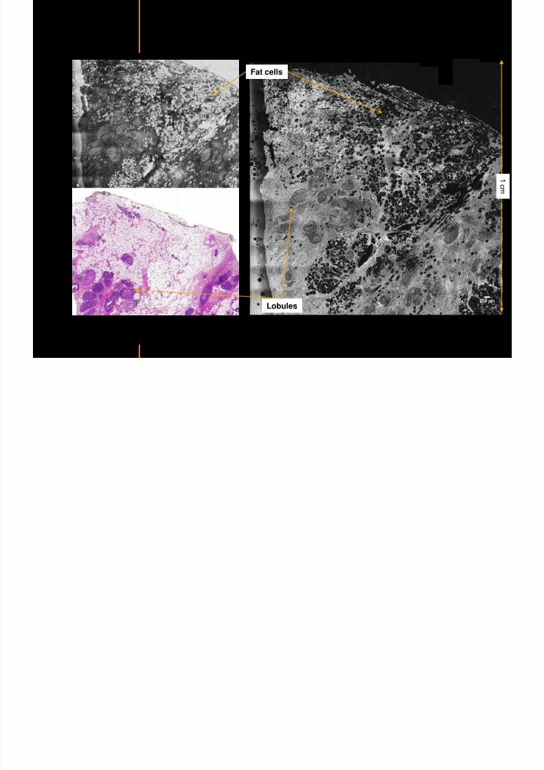
7292019 Atlas of Light-CT images
httpslidepdfcomreaderfullatlas-of-light-ct-images 12118
7292019 Atlas of Light-CT images
httpslidepdfcomreaderfullatlas-of-light-ct-images 13118copy LLTech 2012 ndash wwwlltechimagingcom - No copy without authorization 13
Breast HistologyIdentification of the milk duct structures
Milk Duct End
Fat Cells
7292019 Atlas of Light-CT images
httpslidepdfcomreaderfullatlas-of-light-ct-images 14118copy LLTech 2012 ndash wwwlltechimagingcom - No copy without authorization 14
Healthy breast tissue
Lobule
Galactophorous duct cut longitudinally
Honeycomb configuration of adipocytes
copy LLTech 2012 Courtesy of Hocircpital Tenon Paris France
7292019 Atlas of Light-CT images
httpslidepdfcomreaderfullatlas-of-light-ct-images 15118
7292019 Atlas of Light-CT images
httpslidepdfcomreaderfullatlas-of-light-ct-images 16118copy LLTech 2012 ndash wwwlltechimagingcom - No copy without authorization 16
Invasive adenocarcinoma with in Situ Component
Enlarged ducts filled with cells
proliferation or necrosis
Highly scattering thin trabeculae
aspect of fibrous tissue
surrounding grey cellular zones
copy LLTech 2012 Courtesy of Hocircpital Tenon Paris France
7292019 Atlas of Light-CT images
httpslidepdfcomreaderfullatlas-of-light-ct-images 17118copy LLTech 2012 ndash wwwlltechimagingcom - No copy without authorization 17
Elongated compressed
ductules with slit-like lumen
Well delimited nodule withlobulated appearence
Fibroadenoma
copy LLTech 2012 Courtesy of Hocircpital Tenon Paris France
7292019 Atlas of Light-CT images
httpslidepdfcomreaderfullatlas-of-light-ct-images 18118copy LLTech 2012 ndash wwwlltechimagingcom - No copy without authorization 18
Fibroadenoma
Enlarged ductulescharacteristic of the lesion
copy LLTech 2012 Courtesy of Hocircpital Tenon Paris France
7292019 Atlas of Light-CT images
httpslidepdfcomreaderfullatlas-of-light-ct-images 19118copy LLTech 2012 ndash wwwlltechimagingcom - No copy without authorization 19
Lymphoid zone of the node
Vessel
Adipocytes in the center
of the node
Normal Sentinel Node
copy LLTech 2012 Courtesy of Hocircpital Tenon Paris France
7292019 Atlas of Light-CT images
httpslidepdfcomreaderfullatlas-of-light-ct-images 20118
7292019 Atlas of Light-CT images
httpslidepdfcomreaderfullatlas-of-light-ct-images 21118
copy LLTech 2012 ndash wwwlltechimagingcom - No copy without authorization 21
Normal lymphoid
zone with follicles
(Dark grey)
Metastatsis
(Light grey)
Hypervascularization
due to metastasis
Fibrous enveloppe of the
node
Invaded Sentinel Node
copy LLTech 2012 Courtesy of Hocircpital Tenon Paris France
7292019 Atlas of Light-CT images
httpslidepdfcomreaderfullatlas-of-light-ct-images 22118
copy LLTech 2012 ndash wwwlltechimagingcom - No copy without authorization 22
The Light-CT scanner allows fast tissue processing andpathology examination It completes the toolset available topathologists
When Chemical Fixation is needed
(Mostly Diagnostic)
When Frozen Section is needed
(Mostly Intra-operative)
Biopsy3D
Digital Image
2D
Digital Image
Slide
15-12 mins
Artefacts
Destructive
12-24 hours
Expensive
Destructive
5-8 minutes
Non-Destructive
Tissue Scanner
Light-CT could help to reduce the number of re-excisions in breast
7292019 Atlas of Light-CT images
httpslidepdfcomreaderfullatlas-of-light-ct-images 23118
copy LLTech 2012 ndash wwwlltechimagingcom - No copy without authorization 23
Light CT could help to reduce the number of re excisions in breastcancer surgery whilst preserving the current routines ofpathologists
Per surgery
LLTechrsquos
analysis
Histology Andor
Immunohistochemistry
Further
treatment
Post surgery
Surgical margin analysis
Metastasis analysis (gt 2mm) et
micro metastaisi
PN1mi gt 02 ndash 2 mm lt
SurgeryOKOK
Per surgery Post Surgery
C u r r e n t t e c h n i q u e s
U p t o 4 0 o
f d o u b l e p r o
c e d u r e s
L L T e c h L i g h t - C
T
S i g n i f i c a n t d e c r e a s e o f
d o u b l e p r o c e d u
r e s
Visual
analysis
cryostat
Further
treatmentSurgery
OK
Cancerouscancerous
OK
Poor identification of
metastasis
Up to 40 of double surgical
procedures
Several day analysisFew minutes analysis
Surgeon Pathologist Pathologist
Decrease of double surgical
procedures CancerousCancerous
No surgical
margins analysis
Histology Andor
Immuno
histochemistry
Node
Margin
Several day analysisFew minutes analysisSurgeon Pathologist Pathologist
7292019 Atlas of Light-CT images
httpslidepdfcomreaderfullatlas-of-light-ct-images 24118
copy LLTech 2012 ndash wwwlltechimagingcom - No copy without authorization 24
BRAIN IMAGING
Back to table of contents
7292019 Atlas of Light-CT images
httpslidepdfcomreaderfullatlas-of-light-ct-images 25118
copy LLTech 2012 ndash wwwlltechimagingcom - No copy without authorization 25
Rat brain
Architectural and cellular information seen at various scales
Myelinated fibers visible
1 mm
500 microm
100 microm
7292019 Atlas of Light-CT images
httpslidepdfcomreaderfullatlas-of-light-ct-images 26118
7292019 Atlas of Light-CT images
httpslidepdfcomreaderfullatlas-of-light-ct-images 27118
7292019 Atlas of Light-CT images
httpslidepdfcomreaderfullatlas-of-light-ct-images 28118
copy LLTech 2012 ndash wwwlltechimagingcom - No copy without authorization 28 copy LLTECH 2011
Collagen-rich matrixWide fascicles of
tumour cells
Large capillary
Fibroblastic meningioma grade I
500microm
100microm 200microm200microm
7292019 Atlas of Light-CT images
httpslidepdfcomreaderfullatlas-of-light-ct-images 29118
copy LLTech 2012 ndash wwwlltechimagingcom - No copy without authorization 29
Transitional meningioma grade I
Psammoma surrounding a calcification
Whorls formation
Collagen-rich matrix
500microm
100microm
50microm
50microm
7292019 Atlas of Light-CT images
httpslidepdfcomreaderfullatlas-of-light-ct-images 30118
7292019 Atlas of Light-CT images
httpslidepdfcomreaderfullatlas-of-light-ct-images 31118
copy LLTech 2012 ndash wwwlltechimagingcom - No copy without authorization 31 copy LLTECH 2011
Arrows Fibers transverse cut
Spinal cord normal tissue
500 microm
100 microm
100 microm
7292019 Atlas of Light-CT images
httpslidepdfcomreaderfullatlas-of-light-ct-images 32118
7292019 Atlas of Light-CT images
httpslidepdfcomreaderfullatlas-of-light-ct-images 33118
7292019 Atlas of Light-CT images
httpslidepdfcomreaderfullatlas-of-light-ct-images 34118
copy LLTech 2012 ndash wwwlltechimagingcom - No copy without authorization 34
Back to table of contents
SKIN IMAGING
N l ki h l
7292019 Atlas of Light-CT images
httpslidepdfcomreaderfullatlas-of-light-ct-images 35118
copy LLTech 2012 ndash wwwlltechimagingcom - No copy without authorization 35
Normal skin morphology
Epidermis
Collagen
Pilosebaceous
unit
Sweat gland
Adipocytes
b
a
c
e
d
500 microm
copy LLTech 2012 Courtesy of Hopitaux Universitaires de Genegraveve Genegraveve Switzerland
7292019 Atlas of Light-CT images
httpslidepdfcomreaderfullatlas-of-light-ct-images 36118
copy LLTech 2012 ndash wwwlltechimagingcom - No copy without authorization 36
Hypodermis
Skin morphology layers are well discriminated
5 mm x 8 mm mosaic of native field image
Ageing patient (solar elastosis)
En-face imaging of vertical excision
Epidermis
Dermis
1
3
2
4
7292019 Atlas of Light-CT images
httpslidepdfcomreaderfullatlas-of-light-ct-images 37118
En face skin slicing shows structural and cellular details
7292019 Atlas of Light-CT images
httpslidepdfcomreaderfullatlas-of-light-ct-images 38118
copy LLTech 2012 ndash wwwlltechimagingcom - No copy without authorization 38
Stratum corneum Stratum spinosum
Stratum basale Reticular region
Epidermis
Dermis
En-face skin slicing shows structural and cellular details
Melanin
papillary caps
Keratinocyte
nuclei
Blood vessels
Collagen
Corneocytes
100 microm 100 microm
100 microm100 microm
copy LLTech 2012 Courtesy of Hopitaux Universitaires de Genegraveve Genegraveve Switzerland
7292019 Atlas of Light-CT images
httpslidepdfcomreaderfullatlas-of-light-ct-images 39118
7292019 Atlas of Light-CT images
httpslidepdfcomreaderfullatlas-of-light-ct-images 40118
Vertical reconstruction of skin model
7292019 Atlas of Light-CT images
httpslidepdfcomreaderfullatlas-of-light-ct-images 41118
copy LLTech 2012 ndash wwwlltechimagingcom - No copy without authorization 41
Vertical reconstruction of skin model
50 microm
Stratum corneum
Stratum spinosum
Dermis
Keratinocyte nuclei
copy LLTech 2012
7292019 Atlas of Light-CT images
httpslidepdfcomreaderfullatlas-of-light-ct-images 42118
Normal
7292019 Atlas of Light-CT images
httpslidepdfcomreaderfullatlas-of-light-ct-images 43118
copy LLTech 2012 ndash wwwlltechimagingcom - No copy without authorization 43
skin
b
a
e
c
f
d
500 microm
7292019 Atlas of Light-CT images
httpslidepdfcomreaderfullatlas-of-light-ct-images 44118
7292019 Atlas of Light-CT images
httpslidepdfcomreaderfullatlas-of-light-ct-images 45118
copy LLTech 2012 ndash wwwlltechimagingcom - No copy without authorization 45
Adipocytes
9um depth 27um depth 60um depth
Reconstructed
depth slice
200 microm
50 microm
7292019 Atlas of Light-CT images
httpslidepdfcomreaderfullatlas-of-light-ct-images 46118
Skin pathologies basal cell carcinoma discrimination at
7292019 Atlas of Light-CT images
httpslidepdfcomreaderfullatlas-of-light-ct-images 47118
copy LLTech 2012 ndash wwwlltechimagingcom - No copy without authorization 47
Epidermis
Dermis
Hypodermis
A
AA
C
BCC
PSU
PSU
BCC Basal cell carcinoma
A Adipocytes
C Collagen
PSU Pilosebaceous unit
structural level
1 mm
copy LLTech 2012 Courtesy of Hopitaux Universitaires de Genegraveve Genegraveve Switzerland
C
Zoom on basal cell carcinoma discrimination at cellular level
7292019 Atlas of Light-CT images
httpslidepdfcomreaderfullatlas-of-light-ct-images 48118
copy LLTech 2012 ndash wwwlltechimagingcom - No copy without authorization 48
Zoom on basal cell carcinoma discrimination at cellular level
200 microm
100 microm
Peritumoral stroma
High cell density
copy LLTech 2012 Courtesy of Hopitaux Universitaires de Genegraveve Genegraveve Switzerland
Skin pathologies discrimination of actinic keratosis
7292019 Atlas of Light-CT images
httpslidepdfcomreaderfullatlas-of-light-ct-images 49118
copy LLTech 2012 ndash wwwlltechimagingcom - No copy without authorization 49
Skin pathologies discrimination of actinic keratosis
Enlarged epidermis
Extended epidermal ridges
1 mm
copy LLTech 2012 Courtesy of Hopitaux Universitaires de Genegraveve Genegraveve Switzerland
7292019 Atlas of Light-CT images
httpslidepdfcomreaderfullatlas-of-light-ct-images 50118
copy LLTech 2012 ndash wwwlltechimagingcom - No copy without authorization 50
Basal cell carcinoma in depth imaging
Z = 10 microns 7 x 42 mm FOV Z = 35 microns 7 x 42 mm FOV
BCC
SG Sebaceous glands
Z = 55 microns 7 x 42 mm FOV Z = 75 microns 7 x 42 mm FOV
SG
Application to Mohs surgerycopy LLTech 2012 Courtesy of Hopitaux Universitaires de Genegraveve Genegraveve Switzerland
7292019 Atlas of Light-CT images
httpslidepdfcomreaderfullatlas-of-light-ct-images 51118
copy LLTech 2012 ndash wwwlltechimagingcom - No copy without authorization 51 copy LLTECH 2011
Healthy vs Cheloid scar tissue
Healthy skin tissueFOV 75x5mm
Cheloid scar tissueFOV 65x65mm
copy LLTECH 2011
7292019 Atlas of Light-CT images
httpslidepdfcomreaderfullatlas-of-light-ct-images 52118
copy LLTech 2012 ndash wwwlltechimagingcom - No copy without authorization 52
Dermo-epidermaljunction
Cheloid scar tissue in detail
copy LLTECH 2011
Nodular structureThickcollagenfiberbundles
1mm
600 microm
500 microm
h l d d f
7292019 Atlas of Light-CT images
httpslidepdfcomreaderfullatlas-of-light-ct-images 53118
copy LLTech 2012 ndash wwwlltechimagingcom - No copy without authorization 53
Cheloid scar tissue viewed en face
20microm depth 120microm depth
7292019 Atlas of Light-CT images
httpslidepdfcomreaderfullatlas-of-light-ct-images 54118
Skin in-vivo sweat duct imaging in 3D
7292019 Atlas of Light-CT images
httpslidepdfcomreaderfullatlas-of-light-ct-images 55118
copy LLTech 2012 ndash wwwlltechimagingcom - No copy without authorization 55
g g
Vertical reconstruction from en-face images
Sweat duct 3D reconstruction
Stratum corneum
Stratum spinosum
SD sweat ducts
SD SD
100 microm
7292019 Atlas of Light-CT images
httpslidepdfcomreaderfullatlas-of-light-ct-images 56118
7292019 Atlas of Light-CT images
httpslidepdfcomreaderfullatlas-of-light-ct-images 57118
7292019 Atlas of Light-CT images
httpslidepdfcomreaderfullatlas-of-light-ct-images 58118
copy LLTech 2012 ndash wwwlltechimagingcom - No copy without authorization 58
LUNG IMAGING
Back to table of contents
H L
7292019 Atlas of Light-CT images
httpslidepdfcomreaderfullatlas-of-light-ct-images 59118
copy LLTech 2012 ndash wwwlltechimagingcom - No copy without authorization 59
Zone 1
Zone 2
Zone 3
Zone 4
Zone with
cancerous tissue
Limit of cancerous
stroma
alveoli with
macrophages
Alveoles
Human Lung
500microm
copy LLTech 2012 Courtesy of Hocircpital Hotel-Dieu Paris France
H L Z 1 Z t
7292019 Atlas of Light-CT images
httpslidepdfcomreaderfullatlas-of-light-ct-images 60118
copy LLTech 2012 ndash wwwlltechimagingcom - No copy without authorization 60
Stroma
Human Lung ndash Zone 1 ndash Zoom on cancerous stroma
Cancerous
area
200microm
copy LLTech 2012 Courtesy of Hocircpital Hotel-Dieu Paris France
Human Lung ndash Zone 2 ndash Limit cancerous stroma alveoli
7292019 Atlas of Light-CT images
httpslidepdfcomreaderfullatlas-of-light-ct-images 61118
copy LLTech 2012 ndash wwwlltechimagingcom - No copy without authorization 61
g
with visible macrophages and vessel
Alveoli
Macrophages
Vessel
copy LLTech 2012 Courtesy of Hocircpital Hotel-Dieu Paris France
Human Lung ndash Zone 3 ndash Limit cancerous stroma alveoli
7292019 Atlas of Light-CT images
httpslidepdfcomreaderfullatlas-of-light-ct-images 62118
copy LLTech 2012 ndash wwwlltechimagingcom - No copy without authorization 62
g
with visible macrophages
Macrophages
100microm
copy LLTech 2012 Courtesy of Hocircpital Hotel-Dieu Paris France
Human Lung ndash Zone 4 ndash Alveoli
7292019 Atlas of Light-CT images
httpslidepdfcomreaderfullatlas-of-light-ct-images 63118
copy LLTech 2012 ndash wwwlltechimagingcom - No copy without authorization 63
Human Lung Zone 4 Alveoli
Alveoli
200microm
copy LLTech 2012 Courtesy of Hocircpital Hotel-Dieu Paris France
7292019 Atlas of Light-CT images
httpslidepdfcomreaderfullatlas-of-light-ct-images 64118
copy LLTech 2012 ndash wwwlltechimagingcom - No copy without authorization 64
GI IMAGING
Back to table of contents
7292019 Atlas of Light-CT images
httpslidepdfcomreaderfullatlas-of-light-ct-images 65118
Colonic carcinoma
7292019 Atlas of Light-CT images
httpslidepdfcomreaderfullatlas-of-light-ct-images 66118
copy LLTech 2012 ndash wwwlltechimagingcom - No copy without authorization 66
Colonic Carcinoma
Colonic carcinoma
copy LLTech 2012 Courtesy of MD Anderson Houston USA
Intestines ndash Crohnrsquos disease
7292019 Atlas of Light-CT images
httpslidepdfcomreaderfullatlas-of-light-ct-images 67118
copy LLTech 2012 ndash wwwlltechimagingcom - No copy without authorization 67
Crohnrsquos disease structure not
visible from the surface
Fat cells
200 microm
Intestines Crohn s disease
copy LLTech 2012 Courtesy of Institut Curie Paris France
Rat stomach
7292019 Atlas of Light-CT images
httpslidepdfcomreaderfullatlas-of-light-ct-images 68118
copy LLTech 2012 ndash wwwlltechimagingcom - No copy without authorization 68
Rat stomach
Mucosa withgastric pits
Submucosa with
bright collagen
fibers
Muscularis
propria with
muscle bundles
copy LLTech 2012 Courtesy of Weill Cornell Medical College NY USA
200 microm
7292019 Atlas of Light-CT images
httpslidepdfcomreaderfullatlas-of-light-ct-images 69118
copy LLTech 2012 ndash wwwlltechimagingcom - No copy without authorization 69 copy LLTECH 2011
UROGENITAL IMAGING
Back to table of contents
Rat bladder
7292019 Atlas of Light-CT images
httpslidepdfcomreaderfullatlas-of-light-ct-images 70118
copy LLTech 2012 ndash wwwlltechimagingcom - No copy without authorization 70
Rat bladder
Stratifiedurothelium
Lamina propria
500 microm
Muscular
bundles of the
lamina propria
Serosa
copy LLTech 2012 Courtesy of Weill Cornell Medical College NY USA
100 microm
100 microm
Human bladder low grade papillary urothelial carcinoma
7292019 Atlas of Light-CT images
httpslidepdfcomreaderfullatlas-of-light-ct-images 71118
copy LLTech 2012 ndash wwwlltechimagingcom - No copy without authorization 71
Lesion with fibrovascular cores (arrow) and superficial umbrella cells (arrowheads)
Human bladder low grade papillary urothelial carcinoma
copy LLTech 2012 Courtesy of Weill Cornell Medical College NY USA
100 microm
7292019 Atlas of Light-CT images
httpslidepdfcomreaderfullatlas-of-light-ct-images 72118
Rat prostate
7292019 Atlas of Light-CT images
httpslidepdfcomreaderfullatlas-of-light-ct-images 73118
copy LLTech 2012 ndash wwwlltechimagingcom - No copy without authorization 73
p
copy LLTech 2012 Courtesy of Weill Cornell Medical College NY USA
Papillary folds of the
acinar glands
Prostatic acini
Fibromuscular stromaPeriprostatic fat Blood vessel
1 mm
200 microm
Mouse kidney
7292019 Atlas of Light-CT images
httpslidepdfcomreaderfullatlas-of-light-ct-images 74118
copy LLTech 2012 ndash wwwlltechimagingcom - No copy without authorization 74
1 mm
y
copy LLTech 2012 Courtesy of Institut Biomeacutedical de Bicecirctre Paris France
Renal cortex
Renal medulla
Renal sinus
7292019 Atlas of Light-CT images
httpslidepdfcomreaderfullatlas-of-light-ct-images 75118
copy LLTech 2012 ndash wwwlltechimagingcom - No copy without authorization 75
Mouse kidney ndash zoom on highlighted section of previous slide
800microm
copy LLTech 2012 Courtesy of Hocircpital Kremlin Bicecirctre Paris France
7292019 Atlas of Light-CT images
httpslidepdfcomreaderfullatlas-of-light-ct-images 76118
7292019 Atlas of Light-CT images
httpslidepdfcomreaderfullatlas-of-light-ct-images 77118
7292019 Atlas of Light-CT images
httpslidepdfcomreaderfullatlas-of-light-ct-images 78118
copy LLTech 2012 ndash wwwlltechimagingcom - No copy without authorization 78 copy LLTECH 2011
CORE NEEDLE BIOPSIES
Back to table of contents
Kidney
7292019 Atlas of Light-CT images
httpslidepdfcomreaderfullatlas-of-light-ct-images 79118
copy LLTech 2012 ndash wwwlltechimagingcom - No copy without authorization 79
100microm
50microm
Renal tubules
Vessel
500microm
copy LLTech 2012 Courtesy of Institut Curie Paris France
Breast Invasive lobular carcinoma
7292019 Atlas of Light-CT images
httpslidepdfcomreaderfullatlas-of-light-ct-images 80118
copy LLTech 2012 ndash wwwlltechimagingcom - No copy without authorization 80
500microm
stroma
adipocytes
copy LLTech 2012 Courtesy of Institut Curie Paris France
7292019 Atlas of Light-CT images
httpslidepdfcomreaderfullatlas-of-light-ct-images 81118
Breast Ductal carcinoma in situ
7292019 Atlas of Light-CT images
httpslidepdfcomreaderfullatlas-of-light-ct-images 82118
copy LLTech 2012 ndash wwwlltechimagingcom - No copy without authorization 82
500microm
copy LLTech 2012 Courtesy of Institut Curie Paris France
7292019 Atlas of Light-CT images
httpslidepdfcomreaderfullatlas-of-light-ct-images 83118
copy LLTech 2012 ndash wwwlltechimagingcom - No copy without authorization 83 copy LLTECH 2011
THYROID IMAGING
Back to table of contents
7292019 Atlas of Light-CT images
httpslidepdfcomreaderfullatlas-of-light-ct-images 84118
copy LLTech 2012 ndash wwwlltechimagingcom - No copy without authorization 84
Thyroid
800 microm
200 microm
copy LLTech 2012 Courtesy of Hocircpital Tenon Paris France
7292019 Atlas of Light-CT images
httpslidepdfcomreaderfullatlas-of-light-ct-images 85118
copy LLTech 2012 ndash wwwlltechimagingcom - No copy without authorization 85
Thyroid
800 microm
200 microm
copy LLTech 2012 Courtesy of Hocircpital Tenon Paris France
7292019 Atlas of Light-CT images
httpslidepdfcomreaderfullatlas-of-light-ct-images 86118
copy LLTech 2012 ndash wwwlltechimagingcom - No copy without authorization 86 copy LLTECH 2011
FAT CELLS IMAGING
Back to table of contents
7292019 Atlas of Light-CT images
httpslidepdfcomreaderfullatlas-of-light-ct-images 87118
copy LLTech 2012 ndash wwwlltechimagingcom - No copy without authorization 87
Brown fat cells - adipocytes
100 microm 100 microm
200 microm
10um depth
Adipocytes
45um depth
En face
slices
Reconstructed
depth slice
copy LLTech 2012 Courtesy of Centre de Recherche des Cordeliers Paris France
7292019 Atlas of Light-CT images
httpslidepdfcomreaderfullatlas-of-light-ct-images 88118
copy LLTech 2012 ndash wwwlltechimagingcom - No copy without authorization 88 copy LLTECH 2011
CORNEA AND RETINA
Back to table of contents
Human cornea reconstructed depth slice
7292019 Atlas of Light-CT images
httpslidepdfcomreaderfullatlas-of-light-ct-images 89118
copy LLTech 2012 ndash wwwlltechimagingcom - No copy without authorization 89
50 um
wwwlltechimagingcom
7292019 Atlas of Light-CT images
httpslidepdfcomreaderfullatlas-of-light-ct-images 90118
Large field en face view in stroma
7292019 Atlas of Light-CT images
httpslidepdfcomreaderfullatlas-of-light-ct-images 91118
copy LLTech 2012 ndash wwwlltechimagingcom - No copy without authorization 91
200 um
wwwlltechimagingcom
7292019 Atlas of Light-CT images
httpslidepdfcomreaderfullatlas-of-light-ct-images 92118
Rabbit cornea after LASIK reflectivity profiles reveal
i d tt i d thi k d ith li l l
7292019 Atlas of Light-CT images
httpslidepdfcomreaderfullatlas-of-light-ct-images 93118
copy LLTech 2012 ndash wwwlltechimagingcom - No copy without authorization 93
increased scattering and thickened epithelial layer
LASIK LASIK CONTROL CONTROL
copy LLTech 2012
Newborn mouse anterior segment
7292019 Atlas of Light-CT images
httpslidepdfcomreaderfullatlas-of-light-ct-images 94118
copy LLTech 2012 ndash wwwlltechimagingcom - No copy without authorization 94
Corneal epithelial cells
Basal membrane
Stroma
Descemetrsquos membrane
Aqueous humor
Lens capsule
Lens epithelial cells
Cortical fibers
copy LLTech 2012 Courtesy of ESPCI Paris Francecopy LLTech 2012
Rat retina comparison with histology
7292019 Atlas of Light-CT images
httpslidepdfcomreaderfullatlas-of-light-ct-images 95118
copy LLTech 2012 ndash wwwlltechimagingcom - No copy without authorization 95
Figure 1 Right Ex vivo rat retinaimaged using the f ull field OCT
system of ESPCI Left Histology
for comparison
Figure 1 Right Ex vivo rat retinaimaged using the f ull field OCT
system of ESPCI Left Histology
for comparison
Figure 1 Right Ex vivo rat retinaimaged using the f ull field OCT
system of ESPCI Left Histology
for comparisonField
230x300microm
RPE
PRL
ONL
OPL
INL
IPL
GCL
NFL
Invest Ophthalmol Vis Sci 2004 Nov45(11)4126-31
Grieve K Paques M Dubois A Sahel J Boccara C Le Gargasson JF copy LLTech 2012
Rat retina
7292019 Atlas of Light-CT images
httpslidepdfcomreaderfullatlas-of-light-ct-images 96118
copy LLTech 2012 ndash wwwlltechimagingcom - No copy without authorization 96 copy LLTech 2012 Courtesy of ESPCI Paris France
Nerve fiber layer
Retina
Invest Ophthalmol Vis Sci 2004 Nov45(11)4126-31
Grieve K Paques M Dubois A Sahel J Boccara C Le Gargasson JF
Transscleral image
7292019 Atlas of Light-CT images
httpslidepdfcomreaderfullatlas-of-light-ct-images 97118
7292019 Atlas of Light-CT images
httpslidepdfcomreaderfullatlas-of-light-ct-images 98118
7292019 Atlas of Light-CT images
httpslidepdfcomreaderfullatlas-of-light-ct-images 99118
In vivo imaging of drosophila melanogaster
7292019 Atlas of Light-CT images
httpslidepdfcomreaderfullatlas-of-light-ct-images 100118
copy LLTech 2012 ndash wwwlltechimagingcom - No copy without authorization 100 copy LLTech 2012 Courtesy of ESPCI Paris France
In vivo imaging of drosophila melanogaster ndash four stages of pupation over 72 hours
7292019 Atlas of Light-CT images
httpslidepdfcomreaderfullatlas-of-light-ct-images 101118
copy LLTech 2012 ndash wwwlltechimagingcom - No copy without authorization 101
Prepupa (0-2 h) Transition topupal stage (24 h)
Pupal stage (48 h) Advanced pupal phase
before eclosion (72 h)
copy LLTech 2012 Courtesy of ESPCI Paris France
Pupa 118 4 days 30um depth
100
7292019 Atlas of Light-CT images
httpslidepdfcomreaderfullatlas-of-light-ct-images 102118
copy LLTech 2012 ndash wwwlltechimagingcom - No copy without authorization 102
100 um
Drosophila
7292019 Atlas of Light-CT images
httpslidepdfcomreaderfullatlas-of-light-ct-images 103118
copy LLTech 2012 ndash wwwlltechimagingcom - No copy without authorization 103
200 microm
copy LLTech 2012 Courtesy of ESPCI Paris France
C Elegans ndash Fast identification of anatomy
7292019 Atlas of Light-CT images
httpslidepdfcomreaderfullatlas-of-light-ct-images 104118
copy LLTech 2012 ndash wwwlltechimagingcom - No copy without authorization 104
Body wall - cuticle
gonad arm
30 microm depth
uterus
ovocytes
intestine
100 microm 100 microm
15 microm depth
copy LLTech 2012 Courtesy of ENS Paris France
C elegans ndash Images at higher magnification 40X
7292019 Atlas of Light-CT images
httpslidepdfcomreaderfullatlas-of-light-ct-images 105118
copy LLTech 2012 ndash wwwlltechimagingcom - No copy without authorization 105
3D reconstruction 50 microm
100 microm
copy LLTech 2012 Courtesy of ENS Paris France
Zebra fish eye
7292019 Atlas of Light-CT images
httpslidepdfcomreaderfullatlas-of-light-ct-images 106118
copy LLTech 2012 ndash wwwlltechimagingcom - No copy without authorization 106
Zebra fish eye
100 microm
100 microm
En face slices
Reconstructed depth slice
copy LLTech 2012 Courtesy of Inserm Paris France
10um depth 20um depth 45um depth
7292019 Atlas of Light-CT images
httpslidepdfcomreaderfullatlas-of-light-ct-images 107118
Xenopus Laevis backtail
7292019 Atlas of Light-CT images
httpslidepdfcomreaderfullatlas-of-light-ct-images 108118
copy LLTech 2012 ndash wwwlltechimagingcom - No copy without authorization 108
100 um30um depth 60um depth
7292019 Atlas of Light-CT images
httpslidepdfcomreaderfullatlas-of-light-ct-images 109118
Mouse embryo in vitro 6 5 days old ndash
7292019 Atlas of Light-CT images
httpslidepdfcomreaderfullatlas-of-light-ct-images 110118
copy LLTech 2012 ndash wwwlltechimagingcom - No copy without authorization 110
Mouse embryo in vitro 65 days old
copy LLTech 2012 Courtesy of Ellvire Guiot and nstitut Jacques Monod Paris France
3D imaging of internal cavity
Sabellaria alveolata mouth palps
7292019 Atlas of Light-CT images
httpslidepdfcomreaderfullatlas-of-light-ct-images 111118
copy LLTech 2012 ndash wwwlltechimagingcom - No copy without authorization 111
500 microm
100 microm
Mouth palps
copy LLTech 2012 Courtesy of Museacutee National drsquoHistoire Naturelle Paris France
7292019 Atlas of Light-CT images
httpslidepdfcomreaderfullatlas-of-light-ct-images 112118
copy LLTech 2012 ndash wwwlltechimagingcom - No copy without authorization 112 copy LLTECH 2011
PLANT IMAGING
Back to table of contents
Mouse ear leaf epidermis
7292019 Atlas of Light-CT images
httpslidepdfcomreaderfullatlas-of-light-ct-images 113118
copy LLTech 2012 ndash wwwlltechimagingcom - No copy without authorization 113
100 microm
Epidermal cell
copy LLTech 2012
Stoma
Leaf veins
7292019 Atlas of Light-CT images
httpslidepdfcomreaderfullatlas-of-light-ct-images 114118
copy LLTech 2012 ndash wwwlltechimagingcom - No copy without authorization 114
114copy LLTech 2012
Fibers
Veins
100 microm
500 microm
Apple
7292019 Atlas of Light-CT images
httpslidepdfcomreaderfullatlas-of-light-ct-images 115118
copy LLTech 2012 ndash wwwlltechimagingcom - No copy without authorization 115
115copy LLTech 2012
100 microm
Wax
Skin
Flesh
500 microm
Water
Effect of harpin infiltration on tobacco leaf
7292019 Atlas of Light-CT images
httpslidepdfcomreaderfullatlas-of-light-ct-images 116118
copy LLTech 2012 ndash wwwlltechimagingcom - No copy without authorization 116
116copy LLTech 2012 Courtesy of UPMC Paris France
50 microm
50 microm
50 microm
50 microm
Upper epidermis
10 min after harpin infiltration 30 min after harpin infiltration
Palisade parenchyma
Epidermis cell
Chloroplast
7292019 Atlas of Light-CT images
httpslidepdfcomreaderfullatlas-of-light-ct-images 117118
copy LLTech 2012 ndash wwwlltechimagingcom - No copy without authorization 117
bull Light-CTtrade does not require tissue preparation nor staining of any kind
bull Creates images within minutes using a non-destructive process
bull Offers a 1 microm cellular resolution in 3D
bull Reveals structural and cellular information
bull Mosaicing allows fast visualisation at various scale
bull En-face high-resolution imaging allows vertical and 3D reconstruction
bull In-vivo images show good promise for in-vivo system
bull Clinical and research applications in cancer detection cosmetics biobanking small animal
developmental biology botanyhellip
Contact Details
7292019 Atlas of Light-CT images
httpslidepdfcomreaderfullatlas-of-light-ct-images 118118
USA
LLTech Inc
103 Carnegie Center DriveSuite 300
Princeton NJ 08540
USA
wwwlltechimagingcom
contactlltechfr
Phone + 1 609 955 3506
LLTech SAS
29 rue du Faubourg St-JacquesPeacutepiniegravere Paris Santeacute Cochin
75014
Paris - France
wwwlltechimagingcom
contactlltechfr
Phone +33 1 82 72 61 25
Europe
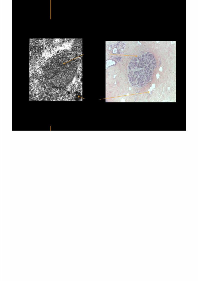
7292019 Atlas of Light-CT images
httpslidepdfcomreaderfullatlas-of-light-ct-images 14118copy LLTech 2012 ndash wwwlltechimagingcom - No copy without authorization 14
Healthy breast tissue
Lobule
Galactophorous duct cut longitudinally
Honeycomb configuration of adipocytes
copy LLTech 2012 Courtesy of Hocircpital Tenon Paris France
7292019 Atlas of Light-CT images
httpslidepdfcomreaderfullatlas-of-light-ct-images 15118
7292019 Atlas of Light-CT images
httpslidepdfcomreaderfullatlas-of-light-ct-images 16118copy LLTech 2012 ndash wwwlltechimagingcom - No copy without authorization 16
Invasive adenocarcinoma with in Situ Component
Enlarged ducts filled with cells
proliferation or necrosis
Highly scattering thin trabeculae
aspect of fibrous tissue
surrounding grey cellular zones
copy LLTech 2012 Courtesy of Hocircpital Tenon Paris France
7292019 Atlas of Light-CT images
httpslidepdfcomreaderfullatlas-of-light-ct-images 17118copy LLTech 2012 ndash wwwlltechimagingcom - No copy without authorization 17
Elongated compressed
ductules with slit-like lumen
Well delimited nodule withlobulated appearence
Fibroadenoma
copy LLTech 2012 Courtesy of Hocircpital Tenon Paris France
7292019 Atlas of Light-CT images
httpslidepdfcomreaderfullatlas-of-light-ct-images 18118copy LLTech 2012 ndash wwwlltechimagingcom - No copy without authorization 18
Fibroadenoma
Enlarged ductulescharacteristic of the lesion
copy LLTech 2012 Courtesy of Hocircpital Tenon Paris France
7292019 Atlas of Light-CT images
httpslidepdfcomreaderfullatlas-of-light-ct-images 19118copy LLTech 2012 ndash wwwlltechimagingcom - No copy without authorization 19
Lymphoid zone of the node
Vessel
Adipocytes in the center
of the node
Normal Sentinel Node
copy LLTech 2012 Courtesy of Hocircpital Tenon Paris France
7292019 Atlas of Light-CT images
httpslidepdfcomreaderfullatlas-of-light-ct-images 20118
7292019 Atlas of Light-CT images
httpslidepdfcomreaderfullatlas-of-light-ct-images 21118
copy LLTech 2012 ndash wwwlltechimagingcom - No copy without authorization 21
Normal lymphoid
zone with follicles
(Dark grey)
Metastatsis
(Light grey)
Hypervascularization
due to metastasis
Fibrous enveloppe of the
node
Invaded Sentinel Node
copy LLTech 2012 Courtesy of Hocircpital Tenon Paris France
7292019 Atlas of Light-CT images
httpslidepdfcomreaderfullatlas-of-light-ct-images 22118
copy LLTech 2012 ndash wwwlltechimagingcom - No copy without authorization 22
The Light-CT scanner allows fast tissue processing andpathology examination It completes the toolset available topathologists
When Chemical Fixation is needed
(Mostly Diagnostic)
When Frozen Section is needed
(Mostly Intra-operative)
Biopsy3D
Digital Image
2D
Digital Image
Slide
15-12 mins
Artefacts
Destructive
12-24 hours
Expensive
Destructive
5-8 minutes
Non-Destructive
Tissue Scanner
Light-CT could help to reduce the number of re-excisions in breast
7292019 Atlas of Light-CT images
httpslidepdfcomreaderfullatlas-of-light-ct-images 23118
copy LLTech 2012 ndash wwwlltechimagingcom - No copy without authorization 23
Light CT could help to reduce the number of re excisions in breastcancer surgery whilst preserving the current routines ofpathologists
Per surgery
LLTechrsquos
analysis
Histology Andor
Immunohistochemistry
Further
treatment
Post surgery
Surgical margin analysis
Metastasis analysis (gt 2mm) et
micro metastaisi
PN1mi gt 02 ndash 2 mm lt
SurgeryOKOK
Per surgery Post Surgery
C u r r e n t t e c h n i q u e s
U p t o 4 0 o
f d o u b l e p r o
c e d u r e s
L L T e c h L i g h t - C
T
S i g n i f i c a n t d e c r e a s e o f
d o u b l e p r o c e d u
r e s
Visual
analysis
cryostat
Further
treatmentSurgery
OK
Cancerouscancerous
OK
Poor identification of
metastasis
Up to 40 of double surgical
procedures
Several day analysisFew minutes analysis
Surgeon Pathologist Pathologist
Decrease of double surgical
procedures CancerousCancerous
No surgical
margins analysis
Histology Andor
Immuno
histochemistry
Node
Margin
Several day analysisFew minutes analysisSurgeon Pathologist Pathologist
7292019 Atlas of Light-CT images
httpslidepdfcomreaderfullatlas-of-light-ct-images 24118
copy LLTech 2012 ndash wwwlltechimagingcom - No copy without authorization 24
BRAIN IMAGING
Back to table of contents
7292019 Atlas of Light-CT images
httpslidepdfcomreaderfullatlas-of-light-ct-images 25118
copy LLTech 2012 ndash wwwlltechimagingcom - No copy without authorization 25
Rat brain
Architectural and cellular information seen at various scales
Myelinated fibers visible
1 mm
500 microm
100 microm
7292019 Atlas of Light-CT images
httpslidepdfcomreaderfullatlas-of-light-ct-images 26118
7292019 Atlas of Light-CT images
httpslidepdfcomreaderfullatlas-of-light-ct-images 27118
7292019 Atlas of Light-CT images
httpslidepdfcomreaderfullatlas-of-light-ct-images 28118
copy LLTech 2012 ndash wwwlltechimagingcom - No copy without authorization 28 copy LLTECH 2011
Collagen-rich matrixWide fascicles of
tumour cells
Large capillary
Fibroblastic meningioma grade I
500microm
100microm 200microm200microm
7292019 Atlas of Light-CT images
httpslidepdfcomreaderfullatlas-of-light-ct-images 29118
copy LLTech 2012 ndash wwwlltechimagingcom - No copy without authorization 29
Transitional meningioma grade I
Psammoma surrounding a calcification
Whorls formation
Collagen-rich matrix
500microm
100microm
50microm
50microm
7292019 Atlas of Light-CT images
httpslidepdfcomreaderfullatlas-of-light-ct-images 30118
7292019 Atlas of Light-CT images
httpslidepdfcomreaderfullatlas-of-light-ct-images 31118
copy LLTech 2012 ndash wwwlltechimagingcom - No copy without authorization 31 copy LLTECH 2011
Arrows Fibers transverse cut
Spinal cord normal tissue
500 microm
100 microm
100 microm
7292019 Atlas of Light-CT images
httpslidepdfcomreaderfullatlas-of-light-ct-images 32118
7292019 Atlas of Light-CT images
httpslidepdfcomreaderfullatlas-of-light-ct-images 33118
7292019 Atlas of Light-CT images
httpslidepdfcomreaderfullatlas-of-light-ct-images 34118
copy LLTech 2012 ndash wwwlltechimagingcom - No copy without authorization 34
Back to table of contents
SKIN IMAGING
N l ki h l
7292019 Atlas of Light-CT images
httpslidepdfcomreaderfullatlas-of-light-ct-images 35118
copy LLTech 2012 ndash wwwlltechimagingcom - No copy without authorization 35
Normal skin morphology
Epidermis
Collagen
Pilosebaceous
unit
Sweat gland
Adipocytes
b
a
c
e
d
500 microm
copy LLTech 2012 Courtesy of Hopitaux Universitaires de Genegraveve Genegraveve Switzerland
7292019 Atlas of Light-CT images
httpslidepdfcomreaderfullatlas-of-light-ct-images 36118
copy LLTech 2012 ndash wwwlltechimagingcom - No copy without authorization 36
Hypodermis
Skin morphology layers are well discriminated
5 mm x 8 mm mosaic of native field image
Ageing patient (solar elastosis)
En-face imaging of vertical excision
Epidermis
Dermis
1
3
2
4
7292019 Atlas of Light-CT images
httpslidepdfcomreaderfullatlas-of-light-ct-images 37118
En face skin slicing shows structural and cellular details
7292019 Atlas of Light-CT images
httpslidepdfcomreaderfullatlas-of-light-ct-images 38118
copy LLTech 2012 ndash wwwlltechimagingcom - No copy without authorization 38
Stratum corneum Stratum spinosum
Stratum basale Reticular region
Epidermis
Dermis
En-face skin slicing shows structural and cellular details
Melanin
papillary caps
Keratinocyte
nuclei
Blood vessels
Collagen
Corneocytes
100 microm 100 microm
100 microm100 microm
copy LLTech 2012 Courtesy of Hopitaux Universitaires de Genegraveve Genegraveve Switzerland
7292019 Atlas of Light-CT images
httpslidepdfcomreaderfullatlas-of-light-ct-images 39118
7292019 Atlas of Light-CT images
httpslidepdfcomreaderfullatlas-of-light-ct-images 40118
Vertical reconstruction of skin model
7292019 Atlas of Light-CT images
httpslidepdfcomreaderfullatlas-of-light-ct-images 41118
copy LLTech 2012 ndash wwwlltechimagingcom - No copy without authorization 41
Vertical reconstruction of skin model
50 microm
Stratum corneum
Stratum spinosum
Dermis
Keratinocyte nuclei
copy LLTech 2012
7292019 Atlas of Light-CT images
httpslidepdfcomreaderfullatlas-of-light-ct-images 42118
Normal
7292019 Atlas of Light-CT images
httpslidepdfcomreaderfullatlas-of-light-ct-images 43118
copy LLTech 2012 ndash wwwlltechimagingcom - No copy without authorization 43
skin
b
a
e
c
f
d
500 microm
7292019 Atlas of Light-CT images
httpslidepdfcomreaderfullatlas-of-light-ct-images 44118
7292019 Atlas of Light-CT images
httpslidepdfcomreaderfullatlas-of-light-ct-images 45118
copy LLTech 2012 ndash wwwlltechimagingcom - No copy without authorization 45
Adipocytes
9um depth 27um depth 60um depth
Reconstructed
depth slice
200 microm
50 microm
7292019 Atlas of Light-CT images
httpslidepdfcomreaderfullatlas-of-light-ct-images 46118
Skin pathologies basal cell carcinoma discrimination at
7292019 Atlas of Light-CT images
httpslidepdfcomreaderfullatlas-of-light-ct-images 47118
copy LLTech 2012 ndash wwwlltechimagingcom - No copy without authorization 47
Epidermis
Dermis
Hypodermis
A
AA
C
BCC
PSU
PSU
BCC Basal cell carcinoma
A Adipocytes
C Collagen
PSU Pilosebaceous unit
structural level
1 mm
copy LLTech 2012 Courtesy of Hopitaux Universitaires de Genegraveve Genegraveve Switzerland
C
Zoom on basal cell carcinoma discrimination at cellular level
7292019 Atlas of Light-CT images
httpslidepdfcomreaderfullatlas-of-light-ct-images 48118
copy LLTech 2012 ndash wwwlltechimagingcom - No copy without authorization 48
Zoom on basal cell carcinoma discrimination at cellular level
200 microm
100 microm
Peritumoral stroma
High cell density
copy LLTech 2012 Courtesy of Hopitaux Universitaires de Genegraveve Genegraveve Switzerland
Skin pathologies discrimination of actinic keratosis
7292019 Atlas of Light-CT images
httpslidepdfcomreaderfullatlas-of-light-ct-images 49118
copy LLTech 2012 ndash wwwlltechimagingcom - No copy without authorization 49
Skin pathologies discrimination of actinic keratosis
Enlarged epidermis
Extended epidermal ridges
1 mm
copy LLTech 2012 Courtesy of Hopitaux Universitaires de Genegraveve Genegraveve Switzerland
7292019 Atlas of Light-CT images
httpslidepdfcomreaderfullatlas-of-light-ct-images 50118
copy LLTech 2012 ndash wwwlltechimagingcom - No copy without authorization 50
Basal cell carcinoma in depth imaging
Z = 10 microns 7 x 42 mm FOV Z = 35 microns 7 x 42 mm FOV
BCC
SG Sebaceous glands
Z = 55 microns 7 x 42 mm FOV Z = 75 microns 7 x 42 mm FOV
SG
Application to Mohs surgerycopy LLTech 2012 Courtesy of Hopitaux Universitaires de Genegraveve Genegraveve Switzerland
7292019 Atlas of Light-CT images
httpslidepdfcomreaderfullatlas-of-light-ct-images 51118
copy LLTech 2012 ndash wwwlltechimagingcom - No copy without authorization 51 copy LLTECH 2011
Healthy vs Cheloid scar tissue
Healthy skin tissueFOV 75x5mm
Cheloid scar tissueFOV 65x65mm
copy LLTECH 2011
7292019 Atlas of Light-CT images
httpslidepdfcomreaderfullatlas-of-light-ct-images 52118
copy LLTech 2012 ndash wwwlltechimagingcom - No copy without authorization 52
Dermo-epidermaljunction
Cheloid scar tissue in detail
copy LLTECH 2011
Nodular structureThickcollagenfiberbundles
1mm
600 microm
500 microm
h l d d f
7292019 Atlas of Light-CT images
httpslidepdfcomreaderfullatlas-of-light-ct-images 53118
copy LLTech 2012 ndash wwwlltechimagingcom - No copy without authorization 53
Cheloid scar tissue viewed en face
20microm depth 120microm depth
7292019 Atlas of Light-CT images
httpslidepdfcomreaderfullatlas-of-light-ct-images 54118
Skin in-vivo sweat duct imaging in 3D
7292019 Atlas of Light-CT images
httpslidepdfcomreaderfullatlas-of-light-ct-images 55118
copy LLTech 2012 ndash wwwlltechimagingcom - No copy without authorization 55
g g
Vertical reconstruction from en-face images
Sweat duct 3D reconstruction
Stratum corneum
Stratum spinosum
SD sweat ducts
SD SD
100 microm
7292019 Atlas of Light-CT images
httpslidepdfcomreaderfullatlas-of-light-ct-images 56118
7292019 Atlas of Light-CT images
httpslidepdfcomreaderfullatlas-of-light-ct-images 57118
7292019 Atlas of Light-CT images
httpslidepdfcomreaderfullatlas-of-light-ct-images 58118
copy LLTech 2012 ndash wwwlltechimagingcom - No copy without authorization 58
LUNG IMAGING
Back to table of contents
H L
7292019 Atlas of Light-CT images
httpslidepdfcomreaderfullatlas-of-light-ct-images 59118
copy LLTech 2012 ndash wwwlltechimagingcom - No copy without authorization 59
Zone 1
Zone 2
Zone 3
Zone 4
Zone with
cancerous tissue
Limit of cancerous
stroma
alveoli with
macrophages
Alveoles
Human Lung
500microm
copy LLTech 2012 Courtesy of Hocircpital Hotel-Dieu Paris France
H L Z 1 Z t
7292019 Atlas of Light-CT images
httpslidepdfcomreaderfullatlas-of-light-ct-images 60118
copy LLTech 2012 ndash wwwlltechimagingcom - No copy without authorization 60
Stroma
Human Lung ndash Zone 1 ndash Zoom on cancerous stroma
Cancerous
area
200microm
copy LLTech 2012 Courtesy of Hocircpital Hotel-Dieu Paris France
Human Lung ndash Zone 2 ndash Limit cancerous stroma alveoli
7292019 Atlas of Light-CT images
httpslidepdfcomreaderfullatlas-of-light-ct-images 61118
copy LLTech 2012 ndash wwwlltechimagingcom - No copy without authorization 61
g
with visible macrophages and vessel
Alveoli
Macrophages
Vessel
copy LLTech 2012 Courtesy of Hocircpital Hotel-Dieu Paris France
Human Lung ndash Zone 3 ndash Limit cancerous stroma alveoli
7292019 Atlas of Light-CT images
httpslidepdfcomreaderfullatlas-of-light-ct-images 62118
copy LLTech 2012 ndash wwwlltechimagingcom - No copy without authorization 62
g
with visible macrophages
Macrophages
100microm
copy LLTech 2012 Courtesy of Hocircpital Hotel-Dieu Paris France
Human Lung ndash Zone 4 ndash Alveoli
7292019 Atlas of Light-CT images
httpslidepdfcomreaderfullatlas-of-light-ct-images 63118
copy LLTech 2012 ndash wwwlltechimagingcom - No copy without authorization 63
Human Lung Zone 4 Alveoli
Alveoli
200microm
copy LLTech 2012 Courtesy of Hocircpital Hotel-Dieu Paris France
7292019 Atlas of Light-CT images
httpslidepdfcomreaderfullatlas-of-light-ct-images 64118
copy LLTech 2012 ndash wwwlltechimagingcom - No copy without authorization 64
GI IMAGING
Back to table of contents
7292019 Atlas of Light-CT images
httpslidepdfcomreaderfullatlas-of-light-ct-images 65118
Colonic carcinoma
7292019 Atlas of Light-CT images
httpslidepdfcomreaderfullatlas-of-light-ct-images 66118
copy LLTech 2012 ndash wwwlltechimagingcom - No copy without authorization 66
Colonic Carcinoma
Colonic carcinoma
copy LLTech 2012 Courtesy of MD Anderson Houston USA
Intestines ndash Crohnrsquos disease
7292019 Atlas of Light-CT images
httpslidepdfcomreaderfullatlas-of-light-ct-images 67118
copy LLTech 2012 ndash wwwlltechimagingcom - No copy without authorization 67
Crohnrsquos disease structure not
visible from the surface
Fat cells
200 microm
Intestines Crohn s disease
copy LLTech 2012 Courtesy of Institut Curie Paris France
Rat stomach
7292019 Atlas of Light-CT images
httpslidepdfcomreaderfullatlas-of-light-ct-images 68118
copy LLTech 2012 ndash wwwlltechimagingcom - No copy without authorization 68
Rat stomach
Mucosa withgastric pits
Submucosa with
bright collagen
fibers
Muscularis
propria with
muscle bundles
copy LLTech 2012 Courtesy of Weill Cornell Medical College NY USA
200 microm
7292019 Atlas of Light-CT images
httpslidepdfcomreaderfullatlas-of-light-ct-images 69118
copy LLTech 2012 ndash wwwlltechimagingcom - No copy without authorization 69 copy LLTECH 2011
UROGENITAL IMAGING
Back to table of contents
Rat bladder
7292019 Atlas of Light-CT images
httpslidepdfcomreaderfullatlas-of-light-ct-images 70118
copy LLTech 2012 ndash wwwlltechimagingcom - No copy without authorization 70
Rat bladder
Stratifiedurothelium
Lamina propria
500 microm
Muscular
bundles of the
lamina propria
Serosa
copy LLTech 2012 Courtesy of Weill Cornell Medical College NY USA
100 microm
100 microm
Human bladder low grade papillary urothelial carcinoma
7292019 Atlas of Light-CT images
httpslidepdfcomreaderfullatlas-of-light-ct-images 71118
copy LLTech 2012 ndash wwwlltechimagingcom - No copy without authorization 71
Lesion with fibrovascular cores (arrow) and superficial umbrella cells (arrowheads)
Human bladder low grade papillary urothelial carcinoma
copy LLTech 2012 Courtesy of Weill Cornell Medical College NY USA
100 microm
7292019 Atlas of Light-CT images
httpslidepdfcomreaderfullatlas-of-light-ct-images 72118
Rat prostate
7292019 Atlas of Light-CT images
httpslidepdfcomreaderfullatlas-of-light-ct-images 73118
copy LLTech 2012 ndash wwwlltechimagingcom - No copy without authorization 73
p
copy LLTech 2012 Courtesy of Weill Cornell Medical College NY USA
Papillary folds of the
acinar glands
Prostatic acini
Fibromuscular stromaPeriprostatic fat Blood vessel
1 mm
200 microm
Mouse kidney
7292019 Atlas of Light-CT images
httpslidepdfcomreaderfullatlas-of-light-ct-images 74118
copy LLTech 2012 ndash wwwlltechimagingcom - No copy without authorization 74
1 mm
y
copy LLTech 2012 Courtesy of Institut Biomeacutedical de Bicecirctre Paris France
Renal cortex
Renal medulla
Renal sinus
7292019 Atlas of Light-CT images
httpslidepdfcomreaderfullatlas-of-light-ct-images 75118
copy LLTech 2012 ndash wwwlltechimagingcom - No copy without authorization 75
Mouse kidney ndash zoom on highlighted section of previous slide
800microm
copy LLTech 2012 Courtesy of Hocircpital Kremlin Bicecirctre Paris France
7292019 Atlas of Light-CT images
httpslidepdfcomreaderfullatlas-of-light-ct-images 76118
7292019 Atlas of Light-CT images
httpslidepdfcomreaderfullatlas-of-light-ct-images 77118
7292019 Atlas of Light-CT images
httpslidepdfcomreaderfullatlas-of-light-ct-images 78118
copy LLTech 2012 ndash wwwlltechimagingcom - No copy without authorization 78 copy LLTECH 2011
CORE NEEDLE BIOPSIES
Back to table of contents
Kidney
7292019 Atlas of Light-CT images
httpslidepdfcomreaderfullatlas-of-light-ct-images 79118
copy LLTech 2012 ndash wwwlltechimagingcom - No copy without authorization 79
100microm
50microm
Renal tubules
Vessel
500microm
copy LLTech 2012 Courtesy of Institut Curie Paris France
Breast Invasive lobular carcinoma
7292019 Atlas of Light-CT images
httpslidepdfcomreaderfullatlas-of-light-ct-images 80118
copy LLTech 2012 ndash wwwlltechimagingcom - No copy without authorization 80
500microm
stroma
adipocytes
copy LLTech 2012 Courtesy of Institut Curie Paris France
7292019 Atlas of Light-CT images
httpslidepdfcomreaderfullatlas-of-light-ct-images 81118
Breast Ductal carcinoma in situ
7292019 Atlas of Light-CT images
httpslidepdfcomreaderfullatlas-of-light-ct-images 82118
copy LLTech 2012 ndash wwwlltechimagingcom - No copy without authorization 82
500microm
copy LLTech 2012 Courtesy of Institut Curie Paris France
7292019 Atlas of Light-CT images
httpslidepdfcomreaderfullatlas-of-light-ct-images 83118
copy LLTech 2012 ndash wwwlltechimagingcom - No copy without authorization 83 copy LLTECH 2011
THYROID IMAGING
Back to table of contents
7292019 Atlas of Light-CT images
httpslidepdfcomreaderfullatlas-of-light-ct-images 84118
copy LLTech 2012 ndash wwwlltechimagingcom - No copy without authorization 84
Thyroid
800 microm
200 microm
copy LLTech 2012 Courtesy of Hocircpital Tenon Paris France
7292019 Atlas of Light-CT images
httpslidepdfcomreaderfullatlas-of-light-ct-images 85118
copy LLTech 2012 ndash wwwlltechimagingcom - No copy without authorization 85
Thyroid
800 microm
200 microm
copy LLTech 2012 Courtesy of Hocircpital Tenon Paris France
7292019 Atlas of Light-CT images
httpslidepdfcomreaderfullatlas-of-light-ct-images 86118
copy LLTech 2012 ndash wwwlltechimagingcom - No copy without authorization 86 copy LLTECH 2011
FAT CELLS IMAGING
Back to table of contents
7292019 Atlas of Light-CT images
httpslidepdfcomreaderfullatlas-of-light-ct-images 87118
copy LLTech 2012 ndash wwwlltechimagingcom - No copy without authorization 87
Brown fat cells - adipocytes
100 microm 100 microm
200 microm
10um depth
Adipocytes
45um depth
En face
slices
Reconstructed
depth slice
copy LLTech 2012 Courtesy of Centre de Recherche des Cordeliers Paris France
7292019 Atlas of Light-CT images
httpslidepdfcomreaderfullatlas-of-light-ct-images 88118
copy LLTech 2012 ndash wwwlltechimagingcom - No copy without authorization 88 copy LLTECH 2011
CORNEA AND RETINA
Back to table of contents
Human cornea reconstructed depth slice
7292019 Atlas of Light-CT images
httpslidepdfcomreaderfullatlas-of-light-ct-images 89118
copy LLTech 2012 ndash wwwlltechimagingcom - No copy without authorization 89
50 um
wwwlltechimagingcom
7292019 Atlas of Light-CT images
httpslidepdfcomreaderfullatlas-of-light-ct-images 90118
Large field en face view in stroma
7292019 Atlas of Light-CT images
httpslidepdfcomreaderfullatlas-of-light-ct-images 91118
copy LLTech 2012 ndash wwwlltechimagingcom - No copy without authorization 91
200 um
wwwlltechimagingcom
7292019 Atlas of Light-CT images
httpslidepdfcomreaderfullatlas-of-light-ct-images 92118
Rabbit cornea after LASIK reflectivity profiles reveal
i d tt i d thi k d ith li l l
7292019 Atlas of Light-CT images
httpslidepdfcomreaderfullatlas-of-light-ct-images 93118
copy LLTech 2012 ndash wwwlltechimagingcom - No copy without authorization 93
increased scattering and thickened epithelial layer
LASIK LASIK CONTROL CONTROL
copy LLTech 2012
Newborn mouse anterior segment
7292019 Atlas of Light-CT images
httpslidepdfcomreaderfullatlas-of-light-ct-images 94118
copy LLTech 2012 ndash wwwlltechimagingcom - No copy without authorization 94
Corneal epithelial cells
Basal membrane
Stroma
Descemetrsquos membrane
Aqueous humor
Lens capsule
Lens epithelial cells
Cortical fibers
copy LLTech 2012 Courtesy of ESPCI Paris Francecopy LLTech 2012
Rat retina comparison with histology
7292019 Atlas of Light-CT images
httpslidepdfcomreaderfullatlas-of-light-ct-images 95118
copy LLTech 2012 ndash wwwlltechimagingcom - No copy without authorization 95
Figure 1 Right Ex vivo rat retinaimaged using the f ull field OCT
system of ESPCI Left Histology
for comparison
Figure 1 Right Ex vivo rat retinaimaged using the f ull field OCT
system of ESPCI Left Histology
for comparison
Figure 1 Right Ex vivo rat retinaimaged using the f ull field OCT
system of ESPCI Left Histology
for comparisonField
230x300microm
RPE
PRL
ONL
OPL
INL
IPL
GCL
NFL
Invest Ophthalmol Vis Sci 2004 Nov45(11)4126-31
Grieve K Paques M Dubois A Sahel J Boccara C Le Gargasson JF copy LLTech 2012
Rat retina
7292019 Atlas of Light-CT images
httpslidepdfcomreaderfullatlas-of-light-ct-images 96118
copy LLTech 2012 ndash wwwlltechimagingcom - No copy without authorization 96 copy LLTech 2012 Courtesy of ESPCI Paris France
Nerve fiber layer
Retina
Invest Ophthalmol Vis Sci 2004 Nov45(11)4126-31
Grieve K Paques M Dubois A Sahel J Boccara C Le Gargasson JF
Transscleral image
7292019 Atlas of Light-CT images
httpslidepdfcomreaderfullatlas-of-light-ct-images 97118
7292019 Atlas of Light-CT images
httpslidepdfcomreaderfullatlas-of-light-ct-images 98118
7292019 Atlas of Light-CT images
httpslidepdfcomreaderfullatlas-of-light-ct-images 99118
In vivo imaging of drosophila melanogaster
7292019 Atlas of Light-CT images
httpslidepdfcomreaderfullatlas-of-light-ct-images 100118
copy LLTech 2012 ndash wwwlltechimagingcom - No copy without authorization 100 copy LLTech 2012 Courtesy of ESPCI Paris France
In vivo imaging of drosophila melanogaster ndash four stages of pupation over 72 hours
7292019 Atlas of Light-CT images
httpslidepdfcomreaderfullatlas-of-light-ct-images 101118
copy LLTech 2012 ndash wwwlltechimagingcom - No copy without authorization 101
Prepupa (0-2 h) Transition topupal stage (24 h)
Pupal stage (48 h) Advanced pupal phase
before eclosion (72 h)
copy LLTech 2012 Courtesy of ESPCI Paris France
Pupa 118 4 days 30um depth
100
7292019 Atlas of Light-CT images
httpslidepdfcomreaderfullatlas-of-light-ct-images 102118
copy LLTech 2012 ndash wwwlltechimagingcom - No copy without authorization 102
100 um
Drosophila
7292019 Atlas of Light-CT images
httpslidepdfcomreaderfullatlas-of-light-ct-images 103118
copy LLTech 2012 ndash wwwlltechimagingcom - No copy without authorization 103
200 microm
copy LLTech 2012 Courtesy of ESPCI Paris France
C Elegans ndash Fast identification of anatomy
7292019 Atlas of Light-CT images
httpslidepdfcomreaderfullatlas-of-light-ct-images 104118
copy LLTech 2012 ndash wwwlltechimagingcom - No copy without authorization 104
Body wall - cuticle
gonad arm
30 microm depth
uterus
ovocytes
intestine
100 microm 100 microm
15 microm depth
copy LLTech 2012 Courtesy of ENS Paris France
C elegans ndash Images at higher magnification 40X
7292019 Atlas of Light-CT images
httpslidepdfcomreaderfullatlas-of-light-ct-images 105118
copy LLTech 2012 ndash wwwlltechimagingcom - No copy without authorization 105
3D reconstruction 50 microm
100 microm
copy LLTech 2012 Courtesy of ENS Paris France
Zebra fish eye
7292019 Atlas of Light-CT images
httpslidepdfcomreaderfullatlas-of-light-ct-images 106118
copy LLTech 2012 ndash wwwlltechimagingcom - No copy without authorization 106
Zebra fish eye
100 microm
100 microm
En face slices
Reconstructed depth slice
copy LLTech 2012 Courtesy of Inserm Paris France
10um depth 20um depth 45um depth
7292019 Atlas of Light-CT images
httpslidepdfcomreaderfullatlas-of-light-ct-images 107118
Xenopus Laevis backtail
7292019 Atlas of Light-CT images
httpslidepdfcomreaderfullatlas-of-light-ct-images 108118
copy LLTech 2012 ndash wwwlltechimagingcom - No copy without authorization 108
100 um30um depth 60um depth
7292019 Atlas of Light-CT images
httpslidepdfcomreaderfullatlas-of-light-ct-images 109118
Mouse embryo in vitro 6 5 days old ndash
7292019 Atlas of Light-CT images
httpslidepdfcomreaderfullatlas-of-light-ct-images 110118
copy LLTech 2012 ndash wwwlltechimagingcom - No copy without authorization 110
Mouse embryo in vitro 65 days old
copy LLTech 2012 Courtesy of Ellvire Guiot and nstitut Jacques Monod Paris France
3D imaging of internal cavity
Sabellaria alveolata mouth palps
7292019 Atlas of Light-CT images
httpslidepdfcomreaderfullatlas-of-light-ct-images 111118
copy LLTech 2012 ndash wwwlltechimagingcom - No copy without authorization 111
500 microm
100 microm
Mouth palps
copy LLTech 2012 Courtesy of Museacutee National drsquoHistoire Naturelle Paris France
7292019 Atlas of Light-CT images
httpslidepdfcomreaderfullatlas-of-light-ct-images 112118
copy LLTech 2012 ndash wwwlltechimagingcom - No copy without authorization 112 copy LLTECH 2011
PLANT IMAGING
Back to table of contents
Mouse ear leaf epidermis
7292019 Atlas of Light-CT images
httpslidepdfcomreaderfullatlas-of-light-ct-images 113118
copy LLTech 2012 ndash wwwlltechimagingcom - No copy without authorization 113
100 microm
Epidermal cell
copy LLTech 2012
Stoma
Leaf veins
7292019 Atlas of Light-CT images
httpslidepdfcomreaderfullatlas-of-light-ct-images 114118
copy LLTech 2012 ndash wwwlltechimagingcom - No copy without authorization 114
114copy LLTech 2012
Fibers
Veins
100 microm
500 microm
Apple
7292019 Atlas of Light-CT images
httpslidepdfcomreaderfullatlas-of-light-ct-images 115118
copy LLTech 2012 ndash wwwlltechimagingcom - No copy without authorization 115
115copy LLTech 2012
100 microm
Wax
Skin
Flesh
500 microm
Water
Effect of harpin infiltration on tobacco leaf
7292019 Atlas of Light-CT images
httpslidepdfcomreaderfullatlas-of-light-ct-images 116118
copy LLTech 2012 ndash wwwlltechimagingcom - No copy without authorization 116
116copy LLTech 2012 Courtesy of UPMC Paris France
50 microm
50 microm
50 microm
50 microm
Upper epidermis
10 min after harpin infiltration 30 min after harpin infiltration
Palisade parenchyma
Epidermis cell
Chloroplast
7292019 Atlas of Light-CT images
httpslidepdfcomreaderfullatlas-of-light-ct-images 117118
copy LLTech 2012 ndash wwwlltechimagingcom - No copy without authorization 117
bull Light-CTtrade does not require tissue preparation nor staining of any kind
bull Creates images within minutes using a non-destructive process
bull Offers a 1 microm cellular resolution in 3D
bull Reveals structural and cellular information
bull Mosaicing allows fast visualisation at various scale
bull En-face high-resolution imaging allows vertical and 3D reconstruction
bull In-vivo images show good promise for in-vivo system
bull Clinical and research applications in cancer detection cosmetics biobanking small animal
developmental biology botanyhellip
Contact Details
7292019 Atlas of Light-CT images
httpslidepdfcomreaderfullatlas-of-light-ct-images 118118
USA
LLTech Inc
103 Carnegie Center DriveSuite 300
Princeton NJ 08540
USA
wwwlltechimagingcom
contactlltechfr
Phone + 1 609 955 3506
LLTech SAS
29 rue du Faubourg St-JacquesPeacutepiniegravere Paris Santeacute Cochin
75014
Paris - France
wwwlltechimagingcom
contactlltechfr
Phone +33 1 82 72 61 25
Europe
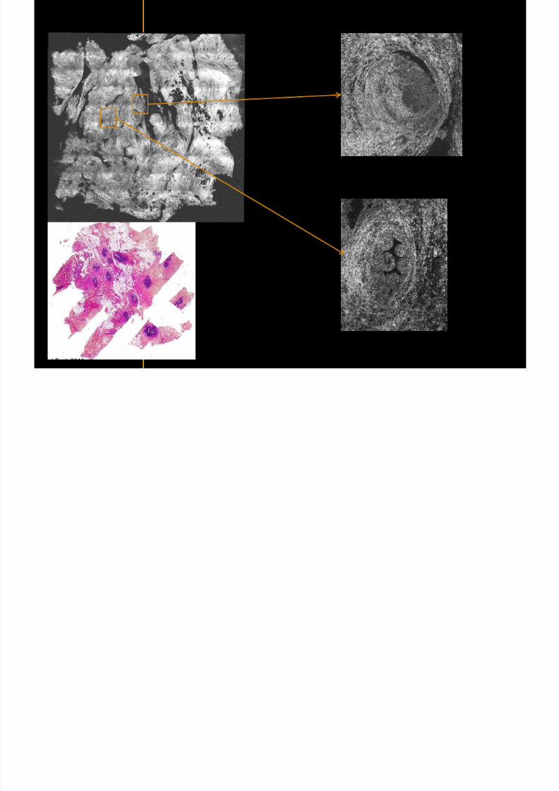
7292019 Atlas of Light-CT images
httpslidepdfcomreaderfullatlas-of-light-ct-images 18118copy LLTech 2012 ndash wwwlltechimagingcom - No copy without authorization 18
Fibroadenoma
Enlarged ductulescharacteristic of the lesion
copy LLTech 2012 Courtesy of Hocircpital Tenon Paris France
7292019 Atlas of Light-CT images
httpslidepdfcomreaderfullatlas-of-light-ct-images 19118copy LLTech 2012 ndash wwwlltechimagingcom - No copy without authorization 19
Lymphoid zone of the node
Vessel
Adipocytes in the center
of the node
Normal Sentinel Node
copy LLTech 2012 Courtesy of Hocircpital Tenon Paris France
7292019 Atlas of Light-CT images
httpslidepdfcomreaderfullatlas-of-light-ct-images 20118
7292019 Atlas of Light-CT images
httpslidepdfcomreaderfullatlas-of-light-ct-images 21118
copy LLTech 2012 ndash wwwlltechimagingcom - No copy without authorization 21
Normal lymphoid
zone with follicles
(Dark grey)
Metastatsis
(Light grey)
Hypervascularization
due to metastasis
Fibrous enveloppe of the
node
Invaded Sentinel Node
copy LLTech 2012 Courtesy of Hocircpital Tenon Paris France
7292019 Atlas of Light-CT images
httpslidepdfcomreaderfullatlas-of-light-ct-images 22118
copy LLTech 2012 ndash wwwlltechimagingcom - No copy without authorization 22
The Light-CT scanner allows fast tissue processing andpathology examination It completes the toolset available topathologists
When Chemical Fixation is needed
(Mostly Diagnostic)
When Frozen Section is needed
(Mostly Intra-operative)
Biopsy3D
Digital Image
2D
Digital Image
Slide
15-12 mins
Artefacts
Destructive
12-24 hours
Expensive
Destructive
5-8 minutes
Non-Destructive
Tissue Scanner
Light-CT could help to reduce the number of re-excisions in breast
7292019 Atlas of Light-CT images
httpslidepdfcomreaderfullatlas-of-light-ct-images 23118
copy LLTech 2012 ndash wwwlltechimagingcom - No copy without authorization 23
Light CT could help to reduce the number of re excisions in breastcancer surgery whilst preserving the current routines ofpathologists
Per surgery
LLTechrsquos
analysis
Histology Andor
Immunohistochemistry
Further
treatment
Post surgery
Surgical margin analysis
Metastasis analysis (gt 2mm) et
micro metastaisi
PN1mi gt 02 ndash 2 mm lt
SurgeryOKOK
Per surgery Post Surgery
C u r r e n t t e c h n i q u e s
U p t o 4 0 o
f d o u b l e p r o
c e d u r e s
L L T e c h L i g h t - C
T
S i g n i f i c a n t d e c r e a s e o f
d o u b l e p r o c e d u
r e s
Visual
analysis
cryostat
Further
treatmentSurgery
OK
Cancerouscancerous
OK
Poor identification of
metastasis
Up to 40 of double surgical
procedures
Several day analysisFew minutes analysis
Surgeon Pathologist Pathologist
Decrease of double surgical
procedures CancerousCancerous
No surgical
margins analysis
Histology Andor
Immuno
histochemistry
Node
Margin
Several day analysisFew minutes analysisSurgeon Pathologist Pathologist
7292019 Atlas of Light-CT images
httpslidepdfcomreaderfullatlas-of-light-ct-images 24118
copy LLTech 2012 ndash wwwlltechimagingcom - No copy without authorization 24
BRAIN IMAGING
Back to table of contents
7292019 Atlas of Light-CT images
httpslidepdfcomreaderfullatlas-of-light-ct-images 25118
copy LLTech 2012 ndash wwwlltechimagingcom - No copy without authorization 25
Rat brain
Architectural and cellular information seen at various scales
Myelinated fibers visible
1 mm
500 microm
100 microm
7292019 Atlas of Light-CT images
httpslidepdfcomreaderfullatlas-of-light-ct-images 26118
7292019 Atlas of Light-CT images
httpslidepdfcomreaderfullatlas-of-light-ct-images 27118
7292019 Atlas of Light-CT images
httpslidepdfcomreaderfullatlas-of-light-ct-images 28118
copy LLTech 2012 ndash wwwlltechimagingcom - No copy without authorization 28 copy LLTECH 2011
Collagen-rich matrixWide fascicles of
tumour cells
Large capillary
Fibroblastic meningioma grade I
500microm
100microm 200microm200microm
7292019 Atlas of Light-CT images
httpslidepdfcomreaderfullatlas-of-light-ct-images 29118
copy LLTech 2012 ndash wwwlltechimagingcom - No copy without authorization 29
Transitional meningioma grade I
Psammoma surrounding a calcification
Whorls formation
Collagen-rich matrix
500microm
100microm
50microm
50microm
7292019 Atlas of Light-CT images
httpslidepdfcomreaderfullatlas-of-light-ct-images 30118
7292019 Atlas of Light-CT images
httpslidepdfcomreaderfullatlas-of-light-ct-images 31118
copy LLTech 2012 ndash wwwlltechimagingcom - No copy without authorization 31 copy LLTECH 2011
Arrows Fibers transverse cut
Spinal cord normal tissue
500 microm
100 microm
100 microm
7292019 Atlas of Light-CT images
httpslidepdfcomreaderfullatlas-of-light-ct-images 32118
7292019 Atlas of Light-CT images
httpslidepdfcomreaderfullatlas-of-light-ct-images 33118
7292019 Atlas of Light-CT images
httpslidepdfcomreaderfullatlas-of-light-ct-images 34118
copy LLTech 2012 ndash wwwlltechimagingcom - No copy without authorization 34
Back to table of contents
SKIN IMAGING
N l ki h l
7292019 Atlas of Light-CT images
httpslidepdfcomreaderfullatlas-of-light-ct-images 35118
copy LLTech 2012 ndash wwwlltechimagingcom - No copy without authorization 35
Normal skin morphology
Epidermis
Collagen
Pilosebaceous
unit
Sweat gland
Adipocytes
b
a
c
e
d
500 microm
copy LLTech 2012 Courtesy of Hopitaux Universitaires de Genegraveve Genegraveve Switzerland
7292019 Atlas of Light-CT images
httpslidepdfcomreaderfullatlas-of-light-ct-images 36118
copy LLTech 2012 ndash wwwlltechimagingcom - No copy without authorization 36
Hypodermis
Skin morphology layers are well discriminated
5 mm x 8 mm mosaic of native field image
Ageing patient (solar elastosis)
En-face imaging of vertical excision
Epidermis
Dermis
1
3
2
4
7292019 Atlas of Light-CT images
httpslidepdfcomreaderfullatlas-of-light-ct-images 37118
En face skin slicing shows structural and cellular details
7292019 Atlas of Light-CT images
httpslidepdfcomreaderfullatlas-of-light-ct-images 38118
copy LLTech 2012 ndash wwwlltechimagingcom - No copy without authorization 38
Stratum corneum Stratum spinosum
Stratum basale Reticular region
Epidermis
Dermis
En-face skin slicing shows structural and cellular details
Melanin
papillary caps
Keratinocyte
nuclei
Blood vessels
Collagen
Corneocytes
100 microm 100 microm
100 microm100 microm
copy LLTech 2012 Courtesy of Hopitaux Universitaires de Genegraveve Genegraveve Switzerland
7292019 Atlas of Light-CT images
httpslidepdfcomreaderfullatlas-of-light-ct-images 39118
7292019 Atlas of Light-CT images
httpslidepdfcomreaderfullatlas-of-light-ct-images 40118
Vertical reconstruction of skin model
7292019 Atlas of Light-CT images
httpslidepdfcomreaderfullatlas-of-light-ct-images 41118
copy LLTech 2012 ndash wwwlltechimagingcom - No copy without authorization 41
Vertical reconstruction of skin model
50 microm
Stratum corneum
Stratum spinosum
Dermis
Keratinocyte nuclei
copy LLTech 2012
7292019 Atlas of Light-CT images
httpslidepdfcomreaderfullatlas-of-light-ct-images 42118
Normal
7292019 Atlas of Light-CT images
httpslidepdfcomreaderfullatlas-of-light-ct-images 43118
copy LLTech 2012 ndash wwwlltechimagingcom - No copy without authorization 43
skin
b
a
e
c
f
d
500 microm
7292019 Atlas of Light-CT images
httpslidepdfcomreaderfullatlas-of-light-ct-images 44118
7292019 Atlas of Light-CT images
httpslidepdfcomreaderfullatlas-of-light-ct-images 45118
copy LLTech 2012 ndash wwwlltechimagingcom - No copy without authorization 45
Adipocytes
9um depth 27um depth 60um depth
Reconstructed
depth slice
200 microm
50 microm
7292019 Atlas of Light-CT images
httpslidepdfcomreaderfullatlas-of-light-ct-images 46118
Skin pathologies basal cell carcinoma discrimination at
7292019 Atlas of Light-CT images
httpslidepdfcomreaderfullatlas-of-light-ct-images 47118
copy LLTech 2012 ndash wwwlltechimagingcom - No copy without authorization 47
Epidermis
Dermis
Hypodermis
A
AA
C
BCC
PSU
PSU
BCC Basal cell carcinoma
A Adipocytes
C Collagen
PSU Pilosebaceous unit
structural level
1 mm
copy LLTech 2012 Courtesy of Hopitaux Universitaires de Genegraveve Genegraveve Switzerland
C
Zoom on basal cell carcinoma discrimination at cellular level
7292019 Atlas of Light-CT images
httpslidepdfcomreaderfullatlas-of-light-ct-images 48118
copy LLTech 2012 ndash wwwlltechimagingcom - No copy without authorization 48
Zoom on basal cell carcinoma discrimination at cellular level
200 microm
100 microm
Peritumoral stroma
High cell density
copy LLTech 2012 Courtesy of Hopitaux Universitaires de Genegraveve Genegraveve Switzerland
Skin pathologies discrimination of actinic keratosis
7292019 Atlas of Light-CT images
httpslidepdfcomreaderfullatlas-of-light-ct-images 49118
copy LLTech 2012 ndash wwwlltechimagingcom - No copy without authorization 49
Skin pathologies discrimination of actinic keratosis
Enlarged epidermis
Extended epidermal ridges
1 mm
copy LLTech 2012 Courtesy of Hopitaux Universitaires de Genegraveve Genegraveve Switzerland
7292019 Atlas of Light-CT images
httpslidepdfcomreaderfullatlas-of-light-ct-images 50118
copy LLTech 2012 ndash wwwlltechimagingcom - No copy without authorization 50
Basal cell carcinoma in depth imaging
Z = 10 microns 7 x 42 mm FOV Z = 35 microns 7 x 42 mm FOV
BCC
SG Sebaceous glands
Z = 55 microns 7 x 42 mm FOV Z = 75 microns 7 x 42 mm FOV
SG
Application to Mohs surgerycopy LLTech 2012 Courtesy of Hopitaux Universitaires de Genegraveve Genegraveve Switzerland
7292019 Atlas of Light-CT images
httpslidepdfcomreaderfullatlas-of-light-ct-images 51118
copy LLTech 2012 ndash wwwlltechimagingcom - No copy without authorization 51 copy LLTECH 2011
Healthy vs Cheloid scar tissue
Healthy skin tissueFOV 75x5mm
Cheloid scar tissueFOV 65x65mm
copy LLTECH 2011
7292019 Atlas of Light-CT images
httpslidepdfcomreaderfullatlas-of-light-ct-images 52118
copy LLTech 2012 ndash wwwlltechimagingcom - No copy without authorization 52
Dermo-epidermaljunction
Cheloid scar tissue in detail
copy LLTECH 2011
Nodular structureThickcollagenfiberbundles
1mm
600 microm
500 microm
h l d d f
7292019 Atlas of Light-CT images
httpslidepdfcomreaderfullatlas-of-light-ct-images 53118
copy LLTech 2012 ndash wwwlltechimagingcom - No copy without authorization 53
Cheloid scar tissue viewed en face
20microm depth 120microm depth
7292019 Atlas of Light-CT images
httpslidepdfcomreaderfullatlas-of-light-ct-images 54118
Skin in-vivo sweat duct imaging in 3D
7292019 Atlas of Light-CT images
httpslidepdfcomreaderfullatlas-of-light-ct-images 55118
copy LLTech 2012 ndash wwwlltechimagingcom - No copy without authorization 55
g g
Vertical reconstruction from en-face images
Sweat duct 3D reconstruction
Stratum corneum
Stratum spinosum
SD sweat ducts
SD SD
100 microm
7292019 Atlas of Light-CT images
httpslidepdfcomreaderfullatlas-of-light-ct-images 56118
7292019 Atlas of Light-CT images
httpslidepdfcomreaderfullatlas-of-light-ct-images 57118
7292019 Atlas of Light-CT images
httpslidepdfcomreaderfullatlas-of-light-ct-images 58118
copy LLTech 2012 ndash wwwlltechimagingcom - No copy without authorization 58
LUNG IMAGING
Back to table of contents
H L
7292019 Atlas of Light-CT images
httpslidepdfcomreaderfullatlas-of-light-ct-images 59118
copy LLTech 2012 ndash wwwlltechimagingcom - No copy without authorization 59
Zone 1
Zone 2
Zone 3
Zone 4
Zone with
cancerous tissue
Limit of cancerous
stroma
alveoli with
macrophages
Alveoles
Human Lung
500microm
copy LLTech 2012 Courtesy of Hocircpital Hotel-Dieu Paris France
H L Z 1 Z t
7292019 Atlas of Light-CT images
httpslidepdfcomreaderfullatlas-of-light-ct-images 60118
copy LLTech 2012 ndash wwwlltechimagingcom - No copy without authorization 60
Stroma
Human Lung ndash Zone 1 ndash Zoom on cancerous stroma
Cancerous
area
200microm
copy LLTech 2012 Courtesy of Hocircpital Hotel-Dieu Paris France
Human Lung ndash Zone 2 ndash Limit cancerous stroma alveoli
7292019 Atlas of Light-CT images
httpslidepdfcomreaderfullatlas-of-light-ct-images 61118
copy LLTech 2012 ndash wwwlltechimagingcom - No copy without authorization 61
g
with visible macrophages and vessel
Alveoli
Macrophages
Vessel
copy LLTech 2012 Courtesy of Hocircpital Hotel-Dieu Paris France
Human Lung ndash Zone 3 ndash Limit cancerous stroma alveoli
7292019 Atlas of Light-CT images
httpslidepdfcomreaderfullatlas-of-light-ct-images 62118
copy LLTech 2012 ndash wwwlltechimagingcom - No copy without authorization 62
g
with visible macrophages
Macrophages
100microm
copy LLTech 2012 Courtesy of Hocircpital Hotel-Dieu Paris France
Human Lung ndash Zone 4 ndash Alveoli
7292019 Atlas of Light-CT images
httpslidepdfcomreaderfullatlas-of-light-ct-images 63118
copy LLTech 2012 ndash wwwlltechimagingcom - No copy without authorization 63
Human Lung Zone 4 Alveoli
Alveoli
200microm
copy LLTech 2012 Courtesy of Hocircpital Hotel-Dieu Paris France
7292019 Atlas of Light-CT images
httpslidepdfcomreaderfullatlas-of-light-ct-images 64118
copy LLTech 2012 ndash wwwlltechimagingcom - No copy without authorization 64
GI IMAGING
Back to table of contents
7292019 Atlas of Light-CT images
httpslidepdfcomreaderfullatlas-of-light-ct-images 65118
Colonic carcinoma
7292019 Atlas of Light-CT images
httpslidepdfcomreaderfullatlas-of-light-ct-images 66118
copy LLTech 2012 ndash wwwlltechimagingcom - No copy without authorization 66
Colonic Carcinoma
Colonic carcinoma
copy LLTech 2012 Courtesy of MD Anderson Houston USA
Intestines ndash Crohnrsquos disease
7292019 Atlas of Light-CT images
httpslidepdfcomreaderfullatlas-of-light-ct-images 67118
copy LLTech 2012 ndash wwwlltechimagingcom - No copy without authorization 67
Crohnrsquos disease structure not
visible from the surface
Fat cells
200 microm
Intestines Crohn s disease
copy LLTech 2012 Courtesy of Institut Curie Paris France
Rat stomach
7292019 Atlas of Light-CT images
httpslidepdfcomreaderfullatlas-of-light-ct-images 68118
copy LLTech 2012 ndash wwwlltechimagingcom - No copy without authorization 68
Rat stomach
Mucosa withgastric pits
Submucosa with
bright collagen
fibers
Muscularis
propria with
muscle bundles
copy LLTech 2012 Courtesy of Weill Cornell Medical College NY USA
200 microm
7292019 Atlas of Light-CT images
httpslidepdfcomreaderfullatlas-of-light-ct-images 69118
copy LLTech 2012 ndash wwwlltechimagingcom - No copy without authorization 69 copy LLTECH 2011
UROGENITAL IMAGING
Back to table of contents
Rat bladder
7292019 Atlas of Light-CT images
httpslidepdfcomreaderfullatlas-of-light-ct-images 70118
copy LLTech 2012 ndash wwwlltechimagingcom - No copy without authorization 70
Rat bladder
Stratifiedurothelium
Lamina propria
500 microm
Muscular
bundles of the
lamina propria
Serosa
copy LLTech 2012 Courtesy of Weill Cornell Medical College NY USA
100 microm
100 microm
Human bladder low grade papillary urothelial carcinoma
7292019 Atlas of Light-CT images
httpslidepdfcomreaderfullatlas-of-light-ct-images 71118
copy LLTech 2012 ndash wwwlltechimagingcom - No copy without authorization 71
Lesion with fibrovascular cores (arrow) and superficial umbrella cells (arrowheads)
Human bladder low grade papillary urothelial carcinoma
copy LLTech 2012 Courtesy of Weill Cornell Medical College NY USA
100 microm
7292019 Atlas of Light-CT images
httpslidepdfcomreaderfullatlas-of-light-ct-images 72118
Rat prostate
7292019 Atlas of Light-CT images
httpslidepdfcomreaderfullatlas-of-light-ct-images 73118
copy LLTech 2012 ndash wwwlltechimagingcom - No copy without authorization 73
p
copy LLTech 2012 Courtesy of Weill Cornell Medical College NY USA
Papillary folds of the
acinar glands
Prostatic acini
Fibromuscular stromaPeriprostatic fat Blood vessel
1 mm
200 microm
Mouse kidney
7292019 Atlas of Light-CT images
httpslidepdfcomreaderfullatlas-of-light-ct-images 74118
copy LLTech 2012 ndash wwwlltechimagingcom - No copy without authorization 74
1 mm
y
copy LLTech 2012 Courtesy of Institut Biomeacutedical de Bicecirctre Paris France
Renal cortex
Renal medulla
Renal sinus
7292019 Atlas of Light-CT images
httpslidepdfcomreaderfullatlas-of-light-ct-images 75118
copy LLTech 2012 ndash wwwlltechimagingcom - No copy without authorization 75
Mouse kidney ndash zoom on highlighted section of previous slide
800microm
copy LLTech 2012 Courtesy of Hocircpital Kremlin Bicecirctre Paris France
7292019 Atlas of Light-CT images
httpslidepdfcomreaderfullatlas-of-light-ct-images 76118
7292019 Atlas of Light-CT images
httpslidepdfcomreaderfullatlas-of-light-ct-images 77118
7292019 Atlas of Light-CT images
httpslidepdfcomreaderfullatlas-of-light-ct-images 78118
copy LLTech 2012 ndash wwwlltechimagingcom - No copy without authorization 78 copy LLTECH 2011
CORE NEEDLE BIOPSIES
Back to table of contents
Kidney
7292019 Atlas of Light-CT images
httpslidepdfcomreaderfullatlas-of-light-ct-images 79118
copy LLTech 2012 ndash wwwlltechimagingcom - No copy without authorization 79
100microm
50microm
Renal tubules
Vessel
500microm
copy LLTech 2012 Courtesy of Institut Curie Paris France
Breast Invasive lobular carcinoma
7292019 Atlas of Light-CT images
httpslidepdfcomreaderfullatlas-of-light-ct-images 80118
copy LLTech 2012 ndash wwwlltechimagingcom - No copy without authorization 80
500microm
stroma
adipocytes
copy LLTech 2012 Courtesy of Institut Curie Paris France
7292019 Atlas of Light-CT images
httpslidepdfcomreaderfullatlas-of-light-ct-images 81118
Breast Ductal carcinoma in situ
7292019 Atlas of Light-CT images
httpslidepdfcomreaderfullatlas-of-light-ct-images 82118
copy LLTech 2012 ndash wwwlltechimagingcom - No copy without authorization 82
500microm
copy LLTech 2012 Courtesy of Institut Curie Paris France
7292019 Atlas of Light-CT images
httpslidepdfcomreaderfullatlas-of-light-ct-images 83118
copy LLTech 2012 ndash wwwlltechimagingcom - No copy without authorization 83 copy LLTECH 2011
THYROID IMAGING
Back to table of contents
7292019 Atlas of Light-CT images
httpslidepdfcomreaderfullatlas-of-light-ct-images 84118
copy LLTech 2012 ndash wwwlltechimagingcom - No copy without authorization 84
Thyroid
800 microm
200 microm
copy LLTech 2012 Courtesy of Hocircpital Tenon Paris France
7292019 Atlas of Light-CT images
httpslidepdfcomreaderfullatlas-of-light-ct-images 85118
copy LLTech 2012 ndash wwwlltechimagingcom - No copy without authorization 85
Thyroid
800 microm
200 microm
copy LLTech 2012 Courtesy of Hocircpital Tenon Paris France
7292019 Atlas of Light-CT images
httpslidepdfcomreaderfullatlas-of-light-ct-images 86118
copy LLTech 2012 ndash wwwlltechimagingcom - No copy without authorization 86 copy LLTECH 2011
FAT CELLS IMAGING
Back to table of contents
7292019 Atlas of Light-CT images
httpslidepdfcomreaderfullatlas-of-light-ct-images 87118
copy LLTech 2012 ndash wwwlltechimagingcom - No copy without authorization 87
Brown fat cells - adipocytes
100 microm 100 microm
200 microm
10um depth
Adipocytes
45um depth
En face
slices
Reconstructed
depth slice
copy LLTech 2012 Courtesy of Centre de Recherche des Cordeliers Paris France
7292019 Atlas of Light-CT images
httpslidepdfcomreaderfullatlas-of-light-ct-images 88118
copy LLTech 2012 ndash wwwlltechimagingcom - No copy without authorization 88 copy LLTECH 2011
CORNEA AND RETINA
Back to table of contents
Human cornea reconstructed depth slice
7292019 Atlas of Light-CT images
httpslidepdfcomreaderfullatlas-of-light-ct-images 89118
copy LLTech 2012 ndash wwwlltechimagingcom - No copy without authorization 89
50 um
wwwlltechimagingcom
7292019 Atlas of Light-CT images
httpslidepdfcomreaderfullatlas-of-light-ct-images 90118
Large field en face view in stroma
7292019 Atlas of Light-CT images
httpslidepdfcomreaderfullatlas-of-light-ct-images 91118
copy LLTech 2012 ndash wwwlltechimagingcom - No copy without authorization 91
200 um
wwwlltechimagingcom
7292019 Atlas of Light-CT images
httpslidepdfcomreaderfullatlas-of-light-ct-images 92118
Rabbit cornea after LASIK reflectivity profiles reveal
i d tt i d thi k d ith li l l
7292019 Atlas of Light-CT images
httpslidepdfcomreaderfullatlas-of-light-ct-images 93118
copy LLTech 2012 ndash wwwlltechimagingcom - No copy without authorization 93
increased scattering and thickened epithelial layer
LASIK LASIK CONTROL CONTROL
copy LLTech 2012
Newborn mouse anterior segment
7292019 Atlas of Light-CT images
httpslidepdfcomreaderfullatlas-of-light-ct-images 94118
copy LLTech 2012 ndash wwwlltechimagingcom - No copy without authorization 94
Corneal epithelial cells
Basal membrane
Stroma
Descemetrsquos membrane
Aqueous humor
Lens capsule
Lens epithelial cells
Cortical fibers
copy LLTech 2012 Courtesy of ESPCI Paris Francecopy LLTech 2012
Rat retina comparison with histology
7292019 Atlas of Light-CT images
httpslidepdfcomreaderfullatlas-of-light-ct-images 95118
copy LLTech 2012 ndash wwwlltechimagingcom - No copy without authorization 95
Figure 1 Right Ex vivo rat retinaimaged using the f ull field OCT
system of ESPCI Left Histology
for comparison
Figure 1 Right Ex vivo rat retinaimaged using the f ull field OCT
system of ESPCI Left Histology
for comparison
Figure 1 Right Ex vivo rat retinaimaged using the f ull field OCT
system of ESPCI Left Histology
for comparisonField
230x300microm
RPE
PRL
ONL
OPL
INL
IPL
GCL
NFL
Invest Ophthalmol Vis Sci 2004 Nov45(11)4126-31
Grieve K Paques M Dubois A Sahel J Boccara C Le Gargasson JF copy LLTech 2012
Rat retina
7292019 Atlas of Light-CT images
httpslidepdfcomreaderfullatlas-of-light-ct-images 96118
copy LLTech 2012 ndash wwwlltechimagingcom - No copy without authorization 96 copy LLTech 2012 Courtesy of ESPCI Paris France
Nerve fiber layer
Retina
Invest Ophthalmol Vis Sci 2004 Nov45(11)4126-31
Grieve K Paques M Dubois A Sahel J Boccara C Le Gargasson JF
Transscleral image
7292019 Atlas of Light-CT images
httpslidepdfcomreaderfullatlas-of-light-ct-images 97118
7292019 Atlas of Light-CT images
httpslidepdfcomreaderfullatlas-of-light-ct-images 98118
7292019 Atlas of Light-CT images
httpslidepdfcomreaderfullatlas-of-light-ct-images 99118
In vivo imaging of drosophila melanogaster
7292019 Atlas of Light-CT images
httpslidepdfcomreaderfullatlas-of-light-ct-images 100118
copy LLTech 2012 ndash wwwlltechimagingcom - No copy without authorization 100 copy LLTech 2012 Courtesy of ESPCI Paris France
In vivo imaging of drosophila melanogaster ndash four stages of pupation over 72 hours
7292019 Atlas of Light-CT images
httpslidepdfcomreaderfullatlas-of-light-ct-images 101118
copy LLTech 2012 ndash wwwlltechimagingcom - No copy without authorization 101
Prepupa (0-2 h) Transition topupal stage (24 h)
Pupal stage (48 h) Advanced pupal phase
before eclosion (72 h)
copy LLTech 2012 Courtesy of ESPCI Paris France
Pupa 118 4 days 30um depth
100
7292019 Atlas of Light-CT images
httpslidepdfcomreaderfullatlas-of-light-ct-images 102118
copy LLTech 2012 ndash wwwlltechimagingcom - No copy without authorization 102
100 um
Drosophila
7292019 Atlas of Light-CT images
httpslidepdfcomreaderfullatlas-of-light-ct-images 103118
copy LLTech 2012 ndash wwwlltechimagingcom - No copy without authorization 103
200 microm
copy LLTech 2012 Courtesy of ESPCI Paris France
C Elegans ndash Fast identification of anatomy
7292019 Atlas of Light-CT images
httpslidepdfcomreaderfullatlas-of-light-ct-images 104118
copy LLTech 2012 ndash wwwlltechimagingcom - No copy without authorization 104
Body wall - cuticle
gonad arm
30 microm depth
uterus
ovocytes
intestine
100 microm 100 microm
15 microm depth
copy LLTech 2012 Courtesy of ENS Paris France
C elegans ndash Images at higher magnification 40X
7292019 Atlas of Light-CT images
httpslidepdfcomreaderfullatlas-of-light-ct-images 105118
copy LLTech 2012 ndash wwwlltechimagingcom - No copy without authorization 105
3D reconstruction 50 microm
100 microm
copy LLTech 2012 Courtesy of ENS Paris France
Zebra fish eye
7292019 Atlas of Light-CT images
httpslidepdfcomreaderfullatlas-of-light-ct-images 106118
copy LLTech 2012 ndash wwwlltechimagingcom - No copy without authorization 106
Zebra fish eye
100 microm
100 microm
En face slices
Reconstructed depth slice
copy LLTech 2012 Courtesy of Inserm Paris France
10um depth 20um depth 45um depth
7292019 Atlas of Light-CT images
httpslidepdfcomreaderfullatlas-of-light-ct-images 107118
Xenopus Laevis backtail
7292019 Atlas of Light-CT images
httpslidepdfcomreaderfullatlas-of-light-ct-images 108118
copy LLTech 2012 ndash wwwlltechimagingcom - No copy without authorization 108
100 um30um depth 60um depth
7292019 Atlas of Light-CT images
httpslidepdfcomreaderfullatlas-of-light-ct-images 109118
Mouse embryo in vitro 6 5 days old ndash
7292019 Atlas of Light-CT images
httpslidepdfcomreaderfullatlas-of-light-ct-images 110118
copy LLTech 2012 ndash wwwlltechimagingcom - No copy without authorization 110
Mouse embryo in vitro 65 days old
copy LLTech 2012 Courtesy of Ellvire Guiot and nstitut Jacques Monod Paris France
3D imaging of internal cavity
Sabellaria alveolata mouth palps
7292019 Atlas of Light-CT images
httpslidepdfcomreaderfullatlas-of-light-ct-images 111118
copy LLTech 2012 ndash wwwlltechimagingcom - No copy without authorization 111
500 microm
100 microm
Mouth palps
copy LLTech 2012 Courtesy of Museacutee National drsquoHistoire Naturelle Paris France
7292019 Atlas of Light-CT images
httpslidepdfcomreaderfullatlas-of-light-ct-images 112118
copy LLTech 2012 ndash wwwlltechimagingcom - No copy without authorization 112 copy LLTECH 2011
PLANT IMAGING
Back to table of contents
Mouse ear leaf epidermis
7292019 Atlas of Light-CT images
httpslidepdfcomreaderfullatlas-of-light-ct-images 113118
copy LLTech 2012 ndash wwwlltechimagingcom - No copy without authorization 113
100 microm
Epidermal cell
copy LLTech 2012
Stoma
Leaf veins
7292019 Atlas of Light-CT images
httpslidepdfcomreaderfullatlas-of-light-ct-images 114118
copy LLTech 2012 ndash wwwlltechimagingcom - No copy without authorization 114
114copy LLTech 2012
Fibers
Veins
100 microm
500 microm
Apple
7292019 Atlas of Light-CT images
httpslidepdfcomreaderfullatlas-of-light-ct-images 115118
copy LLTech 2012 ndash wwwlltechimagingcom - No copy without authorization 115
115copy LLTech 2012
100 microm
Wax
Skin
Flesh
500 microm
Water
Effect of harpin infiltration on tobacco leaf
7292019 Atlas of Light-CT images
httpslidepdfcomreaderfullatlas-of-light-ct-images 116118
copy LLTech 2012 ndash wwwlltechimagingcom - No copy without authorization 116
116copy LLTech 2012 Courtesy of UPMC Paris France
50 microm
50 microm
50 microm
50 microm
Upper epidermis
10 min after harpin infiltration 30 min after harpin infiltration
Palisade parenchyma
Epidermis cell
Chloroplast
7292019 Atlas of Light-CT images
httpslidepdfcomreaderfullatlas-of-light-ct-images 117118
copy LLTech 2012 ndash wwwlltechimagingcom - No copy without authorization 117
bull Light-CTtrade does not require tissue preparation nor staining of any kind
bull Creates images within minutes using a non-destructive process
bull Offers a 1 microm cellular resolution in 3D
bull Reveals structural and cellular information
bull Mosaicing allows fast visualisation at various scale
bull En-face high-resolution imaging allows vertical and 3D reconstruction
bull In-vivo images show good promise for in-vivo system
bull Clinical and research applications in cancer detection cosmetics biobanking small animal
developmental biology botanyhellip
Contact Details
7292019 Atlas of Light-CT images
httpslidepdfcomreaderfullatlas-of-light-ct-images 118118
USA
LLTech Inc
103 Carnegie Center DriveSuite 300
Princeton NJ 08540
USA
wwwlltechimagingcom
contactlltechfr
Phone + 1 609 955 3506
LLTech SAS
29 rue du Faubourg St-JacquesPeacutepiniegravere Paris Santeacute Cochin
75014
Paris - France
wwwlltechimagingcom
contactlltechfr
Phone +33 1 82 72 61 25
Europe
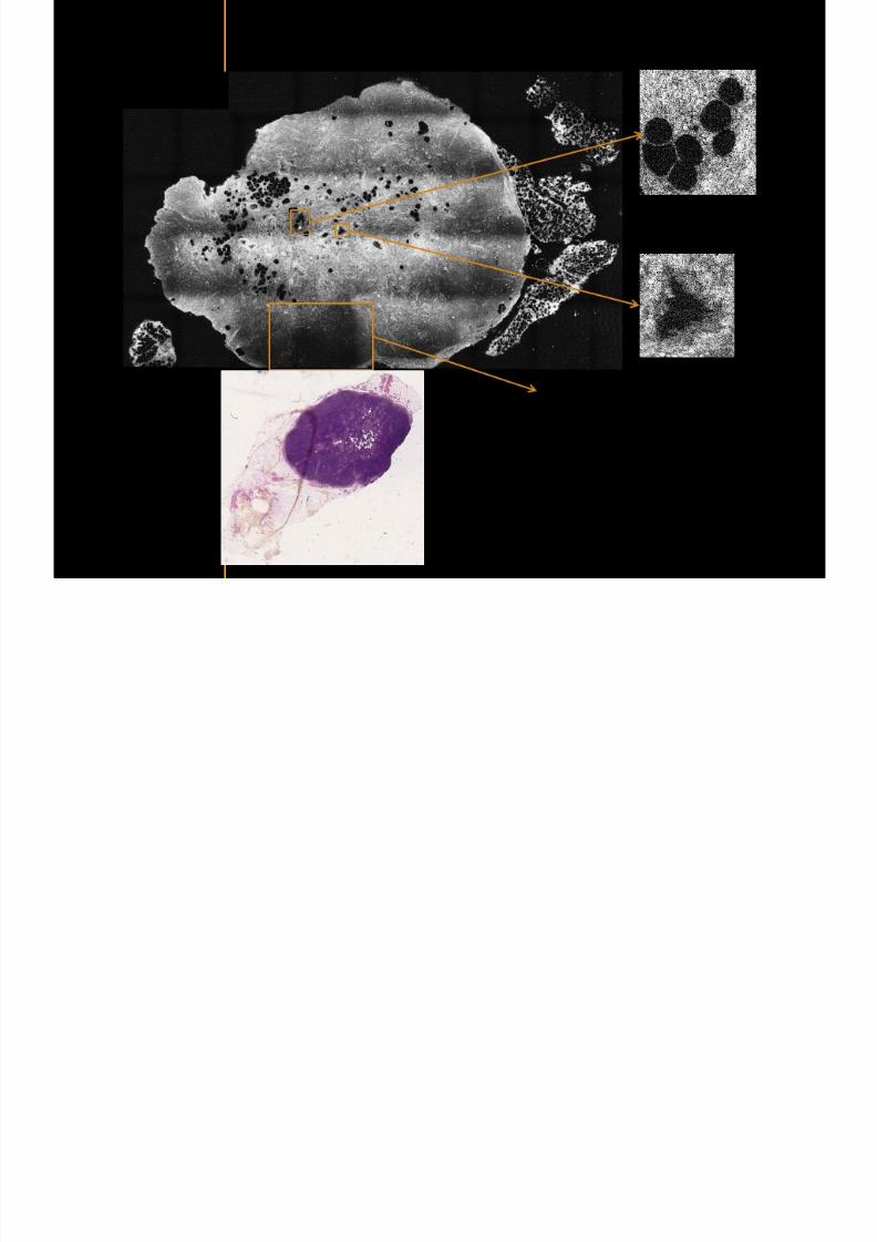
7292019 Atlas of Light-CT images
httpslidepdfcomreaderfullatlas-of-light-ct-images 19118copy LLTech 2012 ndash wwwlltechimagingcom - No copy without authorization 19
Lymphoid zone of the node
Vessel
Adipocytes in the center
of the node
Normal Sentinel Node
copy LLTech 2012 Courtesy of Hocircpital Tenon Paris France
7292019 Atlas of Light-CT images
httpslidepdfcomreaderfullatlas-of-light-ct-images 20118
7292019 Atlas of Light-CT images
httpslidepdfcomreaderfullatlas-of-light-ct-images 21118
copy LLTech 2012 ndash wwwlltechimagingcom - No copy without authorization 21
Normal lymphoid
zone with follicles
(Dark grey)
Metastatsis
(Light grey)
Hypervascularization
due to metastasis
Fibrous enveloppe of the
node
Invaded Sentinel Node
copy LLTech 2012 Courtesy of Hocircpital Tenon Paris France
7292019 Atlas of Light-CT images
httpslidepdfcomreaderfullatlas-of-light-ct-images 22118
copy LLTech 2012 ndash wwwlltechimagingcom - No copy without authorization 22
The Light-CT scanner allows fast tissue processing andpathology examination It completes the toolset available topathologists
When Chemical Fixation is needed
(Mostly Diagnostic)
When Frozen Section is needed
(Mostly Intra-operative)
Biopsy3D
Digital Image
2D
Digital Image
Slide
15-12 mins
Artefacts
Destructive
12-24 hours
Expensive
Destructive
5-8 minutes
Non-Destructive
Tissue Scanner
Light-CT could help to reduce the number of re-excisions in breast
7292019 Atlas of Light-CT images
httpslidepdfcomreaderfullatlas-of-light-ct-images 23118
copy LLTech 2012 ndash wwwlltechimagingcom - No copy without authorization 23
Light CT could help to reduce the number of re excisions in breastcancer surgery whilst preserving the current routines ofpathologists
Per surgery
LLTechrsquos
analysis
Histology Andor
Immunohistochemistry
Further
treatment
Post surgery
Surgical margin analysis
Metastasis analysis (gt 2mm) et
micro metastaisi
PN1mi gt 02 ndash 2 mm lt
SurgeryOKOK
Per surgery Post Surgery
C u r r e n t t e c h n i q u e s
U p t o 4 0 o
f d o u b l e p r o
c e d u r e s
L L T e c h L i g h t - C
T
S i g n i f i c a n t d e c r e a s e o f
d o u b l e p r o c e d u
r e s
Visual
analysis
cryostat
Further
treatmentSurgery
OK
Cancerouscancerous
OK
Poor identification of
metastasis
Up to 40 of double surgical
procedures
Several day analysisFew minutes analysis
Surgeon Pathologist Pathologist
Decrease of double surgical
procedures CancerousCancerous
No surgical
margins analysis
Histology Andor
Immuno
histochemistry
Node
Margin
Several day analysisFew minutes analysisSurgeon Pathologist Pathologist
7292019 Atlas of Light-CT images
httpslidepdfcomreaderfullatlas-of-light-ct-images 24118
copy LLTech 2012 ndash wwwlltechimagingcom - No copy without authorization 24
BRAIN IMAGING
Back to table of contents
7292019 Atlas of Light-CT images
httpslidepdfcomreaderfullatlas-of-light-ct-images 25118
copy LLTech 2012 ndash wwwlltechimagingcom - No copy without authorization 25
Rat brain
Architectural and cellular information seen at various scales
Myelinated fibers visible
1 mm
500 microm
100 microm
7292019 Atlas of Light-CT images
httpslidepdfcomreaderfullatlas-of-light-ct-images 26118
7292019 Atlas of Light-CT images
httpslidepdfcomreaderfullatlas-of-light-ct-images 27118
7292019 Atlas of Light-CT images
httpslidepdfcomreaderfullatlas-of-light-ct-images 28118
copy LLTech 2012 ndash wwwlltechimagingcom - No copy without authorization 28 copy LLTECH 2011
Collagen-rich matrixWide fascicles of
tumour cells
Large capillary
Fibroblastic meningioma grade I
500microm
100microm 200microm200microm
7292019 Atlas of Light-CT images
httpslidepdfcomreaderfullatlas-of-light-ct-images 29118
copy LLTech 2012 ndash wwwlltechimagingcom - No copy without authorization 29
Transitional meningioma grade I
Psammoma surrounding a calcification
Whorls formation
Collagen-rich matrix
500microm
100microm
50microm
50microm
7292019 Atlas of Light-CT images
httpslidepdfcomreaderfullatlas-of-light-ct-images 30118
7292019 Atlas of Light-CT images
httpslidepdfcomreaderfullatlas-of-light-ct-images 31118
copy LLTech 2012 ndash wwwlltechimagingcom - No copy without authorization 31 copy LLTECH 2011
Arrows Fibers transverse cut
Spinal cord normal tissue
500 microm
100 microm
100 microm
7292019 Atlas of Light-CT images
httpslidepdfcomreaderfullatlas-of-light-ct-images 32118
7292019 Atlas of Light-CT images
httpslidepdfcomreaderfullatlas-of-light-ct-images 33118
7292019 Atlas of Light-CT images
httpslidepdfcomreaderfullatlas-of-light-ct-images 34118
copy LLTech 2012 ndash wwwlltechimagingcom - No copy without authorization 34
Back to table of contents
SKIN IMAGING
N l ki h l
7292019 Atlas of Light-CT images
httpslidepdfcomreaderfullatlas-of-light-ct-images 35118
copy LLTech 2012 ndash wwwlltechimagingcom - No copy without authorization 35
Normal skin morphology
Epidermis
Collagen
Pilosebaceous
unit
Sweat gland
Adipocytes
b
a
c
e
d
500 microm
copy LLTech 2012 Courtesy of Hopitaux Universitaires de Genegraveve Genegraveve Switzerland
7292019 Atlas of Light-CT images
httpslidepdfcomreaderfullatlas-of-light-ct-images 36118
copy LLTech 2012 ndash wwwlltechimagingcom - No copy without authorization 36
Hypodermis
Skin morphology layers are well discriminated
5 mm x 8 mm mosaic of native field image
Ageing patient (solar elastosis)
En-face imaging of vertical excision
Epidermis
Dermis
1
3
2
4
7292019 Atlas of Light-CT images
httpslidepdfcomreaderfullatlas-of-light-ct-images 37118
En face skin slicing shows structural and cellular details
7292019 Atlas of Light-CT images
httpslidepdfcomreaderfullatlas-of-light-ct-images 38118
copy LLTech 2012 ndash wwwlltechimagingcom - No copy without authorization 38
Stratum corneum Stratum spinosum
Stratum basale Reticular region
Epidermis
Dermis
En-face skin slicing shows structural and cellular details
Melanin
papillary caps
Keratinocyte
nuclei
Blood vessels
Collagen
Corneocytes
100 microm 100 microm
100 microm100 microm
copy LLTech 2012 Courtesy of Hopitaux Universitaires de Genegraveve Genegraveve Switzerland
7292019 Atlas of Light-CT images
httpslidepdfcomreaderfullatlas-of-light-ct-images 39118
7292019 Atlas of Light-CT images
httpslidepdfcomreaderfullatlas-of-light-ct-images 40118
Vertical reconstruction of skin model
7292019 Atlas of Light-CT images
httpslidepdfcomreaderfullatlas-of-light-ct-images 41118
copy LLTech 2012 ndash wwwlltechimagingcom - No copy without authorization 41
Vertical reconstruction of skin model
50 microm
Stratum corneum
Stratum spinosum
Dermis
Keratinocyte nuclei
copy LLTech 2012
7292019 Atlas of Light-CT images
httpslidepdfcomreaderfullatlas-of-light-ct-images 42118
Normal
7292019 Atlas of Light-CT images
httpslidepdfcomreaderfullatlas-of-light-ct-images 43118
copy LLTech 2012 ndash wwwlltechimagingcom - No copy without authorization 43
skin
b
a
e
c
f
d
500 microm
7292019 Atlas of Light-CT images
httpslidepdfcomreaderfullatlas-of-light-ct-images 44118
7292019 Atlas of Light-CT images
httpslidepdfcomreaderfullatlas-of-light-ct-images 45118
copy LLTech 2012 ndash wwwlltechimagingcom - No copy without authorization 45
Adipocytes
9um depth 27um depth 60um depth
Reconstructed
depth slice
200 microm
50 microm
7292019 Atlas of Light-CT images
httpslidepdfcomreaderfullatlas-of-light-ct-images 46118
Skin pathologies basal cell carcinoma discrimination at
7292019 Atlas of Light-CT images
httpslidepdfcomreaderfullatlas-of-light-ct-images 47118
copy LLTech 2012 ndash wwwlltechimagingcom - No copy without authorization 47
Epidermis
Dermis
Hypodermis
A
AA
C
BCC
PSU
PSU
BCC Basal cell carcinoma
A Adipocytes
C Collagen
PSU Pilosebaceous unit
structural level
1 mm
copy LLTech 2012 Courtesy of Hopitaux Universitaires de Genegraveve Genegraveve Switzerland
C
Zoom on basal cell carcinoma discrimination at cellular level
7292019 Atlas of Light-CT images
httpslidepdfcomreaderfullatlas-of-light-ct-images 48118
copy LLTech 2012 ndash wwwlltechimagingcom - No copy without authorization 48
Zoom on basal cell carcinoma discrimination at cellular level
200 microm
100 microm
Peritumoral stroma
High cell density
copy LLTech 2012 Courtesy of Hopitaux Universitaires de Genegraveve Genegraveve Switzerland
Skin pathologies discrimination of actinic keratosis
7292019 Atlas of Light-CT images
httpslidepdfcomreaderfullatlas-of-light-ct-images 49118
copy LLTech 2012 ndash wwwlltechimagingcom - No copy without authorization 49
Skin pathologies discrimination of actinic keratosis
Enlarged epidermis
Extended epidermal ridges
1 mm
copy LLTech 2012 Courtesy of Hopitaux Universitaires de Genegraveve Genegraveve Switzerland
7292019 Atlas of Light-CT images
httpslidepdfcomreaderfullatlas-of-light-ct-images 50118
copy LLTech 2012 ndash wwwlltechimagingcom - No copy without authorization 50
Basal cell carcinoma in depth imaging
Z = 10 microns 7 x 42 mm FOV Z = 35 microns 7 x 42 mm FOV
BCC
SG Sebaceous glands
Z = 55 microns 7 x 42 mm FOV Z = 75 microns 7 x 42 mm FOV
SG
Application to Mohs surgerycopy LLTech 2012 Courtesy of Hopitaux Universitaires de Genegraveve Genegraveve Switzerland
7292019 Atlas of Light-CT images
httpslidepdfcomreaderfullatlas-of-light-ct-images 51118
copy LLTech 2012 ndash wwwlltechimagingcom - No copy without authorization 51 copy LLTECH 2011
Healthy vs Cheloid scar tissue
Healthy skin tissueFOV 75x5mm
Cheloid scar tissueFOV 65x65mm
copy LLTECH 2011
7292019 Atlas of Light-CT images
httpslidepdfcomreaderfullatlas-of-light-ct-images 52118
copy LLTech 2012 ndash wwwlltechimagingcom - No copy without authorization 52
Dermo-epidermaljunction
Cheloid scar tissue in detail
copy LLTECH 2011
Nodular structureThickcollagenfiberbundles
1mm
600 microm
500 microm
h l d d f
7292019 Atlas of Light-CT images
httpslidepdfcomreaderfullatlas-of-light-ct-images 53118
copy LLTech 2012 ndash wwwlltechimagingcom - No copy without authorization 53
Cheloid scar tissue viewed en face
20microm depth 120microm depth
7292019 Atlas of Light-CT images
httpslidepdfcomreaderfullatlas-of-light-ct-images 54118
Skin in-vivo sweat duct imaging in 3D
7292019 Atlas of Light-CT images
httpslidepdfcomreaderfullatlas-of-light-ct-images 55118
copy LLTech 2012 ndash wwwlltechimagingcom - No copy without authorization 55
g g
Vertical reconstruction from en-face images
Sweat duct 3D reconstruction
Stratum corneum
Stratum spinosum
SD sweat ducts
SD SD
100 microm
7292019 Atlas of Light-CT images
httpslidepdfcomreaderfullatlas-of-light-ct-images 56118
7292019 Atlas of Light-CT images
httpslidepdfcomreaderfullatlas-of-light-ct-images 57118
7292019 Atlas of Light-CT images
httpslidepdfcomreaderfullatlas-of-light-ct-images 58118
copy LLTech 2012 ndash wwwlltechimagingcom - No copy without authorization 58
LUNG IMAGING
Back to table of contents
H L
7292019 Atlas of Light-CT images
httpslidepdfcomreaderfullatlas-of-light-ct-images 59118
copy LLTech 2012 ndash wwwlltechimagingcom - No copy without authorization 59
Zone 1
Zone 2
Zone 3
Zone 4
Zone with
cancerous tissue
Limit of cancerous
stroma
alveoli with
macrophages
Alveoles
Human Lung
500microm
copy LLTech 2012 Courtesy of Hocircpital Hotel-Dieu Paris France
H L Z 1 Z t
7292019 Atlas of Light-CT images
httpslidepdfcomreaderfullatlas-of-light-ct-images 60118
copy LLTech 2012 ndash wwwlltechimagingcom - No copy without authorization 60
Stroma
Human Lung ndash Zone 1 ndash Zoom on cancerous stroma
Cancerous
area
200microm
copy LLTech 2012 Courtesy of Hocircpital Hotel-Dieu Paris France
Human Lung ndash Zone 2 ndash Limit cancerous stroma alveoli
7292019 Atlas of Light-CT images
httpslidepdfcomreaderfullatlas-of-light-ct-images 61118
copy LLTech 2012 ndash wwwlltechimagingcom - No copy without authorization 61
g
with visible macrophages and vessel
Alveoli
Macrophages
Vessel
copy LLTech 2012 Courtesy of Hocircpital Hotel-Dieu Paris France
Human Lung ndash Zone 3 ndash Limit cancerous stroma alveoli
7292019 Atlas of Light-CT images
httpslidepdfcomreaderfullatlas-of-light-ct-images 62118
copy LLTech 2012 ndash wwwlltechimagingcom - No copy without authorization 62
g
with visible macrophages
Macrophages
100microm
copy LLTech 2012 Courtesy of Hocircpital Hotel-Dieu Paris France
Human Lung ndash Zone 4 ndash Alveoli
7292019 Atlas of Light-CT images
httpslidepdfcomreaderfullatlas-of-light-ct-images 63118
copy LLTech 2012 ndash wwwlltechimagingcom - No copy without authorization 63
Human Lung Zone 4 Alveoli
Alveoli
200microm
copy LLTech 2012 Courtesy of Hocircpital Hotel-Dieu Paris France
7292019 Atlas of Light-CT images
httpslidepdfcomreaderfullatlas-of-light-ct-images 64118
copy LLTech 2012 ndash wwwlltechimagingcom - No copy without authorization 64
GI IMAGING
Back to table of contents
7292019 Atlas of Light-CT images
httpslidepdfcomreaderfullatlas-of-light-ct-images 65118
Colonic carcinoma
7292019 Atlas of Light-CT images
httpslidepdfcomreaderfullatlas-of-light-ct-images 66118
copy LLTech 2012 ndash wwwlltechimagingcom - No copy without authorization 66
Colonic Carcinoma
Colonic carcinoma
copy LLTech 2012 Courtesy of MD Anderson Houston USA
Intestines ndash Crohnrsquos disease
7292019 Atlas of Light-CT images
httpslidepdfcomreaderfullatlas-of-light-ct-images 67118
copy LLTech 2012 ndash wwwlltechimagingcom - No copy without authorization 67
Crohnrsquos disease structure not
visible from the surface
Fat cells
200 microm
Intestines Crohn s disease
copy LLTech 2012 Courtesy of Institut Curie Paris France
Rat stomach
7292019 Atlas of Light-CT images
httpslidepdfcomreaderfullatlas-of-light-ct-images 68118
copy LLTech 2012 ndash wwwlltechimagingcom - No copy without authorization 68
Rat stomach
Mucosa withgastric pits
Submucosa with
bright collagen
fibers
Muscularis
propria with
muscle bundles
copy LLTech 2012 Courtesy of Weill Cornell Medical College NY USA
200 microm
7292019 Atlas of Light-CT images
httpslidepdfcomreaderfullatlas-of-light-ct-images 69118
copy LLTech 2012 ndash wwwlltechimagingcom - No copy without authorization 69 copy LLTECH 2011
UROGENITAL IMAGING
Back to table of contents
Rat bladder
7292019 Atlas of Light-CT images
httpslidepdfcomreaderfullatlas-of-light-ct-images 70118
copy LLTech 2012 ndash wwwlltechimagingcom - No copy without authorization 70
Rat bladder
Stratifiedurothelium
Lamina propria
500 microm
Muscular
bundles of the
lamina propria
Serosa
copy LLTech 2012 Courtesy of Weill Cornell Medical College NY USA
100 microm
100 microm
Human bladder low grade papillary urothelial carcinoma
7292019 Atlas of Light-CT images
httpslidepdfcomreaderfullatlas-of-light-ct-images 71118
copy LLTech 2012 ndash wwwlltechimagingcom - No copy without authorization 71
Lesion with fibrovascular cores (arrow) and superficial umbrella cells (arrowheads)
Human bladder low grade papillary urothelial carcinoma
copy LLTech 2012 Courtesy of Weill Cornell Medical College NY USA
100 microm
7292019 Atlas of Light-CT images
httpslidepdfcomreaderfullatlas-of-light-ct-images 72118
Rat prostate
7292019 Atlas of Light-CT images
httpslidepdfcomreaderfullatlas-of-light-ct-images 73118
copy LLTech 2012 ndash wwwlltechimagingcom - No copy without authorization 73
p
copy LLTech 2012 Courtesy of Weill Cornell Medical College NY USA
Papillary folds of the
acinar glands
Prostatic acini
Fibromuscular stromaPeriprostatic fat Blood vessel
1 mm
200 microm
Mouse kidney
7292019 Atlas of Light-CT images
httpslidepdfcomreaderfullatlas-of-light-ct-images 74118
copy LLTech 2012 ndash wwwlltechimagingcom - No copy without authorization 74
1 mm
y
copy LLTech 2012 Courtesy of Institut Biomeacutedical de Bicecirctre Paris France
Renal cortex
Renal medulla
Renal sinus
7292019 Atlas of Light-CT images
httpslidepdfcomreaderfullatlas-of-light-ct-images 75118
copy LLTech 2012 ndash wwwlltechimagingcom - No copy without authorization 75
Mouse kidney ndash zoom on highlighted section of previous slide
800microm
copy LLTech 2012 Courtesy of Hocircpital Kremlin Bicecirctre Paris France
7292019 Atlas of Light-CT images
httpslidepdfcomreaderfullatlas-of-light-ct-images 76118
7292019 Atlas of Light-CT images
httpslidepdfcomreaderfullatlas-of-light-ct-images 77118
7292019 Atlas of Light-CT images
httpslidepdfcomreaderfullatlas-of-light-ct-images 78118
copy LLTech 2012 ndash wwwlltechimagingcom - No copy without authorization 78 copy LLTECH 2011
CORE NEEDLE BIOPSIES
Back to table of contents
Kidney
7292019 Atlas of Light-CT images
httpslidepdfcomreaderfullatlas-of-light-ct-images 79118
copy LLTech 2012 ndash wwwlltechimagingcom - No copy without authorization 79
100microm
50microm
Renal tubules
Vessel
500microm
copy LLTech 2012 Courtesy of Institut Curie Paris France
Breast Invasive lobular carcinoma
7292019 Atlas of Light-CT images
httpslidepdfcomreaderfullatlas-of-light-ct-images 80118
copy LLTech 2012 ndash wwwlltechimagingcom - No copy without authorization 80
500microm
stroma
adipocytes
copy LLTech 2012 Courtesy of Institut Curie Paris France
7292019 Atlas of Light-CT images
httpslidepdfcomreaderfullatlas-of-light-ct-images 81118
Breast Ductal carcinoma in situ
7292019 Atlas of Light-CT images
httpslidepdfcomreaderfullatlas-of-light-ct-images 82118
copy LLTech 2012 ndash wwwlltechimagingcom - No copy without authorization 82
500microm
copy LLTech 2012 Courtesy of Institut Curie Paris France
7292019 Atlas of Light-CT images
httpslidepdfcomreaderfullatlas-of-light-ct-images 83118
copy LLTech 2012 ndash wwwlltechimagingcom - No copy without authorization 83 copy LLTECH 2011
THYROID IMAGING
Back to table of contents
7292019 Atlas of Light-CT images
httpslidepdfcomreaderfullatlas-of-light-ct-images 84118
copy LLTech 2012 ndash wwwlltechimagingcom - No copy without authorization 84
Thyroid
800 microm
200 microm
copy LLTech 2012 Courtesy of Hocircpital Tenon Paris France
7292019 Atlas of Light-CT images
httpslidepdfcomreaderfullatlas-of-light-ct-images 85118
copy LLTech 2012 ndash wwwlltechimagingcom - No copy without authorization 85
Thyroid
800 microm
200 microm
copy LLTech 2012 Courtesy of Hocircpital Tenon Paris France
7292019 Atlas of Light-CT images
httpslidepdfcomreaderfullatlas-of-light-ct-images 86118
copy LLTech 2012 ndash wwwlltechimagingcom - No copy without authorization 86 copy LLTECH 2011
FAT CELLS IMAGING
Back to table of contents
7292019 Atlas of Light-CT images
httpslidepdfcomreaderfullatlas-of-light-ct-images 87118
copy LLTech 2012 ndash wwwlltechimagingcom - No copy without authorization 87
Brown fat cells - adipocytes
100 microm 100 microm
200 microm
10um depth
Adipocytes
45um depth
En face
slices
Reconstructed
depth slice
copy LLTech 2012 Courtesy of Centre de Recherche des Cordeliers Paris France
7292019 Atlas of Light-CT images
httpslidepdfcomreaderfullatlas-of-light-ct-images 88118
copy LLTech 2012 ndash wwwlltechimagingcom - No copy without authorization 88 copy LLTECH 2011
CORNEA AND RETINA
Back to table of contents
Human cornea reconstructed depth slice
7292019 Atlas of Light-CT images
httpslidepdfcomreaderfullatlas-of-light-ct-images 89118
copy LLTech 2012 ndash wwwlltechimagingcom - No copy without authorization 89
50 um
wwwlltechimagingcom
7292019 Atlas of Light-CT images
httpslidepdfcomreaderfullatlas-of-light-ct-images 90118
Large field en face view in stroma
7292019 Atlas of Light-CT images
httpslidepdfcomreaderfullatlas-of-light-ct-images 91118
copy LLTech 2012 ndash wwwlltechimagingcom - No copy without authorization 91
200 um
wwwlltechimagingcom
7292019 Atlas of Light-CT images
httpslidepdfcomreaderfullatlas-of-light-ct-images 92118
Rabbit cornea after LASIK reflectivity profiles reveal
i d tt i d thi k d ith li l l
7292019 Atlas of Light-CT images
httpslidepdfcomreaderfullatlas-of-light-ct-images 93118
copy LLTech 2012 ndash wwwlltechimagingcom - No copy without authorization 93
increased scattering and thickened epithelial layer
LASIK LASIK CONTROL CONTROL
copy LLTech 2012
Newborn mouse anterior segment
7292019 Atlas of Light-CT images
httpslidepdfcomreaderfullatlas-of-light-ct-images 94118
copy LLTech 2012 ndash wwwlltechimagingcom - No copy without authorization 94
Corneal epithelial cells
Basal membrane
Stroma
Descemetrsquos membrane
Aqueous humor
Lens capsule
Lens epithelial cells
Cortical fibers
copy LLTech 2012 Courtesy of ESPCI Paris Francecopy LLTech 2012
Rat retina comparison with histology
7292019 Atlas of Light-CT images
httpslidepdfcomreaderfullatlas-of-light-ct-images 95118
copy LLTech 2012 ndash wwwlltechimagingcom - No copy without authorization 95
Figure 1 Right Ex vivo rat retinaimaged using the f ull field OCT
system of ESPCI Left Histology
for comparison
Figure 1 Right Ex vivo rat retinaimaged using the f ull field OCT
system of ESPCI Left Histology
for comparison
Figure 1 Right Ex vivo rat retinaimaged using the f ull field OCT
system of ESPCI Left Histology
for comparisonField
230x300microm
RPE
PRL
ONL
OPL
INL
IPL
GCL
NFL
Invest Ophthalmol Vis Sci 2004 Nov45(11)4126-31
Grieve K Paques M Dubois A Sahel J Boccara C Le Gargasson JF copy LLTech 2012
Rat retina
7292019 Atlas of Light-CT images
httpslidepdfcomreaderfullatlas-of-light-ct-images 96118
copy LLTech 2012 ndash wwwlltechimagingcom - No copy without authorization 96 copy LLTech 2012 Courtesy of ESPCI Paris France
Nerve fiber layer
Retina
Invest Ophthalmol Vis Sci 2004 Nov45(11)4126-31
Grieve K Paques M Dubois A Sahel J Boccara C Le Gargasson JF
Transscleral image
7292019 Atlas of Light-CT images
httpslidepdfcomreaderfullatlas-of-light-ct-images 97118
7292019 Atlas of Light-CT images
httpslidepdfcomreaderfullatlas-of-light-ct-images 98118
7292019 Atlas of Light-CT images
httpslidepdfcomreaderfullatlas-of-light-ct-images 99118
In vivo imaging of drosophila melanogaster
7292019 Atlas of Light-CT images
httpslidepdfcomreaderfullatlas-of-light-ct-images 100118
copy LLTech 2012 ndash wwwlltechimagingcom - No copy without authorization 100 copy LLTech 2012 Courtesy of ESPCI Paris France
In vivo imaging of drosophila melanogaster ndash four stages of pupation over 72 hours
7292019 Atlas of Light-CT images
httpslidepdfcomreaderfullatlas-of-light-ct-images 101118
copy LLTech 2012 ndash wwwlltechimagingcom - No copy without authorization 101
Prepupa (0-2 h) Transition topupal stage (24 h)
Pupal stage (48 h) Advanced pupal phase
before eclosion (72 h)
copy LLTech 2012 Courtesy of ESPCI Paris France
Pupa 118 4 days 30um depth
100
7292019 Atlas of Light-CT images
httpslidepdfcomreaderfullatlas-of-light-ct-images 102118
copy LLTech 2012 ndash wwwlltechimagingcom - No copy without authorization 102
100 um
Drosophila
7292019 Atlas of Light-CT images
httpslidepdfcomreaderfullatlas-of-light-ct-images 103118
copy LLTech 2012 ndash wwwlltechimagingcom - No copy without authorization 103
200 microm
copy LLTech 2012 Courtesy of ESPCI Paris France
C Elegans ndash Fast identification of anatomy
7292019 Atlas of Light-CT images
httpslidepdfcomreaderfullatlas-of-light-ct-images 104118
copy LLTech 2012 ndash wwwlltechimagingcom - No copy without authorization 104
Body wall - cuticle
gonad arm
30 microm depth
uterus
ovocytes
intestine
100 microm 100 microm
15 microm depth
copy LLTech 2012 Courtesy of ENS Paris France
C elegans ndash Images at higher magnification 40X
7292019 Atlas of Light-CT images
httpslidepdfcomreaderfullatlas-of-light-ct-images 105118
copy LLTech 2012 ndash wwwlltechimagingcom - No copy without authorization 105
3D reconstruction 50 microm
100 microm
copy LLTech 2012 Courtesy of ENS Paris France
Zebra fish eye
7292019 Atlas of Light-CT images
httpslidepdfcomreaderfullatlas-of-light-ct-images 106118
copy LLTech 2012 ndash wwwlltechimagingcom - No copy without authorization 106
Zebra fish eye
100 microm
100 microm
En face slices
Reconstructed depth slice
copy LLTech 2012 Courtesy of Inserm Paris France
10um depth 20um depth 45um depth
7292019 Atlas of Light-CT images
httpslidepdfcomreaderfullatlas-of-light-ct-images 107118
Xenopus Laevis backtail
7292019 Atlas of Light-CT images
httpslidepdfcomreaderfullatlas-of-light-ct-images 108118
copy LLTech 2012 ndash wwwlltechimagingcom - No copy without authorization 108
100 um30um depth 60um depth
7292019 Atlas of Light-CT images
httpslidepdfcomreaderfullatlas-of-light-ct-images 109118
Mouse embryo in vitro 6 5 days old ndash
7292019 Atlas of Light-CT images
httpslidepdfcomreaderfullatlas-of-light-ct-images 110118
copy LLTech 2012 ndash wwwlltechimagingcom - No copy without authorization 110
Mouse embryo in vitro 65 days old
copy LLTech 2012 Courtesy of Ellvire Guiot and nstitut Jacques Monod Paris France
3D imaging of internal cavity
Sabellaria alveolata mouth palps
7292019 Atlas of Light-CT images
httpslidepdfcomreaderfullatlas-of-light-ct-images 111118
copy LLTech 2012 ndash wwwlltechimagingcom - No copy without authorization 111
500 microm
100 microm
Mouth palps
copy LLTech 2012 Courtesy of Museacutee National drsquoHistoire Naturelle Paris France
7292019 Atlas of Light-CT images
httpslidepdfcomreaderfullatlas-of-light-ct-images 112118
copy LLTech 2012 ndash wwwlltechimagingcom - No copy without authorization 112 copy LLTECH 2011
PLANT IMAGING
Back to table of contents
Mouse ear leaf epidermis
7292019 Atlas of Light-CT images
httpslidepdfcomreaderfullatlas-of-light-ct-images 113118
copy LLTech 2012 ndash wwwlltechimagingcom - No copy without authorization 113
100 microm
Epidermal cell
copy LLTech 2012
Stoma
Leaf veins
7292019 Atlas of Light-CT images
httpslidepdfcomreaderfullatlas-of-light-ct-images 114118
copy LLTech 2012 ndash wwwlltechimagingcom - No copy without authorization 114
114copy LLTech 2012
Fibers
Veins
100 microm
500 microm
Apple
7292019 Atlas of Light-CT images
httpslidepdfcomreaderfullatlas-of-light-ct-images 115118
copy LLTech 2012 ndash wwwlltechimagingcom - No copy without authorization 115
115copy LLTech 2012
100 microm
Wax
Skin
Flesh
500 microm
Water
Effect of harpin infiltration on tobacco leaf
7292019 Atlas of Light-CT images
httpslidepdfcomreaderfullatlas-of-light-ct-images 116118
copy LLTech 2012 ndash wwwlltechimagingcom - No copy without authorization 116
116copy LLTech 2012 Courtesy of UPMC Paris France
50 microm
50 microm
50 microm
50 microm
Upper epidermis
10 min after harpin infiltration 30 min after harpin infiltration
Palisade parenchyma
Epidermis cell
Chloroplast
7292019 Atlas of Light-CT images
httpslidepdfcomreaderfullatlas-of-light-ct-images 117118
copy LLTech 2012 ndash wwwlltechimagingcom - No copy without authorization 117
bull Light-CTtrade does not require tissue preparation nor staining of any kind
bull Creates images within minutes using a non-destructive process
bull Offers a 1 microm cellular resolution in 3D
bull Reveals structural and cellular information
bull Mosaicing allows fast visualisation at various scale
bull En-face high-resolution imaging allows vertical and 3D reconstruction
bull In-vivo images show good promise for in-vivo system
bull Clinical and research applications in cancer detection cosmetics biobanking small animal
developmental biology botanyhellip
Contact Details
7292019 Atlas of Light-CT images
httpslidepdfcomreaderfullatlas-of-light-ct-images 118118
USA
LLTech Inc
103 Carnegie Center DriveSuite 300
Princeton NJ 08540
USA
wwwlltechimagingcom
contactlltechfr
Phone + 1 609 955 3506
LLTech SAS
29 rue du Faubourg St-JacquesPeacutepiniegravere Paris Santeacute Cochin
75014
Paris - France
wwwlltechimagingcom
contactlltechfr
Phone +33 1 82 72 61 25
Europe
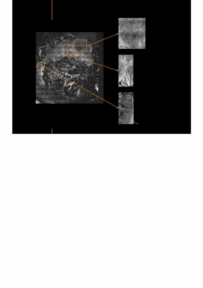
7292019 Atlas of Light-CT images
httpslidepdfcomreaderfullatlas-of-light-ct-images 20118
7292019 Atlas of Light-CT images
httpslidepdfcomreaderfullatlas-of-light-ct-images 21118
copy LLTech 2012 ndash wwwlltechimagingcom - No copy without authorization 21
Normal lymphoid
zone with follicles
(Dark grey)
Metastatsis
(Light grey)
Hypervascularization
due to metastasis
Fibrous enveloppe of the
node
Invaded Sentinel Node
copy LLTech 2012 Courtesy of Hocircpital Tenon Paris France
7292019 Atlas of Light-CT images
httpslidepdfcomreaderfullatlas-of-light-ct-images 22118
copy LLTech 2012 ndash wwwlltechimagingcom - No copy without authorization 22
The Light-CT scanner allows fast tissue processing andpathology examination It completes the toolset available topathologists
When Chemical Fixation is needed
(Mostly Diagnostic)
When Frozen Section is needed
(Mostly Intra-operative)
Biopsy3D
Digital Image
2D
Digital Image
Slide
15-12 mins
Artefacts
Destructive
12-24 hours
Expensive
Destructive
5-8 minutes
Non-Destructive
Tissue Scanner
Light-CT could help to reduce the number of re-excisions in breast
7292019 Atlas of Light-CT images
httpslidepdfcomreaderfullatlas-of-light-ct-images 23118
copy LLTech 2012 ndash wwwlltechimagingcom - No copy without authorization 23
Light CT could help to reduce the number of re excisions in breastcancer surgery whilst preserving the current routines ofpathologists
Per surgery
LLTechrsquos
analysis
Histology Andor
Immunohistochemistry
Further
treatment
Post surgery
Surgical margin analysis
Metastasis analysis (gt 2mm) et
micro metastaisi
PN1mi gt 02 ndash 2 mm lt
SurgeryOKOK
Per surgery Post Surgery
C u r r e n t t e c h n i q u e s
U p t o 4 0 o
f d o u b l e p r o
c e d u r e s
L L T e c h L i g h t - C
T
S i g n i f i c a n t d e c r e a s e o f
d o u b l e p r o c e d u
r e s
Visual
analysis
cryostat
Further
treatmentSurgery
OK
Cancerouscancerous
OK
Poor identification of
metastasis
Up to 40 of double surgical
procedures
Several day analysisFew minutes analysis
Surgeon Pathologist Pathologist
Decrease of double surgical
procedures CancerousCancerous
No surgical
margins analysis
Histology Andor
Immuno
histochemistry
Node
Margin
Several day analysisFew minutes analysisSurgeon Pathologist Pathologist
7292019 Atlas of Light-CT images
httpslidepdfcomreaderfullatlas-of-light-ct-images 24118
copy LLTech 2012 ndash wwwlltechimagingcom - No copy without authorization 24
BRAIN IMAGING
Back to table of contents
7292019 Atlas of Light-CT images
httpslidepdfcomreaderfullatlas-of-light-ct-images 25118
copy LLTech 2012 ndash wwwlltechimagingcom - No copy without authorization 25
Rat brain
Architectural and cellular information seen at various scales
Myelinated fibers visible
1 mm
500 microm
100 microm
7292019 Atlas of Light-CT images
httpslidepdfcomreaderfullatlas-of-light-ct-images 26118
7292019 Atlas of Light-CT images
httpslidepdfcomreaderfullatlas-of-light-ct-images 27118
7292019 Atlas of Light-CT images
httpslidepdfcomreaderfullatlas-of-light-ct-images 28118
copy LLTech 2012 ndash wwwlltechimagingcom - No copy without authorization 28 copy LLTECH 2011
Collagen-rich matrixWide fascicles of
tumour cells
Large capillary
Fibroblastic meningioma grade I
500microm
100microm 200microm200microm
7292019 Atlas of Light-CT images
httpslidepdfcomreaderfullatlas-of-light-ct-images 29118
copy LLTech 2012 ndash wwwlltechimagingcom - No copy without authorization 29
Transitional meningioma grade I
Psammoma surrounding a calcification
Whorls formation
Collagen-rich matrix
500microm
100microm
50microm
50microm
7292019 Atlas of Light-CT images
httpslidepdfcomreaderfullatlas-of-light-ct-images 30118
7292019 Atlas of Light-CT images
httpslidepdfcomreaderfullatlas-of-light-ct-images 31118
copy LLTech 2012 ndash wwwlltechimagingcom - No copy without authorization 31 copy LLTECH 2011
Arrows Fibers transverse cut
Spinal cord normal tissue
500 microm
100 microm
100 microm
7292019 Atlas of Light-CT images
httpslidepdfcomreaderfullatlas-of-light-ct-images 32118
7292019 Atlas of Light-CT images
httpslidepdfcomreaderfullatlas-of-light-ct-images 33118
7292019 Atlas of Light-CT images
httpslidepdfcomreaderfullatlas-of-light-ct-images 34118
copy LLTech 2012 ndash wwwlltechimagingcom - No copy without authorization 34
Back to table of contents
SKIN IMAGING
N l ki h l
7292019 Atlas of Light-CT images
httpslidepdfcomreaderfullatlas-of-light-ct-images 35118
copy LLTech 2012 ndash wwwlltechimagingcom - No copy without authorization 35
Normal skin morphology
Epidermis
Collagen
Pilosebaceous
unit
Sweat gland
Adipocytes
b
a
c
e
d
500 microm
copy LLTech 2012 Courtesy of Hopitaux Universitaires de Genegraveve Genegraveve Switzerland
7292019 Atlas of Light-CT images
httpslidepdfcomreaderfullatlas-of-light-ct-images 36118
copy LLTech 2012 ndash wwwlltechimagingcom - No copy without authorization 36
Hypodermis
Skin morphology layers are well discriminated
5 mm x 8 mm mosaic of native field image
Ageing patient (solar elastosis)
En-face imaging of vertical excision
Epidermis
Dermis
1
3
2
4
7292019 Atlas of Light-CT images
httpslidepdfcomreaderfullatlas-of-light-ct-images 37118
En face skin slicing shows structural and cellular details
7292019 Atlas of Light-CT images
httpslidepdfcomreaderfullatlas-of-light-ct-images 38118
copy LLTech 2012 ndash wwwlltechimagingcom - No copy without authorization 38
Stratum corneum Stratum spinosum
Stratum basale Reticular region
Epidermis
Dermis
En-face skin slicing shows structural and cellular details
Melanin
papillary caps
Keratinocyte
nuclei
Blood vessels
Collagen
Corneocytes
100 microm 100 microm
100 microm100 microm
copy LLTech 2012 Courtesy of Hopitaux Universitaires de Genegraveve Genegraveve Switzerland
7292019 Atlas of Light-CT images
httpslidepdfcomreaderfullatlas-of-light-ct-images 39118
7292019 Atlas of Light-CT images
httpslidepdfcomreaderfullatlas-of-light-ct-images 40118
Vertical reconstruction of skin model
7292019 Atlas of Light-CT images
httpslidepdfcomreaderfullatlas-of-light-ct-images 41118
copy LLTech 2012 ndash wwwlltechimagingcom - No copy without authorization 41
Vertical reconstruction of skin model
50 microm
Stratum corneum
Stratum spinosum
Dermis
Keratinocyte nuclei
copy LLTech 2012
7292019 Atlas of Light-CT images
httpslidepdfcomreaderfullatlas-of-light-ct-images 42118
Normal
7292019 Atlas of Light-CT images
httpslidepdfcomreaderfullatlas-of-light-ct-images 43118
copy LLTech 2012 ndash wwwlltechimagingcom - No copy without authorization 43
skin
b
a
e
c
f
d
500 microm
7292019 Atlas of Light-CT images
httpslidepdfcomreaderfullatlas-of-light-ct-images 44118
7292019 Atlas of Light-CT images
httpslidepdfcomreaderfullatlas-of-light-ct-images 45118
copy LLTech 2012 ndash wwwlltechimagingcom - No copy without authorization 45
Adipocytes
9um depth 27um depth 60um depth
Reconstructed
depth slice
200 microm
50 microm
7292019 Atlas of Light-CT images
httpslidepdfcomreaderfullatlas-of-light-ct-images 46118
Skin pathologies basal cell carcinoma discrimination at
7292019 Atlas of Light-CT images
httpslidepdfcomreaderfullatlas-of-light-ct-images 47118
copy LLTech 2012 ndash wwwlltechimagingcom - No copy without authorization 47
Epidermis
Dermis
Hypodermis
A
AA
C
BCC
PSU
PSU
BCC Basal cell carcinoma
A Adipocytes
C Collagen
PSU Pilosebaceous unit
structural level
1 mm
copy LLTech 2012 Courtesy of Hopitaux Universitaires de Genegraveve Genegraveve Switzerland
C
Zoom on basal cell carcinoma discrimination at cellular level
7292019 Atlas of Light-CT images
httpslidepdfcomreaderfullatlas-of-light-ct-images 48118
copy LLTech 2012 ndash wwwlltechimagingcom - No copy without authorization 48
Zoom on basal cell carcinoma discrimination at cellular level
200 microm
100 microm
Peritumoral stroma
High cell density
copy LLTech 2012 Courtesy of Hopitaux Universitaires de Genegraveve Genegraveve Switzerland
Skin pathologies discrimination of actinic keratosis
7292019 Atlas of Light-CT images
httpslidepdfcomreaderfullatlas-of-light-ct-images 49118
copy LLTech 2012 ndash wwwlltechimagingcom - No copy without authorization 49
Skin pathologies discrimination of actinic keratosis
Enlarged epidermis
Extended epidermal ridges
1 mm
copy LLTech 2012 Courtesy of Hopitaux Universitaires de Genegraveve Genegraveve Switzerland
7292019 Atlas of Light-CT images
httpslidepdfcomreaderfullatlas-of-light-ct-images 50118
copy LLTech 2012 ndash wwwlltechimagingcom - No copy without authorization 50
Basal cell carcinoma in depth imaging
Z = 10 microns 7 x 42 mm FOV Z = 35 microns 7 x 42 mm FOV
BCC
SG Sebaceous glands
Z = 55 microns 7 x 42 mm FOV Z = 75 microns 7 x 42 mm FOV
SG
Application to Mohs surgerycopy LLTech 2012 Courtesy of Hopitaux Universitaires de Genegraveve Genegraveve Switzerland
7292019 Atlas of Light-CT images
httpslidepdfcomreaderfullatlas-of-light-ct-images 51118
copy LLTech 2012 ndash wwwlltechimagingcom - No copy without authorization 51 copy LLTECH 2011
Healthy vs Cheloid scar tissue
Healthy skin tissueFOV 75x5mm
Cheloid scar tissueFOV 65x65mm
copy LLTECH 2011
7292019 Atlas of Light-CT images
httpslidepdfcomreaderfullatlas-of-light-ct-images 52118
copy LLTech 2012 ndash wwwlltechimagingcom - No copy without authorization 52
Dermo-epidermaljunction
Cheloid scar tissue in detail
copy LLTECH 2011
Nodular structureThickcollagenfiberbundles
1mm
600 microm
500 microm
h l d d f
7292019 Atlas of Light-CT images
httpslidepdfcomreaderfullatlas-of-light-ct-images 53118
copy LLTech 2012 ndash wwwlltechimagingcom - No copy without authorization 53
Cheloid scar tissue viewed en face
20microm depth 120microm depth
7292019 Atlas of Light-CT images
httpslidepdfcomreaderfullatlas-of-light-ct-images 54118
Skin in-vivo sweat duct imaging in 3D
7292019 Atlas of Light-CT images
httpslidepdfcomreaderfullatlas-of-light-ct-images 55118
copy LLTech 2012 ndash wwwlltechimagingcom - No copy without authorization 55
g g
Vertical reconstruction from en-face images
Sweat duct 3D reconstruction
Stratum corneum
Stratum spinosum
SD sweat ducts
SD SD
100 microm
7292019 Atlas of Light-CT images
httpslidepdfcomreaderfullatlas-of-light-ct-images 56118
7292019 Atlas of Light-CT images
httpslidepdfcomreaderfullatlas-of-light-ct-images 57118
7292019 Atlas of Light-CT images
httpslidepdfcomreaderfullatlas-of-light-ct-images 58118
copy LLTech 2012 ndash wwwlltechimagingcom - No copy without authorization 58
LUNG IMAGING
Back to table of contents
H L
7292019 Atlas of Light-CT images
httpslidepdfcomreaderfullatlas-of-light-ct-images 59118
copy LLTech 2012 ndash wwwlltechimagingcom - No copy without authorization 59
Zone 1
Zone 2
Zone 3
Zone 4
Zone with
cancerous tissue
Limit of cancerous
stroma
alveoli with
macrophages
Alveoles
Human Lung
500microm
copy LLTech 2012 Courtesy of Hocircpital Hotel-Dieu Paris France
H L Z 1 Z t
7292019 Atlas of Light-CT images
httpslidepdfcomreaderfullatlas-of-light-ct-images 60118
copy LLTech 2012 ndash wwwlltechimagingcom - No copy without authorization 60
Stroma
Human Lung ndash Zone 1 ndash Zoom on cancerous stroma
Cancerous
area
200microm
copy LLTech 2012 Courtesy of Hocircpital Hotel-Dieu Paris France
Human Lung ndash Zone 2 ndash Limit cancerous stroma alveoli
7292019 Atlas of Light-CT images
httpslidepdfcomreaderfullatlas-of-light-ct-images 61118
copy LLTech 2012 ndash wwwlltechimagingcom - No copy without authorization 61
g
with visible macrophages and vessel
Alveoli
Macrophages
Vessel
copy LLTech 2012 Courtesy of Hocircpital Hotel-Dieu Paris France
Human Lung ndash Zone 3 ndash Limit cancerous stroma alveoli
7292019 Atlas of Light-CT images
httpslidepdfcomreaderfullatlas-of-light-ct-images 62118
copy LLTech 2012 ndash wwwlltechimagingcom - No copy without authorization 62
g
with visible macrophages
Macrophages
100microm
copy LLTech 2012 Courtesy of Hocircpital Hotel-Dieu Paris France
Human Lung ndash Zone 4 ndash Alveoli
7292019 Atlas of Light-CT images
httpslidepdfcomreaderfullatlas-of-light-ct-images 63118
copy LLTech 2012 ndash wwwlltechimagingcom - No copy without authorization 63
Human Lung Zone 4 Alveoli
Alveoli
200microm
copy LLTech 2012 Courtesy of Hocircpital Hotel-Dieu Paris France
7292019 Atlas of Light-CT images
httpslidepdfcomreaderfullatlas-of-light-ct-images 64118
copy LLTech 2012 ndash wwwlltechimagingcom - No copy without authorization 64
GI IMAGING
Back to table of contents
7292019 Atlas of Light-CT images
httpslidepdfcomreaderfullatlas-of-light-ct-images 65118
Colonic carcinoma
7292019 Atlas of Light-CT images
httpslidepdfcomreaderfullatlas-of-light-ct-images 66118
copy LLTech 2012 ndash wwwlltechimagingcom - No copy without authorization 66
Colonic Carcinoma
Colonic carcinoma
copy LLTech 2012 Courtesy of MD Anderson Houston USA
Intestines ndash Crohnrsquos disease
7292019 Atlas of Light-CT images
httpslidepdfcomreaderfullatlas-of-light-ct-images 67118
copy LLTech 2012 ndash wwwlltechimagingcom - No copy without authorization 67
Crohnrsquos disease structure not
visible from the surface
Fat cells
200 microm
Intestines Crohn s disease
copy LLTech 2012 Courtesy of Institut Curie Paris France
Rat stomach
7292019 Atlas of Light-CT images
httpslidepdfcomreaderfullatlas-of-light-ct-images 68118
copy LLTech 2012 ndash wwwlltechimagingcom - No copy without authorization 68
Rat stomach
Mucosa withgastric pits
Submucosa with
bright collagen
fibers
Muscularis
propria with
muscle bundles
copy LLTech 2012 Courtesy of Weill Cornell Medical College NY USA
200 microm
7292019 Atlas of Light-CT images
httpslidepdfcomreaderfullatlas-of-light-ct-images 69118
copy LLTech 2012 ndash wwwlltechimagingcom - No copy without authorization 69 copy LLTECH 2011
UROGENITAL IMAGING
Back to table of contents
Rat bladder
7292019 Atlas of Light-CT images
httpslidepdfcomreaderfullatlas-of-light-ct-images 70118
copy LLTech 2012 ndash wwwlltechimagingcom - No copy without authorization 70
Rat bladder
Stratifiedurothelium
Lamina propria
500 microm
Muscular
bundles of the
lamina propria
Serosa
copy LLTech 2012 Courtesy of Weill Cornell Medical College NY USA
100 microm
100 microm
Human bladder low grade papillary urothelial carcinoma
7292019 Atlas of Light-CT images
httpslidepdfcomreaderfullatlas-of-light-ct-images 71118
copy LLTech 2012 ndash wwwlltechimagingcom - No copy without authorization 71
Lesion with fibrovascular cores (arrow) and superficial umbrella cells (arrowheads)
Human bladder low grade papillary urothelial carcinoma
copy LLTech 2012 Courtesy of Weill Cornell Medical College NY USA
100 microm
7292019 Atlas of Light-CT images
httpslidepdfcomreaderfullatlas-of-light-ct-images 72118
Rat prostate
7292019 Atlas of Light-CT images
httpslidepdfcomreaderfullatlas-of-light-ct-images 73118
copy LLTech 2012 ndash wwwlltechimagingcom - No copy without authorization 73
p
copy LLTech 2012 Courtesy of Weill Cornell Medical College NY USA
Papillary folds of the
acinar glands
Prostatic acini
Fibromuscular stromaPeriprostatic fat Blood vessel
1 mm
200 microm
Mouse kidney
7292019 Atlas of Light-CT images
httpslidepdfcomreaderfullatlas-of-light-ct-images 74118
copy LLTech 2012 ndash wwwlltechimagingcom - No copy without authorization 74
1 mm
y
copy LLTech 2012 Courtesy of Institut Biomeacutedical de Bicecirctre Paris France
Renal cortex
Renal medulla
Renal sinus
7292019 Atlas of Light-CT images
httpslidepdfcomreaderfullatlas-of-light-ct-images 75118
copy LLTech 2012 ndash wwwlltechimagingcom - No copy without authorization 75
Mouse kidney ndash zoom on highlighted section of previous slide
800microm
copy LLTech 2012 Courtesy of Hocircpital Kremlin Bicecirctre Paris France
7292019 Atlas of Light-CT images
httpslidepdfcomreaderfullatlas-of-light-ct-images 76118
7292019 Atlas of Light-CT images
httpslidepdfcomreaderfullatlas-of-light-ct-images 77118
7292019 Atlas of Light-CT images
httpslidepdfcomreaderfullatlas-of-light-ct-images 78118
copy LLTech 2012 ndash wwwlltechimagingcom - No copy without authorization 78 copy LLTECH 2011
CORE NEEDLE BIOPSIES
Back to table of contents
Kidney
7292019 Atlas of Light-CT images
httpslidepdfcomreaderfullatlas-of-light-ct-images 79118
copy LLTech 2012 ndash wwwlltechimagingcom - No copy without authorization 79
100microm
50microm
Renal tubules
Vessel
500microm
copy LLTech 2012 Courtesy of Institut Curie Paris France
Breast Invasive lobular carcinoma
7292019 Atlas of Light-CT images
httpslidepdfcomreaderfullatlas-of-light-ct-images 80118
copy LLTech 2012 ndash wwwlltechimagingcom - No copy without authorization 80
500microm
stroma
adipocytes
copy LLTech 2012 Courtesy of Institut Curie Paris France
7292019 Atlas of Light-CT images
httpslidepdfcomreaderfullatlas-of-light-ct-images 81118
Breast Ductal carcinoma in situ
7292019 Atlas of Light-CT images
httpslidepdfcomreaderfullatlas-of-light-ct-images 82118
copy LLTech 2012 ndash wwwlltechimagingcom - No copy without authorization 82
500microm
copy LLTech 2012 Courtesy of Institut Curie Paris France
7292019 Atlas of Light-CT images
httpslidepdfcomreaderfullatlas-of-light-ct-images 83118
copy LLTech 2012 ndash wwwlltechimagingcom - No copy without authorization 83 copy LLTECH 2011
THYROID IMAGING
Back to table of contents
7292019 Atlas of Light-CT images
httpslidepdfcomreaderfullatlas-of-light-ct-images 84118
copy LLTech 2012 ndash wwwlltechimagingcom - No copy without authorization 84
Thyroid
800 microm
200 microm
copy LLTech 2012 Courtesy of Hocircpital Tenon Paris France
7292019 Atlas of Light-CT images
httpslidepdfcomreaderfullatlas-of-light-ct-images 85118
copy LLTech 2012 ndash wwwlltechimagingcom - No copy without authorization 85
Thyroid
800 microm
200 microm
copy LLTech 2012 Courtesy of Hocircpital Tenon Paris France
7292019 Atlas of Light-CT images
httpslidepdfcomreaderfullatlas-of-light-ct-images 86118
copy LLTech 2012 ndash wwwlltechimagingcom - No copy without authorization 86 copy LLTECH 2011
FAT CELLS IMAGING
Back to table of contents
7292019 Atlas of Light-CT images
httpslidepdfcomreaderfullatlas-of-light-ct-images 87118
copy LLTech 2012 ndash wwwlltechimagingcom - No copy without authorization 87
Brown fat cells - adipocytes
100 microm 100 microm
200 microm
10um depth
Adipocytes
45um depth
En face
slices
Reconstructed
depth slice
copy LLTech 2012 Courtesy of Centre de Recherche des Cordeliers Paris France
7292019 Atlas of Light-CT images
httpslidepdfcomreaderfullatlas-of-light-ct-images 88118
copy LLTech 2012 ndash wwwlltechimagingcom - No copy without authorization 88 copy LLTECH 2011
CORNEA AND RETINA
Back to table of contents
Human cornea reconstructed depth slice
7292019 Atlas of Light-CT images
httpslidepdfcomreaderfullatlas-of-light-ct-images 89118
copy LLTech 2012 ndash wwwlltechimagingcom - No copy without authorization 89
50 um
wwwlltechimagingcom
7292019 Atlas of Light-CT images
httpslidepdfcomreaderfullatlas-of-light-ct-images 90118
Large field en face view in stroma
7292019 Atlas of Light-CT images
httpslidepdfcomreaderfullatlas-of-light-ct-images 91118
copy LLTech 2012 ndash wwwlltechimagingcom - No copy without authorization 91
200 um
wwwlltechimagingcom
7292019 Atlas of Light-CT images
httpslidepdfcomreaderfullatlas-of-light-ct-images 92118
Rabbit cornea after LASIK reflectivity profiles reveal
i d tt i d thi k d ith li l l
7292019 Atlas of Light-CT images
httpslidepdfcomreaderfullatlas-of-light-ct-images 93118
copy LLTech 2012 ndash wwwlltechimagingcom - No copy without authorization 93
increased scattering and thickened epithelial layer
LASIK LASIK CONTROL CONTROL
copy LLTech 2012
Newborn mouse anterior segment
7292019 Atlas of Light-CT images
httpslidepdfcomreaderfullatlas-of-light-ct-images 94118
copy LLTech 2012 ndash wwwlltechimagingcom - No copy without authorization 94
Corneal epithelial cells
Basal membrane
Stroma
Descemetrsquos membrane
Aqueous humor
Lens capsule
Lens epithelial cells
Cortical fibers
copy LLTech 2012 Courtesy of ESPCI Paris Francecopy LLTech 2012
Rat retina comparison with histology
7292019 Atlas of Light-CT images
httpslidepdfcomreaderfullatlas-of-light-ct-images 95118
copy LLTech 2012 ndash wwwlltechimagingcom - No copy without authorization 95
Figure 1 Right Ex vivo rat retinaimaged using the f ull field OCT
system of ESPCI Left Histology
for comparison
Figure 1 Right Ex vivo rat retinaimaged using the f ull field OCT
system of ESPCI Left Histology
for comparison
Figure 1 Right Ex vivo rat retinaimaged using the f ull field OCT
system of ESPCI Left Histology
for comparisonField
230x300microm
RPE
PRL
ONL
OPL
INL
IPL
GCL
NFL
Invest Ophthalmol Vis Sci 2004 Nov45(11)4126-31
Grieve K Paques M Dubois A Sahel J Boccara C Le Gargasson JF copy LLTech 2012
Rat retina
7292019 Atlas of Light-CT images
httpslidepdfcomreaderfullatlas-of-light-ct-images 96118
copy LLTech 2012 ndash wwwlltechimagingcom - No copy without authorization 96 copy LLTech 2012 Courtesy of ESPCI Paris France
Nerve fiber layer
Retina
Invest Ophthalmol Vis Sci 2004 Nov45(11)4126-31
Grieve K Paques M Dubois A Sahel J Boccara C Le Gargasson JF
Transscleral image
7292019 Atlas of Light-CT images
httpslidepdfcomreaderfullatlas-of-light-ct-images 97118
7292019 Atlas of Light-CT images
httpslidepdfcomreaderfullatlas-of-light-ct-images 98118
7292019 Atlas of Light-CT images
httpslidepdfcomreaderfullatlas-of-light-ct-images 99118
In vivo imaging of drosophila melanogaster
7292019 Atlas of Light-CT images
httpslidepdfcomreaderfullatlas-of-light-ct-images 100118
copy LLTech 2012 ndash wwwlltechimagingcom - No copy without authorization 100 copy LLTech 2012 Courtesy of ESPCI Paris France
In vivo imaging of drosophila melanogaster ndash four stages of pupation over 72 hours
7292019 Atlas of Light-CT images
httpslidepdfcomreaderfullatlas-of-light-ct-images 101118
copy LLTech 2012 ndash wwwlltechimagingcom - No copy without authorization 101
Prepupa (0-2 h) Transition topupal stage (24 h)
Pupal stage (48 h) Advanced pupal phase
before eclosion (72 h)
copy LLTech 2012 Courtesy of ESPCI Paris France
Pupa 118 4 days 30um depth
100
7292019 Atlas of Light-CT images
httpslidepdfcomreaderfullatlas-of-light-ct-images 102118
copy LLTech 2012 ndash wwwlltechimagingcom - No copy without authorization 102
100 um
Drosophila
7292019 Atlas of Light-CT images
httpslidepdfcomreaderfullatlas-of-light-ct-images 103118
copy LLTech 2012 ndash wwwlltechimagingcom - No copy without authorization 103
200 microm
copy LLTech 2012 Courtesy of ESPCI Paris France
C Elegans ndash Fast identification of anatomy
7292019 Atlas of Light-CT images
httpslidepdfcomreaderfullatlas-of-light-ct-images 104118
copy LLTech 2012 ndash wwwlltechimagingcom - No copy without authorization 104
Body wall - cuticle
gonad arm
30 microm depth
uterus
ovocytes
intestine
100 microm 100 microm
15 microm depth
copy LLTech 2012 Courtesy of ENS Paris France
C elegans ndash Images at higher magnification 40X
7292019 Atlas of Light-CT images
httpslidepdfcomreaderfullatlas-of-light-ct-images 105118
copy LLTech 2012 ndash wwwlltechimagingcom - No copy without authorization 105
3D reconstruction 50 microm
100 microm
copy LLTech 2012 Courtesy of ENS Paris France
Zebra fish eye
7292019 Atlas of Light-CT images
httpslidepdfcomreaderfullatlas-of-light-ct-images 106118
copy LLTech 2012 ndash wwwlltechimagingcom - No copy without authorization 106
Zebra fish eye
100 microm
100 microm
En face slices
Reconstructed depth slice
copy LLTech 2012 Courtesy of Inserm Paris France
10um depth 20um depth 45um depth
7292019 Atlas of Light-CT images
httpslidepdfcomreaderfullatlas-of-light-ct-images 107118
Xenopus Laevis backtail
7292019 Atlas of Light-CT images
httpslidepdfcomreaderfullatlas-of-light-ct-images 108118
copy LLTech 2012 ndash wwwlltechimagingcom - No copy without authorization 108
100 um30um depth 60um depth
7292019 Atlas of Light-CT images
httpslidepdfcomreaderfullatlas-of-light-ct-images 109118
Mouse embryo in vitro 6 5 days old ndash
7292019 Atlas of Light-CT images
httpslidepdfcomreaderfullatlas-of-light-ct-images 110118
copy LLTech 2012 ndash wwwlltechimagingcom - No copy without authorization 110
Mouse embryo in vitro 65 days old
copy LLTech 2012 Courtesy of Ellvire Guiot and nstitut Jacques Monod Paris France
3D imaging of internal cavity
Sabellaria alveolata mouth palps
7292019 Atlas of Light-CT images
httpslidepdfcomreaderfullatlas-of-light-ct-images 111118
copy LLTech 2012 ndash wwwlltechimagingcom - No copy without authorization 111
500 microm
100 microm
Mouth palps
copy LLTech 2012 Courtesy of Museacutee National drsquoHistoire Naturelle Paris France
7292019 Atlas of Light-CT images
httpslidepdfcomreaderfullatlas-of-light-ct-images 112118
copy LLTech 2012 ndash wwwlltechimagingcom - No copy without authorization 112 copy LLTECH 2011
PLANT IMAGING
Back to table of contents
Mouse ear leaf epidermis
7292019 Atlas of Light-CT images
httpslidepdfcomreaderfullatlas-of-light-ct-images 113118
copy LLTech 2012 ndash wwwlltechimagingcom - No copy without authorization 113
100 microm
Epidermal cell
copy LLTech 2012
Stoma
Leaf veins
7292019 Atlas of Light-CT images
httpslidepdfcomreaderfullatlas-of-light-ct-images 114118
copy LLTech 2012 ndash wwwlltechimagingcom - No copy without authorization 114
114copy LLTech 2012
Fibers
Veins
100 microm
500 microm
Apple
7292019 Atlas of Light-CT images
httpslidepdfcomreaderfullatlas-of-light-ct-images 115118
copy LLTech 2012 ndash wwwlltechimagingcom - No copy without authorization 115
115copy LLTech 2012
100 microm
Wax
Skin
Flesh
500 microm
Water
Effect of harpin infiltration on tobacco leaf
7292019 Atlas of Light-CT images
httpslidepdfcomreaderfullatlas-of-light-ct-images 116118
copy LLTech 2012 ndash wwwlltechimagingcom - No copy without authorization 116
116copy LLTech 2012 Courtesy of UPMC Paris France
50 microm
50 microm
50 microm
50 microm
Upper epidermis
10 min after harpin infiltration 30 min after harpin infiltration
Palisade parenchyma
Epidermis cell
Chloroplast
7292019 Atlas of Light-CT images
httpslidepdfcomreaderfullatlas-of-light-ct-images 117118
copy LLTech 2012 ndash wwwlltechimagingcom - No copy without authorization 117
bull Light-CTtrade does not require tissue preparation nor staining of any kind
bull Creates images within minutes using a non-destructive process
bull Offers a 1 microm cellular resolution in 3D
bull Reveals structural and cellular information
bull Mosaicing allows fast visualisation at various scale
bull En-face high-resolution imaging allows vertical and 3D reconstruction
bull In-vivo images show good promise for in-vivo system
bull Clinical and research applications in cancer detection cosmetics biobanking small animal
developmental biology botanyhellip
Contact Details
7292019 Atlas of Light-CT images
httpslidepdfcomreaderfullatlas-of-light-ct-images 118118
USA
LLTech Inc
103 Carnegie Center DriveSuite 300
Princeton NJ 08540
USA
wwwlltechimagingcom
contactlltechfr
Phone + 1 609 955 3506
LLTech SAS
29 rue du Faubourg St-JacquesPeacutepiniegravere Paris Santeacute Cochin
75014
Paris - France
wwwlltechimagingcom
contactlltechfr
Phone +33 1 82 72 61 25
Europe
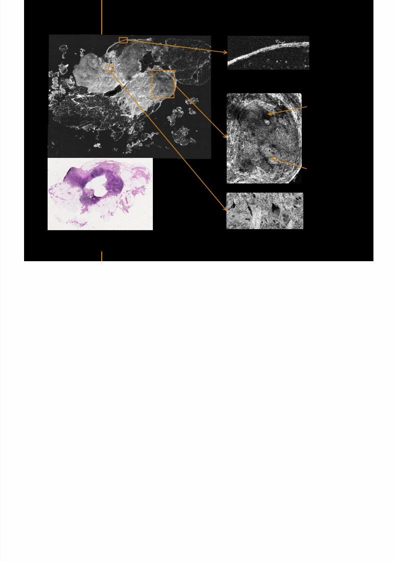
7292019 Atlas of Light-CT images
httpslidepdfcomreaderfullatlas-of-light-ct-images 22118
copy LLTech 2012 ndash wwwlltechimagingcom - No copy without authorization 22
The Light-CT scanner allows fast tissue processing andpathology examination It completes the toolset available topathologists
When Chemical Fixation is needed
(Mostly Diagnostic)
When Frozen Section is needed
(Mostly Intra-operative)
Biopsy3D
Digital Image
2D
Digital Image
Slide
15-12 mins
Artefacts
Destructive
12-24 hours
Expensive
Destructive
5-8 minutes
Non-Destructive
Tissue Scanner
Light-CT could help to reduce the number of re-excisions in breast
7292019 Atlas of Light-CT images
httpslidepdfcomreaderfullatlas-of-light-ct-images 23118
copy LLTech 2012 ndash wwwlltechimagingcom - No copy without authorization 23
Light CT could help to reduce the number of re excisions in breastcancer surgery whilst preserving the current routines ofpathologists
Per surgery
LLTechrsquos
analysis
Histology Andor
Immunohistochemistry
Further
treatment
Post surgery
Surgical margin analysis
Metastasis analysis (gt 2mm) et
micro metastaisi
PN1mi gt 02 ndash 2 mm lt
SurgeryOKOK
Per surgery Post Surgery
C u r r e n t t e c h n i q u e s
U p t o 4 0 o
f d o u b l e p r o
c e d u r e s
L L T e c h L i g h t - C
T
S i g n i f i c a n t d e c r e a s e o f
d o u b l e p r o c e d u
r e s
Visual
analysis
cryostat
Further
treatmentSurgery
OK
Cancerouscancerous
OK
Poor identification of
metastasis
Up to 40 of double surgical
procedures
Several day analysisFew minutes analysis
Surgeon Pathologist Pathologist
Decrease of double surgical
procedures CancerousCancerous
No surgical
margins analysis
Histology Andor
Immuno
histochemistry
Node
Margin
Several day analysisFew minutes analysisSurgeon Pathologist Pathologist
7292019 Atlas of Light-CT images
httpslidepdfcomreaderfullatlas-of-light-ct-images 24118
copy LLTech 2012 ndash wwwlltechimagingcom - No copy without authorization 24
BRAIN IMAGING
Back to table of contents
7292019 Atlas of Light-CT images
httpslidepdfcomreaderfullatlas-of-light-ct-images 25118
copy LLTech 2012 ndash wwwlltechimagingcom - No copy without authorization 25
Rat brain
Architectural and cellular information seen at various scales
Myelinated fibers visible
1 mm
500 microm
100 microm
7292019 Atlas of Light-CT images
httpslidepdfcomreaderfullatlas-of-light-ct-images 26118
7292019 Atlas of Light-CT images
httpslidepdfcomreaderfullatlas-of-light-ct-images 27118
7292019 Atlas of Light-CT images
httpslidepdfcomreaderfullatlas-of-light-ct-images 28118
copy LLTech 2012 ndash wwwlltechimagingcom - No copy without authorization 28 copy LLTECH 2011
Collagen-rich matrixWide fascicles of
tumour cells
Large capillary
Fibroblastic meningioma grade I
500microm
100microm 200microm200microm
7292019 Atlas of Light-CT images
httpslidepdfcomreaderfullatlas-of-light-ct-images 29118
copy LLTech 2012 ndash wwwlltechimagingcom - No copy without authorization 29
Transitional meningioma grade I
Psammoma surrounding a calcification
Whorls formation
Collagen-rich matrix
500microm
100microm
50microm
50microm
7292019 Atlas of Light-CT images
httpslidepdfcomreaderfullatlas-of-light-ct-images 30118
7292019 Atlas of Light-CT images
httpslidepdfcomreaderfullatlas-of-light-ct-images 31118
copy LLTech 2012 ndash wwwlltechimagingcom - No copy without authorization 31 copy LLTECH 2011
Arrows Fibers transverse cut
Spinal cord normal tissue
500 microm
100 microm
100 microm
7292019 Atlas of Light-CT images
httpslidepdfcomreaderfullatlas-of-light-ct-images 32118
7292019 Atlas of Light-CT images
httpslidepdfcomreaderfullatlas-of-light-ct-images 33118
7292019 Atlas of Light-CT images
httpslidepdfcomreaderfullatlas-of-light-ct-images 34118
copy LLTech 2012 ndash wwwlltechimagingcom - No copy without authorization 34
Back to table of contents
SKIN IMAGING
N l ki h l
7292019 Atlas of Light-CT images
httpslidepdfcomreaderfullatlas-of-light-ct-images 35118
copy LLTech 2012 ndash wwwlltechimagingcom - No copy without authorization 35
Normal skin morphology
Epidermis
Collagen
Pilosebaceous
unit
Sweat gland
Adipocytes
b
a
c
e
d
500 microm
copy LLTech 2012 Courtesy of Hopitaux Universitaires de Genegraveve Genegraveve Switzerland
7292019 Atlas of Light-CT images
httpslidepdfcomreaderfullatlas-of-light-ct-images 36118
copy LLTech 2012 ndash wwwlltechimagingcom - No copy without authorization 36
Hypodermis
Skin morphology layers are well discriminated
5 mm x 8 mm mosaic of native field image
Ageing patient (solar elastosis)
En-face imaging of vertical excision
Epidermis
Dermis
1
3
2
4
7292019 Atlas of Light-CT images
httpslidepdfcomreaderfullatlas-of-light-ct-images 37118
En face skin slicing shows structural and cellular details
7292019 Atlas of Light-CT images
httpslidepdfcomreaderfullatlas-of-light-ct-images 38118
copy LLTech 2012 ndash wwwlltechimagingcom - No copy without authorization 38
Stratum corneum Stratum spinosum
Stratum basale Reticular region
Epidermis
Dermis
En-face skin slicing shows structural and cellular details
Melanin
papillary caps
Keratinocyte
nuclei
Blood vessels
Collagen
Corneocytes
100 microm 100 microm
100 microm100 microm
copy LLTech 2012 Courtesy of Hopitaux Universitaires de Genegraveve Genegraveve Switzerland
7292019 Atlas of Light-CT images
httpslidepdfcomreaderfullatlas-of-light-ct-images 39118
7292019 Atlas of Light-CT images
httpslidepdfcomreaderfullatlas-of-light-ct-images 40118
Vertical reconstruction of skin model
7292019 Atlas of Light-CT images
httpslidepdfcomreaderfullatlas-of-light-ct-images 41118
copy LLTech 2012 ndash wwwlltechimagingcom - No copy without authorization 41
Vertical reconstruction of skin model
50 microm
Stratum corneum
Stratum spinosum
Dermis
Keratinocyte nuclei
copy LLTech 2012
7292019 Atlas of Light-CT images
httpslidepdfcomreaderfullatlas-of-light-ct-images 42118
Normal
7292019 Atlas of Light-CT images
httpslidepdfcomreaderfullatlas-of-light-ct-images 43118
copy LLTech 2012 ndash wwwlltechimagingcom - No copy without authorization 43
skin
b
a
e
c
f
d
500 microm
7292019 Atlas of Light-CT images
httpslidepdfcomreaderfullatlas-of-light-ct-images 44118
7292019 Atlas of Light-CT images
httpslidepdfcomreaderfullatlas-of-light-ct-images 45118
copy LLTech 2012 ndash wwwlltechimagingcom - No copy without authorization 45
Adipocytes
9um depth 27um depth 60um depth
Reconstructed
depth slice
200 microm
50 microm
7292019 Atlas of Light-CT images
httpslidepdfcomreaderfullatlas-of-light-ct-images 46118
Skin pathologies basal cell carcinoma discrimination at
7292019 Atlas of Light-CT images
httpslidepdfcomreaderfullatlas-of-light-ct-images 47118
copy LLTech 2012 ndash wwwlltechimagingcom - No copy without authorization 47
Epidermis
Dermis
Hypodermis
A
AA
C
BCC
PSU
PSU
BCC Basal cell carcinoma
A Adipocytes
C Collagen
PSU Pilosebaceous unit
structural level
1 mm
copy LLTech 2012 Courtesy of Hopitaux Universitaires de Genegraveve Genegraveve Switzerland
C
Zoom on basal cell carcinoma discrimination at cellular level
7292019 Atlas of Light-CT images
httpslidepdfcomreaderfullatlas-of-light-ct-images 48118
copy LLTech 2012 ndash wwwlltechimagingcom - No copy without authorization 48
Zoom on basal cell carcinoma discrimination at cellular level
200 microm
100 microm
Peritumoral stroma
High cell density
copy LLTech 2012 Courtesy of Hopitaux Universitaires de Genegraveve Genegraveve Switzerland
Skin pathologies discrimination of actinic keratosis
7292019 Atlas of Light-CT images
httpslidepdfcomreaderfullatlas-of-light-ct-images 49118
copy LLTech 2012 ndash wwwlltechimagingcom - No copy without authorization 49
Skin pathologies discrimination of actinic keratosis
Enlarged epidermis
Extended epidermal ridges
1 mm
copy LLTech 2012 Courtesy of Hopitaux Universitaires de Genegraveve Genegraveve Switzerland
7292019 Atlas of Light-CT images
httpslidepdfcomreaderfullatlas-of-light-ct-images 50118
copy LLTech 2012 ndash wwwlltechimagingcom - No copy without authorization 50
Basal cell carcinoma in depth imaging
Z = 10 microns 7 x 42 mm FOV Z = 35 microns 7 x 42 mm FOV
BCC
SG Sebaceous glands
Z = 55 microns 7 x 42 mm FOV Z = 75 microns 7 x 42 mm FOV
SG
Application to Mohs surgerycopy LLTech 2012 Courtesy of Hopitaux Universitaires de Genegraveve Genegraveve Switzerland
7292019 Atlas of Light-CT images
httpslidepdfcomreaderfullatlas-of-light-ct-images 51118
copy LLTech 2012 ndash wwwlltechimagingcom - No copy without authorization 51 copy LLTECH 2011
Healthy vs Cheloid scar tissue
Healthy skin tissueFOV 75x5mm
Cheloid scar tissueFOV 65x65mm
copy LLTECH 2011
7292019 Atlas of Light-CT images
httpslidepdfcomreaderfullatlas-of-light-ct-images 52118
copy LLTech 2012 ndash wwwlltechimagingcom - No copy without authorization 52
Dermo-epidermaljunction
Cheloid scar tissue in detail
copy LLTECH 2011
Nodular structureThickcollagenfiberbundles
1mm
600 microm
500 microm
h l d d f
7292019 Atlas of Light-CT images
httpslidepdfcomreaderfullatlas-of-light-ct-images 53118
copy LLTech 2012 ndash wwwlltechimagingcom - No copy without authorization 53
Cheloid scar tissue viewed en face
20microm depth 120microm depth
7292019 Atlas of Light-CT images
httpslidepdfcomreaderfullatlas-of-light-ct-images 54118
Skin in-vivo sweat duct imaging in 3D
7292019 Atlas of Light-CT images
httpslidepdfcomreaderfullatlas-of-light-ct-images 55118
copy LLTech 2012 ndash wwwlltechimagingcom - No copy without authorization 55
g g
Vertical reconstruction from en-face images
Sweat duct 3D reconstruction
Stratum corneum
Stratum spinosum
SD sweat ducts
SD SD
100 microm
7292019 Atlas of Light-CT images
httpslidepdfcomreaderfullatlas-of-light-ct-images 56118
7292019 Atlas of Light-CT images
httpslidepdfcomreaderfullatlas-of-light-ct-images 57118
7292019 Atlas of Light-CT images
httpslidepdfcomreaderfullatlas-of-light-ct-images 58118
copy LLTech 2012 ndash wwwlltechimagingcom - No copy without authorization 58
LUNG IMAGING
Back to table of contents
H L
7292019 Atlas of Light-CT images
httpslidepdfcomreaderfullatlas-of-light-ct-images 59118
copy LLTech 2012 ndash wwwlltechimagingcom - No copy without authorization 59
Zone 1
Zone 2
Zone 3
Zone 4
Zone with
cancerous tissue
Limit of cancerous
stroma
alveoli with
macrophages
Alveoles
Human Lung
500microm
copy LLTech 2012 Courtesy of Hocircpital Hotel-Dieu Paris France
H L Z 1 Z t
7292019 Atlas of Light-CT images
httpslidepdfcomreaderfullatlas-of-light-ct-images 60118
copy LLTech 2012 ndash wwwlltechimagingcom - No copy without authorization 60
Stroma
Human Lung ndash Zone 1 ndash Zoom on cancerous stroma
Cancerous
area
200microm
copy LLTech 2012 Courtesy of Hocircpital Hotel-Dieu Paris France
Human Lung ndash Zone 2 ndash Limit cancerous stroma alveoli
7292019 Atlas of Light-CT images
httpslidepdfcomreaderfullatlas-of-light-ct-images 61118
copy LLTech 2012 ndash wwwlltechimagingcom - No copy without authorization 61
g
with visible macrophages and vessel
Alveoli
Macrophages
Vessel
copy LLTech 2012 Courtesy of Hocircpital Hotel-Dieu Paris France
Human Lung ndash Zone 3 ndash Limit cancerous stroma alveoli
7292019 Atlas of Light-CT images
httpslidepdfcomreaderfullatlas-of-light-ct-images 62118
copy LLTech 2012 ndash wwwlltechimagingcom - No copy without authorization 62
g
with visible macrophages
Macrophages
100microm
copy LLTech 2012 Courtesy of Hocircpital Hotel-Dieu Paris France
Human Lung ndash Zone 4 ndash Alveoli
7292019 Atlas of Light-CT images
httpslidepdfcomreaderfullatlas-of-light-ct-images 63118
copy LLTech 2012 ndash wwwlltechimagingcom - No copy without authorization 63
Human Lung Zone 4 Alveoli
Alveoli
200microm
copy LLTech 2012 Courtesy of Hocircpital Hotel-Dieu Paris France
7292019 Atlas of Light-CT images
httpslidepdfcomreaderfullatlas-of-light-ct-images 64118
copy LLTech 2012 ndash wwwlltechimagingcom - No copy without authorization 64
GI IMAGING
Back to table of contents
7292019 Atlas of Light-CT images
httpslidepdfcomreaderfullatlas-of-light-ct-images 65118
Colonic carcinoma
7292019 Atlas of Light-CT images
httpslidepdfcomreaderfullatlas-of-light-ct-images 66118
copy LLTech 2012 ndash wwwlltechimagingcom - No copy without authorization 66
Colonic Carcinoma
Colonic carcinoma
copy LLTech 2012 Courtesy of MD Anderson Houston USA
Intestines ndash Crohnrsquos disease
7292019 Atlas of Light-CT images
httpslidepdfcomreaderfullatlas-of-light-ct-images 67118
copy LLTech 2012 ndash wwwlltechimagingcom - No copy without authorization 67
Crohnrsquos disease structure not
visible from the surface
Fat cells
200 microm
Intestines Crohn s disease
copy LLTech 2012 Courtesy of Institut Curie Paris France
Rat stomach
7292019 Atlas of Light-CT images
httpslidepdfcomreaderfullatlas-of-light-ct-images 68118
copy LLTech 2012 ndash wwwlltechimagingcom - No copy without authorization 68
Rat stomach
Mucosa withgastric pits
Submucosa with
bright collagen
fibers
Muscularis
propria with
muscle bundles
copy LLTech 2012 Courtesy of Weill Cornell Medical College NY USA
200 microm
7292019 Atlas of Light-CT images
httpslidepdfcomreaderfullatlas-of-light-ct-images 69118
copy LLTech 2012 ndash wwwlltechimagingcom - No copy without authorization 69 copy LLTECH 2011
UROGENITAL IMAGING
Back to table of contents
Rat bladder
7292019 Atlas of Light-CT images
httpslidepdfcomreaderfullatlas-of-light-ct-images 70118
copy LLTech 2012 ndash wwwlltechimagingcom - No copy without authorization 70
Rat bladder
Stratifiedurothelium
Lamina propria
500 microm
Muscular
bundles of the
lamina propria
Serosa
copy LLTech 2012 Courtesy of Weill Cornell Medical College NY USA
100 microm
100 microm
Human bladder low grade papillary urothelial carcinoma
7292019 Atlas of Light-CT images
httpslidepdfcomreaderfullatlas-of-light-ct-images 71118
copy LLTech 2012 ndash wwwlltechimagingcom - No copy without authorization 71
Lesion with fibrovascular cores (arrow) and superficial umbrella cells (arrowheads)
Human bladder low grade papillary urothelial carcinoma
copy LLTech 2012 Courtesy of Weill Cornell Medical College NY USA
100 microm
7292019 Atlas of Light-CT images
httpslidepdfcomreaderfullatlas-of-light-ct-images 72118
Rat prostate
7292019 Atlas of Light-CT images
httpslidepdfcomreaderfullatlas-of-light-ct-images 73118
copy LLTech 2012 ndash wwwlltechimagingcom - No copy without authorization 73
p
copy LLTech 2012 Courtesy of Weill Cornell Medical College NY USA
Papillary folds of the
acinar glands
Prostatic acini
Fibromuscular stromaPeriprostatic fat Blood vessel
1 mm
200 microm
Mouse kidney
7292019 Atlas of Light-CT images
httpslidepdfcomreaderfullatlas-of-light-ct-images 74118
copy LLTech 2012 ndash wwwlltechimagingcom - No copy without authorization 74
1 mm
y
copy LLTech 2012 Courtesy of Institut Biomeacutedical de Bicecirctre Paris France
Renal cortex
Renal medulla
Renal sinus
7292019 Atlas of Light-CT images
httpslidepdfcomreaderfullatlas-of-light-ct-images 75118
copy LLTech 2012 ndash wwwlltechimagingcom - No copy without authorization 75
Mouse kidney ndash zoom on highlighted section of previous slide
800microm
copy LLTech 2012 Courtesy of Hocircpital Kremlin Bicecirctre Paris France
7292019 Atlas of Light-CT images
httpslidepdfcomreaderfullatlas-of-light-ct-images 76118
7292019 Atlas of Light-CT images
httpslidepdfcomreaderfullatlas-of-light-ct-images 77118
7292019 Atlas of Light-CT images
httpslidepdfcomreaderfullatlas-of-light-ct-images 78118
copy LLTech 2012 ndash wwwlltechimagingcom - No copy without authorization 78 copy LLTECH 2011
CORE NEEDLE BIOPSIES
Back to table of contents
Kidney
7292019 Atlas of Light-CT images
httpslidepdfcomreaderfullatlas-of-light-ct-images 79118
copy LLTech 2012 ndash wwwlltechimagingcom - No copy without authorization 79
100microm
50microm
Renal tubules
Vessel
500microm
copy LLTech 2012 Courtesy of Institut Curie Paris France
Breast Invasive lobular carcinoma
7292019 Atlas of Light-CT images
httpslidepdfcomreaderfullatlas-of-light-ct-images 80118
copy LLTech 2012 ndash wwwlltechimagingcom - No copy without authorization 80
500microm
stroma
adipocytes
copy LLTech 2012 Courtesy of Institut Curie Paris France
7292019 Atlas of Light-CT images
httpslidepdfcomreaderfullatlas-of-light-ct-images 81118
Breast Ductal carcinoma in situ
7292019 Atlas of Light-CT images
httpslidepdfcomreaderfullatlas-of-light-ct-images 82118
copy LLTech 2012 ndash wwwlltechimagingcom - No copy without authorization 82
500microm
copy LLTech 2012 Courtesy of Institut Curie Paris France
7292019 Atlas of Light-CT images
httpslidepdfcomreaderfullatlas-of-light-ct-images 83118
copy LLTech 2012 ndash wwwlltechimagingcom - No copy without authorization 83 copy LLTECH 2011
THYROID IMAGING
Back to table of contents
7292019 Atlas of Light-CT images
httpslidepdfcomreaderfullatlas-of-light-ct-images 84118
copy LLTech 2012 ndash wwwlltechimagingcom - No copy without authorization 84
Thyroid
800 microm
200 microm
copy LLTech 2012 Courtesy of Hocircpital Tenon Paris France
7292019 Atlas of Light-CT images
httpslidepdfcomreaderfullatlas-of-light-ct-images 85118
copy LLTech 2012 ndash wwwlltechimagingcom - No copy without authorization 85
Thyroid
800 microm
200 microm
copy LLTech 2012 Courtesy of Hocircpital Tenon Paris France
7292019 Atlas of Light-CT images
httpslidepdfcomreaderfullatlas-of-light-ct-images 86118
copy LLTech 2012 ndash wwwlltechimagingcom - No copy without authorization 86 copy LLTECH 2011
FAT CELLS IMAGING
Back to table of contents
7292019 Atlas of Light-CT images
httpslidepdfcomreaderfullatlas-of-light-ct-images 87118
copy LLTech 2012 ndash wwwlltechimagingcom - No copy without authorization 87
Brown fat cells - adipocytes
100 microm 100 microm
200 microm
10um depth
Adipocytes
45um depth
En face
slices
Reconstructed
depth slice
copy LLTech 2012 Courtesy of Centre de Recherche des Cordeliers Paris France
7292019 Atlas of Light-CT images
httpslidepdfcomreaderfullatlas-of-light-ct-images 88118
copy LLTech 2012 ndash wwwlltechimagingcom - No copy without authorization 88 copy LLTECH 2011
CORNEA AND RETINA
Back to table of contents
Human cornea reconstructed depth slice
7292019 Atlas of Light-CT images
httpslidepdfcomreaderfullatlas-of-light-ct-images 89118
copy LLTech 2012 ndash wwwlltechimagingcom - No copy without authorization 89
50 um
wwwlltechimagingcom
7292019 Atlas of Light-CT images
httpslidepdfcomreaderfullatlas-of-light-ct-images 90118
Large field en face view in stroma
7292019 Atlas of Light-CT images
httpslidepdfcomreaderfullatlas-of-light-ct-images 91118
copy LLTech 2012 ndash wwwlltechimagingcom - No copy without authorization 91
200 um
wwwlltechimagingcom
7292019 Atlas of Light-CT images
httpslidepdfcomreaderfullatlas-of-light-ct-images 92118
Rabbit cornea after LASIK reflectivity profiles reveal
i d tt i d thi k d ith li l l
7292019 Atlas of Light-CT images
httpslidepdfcomreaderfullatlas-of-light-ct-images 93118
copy LLTech 2012 ndash wwwlltechimagingcom - No copy without authorization 93
increased scattering and thickened epithelial layer
LASIK LASIK CONTROL CONTROL
copy LLTech 2012
Newborn mouse anterior segment
7292019 Atlas of Light-CT images
httpslidepdfcomreaderfullatlas-of-light-ct-images 94118
copy LLTech 2012 ndash wwwlltechimagingcom - No copy without authorization 94
Corneal epithelial cells
Basal membrane
Stroma
Descemetrsquos membrane
Aqueous humor
Lens capsule
Lens epithelial cells
Cortical fibers
copy LLTech 2012 Courtesy of ESPCI Paris Francecopy LLTech 2012
Rat retina comparison with histology
7292019 Atlas of Light-CT images
httpslidepdfcomreaderfullatlas-of-light-ct-images 95118
copy LLTech 2012 ndash wwwlltechimagingcom - No copy without authorization 95
Figure 1 Right Ex vivo rat retinaimaged using the f ull field OCT
system of ESPCI Left Histology
for comparison
Figure 1 Right Ex vivo rat retinaimaged using the f ull field OCT
system of ESPCI Left Histology
for comparison
Figure 1 Right Ex vivo rat retinaimaged using the f ull field OCT
system of ESPCI Left Histology
for comparisonField
230x300microm
RPE
PRL
ONL
OPL
INL
IPL
GCL
NFL
Invest Ophthalmol Vis Sci 2004 Nov45(11)4126-31
Grieve K Paques M Dubois A Sahel J Boccara C Le Gargasson JF copy LLTech 2012
Rat retina
7292019 Atlas of Light-CT images
httpslidepdfcomreaderfullatlas-of-light-ct-images 96118
copy LLTech 2012 ndash wwwlltechimagingcom - No copy without authorization 96 copy LLTech 2012 Courtesy of ESPCI Paris France
Nerve fiber layer
Retina
Invest Ophthalmol Vis Sci 2004 Nov45(11)4126-31
Grieve K Paques M Dubois A Sahel J Boccara C Le Gargasson JF
Transscleral image
7292019 Atlas of Light-CT images
httpslidepdfcomreaderfullatlas-of-light-ct-images 97118
7292019 Atlas of Light-CT images
httpslidepdfcomreaderfullatlas-of-light-ct-images 98118
7292019 Atlas of Light-CT images
httpslidepdfcomreaderfullatlas-of-light-ct-images 99118
In vivo imaging of drosophila melanogaster
7292019 Atlas of Light-CT images
httpslidepdfcomreaderfullatlas-of-light-ct-images 100118
copy LLTech 2012 ndash wwwlltechimagingcom - No copy without authorization 100 copy LLTech 2012 Courtesy of ESPCI Paris France
In vivo imaging of drosophila melanogaster ndash four stages of pupation over 72 hours
7292019 Atlas of Light-CT images
httpslidepdfcomreaderfullatlas-of-light-ct-images 101118
copy LLTech 2012 ndash wwwlltechimagingcom - No copy without authorization 101
Prepupa (0-2 h) Transition topupal stage (24 h)
Pupal stage (48 h) Advanced pupal phase
before eclosion (72 h)
copy LLTech 2012 Courtesy of ESPCI Paris France
Pupa 118 4 days 30um depth
100
7292019 Atlas of Light-CT images
httpslidepdfcomreaderfullatlas-of-light-ct-images 102118
copy LLTech 2012 ndash wwwlltechimagingcom - No copy without authorization 102
100 um
Drosophila
7292019 Atlas of Light-CT images
httpslidepdfcomreaderfullatlas-of-light-ct-images 103118
copy LLTech 2012 ndash wwwlltechimagingcom - No copy without authorization 103
200 microm
copy LLTech 2012 Courtesy of ESPCI Paris France
C Elegans ndash Fast identification of anatomy
7292019 Atlas of Light-CT images
httpslidepdfcomreaderfullatlas-of-light-ct-images 104118
copy LLTech 2012 ndash wwwlltechimagingcom - No copy without authorization 104
Body wall - cuticle
gonad arm
30 microm depth
uterus
ovocytes
intestine
100 microm 100 microm
15 microm depth
copy LLTech 2012 Courtesy of ENS Paris France
C elegans ndash Images at higher magnification 40X
7292019 Atlas of Light-CT images
httpslidepdfcomreaderfullatlas-of-light-ct-images 105118
copy LLTech 2012 ndash wwwlltechimagingcom - No copy without authorization 105
3D reconstruction 50 microm
100 microm
copy LLTech 2012 Courtesy of ENS Paris France
Zebra fish eye
7292019 Atlas of Light-CT images
httpslidepdfcomreaderfullatlas-of-light-ct-images 106118
copy LLTech 2012 ndash wwwlltechimagingcom - No copy without authorization 106
Zebra fish eye
100 microm
100 microm
En face slices
Reconstructed depth slice
copy LLTech 2012 Courtesy of Inserm Paris France
10um depth 20um depth 45um depth
7292019 Atlas of Light-CT images
httpslidepdfcomreaderfullatlas-of-light-ct-images 107118
Xenopus Laevis backtail
7292019 Atlas of Light-CT images
httpslidepdfcomreaderfullatlas-of-light-ct-images 108118
copy LLTech 2012 ndash wwwlltechimagingcom - No copy without authorization 108
100 um30um depth 60um depth
7292019 Atlas of Light-CT images
httpslidepdfcomreaderfullatlas-of-light-ct-images 109118
Mouse embryo in vitro 6 5 days old ndash
7292019 Atlas of Light-CT images
httpslidepdfcomreaderfullatlas-of-light-ct-images 110118
copy LLTech 2012 ndash wwwlltechimagingcom - No copy without authorization 110
Mouse embryo in vitro 65 days old
copy LLTech 2012 Courtesy of Ellvire Guiot and nstitut Jacques Monod Paris France
3D imaging of internal cavity
Sabellaria alveolata mouth palps
7292019 Atlas of Light-CT images
httpslidepdfcomreaderfullatlas-of-light-ct-images 111118
copy LLTech 2012 ndash wwwlltechimagingcom - No copy without authorization 111
500 microm
100 microm
Mouth palps
copy LLTech 2012 Courtesy of Museacutee National drsquoHistoire Naturelle Paris France
7292019 Atlas of Light-CT images
httpslidepdfcomreaderfullatlas-of-light-ct-images 112118
copy LLTech 2012 ndash wwwlltechimagingcom - No copy without authorization 112 copy LLTECH 2011
PLANT IMAGING
Back to table of contents
Mouse ear leaf epidermis
7292019 Atlas of Light-CT images
httpslidepdfcomreaderfullatlas-of-light-ct-images 113118
copy LLTech 2012 ndash wwwlltechimagingcom - No copy without authorization 113
100 microm
Epidermal cell
copy LLTech 2012
Stoma
Leaf veins
7292019 Atlas of Light-CT images
httpslidepdfcomreaderfullatlas-of-light-ct-images 114118
copy LLTech 2012 ndash wwwlltechimagingcom - No copy without authorization 114
114copy LLTech 2012
Fibers
Veins
100 microm
500 microm
Apple
7292019 Atlas of Light-CT images
httpslidepdfcomreaderfullatlas-of-light-ct-images 115118
copy LLTech 2012 ndash wwwlltechimagingcom - No copy without authorization 115
115copy LLTech 2012
100 microm
Wax
Skin
Flesh
500 microm
Water
Effect of harpin infiltration on tobacco leaf
7292019 Atlas of Light-CT images
httpslidepdfcomreaderfullatlas-of-light-ct-images 116118
copy LLTech 2012 ndash wwwlltechimagingcom - No copy without authorization 116
116copy LLTech 2012 Courtesy of UPMC Paris France
50 microm
50 microm
50 microm
50 microm
Upper epidermis
10 min after harpin infiltration 30 min after harpin infiltration
Palisade parenchyma
Epidermis cell
Chloroplast
7292019 Atlas of Light-CT images
httpslidepdfcomreaderfullatlas-of-light-ct-images 117118
copy LLTech 2012 ndash wwwlltechimagingcom - No copy without authorization 117
bull Light-CTtrade does not require tissue preparation nor staining of any kind
bull Creates images within minutes using a non-destructive process
bull Offers a 1 microm cellular resolution in 3D
bull Reveals structural and cellular information
bull Mosaicing allows fast visualisation at various scale
bull En-face high-resolution imaging allows vertical and 3D reconstruction
bull In-vivo images show good promise for in-vivo system
bull Clinical and research applications in cancer detection cosmetics biobanking small animal
developmental biology botanyhellip
Contact Details
7292019 Atlas of Light-CT images
httpslidepdfcomreaderfullatlas-of-light-ct-images 118118
USA
LLTech Inc
103 Carnegie Center DriveSuite 300
Princeton NJ 08540
USA
wwwlltechimagingcom
contactlltechfr
Phone + 1 609 955 3506
LLTech SAS
29 rue du Faubourg St-JacquesPeacutepiniegravere Paris Santeacute Cochin
75014
Paris - France
wwwlltechimagingcom
contactlltechfr
Phone +33 1 82 72 61 25
Europe
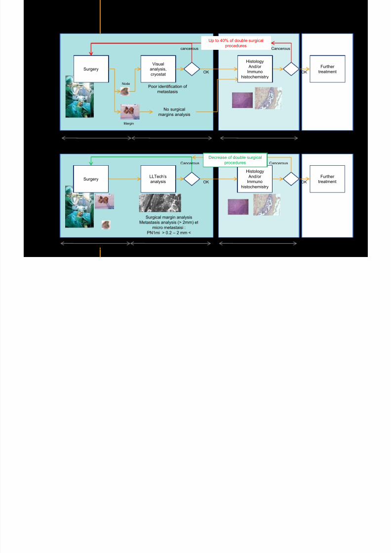
7292019 Atlas of Light-CT images
httpslidepdfcomreaderfullatlas-of-light-ct-images 24118
copy LLTech 2012 ndash wwwlltechimagingcom - No copy without authorization 24
BRAIN IMAGING
Back to table of contents
7292019 Atlas of Light-CT images
httpslidepdfcomreaderfullatlas-of-light-ct-images 25118
copy LLTech 2012 ndash wwwlltechimagingcom - No copy without authorization 25
Rat brain
Architectural and cellular information seen at various scales
Myelinated fibers visible
1 mm
500 microm
100 microm
7292019 Atlas of Light-CT images
httpslidepdfcomreaderfullatlas-of-light-ct-images 26118
7292019 Atlas of Light-CT images
httpslidepdfcomreaderfullatlas-of-light-ct-images 27118
7292019 Atlas of Light-CT images
httpslidepdfcomreaderfullatlas-of-light-ct-images 28118
copy LLTech 2012 ndash wwwlltechimagingcom - No copy without authorization 28 copy LLTECH 2011
Collagen-rich matrixWide fascicles of
tumour cells
Large capillary
Fibroblastic meningioma grade I
500microm
100microm 200microm200microm
7292019 Atlas of Light-CT images
httpslidepdfcomreaderfullatlas-of-light-ct-images 29118
copy LLTech 2012 ndash wwwlltechimagingcom - No copy without authorization 29
Transitional meningioma grade I
Psammoma surrounding a calcification
Whorls formation
Collagen-rich matrix
500microm
100microm
50microm
50microm
7292019 Atlas of Light-CT images
httpslidepdfcomreaderfullatlas-of-light-ct-images 30118
7292019 Atlas of Light-CT images
httpslidepdfcomreaderfullatlas-of-light-ct-images 31118
copy LLTech 2012 ndash wwwlltechimagingcom - No copy without authorization 31 copy LLTECH 2011
Arrows Fibers transverse cut
Spinal cord normal tissue
500 microm
100 microm
100 microm
7292019 Atlas of Light-CT images
httpslidepdfcomreaderfullatlas-of-light-ct-images 32118
7292019 Atlas of Light-CT images
httpslidepdfcomreaderfullatlas-of-light-ct-images 33118
7292019 Atlas of Light-CT images
httpslidepdfcomreaderfullatlas-of-light-ct-images 34118
copy LLTech 2012 ndash wwwlltechimagingcom - No copy without authorization 34
Back to table of contents
SKIN IMAGING
N l ki h l
7292019 Atlas of Light-CT images
httpslidepdfcomreaderfullatlas-of-light-ct-images 35118
copy LLTech 2012 ndash wwwlltechimagingcom - No copy without authorization 35
Normal skin morphology
Epidermis
Collagen
Pilosebaceous
unit
Sweat gland
Adipocytes
b
a
c
e
d
500 microm
copy LLTech 2012 Courtesy of Hopitaux Universitaires de Genegraveve Genegraveve Switzerland
7292019 Atlas of Light-CT images
httpslidepdfcomreaderfullatlas-of-light-ct-images 36118
copy LLTech 2012 ndash wwwlltechimagingcom - No copy without authorization 36
Hypodermis
Skin morphology layers are well discriminated
5 mm x 8 mm mosaic of native field image
Ageing patient (solar elastosis)
En-face imaging of vertical excision
Epidermis
Dermis
1
3
2
4
7292019 Atlas of Light-CT images
httpslidepdfcomreaderfullatlas-of-light-ct-images 37118
En face skin slicing shows structural and cellular details
7292019 Atlas of Light-CT images
httpslidepdfcomreaderfullatlas-of-light-ct-images 38118
copy LLTech 2012 ndash wwwlltechimagingcom - No copy without authorization 38
Stratum corneum Stratum spinosum
Stratum basale Reticular region
Epidermis
Dermis
En-face skin slicing shows structural and cellular details
Melanin
papillary caps
Keratinocyte
nuclei
Blood vessels
Collagen
Corneocytes
100 microm 100 microm
100 microm100 microm
copy LLTech 2012 Courtesy of Hopitaux Universitaires de Genegraveve Genegraveve Switzerland
7292019 Atlas of Light-CT images
httpslidepdfcomreaderfullatlas-of-light-ct-images 39118
7292019 Atlas of Light-CT images
httpslidepdfcomreaderfullatlas-of-light-ct-images 40118
Vertical reconstruction of skin model
7292019 Atlas of Light-CT images
httpslidepdfcomreaderfullatlas-of-light-ct-images 41118
copy LLTech 2012 ndash wwwlltechimagingcom - No copy without authorization 41
Vertical reconstruction of skin model
50 microm
Stratum corneum
Stratum spinosum
Dermis
Keratinocyte nuclei
copy LLTech 2012
7292019 Atlas of Light-CT images
httpslidepdfcomreaderfullatlas-of-light-ct-images 42118
Normal
7292019 Atlas of Light-CT images
httpslidepdfcomreaderfullatlas-of-light-ct-images 43118
copy LLTech 2012 ndash wwwlltechimagingcom - No copy without authorization 43
skin
b
a
e
c
f
d
500 microm
7292019 Atlas of Light-CT images
httpslidepdfcomreaderfullatlas-of-light-ct-images 44118
7292019 Atlas of Light-CT images
httpslidepdfcomreaderfullatlas-of-light-ct-images 45118
copy LLTech 2012 ndash wwwlltechimagingcom - No copy without authorization 45
Adipocytes
9um depth 27um depth 60um depth
Reconstructed
depth slice
200 microm
50 microm
7292019 Atlas of Light-CT images
httpslidepdfcomreaderfullatlas-of-light-ct-images 46118
Skin pathologies basal cell carcinoma discrimination at
7292019 Atlas of Light-CT images
httpslidepdfcomreaderfullatlas-of-light-ct-images 47118
copy LLTech 2012 ndash wwwlltechimagingcom - No copy without authorization 47
Epidermis
Dermis
Hypodermis
A
AA
C
BCC
PSU
PSU
BCC Basal cell carcinoma
A Adipocytes
C Collagen
PSU Pilosebaceous unit
structural level
1 mm
copy LLTech 2012 Courtesy of Hopitaux Universitaires de Genegraveve Genegraveve Switzerland
C
Zoom on basal cell carcinoma discrimination at cellular level
7292019 Atlas of Light-CT images
httpslidepdfcomreaderfullatlas-of-light-ct-images 48118
copy LLTech 2012 ndash wwwlltechimagingcom - No copy without authorization 48
Zoom on basal cell carcinoma discrimination at cellular level
200 microm
100 microm
Peritumoral stroma
High cell density
copy LLTech 2012 Courtesy of Hopitaux Universitaires de Genegraveve Genegraveve Switzerland
Skin pathologies discrimination of actinic keratosis
7292019 Atlas of Light-CT images
httpslidepdfcomreaderfullatlas-of-light-ct-images 49118
copy LLTech 2012 ndash wwwlltechimagingcom - No copy without authorization 49
Skin pathologies discrimination of actinic keratosis
Enlarged epidermis
Extended epidermal ridges
1 mm
copy LLTech 2012 Courtesy of Hopitaux Universitaires de Genegraveve Genegraveve Switzerland
7292019 Atlas of Light-CT images
httpslidepdfcomreaderfullatlas-of-light-ct-images 50118
copy LLTech 2012 ndash wwwlltechimagingcom - No copy without authorization 50
Basal cell carcinoma in depth imaging
Z = 10 microns 7 x 42 mm FOV Z = 35 microns 7 x 42 mm FOV
BCC
SG Sebaceous glands
Z = 55 microns 7 x 42 mm FOV Z = 75 microns 7 x 42 mm FOV
SG
Application to Mohs surgerycopy LLTech 2012 Courtesy of Hopitaux Universitaires de Genegraveve Genegraveve Switzerland
7292019 Atlas of Light-CT images
httpslidepdfcomreaderfullatlas-of-light-ct-images 51118
copy LLTech 2012 ndash wwwlltechimagingcom - No copy without authorization 51 copy LLTECH 2011
Healthy vs Cheloid scar tissue
Healthy skin tissueFOV 75x5mm
Cheloid scar tissueFOV 65x65mm
copy LLTECH 2011
7292019 Atlas of Light-CT images
httpslidepdfcomreaderfullatlas-of-light-ct-images 52118
copy LLTech 2012 ndash wwwlltechimagingcom - No copy without authorization 52
Dermo-epidermaljunction
Cheloid scar tissue in detail
copy LLTECH 2011
Nodular structureThickcollagenfiberbundles
1mm
600 microm
500 microm
h l d d f
7292019 Atlas of Light-CT images
httpslidepdfcomreaderfullatlas-of-light-ct-images 53118
copy LLTech 2012 ndash wwwlltechimagingcom - No copy without authorization 53
Cheloid scar tissue viewed en face
20microm depth 120microm depth
7292019 Atlas of Light-CT images
httpslidepdfcomreaderfullatlas-of-light-ct-images 54118
Skin in-vivo sweat duct imaging in 3D
7292019 Atlas of Light-CT images
httpslidepdfcomreaderfullatlas-of-light-ct-images 55118
copy LLTech 2012 ndash wwwlltechimagingcom - No copy without authorization 55
g g
Vertical reconstruction from en-face images
Sweat duct 3D reconstruction
Stratum corneum
Stratum spinosum
SD sweat ducts
SD SD
100 microm
7292019 Atlas of Light-CT images
httpslidepdfcomreaderfullatlas-of-light-ct-images 56118
7292019 Atlas of Light-CT images
httpslidepdfcomreaderfullatlas-of-light-ct-images 57118
7292019 Atlas of Light-CT images
httpslidepdfcomreaderfullatlas-of-light-ct-images 58118
copy LLTech 2012 ndash wwwlltechimagingcom - No copy without authorization 58
LUNG IMAGING
Back to table of contents
H L
7292019 Atlas of Light-CT images
httpslidepdfcomreaderfullatlas-of-light-ct-images 59118
copy LLTech 2012 ndash wwwlltechimagingcom - No copy without authorization 59
Zone 1
Zone 2
Zone 3
Zone 4
Zone with
cancerous tissue
Limit of cancerous
stroma
alveoli with
macrophages
Alveoles
Human Lung
500microm
copy LLTech 2012 Courtesy of Hocircpital Hotel-Dieu Paris France
H L Z 1 Z t
7292019 Atlas of Light-CT images
httpslidepdfcomreaderfullatlas-of-light-ct-images 60118
copy LLTech 2012 ndash wwwlltechimagingcom - No copy without authorization 60
Stroma
Human Lung ndash Zone 1 ndash Zoom on cancerous stroma
Cancerous
area
200microm
copy LLTech 2012 Courtesy of Hocircpital Hotel-Dieu Paris France
Human Lung ndash Zone 2 ndash Limit cancerous stroma alveoli
7292019 Atlas of Light-CT images
httpslidepdfcomreaderfullatlas-of-light-ct-images 61118
copy LLTech 2012 ndash wwwlltechimagingcom - No copy without authorization 61
g
with visible macrophages and vessel
Alveoli
Macrophages
Vessel
copy LLTech 2012 Courtesy of Hocircpital Hotel-Dieu Paris France
Human Lung ndash Zone 3 ndash Limit cancerous stroma alveoli
7292019 Atlas of Light-CT images
httpslidepdfcomreaderfullatlas-of-light-ct-images 62118
copy LLTech 2012 ndash wwwlltechimagingcom - No copy without authorization 62
g
with visible macrophages
Macrophages
100microm
copy LLTech 2012 Courtesy of Hocircpital Hotel-Dieu Paris France
Human Lung ndash Zone 4 ndash Alveoli
7292019 Atlas of Light-CT images
httpslidepdfcomreaderfullatlas-of-light-ct-images 63118
copy LLTech 2012 ndash wwwlltechimagingcom - No copy without authorization 63
Human Lung Zone 4 Alveoli
Alveoli
200microm
copy LLTech 2012 Courtesy of Hocircpital Hotel-Dieu Paris France
7292019 Atlas of Light-CT images
httpslidepdfcomreaderfullatlas-of-light-ct-images 64118
copy LLTech 2012 ndash wwwlltechimagingcom - No copy without authorization 64
GI IMAGING
Back to table of contents
7292019 Atlas of Light-CT images
httpslidepdfcomreaderfullatlas-of-light-ct-images 65118
Colonic carcinoma
7292019 Atlas of Light-CT images
httpslidepdfcomreaderfullatlas-of-light-ct-images 66118
copy LLTech 2012 ndash wwwlltechimagingcom - No copy without authorization 66
Colonic Carcinoma
Colonic carcinoma
copy LLTech 2012 Courtesy of MD Anderson Houston USA
Intestines ndash Crohnrsquos disease
7292019 Atlas of Light-CT images
httpslidepdfcomreaderfullatlas-of-light-ct-images 67118
copy LLTech 2012 ndash wwwlltechimagingcom - No copy without authorization 67
Crohnrsquos disease structure not
visible from the surface
Fat cells
200 microm
Intestines Crohn s disease
copy LLTech 2012 Courtesy of Institut Curie Paris France
Rat stomach
7292019 Atlas of Light-CT images
httpslidepdfcomreaderfullatlas-of-light-ct-images 68118
copy LLTech 2012 ndash wwwlltechimagingcom - No copy without authorization 68
Rat stomach
Mucosa withgastric pits
Submucosa with
bright collagen
fibers
Muscularis
propria with
muscle bundles
copy LLTech 2012 Courtesy of Weill Cornell Medical College NY USA
200 microm
7292019 Atlas of Light-CT images
httpslidepdfcomreaderfullatlas-of-light-ct-images 69118
copy LLTech 2012 ndash wwwlltechimagingcom - No copy without authorization 69 copy LLTECH 2011
UROGENITAL IMAGING
Back to table of contents
Rat bladder
7292019 Atlas of Light-CT images
httpslidepdfcomreaderfullatlas-of-light-ct-images 70118
copy LLTech 2012 ndash wwwlltechimagingcom - No copy without authorization 70
Rat bladder
Stratifiedurothelium
Lamina propria
500 microm
Muscular
bundles of the
lamina propria
Serosa
copy LLTech 2012 Courtesy of Weill Cornell Medical College NY USA
100 microm
100 microm
Human bladder low grade papillary urothelial carcinoma
7292019 Atlas of Light-CT images
httpslidepdfcomreaderfullatlas-of-light-ct-images 71118
copy LLTech 2012 ndash wwwlltechimagingcom - No copy without authorization 71
Lesion with fibrovascular cores (arrow) and superficial umbrella cells (arrowheads)
Human bladder low grade papillary urothelial carcinoma
copy LLTech 2012 Courtesy of Weill Cornell Medical College NY USA
100 microm
7292019 Atlas of Light-CT images
httpslidepdfcomreaderfullatlas-of-light-ct-images 72118
Rat prostate
7292019 Atlas of Light-CT images
httpslidepdfcomreaderfullatlas-of-light-ct-images 73118
copy LLTech 2012 ndash wwwlltechimagingcom - No copy without authorization 73
p
copy LLTech 2012 Courtesy of Weill Cornell Medical College NY USA
Papillary folds of the
acinar glands
Prostatic acini
Fibromuscular stromaPeriprostatic fat Blood vessel
1 mm
200 microm
Mouse kidney
7292019 Atlas of Light-CT images
httpslidepdfcomreaderfullatlas-of-light-ct-images 74118
copy LLTech 2012 ndash wwwlltechimagingcom - No copy without authorization 74
1 mm
y
copy LLTech 2012 Courtesy of Institut Biomeacutedical de Bicecirctre Paris France
Renal cortex
Renal medulla
Renal sinus
7292019 Atlas of Light-CT images
httpslidepdfcomreaderfullatlas-of-light-ct-images 75118
copy LLTech 2012 ndash wwwlltechimagingcom - No copy without authorization 75
Mouse kidney ndash zoom on highlighted section of previous slide
800microm
copy LLTech 2012 Courtesy of Hocircpital Kremlin Bicecirctre Paris France
7292019 Atlas of Light-CT images
httpslidepdfcomreaderfullatlas-of-light-ct-images 76118
7292019 Atlas of Light-CT images
httpslidepdfcomreaderfullatlas-of-light-ct-images 77118
7292019 Atlas of Light-CT images
httpslidepdfcomreaderfullatlas-of-light-ct-images 78118
copy LLTech 2012 ndash wwwlltechimagingcom - No copy without authorization 78 copy LLTECH 2011
CORE NEEDLE BIOPSIES
Back to table of contents
Kidney
7292019 Atlas of Light-CT images
httpslidepdfcomreaderfullatlas-of-light-ct-images 79118
copy LLTech 2012 ndash wwwlltechimagingcom - No copy without authorization 79
100microm
50microm
Renal tubules
Vessel
500microm
copy LLTech 2012 Courtesy of Institut Curie Paris France
Breast Invasive lobular carcinoma
7292019 Atlas of Light-CT images
httpslidepdfcomreaderfullatlas-of-light-ct-images 80118
copy LLTech 2012 ndash wwwlltechimagingcom - No copy without authorization 80
500microm
stroma
adipocytes
copy LLTech 2012 Courtesy of Institut Curie Paris France
7292019 Atlas of Light-CT images
httpslidepdfcomreaderfullatlas-of-light-ct-images 81118
Breast Ductal carcinoma in situ
7292019 Atlas of Light-CT images
httpslidepdfcomreaderfullatlas-of-light-ct-images 82118
copy LLTech 2012 ndash wwwlltechimagingcom - No copy without authorization 82
500microm
copy LLTech 2012 Courtesy of Institut Curie Paris France
7292019 Atlas of Light-CT images
httpslidepdfcomreaderfullatlas-of-light-ct-images 83118
copy LLTech 2012 ndash wwwlltechimagingcom - No copy without authorization 83 copy LLTECH 2011
THYROID IMAGING
Back to table of contents
7292019 Atlas of Light-CT images
httpslidepdfcomreaderfullatlas-of-light-ct-images 84118
copy LLTech 2012 ndash wwwlltechimagingcom - No copy without authorization 84
Thyroid
800 microm
200 microm
copy LLTech 2012 Courtesy of Hocircpital Tenon Paris France
7292019 Atlas of Light-CT images
httpslidepdfcomreaderfullatlas-of-light-ct-images 85118
copy LLTech 2012 ndash wwwlltechimagingcom - No copy without authorization 85
Thyroid
800 microm
200 microm
copy LLTech 2012 Courtesy of Hocircpital Tenon Paris France
7292019 Atlas of Light-CT images
httpslidepdfcomreaderfullatlas-of-light-ct-images 86118
copy LLTech 2012 ndash wwwlltechimagingcom - No copy without authorization 86 copy LLTECH 2011
FAT CELLS IMAGING
Back to table of contents
7292019 Atlas of Light-CT images
httpslidepdfcomreaderfullatlas-of-light-ct-images 87118
copy LLTech 2012 ndash wwwlltechimagingcom - No copy without authorization 87
Brown fat cells - adipocytes
100 microm 100 microm
200 microm
10um depth
Adipocytes
45um depth
En face
slices
Reconstructed
depth slice
copy LLTech 2012 Courtesy of Centre de Recherche des Cordeliers Paris France
7292019 Atlas of Light-CT images
httpslidepdfcomreaderfullatlas-of-light-ct-images 88118
copy LLTech 2012 ndash wwwlltechimagingcom - No copy without authorization 88 copy LLTECH 2011
CORNEA AND RETINA
Back to table of contents
Human cornea reconstructed depth slice
7292019 Atlas of Light-CT images
httpslidepdfcomreaderfullatlas-of-light-ct-images 89118
copy LLTech 2012 ndash wwwlltechimagingcom - No copy without authorization 89
50 um
wwwlltechimagingcom
7292019 Atlas of Light-CT images
httpslidepdfcomreaderfullatlas-of-light-ct-images 90118
Large field en face view in stroma
7292019 Atlas of Light-CT images
httpslidepdfcomreaderfullatlas-of-light-ct-images 91118
copy LLTech 2012 ndash wwwlltechimagingcom - No copy without authorization 91
200 um
wwwlltechimagingcom
7292019 Atlas of Light-CT images
httpslidepdfcomreaderfullatlas-of-light-ct-images 92118
Rabbit cornea after LASIK reflectivity profiles reveal
i d tt i d thi k d ith li l l
7292019 Atlas of Light-CT images
httpslidepdfcomreaderfullatlas-of-light-ct-images 93118
copy LLTech 2012 ndash wwwlltechimagingcom - No copy without authorization 93
increased scattering and thickened epithelial layer
LASIK LASIK CONTROL CONTROL
copy LLTech 2012
Newborn mouse anterior segment
7292019 Atlas of Light-CT images
httpslidepdfcomreaderfullatlas-of-light-ct-images 94118
copy LLTech 2012 ndash wwwlltechimagingcom - No copy without authorization 94
Corneal epithelial cells
Basal membrane
Stroma
Descemetrsquos membrane
Aqueous humor
Lens capsule
Lens epithelial cells
Cortical fibers
copy LLTech 2012 Courtesy of ESPCI Paris Francecopy LLTech 2012
Rat retina comparison with histology
7292019 Atlas of Light-CT images
httpslidepdfcomreaderfullatlas-of-light-ct-images 95118
copy LLTech 2012 ndash wwwlltechimagingcom - No copy without authorization 95
Figure 1 Right Ex vivo rat retinaimaged using the f ull field OCT
system of ESPCI Left Histology
for comparison
Figure 1 Right Ex vivo rat retinaimaged using the f ull field OCT
system of ESPCI Left Histology
for comparison
Figure 1 Right Ex vivo rat retinaimaged using the f ull field OCT
system of ESPCI Left Histology
for comparisonField
230x300microm
RPE
PRL
ONL
OPL
INL
IPL
GCL
NFL
Invest Ophthalmol Vis Sci 2004 Nov45(11)4126-31
Grieve K Paques M Dubois A Sahel J Boccara C Le Gargasson JF copy LLTech 2012
Rat retina
7292019 Atlas of Light-CT images
httpslidepdfcomreaderfullatlas-of-light-ct-images 96118
copy LLTech 2012 ndash wwwlltechimagingcom - No copy without authorization 96 copy LLTech 2012 Courtesy of ESPCI Paris France
Nerve fiber layer
Retina
Invest Ophthalmol Vis Sci 2004 Nov45(11)4126-31
Grieve K Paques M Dubois A Sahel J Boccara C Le Gargasson JF
Transscleral image
7292019 Atlas of Light-CT images
httpslidepdfcomreaderfullatlas-of-light-ct-images 97118
7292019 Atlas of Light-CT images
httpslidepdfcomreaderfullatlas-of-light-ct-images 98118
7292019 Atlas of Light-CT images
httpslidepdfcomreaderfullatlas-of-light-ct-images 99118
In vivo imaging of drosophila melanogaster
7292019 Atlas of Light-CT images
httpslidepdfcomreaderfullatlas-of-light-ct-images 100118
copy LLTech 2012 ndash wwwlltechimagingcom - No copy without authorization 100 copy LLTech 2012 Courtesy of ESPCI Paris France
In vivo imaging of drosophila melanogaster ndash four stages of pupation over 72 hours
7292019 Atlas of Light-CT images
httpslidepdfcomreaderfullatlas-of-light-ct-images 101118
copy LLTech 2012 ndash wwwlltechimagingcom - No copy without authorization 101
Prepupa (0-2 h) Transition topupal stage (24 h)
Pupal stage (48 h) Advanced pupal phase
before eclosion (72 h)
copy LLTech 2012 Courtesy of ESPCI Paris France
Pupa 118 4 days 30um depth
100
7292019 Atlas of Light-CT images
httpslidepdfcomreaderfullatlas-of-light-ct-images 102118
copy LLTech 2012 ndash wwwlltechimagingcom - No copy without authorization 102
100 um
Drosophila
7292019 Atlas of Light-CT images
httpslidepdfcomreaderfullatlas-of-light-ct-images 103118
copy LLTech 2012 ndash wwwlltechimagingcom - No copy without authorization 103
200 microm
copy LLTech 2012 Courtesy of ESPCI Paris France
C Elegans ndash Fast identification of anatomy
7292019 Atlas of Light-CT images
httpslidepdfcomreaderfullatlas-of-light-ct-images 104118
copy LLTech 2012 ndash wwwlltechimagingcom - No copy without authorization 104
Body wall - cuticle
gonad arm
30 microm depth
uterus
ovocytes
intestine
100 microm 100 microm
15 microm depth
copy LLTech 2012 Courtesy of ENS Paris France
C elegans ndash Images at higher magnification 40X
7292019 Atlas of Light-CT images
httpslidepdfcomreaderfullatlas-of-light-ct-images 105118
copy LLTech 2012 ndash wwwlltechimagingcom - No copy without authorization 105
3D reconstruction 50 microm
100 microm
copy LLTech 2012 Courtesy of ENS Paris France
Zebra fish eye
7292019 Atlas of Light-CT images
httpslidepdfcomreaderfullatlas-of-light-ct-images 106118
copy LLTech 2012 ndash wwwlltechimagingcom - No copy without authorization 106
Zebra fish eye
100 microm
100 microm
En face slices
Reconstructed depth slice
copy LLTech 2012 Courtesy of Inserm Paris France
10um depth 20um depth 45um depth
7292019 Atlas of Light-CT images
httpslidepdfcomreaderfullatlas-of-light-ct-images 107118
Xenopus Laevis backtail
7292019 Atlas of Light-CT images
httpslidepdfcomreaderfullatlas-of-light-ct-images 108118
copy LLTech 2012 ndash wwwlltechimagingcom - No copy without authorization 108
100 um30um depth 60um depth
7292019 Atlas of Light-CT images
httpslidepdfcomreaderfullatlas-of-light-ct-images 109118
Mouse embryo in vitro 6 5 days old ndash
7292019 Atlas of Light-CT images
httpslidepdfcomreaderfullatlas-of-light-ct-images 110118
copy LLTech 2012 ndash wwwlltechimagingcom - No copy without authorization 110
Mouse embryo in vitro 65 days old
copy LLTech 2012 Courtesy of Ellvire Guiot and nstitut Jacques Monod Paris France
3D imaging of internal cavity
Sabellaria alveolata mouth palps
7292019 Atlas of Light-CT images
httpslidepdfcomreaderfullatlas-of-light-ct-images 111118
copy LLTech 2012 ndash wwwlltechimagingcom - No copy without authorization 111
500 microm
100 microm
Mouth palps
copy LLTech 2012 Courtesy of Museacutee National drsquoHistoire Naturelle Paris France
7292019 Atlas of Light-CT images
httpslidepdfcomreaderfullatlas-of-light-ct-images 112118
copy LLTech 2012 ndash wwwlltechimagingcom - No copy without authorization 112 copy LLTECH 2011
PLANT IMAGING
Back to table of contents
Mouse ear leaf epidermis
7292019 Atlas of Light-CT images
httpslidepdfcomreaderfullatlas-of-light-ct-images 113118
copy LLTech 2012 ndash wwwlltechimagingcom - No copy without authorization 113
100 microm
Epidermal cell
copy LLTech 2012
Stoma
Leaf veins
7292019 Atlas of Light-CT images
httpslidepdfcomreaderfullatlas-of-light-ct-images 114118
copy LLTech 2012 ndash wwwlltechimagingcom - No copy without authorization 114
114copy LLTech 2012
Fibers
Veins
100 microm
500 microm
Apple
7292019 Atlas of Light-CT images
httpslidepdfcomreaderfullatlas-of-light-ct-images 115118
copy LLTech 2012 ndash wwwlltechimagingcom - No copy without authorization 115
115copy LLTech 2012
100 microm
Wax
Skin
Flesh
500 microm
Water
Effect of harpin infiltration on tobacco leaf
7292019 Atlas of Light-CT images
httpslidepdfcomreaderfullatlas-of-light-ct-images 116118
copy LLTech 2012 ndash wwwlltechimagingcom - No copy without authorization 116
116copy LLTech 2012 Courtesy of UPMC Paris France
50 microm
50 microm
50 microm
50 microm
Upper epidermis
10 min after harpin infiltration 30 min after harpin infiltration
Palisade parenchyma
Epidermis cell
Chloroplast
7292019 Atlas of Light-CT images
httpslidepdfcomreaderfullatlas-of-light-ct-images 117118
copy LLTech 2012 ndash wwwlltechimagingcom - No copy without authorization 117
bull Light-CTtrade does not require tissue preparation nor staining of any kind
bull Creates images within minutes using a non-destructive process
bull Offers a 1 microm cellular resolution in 3D
bull Reveals structural and cellular information
bull Mosaicing allows fast visualisation at various scale
bull En-face high-resolution imaging allows vertical and 3D reconstruction
bull In-vivo images show good promise for in-vivo system
bull Clinical and research applications in cancer detection cosmetics biobanking small animal
developmental biology botanyhellip
Contact Details
7292019 Atlas of Light-CT images
httpslidepdfcomreaderfullatlas-of-light-ct-images 118118
USA
LLTech Inc
103 Carnegie Center DriveSuite 300
Princeton NJ 08540
USA
wwwlltechimagingcom
contactlltechfr
Phone + 1 609 955 3506
LLTech SAS
29 rue du Faubourg St-JacquesPeacutepiniegravere Paris Santeacute Cochin
75014
Paris - France
wwwlltechimagingcom
contactlltechfr
Phone +33 1 82 72 61 25
Europe
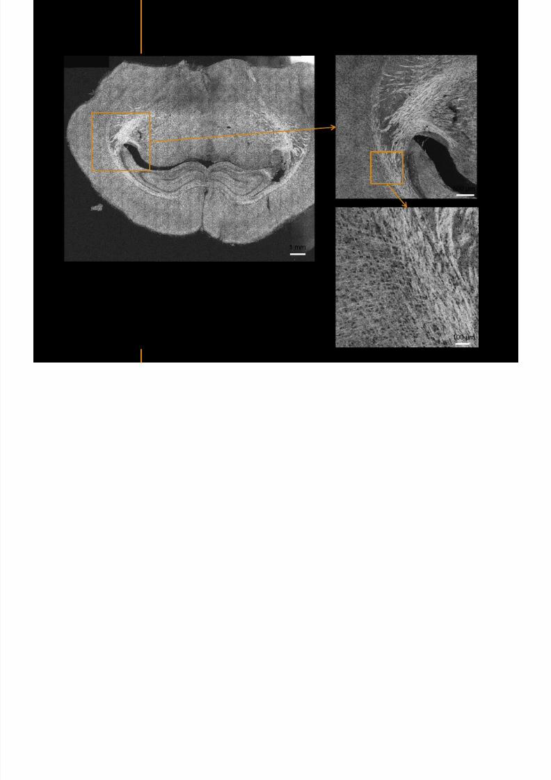
7292019 Atlas of Light-CT images
httpslidepdfcomreaderfullatlas-of-light-ct-images 25118
copy LLTech 2012 ndash wwwlltechimagingcom - No copy without authorization 25
Rat brain
Architectural and cellular information seen at various scales
Myelinated fibers visible
1 mm
500 microm
100 microm
7292019 Atlas of Light-CT images
httpslidepdfcomreaderfullatlas-of-light-ct-images 26118
7292019 Atlas of Light-CT images
httpslidepdfcomreaderfullatlas-of-light-ct-images 27118
7292019 Atlas of Light-CT images
httpslidepdfcomreaderfullatlas-of-light-ct-images 28118
copy LLTech 2012 ndash wwwlltechimagingcom - No copy without authorization 28 copy LLTECH 2011
Collagen-rich matrixWide fascicles of
tumour cells
Large capillary
Fibroblastic meningioma grade I
500microm
100microm 200microm200microm
7292019 Atlas of Light-CT images
httpslidepdfcomreaderfullatlas-of-light-ct-images 29118
copy LLTech 2012 ndash wwwlltechimagingcom - No copy without authorization 29
Transitional meningioma grade I
Psammoma surrounding a calcification
Whorls formation
Collagen-rich matrix
500microm
100microm
50microm
50microm
7292019 Atlas of Light-CT images
httpslidepdfcomreaderfullatlas-of-light-ct-images 30118
7292019 Atlas of Light-CT images
httpslidepdfcomreaderfullatlas-of-light-ct-images 31118
copy LLTech 2012 ndash wwwlltechimagingcom - No copy without authorization 31 copy LLTECH 2011
Arrows Fibers transverse cut
Spinal cord normal tissue
500 microm
100 microm
100 microm
7292019 Atlas of Light-CT images
httpslidepdfcomreaderfullatlas-of-light-ct-images 32118
7292019 Atlas of Light-CT images
httpslidepdfcomreaderfullatlas-of-light-ct-images 33118
7292019 Atlas of Light-CT images
httpslidepdfcomreaderfullatlas-of-light-ct-images 34118
copy LLTech 2012 ndash wwwlltechimagingcom - No copy without authorization 34
Back to table of contents
SKIN IMAGING
N l ki h l
7292019 Atlas of Light-CT images
httpslidepdfcomreaderfullatlas-of-light-ct-images 35118
copy LLTech 2012 ndash wwwlltechimagingcom - No copy without authorization 35
Normal skin morphology
Epidermis
Collagen
Pilosebaceous
unit
Sweat gland
Adipocytes
b
a
c
e
d
500 microm
copy LLTech 2012 Courtesy of Hopitaux Universitaires de Genegraveve Genegraveve Switzerland
7292019 Atlas of Light-CT images
httpslidepdfcomreaderfullatlas-of-light-ct-images 36118
copy LLTech 2012 ndash wwwlltechimagingcom - No copy without authorization 36
Hypodermis
Skin morphology layers are well discriminated
5 mm x 8 mm mosaic of native field image
Ageing patient (solar elastosis)
En-face imaging of vertical excision
Epidermis
Dermis
1
3
2
4
7292019 Atlas of Light-CT images
httpslidepdfcomreaderfullatlas-of-light-ct-images 37118
En face skin slicing shows structural and cellular details
7292019 Atlas of Light-CT images
httpslidepdfcomreaderfullatlas-of-light-ct-images 38118
copy LLTech 2012 ndash wwwlltechimagingcom - No copy without authorization 38
Stratum corneum Stratum spinosum
Stratum basale Reticular region
Epidermis
Dermis
En-face skin slicing shows structural and cellular details
Melanin
papillary caps
Keratinocyte
nuclei
Blood vessels
Collagen
Corneocytes
100 microm 100 microm
100 microm100 microm
copy LLTech 2012 Courtesy of Hopitaux Universitaires de Genegraveve Genegraveve Switzerland
7292019 Atlas of Light-CT images
httpslidepdfcomreaderfullatlas-of-light-ct-images 39118
7292019 Atlas of Light-CT images
httpslidepdfcomreaderfullatlas-of-light-ct-images 40118
Vertical reconstruction of skin model
7292019 Atlas of Light-CT images
httpslidepdfcomreaderfullatlas-of-light-ct-images 41118
copy LLTech 2012 ndash wwwlltechimagingcom - No copy without authorization 41
Vertical reconstruction of skin model
50 microm
Stratum corneum
Stratum spinosum
Dermis
Keratinocyte nuclei
copy LLTech 2012
7292019 Atlas of Light-CT images
httpslidepdfcomreaderfullatlas-of-light-ct-images 42118
Normal
7292019 Atlas of Light-CT images
httpslidepdfcomreaderfullatlas-of-light-ct-images 43118
copy LLTech 2012 ndash wwwlltechimagingcom - No copy without authorization 43
skin
b
a
e
c
f
d
500 microm
7292019 Atlas of Light-CT images
httpslidepdfcomreaderfullatlas-of-light-ct-images 44118
7292019 Atlas of Light-CT images
httpslidepdfcomreaderfullatlas-of-light-ct-images 45118
copy LLTech 2012 ndash wwwlltechimagingcom - No copy without authorization 45
Adipocytes
9um depth 27um depth 60um depth
Reconstructed
depth slice
200 microm
50 microm
7292019 Atlas of Light-CT images
httpslidepdfcomreaderfullatlas-of-light-ct-images 46118
Skin pathologies basal cell carcinoma discrimination at
7292019 Atlas of Light-CT images
httpslidepdfcomreaderfullatlas-of-light-ct-images 47118
copy LLTech 2012 ndash wwwlltechimagingcom - No copy without authorization 47
Epidermis
Dermis
Hypodermis
A
AA
C
BCC
PSU
PSU
BCC Basal cell carcinoma
A Adipocytes
C Collagen
PSU Pilosebaceous unit
structural level
1 mm
copy LLTech 2012 Courtesy of Hopitaux Universitaires de Genegraveve Genegraveve Switzerland
C
Zoom on basal cell carcinoma discrimination at cellular level
7292019 Atlas of Light-CT images
httpslidepdfcomreaderfullatlas-of-light-ct-images 48118
copy LLTech 2012 ndash wwwlltechimagingcom - No copy without authorization 48
Zoom on basal cell carcinoma discrimination at cellular level
200 microm
100 microm
Peritumoral stroma
High cell density
copy LLTech 2012 Courtesy of Hopitaux Universitaires de Genegraveve Genegraveve Switzerland
Skin pathologies discrimination of actinic keratosis
7292019 Atlas of Light-CT images
httpslidepdfcomreaderfullatlas-of-light-ct-images 49118
copy LLTech 2012 ndash wwwlltechimagingcom - No copy without authorization 49
Skin pathologies discrimination of actinic keratosis
Enlarged epidermis
Extended epidermal ridges
1 mm
copy LLTech 2012 Courtesy of Hopitaux Universitaires de Genegraveve Genegraveve Switzerland
7292019 Atlas of Light-CT images
httpslidepdfcomreaderfullatlas-of-light-ct-images 50118
copy LLTech 2012 ndash wwwlltechimagingcom - No copy without authorization 50
Basal cell carcinoma in depth imaging
Z = 10 microns 7 x 42 mm FOV Z = 35 microns 7 x 42 mm FOV
BCC
SG Sebaceous glands
Z = 55 microns 7 x 42 mm FOV Z = 75 microns 7 x 42 mm FOV
SG
Application to Mohs surgerycopy LLTech 2012 Courtesy of Hopitaux Universitaires de Genegraveve Genegraveve Switzerland
7292019 Atlas of Light-CT images
httpslidepdfcomreaderfullatlas-of-light-ct-images 51118
copy LLTech 2012 ndash wwwlltechimagingcom - No copy without authorization 51 copy LLTECH 2011
Healthy vs Cheloid scar tissue
Healthy skin tissueFOV 75x5mm
Cheloid scar tissueFOV 65x65mm
copy LLTECH 2011
7292019 Atlas of Light-CT images
httpslidepdfcomreaderfullatlas-of-light-ct-images 52118
copy LLTech 2012 ndash wwwlltechimagingcom - No copy without authorization 52
Dermo-epidermaljunction
Cheloid scar tissue in detail
copy LLTECH 2011
Nodular structureThickcollagenfiberbundles
1mm
600 microm
500 microm
h l d d f
7292019 Atlas of Light-CT images
httpslidepdfcomreaderfullatlas-of-light-ct-images 53118
copy LLTech 2012 ndash wwwlltechimagingcom - No copy without authorization 53
Cheloid scar tissue viewed en face
20microm depth 120microm depth
7292019 Atlas of Light-CT images
httpslidepdfcomreaderfullatlas-of-light-ct-images 54118
Skin in-vivo sweat duct imaging in 3D
7292019 Atlas of Light-CT images
httpslidepdfcomreaderfullatlas-of-light-ct-images 55118
copy LLTech 2012 ndash wwwlltechimagingcom - No copy without authorization 55
g g
Vertical reconstruction from en-face images
Sweat duct 3D reconstruction
Stratum corneum
Stratum spinosum
SD sweat ducts
SD SD
100 microm
7292019 Atlas of Light-CT images
httpslidepdfcomreaderfullatlas-of-light-ct-images 56118
7292019 Atlas of Light-CT images
httpslidepdfcomreaderfullatlas-of-light-ct-images 57118
7292019 Atlas of Light-CT images
httpslidepdfcomreaderfullatlas-of-light-ct-images 58118
copy LLTech 2012 ndash wwwlltechimagingcom - No copy without authorization 58
LUNG IMAGING
Back to table of contents
H L
7292019 Atlas of Light-CT images
httpslidepdfcomreaderfullatlas-of-light-ct-images 59118
copy LLTech 2012 ndash wwwlltechimagingcom - No copy without authorization 59
Zone 1
Zone 2
Zone 3
Zone 4
Zone with
cancerous tissue
Limit of cancerous
stroma
alveoli with
macrophages
Alveoles
Human Lung
500microm
copy LLTech 2012 Courtesy of Hocircpital Hotel-Dieu Paris France
H L Z 1 Z t
7292019 Atlas of Light-CT images
httpslidepdfcomreaderfullatlas-of-light-ct-images 60118
copy LLTech 2012 ndash wwwlltechimagingcom - No copy without authorization 60
Stroma
Human Lung ndash Zone 1 ndash Zoom on cancerous stroma
Cancerous
area
200microm
copy LLTech 2012 Courtesy of Hocircpital Hotel-Dieu Paris France
Human Lung ndash Zone 2 ndash Limit cancerous stroma alveoli
7292019 Atlas of Light-CT images
httpslidepdfcomreaderfullatlas-of-light-ct-images 61118
copy LLTech 2012 ndash wwwlltechimagingcom - No copy without authorization 61
g
with visible macrophages and vessel
Alveoli
Macrophages
Vessel
copy LLTech 2012 Courtesy of Hocircpital Hotel-Dieu Paris France
Human Lung ndash Zone 3 ndash Limit cancerous stroma alveoli
7292019 Atlas of Light-CT images
httpslidepdfcomreaderfullatlas-of-light-ct-images 62118
copy LLTech 2012 ndash wwwlltechimagingcom - No copy without authorization 62
g
with visible macrophages
Macrophages
100microm
copy LLTech 2012 Courtesy of Hocircpital Hotel-Dieu Paris France
Human Lung ndash Zone 4 ndash Alveoli
7292019 Atlas of Light-CT images
httpslidepdfcomreaderfullatlas-of-light-ct-images 63118
copy LLTech 2012 ndash wwwlltechimagingcom - No copy without authorization 63
Human Lung Zone 4 Alveoli
Alveoli
200microm
copy LLTech 2012 Courtesy of Hocircpital Hotel-Dieu Paris France
7292019 Atlas of Light-CT images
httpslidepdfcomreaderfullatlas-of-light-ct-images 64118
copy LLTech 2012 ndash wwwlltechimagingcom - No copy without authorization 64
GI IMAGING
Back to table of contents
7292019 Atlas of Light-CT images
httpslidepdfcomreaderfullatlas-of-light-ct-images 65118
Colonic carcinoma
7292019 Atlas of Light-CT images
httpslidepdfcomreaderfullatlas-of-light-ct-images 66118
copy LLTech 2012 ndash wwwlltechimagingcom - No copy without authorization 66
Colonic Carcinoma
Colonic carcinoma
copy LLTech 2012 Courtesy of MD Anderson Houston USA
Intestines ndash Crohnrsquos disease
7292019 Atlas of Light-CT images
httpslidepdfcomreaderfullatlas-of-light-ct-images 67118
copy LLTech 2012 ndash wwwlltechimagingcom - No copy without authorization 67
Crohnrsquos disease structure not
visible from the surface
Fat cells
200 microm
Intestines Crohn s disease
copy LLTech 2012 Courtesy of Institut Curie Paris France
Rat stomach
7292019 Atlas of Light-CT images
httpslidepdfcomreaderfullatlas-of-light-ct-images 68118
copy LLTech 2012 ndash wwwlltechimagingcom - No copy without authorization 68
Rat stomach
Mucosa withgastric pits
Submucosa with
bright collagen
fibers
Muscularis
propria with
muscle bundles
copy LLTech 2012 Courtesy of Weill Cornell Medical College NY USA
200 microm
7292019 Atlas of Light-CT images
httpslidepdfcomreaderfullatlas-of-light-ct-images 69118
copy LLTech 2012 ndash wwwlltechimagingcom - No copy without authorization 69 copy LLTECH 2011
UROGENITAL IMAGING
Back to table of contents
Rat bladder
7292019 Atlas of Light-CT images
httpslidepdfcomreaderfullatlas-of-light-ct-images 70118
copy LLTech 2012 ndash wwwlltechimagingcom - No copy without authorization 70
Rat bladder
Stratifiedurothelium
Lamina propria
500 microm
Muscular
bundles of the
lamina propria
Serosa
copy LLTech 2012 Courtesy of Weill Cornell Medical College NY USA
100 microm
100 microm
Human bladder low grade papillary urothelial carcinoma
7292019 Atlas of Light-CT images
httpslidepdfcomreaderfullatlas-of-light-ct-images 71118
copy LLTech 2012 ndash wwwlltechimagingcom - No copy without authorization 71
Lesion with fibrovascular cores (arrow) and superficial umbrella cells (arrowheads)
Human bladder low grade papillary urothelial carcinoma
copy LLTech 2012 Courtesy of Weill Cornell Medical College NY USA
100 microm
7292019 Atlas of Light-CT images
httpslidepdfcomreaderfullatlas-of-light-ct-images 72118
Rat prostate
7292019 Atlas of Light-CT images
httpslidepdfcomreaderfullatlas-of-light-ct-images 73118
copy LLTech 2012 ndash wwwlltechimagingcom - No copy without authorization 73
p
copy LLTech 2012 Courtesy of Weill Cornell Medical College NY USA
Papillary folds of the
acinar glands
Prostatic acini
Fibromuscular stromaPeriprostatic fat Blood vessel
1 mm
200 microm
Mouse kidney
7292019 Atlas of Light-CT images
httpslidepdfcomreaderfullatlas-of-light-ct-images 74118
copy LLTech 2012 ndash wwwlltechimagingcom - No copy without authorization 74
1 mm
y
copy LLTech 2012 Courtesy of Institut Biomeacutedical de Bicecirctre Paris France
Renal cortex
Renal medulla
Renal sinus
7292019 Atlas of Light-CT images
httpslidepdfcomreaderfullatlas-of-light-ct-images 75118
copy LLTech 2012 ndash wwwlltechimagingcom - No copy without authorization 75
Mouse kidney ndash zoom on highlighted section of previous slide
800microm
copy LLTech 2012 Courtesy of Hocircpital Kremlin Bicecirctre Paris France
7292019 Atlas of Light-CT images
httpslidepdfcomreaderfullatlas-of-light-ct-images 76118
7292019 Atlas of Light-CT images
httpslidepdfcomreaderfullatlas-of-light-ct-images 77118
7292019 Atlas of Light-CT images
httpslidepdfcomreaderfullatlas-of-light-ct-images 78118
copy LLTech 2012 ndash wwwlltechimagingcom - No copy without authorization 78 copy LLTECH 2011
CORE NEEDLE BIOPSIES
Back to table of contents
Kidney
7292019 Atlas of Light-CT images
httpslidepdfcomreaderfullatlas-of-light-ct-images 79118
copy LLTech 2012 ndash wwwlltechimagingcom - No copy without authorization 79
100microm
50microm
Renal tubules
Vessel
500microm
copy LLTech 2012 Courtesy of Institut Curie Paris France
Breast Invasive lobular carcinoma
7292019 Atlas of Light-CT images
httpslidepdfcomreaderfullatlas-of-light-ct-images 80118
copy LLTech 2012 ndash wwwlltechimagingcom - No copy without authorization 80
500microm
stroma
adipocytes
copy LLTech 2012 Courtesy of Institut Curie Paris France
7292019 Atlas of Light-CT images
httpslidepdfcomreaderfullatlas-of-light-ct-images 81118
Breast Ductal carcinoma in situ
7292019 Atlas of Light-CT images
httpslidepdfcomreaderfullatlas-of-light-ct-images 82118
copy LLTech 2012 ndash wwwlltechimagingcom - No copy without authorization 82
500microm
copy LLTech 2012 Courtesy of Institut Curie Paris France
7292019 Atlas of Light-CT images
httpslidepdfcomreaderfullatlas-of-light-ct-images 83118
copy LLTech 2012 ndash wwwlltechimagingcom - No copy without authorization 83 copy LLTECH 2011
THYROID IMAGING
Back to table of contents
7292019 Atlas of Light-CT images
httpslidepdfcomreaderfullatlas-of-light-ct-images 84118
copy LLTech 2012 ndash wwwlltechimagingcom - No copy without authorization 84
Thyroid
800 microm
200 microm
copy LLTech 2012 Courtesy of Hocircpital Tenon Paris France
7292019 Atlas of Light-CT images
httpslidepdfcomreaderfullatlas-of-light-ct-images 85118
copy LLTech 2012 ndash wwwlltechimagingcom - No copy without authorization 85
Thyroid
800 microm
200 microm
copy LLTech 2012 Courtesy of Hocircpital Tenon Paris France
7292019 Atlas of Light-CT images
httpslidepdfcomreaderfullatlas-of-light-ct-images 86118
copy LLTech 2012 ndash wwwlltechimagingcom - No copy without authorization 86 copy LLTECH 2011
FAT CELLS IMAGING
Back to table of contents
7292019 Atlas of Light-CT images
httpslidepdfcomreaderfullatlas-of-light-ct-images 87118
copy LLTech 2012 ndash wwwlltechimagingcom - No copy without authorization 87
Brown fat cells - adipocytes
100 microm 100 microm
200 microm
10um depth
Adipocytes
45um depth
En face
slices
Reconstructed
depth slice
copy LLTech 2012 Courtesy of Centre de Recherche des Cordeliers Paris France
7292019 Atlas of Light-CT images
httpslidepdfcomreaderfullatlas-of-light-ct-images 88118
copy LLTech 2012 ndash wwwlltechimagingcom - No copy without authorization 88 copy LLTECH 2011
CORNEA AND RETINA
Back to table of contents
Human cornea reconstructed depth slice
7292019 Atlas of Light-CT images
httpslidepdfcomreaderfullatlas-of-light-ct-images 89118
copy LLTech 2012 ndash wwwlltechimagingcom - No copy without authorization 89
50 um
wwwlltechimagingcom
7292019 Atlas of Light-CT images
httpslidepdfcomreaderfullatlas-of-light-ct-images 90118
Large field en face view in stroma
7292019 Atlas of Light-CT images
httpslidepdfcomreaderfullatlas-of-light-ct-images 91118
copy LLTech 2012 ndash wwwlltechimagingcom - No copy without authorization 91
200 um
wwwlltechimagingcom
7292019 Atlas of Light-CT images
httpslidepdfcomreaderfullatlas-of-light-ct-images 92118
Rabbit cornea after LASIK reflectivity profiles reveal
i d tt i d thi k d ith li l l
7292019 Atlas of Light-CT images
httpslidepdfcomreaderfullatlas-of-light-ct-images 93118
copy LLTech 2012 ndash wwwlltechimagingcom - No copy without authorization 93
increased scattering and thickened epithelial layer
LASIK LASIK CONTROL CONTROL
copy LLTech 2012
Newborn mouse anterior segment
7292019 Atlas of Light-CT images
httpslidepdfcomreaderfullatlas-of-light-ct-images 94118
copy LLTech 2012 ndash wwwlltechimagingcom - No copy without authorization 94
Corneal epithelial cells
Basal membrane
Stroma
Descemetrsquos membrane
Aqueous humor
Lens capsule
Lens epithelial cells
Cortical fibers
copy LLTech 2012 Courtesy of ESPCI Paris Francecopy LLTech 2012
Rat retina comparison with histology
7292019 Atlas of Light-CT images
httpslidepdfcomreaderfullatlas-of-light-ct-images 95118
copy LLTech 2012 ndash wwwlltechimagingcom - No copy without authorization 95
Figure 1 Right Ex vivo rat retinaimaged using the f ull field OCT
system of ESPCI Left Histology
for comparison
Figure 1 Right Ex vivo rat retinaimaged using the f ull field OCT
system of ESPCI Left Histology
for comparison
Figure 1 Right Ex vivo rat retinaimaged using the f ull field OCT
system of ESPCI Left Histology
for comparisonField
230x300microm
RPE
PRL
ONL
OPL
INL
IPL
GCL
NFL
Invest Ophthalmol Vis Sci 2004 Nov45(11)4126-31
Grieve K Paques M Dubois A Sahel J Boccara C Le Gargasson JF copy LLTech 2012
Rat retina
7292019 Atlas of Light-CT images
httpslidepdfcomreaderfullatlas-of-light-ct-images 96118
copy LLTech 2012 ndash wwwlltechimagingcom - No copy without authorization 96 copy LLTech 2012 Courtesy of ESPCI Paris France
Nerve fiber layer
Retina
Invest Ophthalmol Vis Sci 2004 Nov45(11)4126-31
Grieve K Paques M Dubois A Sahel J Boccara C Le Gargasson JF
Transscleral image
7292019 Atlas of Light-CT images
httpslidepdfcomreaderfullatlas-of-light-ct-images 97118
7292019 Atlas of Light-CT images
httpslidepdfcomreaderfullatlas-of-light-ct-images 98118
7292019 Atlas of Light-CT images
httpslidepdfcomreaderfullatlas-of-light-ct-images 99118
In vivo imaging of drosophila melanogaster
7292019 Atlas of Light-CT images
httpslidepdfcomreaderfullatlas-of-light-ct-images 100118
copy LLTech 2012 ndash wwwlltechimagingcom - No copy without authorization 100 copy LLTech 2012 Courtesy of ESPCI Paris France
In vivo imaging of drosophila melanogaster ndash four stages of pupation over 72 hours
7292019 Atlas of Light-CT images
httpslidepdfcomreaderfullatlas-of-light-ct-images 101118
copy LLTech 2012 ndash wwwlltechimagingcom - No copy without authorization 101
Prepupa (0-2 h) Transition topupal stage (24 h)
Pupal stage (48 h) Advanced pupal phase
before eclosion (72 h)
copy LLTech 2012 Courtesy of ESPCI Paris France
Pupa 118 4 days 30um depth
100
7292019 Atlas of Light-CT images
httpslidepdfcomreaderfullatlas-of-light-ct-images 102118
copy LLTech 2012 ndash wwwlltechimagingcom - No copy without authorization 102
100 um
Drosophila
7292019 Atlas of Light-CT images
httpslidepdfcomreaderfullatlas-of-light-ct-images 103118
copy LLTech 2012 ndash wwwlltechimagingcom - No copy without authorization 103
200 microm
copy LLTech 2012 Courtesy of ESPCI Paris France
C Elegans ndash Fast identification of anatomy
7292019 Atlas of Light-CT images
httpslidepdfcomreaderfullatlas-of-light-ct-images 104118
copy LLTech 2012 ndash wwwlltechimagingcom - No copy without authorization 104
Body wall - cuticle
gonad arm
30 microm depth
uterus
ovocytes
intestine
100 microm 100 microm
15 microm depth
copy LLTech 2012 Courtesy of ENS Paris France
C elegans ndash Images at higher magnification 40X
7292019 Atlas of Light-CT images
httpslidepdfcomreaderfullatlas-of-light-ct-images 105118
copy LLTech 2012 ndash wwwlltechimagingcom - No copy without authorization 105
3D reconstruction 50 microm
100 microm
copy LLTech 2012 Courtesy of ENS Paris France
Zebra fish eye
7292019 Atlas of Light-CT images
httpslidepdfcomreaderfullatlas-of-light-ct-images 106118
copy LLTech 2012 ndash wwwlltechimagingcom - No copy without authorization 106
Zebra fish eye
100 microm
100 microm
En face slices
Reconstructed depth slice
copy LLTech 2012 Courtesy of Inserm Paris France
10um depth 20um depth 45um depth
7292019 Atlas of Light-CT images
httpslidepdfcomreaderfullatlas-of-light-ct-images 107118
Xenopus Laevis backtail
7292019 Atlas of Light-CT images
httpslidepdfcomreaderfullatlas-of-light-ct-images 108118
copy LLTech 2012 ndash wwwlltechimagingcom - No copy without authorization 108
100 um30um depth 60um depth
7292019 Atlas of Light-CT images
httpslidepdfcomreaderfullatlas-of-light-ct-images 109118
Mouse embryo in vitro 6 5 days old ndash
7292019 Atlas of Light-CT images
httpslidepdfcomreaderfullatlas-of-light-ct-images 110118
copy LLTech 2012 ndash wwwlltechimagingcom - No copy without authorization 110
Mouse embryo in vitro 65 days old
copy LLTech 2012 Courtesy of Ellvire Guiot and nstitut Jacques Monod Paris France
3D imaging of internal cavity
Sabellaria alveolata mouth palps
7292019 Atlas of Light-CT images
httpslidepdfcomreaderfullatlas-of-light-ct-images 111118
copy LLTech 2012 ndash wwwlltechimagingcom - No copy without authorization 111
500 microm
100 microm
Mouth palps
copy LLTech 2012 Courtesy of Museacutee National drsquoHistoire Naturelle Paris France
7292019 Atlas of Light-CT images
httpslidepdfcomreaderfullatlas-of-light-ct-images 112118
copy LLTech 2012 ndash wwwlltechimagingcom - No copy without authorization 112 copy LLTECH 2011
PLANT IMAGING
Back to table of contents
Mouse ear leaf epidermis
7292019 Atlas of Light-CT images
httpslidepdfcomreaderfullatlas-of-light-ct-images 113118
copy LLTech 2012 ndash wwwlltechimagingcom - No copy without authorization 113
100 microm
Epidermal cell
copy LLTech 2012
Stoma
Leaf veins
7292019 Atlas of Light-CT images
httpslidepdfcomreaderfullatlas-of-light-ct-images 114118
copy LLTech 2012 ndash wwwlltechimagingcom - No copy without authorization 114
114copy LLTech 2012
Fibers
Veins
100 microm
500 microm
Apple
7292019 Atlas of Light-CT images
httpslidepdfcomreaderfullatlas-of-light-ct-images 115118
copy LLTech 2012 ndash wwwlltechimagingcom - No copy without authorization 115
115copy LLTech 2012
100 microm
Wax
Skin
Flesh
500 microm
Water
Effect of harpin infiltration on tobacco leaf
7292019 Atlas of Light-CT images
httpslidepdfcomreaderfullatlas-of-light-ct-images 116118
copy LLTech 2012 ndash wwwlltechimagingcom - No copy without authorization 116
116copy LLTech 2012 Courtesy of UPMC Paris France
50 microm
50 microm
50 microm
50 microm
Upper epidermis
10 min after harpin infiltration 30 min after harpin infiltration
Palisade parenchyma
Epidermis cell
Chloroplast
7292019 Atlas of Light-CT images
httpslidepdfcomreaderfullatlas-of-light-ct-images 117118
copy LLTech 2012 ndash wwwlltechimagingcom - No copy without authorization 117
bull Light-CTtrade does not require tissue preparation nor staining of any kind
bull Creates images within minutes using a non-destructive process
bull Offers a 1 microm cellular resolution in 3D
bull Reveals structural and cellular information
bull Mosaicing allows fast visualisation at various scale
bull En-face high-resolution imaging allows vertical and 3D reconstruction
bull In-vivo images show good promise for in-vivo system
bull Clinical and research applications in cancer detection cosmetics biobanking small animal
developmental biology botanyhellip
Contact Details
7292019 Atlas of Light-CT images
httpslidepdfcomreaderfullatlas-of-light-ct-images 118118
USA
LLTech Inc
103 Carnegie Center DriveSuite 300
Princeton NJ 08540
USA
wwwlltechimagingcom
contactlltechfr
Phone + 1 609 955 3506
LLTech SAS
29 rue du Faubourg St-JacquesPeacutepiniegravere Paris Santeacute Cochin
75014
Paris - France
wwwlltechimagingcom
contactlltechfr
Phone +33 1 82 72 61 25
Europe
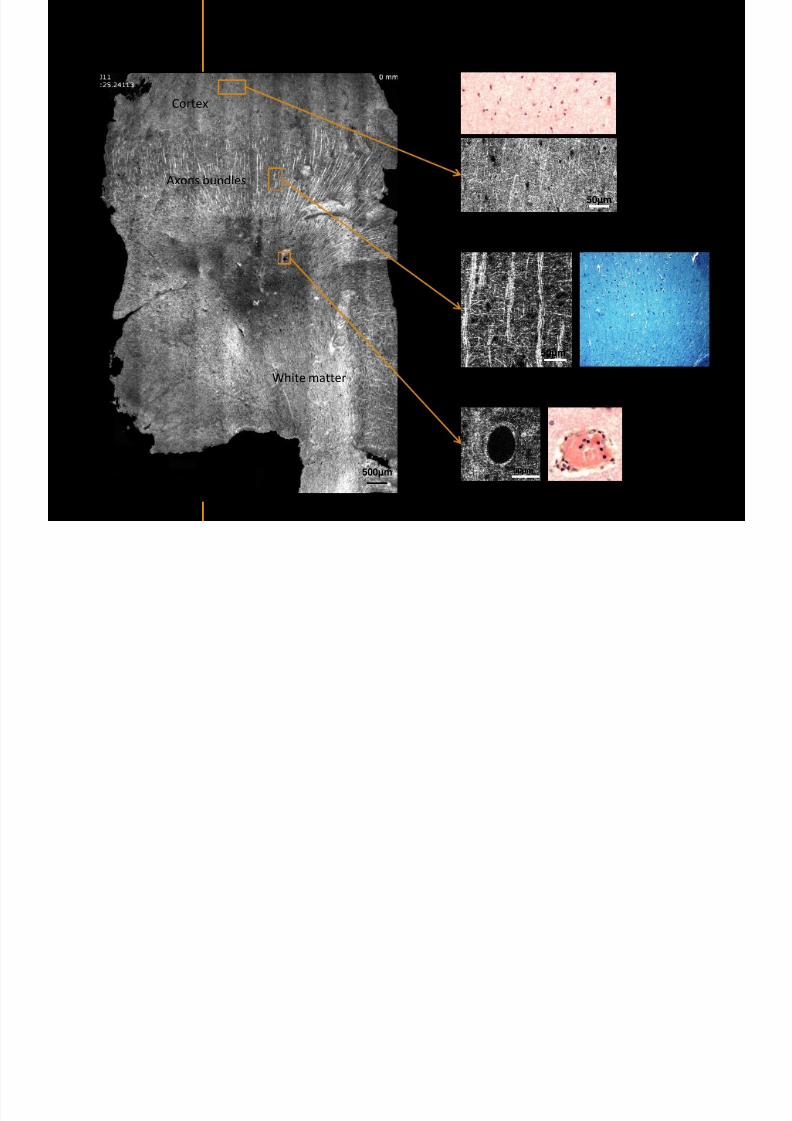
7292019 Atlas of Light-CT images
httpslidepdfcomreaderfullatlas-of-light-ct-images 26118
7292019 Atlas of Light-CT images
httpslidepdfcomreaderfullatlas-of-light-ct-images 27118
7292019 Atlas of Light-CT images
httpslidepdfcomreaderfullatlas-of-light-ct-images 28118
copy LLTech 2012 ndash wwwlltechimagingcom - No copy without authorization 28 copy LLTECH 2011
Collagen-rich matrixWide fascicles of
tumour cells
Large capillary
Fibroblastic meningioma grade I
500microm
100microm 200microm200microm
7292019 Atlas of Light-CT images
httpslidepdfcomreaderfullatlas-of-light-ct-images 29118
copy LLTech 2012 ndash wwwlltechimagingcom - No copy without authorization 29
Transitional meningioma grade I
Psammoma surrounding a calcification
Whorls formation
Collagen-rich matrix
500microm
100microm
50microm
50microm
7292019 Atlas of Light-CT images
httpslidepdfcomreaderfullatlas-of-light-ct-images 30118
7292019 Atlas of Light-CT images
httpslidepdfcomreaderfullatlas-of-light-ct-images 31118
copy LLTech 2012 ndash wwwlltechimagingcom - No copy without authorization 31 copy LLTECH 2011
Arrows Fibers transverse cut
Spinal cord normal tissue
500 microm
100 microm
100 microm
7292019 Atlas of Light-CT images
httpslidepdfcomreaderfullatlas-of-light-ct-images 32118
7292019 Atlas of Light-CT images
httpslidepdfcomreaderfullatlas-of-light-ct-images 33118
7292019 Atlas of Light-CT images
httpslidepdfcomreaderfullatlas-of-light-ct-images 34118
copy LLTech 2012 ndash wwwlltechimagingcom - No copy without authorization 34
Back to table of contents
SKIN IMAGING
N l ki h l
7292019 Atlas of Light-CT images
httpslidepdfcomreaderfullatlas-of-light-ct-images 35118
copy LLTech 2012 ndash wwwlltechimagingcom - No copy without authorization 35
Normal skin morphology
Epidermis
Collagen
Pilosebaceous
unit
Sweat gland
Adipocytes
b
a
c
e
d
500 microm
copy LLTech 2012 Courtesy of Hopitaux Universitaires de Genegraveve Genegraveve Switzerland
7292019 Atlas of Light-CT images
httpslidepdfcomreaderfullatlas-of-light-ct-images 36118
copy LLTech 2012 ndash wwwlltechimagingcom - No copy without authorization 36
Hypodermis
Skin morphology layers are well discriminated
5 mm x 8 mm mosaic of native field image
Ageing patient (solar elastosis)
En-face imaging of vertical excision
Epidermis
Dermis
1
3
2
4
7292019 Atlas of Light-CT images
httpslidepdfcomreaderfullatlas-of-light-ct-images 37118
En face skin slicing shows structural and cellular details
7292019 Atlas of Light-CT images
httpslidepdfcomreaderfullatlas-of-light-ct-images 38118
copy LLTech 2012 ndash wwwlltechimagingcom - No copy without authorization 38
Stratum corneum Stratum spinosum
Stratum basale Reticular region
Epidermis
Dermis
En-face skin slicing shows structural and cellular details
Melanin
papillary caps
Keratinocyte
nuclei
Blood vessels
Collagen
Corneocytes
100 microm 100 microm
100 microm100 microm
copy LLTech 2012 Courtesy of Hopitaux Universitaires de Genegraveve Genegraveve Switzerland
7292019 Atlas of Light-CT images
httpslidepdfcomreaderfullatlas-of-light-ct-images 39118
7292019 Atlas of Light-CT images
httpslidepdfcomreaderfullatlas-of-light-ct-images 40118
Vertical reconstruction of skin model
7292019 Atlas of Light-CT images
httpslidepdfcomreaderfullatlas-of-light-ct-images 41118
copy LLTech 2012 ndash wwwlltechimagingcom - No copy without authorization 41
Vertical reconstruction of skin model
50 microm
Stratum corneum
Stratum spinosum
Dermis
Keratinocyte nuclei
copy LLTech 2012
7292019 Atlas of Light-CT images
httpslidepdfcomreaderfullatlas-of-light-ct-images 42118
Normal
7292019 Atlas of Light-CT images
httpslidepdfcomreaderfullatlas-of-light-ct-images 43118
copy LLTech 2012 ndash wwwlltechimagingcom - No copy without authorization 43
skin
b
a
e
c
f
d
500 microm
7292019 Atlas of Light-CT images
httpslidepdfcomreaderfullatlas-of-light-ct-images 44118
7292019 Atlas of Light-CT images
httpslidepdfcomreaderfullatlas-of-light-ct-images 45118
copy LLTech 2012 ndash wwwlltechimagingcom - No copy without authorization 45
Adipocytes
9um depth 27um depth 60um depth
Reconstructed
depth slice
200 microm
50 microm
7292019 Atlas of Light-CT images
httpslidepdfcomreaderfullatlas-of-light-ct-images 46118
Skin pathologies basal cell carcinoma discrimination at
7292019 Atlas of Light-CT images
httpslidepdfcomreaderfullatlas-of-light-ct-images 47118
copy LLTech 2012 ndash wwwlltechimagingcom - No copy without authorization 47
Epidermis
Dermis
Hypodermis
A
AA
C
BCC
PSU
PSU
BCC Basal cell carcinoma
A Adipocytes
C Collagen
PSU Pilosebaceous unit
structural level
1 mm
copy LLTech 2012 Courtesy of Hopitaux Universitaires de Genegraveve Genegraveve Switzerland
C
Zoom on basal cell carcinoma discrimination at cellular level
7292019 Atlas of Light-CT images
httpslidepdfcomreaderfullatlas-of-light-ct-images 48118
copy LLTech 2012 ndash wwwlltechimagingcom - No copy without authorization 48
Zoom on basal cell carcinoma discrimination at cellular level
200 microm
100 microm
Peritumoral stroma
High cell density
copy LLTech 2012 Courtesy of Hopitaux Universitaires de Genegraveve Genegraveve Switzerland
Skin pathologies discrimination of actinic keratosis
7292019 Atlas of Light-CT images
httpslidepdfcomreaderfullatlas-of-light-ct-images 49118
copy LLTech 2012 ndash wwwlltechimagingcom - No copy without authorization 49
Skin pathologies discrimination of actinic keratosis
Enlarged epidermis
Extended epidermal ridges
1 mm
copy LLTech 2012 Courtesy of Hopitaux Universitaires de Genegraveve Genegraveve Switzerland
7292019 Atlas of Light-CT images
httpslidepdfcomreaderfullatlas-of-light-ct-images 50118
copy LLTech 2012 ndash wwwlltechimagingcom - No copy without authorization 50
Basal cell carcinoma in depth imaging
Z = 10 microns 7 x 42 mm FOV Z = 35 microns 7 x 42 mm FOV
BCC
SG Sebaceous glands
Z = 55 microns 7 x 42 mm FOV Z = 75 microns 7 x 42 mm FOV
SG
Application to Mohs surgerycopy LLTech 2012 Courtesy of Hopitaux Universitaires de Genegraveve Genegraveve Switzerland
7292019 Atlas of Light-CT images
httpslidepdfcomreaderfullatlas-of-light-ct-images 51118
copy LLTech 2012 ndash wwwlltechimagingcom - No copy without authorization 51 copy LLTECH 2011
Healthy vs Cheloid scar tissue
Healthy skin tissueFOV 75x5mm
Cheloid scar tissueFOV 65x65mm
copy LLTECH 2011
7292019 Atlas of Light-CT images
httpslidepdfcomreaderfullatlas-of-light-ct-images 52118
copy LLTech 2012 ndash wwwlltechimagingcom - No copy without authorization 52
Dermo-epidermaljunction
Cheloid scar tissue in detail
copy LLTECH 2011
Nodular structureThickcollagenfiberbundles
1mm
600 microm
500 microm
h l d d f
7292019 Atlas of Light-CT images
httpslidepdfcomreaderfullatlas-of-light-ct-images 53118
copy LLTech 2012 ndash wwwlltechimagingcom - No copy without authorization 53
Cheloid scar tissue viewed en face
20microm depth 120microm depth
7292019 Atlas of Light-CT images
httpslidepdfcomreaderfullatlas-of-light-ct-images 54118
Skin in-vivo sweat duct imaging in 3D
7292019 Atlas of Light-CT images
httpslidepdfcomreaderfullatlas-of-light-ct-images 55118
copy LLTech 2012 ndash wwwlltechimagingcom - No copy without authorization 55
g g
Vertical reconstruction from en-face images
Sweat duct 3D reconstruction
Stratum corneum
Stratum spinosum
SD sweat ducts
SD SD
100 microm
7292019 Atlas of Light-CT images
httpslidepdfcomreaderfullatlas-of-light-ct-images 56118
7292019 Atlas of Light-CT images
httpslidepdfcomreaderfullatlas-of-light-ct-images 57118
7292019 Atlas of Light-CT images
httpslidepdfcomreaderfullatlas-of-light-ct-images 58118
copy LLTech 2012 ndash wwwlltechimagingcom - No copy without authorization 58
LUNG IMAGING
Back to table of contents
H L
7292019 Atlas of Light-CT images
httpslidepdfcomreaderfullatlas-of-light-ct-images 59118
copy LLTech 2012 ndash wwwlltechimagingcom - No copy without authorization 59
Zone 1
Zone 2
Zone 3
Zone 4
Zone with
cancerous tissue
Limit of cancerous
stroma
alveoli with
macrophages
Alveoles
Human Lung
500microm
copy LLTech 2012 Courtesy of Hocircpital Hotel-Dieu Paris France
H L Z 1 Z t
7292019 Atlas of Light-CT images
httpslidepdfcomreaderfullatlas-of-light-ct-images 60118
copy LLTech 2012 ndash wwwlltechimagingcom - No copy without authorization 60
Stroma
Human Lung ndash Zone 1 ndash Zoom on cancerous stroma
Cancerous
area
200microm
copy LLTech 2012 Courtesy of Hocircpital Hotel-Dieu Paris France
Human Lung ndash Zone 2 ndash Limit cancerous stroma alveoli
7292019 Atlas of Light-CT images
httpslidepdfcomreaderfullatlas-of-light-ct-images 61118
copy LLTech 2012 ndash wwwlltechimagingcom - No copy without authorization 61
g
with visible macrophages and vessel
Alveoli
Macrophages
Vessel
copy LLTech 2012 Courtesy of Hocircpital Hotel-Dieu Paris France
Human Lung ndash Zone 3 ndash Limit cancerous stroma alveoli
7292019 Atlas of Light-CT images
httpslidepdfcomreaderfullatlas-of-light-ct-images 62118
copy LLTech 2012 ndash wwwlltechimagingcom - No copy without authorization 62
g
with visible macrophages
Macrophages
100microm
copy LLTech 2012 Courtesy of Hocircpital Hotel-Dieu Paris France
Human Lung ndash Zone 4 ndash Alveoli
7292019 Atlas of Light-CT images
httpslidepdfcomreaderfullatlas-of-light-ct-images 63118
copy LLTech 2012 ndash wwwlltechimagingcom - No copy without authorization 63
Human Lung Zone 4 Alveoli
Alveoli
200microm
copy LLTech 2012 Courtesy of Hocircpital Hotel-Dieu Paris France
7292019 Atlas of Light-CT images
httpslidepdfcomreaderfullatlas-of-light-ct-images 64118
copy LLTech 2012 ndash wwwlltechimagingcom - No copy without authorization 64
GI IMAGING
Back to table of contents
7292019 Atlas of Light-CT images
httpslidepdfcomreaderfullatlas-of-light-ct-images 65118
Colonic carcinoma
7292019 Atlas of Light-CT images
httpslidepdfcomreaderfullatlas-of-light-ct-images 66118
copy LLTech 2012 ndash wwwlltechimagingcom - No copy without authorization 66
Colonic Carcinoma
Colonic carcinoma
copy LLTech 2012 Courtesy of MD Anderson Houston USA
Intestines ndash Crohnrsquos disease
7292019 Atlas of Light-CT images
httpslidepdfcomreaderfullatlas-of-light-ct-images 67118
copy LLTech 2012 ndash wwwlltechimagingcom - No copy without authorization 67
Crohnrsquos disease structure not
visible from the surface
Fat cells
200 microm
Intestines Crohn s disease
copy LLTech 2012 Courtesy of Institut Curie Paris France
Rat stomach
7292019 Atlas of Light-CT images
httpslidepdfcomreaderfullatlas-of-light-ct-images 68118
copy LLTech 2012 ndash wwwlltechimagingcom - No copy without authorization 68
Rat stomach
Mucosa withgastric pits
Submucosa with
bright collagen
fibers
Muscularis
propria with
muscle bundles
copy LLTech 2012 Courtesy of Weill Cornell Medical College NY USA
200 microm
7292019 Atlas of Light-CT images
httpslidepdfcomreaderfullatlas-of-light-ct-images 69118
copy LLTech 2012 ndash wwwlltechimagingcom - No copy without authorization 69 copy LLTECH 2011
UROGENITAL IMAGING
Back to table of contents
Rat bladder
7292019 Atlas of Light-CT images
httpslidepdfcomreaderfullatlas-of-light-ct-images 70118
copy LLTech 2012 ndash wwwlltechimagingcom - No copy without authorization 70
Rat bladder
Stratifiedurothelium
Lamina propria
500 microm
Muscular
bundles of the
lamina propria
Serosa
copy LLTech 2012 Courtesy of Weill Cornell Medical College NY USA
100 microm
100 microm
Human bladder low grade papillary urothelial carcinoma
7292019 Atlas of Light-CT images
httpslidepdfcomreaderfullatlas-of-light-ct-images 71118
copy LLTech 2012 ndash wwwlltechimagingcom - No copy without authorization 71
Lesion with fibrovascular cores (arrow) and superficial umbrella cells (arrowheads)
Human bladder low grade papillary urothelial carcinoma
copy LLTech 2012 Courtesy of Weill Cornell Medical College NY USA
100 microm
7292019 Atlas of Light-CT images
httpslidepdfcomreaderfullatlas-of-light-ct-images 72118
Rat prostate
7292019 Atlas of Light-CT images
httpslidepdfcomreaderfullatlas-of-light-ct-images 73118
copy LLTech 2012 ndash wwwlltechimagingcom - No copy without authorization 73
p
copy LLTech 2012 Courtesy of Weill Cornell Medical College NY USA
Papillary folds of the
acinar glands
Prostatic acini
Fibromuscular stromaPeriprostatic fat Blood vessel
1 mm
200 microm
Mouse kidney
7292019 Atlas of Light-CT images
httpslidepdfcomreaderfullatlas-of-light-ct-images 74118
copy LLTech 2012 ndash wwwlltechimagingcom - No copy without authorization 74
1 mm
y
copy LLTech 2012 Courtesy of Institut Biomeacutedical de Bicecirctre Paris France
Renal cortex
Renal medulla
Renal sinus
7292019 Atlas of Light-CT images
httpslidepdfcomreaderfullatlas-of-light-ct-images 75118
copy LLTech 2012 ndash wwwlltechimagingcom - No copy without authorization 75
Mouse kidney ndash zoom on highlighted section of previous slide
800microm
copy LLTech 2012 Courtesy of Hocircpital Kremlin Bicecirctre Paris France
7292019 Atlas of Light-CT images
httpslidepdfcomreaderfullatlas-of-light-ct-images 76118
7292019 Atlas of Light-CT images
httpslidepdfcomreaderfullatlas-of-light-ct-images 77118
7292019 Atlas of Light-CT images
httpslidepdfcomreaderfullatlas-of-light-ct-images 78118
copy LLTech 2012 ndash wwwlltechimagingcom - No copy without authorization 78 copy LLTECH 2011
CORE NEEDLE BIOPSIES
Back to table of contents
Kidney
7292019 Atlas of Light-CT images
httpslidepdfcomreaderfullatlas-of-light-ct-images 79118
copy LLTech 2012 ndash wwwlltechimagingcom - No copy without authorization 79
100microm
50microm
Renal tubules
Vessel
500microm
copy LLTech 2012 Courtesy of Institut Curie Paris France
Breast Invasive lobular carcinoma
7292019 Atlas of Light-CT images
httpslidepdfcomreaderfullatlas-of-light-ct-images 80118
copy LLTech 2012 ndash wwwlltechimagingcom - No copy without authorization 80
500microm
stroma
adipocytes
copy LLTech 2012 Courtesy of Institut Curie Paris France
7292019 Atlas of Light-CT images
httpslidepdfcomreaderfullatlas-of-light-ct-images 81118
Breast Ductal carcinoma in situ
7292019 Atlas of Light-CT images
httpslidepdfcomreaderfullatlas-of-light-ct-images 82118
copy LLTech 2012 ndash wwwlltechimagingcom - No copy without authorization 82
500microm
copy LLTech 2012 Courtesy of Institut Curie Paris France
7292019 Atlas of Light-CT images
httpslidepdfcomreaderfullatlas-of-light-ct-images 83118
copy LLTech 2012 ndash wwwlltechimagingcom - No copy without authorization 83 copy LLTECH 2011
THYROID IMAGING
Back to table of contents
7292019 Atlas of Light-CT images
httpslidepdfcomreaderfullatlas-of-light-ct-images 84118
copy LLTech 2012 ndash wwwlltechimagingcom - No copy without authorization 84
Thyroid
800 microm
200 microm
copy LLTech 2012 Courtesy of Hocircpital Tenon Paris France
7292019 Atlas of Light-CT images
httpslidepdfcomreaderfullatlas-of-light-ct-images 85118
copy LLTech 2012 ndash wwwlltechimagingcom - No copy without authorization 85
Thyroid
800 microm
200 microm
copy LLTech 2012 Courtesy of Hocircpital Tenon Paris France
7292019 Atlas of Light-CT images
httpslidepdfcomreaderfullatlas-of-light-ct-images 86118
copy LLTech 2012 ndash wwwlltechimagingcom - No copy without authorization 86 copy LLTECH 2011
FAT CELLS IMAGING
Back to table of contents
7292019 Atlas of Light-CT images
httpslidepdfcomreaderfullatlas-of-light-ct-images 87118
copy LLTech 2012 ndash wwwlltechimagingcom - No copy without authorization 87
Brown fat cells - adipocytes
100 microm 100 microm
200 microm
10um depth
Adipocytes
45um depth
En face
slices
Reconstructed
depth slice
copy LLTech 2012 Courtesy of Centre de Recherche des Cordeliers Paris France
7292019 Atlas of Light-CT images
httpslidepdfcomreaderfullatlas-of-light-ct-images 88118
copy LLTech 2012 ndash wwwlltechimagingcom - No copy without authorization 88 copy LLTECH 2011
CORNEA AND RETINA
Back to table of contents
Human cornea reconstructed depth slice
7292019 Atlas of Light-CT images
httpslidepdfcomreaderfullatlas-of-light-ct-images 89118
copy LLTech 2012 ndash wwwlltechimagingcom - No copy without authorization 89
50 um
wwwlltechimagingcom
7292019 Atlas of Light-CT images
httpslidepdfcomreaderfullatlas-of-light-ct-images 90118
Large field en face view in stroma
7292019 Atlas of Light-CT images
httpslidepdfcomreaderfullatlas-of-light-ct-images 91118
copy LLTech 2012 ndash wwwlltechimagingcom - No copy without authorization 91
200 um
wwwlltechimagingcom
7292019 Atlas of Light-CT images
httpslidepdfcomreaderfullatlas-of-light-ct-images 92118
Rabbit cornea after LASIK reflectivity profiles reveal
i d tt i d thi k d ith li l l
7292019 Atlas of Light-CT images
httpslidepdfcomreaderfullatlas-of-light-ct-images 93118
copy LLTech 2012 ndash wwwlltechimagingcom - No copy without authorization 93
increased scattering and thickened epithelial layer
LASIK LASIK CONTROL CONTROL
copy LLTech 2012
Newborn mouse anterior segment
7292019 Atlas of Light-CT images
httpslidepdfcomreaderfullatlas-of-light-ct-images 94118
copy LLTech 2012 ndash wwwlltechimagingcom - No copy without authorization 94
Corneal epithelial cells
Basal membrane
Stroma
Descemetrsquos membrane
Aqueous humor
Lens capsule
Lens epithelial cells
Cortical fibers
copy LLTech 2012 Courtesy of ESPCI Paris Francecopy LLTech 2012
Rat retina comparison with histology
7292019 Atlas of Light-CT images
httpslidepdfcomreaderfullatlas-of-light-ct-images 95118
copy LLTech 2012 ndash wwwlltechimagingcom - No copy without authorization 95
Figure 1 Right Ex vivo rat retinaimaged using the f ull field OCT
system of ESPCI Left Histology
for comparison
Figure 1 Right Ex vivo rat retinaimaged using the f ull field OCT
system of ESPCI Left Histology
for comparison
Figure 1 Right Ex vivo rat retinaimaged using the f ull field OCT
system of ESPCI Left Histology
for comparisonField
230x300microm
RPE
PRL
ONL
OPL
INL
IPL
GCL
NFL
Invest Ophthalmol Vis Sci 2004 Nov45(11)4126-31
Grieve K Paques M Dubois A Sahel J Boccara C Le Gargasson JF copy LLTech 2012
Rat retina
7292019 Atlas of Light-CT images
httpslidepdfcomreaderfullatlas-of-light-ct-images 96118
copy LLTech 2012 ndash wwwlltechimagingcom - No copy without authorization 96 copy LLTech 2012 Courtesy of ESPCI Paris France
Nerve fiber layer
Retina
Invest Ophthalmol Vis Sci 2004 Nov45(11)4126-31
Grieve K Paques M Dubois A Sahel J Boccara C Le Gargasson JF
Transscleral image
7292019 Atlas of Light-CT images
httpslidepdfcomreaderfullatlas-of-light-ct-images 97118
7292019 Atlas of Light-CT images
httpslidepdfcomreaderfullatlas-of-light-ct-images 98118
7292019 Atlas of Light-CT images
httpslidepdfcomreaderfullatlas-of-light-ct-images 99118
In vivo imaging of drosophila melanogaster
7292019 Atlas of Light-CT images
httpslidepdfcomreaderfullatlas-of-light-ct-images 100118
copy LLTech 2012 ndash wwwlltechimagingcom - No copy without authorization 100 copy LLTech 2012 Courtesy of ESPCI Paris France
In vivo imaging of drosophila melanogaster ndash four stages of pupation over 72 hours
7292019 Atlas of Light-CT images
httpslidepdfcomreaderfullatlas-of-light-ct-images 101118
copy LLTech 2012 ndash wwwlltechimagingcom - No copy without authorization 101
Prepupa (0-2 h) Transition topupal stage (24 h)
Pupal stage (48 h) Advanced pupal phase
before eclosion (72 h)
copy LLTech 2012 Courtesy of ESPCI Paris France
Pupa 118 4 days 30um depth
100
7292019 Atlas of Light-CT images
httpslidepdfcomreaderfullatlas-of-light-ct-images 102118
copy LLTech 2012 ndash wwwlltechimagingcom - No copy without authorization 102
100 um
Drosophila
7292019 Atlas of Light-CT images
httpslidepdfcomreaderfullatlas-of-light-ct-images 103118
copy LLTech 2012 ndash wwwlltechimagingcom - No copy without authorization 103
200 microm
copy LLTech 2012 Courtesy of ESPCI Paris France
C Elegans ndash Fast identification of anatomy
7292019 Atlas of Light-CT images
httpslidepdfcomreaderfullatlas-of-light-ct-images 104118
copy LLTech 2012 ndash wwwlltechimagingcom - No copy without authorization 104
Body wall - cuticle
gonad arm
30 microm depth
uterus
ovocytes
intestine
100 microm 100 microm
15 microm depth
copy LLTech 2012 Courtesy of ENS Paris France
C elegans ndash Images at higher magnification 40X
7292019 Atlas of Light-CT images
httpslidepdfcomreaderfullatlas-of-light-ct-images 105118
copy LLTech 2012 ndash wwwlltechimagingcom - No copy without authorization 105
3D reconstruction 50 microm
100 microm
copy LLTech 2012 Courtesy of ENS Paris France
Zebra fish eye
7292019 Atlas of Light-CT images
httpslidepdfcomreaderfullatlas-of-light-ct-images 106118
copy LLTech 2012 ndash wwwlltechimagingcom - No copy without authorization 106
Zebra fish eye
100 microm
100 microm
En face slices
Reconstructed depth slice
copy LLTech 2012 Courtesy of Inserm Paris France
10um depth 20um depth 45um depth
7292019 Atlas of Light-CT images
httpslidepdfcomreaderfullatlas-of-light-ct-images 107118
Xenopus Laevis backtail
7292019 Atlas of Light-CT images
httpslidepdfcomreaderfullatlas-of-light-ct-images 108118
copy LLTech 2012 ndash wwwlltechimagingcom - No copy without authorization 108
100 um30um depth 60um depth
7292019 Atlas of Light-CT images
httpslidepdfcomreaderfullatlas-of-light-ct-images 109118
Mouse embryo in vitro 6 5 days old ndash
7292019 Atlas of Light-CT images
httpslidepdfcomreaderfullatlas-of-light-ct-images 110118
copy LLTech 2012 ndash wwwlltechimagingcom - No copy without authorization 110
Mouse embryo in vitro 65 days old
copy LLTech 2012 Courtesy of Ellvire Guiot and nstitut Jacques Monod Paris France
3D imaging of internal cavity
Sabellaria alveolata mouth palps
7292019 Atlas of Light-CT images
httpslidepdfcomreaderfullatlas-of-light-ct-images 111118
copy LLTech 2012 ndash wwwlltechimagingcom - No copy without authorization 111
500 microm
100 microm
Mouth palps
copy LLTech 2012 Courtesy of Museacutee National drsquoHistoire Naturelle Paris France
7292019 Atlas of Light-CT images
httpslidepdfcomreaderfullatlas-of-light-ct-images 112118
copy LLTech 2012 ndash wwwlltechimagingcom - No copy without authorization 112 copy LLTECH 2011
PLANT IMAGING
Back to table of contents
Mouse ear leaf epidermis
7292019 Atlas of Light-CT images
httpslidepdfcomreaderfullatlas-of-light-ct-images 113118
copy LLTech 2012 ndash wwwlltechimagingcom - No copy without authorization 113
100 microm
Epidermal cell
copy LLTech 2012
Stoma
Leaf veins
7292019 Atlas of Light-CT images
httpslidepdfcomreaderfullatlas-of-light-ct-images 114118
copy LLTech 2012 ndash wwwlltechimagingcom - No copy without authorization 114
114copy LLTech 2012
Fibers
Veins
100 microm
500 microm
Apple
7292019 Atlas of Light-CT images
httpslidepdfcomreaderfullatlas-of-light-ct-images 115118
copy LLTech 2012 ndash wwwlltechimagingcom - No copy without authorization 115
115copy LLTech 2012
100 microm
Wax
Skin
Flesh
500 microm
Water
Effect of harpin infiltration on tobacco leaf
7292019 Atlas of Light-CT images
httpslidepdfcomreaderfullatlas-of-light-ct-images 116118
copy LLTech 2012 ndash wwwlltechimagingcom - No copy without authorization 116
116copy LLTech 2012 Courtesy of UPMC Paris France
50 microm
50 microm
50 microm
50 microm
Upper epidermis
10 min after harpin infiltration 30 min after harpin infiltration
Palisade parenchyma
Epidermis cell
Chloroplast
7292019 Atlas of Light-CT images
httpslidepdfcomreaderfullatlas-of-light-ct-images 117118
copy LLTech 2012 ndash wwwlltechimagingcom - No copy without authorization 117
bull Light-CTtrade does not require tissue preparation nor staining of any kind
bull Creates images within minutes using a non-destructive process
bull Offers a 1 microm cellular resolution in 3D
bull Reveals structural and cellular information
bull Mosaicing allows fast visualisation at various scale
bull En-face high-resolution imaging allows vertical and 3D reconstruction
bull In-vivo images show good promise for in-vivo system
bull Clinical and research applications in cancer detection cosmetics biobanking small animal
developmental biology botanyhellip
Contact Details
7292019 Atlas of Light-CT images
httpslidepdfcomreaderfullatlas-of-light-ct-images 118118
USA
LLTech Inc
103 Carnegie Center DriveSuite 300
Princeton NJ 08540
USA
wwwlltechimagingcom
contactlltechfr
Phone + 1 609 955 3506
LLTech SAS
29 rue du Faubourg St-JacquesPeacutepiniegravere Paris Santeacute Cochin
75014
Paris - France
wwwlltechimagingcom
contactlltechfr
Phone +33 1 82 72 61 25
Europe
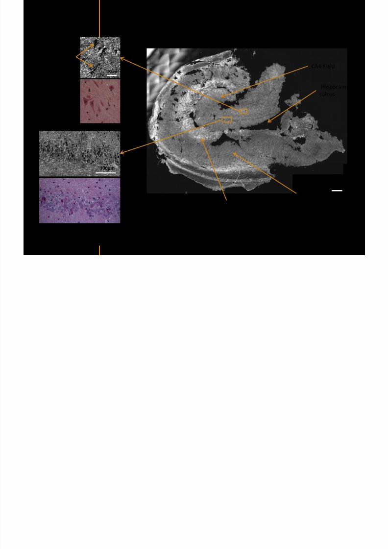
7292019 Atlas of Light-CT images
httpslidepdfcomreaderfullatlas-of-light-ct-images 29118
copy LLTech 2012 ndash wwwlltechimagingcom - No copy without authorization 29
Transitional meningioma grade I
Psammoma surrounding a calcification
Whorls formation
Collagen-rich matrix
500microm
100microm
50microm
50microm
7292019 Atlas of Light-CT images
httpslidepdfcomreaderfullatlas-of-light-ct-images 30118
7292019 Atlas of Light-CT images
httpslidepdfcomreaderfullatlas-of-light-ct-images 31118
copy LLTech 2012 ndash wwwlltechimagingcom - No copy without authorization 31 copy LLTECH 2011
Arrows Fibers transverse cut
Spinal cord normal tissue
500 microm
100 microm
100 microm
7292019 Atlas of Light-CT images
httpslidepdfcomreaderfullatlas-of-light-ct-images 32118
7292019 Atlas of Light-CT images
httpslidepdfcomreaderfullatlas-of-light-ct-images 33118
7292019 Atlas of Light-CT images
httpslidepdfcomreaderfullatlas-of-light-ct-images 34118
copy LLTech 2012 ndash wwwlltechimagingcom - No copy without authorization 34
Back to table of contents
SKIN IMAGING
N l ki h l
7292019 Atlas of Light-CT images
httpslidepdfcomreaderfullatlas-of-light-ct-images 35118
copy LLTech 2012 ndash wwwlltechimagingcom - No copy without authorization 35
Normal skin morphology
Epidermis
Collagen
Pilosebaceous
unit
Sweat gland
Adipocytes
b
a
c
e
d
500 microm
copy LLTech 2012 Courtesy of Hopitaux Universitaires de Genegraveve Genegraveve Switzerland
7292019 Atlas of Light-CT images
httpslidepdfcomreaderfullatlas-of-light-ct-images 36118
copy LLTech 2012 ndash wwwlltechimagingcom - No copy without authorization 36
Hypodermis
Skin morphology layers are well discriminated
5 mm x 8 mm mosaic of native field image
Ageing patient (solar elastosis)
En-face imaging of vertical excision
Epidermis
Dermis
1
3
2
4
7292019 Atlas of Light-CT images
httpslidepdfcomreaderfullatlas-of-light-ct-images 37118
En face skin slicing shows structural and cellular details
7292019 Atlas of Light-CT images
httpslidepdfcomreaderfullatlas-of-light-ct-images 38118
copy LLTech 2012 ndash wwwlltechimagingcom - No copy without authorization 38
Stratum corneum Stratum spinosum
Stratum basale Reticular region
Epidermis
Dermis
En-face skin slicing shows structural and cellular details
Melanin
papillary caps
Keratinocyte
nuclei
Blood vessels
Collagen
Corneocytes
100 microm 100 microm
100 microm100 microm
copy LLTech 2012 Courtesy of Hopitaux Universitaires de Genegraveve Genegraveve Switzerland
7292019 Atlas of Light-CT images
httpslidepdfcomreaderfullatlas-of-light-ct-images 39118
7292019 Atlas of Light-CT images
httpslidepdfcomreaderfullatlas-of-light-ct-images 40118
Vertical reconstruction of skin model
7292019 Atlas of Light-CT images
httpslidepdfcomreaderfullatlas-of-light-ct-images 41118
copy LLTech 2012 ndash wwwlltechimagingcom - No copy without authorization 41
Vertical reconstruction of skin model
50 microm
Stratum corneum
Stratum spinosum
Dermis
Keratinocyte nuclei
copy LLTech 2012
7292019 Atlas of Light-CT images
httpslidepdfcomreaderfullatlas-of-light-ct-images 42118
Normal
7292019 Atlas of Light-CT images
httpslidepdfcomreaderfullatlas-of-light-ct-images 43118
copy LLTech 2012 ndash wwwlltechimagingcom - No copy without authorization 43
skin
b
a
e
c
f
d
500 microm
7292019 Atlas of Light-CT images
httpslidepdfcomreaderfullatlas-of-light-ct-images 44118
7292019 Atlas of Light-CT images
httpslidepdfcomreaderfullatlas-of-light-ct-images 45118
copy LLTech 2012 ndash wwwlltechimagingcom - No copy without authorization 45
Adipocytes
9um depth 27um depth 60um depth
Reconstructed
depth slice
200 microm
50 microm
7292019 Atlas of Light-CT images
httpslidepdfcomreaderfullatlas-of-light-ct-images 46118
Skin pathologies basal cell carcinoma discrimination at
7292019 Atlas of Light-CT images
httpslidepdfcomreaderfullatlas-of-light-ct-images 47118
copy LLTech 2012 ndash wwwlltechimagingcom - No copy without authorization 47
Epidermis
Dermis
Hypodermis
A
AA
C
BCC
PSU
PSU
BCC Basal cell carcinoma
A Adipocytes
C Collagen
PSU Pilosebaceous unit
structural level
1 mm
copy LLTech 2012 Courtesy of Hopitaux Universitaires de Genegraveve Genegraveve Switzerland
C
Zoom on basal cell carcinoma discrimination at cellular level
7292019 Atlas of Light-CT images
httpslidepdfcomreaderfullatlas-of-light-ct-images 48118
copy LLTech 2012 ndash wwwlltechimagingcom - No copy without authorization 48
Zoom on basal cell carcinoma discrimination at cellular level
200 microm
100 microm
Peritumoral stroma
High cell density
copy LLTech 2012 Courtesy of Hopitaux Universitaires de Genegraveve Genegraveve Switzerland
Skin pathologies discrimination of actinic keratosis
7292019 Atlas of Light-CT images
httpslidepdfcomreaderfullatlas-of-light-ct-images 49118
copy LLTech 2012 ndash wwwlltechimagingcom - No copy without authorization 49
Skin pathologies discrimination of actinic keratosis
Enlarged epidermis
Extended epidermal ridges
1 mm
copy LLTech 2012 Courtesy of Hopitaux Universitaires de Genegraveve Genegraveve Switzerland
7292019 Atlas of Light-CT images
httpslidepdfcomreaderfullatlas-of-light-ct-images 50118
copy LLTech 2012 ndash wwwlltechimagingcom - No copy without authorization 50
Basal cell carcinoma in depth imaging
Z = 10 microns 7 x 42 mm FOV Z = 35 microns 7 x 42 mm FOV
BCC
SG Sebaceous glands
Z = 55 microns 7 x 42 mm FOV Z = 75 microns 7 x 42 mm FOV
SG
Application to Mohs surgerycopy LLTech 2012 Courtesy of Hopitaux Universitaires de Genegraveve Genegraveve Switzerland
7292019 Atlas of Light-CT images
httpslidepdfcomreaderfullatlas-of-light-ct-images 51118
copy LLTech 2012 ndash wwwlltechimagingcom - No copy without authorization 51 copy LLTECH 2011
Healthy vs Cheloid scar tissue
Healthy skin tissueFOV 75x5mm
Cheloid scar tissueFOV 65x65mm
copy LLTECH 2011
7292019 Atlas of Light-CT images
httpslidepdfcomreaderfullatlas-of-light-ct-images 52118
copy LLTech 2012 ndash wwwlltechimagingcom - No copy without authorization 52
Dermo-epidermaljunction
Cheloid scar tissue in detail
copy LLTECH 2011
Nodular structureThickcollagenfiberbundles
1mm
600 microm
500 microm
h l d d f
7292019 Atlas of Light-CT images
httpslidepdfcomreaderfullatlas-of-light-ct-images 53118
copy LLTech 2012 ndash wwwlltechimagingcom - No copy without authorization 53
Cheloid scar tissue viewed en face
20microm depth 120microm depth
7292019 Atlas of Light-CT images
httpslidepdfcomreaderfullatlas-of-light-ct-images 54118
Skin in-vivo sweat duct imaging in 3D
7292019 Atlas of Light-CT images
httpslidepdfcomreaderfullatlas-of-light-ct-images 55118
copy LLTech 2012 ndash wwwlltechimagingcom - No copy without authorization 55
g g
Vertical reconstruction from en-face images
Sweat duct 3D reconstruction
Stratum corneum
Stratum spinosum
SD sweat ducts
SD SD
100 microm
7292019 Atlas of Light-CT images
httpslidepdfcomreaderfullatlas-of-light-ct-images 56118
7292019 Atlas of Light-CT images
httpslidepdfcomreaderfullatlas-of-light-ct-images 57118
7292019 Atlas of Light-CT images
httpslidepdfcomreaderfullatlas-of-light-ct-images 58118
copy LLTech 2012 ndash wwwlltechimagingcom - No copy without authorization 58
LUNG IMAGING
Back to table of contents
H L
7292019 Atlas of Light-CT images
httpslidepdfcomreaderfullatlas-of-light-ct-images 59118
copy LLTech 2012 ndash wwwlltechimagingcom - No copy without authorization 59
Zone 1
Zone 2
Zone 3
Zone 4
Zone with
cancerous tissue
Limit of cancerous
stroma
alveoli with
macrophages
Alveoles
Human Lung
500microm
copy LLTech 2012 Courtesy of Hocircpital Hotel-Dieu Paris France
H L Z 1 Z t
7292019 Atlas of Light-CT images
httpslidepdfcomreaderfullatlas-of-light-ct-images 60118
copy LLTech 2012 ndash wwwlltechimagingcom - No copy without authorization 60
Stroma
Human Lung ndash Zone 1 ndash Zoom on cancerous stroma
Cancerous
area
200microm
copy LLTech 2012 Courtesy of Hocircpital Hotel-Dieu Paris France
Human Lung ndash Zone 2 ndash Limit cancerous stroma alveoli
7292019 Atlas of Light-CT images
httpslidepdfcomreaderfullatlas-of-light-ct-images 61118
copy LLTech 2012 ndash wwwlltechimagingcom - No copy without authorization 61
g
with visible macrophages and vessel
Alveoli
Macrophages
Vessel
copy LLTech 2012 Courtesy of Hocircpital Hotel-Dieu Paris France
Human Lung ndash Zone 3 ndash Limit cancerous stroma alveoli
7292019 Atlas of Light-CT images
httpslidepdfcomreaderfullatlas-of-light-ct-images 62118
copy LLTech 2012 ndash wwwlltechimagingcom - No copy without authorization 62
g
with visible macrophages
Macrophages
100microm
copy LLTech 2012 Courtesy of Hocircpital Hotel-Dieu Paris France
Human Lung ndash Zone 4 ndash Alveoli
7292019 Atlas of Light-CT images
httpslidepdfcomreaderfullatlas-of-light-ct-images 63118
copy LLTech 2012 ndash wwwlltechimagingcom - No copy without authorization 63
Human Lung Zone 4 Alveoli
Alveoli
200microm
copy LLTech 2012 Courtesy of Hocircpital Hotel-Dieu Paris France
7292019 Atlas of Light-CT images
httpslidepdfcomreaderfullatlas-of-light-ct-images 64118
copy LLTech 2012 ndash wwwlltechimagingcom - No copy without authorization 64
GI IMAGING
Back to table of contents
7292019 Atlas of Light-CT images
httpslidepdfcomreaderfullatlas-of-light-ct-images 65118
Colonic carcinoma
7292019 Atlas of Light-CT images
httpslidepdfcomreaderfullatlas-of-light-ct-images 66118
copy LLTech 2012 ndash wwwlltechimagingcom - No copy without authorization 66
Colonic Carcinoma
Colonic carcinoma
copy LLTech 2012 Courtesy of MD Anderson Houston USA
Intestines ndash Crohnrsquos disease
7292019 Atlas of Light-CT images
httpslidepdfcomreaderfullatlas-of-light-ct-images 67118
copy LLTech 2012 ndash wwwlltechimagingcom - No copy without authorization 67
Crohnrsquos disease structure not
visible from the surface
Fat cells
200 microm
Intestines Crohn s disease
copy LLTech 2012 Courtesy of Institut Curie Paris France
Rat stomach
7292019 Atlas of Light-CT images
httpslidepdfcomreaderfullatlas-of-light-ct-images 68118
copy LLTech 2012 ndash wwwlltechimagingcom - No copy without authorization 68
Rat stomach
Mucosa withgastric pits
Submucosa with
bright collagen
fibers
Muscularis
propria with
muscle bundles
copy LLTech 2012 Courtesy of Weill Cornell Medical College NY USA
200 microm
7292019 Atlas of Light-CT images
httpslidepdfcomreaderfullatlas-of-light-ct-images 69118
copy LLTech 2012 ndash wwwlltechimagingcom - No copy without authorization 69 copy LLTECH 2011
UROGENITAL IMAGING
Back to table of contents
Rat bladder
7292019 Atlas of Light-CT images
httpslidepdfcomreaderfullatlas-of-light-ct-images 70118
copy LLTech 2012 ndash wwwlltechimagingcom - No copy without authorization 70
Rat bladder
Stratifiedurothelium
Lamina propria
500 microm
Muscular
bundles of the
lamina propria
Serosa
copy LLTech 2012 Courtesy of Weill Cornell Medical College NY USA
100 microm
100 microm
Human bladder low grade papillary urothelial carcinoma
7292019 Atlas of Light-CT images
httpslidepdfcomreaderfullatlas-of-light-ct-images 71118
copy LLTech 2012 ndash wwwlltechimagingcom - No copy without authorization 71
Lesion with fibrovascular cores (arrow) and superficial umbrella cells (arrowheads)
Human bladder low grade papillary urothelial carcinoma
copy LLTech 2012 Courtesy of Weill Cornell Medical College NY USA
100 microm
7292019 Atlas of Light-CT images
httpslidepdfcomreaderfullatlas-of-light-ct-images 72118
Rat prostate
7292019 Atlas of Light-CT images
httpslidepdfcomreaderfullatlas-of-light-ct-images 73118
copy LLTech 2012 ndash wwwlltechimagingcom - No copy without authorization 73
p
copy LLTech 2012 Courtesy of Weill Cornell Medical College NY USA
Papillary folds of the
acinar glands
Prostatic acini
Fibromuscular stromaPeriprostatic fat Blood vessel
1 mm
200 microm
Mouse kidney
7292019 Atlas of Light-CT images
httpslidepdfcomreaderfullatlas-of-light-ct-images 74118
copy LLTech 2012 ndash wwwlltechimagingcom - No copy without authorization 74
1 mm
y
copy LLTech 2012 Courtesy of Institut Biomeacutedical de Bicecirctre Paris France
Renal cortex
Renal medulla
Renal sinus
7292019 Atlas of Light-CT images
httpslidepdfcomreaderfullatlas-of-light-ct-images 75118
copy LLTech 2012 ndash wwwlltechimagingcom - No copy without authorization 75
Mouse kidney ndash zoom on highlighted section of previous slide
800microm
copy LLTech 2012 Courtesy of Hocircpital Kremlin Bicecirctre Paris France
7292019 Atlas of Light-CT images
httpslidepdfcomreaderfullatlas-of-light-ct-images 76118
7292019 Atlas of Light-CT images
httpslidepdfcomreaderfullatlas-of-light-ct-images 77118
7292019 Atlas of Light-CT images
httpslidepdfcomreaderfullatlas-of-light-ct-images 78118
copy LLTech 2012 ndash wwwlltechimagingcom - No copy without authorization 78 copy LLTECH 2011
CORE NEEDLE BIOPSIES
Back to table of contents
Kidney
7292019 Atlas of Light-CT images
httpslidepdfcomreaderfullatlas-of-light-ct-images 79118
copy LLTech 2012 ndash wwwlltechimagingcom - No copy without authorization 79
100microm
50microm
Renal tubules
Vessel
500microm
copy LLTech 2012 Courtesy of Institut Curie Paris France
Breast Invasive lobular carcinoma
7292019 Atlas of Light-CT images
httpslidepdfcomreaderfullatlas-of-light-ct-images 80118
copy LLTech 2012 ndash wwwlltechimagingcom - No copy without authorization 80
500microm
stroma
adipocytes
copy LLTech 2012 Courtesy of Institut Curie Paris France
7292019 Atlas of Light-CT images
httpslidepdfcomreaderfullatlas-of-light-ct-images 81118
Breast Ductal carcinoma in situ
7292019 Atlas of Light-CT images
httpslidepdfcomreaderfullatlas-of-light-ct-images 82118
copy LLTech 2012 ndash wwwlltechimagingcom - No copy without authorization 82
500microm
copy LLTech 2012 Courtesy of Institut Curie Paris France
7292019 Atlas of Light-CT images
httpslidepdfcomreaderfullatlas-of-light-ct-images 83118
copy LLTech 2012 ndash wwwlltechimagingcom - No copy without authorization 83 copy LLTECH 2011
THYROID IMAGING
Back to table of contents
7292019 Atlas of Light-CT images
httpslidepdfcomreaderfullatlas-of-light-ct-images 84118
copy LLTech 2012 ndash wwwlltechimagingcom - No copy without authorization 84
Thyroid
800 microm
200 microm
copy LLTech 2012 Courtesy of Hocircpital Tenon Paris France
7292019 Atlas of Light-CT images
httpslidepdfcomreaderfullatlas-of-light-ct-images 85118
copy LLTech 2012 ndash wwwlltechimagingcom - No copy without authorization 85
Thyroid
800 microm
200 microm
copy LLTech 2012 Courtesy of Hocircpital Tenon Paris France
7292019 Atlas of Light-CT images
httpslidepdfcomreaderfullatlas-of-light-ct-images 86118
copy LLTech 2012 ndash wwwlltechimagingcom - No copy without authorization 86 copy LLTECH 2011
FAT CELLS IMAGING
Back to table of contents
7292019 Atlas of Light-CT images
httpslidepdfcomreaderfullatlas-of-light-ct-images 87118
copy LLTech 2012 ndash wwwlltechimagingcom - No copy without authorization 87
Brown fat cells - adipocytes
100 microm 100 microm
200 microm
10um depth
Adipocytes
45um depth
En face
slices
Reconstructed
depth slice
copy LLTech 2012 Courtesy of Centre de Recherche des Cordeliers Paris France
7292019 Atlas of Light-CT images
httpslidepdfcomreaderfullatlas-of-light-ct-images 88118
copy LLTech 2012 ndash wwwlltechimagingcom - No copy without authorization 88 copy LLTECH 2011
CORNEA AND RETINA
Back to table of contents
Human cornea reconstructed depth slice
7292019 Atlas of Light-CT images
httpslidepdfcomreaderfullatlas-of-light-ct-images 89118
copy LLTech 2012 ndash wwwlltechimagingcom - No copy without authorization 89
50 um
wwwlltechimagingcom
7292019 Atlas of Light-CT images
httpslidepdfcomreaderfullatlas-of-light-ct-images 90118
Large field en face view in stroma
7292019 Atlas of Light-CT images
httpslidepdfcomreaderfullatlas-of-light-ct-images 91118
copy LLTech 2012 ndash wwwlltechimagingcom - No copy without authorization 91
200 um
wwwlltechimagingcom
7292019 Atlas of Light-CT images
httpslidepdfcomreaderfullatlas-of-light-ct-images 92118
Rabbit cornea after LASIK reflectivity profiles reveal
i d tt i d thi k d ith li l l
7292019 Atlas of Light-CT images
httpslidepdfcomreaderfullatlas-of-light-ct-images 93118
copy LLTech 2012 ndash wwwlltechimagingcom - No copy without authorization 93
increased scattering and thickened epithelial layer
LASIK LASIK CONTROL CONTROL
copy LLTech 2012
Newborn mouse anterior segment
7292019 Atlas of Light-CT images
httpslidepdfcomreaderfullatlas-of-light-ct-images 94118
copy LLTech 2012 ndash wwwlltechimagingcom - No copy without authorization 94
Corneal epithelial cells
Basal membrane
Stroma
Descemetrsquos membrane
Aqueous humor
Lens capsule
Lens epithelial cells
Cortical fibers
copy LLTech 2012 Courtesy of ESPCI Paris Francecopy LLTech 2012
Rat retina comparison with histology
7292019 Atlas of Light-CT images
httpslidepdfcomreaderfullatlas-of-light-ct-images 95118
copy LLTech 2012 ndash wwwlltechimagingcom - No copy without authorization 95
Figure 1 Right Ex vivo rat retinaimaged using the f ull field OCT
system of ESPCI Left Histology
for comparison
Figure 1 Right Ex vivo rat retinaimaged using the f ull field OCT
system of ESPCI Left Histology
for comparison
Figure 1 Right Ex vivo rat retinaimaged using the f ull field OCT
system of ESPCI Left Histology
for comparisonField
230x300microm
RPE
PRL
ONL
OPL
INL
IPL
GCL
NFL
Invest Ophthalmol Vis Sci 2004 Nov45(11)4126-31
Grieve K Paques M Dubois A Sahel J Boccara C Le Gargasson JF copy LLTech 2012
Rat retina
7292019 Atlas of Light-CT images
httpslidepdfcomreaderfullatlas-of-light-ct-images 96118
copy LLTech 2012 ndash wwwlltechimagingcom - No copy without authorization 96 copy LLTech 2012 Courtesy of ESPCI Paris France
Nerve fiber layer
Retina
Invest Ophthalmol Vis Sci 2004 Nov45(11)4126-31
Grieve K Paques M Dubois A Sahel J Boccara C Le Gargasson JF
Transscleral image
7292019 Atlas of Light-CT images
httpslidepdfcomreaderfullatlas-of-light-ct-images 97118
7292019 Atlas of Light-CT images
httpslidepdfcomreaderfullatlas-of-light-ct-images 98118
7292019 Atlas of Light-CT images
httpslidepdfcomreaderfullatlas-of-light-ct-images 99118
In vivo imaging of drosophila melanogaster
7292019 Atlas of Light-CT images
httpslidepdfcomreaderfullatlas-of-light-ct-images 100118
copy LLTech 2012 ndash wwwlltechimagingcom - No copy without authorization 100 copy LLTech 2012 Courtesy of ESPCI Paris France
In vivo imaging of drosophila melanogaster ndash four stages of pupation over 72 hours
7292019 Atlas of Light-CT images
httpslidepdfcomreaderfullatlas-of-light-ct-images 101118
copy LLTech 2012 ndash wwwlltechimagingcom - No copy without authorization 101
Prepupa (0-2 h) Transition topupal stage (24 h)
Pupal stage (48 h) Advanced pupal phase
before eclosion (72 h)
copy LLTech 2012 Courtesy of ESPCI Paris France
Pupa 118 4 days 30um depth
100
7292019 Atlas of Light-CT images
httpslidepdfcomreaderfullatlas-of-light-ct-images 102118
copy LLTech 2012 ndash wwwlltechimagingcom - No copy without authorization 102
100 um
Drosophila
7292019 Atlas of Light-CT images
httpslidepdfcomreaderfullatlas-of-light-ct-images 103118
copy LLTech 2012 ndash wwwlltechimagingcom - No copy without authorization 103
200 microm
copy LLTech 2012 Courtesy of ESPCI Paris France
C Elegans ndash Fast identification of anatomy
7292019 Atlas of Light-CT images
httpslidepdfcomreaderfullatlas-of-light-ct-images 104118
copy LLTech 2012 ndash wwwlltechimagingcom - No copy without authorization 104
Body wall - cuticle
gonad arm
30 microm depth
uterus
ovocytes
intestine
100 microm 100 microm
15 microm depth
copy LLTech 2012 Courtesy of ENS Paris France
C elegans ndash Images at higher magnification 40X
7292019 Atlas of Light-CT images
httpslidepdfcomreaderfullatlas-of-light-ct-images 105118
copy LLTech 2012 ndash wwwlltechimagingcom - No copy without authorization 105
3D reconstruction 50 microm
100 microm
copy LLTech 2012 Courtesy of ENS Paris France
Zebra fish eye
7292019 Atlas of Light-CT images
httpslidepdfcomreaderfullatlas-of-light-ct-images 106118
copy LLTech 2012 ndash wwwlltechimagingcom - No copy without authorization 106
Zebra fish eye
100 microm
100 microm
En face slices
Reconstructed depth slice
copy LLTech 2012 Courtesy of Inserm Paris France
10um depth 20um depth 45um depth
7292019 Atlas of Light-CT images
httpslidepdfcomreaderfullatlas-of-light-ct-images 107118
Xenopus Laevis backtail
7292019 Atlas of Light-CT images
httpslidepdfcomreaderfullatlas-of-light-ct-images 108118
copy LLTech 2012 ndash wwwlltechimagingcom - No copy without authorization 108
100 um30um depth 60um depth
7292019 Atlas of Light-CT images
httpslidepdfcomreaderfullatlas-of-light-ct-images 109118
Mouse embryo in vitro 6 5 days old ndash
7292019 Atlas of Light-CT images
httpslidepdfcomreaderfullatlas-of-light-ct-images 110118
copy LLTech 2012 ndash wwwlltechimagingcom - No copy without authorization 110
Mouse embryo in vitro 65 days old
copy LLTech 2012 Courtesy of Ellvire Guiot and nstitut Jacques Monod Paris France
3D imaging of internal cavity
Sabellaria alveolata mouth palps
7292019 Atlas of Light-CT images
httpslidepdfcomreaderfullatlas-of-light-ct-images 111118
copy LLTech 2012 ndash wwwlltechimagingcom - No copy without authorization 111
500 microm
100 microm
Mouth palps
copy LLTech 2012 Courtesy of Museacutee National drsquoHistoire Naturelle Paris France
7292019 Atlas of Light-CT images
httpslidepdfcomreaderfullatlas-of-light-ct-images 112118
copy LLTech 2012 ndash wwwlltechimagingcom - No copy without authorization 112 copy LLTECH 2011
PLANT IMAGING
Back to table of contents
Mouse ear leaf epidermis
7292019 Atlas of Light-CT images
httpslidepdfcomreaderfullatlas-of-light-ct-images 113118
copy LLTech 2012 ndash wwwlltechimagingcom - No copy without authorization 113
100 microm
Epidermal cell
copy LLTech 2012
Stoma
Leaf veins
7292019 Atlas of Light-CT images
httpslidepdfcomreaderfullatlas-of-light-ct-images 114118
copy LLTech 2012 ndash wwwlltechimagingcom - No copy without authorization 114
114copy LLTech 2012
Fibers
Veins
100 microm
500 microm
Apple
7292019 Atlas of Light-CT images
httpslidepdfcomreaderfullatlas-of-light-ct-images 115118
copy LLTech 2012 ndash wwwlltechimagingcom - No copy without authorization 115
115copy LLTech 2012
100 microm
Wax
Skin
Flesh
500 microm
Water
Effect of harpin infiltration on tobacco leaf
7292019 Atlas of Light-CT images
httpslidepdfcomreaderfullatlas-of-light-ct-images 116118
copy LLTech 2012 ndash wwwlltechimagingcom - No copy without authorization 116
116copy LLTech 2012 Courtesy of UPMC Paris France
50 microm
50 microm
50 microm
50 microm
Upper epidermis
10 min after harpin infiltration 30 min after harpin infiltration
Palisade parenchyma
Epidermis cell
Chloroplast
7292019 Atlas of Light-CT images
httpslidepdfcomreaderfullatlas-of-light-ct-images 117118
copy LLTech 2012 ndash wwwlltechimagingcom - No copy without authorization 117
bull Light-CTtrade does not require tissue preparation nor staining of any kind
bull Creates images within minutes using a non-destructive process
bull Offers a 1 microm cellular resolution in 3D
bull Reveals structural and cellular information
bull Mosaicing allows fast visualisation at various scale
bull En-face high-resolution imaging allows vertical and 3D reconstruction
bull In-vivo images show good promise for in-vivo system
bull Clinical and research applications in cancer detection cosmetics biobanking small animal
developmental biology botanyhellip
Contact Details
7292019 Atlas of Light-CT images
httpslidepdfcomreaderfullatlas-of-light-ct-images 118118
USA
LLTech Inc
103 Carnegie Center DriveSuite 300
Princeton NJ 08540
USA
wwwlltechimagingcom
contactlltechfr
Phone + 1 609 955 3506
LLTech SAS
29 rue du Faubourg St-JacquesPeacutepiniegravere Paris Santeacute Cochin
75014
Paris - France
wwwlltechimagingcom
contactlltechfr
Phone +33 1 82 72 61 25
Europe
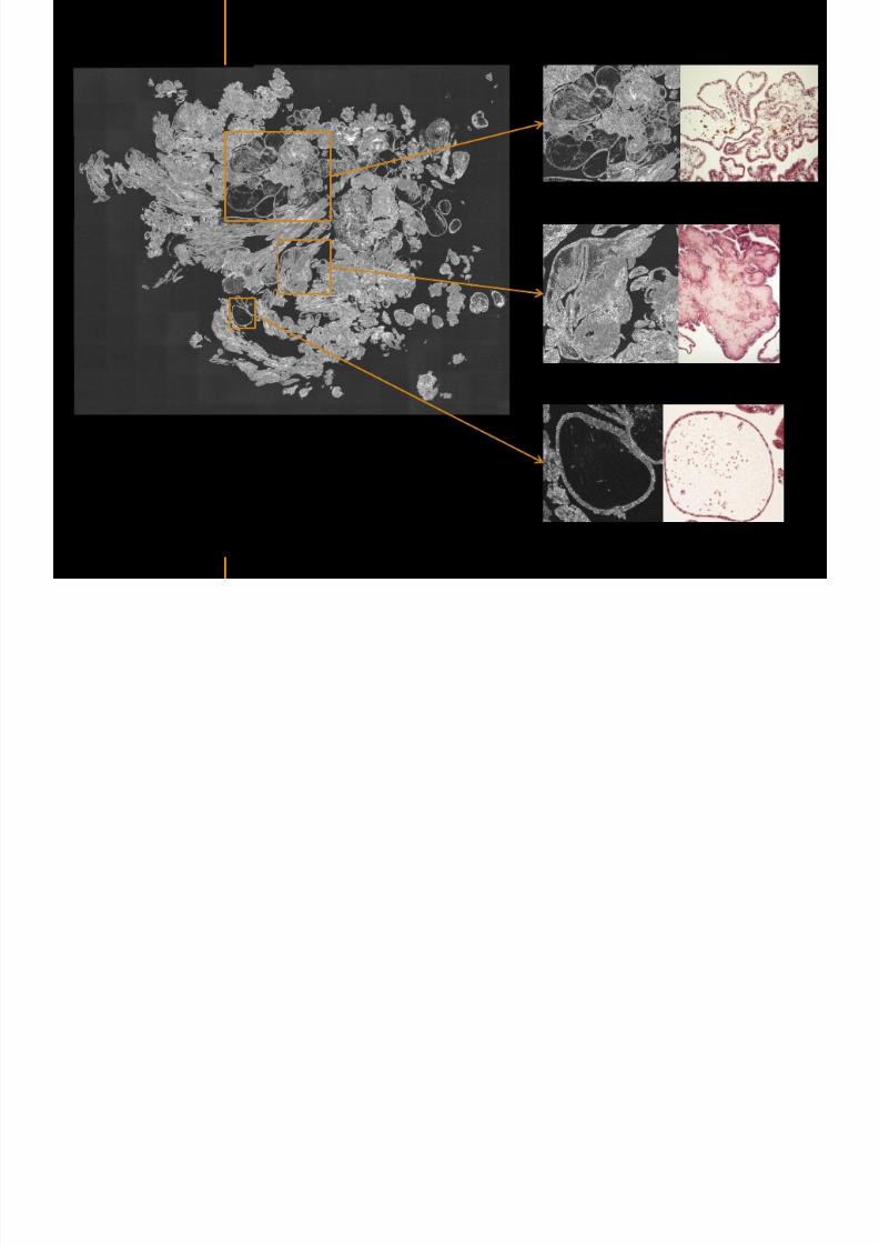
7292019 Atlas of Light-CT images
httpslidepdfcomreaderfullatlas-of-light-ct-images 36118
copy LLTech 2012 ndash wwwlltechimagingcom - No copy without authorization 36
Hypodermis
Skin morphology layers are well discriminated
5 mm x 8 mm mosaic of native field image
Ageing patient (solar elastosis)
En-face imaging of vertical excision
Epidermis
Dermis
1
3
2
4
7292019 Atlas of Light-CT images
httpslidepdfcomreaderfullatlas-of-light-ct-images 37118
En face skin slicing shows structural and cellular details
7292019 Atlas of Light-CT images
httpslidepdfcomreaderfullatlas-of-light-ct-images 38118
copy LLTech 2012 ndash wwwlltechimagingcom - No copy without authorization 38
Stratum corneum Stratum spinosum
Stratum basale Reticular region
Epidermis
Dermis
En-face skin slicing shows structural and cellular details
Melanin
papillary caps
Keratinocyte
nuclei
Blood vessels
Collagen
Corneocytes
100 microm 100 microm
100 microm100 microm
copy LLTech 2012 Courtesy of Hopitaux Universitaires de Genegraveve Genegraveve Switzerland
7292019 Atlas of Light-CT images
httpslidepdfcomreaderfullatlas-of-light-ct-images 39118
7292019 Atlas of Light-CT images
httpslidepdfcomreaderfullatlas-of-light-ct-images 40118
Vertical reconstruction of skin model
7292019 Atlas of Light-CT images
httpslidepdfcomreaderfullatlas-of-light-ct-images 41118
copy LLTech 2012 ndash wwwlltechimagingcom - No copy without authorization 41
Vertical reconstruction of skin model
50 microm
Stratum corneum
Stratum spinosum
Dermis
Keratinocyte nuclei
copy LLTech 2012
7292019 Atlas of Light-CT images
httpslidepdfcomreaderfullatlas-of-light-ct-images 42118
Normal
7292019 Atlas of Light-CT images
httpslidepdfcomreaderfullatlas-of-light-ct-images 43118
copy LLTech 2012 ndash wwwlltechimagingcom - No copy without authorization 43
skin
b
a
e
c
f
d
500 microm
7292019 Atlas of Light-CT images
httpslidepdfcomreaderfullatlas-of-light-ct-images 44118
7292019 Atlas of Light-CT images
httpslidepdfcomreaderfullatlas-of-light-ct-images 45118
copy LLTech 2012 ndash wwwlltechimagingcom - No copy without authorization 45
Adipocytes
9um depth 27um depth 60um depth
Reconstructed
depth slice
200 microm
50 microm
7292019 Atlas of Light-CT images
httpslidepdfcomreaderfullatlas-of-light-ct-images 46118
Skin pathologies basal cell carcinoma discrimination at
7292019 Atlas of Light-CT images
httpslidepdfcomreaderfullatlas-of-light-ct-images 47118
copy LLTech 2012 ndash wwwlltechimagingcom - No copy without authorization 47
Epidermis
Dermis
Hypodermis
A
AA
C
BCC
PSU
PSU
BCC Basal cell carcinoma
A Adipocytes
C Collagen
PSU Pilosebaceous unit
structural level
1 mm
copy LLTech 2012 Courtesy of Hopitaux Universitaires de Genegraveve Genegraveve Switzerland
C
Zoom on basal cell carcinoma discrimination at cellular level
7292019 Atlas of Light-CT images
httpslidepdfcomreaderfullatlas-of-light-ct-images 48118
copy LLTech 2012 ndash wwwlltechimagingcom - No copy without authorization 48
Zoom on basal cell carcinoma discrimination at cellular level
200 microm
100 microm
Peritumoral stroma
High cell density
copy LLTech 2012 Courtesy of Hopitaux Universitaires de Genegraveve Genegraveve Switzerland
Skin pathologies discrimination of actinic keratosis
7292019 Atlas of Light-CT images
httpslidepdfcomreaderfullatlas-of-light-ct-images 49118
copy LLTech 2012 ndash wwwlltechimagingcom - No copy without authorization 49
Skin pathologies discrimination of actinic keratosis
Enlarged epidermis
Extended epidermal ridges
1 mm
copy LLTech 2012 Courtesy of Hopitaux Universitaires de Genegraveve Genegraveve Switzerland
7292019 Atlas of Light-CT images
httpslidepdfcomreaderfullatlas-of-light-ct-images 50118
copy LLTech 2012 ndash wwwlltechimagingcom - No copy without authorization 50
Basal cell carcinoma in depth imaging
Z = 10 microns 7 x 42 mm FOV Z = 35 microns 7 x 42 mm FOV
BCC
SG Sebaceous glands
Z = 55 microns 7 x 42 mm FOV Z = 75 microns 7 x 42 mm FOV
SG
Application to Mohs surgerycopy LLTech 2012 Courtesy of Hopitaux Universitaires de Genegraveve Genegraveve Switzerland
7292019 Atlas of Light-CT images
httpslidepdfcomreaderfullatlas-of-light-ct-images 51118
copy LLTech 2012 ndash wwwlltechimagingcom - No copy without authorization 51 copy LLTECH 2011
Healthy vs Cheloid scar tissue
Healthy skin tissueFOV 75x5mm
Cheloid scar tissueFOV 65x65mm
copy LLTECH 2011
7292019 Atlas of Light-CT images
httpslidepdfcomreaderfullatlas-of-light-ct-images 52118
copy LLTech 2012 ndash wwwlltechimagingcom - No copy without authorization 52
Dermo-epidermaljunction
Cheloid scar tissue in detail
copy LLTECH 2011
Nodular structureThickcollagenfiberbundles
1mm
600 microm
500 microm
h l d d f
7292019 Atlas of Light-CT images
httpslidepdfcomreaderfullatlas-of-light-ct-images 53118
copy LLTech 2012 ndash wwwlltechimagingcom - No copy without authorization 53
Cheloid scar tissue viewed en face
20microm depth 120microm depth
7292019 Atlas of Light-CT images
httpslidepdfcomreaderfullatlas-of-light-ct-images 54118
Skin in-vivo sweat duct imaging in 3D
7292019 Atlas of Light-CT images
httpslidepdfcomreaderfullatlas-of-light-ct-images 55118
copy LLTech 2012 ndash wwwlltechimagingcom - No copy without authorization 55
g g
Vertical reconstruction from en-face images
Sweat duct 3D reconstruction
Stratum corneum
Stratum spinosum
SD sweat ducts
SD SD
100 microm
7292019 Atlas of Light-CT images
httpslidepdfcomreaderfullatlas-of-light-ct-images 56118
7292019 Atlas of Light-CT images
httpslidepdfcomreaderfullatlas-of-light-ct-images 57118
7292019 Atlas of Light-CT images
httpslidepdfcomreaderfullatlas-of-light-ct-images 58118
copy LLTech 2012 ndash wwwlltechimagingcom - No copy without authorization 58
LUNG IMAGING
Back to table of contents
H L
7292019 Atlas of Light-CT images
httpslidepdfcomreaderfullatlas-of-light-ct-images 59118
copy LLTech 2012 ndash wwwlltechimagingcom - No copy without authorization 59
Zone 1
Zone 2
Zone 3
Zone 4
Zone with
cancerous tissue
Limit of cancerous
stroma
alveoli with
macrophages
Alveoles
Human Lung
500microm
copy LLTech 2012 Courtesy of Hocircpital Hotel-Dieu Paris France
H L Z 1 Z t
7292019 Atlas of Light-CT images
httpslidepdfcomreaderfullatlas-of-light-ct-images 60118
copy LLTech 2012 ndash wwwlltechimagingcom - No copy without authorization 60
Stroma
Human Lung ndash Zone 1 ndash Zoom on cancerous stroma
Cancerous
area
200microm
copy LLTech 2012 Courtesy of Hocircpital Hotel-Dieu Paris France
Human Lung ndash Zone 2 ndash Limit cancerous stroma alveoli
7292019 Atlas of Light-CT images
httpslidepdfcomreaderfullatlas-of-light-ct-images 61118
copy LLTech 2012 ndash wwwlltechimagingcom - No copy without authorization 61
g
with visible macrophages and vessel
Alveoli
Macrophages
Vessel
copy LLTech 2012 Courtesy of Hocircpital Hotel-Dieu Paris France
Human Lung ndash Zone 3 ndash Limit cancerous stroma alveoli
7292019 Atlas of Light-CT images
httpslidepdfcomreaderfullatlas-of-light-ct-images 62118
copy LLTech 2012 ndash wwwlltechimagingcom - No copy without authorization 62
g
with visible macrophages
Macrophages
100microm
copy LLTech 2012 Courtesy of Hocircpital Hotel-Dieu Paris France
Human Lung ndash Zone 4 ndash Alveoli
7292019 Atlas of Light-CT images
httpslidepdfcomreaderfullatlas-of-light-ct-images 63118
copy LLTech 2012 ndash wwwlltechimagingcom - No copy without authorization 63
Human Lung Zone 4 Alveoli
Alveoli
200microm
copy LLTech 2012 Courtesy of Hocircpital Hotel-Dieu Paris France
7292019 Atlas of Light-CT images
httpslidepdfcomreaderfullatlas-of-light-ct-images 64118
copy LLTech 2012 ndash wwwlltechimagingcom - No copy without authorization 64
GI IMAGING
Back to table of contents
7292019 Atlas of Light-CT images
httpslidepdfcomreaderfullatlas-of-light-ct-images 65118
Colonic carcinoma
7292019 Atlas of Light-CT images
httpslidepdfcomreaderfullatlas-of-light-ct-images 66118
copy LLTech 2012 ndash wwwlltechimagingcom - No copy without authorization 66
Colonic Carcinoma
Colonic carcinoma
copy LLTech 2012 Courtesy of MD Anderson Houston USA
Intestines ndash Crohnrsquos disease
7292019 Atlas of Light-CT images
httpslidepdfcomreaderfullatlas-of-light-ct-images 67118
copy LLTech 2012 ndash wwwlltechimagingcom - No copy without authorization 67
Crohnrsquos disease structure not
visible from the surface
Fat cells
200 microm
Intestines Crohn s disease
copy LLTech 2012 Courtesy of Institut Curie Paris France
Rat stomach
7292019 Atlas of Light-CT images
httpslidepdfcomreaderfullatlas-of-light-ct-images 68118
copy LLTech 2012 ndash wwwlltechimagingcom - No copy without authorization 68
Rat stomach
Mucosa withgastric pits
Submucosa with
bright collagen
fibers
Muscularis
propria with
muscle bundles
copy LLTech 2012 Courtesy of Weill Cornell Medical College NY USA
200 microm
7292019 Atlas of Light-CT images
httpslidepdfcomreaderfullatlas-of-light-ct-images 69118
copy LLTech 2012 ndash wwwlltechimagingcom - No copy without authorization 69 copy LLTECH 2011
UROGENITAL IMAGING
Back to table of contents
Rat bladder
7292019 Atlas of Light-CT images
httpslidepdfcomreaderfullatlas-of-light-ct-images 70118
copy LLTech 2012 ndash wwwlltechimagingcom - No copy without authorization 70
Rat bladder
Stratifiedurothelium
Lamina propria
500 microm
Muscular
bundles of the
lamina propria
Serosa
copy LLTech 2012 Courtesy of Weill Cornell Medical College NY USA
100 microm
100 microm
Human bladder low grade papillary urothelial carcinoma
7292019 Atlas of Light-CT images
httpslidepdfcomreaderfullatlas-of-light-ct-images 71118
copy LLTech 2012 ndash wwwlltechimagingcom - No copy without authorization 71
Lesion with fibrovascular cores (arrow) and superficial umbrella cells (arrowheads)
Human bladder low grade papillary urothelial carcinoma
copy LLTech 2012 Courtesy of Weill Cornell Medical College NY USA
100 microm
7292019 Atlas of Light-CT images
httpslidepdfcomreaderfullatlas-of-light-ct-images 72118
Rat prostate
7292019 Atlas of Light-CT images
httpslidepdfcomreaderfullatlas-of-light-ct-images 73118
copy LLTech 2012 ndash wwwlltechimagingcom - No copy without authorization 73
p
copy LLTech 2012 Courtesy of Weill Cornell Medical College NY USA
Papillary folds of the
acinar glands
Prostatic acini
Fibromuscular stromaPeriprostatic fat Blood vessel
1 mm
200 microm
Mouse kidney
7292019 Atlas of Light-CT images
httpslidepdfcomreaderfullatlas-of-light-ct-images 74118
copy LLTech 2012 ndash wwwlltechimagingcom - No copy without authorization 74
1 mm
y
copy LLTech 2012 Courtesy of Institut Biomeacutedical de Bicecirctre Paris France
Renal cortex
Renal medulla
Renal sinus
7292019 Atlas of Light-CT images
httpslidepdfcomreaderfullatlas-of-light-ct-images 75118
copy LLTech 2012 ndash wwwlltechimagingcom - No copy without authorization 75
Mouse kidney ndash zoom on highlighted section of previous slide
800microm
copy LLTech 2012 Courtesy of Hocircpital Kremlin Bicecirctre Paris France
7292019 Atlas of Light-CT images
httpslidepdfcomreaderfullatlas-of-light-ct-images 76118
7292019 Atlas of Light-CT images
httpslidepdfcomreaderfullatlas-of-light-ct-images 77118
7292019 Atlas of Light-CT images
httpslidepdfcomreaderfullatlas-of-light-ct-images 78118
copy LLTech 2012 ndash wwwlltechimagingcom - No copy without authorization 78 copy LLTECH 2011
CORE NEEDLE BIOPSIES
Back to table of contents
Kidney
7292019 Atlas of Light-CT images
httpslidepdfcomreaderfullatlas-of-light-ct-images 79118
copy LLTech 2012 ndash wwwlltechimagingcom - No copy without authorization 79
100microm
50microm
Renal tubules
Vessel
500microm
copy LLTech 2012 Courtesy of Institut Curie Paris France
Breast Invasive lobular carcinoma
7292019 Atlas of Light-CT images
httpslidepdfcomreaderfullatlas-of-light-ct-images 80118
copy LLTech 2012 ndash wwwlltechimagingcom - No copy without authorization 80
500microm
stroma
adipocytes
copy LLTech 2012 Courtesy of Institut Curie Paris France
7292019 Atlas of Light-CT images
httpslidepdfcomreaderfullatlas-of-light-ct-images 81118
Breast Ductal carcinoma in situ
7292019 Atlas of Light-CT images
httpslidepdfcomreaderfullatlas-of-light-ct-images 82118
copy LLTech 2012 ndash wwwlltechimagingcom - No copy without authorization 82
500microm
copy LLTech 2012 Courtesy of Institut Curie Paris France
7292019 Atlas of Light-CT images
httpslidepdfcomreaderfullatlas-of-light-ct-images 83118
copy LLTech 2012 ndash wwwlltechimagingcom - No copy without authorization 83 copy LLTECH 2011
THYROID IMAGING
Back to table of contents
7292019 Atlas of Light-CT images
httpslidepdfcomreaderfullatlas-of-light-ct-images 84118
copy LLTech 2012 ndash wwwlltechimagingcom - No copy without authorization 84
Thyroid
800 microm
200 microm
copy LLTech 2012 Courtesy of Hocircpital Tenon Paris France
7292019 Atlas of Light-CT images
httpslidepdfcomreaderfullatlas-of-light-ct-images 85118
copy LLTech 2012 ndash wwwlltechimagingcom - No copy without authorization 85
Thyroid
800 microm
200 microm
copy LLTech 2012 Courtesy of Hocircpital Tenon Paris France
7292019 Atlas of Light-CT images
httpslidepdfcomreaderfullatlas-of-light-ct-images 86118
copy LLTech 2012 ndash wwwlltechimagingcom - No copy without authorization 86 copy LLTECH 2011
FAT CELLS IMAGING
Back to table of contents
7292019 Atlas of Light-CT images
httpslidepdfcomreaderfullatlas-of-light-ct-images 87118
copy LLTech 2012 ndash wwwlltechimagingcom - No copy without authorization 87
Brown fat cells - adipocytes
100 microm 100 microm
200 microm
10um depth
Adipocytes
45um depth
En face
slices
Reconstructed
depth slice
copy LLTech 2012 Courtesy of Centre de Recherche des Cordeliers Paris France
7292019 Atlas of Light-CT images
httpslidepdfcomreaderfullatlas-of-light-ct-images 88118
copy LLTech 2012 ndash wwwlltechimagingcom - No copy without authorization 88 copy LLTECH 2011
CORNEA AND RETINA
Back to table of contents
Human cornea reconstructed depth slice
7292019 Atlas of Light-CT images
httpslidepdfcomreaderfullatlas-of-light-ct-images 89118
copy LLTech 2012 ndash wwwlltechimagingcom - No copy without authorization 89
50 um
wwwlltechimagingcom
7292019 Atlas of Light-CT images
httpslidepdfcomreaderfullatlas-of-light-ct-images 90118
Large field en face view in stroma
7292019 Atlas of Light-CT images
httpslidepdfcomreaderfullatlas-of-light-ct-images 91118
copy LLTech 2012 ndash wwwlltechimagingcom - No copy without authorization 91
200 um
wwwlltechimagingcom
7292019 Atlas of Light-CT images
httpslidepdfcomreaderfullatlas-of-light-ct-images 92118
Rabbit cornea after LASIK reflectivity profiles reveal
i d tt i d thi k d ith li l l
7292019 Atlas of Light-CT images
httpslidepdfcomreaderfullatlas-of-light-ct-images 93118
copy LLTech 2012 ndash wwwlltechimagingcom - No copy without authorization 93
increased scattering and thickened epithelial layer
LASIK LASIK CONTROL CONTROL
copy LLTech 2012
Newborn mouse anterior segment
7292019 Atlas of Light-CT images
httpslidepdfcomreaderfullatlas-of-light-ct-images 94118
copy LLTech 2012 ndash wwwlltechimagingcom - No copy without authorization 94
Corneal epithelial cells
Basal membrane
Stroma
Descemetrsquos membrane
Aqueous humor
Lens capsule
Lens epithelial cells
Cortical fibers
copy LLTech 2012 Courtesy of ESPCI Paris Francecopy LLTech 2012
Rat retina comparison with histology
7292019 Atlas of Light-CT images
httpslidepdfcomreaderfullatlas-of-light-ct-images 95118
copy LLTech 2012 ndash wwwlltechimagingcom - No copy without authorization 95
Figure 1 Right Ex vivo rat retinaimaged using the f ull field OCT
system of ESPCI Left Histology
for comparison
Figure 1 Right Ex vivo rat retinaimaged using the f ull field OCT
system of ESPCI Left Histology
for comparison
Figure 1 Right Ex vivo rat retinaimaged using the f ull field OCT
system of ESPCI Left Histology
for comparisonField
230x300microm
RPE
PRL
ONL
OPL
INL
IPL
GCL
NFL
Invest Ophthalmol Vis Sci 2004 Nov45(11)4126-31
Grieve K Paques M Dubois A Sahel J Boccara C Le Gargasson JF copy LLTech 2012
Rat retina
7292019 Atlas of Light-CT images
httpslidepdfcomreaderfullatlas-of-light-ct-images 96118
copy LLTech 2012 ndash wwwlltechimagingcom - No copy without authorization 96 copy LLTech 2012 Courtesy of ESPCI Paris France
Nerve fiber layer
Retina
Invest Ophthalmol Vis Sci 2004 Nov45(11)4126-31
Grieve K Paques M Dubois A Sahel J Boccara C Le Gargasson JF
Transscleral image
7292019 Atlas of Light-CT images
httpslidepdfcomreaderfullatlas-of-light-ct-images 97118
7292019 Atlas of Light-CT images
httpslidepdfcomreaderfullatlas-of-light-ct-images 98118
7292019 Atlas of Light-CT images
httpslidepdfcomreaderfullatlas-of-light-ct-images 99118
In vivo imaging of drosophila melanogaster
7292019 Atlas of Light-CT images
httpslidepdfcomreaderfullatlas-of-light-ct-images 100118
copy LLTech 2012 ndash wwwlltechimagingcom - No copy without authorization 100 copy LLTech 2012 Courtesy of ESPCI Paris France
In vivo imaging of drosophila melanogaster ndash four stages of pupation over 72 hours
7292019 Atlas of Light-CT images
httpslidepdfcomreaderfullatlas-of-light-ct-images 101118
copy LLTech 2012 ndash wwwlltechimagingcom - No copy without authorization 101
Prepupa (0-2 h) Transition topupal stage (24 h)
Pupal stage (48 h) Advanced pupal phase
before eclosion (72 h)
copy LLTech 2012 Courtesy of ESPCI Paris France
Pupa 118 4 days 30um depth
100
7292019 Atlas of Light-CT images
httpslidepdfcomreaderfullatlas-of-light-ct-images 102118
copy LLTech 2012 ndash wwwlltechimagingcom - No copy without authorization 102
100 um
Drosophila
7292019 Atlas of Light-CT images
httpslidepdfcomreaderfullatlas-of-light-ct-images 103118
copy LLTech 2012 ndash wwwlltechimagingcom - No copy without authorization 103
200 microm
copy LLTech 2012 Courtesy of ESPCI Paris France
C Elegans ndash Fast identification of anatomy
7292019 Atlas of Light-CT images
httpslidepdfcomreaderfullatlas-of-light-ct-images 104118
copy LLTech 2012 ndash wwwlltechimagingcom - No copy without authorization 104
Body wall - cuticle
gonad arm
30 microm depth
uterus
ovocytes
intestine
100 microm 100 microm
15 microm depth
copy LLTech 2012 Courtesy of ENS Paris France
C elegans ndash Images at higher magnification 40X
7292019 Atlas of Light-CT images
httpslidepdfcomreaderfullatlas-of-light-ct-images 105118
copy LLTech 2012 ndash wwwlltechimagingcom - No copy without authorization 105
3D reconstruction 50 microm
100 microm
copy LLTech 2012 Courtesy of ENS Paris France
Zebra fish eye
7292019 Atlas of Light-CT images
httpslidepdfcomreaderfullatlas-of-light-ct-images 106118
copy LLTech 2012 ndash wwwlltechimagingcom - No copy without authorization 106
Zebra fish eye
100 microm
100 microm
En face slices
Reconstructed depth slice
copy LLTech 2012 Courtesy of Inserm Paris France
10um depth 20um depth 45um depth
7292019 Atlas of Light-CT images
httpslidepdfcomreaderfullatlas-of-light-ct-images 107118
Xenopus Laevis backtail
7292019 Atlas of Light-CT images
httpslidepdfcomreaderfullatlas-of-light-ct-images 108118
copy LLTech 2012 ndash wwwlltechimagingcom - No copy without authorization 108
100 um30um depth 60um depth
7292019 Atlas of Light-CT images
httpslidepdfcomreaderfullatlas-of-light-ct-images 109118
Mouse embryo in vitro 6 5 days old ndash
7292019 Atlas of Light-CT images
httpslidepdfcomreaderfullatlas-of-light-ct-images 110118
copy LLTech 2012 ndash wwwlltechimagingcom - No copy without authorization 110
Mouse embryo in vitro 65 days old
copy LLTech 2012 Courtesy of Ellvire Guiot and nstitut Jacques Monod Paris France
3D imaging of internal cavity
Sabellaria alveolata mouth palps
7292019 Atlas of Light-CT images
httpslidepdfcomreaderfullatlas-of-light-ct-images 111118
copy LLTech 2012 ndash wwwlltechimagingcom - No copy without authorization 111
500 microm
100 microm
Mouth palps
copy LLTech 2012 Courtesy of Museacutee National drsquoHistoire Naturelle Paris France
7292019 Atlas of Light-CT images
httpslidepdfcomreaderfullatlas-of-light-ct-images 112118
copy LLTech 2012 ndash wwwlltechimagingcom - No copy without authorization 112 copy LLTECH 2011
PLANT IMAGING
Back to table of contents
Mouse ear leaf epidermis
7292019 Atlas of Light-CT images
httpslidepdfcomreaderfullatlas-of-light-ct-images 113118
copy LLTech 2012 ndash wwwlltechimagingcom - No copy without authorization 113
100 microm
Epidermal cell
copy LLTech 2012
Stoma
Leaf veins
7292019 Atlas of Light-CT images
httpslidepdfcomreaderfullatlas-of-light-ct-images 114118
copy LLTech 2012 ndash wwwlltechimagingcom - No copy without authorization 114
114copy LLTech 2012
Fibers
Veins
100 microm
500 microm
Apple
7292019 Atlas of Light-CT images
httpslidepdfcomreaderfullatlas-of-light-ct-images 115118
copy LLTech 2012 ndash wwwlltechimagingcom - No copy without authorization 115
115copy LLTech 2012
100 microm
Wax
Skin
Flesh
500 microm
Water
Effect of harpin infiltration on tobacco leaf
7292019 Atlas of Light-CT images
httpslidepdfcomreaderfullatlas-of-light-ct-images 116118
copy LLTech 2012 ndash wwwlltechimagingcom - No copy without authorization 116
116copy LLTech 2012 Courtesy of UPMC Paris France
50 microm
50 microm
50 microm
50 microm
Upper epidermis
10 min after harpin infiltration 30 min after harpin infiltration
Palisade parenchyma
Epidermis cell
Chloroplast
7292019 Atlas of Light-CT images
httpslidepdfcomreaderfullatlas-of-light-ct-images 117118
copy LLTech 2012 ndash wwwlltechimagingcom - No copy without authorization 117
bull Light-CTtrade does not require tissue preparation nor staining of any kind
bull Creates images within minutes using a non-destructive process
bull Offers a 1 microm cellular resolution in 3D
bull Reveals structural and cellular information
bull Mosaicing allows fast visualisation at various scale
bull En-face high-resolution imaging allows vertical and 3D reconstruction
bull In-vivo images show good promise for in-vivo system
bull Clinical and research applications in cancer detection cosmetics biobanking small animal
developmental biology botanyhellip
Contact Details
7292019 Atlas of Light-CT images
httpslidepdfcomreaderfullatlas-of-light-ct-images 118118
USA
LLTech Inc
103 Carnegie Center DriveSuite 300
Princeton NJ 08540
USA
wwwlltechimagingcom
contactlltechfr
Phone + 1 609 955 3506
LLTech SAS
29 rue du Faubourg St-JacquesPeacutepiniegravere Paris Santeacute Cochin
75014
Paris - France
wwwlltechimagingcom
contactlltechfr
Phone +33 1 82 72 61 25
Europe
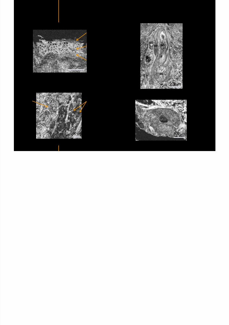
7292019 Atlas of Light-CT images
httpslidepdfcomreaderfullatlas-of-light-ct-images 39118
7292019 Atlas of Light-CT images
httpslidepdfcomreaderfullatlas-of-light-ct-images 40118
Vertical reconstruction of skin model
7292019 Atlas of Light-CT images
httpslidepdfcomreaderfullatlas-of-light-ct-images 41118
copy LLTech 2012 ndash wwwlltechimagingcom - No copy without authorization 41
Vertical reconstruction of skin model
50 microm
Stratum corneum
Stratum spinosum
Dermis
Keratinocyte nuclei
copy LLTech 2012
7292019 Atlas of Light-CT images
httpslidepdfcomreaderfullatlas-of-light-ct-images 42118
Normal
7292019 Atlas of Light-CT images
httpslidepdfcomreaderfullatlas-of-light-ct-images 43118
copy LLTech 2012 ndash wwwlltechimagingcom - No copy without authorization 43
skin
b
a
e
c
f
d
500 microm
7292019 Atlas of Light-CT images
httpslidepdfcomreaderfullatlas-of-light-ct-images 44118
7292019 Atlas of Light-CT images
httpslidepdfcomreaderfullatlas-of-light-ct-images 45118
copy LLTech 2012 ndash wwwlltechimagingcom - No copy without authorization 45
Adipocytes
9um depth 27um depth 60um depth
Reconstructed
depth slice
200 microm
50 microm
7292019 Atlas of Light-CT images
httpslidepdfcomreaderfullatlas-of-light-ct-images 46118
Skin pathologies basal cell carcinoma discrimination at
7292019 Atlas of Light-CT images
httpslidepdfcomreaderfullatlas-of-light-ct-images 47118
copy LLTech 2012 ndash wwwlltechimagingcom - No copy without authorization 47
Epidermis
Dermis
Hypodermis
A
AA
C
BCC
PSU
PSU
BCC Basal cell carcinoma
A Adipocytes
C Collagen
PSU Pilosebaceous unit
structural level
1 mm
copy LLTech 2012 Courtesy of Hopitaux Universitaires de Genegraveve Genegraveve Switzerland
C
Zoom on basal cell carcinoma discrimination at cellular level
7292019 Atlas of Light-CT images
httpslidepdfcomreaderfullatlas-of-light-ct-images 48118
copy LLTech 2012 ndash wwwlltechimagingcom - No copy without authorization 48
Zoom on basal cell carcinoma discrimination at cellular level
200 microm
100 microm
Peritumoral stroma
High cell density
copy LLTech 2012 Courtesy of Hopitaux Universitaires de Genegraveve Genegraveve Switzerland
Skin pathologies discrimination of actinic keratosis
7292019 Atlas of Light-CT images
httpslidepdfcomreaderfullatlas-of-light-ct-images 49118
copy LLTech 2012 ndash wwwlltechimagingcom - No copy without authorization 49
Skin pathologies discrimination of actinic keratosis
Enlarged epidermis
Extended epidermal ridges
1 mm
copy LLTech 2012 Courtesy of Hopitaux Universitaires de Genegraveve Genegraveve Switzerland
7292019 Atlas of Light-CT images
httpslidepdfcomreaderfullatlas-of-light-ct-images 50118
copy LLTech 2012 ndash wwwlltechimagingcom - No copy without authorization 50
Basal cell carcinoma in depth imaging
Z = 10 microns 7 x 42 mm FOV Z = 35 microns 7 x 42 mm FOV
BCC
SG Sebaceous glands
Z = 55 microns 7 x 42 mm FOV Z = 75 microns 7 x 42 mm FOV
SG
Application to Mohs surgerycopy LLTech 2012 Courtesy of Hopitaux Universitaires de Genegraveve Genegraveve Switzerland
7292019 Atlas of Light-CT images
httpslidepdfcomreaderfullatlas-of-light-ct-images 51118
copy LLTech 2012 ndash wwwlltechimagingcom - No copy without authorization 51 copy LLTECH 2011
Healthy vs Cheloid scar tissue
Healthy skin tissueFOV 75x5mm
Cheloid scar tissueFOV 65x65mm
copy LLTECH 2011
7292019 Atlas of Light-CT images
httpslidepdfcomreaderfullatlas-of-light-ct-images 52118
copy LLTech 2012 ndash wwwlltechimagingcom - No copy without authorization 52
Dermo-epidermaljunction
Cheloid scar tissue in detail
copy LLTECH 2011
Nodular structureThickcollagenfiberbundles
1mm
600 microm
500 microm
h l d d f
7292019 Atlas of Light-CT images
httpslidepdfcomreaderfullatlas-of-light-ct-images 53118
copy LLTech 2012 ndash wwwlltechimagingcom - No copy without authorization 53
Cheloid scar tissue viewed en face
20microm depth 120microm depth
7292019 Atlas of Light-CT images
httpslidepdfcomreaderfullatlas-of-light-ct-images 54118
Skin in-vivo sweat duct imaging in 3D
7292019 Atlas of Light-CT images
httpslidepdfcomreaderfullatlas-of-light-ct-images 55118
copy LLTech 2012 ndash wwwlltechimagingcom - No copy without authorization 55
g g
Vertical reconstruction from en-face images
Sweat duct 3D reconstruction
Stratum corneum
Stratum spinosum
SD sweat ducts
SD SD
100 microm
7292019 Atlas of Light-CT images
httpslidepdfcomreaderfullatlas-of-light-ct-images 56118
7292019 Atlas of Light-CT images
httpslidepdfcomreaderfullatlas-of-light-ct-images 57118
7292019 Atlas of Light-CT images
httpslidepdfcomreaderfullatlas-of-light-ct-images 58118
copy LLTech 2012 ndash wwwlltechimagingcom - No copy without authorization 58
LUNG IMAGING
Back to table of contents
H L
7292019 Atlas of Light-CT images
httpslidepdfcomreaderfullatlas-of-light-ct-images 59118
copy LLTech 2012 ndash wwwlltechimagingcom - No copy without authorization 59
Zone 1
Zone 2
Zone 3
Zone 4
Zone with
cancerous tissue
Limit of cancerous
stroma
alveoli with
macrophages
Alveoles
Human Lung
500microm
copy LLTech 2012 Courtesy of Hocircpital Hotel-Dieu Paris France
H L Z 1 Z t
7292019 Atlas of Light-CT images
httpslidepdfcomreaderfullatlas-of-light-ct-images 60118
copy LLTech 2012 ndash wwwlltechimagingcom - No copy without authorization 60
Stroma
Human Lung ndash Zone 1 ndash Zoom on cancerous stroma
Cancerous
area
200microm
copy LLTech 2012 Courtesy of Hocircpital Hotel-Dieu Paris France
Human Lung ndash Zone 2 ndash Limit cancerous stroma alveoli
7292019 Atlas of Light-CT images
httpslidepdfcomreaderfullatlas-of-light-ct-images 61118
copy LLTech 2012 ndash wwwlltechimagingcom - No copy without authorization 61
g
with visible macrophages and vessel
Alveoli
Macrophages
Vessel
copy LLTech 2012 Courtesy of Hocircpital Hotel-Dieu Paris France
Human Lung ndash Zone 3 ndash Limit cancerous stroma alveoli
7292019 Atlas of Light-CT images
httpslidepdfcomreaderfullatlas-of-light-ct-images 62118
copy LLTech 2012 ndash wwwlltechimagingcom - No copy without authorization 62
g
with visible macrophages
Macrophages
100microm
copy LLTech 2012 Courtesy of Hocircpital Hotel-Dieu Paris France
Human Lung ndash Zone 4 ndash Alveoli
7292019 Atlas of Light-CT images
httpslidepdfcomreaderfullatlas-of-light-ct-images 63118
copy LLTech 2012 ndash wwwlltechimagingcom - No copy without authorization 63
Human Lung Zone 4 Alveoli
Alveoli
200microm
copy LLTech 2012 Courtesy of Hocircpital Hotel-Dieu Paris France
7292019 Atlas of Light-CT images
httpslidepdfcomreaderfullatlas-of-light-ct-images 64118
copy LLTech 2012 ndash wwwlltechimagingcom - No copy without authorization 64
GI IMAGING
Back to table of contents
7292019 Atlas of Light-CT images
httpslidepdfcomreaderfullatlas-of-light-ct-images 65118
Colonic carcinoma
7292019 Atlas of Light-CT images
httpslidepdfcomreaderfullatlas-of-light-ct-images 66118
copy LLTech 2012 ndash wwwlltechimagingcom - No copy without authorization 66
Colonic Carcinoma
Colonic carcinoma
copy LLTech 2012 Courtesy of MD Anderson Houston USA
Intestines ndash Crohnrsquos disease
7292019 Atlas of Light-CT images
httpslidepdfcomreaderfullatlas-of-light-ct-images 67118
copy LLTech 2012 ndash wwwlltechimagingcom - No copy without authorization 67
Crohnrsquos disease structure not
visible from the surface
Fat cells
200 microm
Intestines Crohn s disease
copy LLTech 2012 Courtesy of Institut Curie Paris France
Rat stomach
7292019 Atlas of Light-CT images
httpslidepdfcomreaderfullatlas-of-light-ct-images 68118
copy LLTech 2012 ndash wwwlltechimagingcom - No copy without authorization 68
Rat stomach
Mucosa withgastric pits
Submucosa with
bright collagen
fibers
Muscularis
propria with
muscle bundles
copy LLTech 2012 Courtesy of Weill Cornell Medical College NY USA
200 microm
7292019 Atlas of Light-CT images
httpslidepdfcomreaderfullatlas-of-light-ct-images 69118
copy LLTech 2012 ndash wwwlltechimagingcom - No copy without authorization 69 copy LLTECH 2011
UROGENITAL IMAGING
Back to table of contents
Rat bladder
7292019 Atlas of Light-CT images
httpslidepdfcomreaderfullatlas-of-light-ct-images 70118
copy LLTech 2012 ndash wwwlltechimagingcom - No copy without authorization 70
Rat bladder
Stratifiedurothelium
Lamina propria
500 microm
Muscular
bundles of the
lamina propria
Serosa
copy LLTech 2012 Courtesy of Weill Cornell Medical College NY USA
100 microm
100 microm
Human bladder low grade papillary urothelial carcinoma
7292019 Atlas of Light-CT images
httpslidepdfcomreaderfullatlas-of-light-ct-images 71118
copy LLTech 2012 ndash wwwlltechimagingcom - No copy without authorization 71
Lesion with fibrovascular cores (arrow) and superficial umbrella cells (arrowheads)
Human bladder low grade papillary urothelial carcinoma
copy LLTech 2012 Courtesy of Weill Cornell Medical College NY USA
100 microm
7292019 Atlas of Light-CT images
httpslidepdfcomreaderfullatlas-of-light-ct-images 72118
Rat prostate
7292019 Atlas of Light-CT images
httpslidepdfcomreaderfullatlas-of-light-ct-images 73118
copy LLTech 2012 ndash wwwlltechimagingcom - No copy without authorization 73
p
copy LLTech 2012 Courtesy of Weill Cornell Medical College NY USA
Papillary folds of the
acinar glands
Prostatic acini
Fibromuscular stromaPeriprostatic fat Blood vessel
1 mm
200 microm
Mouse kidney
7292019 Atlas of Light-CT images
httpslidepdfcomreaderfullatlas-of-light-ct-images 74118
copy LLTech 2012 ndash wwwlltechimagingcom - No copy without authorization 74
1 mm
y
copy LLTech 2012 Courtesy of Institut Biomeacutedical de Bicecirctre Paris France
Renal cortex
Renal medulla
Renal sinus
7292019 Atlas of Light-CT images
httpslidepdfcomreaderfullatlas-of-light-ct-images 75118
copy LLTech 2012 ndash wwwlltechimagingcom - No copy without authorization 75
Mouse kidney ndash zoom on highlighted section of previous slide
800microm
copy LLTech 2012 Courtesy of Hocircpital Kremlin Bicecirctre Paris France
7292019 Atlas of Light-CT images
httpslidepdfcomreaderfullatlas-of-light-ct-images 76118
7292019 Atlas of Light-CT images
httpslidepdfcomreaderfullatlas-of-light-ct-images 77118
7292019 Atlas of Light-CT images
httpslidepdfcomreaderfullatlas-of-light-ct-images 78118
copy LLTech 2012 ndash wwwlltechimagingcom - No copy without authorization 78 copy LLTECH 2011
CORE NEEDLE BIOPSIES
Back to table of contents
Kidney
7292019 Atlas of Light-CT images
httpslidepdfcomreaderfullatlas-of-light-ct-images 79118
copy LLTech 2012 ndash wwwlltechimagingcom - No copy without authorization 79
100microm
50microm
Renal tubules
Vessel
500microm
copy LLTech 2012 Courtesy of Institut Curie Paris France
Breast Invasive lobular carcinoma
7292019 Atlas of Light-CT images
httpslidepdfcomreaderfullatlas-of-light-ct-images 80118
copy LLTech 2012 ndash wwwlltechimagingcom - No copy without authorization 80
500microm
stroma
adipocytes
copy LLTech 2012 Courtesy of Institut Curie Paris France
7292019 Atlas of Light-CT images
httpslidepdfcomreaderfullatlas-of-light-ct-images 81118
Breast Ductal carcinoma in situ
7292019 Atlas of Light-CT images
httpslidepdfcomreaderfullatlas-of-light-ct-images 82118
copy LLTech 2012 ndash wwwlltechimagingcom - No copy without authorization 82
500microm
copy LLTech 2012 Courtesy of Institut Curie Paris France
7292019 Atlas of Light-CT images
httpslidepdfcomreaderfullatlas-of-light-ct-images 83118
copy LLTech 2012 ndash wwwlltechimagingcom - No copy without authorization 83 copy LLTECH 2011
THYROID IMAGING
Back to table of contents
7292019 Atlas of Light-CT images
httpslidepdfcomreaderfullatlas-of-light-ct-images 84118
copy LLTech 2012 ndash wwwlltechimagingcom - No copy without authorization 84
Thyroid
800 microm
200 microm
copy LLTech 2012 Courtesy of Hocircpital Tenon Paris France
7292019 Atlas of Light-CT images
httpslidepdfcomreaderfullatlas-of-light-ct-images 85118
copy LLTech 2012 ndash wwwlltechimagingcom - No copy without authorization 85
Thyroid
800 microm
200 microm
copy LLTech 2012 Courtesy of Hocircpital Tenon Paris France
7292019 Atlas of Light-CT images
httpslidepdfcomreaderfullatlas-of-light-ct-images 86118
copy LLTech 2012 ndash wwwlltechimagingcom - No copy without authorization 86 copy LLTECH 2011
FAT CELLS IMAGING
Back to table of contents
7292019 Atlas of Light-CT images
httpslidepdfcomreaderfullatlas-of-light-ct-images 87118
copy LLTech 2012 ndash wwwlltechimagingcom - No copy without authorization 87
Brown fat cells - adipocytes
100 microm 100 microm
200 microm
10um depth
Adipocytes
45um depth
En face
slices
Reconstructed
depth slice
copy LLTech 2012 Courtesy of Centre de Recherche des Cordeliers Paris France
7292019 Atlas of Light-CT images
httpslidepdfcomreaderfullatlas-of-light-ct-images 88118
copy LLTech 2012 ndash wwwlltechimagingcom - No copy without authorization 88 copy LLTECH 2011
CORNEA AND RETINA
Back to table of contents
Human cornea reconstructed depth slice
7292019 Atlas of Light-CT images
httpslidepdfcomreaderfullatlas-of-light-ct-images 89118
copy LLTech 2012 ndash wwwlltechimagingcom - No copy without authorization 89
50 um
wwwlltechimagingcom
7292019 Atlas of Light-CT images
httpslidepdfcomreaderfullatlas-of-light-ct-images 90118
Large field en face view in stroma
7292019 Atlas of Light-CT images
httpslidepdfcomreaderfullatlas-of-light-ct-images 91118
copy LLTech 2012 ndash wwwlltechimagingcom - No copy without authorization 91
200 um
wwwlltechimagingcom
7292019 Atlas of Light-CT images
httpslidepdfcomreaderfullatlas-of-light-ct-images 92118
Rabbit cornea after LASIK reflectivity profiles reveal
i d tt i d thi k d ith li l l
7292019 Atlas of Light-CT images
httpslidepdfcomreaderfullatlas-of-light-ct-images 93118
copy LLTech 2012 ndash wwwlltechimagingcom - No copy without authorization 93
increased scattering and thickened epithelial layer
LASIK LASIK CONTROL CONTROL
copy LLTech 2012
Newborn mouse anterior segment
7292019 Atlas of Light-CT images
httpslidepdfcomreaderfullatlas-of-light-ct-images 94118
copy LLTech 2012 ndash wwwlltechimagingcom - No copy without authorization 94
Corneal epithelial cells
Basal membrane
Stroma
Descemetrsquos membrane
Aqueous humor
Lens capsule
Lens epithelial cells
Cortical fibers
copy LLTech 2012 Courtesy of ESPCI Paris Francecopy LLTech 2012
Rat retina comparison with histology
7292019 Atlas of Light-CT images
httpslidepdfcomreaderfullatlas-of-light-ct-images 95118
copy LLTech 2012 ndash wwwlltechimagingcom - No copy without authorization 95
Figure 1 Right Ex vivo rat retinaimaged using the f ull field OCT
system of ESPCI Left Histology
for comparison
Figure 1 Right Ex vivo rat retinaimaged using the f ull field OCT
system of ESPCI Left Histology
for comparison
Figure 1 Right Ex vivo rat retinaimaged using the f ull field OCT
system of ESPCI Left Histology
for comparisonField
230x300microm
RPE
PRL
ONL
OPL
INL
IPL
GCL
NFL
Invest Ophthalmol Vis Sci 2004 Nov45(11)4126-31
Grieve K Paques M Dubois A Sahel J Boccara C Le Gargasson JF copy LLTech 2012
Rat retina
7292019 Atlas of Light-CT images
httpslidepdfcomreaderfullatlas-of-light-ct-images 96118
copy LLTech 2012 ndash wwwlltechimagingcom - No copy without authorization 96 copy LLTech 2012 Courtesy of ESPCI Paris France
Nerve fiber layer
Retina
Invest Ophthalmol Vis Sci 2004 Nov45(11)4126-31
Grieve K Paques M Dubois A Sahel J Boccara C Le Gargasson JF
Transscleral image
7292019 Atlas of Light-CT images
httpslidepdfcomreaderfullatlas-of-light-ct-images 97118
7292019 Atlas of Light-CT images
httpslidepdfcomreaderfullatlas-of-light-ct-images 98118
7292019 Atlas of Light-CT images
httpslidepdfcomreaderfullatlas-of-light-ct-images 99118
In vivo imaging of drosophila melanogaster
7292019 Atlas of Light-CT images
httpslidepdfcomreaderfullatlas-of-light-ct-images 100118
copy LLTech 2012 ndash wwwlltechimagingcom - No copy without authorization 100 copy LLTech 2012 Courtesy of ESPCI Paris France
In vivo imaging of drosophila melanogaster ndash four stages of pupation over 72 hours
7292019 Atlas of Light-CT images
httpslidepdfcomreaderfullatlas-of-light-ct-images 101118
copy LLTech 2012 ndash wwwlltechimagingcom - No copy without authorization 101
Prepupa (0-2 h) Transition topupal stage (24 h)
Pupal stage (48 h) Advanced pupal phase
before eclosion (72 h)
copy LLTech 2012 Courtesy of ESPCI Paris France
Pupa 118 4 days 30um depth
100
7292019 Atlas of Light-CT images
httpslidepdfcomreaderfullatlas-of-light-ct-images 102118
copy LLTech 2012 ndash wwwlltechimagingcom - No copy without authorization 102
100 um
Drosophila
7292019 Atlas of Light-CT images
httpslidepdfcomreaderfullatlas-of-light-ct-images 103118
copy LLTech 2012 ndash wwwlltechimagingcom - No copy without authorization 103
200 microm
copy LLTech 2012 Courtesy of ESPCI Paris France
C Elegans ndash Fast identification of anatomy
7292019 Atlas of Light-CT images
httpslidepdfcomreaderfullatlas-of-light-ct-images 104118
copy LLTech 2012 ndash wwwlltechimagingcom - No copy without authorization 104
Body wall - cuticle
gonad arm
30 microm depth
uterus
ovocytes
intestine
100 microm 100 microm
15 microm depth
copy LLTech 2012 Courtesy of ENS Paris France
C elegans ndash Images at higher magnification 40X
7292019 Atlas of Light-CT images
httpslidepdfcomreaderfullatlas-of-light-ct-images 105118
copy LLTech 2012 ndash wwwlltechimagingcom - No copy without authorization 105
3D reconstruction 50 microm
100 microm
copy LLTech 2012 Courtesy of ENS Paris France
Zebra fish eye
7292019 Atlas of Light-CT images
httpslidepdfcomreaderfullatlas-of-light-ct-images 106118
copy LLTech 2012 ndash wwwlltechimagingcom - No copy without authorization 106
Zebra fish eye
100 microm
100 microm
En face slices
Reconstructed depth slice
copy LLTech 2012 Courtesy of Inserm Paris France
10um depth 20um depth 45um depth
7292019 Atlas of Light-CT images
httpslidepdfcomreaderfullatlas-of-light-ct-images 107118
Xenopus Laevis backtail
7292019 Atlas of Light-CT images
httpslidepdfcomreaderfullatlas-of-light-ct-images 108118
copy LLTech 2012 ndash wwwlltechimagingcom - No copy without authorization 108
100 um30um depth 60um depth
7292019 Atlas of Light-CT images
httpslidepdfcomreaderfullatlas-of-light-ct-images 109118
Mouse embryo in vitro 6 5 days old ndash
7292019 Atlas of Light-CT images
httpslidepdfcomreaderfullatlas-of-light-ct-images 110118
copy LLTech 2012 ndash wwwlltechimagingcom - No copy without authorization 110
Mouse embryo in vitro 65 days old
copy LLTech 2012 Courtesy of Ellvire Guiot and nstitut Jacques Monod Paris France
3D imaging of internal cavity
Sabellaria alveolata mouth palps
7292019 Atlas of Light-CT images
httpslidepdfcomreaderfullatlas-of-light-ct-images 111118
copy LLTech 2012 ndash wwwlltechimagingcom - No copy without authorization 111
500 microm
100 microm
Mouth palps
copy LLTech 2012 Courtesy of Museacutee National drsquoHistoire Naturelle Paris France
7292019 Atlas of Light-CT images
httpslidepdfcomreaderfullatlas-of-light-ct-images 112118
copy LLTech 2012 ndash wwwlltechimagingcom - No copy without authorization 112 copy LLTECH 2011
PLANT IMAGING
Back to table of contents
Mouse ear leaf epidermis
7292019 Atlas of Light-CT images
httpslidepdfcomreaderfullatlas-of-light-ct-images 113118
copy LLTech 2012 ndash wwwlltechimagingcom - No copy without authorization 113
100 microm
Epidermal cell
copy LLTech 2012
Stoma
Leaf veins
7292019 Atlas of Light-CT images
httpslidepdfcomreaderfullatlas-of-light-ct-images 114118
copy LLTech 2012 ndash wwwlltechimagingcom - No copy without authorization 114
114copy LLTech 2012
Fibers
Veins
100 microm
500 microm
Apple
7292019 Atlas of Light-CT images
httpslidepdfcomreaderfullatlas-of-light-ct-images 115118
copy LLTech 2012 ndash wwwlltechimagingcom - No copy without authorization 115
115copy LLTech 2012
100 microm
Wax
Skin
Flesh
500 microm
Water
Effect of harpin infiltration on tobacco leaf
7292019 Atlas of Light-CT images
httpslidepdfcomreaderfullatlas-of-light-ct-images 116118
copy LLTech 2012 ndash wwwlltechimagingcom - No copy without authorization 116
116copy LLTech 2012 Courtesy of UPMC Paris France
50 microm
50 microm
50 microm
50 microm
Upper epidermis
10 min after harpin infiltration 30 min after harpin infiltration
Palisade parenchyma
Epidermis cell
Chloroplast
7292019 Atlas of Light-CT images
httpslidepdfcomreaderfullatlas-of-light-ct-images 117118
copy LLTech 2012 ndash wwwlltechimagingcom - No copy without authorization 117
bull Light-CTtrade does not require tissue preparation nor staining of any kind
bull Creates images within minutes using a non-destructive process
bull Offers a 1 microm cellular resolution in 3D
bull Reveals structural and cellular information
bull Mosaicing allows fast visualisation at various scale
bull En-face high-resolution imaging allows vertical and 3D reconstruction
bull In-vivo images show good promise for in-vivo system
bull Clinical and research applications in cancer detection cosmetics biobanking small animal
developmental biology botanyhellip
Contact Details
7292019 Atlas of Light-CT images
httpslidepdfcomreaderfullatlas-of-light-ct-images 118118
USA
LLTech Inc
103 Carnegie Center DriveSuite 300
Princeton NJ 08540
USA
wwwlltechimagingcom
contactlltechfr
Phone + 1 609 955 3506
LLTech SAS
29 rue du Faubourg St-JacquesPeacutepiniegravere Paris Santeacute Cochin
75014
Paris - France
wwwlltechimagingcom
contactlltechfr
Phone +33 1 82 72 61 25
Europe
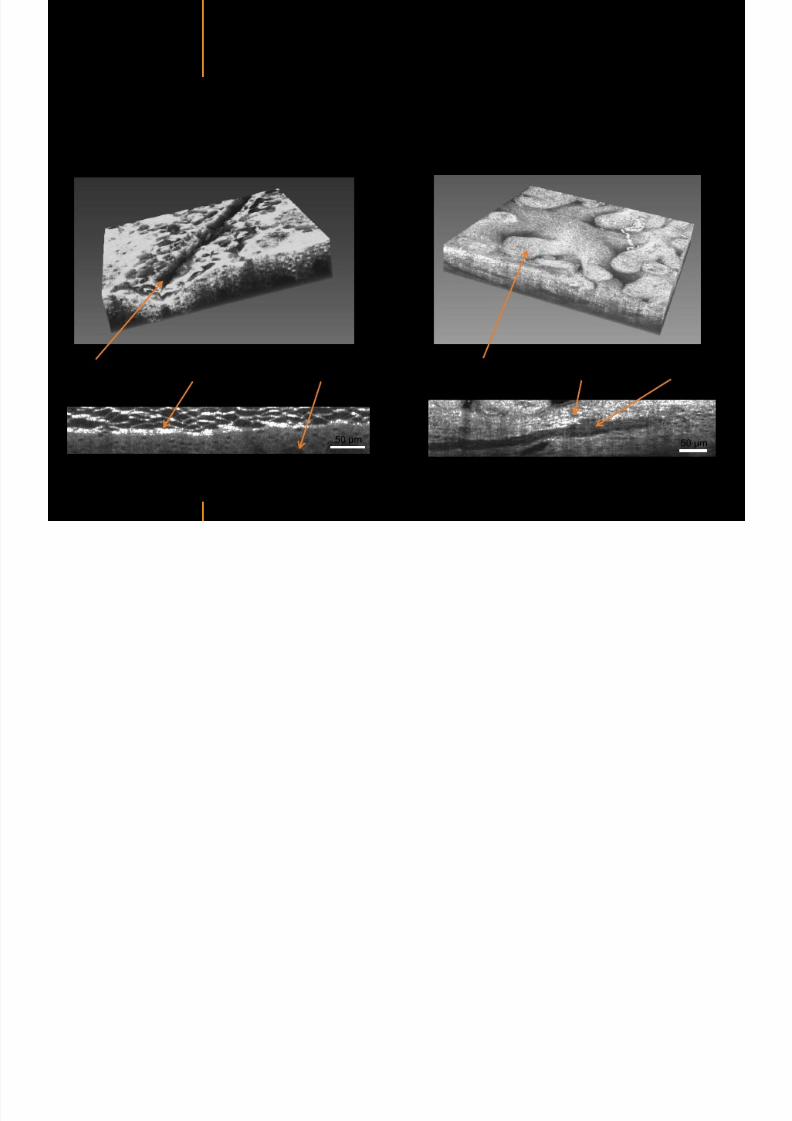
7292019 Atlas of Light-CT images
httpslidepdfcomreaderfullatlas-of-light-ct-images 42118
Normal
7292019 Atlas of Light-CT images
httpslidepdfcomreaderfullatlas-of-light-ct-images 43118
copy LLTech 2012 ndash wwwlltechimagingcom - No copy without authorization 43
skin
b
a
e
c
f
d
500 microm
7292019 Atlas of Light-CT images
httpslidepdfcomreaderfullatlas-of-light-ct-images 44118
7292019 Atlas of Light-CT images
httpslidepdfcomreaderfullatlas-of-light-ct-images 45118
copy LLTech 2012 ndash wwwlltechimagingcom - No copy without authorization 45
Adipocytes
9um depth 27um depth 60um depth
Reconstructed
depth slice
200 microm
50 microm
7292019 Atlas of Light-CT images
httpslidepdfcomreaderfullatlas-of-light-ct-images 46118
Skin pathologies basal cell carcinoma discrimination at
7292019 Atlas of Light-CT images
httpslidepdfcomreaderfullatlas-of-light-ct-images 47118
copy LLTech 2012 ndash wwwlltechimagingcom - No copy without authorization 47
Epidermis
Dermis
Hypodermis
A
AA
C
BCC
PSU
PSU
BCC Basal cell carcinoma
A Adipocytes
C Collagen
PSU Pilosebaceous unit
structural level
1 mm
copy LLTech 2012 Courtesy of Hopitaux Universitaires de Genegraveve Genegraveve Switzerland
C
Zoom on basal cell carcinoma discrimination at cellular level
7292019 Atlas of Light-CT images
httpslidepdfcomreaderfullatlas-of-light-ct-images 48118
copy LLTech 2012 ndash wwwlltechimagingcom - No copy without authorization 48
Zoom on basal cell carcinoma discrimination at cellular level
200 microm
100 microm
Peritumoral stroma
High cell density
copy LLTech 2012 Courtesy of Hopitaux Universitaires de Genegraveve Genegraveve Switzerland
Skin pathologies discrimination of actinic keratosis
7292019 Atlas of Light-CT images
httpslidepdfcomreaderfullatlas-of-light-ct-images 49118
copy LLTech 2012 ndash wwwlltechimagingcom - No copy without authorization 49
Skin pathologies discrimination of actinic keratosis
Enlarged epidermis
Extended epidermal ridges
1 mm
copy LLTech 2012 Courtesy of Hopitaux Universitaires de Genegraveve Genegraveve Switzerland
7292019 Atlas of Light-CT images
httpslidepdfcomreaderfullatlas-of-light-ct-images 50118
copy LLTech 2012 ndash wwwlltechimagingcom - No copy without authorization 50
Basal cell carcinoma in depth imaging
Z = 10 microns 7 x 42 mm FOV Z = 35 microns 7 x 42 mm FOV
BCC
SG Sebaceous glands
Z = 55 microns 7 x 42 mm FOV Z = 75 microns 7 x 42 mm FOV
SG
Application to Mohs surgerycopy LLTech 2012 Courtesy of Hopitaux Universitaires de Genegraveve Genegraveve Switzerland
7292019 Atlas of Light-CT images
httpslidepdfcomreaderfullatlas-of-light-ct-images 51118
copy LLTech 2012 ndash wwwlltechimagingcom - No copy without authorization 51 copy LLTECH 2011
Healthy vs Cheloid scar tissue
Healthy skin tissueFOV 75x5mm
Cheloid scar tissueFOV 65x65mm
copy LLTECH 2011
7292019 Atlas of Light-CT images
httpslidepdfcomreaderfullatlas-of-light-ct-images 52118
copy LLTech 2012 ndash wwwlltechimagingcom - No copy without authorization 52
Dermo-epidermaljunction
Cheloid scar tissue in detail
copy LLTECH 2011
Nodular structureThickcollagenfiberbundles
1mm
600 microm
500 microm
h l d d f
7292019 Atlas of Light-CT images
httpslidepdfcomreaderfullatlas-of-light-ct-images 53118
copy LLTech 2012 ndash wwwlltechimagingcom - No copy without authorization 53
Cheloid scar tissue viewed en face
20microm depth 120microm depth
7292019 Atlas of Light-CT images
httpslidepdfcomreaderfullatlas-of-light-ct-images 54118
Skin in-vivo sweat duct imaging in 3D
7292019 Atlas of Light-CT images
httpslidepdfcomreaderfullatlas-of-light-ct-images 55118
copy LLTech 2012 ndash wwwlltechimagingcom - No copy without authorization 55
g g
Vertical reconstruction from en-face images
Sweat duct 3D reconstruction
Stratum corneum
Stratum spinosum
SD sweat ducts
SD SD
100 microm
7292019 Atlas of Light-CT images
httpslidepdfcomreaderfullatlas-of-light-ct-images 56118
7292019 Atlas of Light-CT images
httpslidepdfcomreaderfullatlas-of-light-ct-images 57118
7292019 Atlas of Light-CT images
httpslidepdfcomreaderfullatlas-of-light-ct-images 58118
copy LLTech 2012 ndash wwwlltechimagingcom - No copy without authorization 58
LUNG IMAGING
Back to table of contents
H L
7292019 Atlas of Light-CT images
httpslidepdfcomreaderfullatlas-of-light-ct-images 59118
copy LLTech 2012 ndash wwwlltechimagingcom - No copy without authorization 59
Zone 1
Zone 2
Zone 3
Zone 4
Zone with
cancerous tissue
Limit of cancerous
stroma
alveoli with
macrophages
Alveoles
Human Lung
500microm
copy LLTech 2012 Courtesy of Hocircpital Hotel-Dieu Paris France
H L Z 1 Z t
7292019 Atlas of Light-CT images
httpslidepdfcomreaderfullatlas-of-light-ct-images 60118
copy LLTech 2012 ndash wwwlltechimagingcom - No copy without authorization 60
Stroma
Human Lung ndash Zone 1 ndash Zoom on cancerous stroma
Cancerous
area
200microm
copy LLTech 2012 Courtesy of Hocircpital Hotel-Dieu Paris France
Human Lung ndash Zone 2 ndash Limit cancerous stroma alveoli
7292019 Atlas of Light-CT images
httpslidepdfcomreaderfullatlas-of-light-ct-images 61118
copy LLTech 2012 ndash wwwlltechimagingcom - No copy without authorization 61
g
with visible macrophages and vessel
Alveoli
Macrophages
Vessel
copy LLTech 2012 Courtesy of Hocircpital Hotel-Dieu Paris France
Human Lung ndash Zone 3 ndash Limit cancerous stroma alveoli
7292019 Atlas of Light-CT images
httpslidepdfcomreaderfullatlas-of-light-ct-images 62118
copy LLTech 2012 ndash wwwlltechimagingcom - No copy without authorization 62
g
with visible macrophages
Macrophages
100microm
copy LLTech 2012 Courtesy of Hocircpital Hotel-Dieu Paris France
Human Lung ndash Zone 4 ndash Alveoli
7292019 Atlas of Light-CT images
httpslidepdfcomreaderfullatlas-of-light-ct-images 63118
copy LLTech 2012 ndash wwwlltechimagingcom - No copy without authorization 63
Human Lung Zone 4 Alveoli
Alveoli
200microm
copy LLTech 2012 Courtesy of Hocircpital Hotel-Dieu Paris France
7292019 Atlas of Light-CT images
httpslidepdfcomreaderfullatlas-of-light-ct-images 64118
copy LLTech 2012 ndash wwwlltechimagingcom - No copy without authorization 64
GI IMAGING
Back to table of contents
7292019 Atlas of Light-CT images
httpslidepdfcomreaderfullatlas-of-light-ct-images 65118
Colonic carcinoma
7292019 Atlas of Light-CT images
httpslidepdfcomreaderfullatlas-of-light-ct-images 66118
copy LLTech 2012 ndash wwwlltechimagingcom - No copy without authorization 66
Colonic Carcinoma
Colonic carcinoma
copy LLTech 2012 Courtesy of MD Anderson Houston USA
Intestines ndash Crohnrsquos disease
7292019 Atlas of Light-CT images
httpslidepdfcomreaderfullatlas-of-light-ct-images 67118
copy LLTech 2012 ndash wwwlltechimagingcom - No copy without authorization 67
Crohnrsquos disease structure not
visible from the surface
Fat cells
200 microm
Intestines Crohn s disease
copy LLTech 2012 Courtesy of Institut Curie Paris France
Rat stomach
7292019 Atlas of Light-CT images
httpslidepdfcomreaderfullatlas-of-light-ct-images 68118
copy LLTech 2012 ndash wwwlltechimagingcom - No copy without authorization 68
Rat stomach
Mucosa withgastric pits
Submucosa with
bright collagen
fibers
Muscularis
propria with
muscle bundles
copy LLTech 2012 Courtesy of Weill Cornell Medical College NY USA
200 microm
7292019 Atlas of Light-CT images
httpslidepdfcomreaderfullatlas-of-light-ct-images 69118
copy LLTech 2012 ndash wwwlltechimagingcom - No copy without authorization 69 copy LLTECH 2011
UROGENITAL IMAGING
Back to table of contents
Rat bladder
7292019 Atlas of Light-CT images
httpslidepdfcomreaderfullatlas-of-light-ct-images 70118
copy LLTech 2012 ndash wwwlltechimagingcom - No copy without authorization 70
Rat bladder
Stratifiedurothelium
Lamina propria
500 microm
Muscular
bundles of the
lamina propria
Serosa
copy LLTech 2012 Courtesy of Weill Cornell Medical College NY USA
100 microm
100 microm
Human bladder low grade papillary urothelial carcinoma
7292019 Atlas of Light-CT images
httpslidepdfcomreaderfullatlas-of-light-ct-images 71118
copy LLTech 2012 ndash wwwlltechimagingcom - No copy without authorization 71
Lesion with fibrovascular cores (arrow) and superficial umbrella cells (arrowheads)
Human bladder low grade papillary urothelial carcinoma
copy LLTech 2012 Courtesy of Weill Cornell Medical College NY USA
100 microm
7292019 Atlas of Light-CT images
httpslidepdfcomreaderfullatlas-of-light-ct-images 72118
Rat prostate
7292019 Atlas of Light-CT images
httpslidepdfcomreaderfullatlas-of-light-ct-images 73118
copy LLTech 2012 ndash wwwlltechimagingcom - No copy without authorization 73
p
copy LLTech 2012 Courtesy of Weill Cornell Medical College NY USA
Papillary folds of the
acinar glands
Prostatic acini
Fibromuscular stromaPeriprostatic fat Blood vessel
1 mm
200 microm
Mouse kidney
7292019 Atlas of Light-CT images
httpslidepdfcomreaderfullatlas-of-light-ct-images 74118
copy LLTech 2012 ndash wwwlltechimagingcom - No copy without authorization 74
1 mm
y
copy LLTech 2012 Courtesy of Institut Biomeacutedical de Bicecirctre Paris France
Renal cortex
Renal medulla
Renal sinus
7292019 Atlas of Light-CT images
httpslidepdfcomreaderfullatlas-of-light-ct-images 75118
copy LLTech 2012 ndash wwwlltechimagingcom - No copy without authorization 75
Mouse kidney ndash zoom on highlighted section of previous slide
800microm
copy LLTech 2012 Courtesy of Hocircpital Kremlin Bicecirctre Paris France
7292019 Atlas of Light-CT images
httpslidepdfcomreaderfullatlas-of-light-ct-images 76118
7292019 Atlas of Light-CT images
httpslidepdfcomreaderfullatlas-of-light-ct-images 77118
7292019 Atlas of Light-CT images
httpslidepdfcomreaderfullatlas-of-light-ct-images 78118
copy LLTech 2012 ndash wwwlltechimagingcom - No copy without authorization 78 copy LLTECH 2011
CORE NEEDLE BIOPSIES
Back to table of contents
Kidney
7292019 Atlas of Light-CT images
httpslidepdfcomreaderfullatlas-of-light-ct-images 79118
copy LLTech 2012 ndash wwwlltechimagingcom - No copy without authorization 79
100microm
50microm
Renal tubules
Vessel
500microm
copy LLTech 2012 Courtesy of Institut Curie Paris France
Breast Invasive lobular carcinoma
7292019 Atlas of Light-CT images
httpslidepdfcomreaderfullatlas-of-light-ct-images 80118
copy LLTech 2012 ndash wwwlltechimagingcom - No copy without authorization 80
500microm
stroma
adipocytes
copy LLTech 2012 Courtesy of Institut Curie Paris France
7292019 Atlas of Light-CT images
httpslidepdfcomreaderfullatlas-of-light-ct-images 81118
Breast Ductal carcinoma in situ
7292019 Atlas of Light-CT images
httpslidepdfcomreaderfullatlas-of-light-ct-images 82118
copy LLTech 2012 ndash wwwlltechimagingcom - No copy without authorization 82
500microm
copy LLTech 2012 Courtesy of Institut Curie Paris France
7292019 Atlas of Light-CT images
httpslidepdfcomreaderfullatlas-of-light-ct-images 83118
copy LLTech 2012 ndash wwwlltechimagingcom - No copy without authorization 83 copy LLTECH 2011
THYROID IMAGING
Back to table of contents
7292019 Atlas of Light-CT images
httpslidepdfcomreaderfullatlas-of-light-ct-images 84118
copy LLTech 2012 ndash wwwlltechimagingcom - No copy without authorization 84
Thyroid
800 microm
200 microm
copy LLTech 2012 Courtesy of Hocircpital Tenon Paris France
7292019 Atlas of Light-CT images
httpslidepdfcomreaderfullatlas-of-light-ct-images 85118
copy LLTech 2012 ndash wwwlltechimagingcom - No copy without authorization 85
Thyroid
800 microm
200 microm
copy LLTech 2012 Courtesy of Hocircpital Tenon Paris France
7292019 Atlas of Light-CT images
httpslidepdfcomreaderfullatlas-of-light-ct-images 86118
copy LLTech 2012 ndash wwwlltechimagingcom - No copy without authorization 86 copy LLTECH 2011
FAT CELLS IMAGING
Back to table of contents
7292019 Atlas of Light-CT images
httpslidepdfcomreaderfullatlas-of-light-ct-images 87118
copy LLTech 2012 ndash wwwlltechimagingcom - No copy without authorization 87
Brown fat cells - adipocytes
100 microm 100 microm
200 microm
10um depth
Adipocytes
45um depth
En face
slices
Reconstructed
depth slice
copy LLTech 2012 Courtesy of Centre de Recherche des Cordeliers Paris France
7292019 Atlas of Light-CT images
httpslidepdfcomreaderfullatlas-of-light-ct-images 88118
copy LLTech 2012 ndash wwwlltechimagingcom - No copy without authorization 88 copy LLTECH 2011
CORNEA AND RETINA
Back to table of contents
Human cornea reconstructed depth slice
7292019 Atlas of Light-CT images
httpslidepdfcomreaderfullatlas-of-light-ct-images 89118
copy LLTech 2012 ndash wwwlltechimagingcom - No copy without authorization 89
50 um
wwwlltechimagingcom
7292019 Atlas of Light-CT images
httpslidepdfcomreaderfullatlas-of-light-ct-images 90118
Large field en face view in stroma
7292019 Atlas of Light-CT images
httpslidepdfcomreaderfullatlas-of-light-ct-images 91118
copy LLTech 2012 ndash wwwlltechimagingcom - No copy without authorization 91
200 um
wwwlltechimagingcom
7292019 Atlas of Light-CT images
httpslidepdfcomreaderfullatlas-of-light-ct-images 92118
Rabbit cornea after LASIK reflectivity profiles reveal
i d tt i d thi k d ith li l l
7292019 Atlas of Light-CT images
httpslidepdfcomreaderfullatlas-of-light-ct-images 93118
copy LLTech 2012 ndash wwwlltechimagingcom - No copy without authorization 93
increased scattering and thickened epithelial layer
LASIK LASIK CONTROL CONTROL
copy LLTech 2012
Newborn mouse anterior segment
7292019 Atlas of Light-CT images
httpslidepdfcomreaderfullatlas-of-light-ct-images 94118
copy LLTech 2012 ndash wwwlltechimagingcom - No copy without authorization 94
Corneal epithelial cells
Basal membrane
Stroma
Descemetrsquos membrane
Aqueous humor
Lens capsule
Lens epithelial cells
Cortical fibers
copy LLTech 2012 Courtesy of ESPCI Paris Francecopy LLTech 2012
Rat retina comparison with histology
7292019 Atlas of Light-CT images
httpslidepdfcomreaderfullatlas-of-light-ct-images 95118
copy LLTech 2012 ndash wwwlltechimagingcom - No copy without authorization 95
Figure 1 Right Ex vivo rat retinaimaged using the f ull field OCT
system of ESPCI Left Histology
for comparison
Figure 1 Right Ex vivo rat retinaimaged using the f ull field OCT
system of ESPCI Left Histology
for comparison
Figure 1 Right Ex vivo rat retinaimaged using the f ull field OCT
system of ESPCI Left Histology
for comparisonField
230x300microm
RPE
PRL
ONL
OPL
INL
IPL
GCL
NFL
Invest Ophthalmol Vis Sci 2004 Nov45(11)4126-31
Grieve K Paques M Dubois A Sahel J Boccara C Le Gargasson JF copy LLTech 2012
Rat retina
7292019 Atlas of Light-CT images
httpslidepdfcomreaderfullatlas-of-light-ct-images 96118
copy LLTech 2012 ndash wwwlltechimagingcom - No copy without authorization 96 copy LLTech 2012 Courtesy of ESPCI Paris France
Nerve fiber layer
Retina
Invest Ophthalmol Vis Sci 2004 Nov45(11)4126-31
Grieve K Paques M Dubois A Sahel J Boccara C Le Gargasson JF
Transscleral image
7292019 Atlas of Light-CT images
httpslidepdfcomreaderfullatlas-of-light-ct-images 97118
7292019 Atlas of Light-CT images
httpslidepdfcomreaderfullatlas-of-light-ct-images 98118
7292019 Atlas of Light-CT images
httpslidepdfcomreaderfullatlas-of-light-ct-images 99118
In vivo imaging of drosophila melanogaster
7292019 Atlas of Light-CT images
httpslidepdfcomreaderfullatlas-of-light-ct-images 100118
copy LLTech 2012 ndash wwwlltechimagingcom - No copy without authorization 100 copy LLTech 2012 Courtesy of ESPCI Paris France
In vivo imaging of drosophila melanogaster ndash four stages of pupation over 72 hours
7292019 Atlas of Light-CT images
httpslidepdfcomreaderfullatlas-of-light-ct-images 101118
copy LLTech 2012 ndash wwwlltechimagingcom - No copy without authorization 101
Prepupa (0-2 h) Transition topupal stage (24 h)
Pupal stage (48 h) Advanced pupal phase
before eclosion (72 h)
copy LLTech 2012 Courtesy of ESPCI Paris France
Pupa 118 4 days 30um depth
100
7292019 Atlas of Light-CT images
httpslidepdfcomreaderfullatlas-of-light-ct-images 102118
copy LLTech 2012 ndash wwwlltechimagingcom - No copy without authorization 102
100 um
Drosophila
7292019 Atlas of Light-CT images
httpslidepdfcomreaderfullatlas-of-light-ct-images 103118
copy LLTech 2012 ndash wwwlltechimagingcom - No copy without authorization 103
200 microm
copy LLTech 2012 Courtesy of ESPCI Paris France
C Elegans ndash Fast identification of anatomy
7292019 Atlas of Light-CT images
httpslidepdfcomreaderfullatlas-of-light-ct-images 104118
copy LLTech 2012 ndash wwwlltechimagingcom - No copy without authorization 104
Body wall - cuticle
gonad arm
30 microm depth
uterus
ovocytes
intestine
100 microm 100 microm
15 microm depth
copy LLTech 2012 Courtesy of ENS Paris France
C elegans ndash Images at higher magnification 40X
7292019 Atlas of Light-CT images
httpslidepdfcomreaderfullatlas-of-light-ct-images 105118
copy LLTech 2012 ndash wwwlltechimagingcom - No copy without authorization 105
3D reconstruction 50 microm
100 microm
copy LLTech 2012 Courtesy of ENS Paris France
Zebra fish eye
7292019 Atlas of Light-CT images
httpslidepdfcomreaderfullatlas-of-light-ct-images 106118
copy LLTech 2012 ndash wwwlltechimagingcom - No copy without authorization 106
Zebra fish eye
100 microm
100 microm
En face slices
Reconstructed depth slice
copy LLTech 2012 Courtesy of Inserm Paris France
10um depth 20um depth 45um depth
7292019 Atlas of Light-CT images
httpslidepdfcomreaderfullatlas-of-light-ct-images 107118
Xenopus Laevis backtail
7292019 Atlas of Light-CT images
httpslidepdfcomreaderfullatlas-of-light-ct-images 108118
copy LLTech 2012 ndash wwwlltechimagingcom - No copy without authorization 108
100 um30um depth 60um depth
7292019 Atlas of Light-CT images
httpslidepdfcomreaderfullatlas-of-light-ct-images 109118
Mouse embryo in vitro 6 5 days old ndash
7292019 Atlas of Light-CT images
httpslidepdfcomreaderfullatlas-of-light-ct-images 110118
copy LLTech 2012 ndash wwwlltechimagingcom - No copy without authorization 110
Mouse embryo in vitro 65 days old
copy LLTech 2012 Courtesy of Ellvire Guiot and nstitut Jacques Monod Paris France
3D imaging of internal cavity
Sabellaria alveolata mouth palps
7292019 Atlas of Light-CT images
httpslidepdfcomreaderfullatlas-of-light-ct-images 111118
copy LLTech 2012 ndash wwwlltechimagingcom - No copy without authorization 111
500 microm
100 microm
Mouth palps
copy LLTech 2012 Courtesy of Museacutee National drsquoHistoire Naturelle Paris France
7292019 Atlas of Light-CT images
httpslidepdfcomreaderfullatlas-of-light-ct-images 112118
copy LLTech 2012 ndash wwwlltechimagingcom - No copy without authorization 112 copy LLTECH 2011
PLANT IMAGING
Back to table of contents
Mouse ear leaf epidermis
7292019 Atlas of Light-CT images
httpslidepdfcomreaderfullatlas-of-light-ct-images 113118
copy LLTech 2012 ndash wwwlltechimagingcom - No copy without authorization 113
100 microm
Epidermal cell
copy LLTech 2012
Stoma
Leaf veins
7292019 Atlas of Light-CT images
httpslidepdfcomreaderfullatlas-of-light-ct-images 114118
copy LLTech 2012 ndash wwwlltechimagingcom - No copy without authorization 114
114copy LLTech 2012
Fibers
Veins
100 microm
500 microm
Apple
7292019 Atlas of Light-CT images
httpslidepdfcomreaderfullatlas-of-light-ct-images 115118
copy LLTech 2012 ndash wwwlltechimagingcom - No copy without authorization 115
115copy LLTech 2012
100 microm
Wax
Skin
Flesh
500 microm
Water
Effect of harpin infiltration on tobacco leaf
7292019 Atlas of Light-CT images
httpslidepdfcomreaderfullatlas-of-light-ct-images 116118
copy LLTech 2012 ndash wwwlltechimagingcom - No copy without authorization 116
116copy LLTech 2012 Courtesy of UPMC Paris France
50 microm
50 microm
50 microm
50 microm
Upper epidermis
10 min after harpin infiltration 30 min after harpin infiltration
Palisade parenchyma
Epidermis cell
Chloroplast
7292019 Atlas of Light-CT images
httpslidepdfcomreaderfullatlas-of-light-ct-images 117118
copy LLTech 2012 ndash wwwlltechimagingcom - No copy without authorization 117
bull Light-CTtrade does not require tissue preparation nor staining of any kind
bull Creates images within minutes using a non-destructive process
bull Offers a 1 microm cellular resolution in 3D
bull Reveals structural and cellular information
bull Mosaicing allows fast visualisation at various scale
bull En-face high-resolution imaging allows vertical and 3D reconstruction
bull In-vivo images show good promise for in-vivo system
bull Clinical and research applications in cancer detection cosmetics biobanking small animal
developmental biology botanyhellip
Contact Details
7292019 Atlas of Light-CT images
httpslidepdfcomreaderfullatlas-of-light-ct-images 118118
USA
LLTech Inc
103 Carnegie Center DriveSuite 300
Princeton NJ 08540
USA
wwwlltechimagingcom
contactlltechfr
Phone + 1 609 955 3506
LLTech SAS
29 rue du Faubourg St-JacquesPeacutepiniegravere Paris Santeacute Cochin
75014
Paris - France
wwwlltechimagingcom
contactlltechfr
Phone +33 1 82 72 61 25
Europe
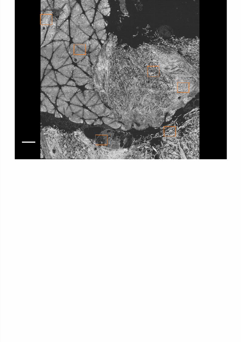
7292019 Atlas of Light-CT images
httpslidepdfcomreaderfullatlas-of-light-ct-images 43118
copy LLTech 2012 ndash wwwlltechimagingcom - No copy without authorization 43
skin
b
a
e
c
f
d
500 microm
7292019 Atlas of Light-CT images
httpslidepdfcomreaderfullatlas-of-light-ct-images 44118
7292019 Atlas of Light-CT images
httpslidepdfcomreaderfullatlas-of-light-ct-images 45118
copy LLTech 2012 ndash wwwlltechimagingcom - No copy without authorization 45
Adipocytes
9um depth 27um depth 60um depth
Reconstructed
depth slice
200 microm
50 microm
7292019 Atlas of Light-CT images
httpslidepdfcomreaderfullatlas-of-light-ct-images 46118
Skin pathologies basal cell carcinoma discrimination at
7292019 Atlas of Light-CT images
httpslidepdfcomreaderfullatlas-of-light-ct-images 47118
copy LLTech 2012 ndash wwwlltechimagingcom - No copy without authorization 47
Epidermis
Dermis
Hypodermis
A
AA
C
BCC
PSU
PSU
BCC Basal cell carcinoma
A Adipocytes
C Collagen
PSU Pilosebaceous unit
structural level
1 mm
copy LLTech 2012 Courtesy of Hopitaux Universitaires de Genegraveve Genegraveve Switzerland
C
Zoom on basal cell carcinoma discrimination at cellular level
7292019 Atlas of Light-CT images
httpslidepdfcomreaderfullatlas-of-light-ct-images 48118
copy LLTech 2012 ndash wwwlltechimagingcom - No copy without authorization 48
Zoom on basal cell carcinoma discrimination at cellular level
200 microm
100 microm
Peritumoral stroma
High cell density
copy LLTech 2012 Courtesy of Hopitaux Universitaires de Genegraveve Genegraveve Switzerland
Skin pathologies discrimination of actinic keratosis
7292019 Atlas of Light-CT images
httpslidepdfcomreaderfullatlas-of-light-ct-images 49118
copy LLTech 2012 ndash wwwlltechimagingcom - No copy without authorization 49
Skin pathologies discrimination of actinic keratosis
Enlarged epidermis
Extended epidermal ridges
1 mm
copy LLTech 2012 Courtesy of Hopitaux Universitaires de Genegraveve Genegraveve Switzerland
7292019 Atlas of Light-CT images
httpslidepdfcomreaderfullatlas-of-light-ct-images 50118
copy LLTech 2012 ndash wwwlltechimagingcom - No copy without authorization 50
Basal cell carcinoma in depth imaging
Z = 10 microns 7 x 42 mm FOV Z = 35 microns 7 x 42 mm FOV
BCC
SG Sebaceous glands
Z = 55 microns 7 x 42 mm FOV Z = 75 microns 7 x 42 mm FOV
SG
Application to Mohs surgerycopy LLTech 2012 Courtesy of Hopitaux Universitaires de Genegraveve Genegraveve Switzerland
7292019 Atlas of Light-CT images
httpslidepdfcomreaderfullatlas-of-light-ct-images 51118
copy LLTech 2012 ndash wwwlltechimagingcom - No copy without authorization 51 copy LLTECH 2011
Healthy vs Cheloid scar tissue
Healthy skin tissueFOV 75x5mm
Cheloid scar tissueFOV 65x65mm
copy LLTECH 2011
7292019 Atlas of Light-CT images
httpslidepdfcomreaderfullatlas-of-light-ct-images 52118
copy LLTech 2012 ndash wwwlltechimagingcom - No copy without authorization 52
Dermo-epidermaljunction
Cheloid scar tissue in detail
copy LLTECH 2011
Nodular structureThickcollagenfiberbundles
1mm
600 microm
500 microm
h l d d f
7292019 Atlas of Light-CT images
httpslidepdfcomreaderfullatlas-of-light-ct-images 53118
copy LLTech 2012 ndash wwwlltechimagingcom - No copy without authorization 53
Cheloid scar tissue viewed en face
20microm depth 120microm depth
7292019 Atlas of Light-CT images
httpslidepdfcomreaderfullatlas-of-light-ct-images 54118
Skin in-vivo sweat duct imaging in 3D
7292019 Atlas of Light-CT images
httpslidepdfcomreaderfullatlas-of-light-ct-images 55118
copy LLTech 2012 ndash wwwlltechimagingcom - No copy without authorization 55
g g
Vertical reconstruction from en-face images
Sweat duct 3D reconstruction
Stratum corneum
Stratum spinosum
SD sweat ducts
SD SD
100 microm
7292019 Atlas of Light-CT images
httpslidepdfcomreaderfullatlas-of-light-ct-images 56118
7292019 Atlas of Light-CT images
httpslidepdfcomreaderfullatlas-of-light-ct-images 57118
7292019 Atlas of Light-CT images
httpslidepdfcomreaderfullatlas-of-light-ct-images 58118
copy LLTech 2012 ndash wwwlltechimagingcom - No copy without authorization 58
LUNG IMAGING
Back to table of contents
H L
7292019 Atlas of Light-CT images
httpslidepdfcomreaderfullatlas-of-light-ct-images 59118
copy LLTech 2012 ndash wwwlltechimagingcom - No copy without authorization 59
Zone 1
Zone 2
Zone 3
Zone 4
Zone with
cancerous tissue
Limit of cancerous
stroma
alveoli with
macrophages
Alveoles
Human Lung
500microm
copy LLTech 2012 Courtesy of Hocircpital Hotel-Dieu Paris France
H L Z 1 Z t
7292019 Atlas of Light-CT images
httpslidepdfcomreaderfullatlas-of-light-ct-images 60118
copy LLTech 2012 ndash wwwlltechimagingcom - No copy without authorization 60
Stroma
Human Lung ndash Zone 1 ndash Zoom on cancerous stroma
Cancerous
area
200microm
copy LLTech 2012 Courtesy of Hocircpital Hotel-Dieu Paris France
Human Lung ndash Zone 2 ndash Limit cancerous stroma alveoli
7292019 Atlas of Light-CT images
httpslidepdfcomreaderfullatlas-of-light-ct-images 61118
copy LLTech 2012 ndash wwwlltechimagingcom - No copy without authorization 61
g
with visible macrophages and vessel
Alveoli
Macrophages
Vessel
copy LLTech 2012 Courtesy of Hocircpital Hotel-Dieu Paris France
Human Lung ndash Zone 3 ndash Limit cancerous stroma alveoli
7292019 Atlas of Light-CT images
httpslidepdfcomreaderfullatlas-of-light-ct-images 62118
copy LLTech 2012 ndash wwwlltechimagingcom - No copy without authorization 62
g
with visible macrophages
Macrophages
100microm
copy LLTech 2012 Courtesy of Hocircpital Hotel-Dieu Paris France
Human Lung ndash Zone 4 ndash Alveoli
7292019 Atlas of Light-CT images
httpslidepdfcomreaderfullatlas-of-light-ct-images 63118
copy LLTech 2012 ndash wwwlltechimagingcom - No copy without authorization 63
Human Lung Zone 4 Alveoli
Alveoli
200microm
copy LLTech 2012 Courtesy of Hocircpital Hotel-Dieu Paris France
7292019 Atlas of Light-CT images
httpslidepdfcomreaderfullatlas-of-light-ct-images 64118
copy LLTech 2012 ndash wwwlltechimagingcom - No copy without authorization 64
GI IMAGING
Back to table of contents
7292019 Atlas of Light-CT images
httpslidepdfcomreaderfullatlas-of-light-ct-images 65118
Colonic carcinoma
7292019 Atlas of Light-CT images
httpslidepdfcomreaderfullatlas-of-light-ct-images 66118
copy LLTech 2012 ndash wwwlltechimagingcom - No copy without authorization 66
Colonic Carcinoma
Colonic carcinoma
copy LLTech 2012 Courtesy of MD Anderson Houston USA
Intestines ndash Crohnrsquos disease
7292019 Atlas of Light-CT images
httpslidepdfcomreaderfullatlas-of-light-ct-images 67118
copy LLTech 2012 ndash wwwlltechimagingcom - No copy without authorization 67
Crohnrsquos disease structure not
visible from the surface
Fat cells
200 microm
Intestines Crohn s disease
copy LLTech 2012 Courtesy of Institut Curie Paris France
Rat stomach
7292019 Atlas of Light-CT images
httpslidepdfcomreaderfullatlas-of-light-ct-images 68118
copy LLTech 2012 ndash wwwlltechimagingcom - No copy without authorization 68
Rat stomach
Mucosa withgastric pits
Submucosa with
bright collagen
fibers
Muscularis
propria with
muscle bundles
copy LLTech 2012 Courtesy of Weill Cornell Medical College NY USA
200 microm
7292019 Atlas of Light-CT images
httpslidepdfcomreaderfullatlas-of-light-ct-images 69118
copy LLTech 2012 ndash wwwlltechimagingcom - No copy without authorization 69 copy LLTECH 2011
UROGENITAL IMAGING
Back to table of contents
Rat bladder
7292019 Atlas of Light-CT images
httpslidepdfcomreaderfullatlas-of-light-ct-images 70118
copy LLTech 2012 ndash wwwlltechimagingcom - No copy without authorization 70
Rat bladder
Stratifiedurothelium
Lamina propria
500 microm
Muscular
bundles of the
lamina propria
Serosa
copy LLTech 2012 Courtesy of Weill Cornell Medical College NY USA
100 microm
100 microm
Human bladder low grade papillary urothelial carcinoma
7292019 Atlas of Light-CT images
httpslidepdfcomreaderfullatlas-of-light-ct-images 71118
copy LLTech 2012 ndash wwwlltechimagingcom - No copy without authorization 71
Lesion with fibrovascular cores (arrow) and superficial umbrella cells (arrowheads)
Human bladder low grade papillary urothelial carcinoma
copy LLTech 2012 Courtesy of Weill Cornell Medical College NY USA
100 microm
7292019 Atlas of Light-CT images
httpslidepdfcomreaderfullatlas-of-light-ct-images 72118
Rat prostate
7292019 Atlas of Light-CT images
httpslidepdfcomreaderfullatlas-of-light-ct-images 73118
copy LLTech 2012 ndash wwwlltechimagingcom - No copy without authorization 73
p
copy LLTech 2012 Courtesy of Weill Cornell Medical College NY USA
Papillary folds of the
acinar glands
Prostatic acini
Fibromuscular stromaPeriprostatic fat Blood vessel
1 mm
200 microm
Mouse kidney
7292019 Atlas of Light-CT images
httpslidepdfcomreaderfullatlas-of-light-ct-images 74118
copy LLTech 2012 ndash wwwlltechimagingcom - No copy without authorization 74
1 mm
y
copy LLTech 2012 Courtesy of Institut Biomeacutedical de Bicecirctre Paris France
Renal cortex
Renal medulla
Renal sinus
7292019 Atlas of Light-CT images
httpslidepdfcomreaderfullatlas-of-light-ct-images 75118
copy LLTech 2012 ndash wwwlltechimagingcom - No copy without authorization 75
Mouse kidney ndash zoom on highlighted section of previous slide
800microm
copy LLTech 2012 Courtesy of Hocircpital Kremlin Bicecirctre Paris France
7292019 Atlas of Light-CT images
httpslidepdfcomreaderfullatlas-of-light-ct-images 76118
7292019 Atlas of Light-CT images
httpslidepdfcomreaderfullatlas-of-light-ct-images 77118
7292019 Atlas of Light-CT images
httpslidepdfcomreaderfullatlas-of-light-ct-images 78118
copy LLTech 2012 ndash wwwlltechimagingcom - No copy without authorization 78 copy LLTECH 2011
CORE NEEDLE BIOPSIES
Back to table of contents
Kidney
7292019 Atlas of Light-CT images
httpslidepdfcomreaderfullatlas-of-light-ct-images 79118
copy LLTech 2012 ndash wwwlltechimagingcom - No copy without authorization 79
100microm
50microm
Renal tubules
Vessel
500microm
copy LLTech 2012 Courtesy of Institut Curie Paris France
Breast Invasive lobular carcinoma
7292019 Atlas of Light-CT images
httpslidepdfcomreaderfullatlas-of-light-ct-images 80118
copy LLTech 2012 ndash wwwlltechimagingcom - No copy without authorization 80
500microm
stroma
adipocytes
copy LLTech 2012 Courtesy of Institut Curie Paris France
7292019 Atlas of Light-CT images
httpslidepdfcomreaderfullatlas-of-light-ct-images 81118
Breast Ductal carcinoma in situ
7292019 Atlas of Light-CT images
httpslidepdfcomreaderfullatlas-of-light-ct-images 82118
copy LLTech 2012 ndash wwwlltechimagingcom - No copy without authorization 82
500microm
copy LLTech 2012 Courtesy of Institut Curie Paris France
7292019 Atlas of Light-CT images
httpslidepdfcomreaderfullatlas-of-light-ct-images 83118
copy LLTech 2012 ndash wwwlltechimagingcom - No copy without authorization 83 copy LLTECH 2011
THYROID IMAGING
Back to table of contents
7292019 Atlas of Light-CT images
httpslidepdfcomreaderfullatlas-of-light-ct-images 84118
copy LLTech 2012 ndash wwwlltechimagingcom - No copy without authorization 84
Thyroid
800 microm
200 microm
copy LLTech 2012 Courtesy of Hocircpital Tenon Paris France
7292019 Atlas of Light-CT images
httpslidepdfcomreaderfullatlas-of-light-ct-images 85118
copy LLTech 2012 ndash wwwlltechimagingcom - No copy without authorization 85
Thyroid
800 microm
200 microm
copy LLTech 2012 Courtesy of Hocircpital Tenon Paris France
7292019 Atlas of Light-CT images
httpslidepdfcomreaderfullatlas-of-light-ct-images 86118
copy LLTech 2012 ndash wwwlltechimagingcom - No copy without authorization 86 copy LLTECH 2011
FAT CELLS IMAGING
Back to table of contents
7292019 Atlas of Light-CT images
httpslidepdfcomreaderfullatlas-of-light-ct-images 87118
copy LLTech 2012 ndash wwwlltechimagingcom - No copy without authorization 87
Brown fat cells - adipocytes
100 microm 100 microm
200 microm
10um depth
Adipocytes
45um depth
En face
slices
Reconstructed
depth slice
copy LLTech 2012 Courtesy of Centre de Recherche des Cordeliers Paris France
7292019 Atlas of Light-CT images
httpslidepdfcomreaderfullatlas-of-light-ct-images 88118
copy LLTech 2012 ndash wwwlltechimagingcom - No copy without authorization 88 copy LLTECH 2011
CORNEA AND RETINA
Back to table of contents
Human cornea reconstructed depth slice
7292019 Atlas of Light-CT images
httpslidepdfcomreaderfullatlas-of-light-ct-images 89118
copy LLTech 2012 ndash wwwlltechimagingcom - No copy without authorization 89
50 um
wwwlltechimagingcom
7292019 Atlas of Light-CT images
httpslidepdfcomreaderfullatlas-of-light-ct-images 90118
Large field en face view in stroma
7292019 Atlas of Light-CT images
httpslidepdfcomreaderfullatlas-of-light-ct-images 91118
copy LLTech 2012 ndash wwwlltechimagingcom - No copy without authorization 91
200 um
wwwlltechimagingcom
7292019 Atlas of Light-CT images
httpslidepdfcomreaderfullatlas-of-light-ct-images 92118
Rabbit cornea after LASIK reflectivity profiles reveal
i d tt i d thi k d ith li l l
7292019 Atlas of Light-CT images
httpslidepdfcomreaderfullatlas-of-light-ct-images 93118
copy LLTech 2012 ndash wwwlltechimagingcom - No copy without authorization 93
increased scattering and thickened epithelial layer
LASIK LASIK CONTROL CONTROL
copy LLTech 2012
Newborn mouse anterior segment
7292019 Atlas of Light-CT images
httpslidepdfcomreaderfullatlas-of-light-ct-images 94118
copy LLTech 2012 ndash wwwlltechimagingcom - No copy without authorization 94
Corneal epithelial cells
Basal membrane
Stroma
Descemetrsquos membrane
Aqueous humor
Lens capsule
Lens epithelial cells
Cortical fibers
copy LLTech 2012 Courtesy of ESPCI Paris Francecopy LLTech 2012
Rat retina comparison with histology
7292019 Atlas of Light-CT images
httpslidepdfcomreaderfullatlas-of-light-ct-images 95118
copy LLTech 2012 ndash wwwlltechimagingcom - No copy without authorization 95
Figure 1 Right Ex vivo rat retinaimaged using the f ull field OCT
system of ESPCI Left Histology
for comparison
Figure 1 Right Ex vivo rat retinaimaged using the f ull field OCT
system of ESPCI Left Histology
for comparison
Figure 1 Right Ex vivo rat retinaimaged using the f ull field OCT
system of ESPCI Left Histology
for comparisonField
230x300microm
RPE
PRL
ONL
OPL
INL
IPL
GCL
NFL
Invest Ophthalmol Vis Sci 2004 Nov45(11)4126-31
Grieve K Paques M Dubois A Sahel J Boccara C Le Gargasson JF copy LLTech 2012
Rat retina
7292019 Atlas of Light-CT images
httpslidepdfcomreaderfullatlas-of-light-ct-images 96118
copy LLTech 2012 ndash wwwlltechimagingcom - No copy without authorization 96 copy LLTech 2012 Courtesy of ESPCI Paris France
Nerve fiber layer
Retina
Invest Ophthalmol Vis Sci 2004 Nov45(11)4126-31
Grieve K Paques M Dubois A Sahel J Boccara C Le Gargasson JF
Transscleral image
7292019 Atlas of Light-CT images
httpslidepdfcomreaderfullatlas-of-light-ct-images 97118
7292019 Atlas of Light-CT images
httpslidepdfcomreaderfullatlas-of-light-ct-images 98118
7292019 Atlas of Light-CT images
httpslidepdfcomreaderfullatlas-of-light-ct-images 99118
In vivo imaging of drosophila melanogaster
7292019 Atlas of Light-CT images
httpslidepdfcomreaderfullatlas-of-light-ct-images 100118
copy LLTech 2012 ndash wwwlltechimagingcom - No copy without authorization 100 copy LLTech 2012 Courtesy of ESPCI Paris France
In vivo imaging of drosophila melanogaster ndash four stages of pupation over 72 hours
7292019 Atlas of Light-CT images
httpslidepdfcomreaderfullatlas-of-light-ct-images 101118
copy LLTech 2012 ndash wwwlltechimagingcom - No copy without authorization 101
Prepupa (0-2 h) Transition topupal stage (24 h)
Pupal stage (48 h) Advanced pupal phase
before eclosion (72 h)
copy LLTech 2012 Courtesy of ESPCI Paris France
Pupa 118 4 days 30um depth
100
7292019 Atlas of Light-CT images
httpslidepdfcomreaderfullatlas-of-light-ct-images 102118
copy LLTech 2012 ndash wwwlltechimagingcom - No copy without authorization 102
100 um
Drosophila
7292019 Atlas of Light-CT images
httpslidepdfcomreaderfullatlas-of-light-ct-images 103118
copy LLTech 2012 ndash wwwlltechimagingcom - No copy without authorization 103
200 microm
copy LLTech 2012 Courtesy of ESPCI Paris France
C Elegans ndash Fast identification of anatomy
7292019 Atlas of Light-CT images
httpslidepdfcomreaderfullatlas-of-light-ct-images 104118
copy LLTech 2012 ndash wwwlltechimagingcom - No copy without authorization 104
Body wall - cuticle
gonad arm
30 microm depth
uterus
ovocytes
intestine
100 microm 100 microm
15 microm depth
copy LLTech 2012 Courtesy of ENS Paris France
C elegans ndash Images at higher magnification 40X
7292019 Atlas of Light-CT images
httpslidepdfcomreaderfullatlas-of-light-ct-images 105118
copy LLTech 2012 ndash wwwlltechimagingcom - No copy without authorization 105
3D reconstruction 50 microm
100 microm
copy LLTech 2012 Courtesy of ENS Paris France
Zebra fish eye
7292019 Atlas of Light-CT images
httpslidepdfcomreaderfullatlas-of-light-ct-images 106118
copy LLTech 2012 ndash wwwlltechimagingcom - No copy without authorization 106
Zebra fish eye
100 microm
100 microm
En face slices
Reconstructed depth slice
copy LLTech 2012 Courtesy of Inserm Paris France
10um depth 20um depth 45um depth
7292019 Atlas of Light-CT images
httpslidepdfcomreaderfullatlas-of-light-ct-images 107118
Xenopus Laevis backtail
7292019 Atlas of Light-CT images
httpslidepdfcomreaderfullatlas-of-light-ct-images 108118
copy LLTech 2012 ndash wwwlltechimagingcom - No copy without authorization 108
100 um30um depth 60um depth
7292019 Atlas of Light-CT images
httpslidepdfcomreaderfullatlas-of-light-ct-images 109118
Mouse embryo in vitro 6 5 days old ndash
7292019 Atlas of Light-CT images
httpslidepdfcomreaderfullatlas-of-light-ct-images 110118
copy LLTech 2012 ndash wwwlltechimagingcom - No copy without authorization 110
Mouse embryo in vitro 65 days old
copy LLTech 2012 Courtesy of Ellvire Guiot and nstitut Jacques Monod Paris France
3D imaging of internal cavity
Sabellaria alveolata mouth palps
7292019 Atlas of Light-CT images
httpslidepdfcomreaderfullatlas-of-light-ct-images 111118
copy LLTech 2012 ndash wwwlltechimagingcom - No copy without authorization 111
500 microm
100 microm
Mouth palps
copy LLTech 2012 Courtesy of Museacutee National drsquoHistoire Naturelle Paris France
7292019 Atlas of Light-CT images
httpslidepdfcomreaderfullatlas-of-light-ct-images 112118
copy LLTech 2012 ndash wwwlltechimagingcom - No copy without authorization 112 copy LLTECH 2011
PLANT IMAGING
Back to table of contents
Mouse ear leaf epidermis
7292019 Atlas of Light-CT images
httpslidepdfcomreaderfullatlas-of-light-ct-images 113118
copy LLTech 2012 ndash wwwlltechimagingcom - No copy without authorization 113
100 microm
Epidermal cell
copy LLTech 2012
Stoma
Leaf veins
7292019 Atlas of Light-CT images
httpslidepdfcomreaderfullatlas-of-light-ct-images 114118
copy LLTech 2012 ndash wwwlltechimagingcom - No copy without authorization 114
114copy LLTech 2012
Fibers
Veins
100 microm
500 microm
Apple
7292019 Atlas of Light-CT images
httpslidepdfcomreaderfullatlas-of-light-ct-images 115118
copy LLTech 2012 ndash wwwlltechimagingcom - No copy without authorization 115
115copy LLTech 2012
100 microm
Wax
Skin
Flesh
500 microm
Water
Effect of harpin infiltration on tobacco leaf
7292019 Atlas of Light-CT images
httpslidepdfcomreaderfullatlas-of-light-ct-images 116118
copy LLTech 2012 ndash wwwlltechimagingcom - No copy without authorization 116
116copy LLTech 2012 Courtesy of UPMC Paris France
50 microm
50 microm
50 microm
50 microm
Upper epidermis
10 min after harpin infiltration 30 min after harpin infiltration
Palisade parenchyma
Epidermis cell
Chloroplast
7292019 Atlas of Light-CT images
httpslidepdfcomreaderfullatlas-of-light-ct-images 117118
copy LLTech 2012 ndash wwwlltechimagingcom - No copy without authorization 117
bull Light-CTtrade does not require tissue preparation nor staining of any kind
bull Creates images within minutes using a non-destructive process
bull Offers a 1 microm cellular resolution in 3D
bull Reveals structural and cellular information
bull Mosaicing allows fast visualisation at various scale
bull En-face high-resolution imaging allows vertical and 3D reconstruction
bull In-vivo images show good promise for in-vivo system
bull Clinical and research applications in cancer detection cosmetics biobanking small animal
developmental biology botanyhellip
Contact Details
7292019 Atlas of Light-CT images
httpslidepdfcomreaderfullatlas-of-light-ct-images 118118
USA
LLTech Inc
103 Carnegie Center DriveSuite 300
Princeton NJ 08540
USA
wwwlltechimagingcom
contactlltechfr
Phone + 1 609 955 3506
LLTech SAS
29 rue du Faubourg St-JacquesPeacutepiniegravere Paris Santeacute Cochin
75014
Paris - France
wwwlltechimagingcom
contactlltechfr
Phone +33 1 82 72 61 25
Europe
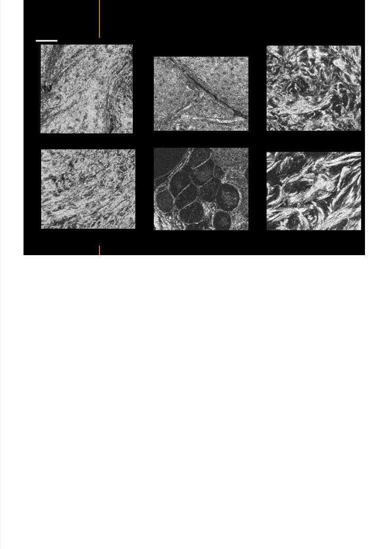
7292019 Atlas of Light-CT images
httpslidepdfcomreaderfullatlas-of-light-ct-images 46118
Skin pathologies basal cell carcinoma discrimination at
7292019 Atlas of Light-CT images
httpslidepdfcomreaderfullatlas-of-light-ct-images 47118
copy LLTech 2012 ndash wwwlltechimagingcom - No copy without authorization 47
Epidermis
Dermis
Hypodermis
A
AA
C
BCC
PSU
PSU
BCC Basal cell carcinoma
A Adipocytes
C Collagen
PSU Pilosebaceous unit
structural level
1 mm
copy LLTech 2012 Courtesy of Hopitaux Universitaires de Genegraveve Genegraveve Switzerland
C
Zoom on basal cell carcinoma discrimination at cellular level
7292019 Atlas of Light-CT images
httpslidepdfcomreaderfullatlas-of-light-ct-images 48118
copy LLTech 2012 ndash wwwlltechimagingcom - No copy without authorization 48
Zoom on basal cell carcinoma discrimination at cellular level
200 microm
100 microm
Peritumoral stroma
High cell density
copy LLTech 2012 Courtesy of Hopitaux Universitaires de Genegraveve Genegraveve Switzerland
Skin pathologies discrimination of actinic keratosis
7292019 Atlas of Light-CT images
httpslidepdfcomreaderfullatlas-of-light-ct-images 49118
copy LLTech 2012 ndash wwwlltechimagingcom - No copy without authorization 49
Skin pathologies discrimination of actinic keratosis
Enlarged epidermis
Extended epidermal ridges
1 mm
copy LLTech 2012 Courtesy of Hopitaux Universitaires de Genegraveve Genegraveve Switzerland
7292019 Atlas of Light-CT images
httpslidepdfcomreaderfullatlas-of-light-ct-images 50118
copy LLTech 2012 ndash wwwlltechimagingcom - No copy without authorization 50
Basal cell carcinoma in depth imaging
Z = 10 microns 7 x 42 mm FOV Z = 35 microns 7 x 42 mm FOV
BCC
SG Sebaceous glands
Z = 55 microns 7 x 42 mm FOV Z = 75 microns 7 x 42 mm FOV
SG
Application to Mohs surgerycopy LLTech 2012 Courtesy of Hopitaux Universitaires de Genegraveve Genegraveve Switzerland
7292019 Atlas of Light-CT images
httpslidepdfcomreaderfullatlas-of-light-ct-images 51118
copy LLTech 2012 ndash wwwlltechimagingcom - No copy without authorization 51 copy LLTECH 2011
Healthy vs Cheloid scar tissue
Healthy skin tissueFOV 75x5mm
Cheloid scar tissueFOV 65x65mm
copy LLTECH 2011
7292019 Atlas of Light-CT images
httpslidepdfcomreaderfullatlas-of-light-ct-images 52118
copy LLTech 2012 ndash wwwlltechimagingcom - No copy without authorization 52
Dermo-epidermaljunction
Cheloid scar tissue in detail
copy LLTECH 2011
Nodular structureThickcollagenfiberbundles
1mm
600 microm
500 microm
h l d d f
7292019 Atlas of Light-CT images
httpslidepdfcomreaderfullatlas-of-light-ct-images 53118
copy LLTech 2012 ndash wwwlltechimagingcom - No copy without authorization 53
Cheloid scar tissue viewed en face
20microm depth 120microm depth
7292019 Atlas of Light-CT images
httpslidepdfcomreaderfullatlas-of-light-ct-images 54118
Skin in-vivo sweat duct imaging in 3D
7292019 Atlas of Light-CT images
httpslidepdfcomreaderfullatlas-of-light-ct-images 55118
copy LLTech 2012 ndash wwwlltechimagingcom - No copy without authorization 55
g g
Vertical reconstruction from en-face images
Sweat duct 3D reconstruction
Stratum corneum
Stratum spinosum
SD sweat ducts
SD SD
100 microm
7292019 Atlas of Light-CT images
httpslidepdfcomreaderfullatlas-of-light-ct-images 56118
7292019 Atlas of Light-CT images
httpslidepdfcomreaderfullatlas-of-light-ct-images 57118
7292019 Atlas of Light-CT images
httpslidepdfcomreaderfullatlas-of-light-ct-images 58118
copy LLTech 2012 ndash wwwlltechimagingcom - No copy without authorization 58
LUNG IMAGING
Back to table of contents
H L
7292019 Atlas of Light-CT images
httpslidepdfcomreaderfullatlas-of-light-ct-images 59118
copy LLTech 2012 ndash wwwlltechimagingcom - No copy without authorization 59
Zone 1
Zone 2
Zone 3
Zone 4
Zone with
cancerous tissue
Limit of cancerous
stroma
alveoli with
macrophages
Alveoles
Human Lung
500microm
copy LLTech 2012 Courtesy of Hocircpital Hotel-Dieu Paris France
H L Z 1 Z t
7292019 Atlas of Light-CT images
httpslidepdfcomreaderfullatlas-of-light-ct-images 60118
copy LLTech 2012 ndash wwwlltechimagingcom - No copy without authorization 60
Stroma
Human Lung ndash Zone 1 ndash Zoom on cancerous stroma
Cancerous
area
200microm
copy LLTech 2012 Courtesy of Hocircpital Hotel-Dieu Paris France
Human Lung ndash Zone 2 ndash Limit cancerous stroma alveoli
7292019 Atlas of Light-CT images
httpslidepdfcomreaderfullatlas-of-light-ct-images 61118
copy LLTech 2012 ndash wwwlltechimagingcom - No copy without authorization 61
g
with visible macrophages and vessel
Alveoli
Macrophages
Vessel
copy LLTech 2012 Courtesy of Hocircpital Hotel-Dieu Paris France
Human Lung ndash Zone 3 ndash Limit cancerous stroma alveoli
7292019 Atlas of Light-CT images
httpslidepdfcomreaderfullatlas-of-light-ct-images 62118
copy LLTech 2012 ndash wwwlltechimagingcom - No copy without authorization 62
g
with visible macrophages
Macrophages
100microm
copy LLTech 2012 Courtesy of Hocircpital Hotel-Dieu Paris France
Human Lung ndash Zone 4 ndash Alveoli
7292019 Atlas of Light-CT images
httpslidepdfcomreaderfullatlas-of-light-ct-images 63118
copy LLTech 2012 ndash wwwlltechimagingcom - No copy without authorization 63
Human Lung Zone 4 Alveoli
Alveoli
200microm
copy LLTech 2012 Courtesy of Hocircpital Hotel-Dieu Paris France
7292019 Atlas of Light-CT images
httpslidepdfcomreaderfullatlas-of-light-ct-images 64118
copy LLTech 2012 ndash wwwlltechimagingcom - No copy without authorization 64
GI IMAGING
Back to table of contents
7292019 Atlas of Light-CT images
httpslidepdfcomreaderfullatlas-of-light-ct-images 65118
Colonic carcinoma
7292019 Atlas of Light-CT images
httpslidepdfcomreaderfullatlas-of-light-ct-images 66118
copy LLTech 2012 ndash wwwlltechimagingcom - No copy without authorization 66
Colonic Carcinoma
Colonic carcinoma
copy LLTech 2012 Courtesy of MD Anderson Houston USA
Intestines ndash Crohnrsquos disease
7292019 Atlas of Light-CT images
httpslidepdfcomreaderfullatlas-of-light-ct-images 67118
copy LLTech 2012 ndash wwwlltechimagingcom - No copy without authorization 67
Crohnrsquos disease structure not
visible from the surface
Fat cells
200 microm
Intestines Crohn s disease
copy LLTech 2012 Courtesy of Institut Curie Paris France
Rat stomach
7292019 Atlas of Light-CT images
httpslidepdfcomreaderfullatlas-of-light-ct-images 68118
copy LLTech 2012 ndash wwwlltechimagingcom - No copy without authorization 68
Rat stomach
Mucosa withgastric pits
Submucosa with
bright collagen
fibers
Muscularis
propria with
muscle bundles
copy LLTech 2012 Courtesy of Weill Cornell Medical College NY USA
200 microm
7292019 Atlas of Light-CT images
httpslidepdfcomreaderfullatlas-of-light-ct-images 69118
copy LLTech 2012 ndash wwwlltechimagingcom - No copy without authorization 69 copy LLTECH 2011
UROGENITAL IMAGING
Back to table of contents
Rat bladder
7292019 Atlas of Light-CT images
httpslidepdfcomreaderfullatlas-of-light-ct-images 70118
copy LLTech 2012 ndash wwwlltechimagingcom - No copy without authorization 70
Rat bladder
Stratifiedurothelium
Lamina propria
500 microm
Muscular
bundles of the
lamina propria
Serosa
copy LLTech 2012 Courtesy of Weill Cornell Medical College NY USA
100 microm
100 microm
Human bladder low grade papillary urothelial carcinoma
7292019 Atlas of Light-CT images
httpslidepdfcomreaderfullatlas-of-light-ct-images 71118
copy LLTech 2012 ndash wwwlltechimagingcom - No copy without authorization 71
Lesion with fibrovascular cores (arrow) and superficial umbrella cells (arrowheads)
Human bladder low grade papillary urothelial carcinoma
copy LLTech 2012 Courtesy of Weill Cornell Medical College NY USA
100 microm
7292019 Atlas of Light-CT images
httpslidepdfcomreaderfullatlas-of-light-ct-images 72118
Rat prostate
7292019 Atlas of Light-CT images
httpslidepdfcomreaderfullatlas-of-light-ct-images 73118
copy LLTech 2012 ndash wwwlltechimagingcom - No copy without authorization 73
p
copy LLTech 2012 Courtesy of Weill Cornell Medical College NY USA
Papillary folds of the
acinar glands
Prostatic acini
Fibromuscular stromaPeriprostatic fat Blood vessel
1 mm
200 microm
Mouse kidney
7292019 Atlas of Light-CT images
httpslidepdfcomreaderfullatlas-of-light-ct-images 74118
copy LLTech 2012 ndash wwwlltechimagingcom - No copy without authorization 74
1 mm
y
copy LLTech 2012 Courtesy of Institut Biomeacutedical de Bicecirctre Paris France
Renal cortex
Renal medulla
Renal sinus
7292019 Atlas of Light-CT images
httpslidepdfcomreaderfullatlas-of-light-ct-images 75118
copy LLTech 2012 ndash wwwlltechimagingcom - No copy without authorization 75
Mouse kidney ndash zoom on highlighted section of previous slide
800microm
copy LLTech 2012 Courtesy of Hocircpital Kremlin Bicecirctre Paris France
7292019 Atlas of Light-CT images
httpslidepdfcomreaderfullatlas-of-light-ct-images 76118
7292019 Atlas of Light-CT images
httpslidepdfcomreaderfullatlas-of-light-ct-images 77118
7292019 Atlas of Light-CT images
httpslidepdfcomreaderfullatlas-of-light-ct-images 78118
copy LLTech 2012 ndash wwwlltechimagingcom - No copy without authorization 78 copy LLTECH 2011
CORE NEEDLE BIOPSIES
Back to table of contents
Kidney
7292019 Atlas of Light-CT images
httpslidepdfcomreaderfullatlas-of-light-ct-images 79118
copy LLTech 2012 ndash wwwlltechimagingcom - No copy without authorization 79
100microm
50microm
Renal tubules
Vessel
500microm
copy LLTech 2012 Courtesy of Institut Curie Paris France
Breast Invasive lobular carcinoma
7292019 Atlas of Light-CT images
httpslidepdfcomreaderfullatlas-of-light-ct-images 80118
copy LLTech 2012 ndash wwwlltechimagingcom - No copy without authorization 80
500microm
stroma
adipocytes
copy LLTech 2012 Courtesy of Institut Curie Paris France
7292019 Atlas of Light-CT images
httpslidepdfcomreaderfullatlas-of-light-ct-images 81118
Breast Ductal carcinoma in situ
7292019 Atlas of Light-CT images
httpslidepdfcomreaderfullatlas-of-light-ct-images 82118
copy LLTech 2012 ndash wwwlltechimagingcom - No copy without authorization 82
500microm
copy LLTech 2012 Courtesy of Institut Curie Paris France
7292019 Atlas of Light-CT images
httpslidepdfcomreaderfullatlas-of-light-ct-images 83118
copy LLTech 2012 ndash wwwlltechimagingcom - No copy without authorization 83 copy LLTECH 2011
THYROID IMAGING
Back to table of contents
7292019 Atlas of Light-CT images
httpslidepdfcomreaderfullatlas-of-light-ct-images 84118
copy LLTech 2012 ndash wwwlltechimagingcom - No copy without authorization 84
Thyroid
800 microm
200 microm
copy LLTech 2012 Courtesy of Hocircpital Tenon Paris France
7292019 Atlas of Light-CT images
httpslidepdfcomreaderfullatlas-of-light-ct-images 85118
copy LLTech 2012 ndash wwwlltechimagingcom - No copy without authorization 85
Thyroid
800 microm
200 microm
copy LLTech 2012 Courtesy of Hocircpital Tenon Paris France
7292019 Atlas of Light-CT images
httpslidepdfcomreaderfullatlas-of-light-ct-images 86118
copy LLTech 2012 ndash wwwlltechimagingcom - No copy without authorization 86 copy LLTECH 2011
FAT CELLS IMAGING
Back to table of contents
7292019 Atlas of Light-CT images
httpslidepdfcomreaderfullatlas-of-light-ct-images 87118
copy LLTech 2012 ndash wwwlltechimagingcom - No copy without authorization 87
Brown fat cells - adipocytes
100 microm 100 microm
200 microm
10um depth
Adipocytes
45um depth
En face
slices
Reconstructed
depth slice
copy LLTech 2012 Courtesy of Centre de Recherche des Cordeliers Paris France
7292019 Atlas of Light-CT images
httpslidepdfcomreaderfullatlas-of-light-ct-images 88118
copy LLTech 2012 ndash wwwlltechimagingcom - No copy without authorization 88 copy LLTECH 2011
CORNEA AND RETINA
Back to table of contents
Human cornea reconstructed depth slice
7292019 Atlas of Light-CT images
httpslidepdfcomreaderfullatlas-of-light-ct-images 89118
copy LLTech 2012 ndash wwwlltechimagingcom - No copy without authorization 89
50 um
wwwlltechimagingcom
7292019 Atlas of Light-CT images
httpslidepdfcomreaderfullatlas-of-light-ct-images 90118
Large field en face view in stroma
7292019 Atlas of Light-CT images
httpslidepdfcomreaderfullatlas-of-light-ct-images 91118
copy LLTech 2012 ndash wwwlltechimagingcom - No copy without authorization 91
200 um
wwwlltechimagingcom
7292019 Atlas of Light-CT images
httpslidepdfcomreaderfullatlas-of-light-ct-images 92118
Rabbit cornea after LASIK reflectivity profiles reveal
i d tt i d thi k d ith li l l
7292019 Atlas of Light-CT images
httpslidepdfcomreaderfullatlas-of-light-ct-images 93118
copy LLTech 2012 ndash wwwlltechimagingcom - No copy without authorization 93
increased scattering and thickened epithelial layer
LASIK LASIK CONTROL CONTROL
copy LLTech 2012
Newborn mouse anterior segment
7292019 Atlas of Light-CT images
httpslidepdfcomreaderfullatlas-of-light-ct-images 94118
copy LLTech 2012 ndash wwwlltechimagingcom - No copy without authorization 94
Corneal epithelial cells
Basal membrane
Stroma
Descemetrsquos membrane
Aqueous humor
Lens capsule
Lens epithelial cells
Cortical fibers
copy LLTech 2012 Courtesy of ESPCI Paris Francecopy LLTech 2012
Rat retina comparison with histology
7292019 Atlas of Light-CT images
httpslidepdfcomreaderfullatlas-of-light-ct-images 95118
copy LLTech 2012 ndash wwwlltechimagingcom - No copy without authorization 95
Figure 1 Right Ex vivo rat retinaimaged using the f ull field OCT
system of ESPCI Left Histology
for comparison
Figure 1 Right Ex vivo rat retinaimaged using the f ull field OCT
system of ESPCI Left Histology
for comparison
Figure 1 Right Ex vivo rat retinaimaged using the f ull field OCT
system of ESPCI Left Histology
for comparisonField
230x300microm
RPE
PRL
ONL
OPL
INL
IPL
GCL
NFL
Invest Ophthalmol Vis Sci 2004 Nov45(11)4126-31
Grieve K Paques M Dubois A Sahel J Boccara C Le Gargasson JF copy LLTech 2012
Rat retina
7292019 Atlas of Light-CT images
httpslidepdfcomreaderfullatlas-of-light-ct-images 96118
copy LLTech 2012 ndash wwwlltechimagingcom - No copy without authorization 96 copy LLTech 2012 Courtesy of ESPCI Paris France
Nerve fiber layer
Retina
Invest Ophthalmol Vis Sci 2004 Nov45(11)4126-31
Grieve K Paques M Dubois A Sahel J Boccara C Le Gargasson JF
Transscleral image
7292019 Atlas of Light-CT images
httpslidepdfcomreaderfullatlas-of-light-ct-images 97118
7292019 Atlas of Light-CT images
httpslidepdfcomreaderfullatlas-of-light-ct-images 98118
7292019 Atlas of Light-CT images
httpslidepdfcomreaderfullatlas-of-light-ct-images 99118
In vivo imaging of drosophila melanogaster
7292019 Atlas of Light-CT images
httpslidepdfcomreaderfullatlas-of-light-ct-images 100118
copy LLTech 2012 ndash wwwlltechimagingcom - No copy without authorization 100 copy LLTech 2012 Courtesy of ESPCI Paris France
In vivo imaging of drosophila melanogaster ndash four stages of pupation over 72 hours
7292019 Atlas of Light-CT images
httpslidepdfcomreaderfullatlas-of-light-ct-images 101118
copy LLTech 2012 ndash wwwlltechimagingcom - No copy without authorization 101
Prepupa (0-2 h) Transition topupal stage (24 h)
Pupal stage (48 h) Advanced pupal phase
before eclosion (72 h)
copy LLTech 2012 Courtesy of ESPCI Paris France
Pupa 118 4 days 30um depth
100
7292019 Atlas of Light-CT images
httpslidepdfcomreaderfullatlas-of-light-ct-images 102118
copy LLTech 2012 ndash wwwlltechimagingcom - No copy without authorization 102
100 um
Drosophila
7292019 Atlas of Light-CT images
httpslidepdfcomreaderfullatlas-of-light-ct-images 103118
copy LLTech 2012 ndash wwwlltechimagingcom - No copy without authorization 103
200 microm
copy LLTech 2012 Courtesy of ESPCI Paris France
C Elegans ndash Fast identification of anatomy
7292019 Atlas of Light-CT images
httpslidepdfcomreaderfullatlas-of-light-ct-images 104118
copy LLTech 2012 ndash wwwlltechimagingcom - No copy without authorization 104
Body wall - cuticle
gonad arm
30 microm depth
uterus
ovocytes
intestine
100 microm 100 microm
15 microm depth
copy LLTech 2012 Courtesy of ENS Paris France
C elegans ndash Images at higher magnification 40X
7292019 Atlas of Light-CT images
httpslidepdfcomreaderfullatlas-of-light-ct-images 105118
copy LLTech 2012 ndash wwwlltechimagingcom - No copy without authorization 105
3D reconstruction 50 microm
100 microm
copy LLTech 2012 Courtesy of ENS Paris France
Zebra fish eye
7292019 Atlas of Light-CT images
httpslidepdfcomreaderfullatlas-of-light-ct-images 106118
copy LLTech 2012 ndash wwwlltechimagingcom - No copy without authorization 106
Zebra fish eye
100 microm
100 microm
En face slices
Reconstructed depth slice
copy LLTech 2012 Courtesy of Inserm Paris France
10um depth 20um depth 45um depth
7292019 Atlas of Light-CT images
httpslidepdfcomreaderfullatlas-of-light-ct-images 107118
Xenopus Laevis backtail
7292019 Atlas of Light-CT images
httpslidepdfcomreaderfullatlas-of-light-ct-images 108118
copy LLTech 2012 ndash wwwlltechimagingcom - No copy without authorization 108
100 um30um depth 60um depth
7292019 Atlas of Light-CT images
httpslidepdfcomreaderfullatlas-of-light-ct-images 109118
Mouse embryo in vitro 6 5 days old ndash
7292019 Atlas of Light-CT images
httpslidepdfcomreaderfullatlas-of-light-ct-images 110118
copy LLTech 2012 ndash wwwlltechimagingcom - No copy without authorization 110
Mouse embryo in vitro 65 days old
copy LLTech 2012 Courtesy of Ellvire Guiot and nstitut Jacques Monod Paris France
3D imaging of internal cavity
Sabellaria alveolata mouth palps
7292019 Atlas of Light-CT images
httpslidepdfcomreaderfullatlas-of-light-ct-images 111118
copy LLTech 2012 ndash wwwlltechimagingcom - No copy without authorization 111
500 microm
100 microm
Mouth palps
copy LLTech 2012 Courtesy of Museacutee National drsquoHistoire Naturelle Paris France
7292019 Atlas of Light-CT images
httpslidepdfcomreaderfullatlas-of-light-ct-images 112118
copy LLTech 2012 ndash wwwlltechimagingcom - No copy without authorization 112 copy LLTECH 2011
PLANT IMAGING
Back to table of contents
Mouse ear leaf epidermis
7292019 Atlas of Light-CT images
httpslidepdfcomreaderfullatlas-of-light-ct-images 113118
copy LLTech 2012 ndash wwwlltechimagingcom - No copy without authorization 113
100 microm
Epidermal cell
copy LLTech 2012
Stoma
Leaf veins
7292019 Atlas of Light-CT images
httpslidepdfcomreaderfullatlas-of-light-ct-images 114118
copy LLTech 2012 ndash wwwlltechimagingcom - No copy without authorization 114
114copy LLTech 2012
Fibers
Veins
100 microm
500 microm
Apple
7292019 Atlas of Light-CT images
httpslidepdfcomreaderfullatlas-of-light-ct-images 115118
copy LLTech 2012 ndash wwwlltechimagingcom - No copy without authorization 115
115copy LLTech 2012
100 microm
Wax
Skin
Flesh
500 microm
Water
Effect of harpin infiltration on tobacco leaf
7292019 Atlas of Light-CT images
httpslidepdfcomreaderfullatlas-of-light-ct-images 116118
copy LLTech 2012 ndash wwwlltechimagingcom - No copy without authorization 116
116copy LLTech 2012 Courtesy of UPMC Paris France
50 microm
50 microm
50 microm
50 microm
Upper epidermis
10 min after harpin infiltration 30 min after harpin infiltration
Palisade parenchyma
Epidermis cell
Chloroplast
7292019 Atlas of Light-CT images
httpslidepdfcomreaderfullatlas-of-light-ct-images 117118
copy LLTech 2012 ndash wwwlltechimagingcom - No copy without authorization 117
bull Light-CTtrade does not require tissue preparation nor staining of any kind
bull Creates images within minutes using a non-destructive process
bull Offers a 1 microm cellular resolution in 3D
bull Reveals structural and cellular information
bull Mosaicing allows fast visualisation at various scale
bull En-face high-resolution imaging allows vertical and 3D reconstruction
bull In-vivo images show good promise for in-vivo system
bull Clinical and research applications in cancer detection cosmetics biobanking small animal
developmental biology botanyhellip
Contact Details
7292019 Atlas of Light-CT images
httpslidepdfcomreaderfullatlas-of-light-ct-images 118118
USA
LLTech Inc
103 Carnegie Center DriveSuite 300
Princeton NJ 08540
USA
wwwlltechimagingcom
contactlltechfr
Phone + 1 609 955 3506
LLTech SAS
29 rue du Faubourg St-JacquesPeacutepiniegravere Paris Santeacute Cochin
75014
Paris - France
wwwlltechimagingcom
contactlltechfr
Phone +33 1 82 72 61 25
Europe
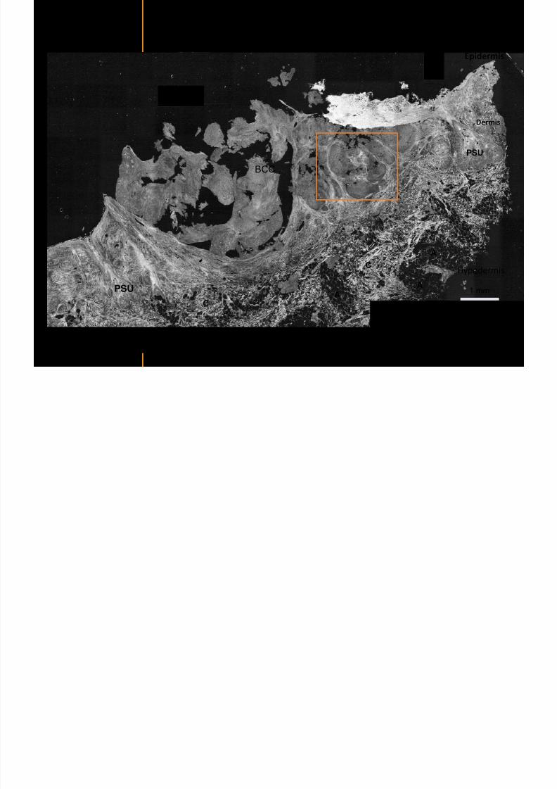
7292019 Atlas of Light-CT images
httpslidepdfcomreaderfullatlas-of-light-ct-images 51118
copy LLTech 2012 ndash wwwlltechimagingcom - No copy without authorization 51 copy LLTECH 2011
Healthy vs Cheloid scar tissue
Healthy skin tissueFOV 75x5mm
Cheloid scar tissueFOV 65x65mm
copy LLTECH 2011
7292019 Atlas of Light-CT images
httpslidepdfcomreaderfullatlas-of-light-ct-images 52118
copy LLTech 2012 ndash wwwlltechimagingcom - No copy without authorization 52
Dermo-epidermaljunction
Cheloid scar tissue in detail
copy LLTECH 2011
Nodular structureThickcollagenfiberbundles
1mm
600 microm
500 microm
h l d d f
7292019 Atlas of Light-CT images
httpslidepdfcomreaderfullatlas-of-light-ct-images 53118
copy LLTech 2012 ndash wwwlltechimagingcom - No copy without authorization 53
Cheloid scar tissue viewed en face
20microm depth 120microm depth
7292019 Atlas of Light-CT images
httpslidepdfcomreaderfullatlas-of-light-ct-images 54118
Skin in-vivo sweat duct imaging in 3D
7292019 Atlas of Light-CT images
httpslidepdfcomreaderfullatlas-of-light-ct-images 55118
copy LLTech 2012 ndash wwwlltechimagingcom - No copy without authorization 55
g g
Vertical reconstruction from en-face images
Sweat duct 3D reconstruction
Stratum corneum
Stratum spinosum
SD sweat ducts
SD SD
100 microm
7292019 Atlas of Light-CT images
httpslidepdfcomreaderfullatlas-of-light-ct-images 56118
7292019 Atlas of Light-CT images
httpslidepdfcomreaderfullatlas-of-light-ct-images 57118
7292019 Atlas of Light-CT images
httpslidepdfcomreaderfullatlas-of-light-ct-images 58118
copy LLTech 2012 ndash wwwlltechimagingcom - No copy without authorization 58
LUNG IMAGING
Back to table of contents
H L
7292019 Atlas of Light-CT images
httpslidepdfcomreaderfullatlas-of-light-ct-images 59118
copy LLTech 2012 ndash wwwlltechimagingcom - No copy without authorization 59
Zone 1
Zone 2
Zone 3
Zone 4
Zone with
cancerous tissue
Limit of cancerous
stroma
alveoli with
macrophages
Alveoles
Human Lung
500microm
copy LLTech 2012 Courtesy of Hocircpital Hotel-Dieu Paris France
H L Z 1 Z t
7292019 Atlas of Light-CT images
httpslidepdfcomreaderfullatlas-of-light-ct-images 60118
copy LLTech 2012 ndash wwwlltechimagingcom - No copy without authorization 60
Stroma
Human Lung ndash Zone 1 ndash Zoom on cancerous stroma
Cancerous
area
200microm
copy LLTech 2012 Courtesy of Hocircpital Hotel-Dieu Paris France
Human Lung ndash Zone 2 ndash Limit cancerous stroma alveoli
7292019 Atlas of Light-CT images
httpslidepdfcomreaderfullatlas-of-light-ct-images 61118
copy LLTech 2012 ndash wwwlltechimagingcom - No copy without authorization 61
g
with visible macrophages and vessel
Alveoli
Macrophages
Vessel
copy LLTech 2012 Courtesy of Hocircpital Hotel-Dieu Paris France
Human Lung ndash Zone 3 ndash Limit cancerous stroma alveoli
7292019 Atlas of Light-CT images
httpslidepdfcomreaderfullatlas-of-light-ct-images 62118
copy LLTech 2012 ndash wwwlltechimagingcom - No copy without authorization 62
g
with visible macrophages
Macrophages
100microm
copy LLTech 2012 Courtesy of Hocircpital Hotel-Dieu Paris France
Human Lung ndash Zone 4 ndash Alveoli
7292019 Atlas of Light-CT images
httpslidepdfcomreaderfullatlas-of-light-ct-images 63118
copy LLTech 2012 ndash wwwlltechimagingcom - No copy without authorization 63
Human Lung Zone 4 Alveoli
Alveoli
200microm
copy LLTech 2012 Courtesy of Hocircpital Hotel-Dieu Paris France
7292019 Atlas of Light-CT images
httpslidepdfcomreaderfullatlas-of-light-ct-images 64118
copy LLTech 2012 ndash wwwlltechimagingcom - No copy without authorization 64
GI IMAGING
Back to table of contents
7292019 Atlas of Light-CT images
httpslidepdfcomreaderfullatlas-of-light-ct-images 65118
Colonic carcinoma
7292019 Atlas of Light-CT images
httpslidepdfcomreaderfullatlas-of-light-ct-images 66118
copy LLTech 2012 ndash wwwlltechimagingcom - No copy without authorization 66
Colonic Carcinoma
Colonic carcinoma
copy LLTech 2012 Courtesy of MD Anderson Houston USA
Intestines ndash Crohnrsquos disease
7292019 Atlas of Light-CT images
httpslidepdfcomreaderfullatlas-of-light-ct-images 67118
copy LLTech 2012 ndash wwwlltechimagingcom - No copy without authorization 67
Crohnrsquos disease structure not
visible from the surface
Fat cells
200 microm
Intestines Crohn s disease
copy LLTech 2012 Courtesy of Institut Curie Paris France
Rat stomach
7292019 Atlas of Light-CT images
httpslidepdfcomreaderfullatlas-of-light-ct-images 68118
copy LLTech 2012 ndash wwwlltechimagingcom - No copy without authorization 68
Rat stomach
Mucosa withgastric pits
Submucosa with
bright collagen
fibers
Muscularis
propria with
muscle bundles
copy LLTech 2012 Courtesy of Weill Cornell Medical College NY USA
200 microm
7292019 Atlas of Light-CT images
httpslidepdfcomreaderfullatlas-of-light-ct-images 69118
copy LLTech 2012 ndash wwwlltechimagingcom - No copy without authorization 69 copy LLTECH 2011
UROGENITAL IMAGING
Back to table of contents
Rat bladder
7292019 Atlas of Light-CT images
httpslidepdfcomreaderfullatlas-of-light-ct-images 70118
copy LLTech 2012 ndash wwwlltechimagingcom - No copy without authorization 70
Rat bladder
Stratifiedurothelium
Lamina propria
500 microm
Muscular
bundles of the
lamina propria
Serosa
copy LLTech 2012 Courtesy of Weill Cornell Medical College NY USA
100 microm
100 microm
Human bladder low grade papillary urothelial carcinoma
7292019 Atlas of Light-CT images
httpslidepdfcomreaderfullatlas-of-light-ct-images 71118
copy LLTech 2012 ndash wwwlltechimagingcom - No copy without authorization 71
Lesion with fibrovascular cores (arrow) and superficial umbrella cells (arrowheads)
Human bladder low grade papillary urothelial carcinoma
copy LLTech 2012 Courtesy of Weill Cornell Medical College NY USA
100 microm
7292019 Atlas of Light-CT images
httpslidepdfcomreaderfullatlas-of-light-ct-images 72118
Rat prostate
7292019 Atlas of Light-CT images
httpslidepdfcomreaderfullatlas-of-light-ct-images 73118
copy LLTech 2012 ndash wwwlltechimagingcom - No copy without authorization 73
p
copy LLTech 2012 Courtesy of Weill Cornell Medical College NY USA
Papillary folds of the
acinar glands
Prostatic acini
Fibromuscular stromaPeriprostatic fat Blood vessel
1 mm
200 microm
Mouse kidney
7292019 Atlas of Light-CT images
httpslidepdfcomreaderfullatlas-of-light-ct-images 74118
copy LLTech 2012 ndash wwwlltechimagingcom - No copy without authorization 74
1 mm
y
copy LLTech 2012 Courtesy of Institut Biomeacutedical de Bicecirctre Paris France
Renal cortex
Renal medulla
Renal sinus
7292019 Atlas of Light-CT images
httpslidepdfcomreaderfullatlas-of-light-ct-images 75118
copy LLTech 2012 ndash wwwlltechimagingcom - No copy without authorization 75
Mouse kidney ndash zoom on highlighted section of previous slide
800microm
copy LLTech 2012 Courtesy of Hocircpital Kremlin Bicecirctre Paris France
7292019 Atlas of Light-CT images
httpslidepdfcomreaderfullatlas-of-light-ct-images 76118
7292019 Atlas of Light-CT images
httpslidepdfcomreaderfullatlas-of-light-ct-images 77118
7292019 Atlas of Light-CT images
httpslidepdfcomreaderfullatlas-of-light-ct-images 78118
copy LLTech 2012 ndash wwwlltechimagingcom - No copy without authorization 78 copy LLTECH 2011
CORE NEEDLE BIOPSIES
Back to table of contents
Kidney
7292019 Atlas of Light-CT images
httpslidepdfcomreaderfullatlas-of-light-ct-images 79118
copy LLTech 2012 ndash wwwlltechimagingcom - No copy without authorization 79
100microm
50microm
Renal tubules
Vessel
500microm
copy LLTech 2012 Courtesy of Institut Curie Paris France
Breast Invasive lobular carcinoma
7292019 Atlas of Light-CT images
httpslidepdfcomreaderfullatlas-of-light-ct-images 80118
copy LLTech 2012 ndash wwwlltechimagingcom - No copy without authorization 80
500microm
stroma
adipocytes
copy LLTech 2012 Courtesy of Institut Curie Paris France
7292019 Atlas of Light-CT images
httpslidepdfcomreaderfullatlas-of-light-ct-images 81118
Breast Ductal carcinoma in situ
7292019 Atlas of Light-CT images
httpslidepdfcomreaderfullatlas-of-light-ct-images 82118
copy LLTech 2012 ndash wwwlltechimagingcom - No copy without authorization 82
500microm
copy LLTech 2012 Courtesy of Institut Curie Paris France
7292019 Atlas of Light-CT images
httpslidepdfcomreaderfullatlas-of-light-ct-images 83118
copy LLTech 2012 ndash wwwlltechimagingcom - No copy without authorization 83 copy LLTECH 2011
THYROID IMAGING
Back to table of contents
7292019 Atlas of Light-CT images
httpslidepdfcomreaderfullatlas-of-light-ct-images 84118
copy LLTech 2012 ndash wwwlltechimagingcom - No copy without authorization 84
Thyroid
800 microm
200 microm
copy LLTech 2012 Courtesy of Hocircpital Tenon Paris France
7292019 Atlas of Light-CT images
httpslidepdfcomreaderfullatlas-of-light-ct-images 85118
copy LLTech 2012 ndash wwwlltechimagingcom - No copy without authorization 85
Thyroid
800 microm
200 microm
copy LLTech 2012 Courtesy of Hocircpital Tenon Paris France
7292019 Atlas of Light-CT images
httpslidepdfcomreaderfullatlas-of-light-ct-images 86118
copy LLTech 2012 ndash wwwlltechimagingcom - No copy without authorization 86 copy LLTECH 2011
FAT CELLS IMAGING
Back to table of contents
7292019 Atlas of Light-CT images
httpslidepdfcomreaderfullatlas-of-light-ct-images 87118
copy LLTech 2012 ndash wwwlltechimagingcom - No copy without authorization 87
Brown fat cells - adipocytes
100 microm 100 microm
200 microm
10um depth
Adipocytes
45um depth
En face
slices
Reconstructed
depth slice
copy LLTech 2012 Courtesy of Centre de Recherche des Cordeliers Paris France
7292019 Atlas of Light-CT images
httpslidepdfcomreaderfullatlas-of-light-ct-images 88118
copy LLTech 2012 ndash wwwlltechimagingcom - No copy without authorization 88 copy LLTECH 2011
CORNEA AND RETINA
Back to table of contents
Human cornea reconstructed depth slice
7292019 Atlas of Light-CT images
httpslidepdfcomreaderfullatlas-of-light-ct-images 89118
copy LLTech 2012 ndash wwwlltechimagingcom - No copy without authorization 89
50 um
wwwlltechimagingcom
7292019 Atlas of Light-CT images
httpslidepdfcomreaderfullatlas-of-light-ct-images 90118
Large field en face view in stroma
7292019 Atlas of Light-CT images
httpslidepdfcomreaderfullatlas-of-light-ct-images 91118
copy LLTech 2012 ndash wwwlltechimagingcom - No copy without authorization 91
200 um
wwwlltechimagingcom
7292019 Atlas of Light-CT images
httpslidepdfcomreaderfullatlas-of-light-ct-images 92118
Rabbit cornea after LASIK reflectivity profiles reveal
i d tt i d thi k d ith li l l
7292019 Atlas of Light-CT images
httpslidepdfcomreaderfullatlas-of-light-ct-images 93118
copy LLTech 2012 ndash wwwlltechimagingcom - No copy without authorization 93
increased scattering and thickened epithelial layer
LASIK LASIK CONTROL CONTROL
copy LLTech 2012
Newborn mouse anterior segment
7292019 Atlas of Light-CT images
httpslidepdfcomreaderfullatlas-of-light-ct-images 94118
copy LLTech 2012 ndash wwwlltechimagingcom - No copy without authorization 94
Corneal epithelial cells
Basal membrane
Stroma
Descemetrsquos membrane
Aqueous humor
Lens capsule
Lens epithelial cells
Cortical fibers
copy LLTech 2012 Courtesy of ESPCI Paris Francecopy LLTech 2012
Rat retina comparison with histology
7292019 Atlas of Light-CT images
httpslidepdfcomreaderfullatlas-of-light-ct-images 95118
copy LLTech 2012 ndash wwwlltechimagingcom - No copy without authorization 95
Figure 1 Right Ex vivo rat retinaimaged using the f ull field OCT
system of ESPCI Left Histology
for comparison
Figure 1 Right Ex vivo rat retinaimaged using the f ull field OCT
system of ESPCI Left Histology
for comparison
Figure 1 Right Ex vivo rat retinaimaged using the f ull field OCT
system of ESPCI Left Histology
for comparisonField
230x300microm
RPE
PRL
ONL
OPL
INL
IPL
GCL
NFL
Invest Ophthalmol Vis Sci 2004 Nov45(11)4126-31
Grieve K Paques M Dubois A Sahel J Boccara C Le Gargasson JF copy LLTech 2012
Rat retina
7292019 Atlas of Light-CT images
httpslidepdfcomreaderfullatlas-of-light-ct-images 96118
copy LLTech 2012 ndash wwwlltechimagingcom - No copy without authorization 96 copy LLTech 2012 Courtesy of ESPCI Paris France
Nerve fiber layer
Retina
Invest Ophthalmol Vis Sci 2004 Nov45(11)4126-31
Grieve K Paques M Dubois A Sahel J Boccara C Le Gargasson JF
Transscleral image
7292019 Atlas of Light-CT images
httpslidepdfcomreaderfullatlas-of-light-ct-images 97118
7292019 Atlas of Light-CT images
httpslidepdfcomreaderfullatlas-of-light-ct-images 98118
7292019 Atlas of Light-CT images
httpslidepdfcomreaderfullatlas-of-light-ct-images 99118
In vivo imaging of drosophila melanogaster
7292019 Atlas of Light-CT images
httpslidepdfcomreaderfullatlas-of-light-ct-images 100118
copy LLTech 2012 ndash wwwlltechimagingcom - No copy without authorization 100 copy LLTech 2012 Courtesy of ESPCI Paris France
In vivo imaging of drosophila melanogaster ndash four stages of pupation over 72 hours
7292019 Atlas of Light-CT images
httpslidepdfcomreaderfullatlas-of-light-ct-images 101118
copy LLTech 2012 ndash wwwlltechimagingcom - No copy without authorization 101
Prepupa (0-2 h) Transition topupal stage (24 h)
Pupal stage (48 h) Advanced pupal phase
before eclosion (72 h)
copy LLTech 2012 Courtesy of ESPCI Paris France
Pupa 118 4 days 30um depth
100
7292019 Atlas of Light-CT images
httpslidepdfcomreaderfullatlas-of-light-ct-images 102118
copy LLTech 2012 ndash wwwlltechimagingcom - No copy without authorization 102
100 um
Drosophila
7292019 Atlas of Light-CT images
httpslidepdfcomreaderfullatlas-of-light-ct-images 103118
copy LLTech 2012 ndash wwwlltechimagingcom - No copy without authorization 103
200 microm
copy LLTech 2012 Courtesy of ESPCI Paris France
C Elegans ndash Fast identification of anatomy
7292019 Atlas of Light-CT images
httpslidepdfcomreaderfullatlas-of-light-ct-images 104118
copy LLTech 2012 ndash wwwlltechimagingcom - No copy without authorization 104
Body wall - cuticle
gonad arm
30 microm depth
uterus
ovocytes
intestine
100 microm 100 microm
15 microm depth
copy LLTech 2012 Courtesy of ENS Paris France
C elegans ndash Images at higher magnification 40X
7292019 Atlas of Light-CT images
httpslidepdfcomreaderfullatlas-of-light-ct-images 105118
copy LLTech 2012 ndash wwwlltechimagingcom - No copy without authorization 105
3D reconstruction 50 microm
100 microm
copy LLTech 2012 Courtesy of ENS Paris France
Zebra fish eye
7292019 Atlas of Light-CT images
httpslidepdfcomreaderfullatlas-of-light-ct-images 106118
copy LLTech 2012 ndash wwwlltechimagingcom - No copy without authorization 106
Zebra fish eye
100 microm
100 microm
En face slices
Reconstructed depth slice
copy LLTech 2012 Courtesy of Inserm Paris France
10um depth 20um depth 45um depth
7292019 Atlas of Light-CT images
httpslidepdfcomreaderfullatlas-of-light-ct-images 107118
Xenopus Laevis backtail
7292019 Atlas of Light-CT images
httpslidepdfcomreaderfullatlas-of-light-ct-images 108118
copy LLTech 2012 ndash wwwlltechimagingcom - No copy without authorization 108
100 um30um depth 60um depth
7292019 Atlas of Light-CT images
httpslidepdfcomreaderfullatlas-of-light-ct-images 109118
Mouse embryo in vitro 6 5 days old ndash
7292019 Atlas of Light-CT images
httpslidepdfcomreaderfullatlas-of-light-ct-images 110118
copy LLTech 2012 ndash wwwlltechimagingcom - No copy without authorization 110
Mouse embryo in vitro 65 days old
copy LLTech 2012 Courtesy of Ellvire Guiot and nstitut Jacques Monod Paris France
3D imaging of internal cavity
Sabellaria alveolata mouth palps
7292019 Atlas of Light-CT images
httpslidepdfcomreaderfullatlas-of-light-ct-images 111118
copy LLTech 2012 ndash wwwlltechimagingcom - No copy without authorization 111
500 microm
100 microm
Mouth palps
copy LLTech 2012 Courtesy of Museacutee National drsquoHistoire Naturelle Paris France
7292019 Atlas of Light-CT images
httpslidepdfcomreaderfullatlas-of-light-ct-images 112118
copy LLTech 2012 ndash wwwlltechimagingcom - No copy without authorization 112 copy LLTECH 2011
PLANT IMAGING
Back to table of contents
Mouse ear leaf epidermis
7292019 Atlas of Light-CT images
httpslidepdfcomreaderfullatlas-of-light-ct-images 113118
copy LLTech 2012 ndash wwwlltechimagingcom - No copy without authorization 113
100 microm
Epidermal cell
copy LLTech 2012
Stoma
Leaf veins
7292019 Atlas of Light-CT images
httpslidepdfcomreaderfullatlas-of-light-ct-images 114118
copy LLTech 2012 ndash wwwlltechimagingcom - No copy without authorization 114
114copy LLTech 2012
Fibers
Veins
100 microm
500 microm
Apple
7292019 Atlas of Light-CT images
httpslidepdfcomreaderfullatlas-of-light-ct-images 115118
copy LLTech 2012 ndash wwwlltechimagingcom - No copy without authorization 115
115copy LLTech 2012
100 microm
Wax
Skin
Flesh
500 microm
Water
Effect of harpin infiltration on tobacco leaf
7292019 Atlas of Light-CT images
httpslidepdfcomreaderfullatlas-of-light-ct-images 116118
copy LLTech 2012 ndash wwwlltechimagingcom - No copy without authorization 116
116copy LLTech 2012 Courtesy of UPMC Paris France
50 microm
50 microm
50 microm
50 microm
Upper epidermis
10 min after harpin infiltration 30 min after harpin infiltration
Palisade parenchyma
Epidermis cell
Chloroplast
7292019 Atlas of Light-CT images
httpslidepdfcomreaderfullatlas-of-light-ct-images 117118
copy LLTech 2012 ndash wwwlltechimagingcom - No copy without authorization 117
bull Light-CTtrade does not require tissue preparation nor staining of any kind
bull Creates images within minutes using a non-destructive process
bull Offers a 1 microm cellular resolution in 3D
bull Reveals structural and cellular information
bull Mosaicing allows fast visualisation at various scale
bull En-face high-resolution imaging allows vertical and 3D reconstruction
bull In-vivo images show good promise for in-vivo system
bull Clinical and research applications in cancer detection cosmetics biobanking small animal
developmental biology botanyhellip
Contact Details
7292019 Atlas of Light-CT images
httpslidepdfcomreaderfullatlas-of-light-ct-images 118118
USA
LLTech Inc
103 Carnegie Center DriveSuite 300
Princeton NJ 08540
USA
wwwlltechimagingcom
contactlltechfr
Phone + 1 609 955 3506
LLTech SAS
29 rue du Faubourg St-JacquesPeacutepiniegravere Paris Santeacute Cochin
75014
Paris - France
wwwlltechimagingcom
contactlltechfr
Phone +33 1 82 72 61 25
Europe
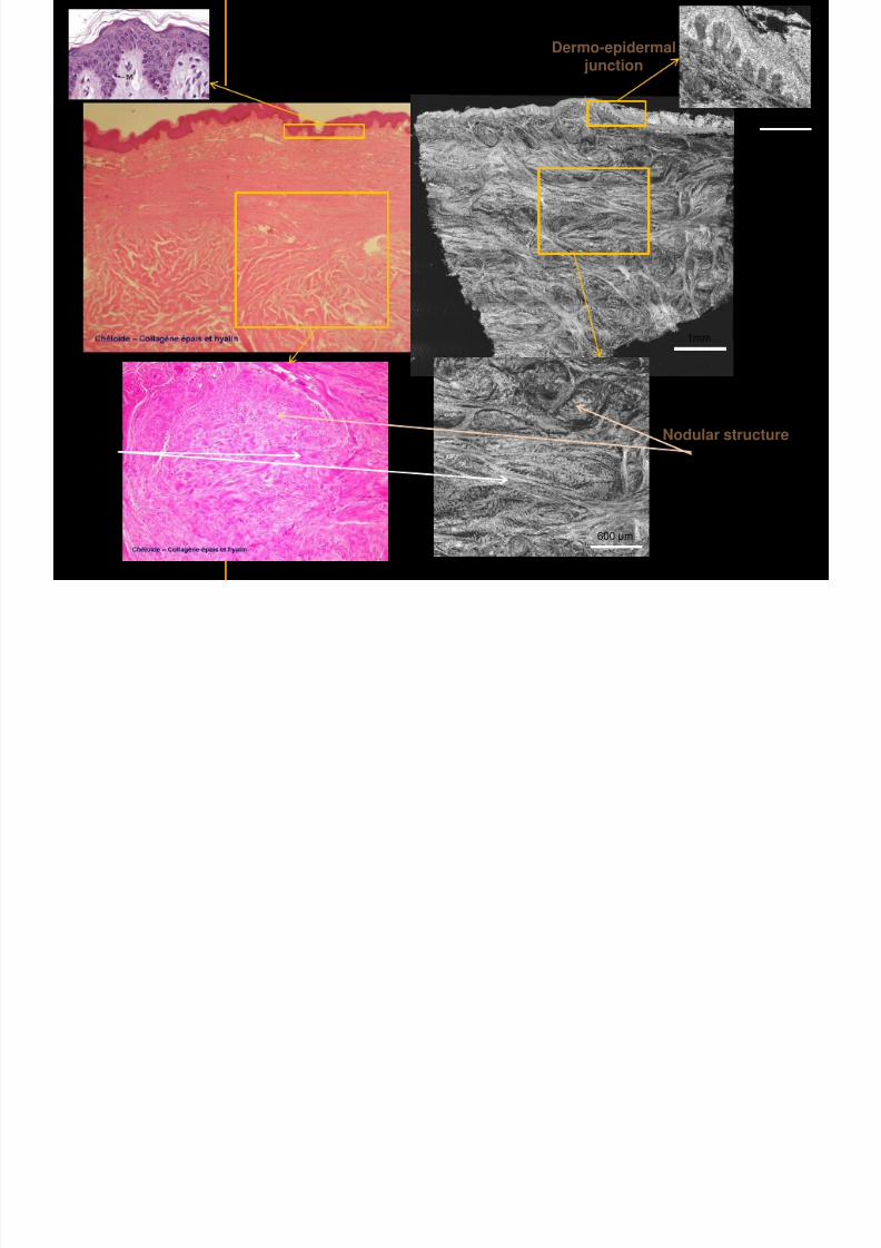
7292019 Atlas of Light-CT images
httpslidepdfcomreaderfullatlas-of-light-ct-images 53118
copy LLTech 2012 ndash wwwlltechimagingcom - No copy without authorization 53
Cheloid scar tissue viewed en face
20microm depth 120microm depth
7292019 Atlas of Light-CT images
httpslidepdfcomreaderfullatlas-of-light-ct-images 54118
Skin in-vivo sweat duct imaging in 3D
7292019 Atlas of Light-CT images
httpslidepdfcomreaderfullatlas-of-light-ct-images 55118
copy LLTech 2012 ndash wwwlltechimagingcom - No copy without authorization 55
g g
Vertical reconstruction from en-face images
Sweat duct 3D reconstruction
Stratum corneum
Stratum spinosum
SD sweat ducts
SD SD
100 microm
7292019 Atlas of Light-CT images
httpslidepdfcomreaderfullatlas-of-light-ct-images 56118
7292019 Atlas of Light-CT images
httpslidepdfcomreaderfullatlas-of-light-ct-images 57118
7292019 Atlas of Light-CT images
httpslidepdfcomreaderfullatlas-of-light-ct-images 58118
copy LLTech 2012 ndash wwwlltechimagingcom - No copy without authorization 58
LUNG IMAGING
Back to table of contents
H L
7292019 Atlas of Light-CT images
httpslidepdfcomreaderfullatlas-of-light-ct-images 59118
copy LLTech 2012 ndash wwwlltechimagingcom - No copy without authorization 59
Zone 1
Zone 2
Zone 3
Zone 4
Zone with
cancerous tissue
Limit of cancerous
stroma
alveoli with
macrophages
Alveoles
Human Lung
500microm
copy LLTech 2012 Courtesy of Hocircpital Hotel-Dieu Paris France
H L Z 1 Z t
7292019 Atlas of Light-CT images
httpslidepdfcomreaderfullatlas-of-light-ct-images 60118
copy LLTech 2012 ndash wwwlltechimagingcom - No copy without authorization 60
Stroma
Human Lung ndash Zone 1 ndash Zoom on cancerous stroma
Cancerous
area
200microm
copy LLTech 2012 Courtesy of Hocircpital Hotel-Dieu Paris France
Human Lung ndash Zone 2 ndash Limit cancerous stroma alveoli
7292019 Atlas of Light-CT images
httpslidepdfcomreaderfullatlas-of-light-ct-images 61118
copy LLTech 2012 ndash wwwlltechimagingcom - No copy without authorization 61
g
with visible macrophages and vessel
Alveoli
Macrophages
Vessel
copy LLTech 2012 Courtesy of Hocircpital Hotel-Dieu Paris France
Human Lung ndash Zone 3 ndash Limit cancerous stroma alveoli
7292019 Atlas of Light-CT images
httpslidepdfcomreaderfullatlas-of-light-ct-images 62118
copy LLTech 2012 ndash wwwlltechimagingcom - No copy without authorization 62
g
with visible macrophages
Macrophages
100microm
copy LLTech 2012 Courtesy of Hocircpital Hotel-Dieu Paris France
Human Lung ndash Zone 4 ndash Alveoli
7292019 Atlas of Light-CT images
httpslidepdfcomreaderfullatlas-of-light-ct-images 63118
copy LLTech 2012 ndash wwwlltechimagingcom - No copy without authorization 63
Human Lung Zone 4 Alveoli
Alveoli
200microm
copy LLTech 2012 Courtesy of Hocircpital Hotel-Dieu Paris France
7292019 Atlas of Light-CT images
httpslidepdfcomreaderfullatlas-of-light-ct-images 64118
copy LLTech 2012 ndash wwwlltechimagingcom - No copy without authorization 64
GI IMAGING
Back to table of contents
7292019 Atlas of Light-CT images
httpslidepdfcomreaderfullatlas-of-light-ct-images 65118
Colonic carcinoma
7292019 Atlas of Light-CT images
httpslidepdfcomreaderfullatlas-of-light-ct-images 66118
copy LLTech 2012 ndash wwwlltechimagingcom - No copy without authorization 66
Colonic Carcinoma
Colonic carcinoma
copy LLTech 2012 Courtesy of MD Anderson Houston USA
Intestines ndash Crohnrsquos disease
7292019 Atlas of Light-CT images
httpslidepdfcomreaderfullatlas-of-light-ct-images 67118
copy LLTech 2012 ndash wwwlltechimagingcom - No copy without authorization 67
Crohnrsquos disease structure not
visible from the surface
Fat cells
200 microm
Intestines Crohn s disease
copy LLTech 2012 Courtesy of Institut Curie Paris France
Rat stomach
7292019 Atlas of Light-CT images
httpslidepdfcomreaderfullatlas-of-light-ct-images 68118
copy LLTech 2012 ndash wwwlltechimagingcom - No copy without authorization 68
Rat stomach
Mucosa withgastric pits
Submucosa with
bright collagen
fibers
Muscularis
propria with
muscle bundles
copy LLTech 2012 Courtesy of Weill Cornell Medical College NY USA
200 microm
7292019 Atlas of Light-CT images
httpslidepdfcomreaderfullatlas-of-light-ct-images 69118
copy LLTech 2012 ndash wwwlltechimagingcom - No copy without authorization 69 copy LLTECH 2011
UROGENITAL IMAGING
Back to table of contents
Rat bladder
7292019 Atlas of Light-CT images
httpslidepdfcomreaderfullatlas-of-light-ct-images 70118
copy LLTech 2012 ndash wwwlltechimagingcom - No copy without authorization 70
Rat bladder
Stratifiedurothelium
Lamina propria
500 microm
Muscular
bundles of the
lamina propria
Serosa
copy LLTech 2012 Courtesy of Weill Cornell Medical College NY USA
100 microm
100 microm
Human bladder low grade papillary urothelial carcinoma
7292019 Atlas of Light-CT images
httpslidepdfcomreaderfullatlas-of-light-ct-images 71118
copy LLTech 2012 ndash wwwlltechimagingcom - No copy without authorization 71
Lesion with fibrovascular cores (arrow) and superficial umbrella cells (arrowheads)
Human bladder low grade papillary urothelial carcinoma
copy LLTech 2012 Courtesy of Weill Cornell Medical College NY USA
100 microm
7292019 Atlas of Light-CT images
httpslidepdfcomreaderfullatlas-of-light-ct-images 72118
Rat prostate
7292019 Atlas of Light-CT images
httpslidepdfcomreaderfullatlas-of-light-ct-images 73118
copy LLTech 2012 ndash wwwlltechimagingcom - No copy without authorization 73
p
copy LLTech 2012 Courtesy of Weill Cornell Medical College NY USA
Papillary folds of the
acinar glands
Prostatic acini
Fibromuscular stromaPeriprostatic fat Blood vessel
1 mm
200 microm
Mouse kidney
7292019 Atlas of Light-CT images
httpslidepdfcomreaderfullatlas-of-light-ct-images 74118
copy LLTech 2012 ndash wwwlltechimagingcom - No copy without authorization 74
1 mm
y
copy LLTech 2012 Courtesy of Institut Biomeacutedical de Bicecirctre Paris France
Renal cortex
Renal medulla
Renal sinus
7292019 Atlas of Light-CT images
httpslidepdfcomreaderfullatlas-of-light-ct-images 75118
copy LLTech 2012 ndash wwwlltechimagingcom - No copy without authorization 75
Mouse kidney ndash zoom on highlighted section of previous slide
800microm
copy LLTech 2012 Courtesy of Hocircpital Kremlin Bicecirctre Paris France
7292019 Atlas of Light-CT images
httpslidepdfcomreaderfullatlas-of-light-ct-images 76118
7292019 Atlas of Light-CT images
httpslidepdfcomreaderfullatlas-of-light-ct-images 77118
7292019 Atlas of Light-CT images
httpslidepdfcomreaderfullatlas-of-light-ct-images 78118
copy LLTech 2012 ndash wwwlltechimagingcom - No copy without authorization 78 copy LLTECH 2011
CORE NEEDLE BIOPSIES
Back to table of contents
Kidney
7292019 Atlas of Light-CT images
httpslidepdfcomreaderfullatlas-of-light-ct-images 79118
copy LLTech 2012 ndash wwwlltechimagingcom - No copy without authorization 79
100microm
50microm
Renal tubules
Vessel
500microm
copy LLTech 2012 Courtesy of Institut Curie Paris France
Breast Invasive lobular carcinoma
7292019 Atlas of Light-CT images
httpslidepdfcomreaderfullatlas-of-light-ct-images 80118
copy LLTech 2012 ndash wwwlltechimagingcom - No copy without authorization 80
500microm
stroma
adipocytes
copy LLTech 2012 Courtesy of Institut Curie Paris France
7292019 Atlas of Light-CT images
httpslidepdfcomreaderfullatlas-of-light-ct-images 81118
Breast Ductal carcinoma in situ
7292019 Atlas of Light-CT images
httpslidepdfcomreaderfullatlas-of-light-ct-images 82118
copy LLTech 2012 ndash wwwlltechimagingcom - No copy without authorization 82
500microm
copy LLTech 2012 Courtesy of Institut Curie Paris France
7292019 Atlas of Light-CT images
httpslidepdfcomreaderfullatlas-of-light-ct-images 83118
copy LLTech 2012 ndash wwwlltechimagingcom - No copy without authorization 83 copy LLTECH 2011
THYROID IMAGING
Back to table of contents
7292019 Atlas of Light-CT images
httpslidepdfcomreaderfullatlas-of-light-ct-images 84118
copy LLTech 2012 ndash wwwlltechimagingcom - No copy without authorization 84
Thyroid
800 microm
200 microm
copy LLTech 2012 Courtesy of Hocircpital Tenon Paris France
7292019 Atlas of Light-CT images
httpslidepdfcomreaderfullatlas-of-light-ct-images 85118
copy LLTech 2012 ndash wwwlltechimagingcom - No copy without authorization 85
Thyroid
800 microm
200 microm
copy LLTech 2012 Courtesy of Hocircpital Tenon Paris France
7292019 Atlas of Light-CT images
httpslidepdfcomreaderfullatlas-of-light-ct-images 86118
copy LLTech 2012 ndash wwwlltechimagingcom - No copy without authorization 86 copy LLTECH 2011
FAT CELLS IMAGING
Back to table of contents
7292019 Atlas of Light-CT images
httpslidepdfcomreaderfullatlas-of-light-ct-images 87118
copy LLTech 2012 ndash wwwlltechimagingcom - No copy without authorization 87
Brown fat cells - adipocytes
100 microm 100 microm
200 microm
10um depth
Adipocytes
45um depth
En face
slices
Reconstructed
depth slice
copy LLTech 2012 Courtesy of Centre de Recherche des Cordeliers Paris France
7292019 Atlas of Light-CT images
httpslidepdfcomreaderfullatlas-of-light-ct-images 88118
copy LLTech 2012 ndash wwwlltechimagingcom - No copy without authorization 88 copy LLTECH 2011
CORNEA AND RETINA
Back to table of contents
Human cornea reconstructed depth slice
7292019 Atlas of Light-CT images
httpslidepdfcomreaderfullatlas-of-light-ct-images 89118
copy LLTech 2012 ndash wwwlltechimagingcom - No copy without authorization 89
50 um
wwwlltechimagingcom
7292019 Atlas of Light-CT images
httpslidepdfcomreaderfullatlas-of-light-ct-images 90118
Large field en face view in stroma
7292019 Atlas of Light-CT images
httpslidepdfcomreaderfullatlas-of-light-ct-images 91118
copy LLTech 2012 ndash wwwlltechimagingcom - No copy without authorization 91
200 um
wwwlltechimagingcom
7292019 Atlas of Light-CT images
httpslidepdfcomreaderfullatlas-of-light-ct-images 92118
Rabbit cornea after LASIK reflectivity profiles reveal
i d tt i d thi k d ith li l l
7292019 Atlas of Light-CT images
httpslidepdfcomreaderfullatlas-of-light-ct-images 93118
copy LLTech 2012 ndash wwwlltechimagingcom - No copy without authorization 93
increased scattering and thickened epithelial layer
LASIK LASIK CONTROL CONTROL
copy LLTech 2012
Newborn mouse anterior segment
7292019 Atlas of Light-CT images
httpslidepdfcomreaderfullatlas-of-light-ct-images 94118
copy LLTech 2012 ndash wwwlltechimagingcom - No copy without authorization 94
Corneal epithelial cells
Basal membrane
Stroma
Descemetrsquos membrane
Aqueous humor
Lens capsule
Lens epithelial cells
Cortical fibers
copy LLTech 2012 Courtesy of ESPCI Paris Francecopy LLTech 2012
Rat retina comparison with histology
7292019 Atlas of Light-CT images
httpslidepdfcomreaderfullatlas-of-light-ct-images 95118
copy LLTech 2012 ndash wwwlltechimagingcom - No copy without authorization 95
Figure 1 Right Ex vivo rat retinaimaged using the f ull field OCT
system of ESPCI Left Histology
for comparison
Figure 1 Right Ex vivo rat retinaimaged using the f ull field OCT
system of ESPCI Left Histology
for comparison
Figure 1 Right Ex vivo rat retinaimaged using the f ull field OCT
system of ESPCI Left Histology
for comparisonField
230x300microm
RPE
PRL
ONL
OPL
INL
IPL
GCL
NFL
Invest Ophthalmol Vis Sci 2004 Nov45(11)4126-31
Grieve K Paques M Dubois A Sahel J Boccara C Le Gargasson JF copy LLTech 2012
Rat retina
7292019 Atlas of Light-CT images
httpslidepdfcomreaderfullatlas-of-light-ct-images 96118
copy LLTech 2012 ndash wwwlltechimagingcom - No copy without authorization 96 copy LLTech 2012 Courtesy of ESPCI Paris France
Nerve fiber layer
Retina
Invest Ophthalmol Vis Sci 2004 Nov45(11)4126-31
Grieve K Paques M Dubois A Sahel J Boccara C Le Gargasson JF
Transscleral image
7292019 Atlas of Light-CT images
httpslidepdfcomreaderfullatlas-of-light-ct-images 97118
7292019 Atlas of Light-CT images
httpslidepdfcomreaderfullatlas-of-light-ct-images 98118
7292019 Atlas of Light-CT images
httpslidepdfcomreaderfullatlas-of-light-ct-images 99118
In vivo imaging of drosophila melanogaster
7292019 Atlas of Light-CT images
httpslidepdfcomreaderfullatlas-of-light-ct-images 100118
copy LLTech 2012 ndash wwwlltechimagingcom - No copy without authorization 100 copy LLTech 2012 Courtesy of ESPCI Paris France
In vivo imaging of drosophila melanogaster ndash four stages of pupation over 72 hours
7292019 Atlas of Light-CT images
httpslidepdfcomreaderfullatlas-of-light-ct-images 101118
copy LLTech 2012 ndash wwwlltechimagingcom - No copy without authorization 101
Prepupa (0-2 h) Transition topupal stage (24 h)
Pupal stage (48 h) Advanced pupal phase
before eclosion (72 h)
copy LLTech 2012 Courtesy of ESPCI Paris France
Pupa 118 4 days 30um depth
100
7292019 Atlas of Light-CT images
httpslidepdfcomreaderfullatlas-of-light-ct-images 102118
copy LLTech 2012 ndash wwwlltechimagingcom - No copy without authorization 102
100 um
Drosophila
7292019 Atlas of Light-CT images
httpslidepdfcomreaderfullatlas-of-light-ct-images 103118
copy LLTech 2012 ndash wwwlltechimagingcom - No copy without authorization 103
200 microm
copy LLTech 2012 Courtesy of ESPCI Paris France
C Elegans ndash Fast identification of anatomy
7292019 Atlas of Light-CT images
httpslidepdfcomreaderfullatlas-of-light-ct-images 104118
copy LLTech 2012 ndash wwwlltechimagingcom - No copy without authorization 104
Body wall - cuticle
gonad arm
30 microm depth
uterus
ovocytes
intestine
100 microm 100 microm
15 microm depth
copy LLTech 2012 Courtesy of ENS Paris France
C elegans ndash Images at higher magnification 40X
7292019 Atlas of Light-CT images
httpslidepdfcomreaderfullatlas-of-light-ct-images 105118
copy LLTech 2012 ndash wwwlltechimagingcom - No copy without authorization 105
3D reconstruction 50 microm
100 microm
copy LLTech 2012 Courtesy of ENS Paris France
Zebra fish eye
7292019 Atlas of Light-CT images
httpslidepdfcomreaderfullatlas-of-light-ct-images 106118
copy LLTech 2012 ndash wwwlltechimagingcom - No copy without authorization 106
Zebra fish eye
100 microm
100 microm
En face slices
Reconstructed depth slice
copy LLTech 2012 Courtesy of Inserm Paris France
10um depth 20um depth 45um depth
7292019 Atlas of Light-CT images
httpslidepdfcomreaderfullatlas-of-light-ct-images 107118
Xenopus Laevis backtail
7292019 Atlas of Light-CT images
httpslidepdfcomreaderfullatlas-of-light-ct-images 108118
copy LLTech 2012 ndash wwwlltechimagingcom - No copy without authorization 108
100 um30um depth 60um depth
7292019 Atlas of Light-CT images
httpslidepdfcomreaderfullatlas-of-light-ct-images 109118
Mouse embryo in vitro 6 5 days old ndash
7292019 Atlas of Light-CT images
httpslidepdfcomreaderfullatlas-of-light-ct-images 110118
copy LLTech 2012 ndash wwwlltechimagingcom - No copy without authorization 110
Mouse embryo in vitro 65 days old
copy LLTech 2012 Courtesy of Ellvire Guiot and nstitut Jacques Monod Paris France
3D imaging of internal cavity
Sabellaria alveolata mouth palps
7292019 Atlas of Light-CT images
httpslidepdfcomreaderfullatlas-of-light-ct-images 111118
copy LLTech 2012 ndash wwwlltechimagingcom - No copy without authorization 111
500 microm
100 microm
Mouth palps
copy LLTech 2012 Courtesy of Museacutee National drsquoHistoire Naturelle Paris France
7292019 Atlas of Light-CT images
httpslidepdfcomreaderfullatlas-of-light-ct-images 112118
copy LLTech 2012 ndash wwwlltechimagingcom - No copy without authorization 112 copy LLTECH 2011
PLANT IMAGING
Back to table of contents
Mouse ear leaf epidermis
7292019 Atlas of Light-CT images
httpslidepdfcomreaderfullatlas-of-light-ct-images 113118
copy LLTech 2012 ndash wwwlltechimagingcom - No copy without authorization 113
100 microm
Epidermal cell
copy LLTech 2012
Stoma
Leaf veins
7292019 Atlas of Light-CT images
httpslidepdfcomreaderfullatlas-of-light-ct-images 114118
copy LLTech 2012 ndash wwwlltechimagingcom - No copy without authorization 114
114copy LLTech 2012
Fibers
Veins
100 microm
500 microm
Apple
7292019 Atlas of Light-CT images
httpslidepdfcomreaderfullatlas-of-light-ct-images 115118
copy LLTech 2012 ndash wwwlltechimagingcom - No copy without authorization 115
115copy LLTech 2012
100 microm
Wax
Skin
Flesh
500 microm
Water
Effect of harpin infiltration on tobacco leaf
7292019 Atlas of Light-CT images
httpslidepdfcomreaderfullatlas-of-light-ct-images 116118
copy LLTech 2012 ndash wwwlltechimagingcom - No copy without authorization 116
116copy LLTech 2012 Courtesy of UPMC Paris France
50 microm
50 microm
50 microm
50 microm
Upper epidermis
10 min after harpin infiltration 30 min after harpin infiltration
Palisade parenchyma
Epidermis cell
Chloroplast
7292019 Atlas of Light-CT images
httpslidepdfcomreaderfullatlas-of-light-ct-images 117118
copy LLTech 2012 ndash wwwlltechimagingcom - No copy without authorization 117
bull Light-CTtrade does not require tissue preparation nor staining of any kind
bull Creates images within minutes using a non-destructive process
bull Offers a 1 microm cellular resolution in 3D
bull Reveals structural and cellular information
bull Mosaicing allows fast visualisation at various scale
bull En-face high-resolution imaging allows vertical and 3D reconstruction
bull In-vivo images show good promise for in-vivo system
bull Clinical and research applications in cancer detection cosmetics biobanking small animal
developmental biology botanyhellip
Contact Details
7292019 Atlas of Light-CT images
httpslidepdfcomreaderfullatlas-of-light-ct-images 118118
USA
LLTech Inc
103 Carnegie Center DriveSuite 300
Princeton NJ 08540
USA
wwwlltechimagingcom
contactlltechfr
Phone + 1 609 955 3506
LLTech SAS
29 rue du Faubourg St-JacquesPeacutepiniegravere Paris Santeacute Cochin
75014
Paris - France
wwwlltechimagingcom
contactlltechfr
Phone +33 1 82 72 61 25
Europe
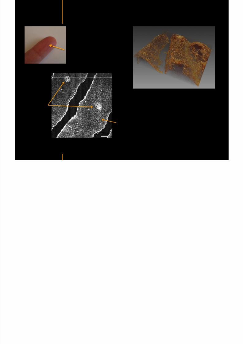
7292019 Atlas of Light-CT images
httpslidepdfcomreaderfullatlas-of-light-ct-images 54118
Skin in-vivo sweat duct imaging in 3D
7292019 Atlas of Light-CT images
httpslidepdfcomreaderfullatlas-of-light-ct-images 55118
copy LLTech 2012 ndash wwwlltechimagingcom - No copy without authorization 55
g g
Vertical reconstruction from en-face images
Sweat duct 3D reconstruction
Stratum corneum
Stratum spinosum
SD sweat ducts
SD SD
100 microm
7292019 Atlas of Light-CT images
httpslidepdfcomreaderfullatlas-of-light-ct-images 56118
7292019 Atlas of Light-CT images
httpslidepdfcomreaderfullatlas-of-light-ct-images 57118
7292019 Atlas of Light-CT images
httpslidepdfcomreaderfullatlas-of-light-ct-images 58118
copy LLTech 2012 ndash wwwlltechimagingcom - No copy without authorization 58
LUNG IMAGING
Back to table of contents
H L
7292019 Atlas of Light-CT images
httpslidepdfcomreaderfullatlas-of-light-ct-images 59118
copy LLTech 2012 ndash wwwlltechimagingcom - No copy without authorization 59
Zone 1
Zone 2
Zone 3
Zone 4
Zone with
cancerous tissue
Limit of cancerous
stroma
alveoli with
macrophages
Alveoles
Human Lung
500microm
copy LLTech 2012 Courtesy of Hocircpital Hotel-Dieu Paris France
H L Z 1 Z t
7292019 Atlas of Light-CT images
httpslidepdfcomreaderfullatlas-of-light-ct-images 60118
copy LLTech 2012 ndash wwwlltechimagingcom - No copy without authorization 60
Stroma
Human Lung ndash Zone 1 ndash Zoom on cancerous stroma
Cancerous
area
200microm
copy LLTech 2012 Courtesy of Hocircpital Hotel-Dieu Paris France
Human Lung ndash Zone 2 ndash Limit cancerous stroma alveoli
7292019 Atlas of Light-CT images
httpslidepdfcomreaderfullatlas-of-light-ct-images 61118
copy LLTech 2012 ndash wwwlltechimagingcom - No copy without authorization 61
g
with visible macrophages and vessel
Alveoli
Macrophages
Vessel
copy LLTech 2012 Courtesy of Hocircpital Hotel-Dieu Paris France
Human Lung ndash Zone 3 ndash Limit cancerous stroma alveoli
7292019 Atlas of Light-CT images
httpslidepdfcomreaderfullatlas-of-light-ct-images 62118
copy LLTech 2012 ndash wwwlltechimagingcom - No copy without authorization 62
g
with visible macrophages
Macrophages
100microm
copy LLTech 2012 Courtesy of Hocircpital Hotel-Dieu Paris France
Human Lung ndash Zone 4 ndash Alveoli
7292019 Atlas of Light-CT images
httpslidepdfcomreaderfullatlas-of-light-ct-images 63118
copy LLTech 2012 ndash wwwlltechimagingcom - No copy without authorization 63
Human Lung Zone 4 Alveoli
Alveoli
200microm
copy LLTech 2012 Courtesy of Hocircpital Hotel-Dieu Paris France
7292019 Atlas of Light-CT images
httpslidepdfcomreaderfullatlas-of-light-ct-images 64118
copy LLTech 2012 ndash wwwlltechimagingcom - No copy without authorization 64
GI IMAGING
Back to table of contents
7292019 Atlas of Light-CT images
httpslidepdfcomreaderfullatlas-of-light-ct-images 65118
Colonic carcinoma
7292019 Atlas of Light-CT images
httpslidepdfcomreaderfullatlas-of-light-ct-images 66118
copy LLTech 2012 ndash wwwlltechimagingcom - No copy without authorization 66
Colonic Carcinoma
Colonic carcinoma
copy LLTech 2012 Courtesy of MD Anderson Houston USA
Intestines ndash Crohnrsquos disease
7292019 Atlas of Light-CT images
httpslidepdfcomreaderfullatlas-of-light-ct-images 67118
copy LLTech 2012 ndash wwwlltechimagingcom - No copy without authorization 67
Crohnrsquos disease structure not
visible from the surface
Fat cells
200 microm
Intestines Crohn s disease
copy LLTech 2012 Courtesy of Institut Curie Paris France
Rat stomach
7292019 Atlas of Light-CT images
httpslidepdfcomreaderfullatlas-of-light-ct-images 68118
copy LLTech 2012 ndash wwwlltechimagingcom - No copy without authorization 68
Rat stomach
Mucosa withgastric pits
Submucosa with
bright collagen
fibers
Muscularis
propria with
muscle bundles
copy LLTech 2012 Courtesy of Weill Cornell Medical College NY USA
200 microm
7292019 Atlas of Light-CT images
httpslidepdfcomreaderfullatlas-of-light-ct-images 69118
copy LLTech 2012 ndash wwwlltechimagingcom - No copy without authorization 69 copy LLTECH 2011
UROGENITAL IMAGING
Back to table of contents
Rat bladder
7292019 Atlas of Light-CT images
httpslidepdfcomreaderfullatlas-of-light-ct-images 70118
copy LLTech 2012 ndash wwwlltechimagingcom - No copy without authorization 70
Rat bladder
Stratifiedurothelium
Lamina propria
500 microm
Muscular
bundles of the
lamina propria
Serosa
copy LLTech 2012 Courtesy of Weill Cornell Medical College NY USA
100 microm
100 microm
Human bladder low grade papillary urothelial carcinoma
7292019 Atlas of Light-CT images
httpslidepdfcomreaderfullatlas-of-light-ct-images 71118
copy LLTech 2012 ndash wwwlltechimagingcom - No copy without authorization 71
Lesion with fibrovascular cores (arrow) and superficial umbrella cells (arrowheads)
Human bladder low grade papillary urothelial carcinoma
copy LLTech 2012 Courtesy of Weill Cornell Medical College NY USA
100 microm
7292019 Atlas of Light-CT images
httpslidepdfcomreaderfullatlas-of-light-ct-images 72118
Rat prostate
7292019 Atlas of Light-CT images
httpslidepdfcomreaderfullatlas-of-light-ct-images 73118
copy LLTech 2012 ndash wwwlltechimagingcom - No copy without authorization 73
p
copy LLTech 2012 Courtesy of Weill Cornell Medical College NY USA
Papillary folds of the
acinar glands
Prostatic acini
Fibromuscular stromaPeriprostatic fat Blood vessel
1 mm
200 microm
Mouse kidney
7292019 Atlas of Light-CT images
httpslidepdfcomreaderfullatlas-of-light-ct-images 74118
copy LLTech 2012 ndash wwwlltechimagingcom - No copy without authorization 74
1 mm
y
copy LLTech 2012 Courtesy of Institut Biomeacutedical de Bicecirctre Paris France
Renal cortex
Renal medulla
Renal sinus
7292019 Atlas of Light-CT images
httpslidepdfcomreaderfullatlas-of-light-ct-images 75118
copy LLTech 2012 ndash wwwlltechimagingcom - No copy without authorization 75
Mouse kidney ndash zoom on highlighted section of previous slide
800microm
copy LLTech 2012 Courtesy of Hocircpital Kremlin Bicecirctre Paris France
7292019 Atlas of Light-CT images
httpslidepdfcomreaderfullatlas-of-light-ct-images 76118
7292019 Atlas of Light-CT images
httpslidepdfcomreaderfullatlas-of-light-ct-images 77118
7292019 Atlas of Light-CT images
httpslidepdfcomreaderfullatlas-of-light-ct-images 78118
copy LLTech 2012 ndash wwwlltechimagingcom - No copy without authorization 78 copy LLTECH 2011
CORE NEEDLE BIOPSIES
Back to table of contents
Kidney
7292019 Atlas of Light-CT images
httpslidepdfcomreaderfullatlas-of-light-ct-images 79118
copy LLTech 2012 ndash wwwlltechimagingcom - No copy without authorization 79
100microm
50microm
Renal tubules
Vessel
500microm
copy LLTech 2012 Courtesy of Institut Curie Paris France
Breast Invasive lobular carcinoma
7292019 Atlas of Light-CT images
httpslidepdfcomreaderfullatlas-of-light-ct-images 80118
copy LLTech 2012 ndash wwwlltechimagingcom - No copy without authorization 80
500microm
stroma
adipocytes
copy LLTech 2012 Courtesy of Institut Curie Paris France
7292019 Atlas of Light-CT images
httpslidepdfcomreaderfullatlas-of-light-ct-images 81118
Breast Ductal carcinoma in situ
7292019 Atlas of Light-CT images
httpslidepdfcomreaderfullatlas-of-light-ct-images 82118
copy LLTech 2012 ndash wwwlltechimagingcom - No copy without authorization 82
500microm
copy LLTech 2012 Courtesy of Institut Curie Paris France
7292019 Atlas of Light-CT images
httpslidepdfcomreaderfullatlas-of-light-ct-images 83118
copy LLTech 2012 ndash wwwlltechimagingcom - No copy without authorization 83 copy LLTECH 2011
THYROID IMAGING
Back to table of contents
7292019 Atlas of Light-CT images
httpslidepdfcomreaderfullatlas-of-light-ct-images 84118
copy LLTech 2012 ndash wwwlltechimagingcom - No copy without authorization 84
Thyroid
800 microm
200 microm
copy LLTech 2012 Courtesy of Hocircpital Tenon Paris France
7292019 Atlas of Light-CT images
httpslidepdfcomreaderfullatlas-of-light-ct-images 85118
copy LLTech 2012 ndash wwwlltechimagingcom - No copy without authorization 85
Thyroid
800 microm
200 microm
copy LLTech 2012 Courtesy of Hocircpital Tenon Paris France
7292019 Atlas of Light-CT images
httpslidepdfcomreaderfullatlas-of-light-ct-images 86118
copy LLTech 2012 ndash wwwlltechimagingcom - No copy without authorization 86 copy LLTECH 2011
FAT CELLS IMAGING
Back to table of contents
7292019 Atlas of Light-CT images
httpslidepdfcomreaderfullatlas-of-light-ct-images 87118
copy LLTech 2012 ndash wwwlltechimagingcom - No copy without authorization 87
Brown fat cells - adipocytes
100 microm 100 microm
200 microm
10um depth
Adipocytes
45um depth
En face
slices
Reconstructed
depth slice
copy LLTech 2012 Courtesy of Centre de Recherche des Cordeliers Paris France
7292019 Atlas of Light-CT images
httpslidepdfcomreaderfullatlas-of-light-ct-images 88118
copy LLTech 2012 ndash wwwlltechimagingcom - No copy without authorization 88 copy LLTECH 2011
CORNEA AND RETINA
Back to table of contents
Human cornea reconstructed depth slice
7292019 Atlas of Light-CT images
httpslidepdfcomreaderfullatlas-of-light-ct-images 89118
copy LLTech 2012 ndash wwwlltechimagingcom - No copy without authorization 89
50 um
wwwlltechimagingcom
7292019 Atlas of Light-CT images
httpslidepdfcomreaderfullatlas-of-light-ct-images 90118
Large field en face view in stroma
7292019 Atlas of Light-CT images
httpslidepdfcomreaderfullatlas-of-light-ct-images 91118
copy LLTech 2012 ndash wwwlltechimagingcom - No copy without authorization 91
200 um
wwwlltechimagingcom
7292019 Atlas of Light-CT images
httpslidepdfcomreaderfullatlas-of-light-ct-images 92118
Rabbit cornea after LASIK reflectivity profiles reveal
i d tt i d thi k d ith li l l
7292019 Atlas of Light-CT images
httpslidepdfcomreaderfullatlas-of-light-ct-images 93118
copy LLTech 2012 ndash wwwlltechimagingcom - No copy without authorization 93
increased scattering and thickened epithelial layer
LASIK LASIK CONTROL CONTROL
copy LLTech 2012
Newborn mouse anterior segment
7292019 Atlas of Light-CT images
httpslidepdfcomreaderfullatlas-of-light-ct-images 94118
copy LLTech 2012 ndash wwwlltechimagingcom - No copy without authorization 94
Corneal epithelial cells
Basal membrane
Stroma
Descemetrsquos membrane
Aqueous humor
Lens capsule
Lens epithelial cells
Cortical fibers
copy LLTech 2012 Courtesy of ESPCI Paris Francecopy LLTech 2012
Rat retina comparison with histology
7292019 Atlas of Light-CT images
httpslidepdfcomreaderfullatlas-of-light-ct-images 95118
copy LLTech 2012 ndash wwwlltechimagingcom - No copy without authorization 95
Figure 1 Right Ex vivo rat retinaimaged using the f ull field OCT
system of ESPCI Left Histology
for comparison
Figure 1 Right Ex vivo rat retinaimaged using the f ull field OCT
system of ESPCI Left Histology
for comparison
Figure 1 Right Ex vivo rat retinaimaged using the f ull field OCT
system of ESPCI Left Histology
for comparisonField
230x300microm
RPE
PRL
ONL
OPL
INL
IPL
GCL
NFL
Invest Ophthalmol Vis Sci 2004 Nov45(11)4126-31
Grieve K Paques M Dubois A Sahel J Boccara C Le Gargasson JF copy LLTech 2012
Rat retina
7292019 Atlas of Light-CT images
httpslidepdfcomreaderfullatlas-of-light-ct-images 96118
copy LLTech 2012 ndash wwwlltechimagingcom - No copy without authorization 96 copy LLTech 2012 Courtesy of ESPCI Paris France
Nerve fiber layer
Retina
Invest Ophthalmol Vis Sci 2004 Nov45(11)4126-31
Grieve K Paques M Dubois A Sahel J Boccara C Le Gargasson JF
Transscleral image
7292019 Atlas of Light-CT images
httpslidepdfcomreaderfullatlas-of-light-ct-images 97118
7292019 Atlas of Light-CT images
httpslidepdfcomreaderfullatlas-of-light-ct-images 98118
7292019 Atlas of Light-CT images
httpslidepdfcomreaderfullatlas-of-light-ct-images 99118
In vivo imaging of drosophila melanogaster
7292019 Atlas of Light-CT images
httpslidepdfcomreaderfullatlas-of-light-ct-images 100118
copy LLTech 2012 ndash wwwlltechimagingcom - No copy without authorization 100 copy LLTech 2012 Courtesy of ESPCI Paris France
In vivo imaging of drosophila melanogaster ndash four stages of pupation over 72 hours
7292019 Atlas of Light-CT images
httpslidepdfcomreaderfullatlas-of-light-ct-images 101118
copy LLTech 2012 ndash wwwlltechimagingcom - No copy without authorization 101
Prepupa (0-2 h) Transition topupal stage (24 h)
Pupal stage (48 h) Advanced pupal phase
before eclosion (72 h)
copy LLTech 2012 Courtesy of ESPCI Paris France
Pupa 118 4 days 30um depth
100
7292019 Atlas of Light-CT images
httpslidepdfcomreaderfullatlas-of-light-ct-images 102118
copy LLTech 2012 ndash wwwlltechimagingcom - No copy without authorization 102
100 um
Drosophila
7292019 Atlas of Light-CT images
httpslidepdfcomreaderfullatlas-of-light-ct-images 103118
copy LLTech 2012 ndash wwwlltechimagingcom - No copy without authorization 103
200 microm
copy LLTech 2012 Courtesy of ESPCI Paris France
C Elegans ndash Fast identification of anatomy
7292019 Atlas of Light-CT images
httpslidepdfcomreaderfullatlas-of-light-ct-images 104118
copy LLTech 2012 ndash wwwlltechimagingcom - No copy without authorization 104
Body wall - cuticle
gonad arm
30 microm depth
uterus
ovocytes
intestine
100 microm 100 microm
15 microm depth
copy LLTech 2012 Courtesy of ENS Paris France
C elegans ndash Images at higher magnification 40X
7292019 Atlas of Light-CT images
httpslidepdfcomreaderfullatlas-of-light-ct-images 105118
copy LLTech 2012 ndash wwwlltechimagingcom - No copy without authorization 105
3D reconstruction 50 microm
100 microm
copy LLTech 2012 Courtesy of ENS Paris France
Zebra fish eye
7292019 Atlas of Light-CT images
httpslidepdfcomreaderfullatlas-of-light-ct-images 106118
copy LLTech 2012 ndash wwwlltechimagingcom - No copy without authorization 106
Zebra fish eye
100 microm
100 microm
En face slices
Reconstructed depth slice
copy LLTech 2012 Courtesy of Inserm Paris France
10um depth 20um depth 45um depth
7292019 Atlas of Light-CT images
httpslidepdfcomreaderfullatlas-of-light-ct-images 107118
Xenopus Laevis backtail
7292019 Atlas of Light-CT images
httpslidepdfcomreaderfullatlas-of-light-ct-images 108118
copy LLTech 2012 ndash wwwlltechimagingcom - No copy without authorization 108
100 um30um depth 60um depth
7292019 Atlas of Light-CT images
httpslidepdfcomreaderfullatlas-of-light-ct-images 109118
Mouse embryo in vitro 6 5 days old ndash
7292019 Atlas of Light-CT images
httpslidepdfcomreaderfullatlas-of-light-ct-images 110118
copy LLTech 2012 ndash wwwlltechimagingcom - No copy without authorization 110
Mouse embryo in vitro 65 days old
copy LLTech 2012 Courtesy of Ellvire Guiot and nstitut Jacques Monod Paris France
3D imaging of internal cavity
Sabellaria alveolata mouth palps
7292019 Atlas of Light-CT images
httpslidepdfcomreaderfullatlas-of-light-ct-images 111118
copy LLTech 2012 ndash wwwlltechimagingcom - No copy without authorization 111
500 microm
100 microm
Mouth palps
copy LLTech 2012 Courtesy of Museacutee National drsquoHistoire Naturelle Paris France
7292019 Atlas of Light-CT images
httpslidepdfcomreaderfullatlas-of-light-ct-images 112118
copy LLTech 2012 ndash wwwlltechimagingcom - No copy without authorization 112 copy LLTECH 2011
PLANT IMAGING
Back to table of contents
Mouse ear leaf epidermis
7292019 Atlas of Light-CT images
httpslidepdfcomreaderfullatlas-of-light-ct-images 113118
copy LLTech 2012 ndash wwwlltechimagingcom - No copy without authorization 113
100 microm
Epidermal cell
copy LLTech 2012
Stoma
Leaf veins
7292019 Atlas of Light-CT images
httpslidepdfcomreaderfullatlas-of-light-ct-images 114118
copy LLTech 2012 ndash wwwlltechimagingcom - No copy without authorization 114
114copy LLTech 2012
Fibers
Veins
100 microm
500 microm
Apple
7292019 Atlas of Light-CT images
httpslidepdfcomreaderfullatlas-of-light-ct-images 115118
copy LLTech 2012 ndash wwwlltechimagingcom - No copy without authorization 115
115copy LLTech 2012
100 microm
Wax
Skin
Flesh
500 microm
Water
Effect of harpin infiltration on tobacco leaf
7292019 Atlas of Light-CT images
httpslidepdfcomreaderfullatlas-of-light-ct-images 116118
copy LLTech 2012 ndash wwwlltechimagingcom - No copy without authorization 116
116copy LLTech 2012 Courtesy of UPMC Paris France
50 microm
50 microm
50 microm
50 microm
Upper epidermis
10 min after harpin infiltration 30 min after harpin infiltration
Palisade parenchyma
Epidermis cell
Chloroplast
7292019 Atlas of Light-CT images
httpslidepdfcomreaderfullatlas-of-light-ct-images 117118
copy LLTech 2012 ndash wwwlltechimagingcom - No copy without authorization 117
bull Light-CTtrade does not require tissue preparation nor staining of any kind
bull Creates images within minutes using a non-destructive process
bull Offers a 1 microm cellular resolution in 3D
bull Reveals structural and cellular information
bull Mosaicing allows fast visualisation at various scale
bull En-face high-resolution imaging allows vertical and 3D reconstruction
bull In-vivo images show good promise for in-vivo system
bull Clinical and research applications in cancer detection cosmetics biobanking small animal
developmental biology botanyhellip
Contact Details
7292019 Atlas of Light-CT images
httpslidepdfcomreaderfullatlas-of-light-ct-images 118118
USA
LLTech Inc
103 Carnegie Center DriveSuite 300
Princeton NJ 08540
USA
wwwlltechimagingcom
contactlltechfr
Phone + 1 609 955 3506
LLTech SAS
29 rue du Faubourg St-JacquesPeacutepiniegravere Paris Santeacute Cochin
75014
Paris - France
wwwlltechimagingcom
contactlltechfr
Phone +33 1 82 72 61 25
Europe
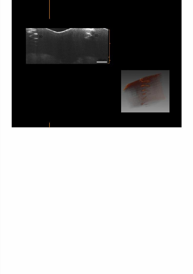
7292019 Atlas of Light-CT images
httpslidepdfcomreaderfullatlas-of-light-ct-images 58118
copy LLTech 2012 ndash wwwlltechimagingcom - No copy without authorization 58
LUNG IMAGING
Back to table of contents
H L
7292019 Atlas of Light-CT images
httpslidepdfcomreaderfullatlas-of-light-ct-images 59118
copy LLTech 2012 ndash wwwlltechimagingcom - No copy without authorization 59
Zone 1
Zone 2
Zone 3
Zone 4
Zone with
cancerous tissue
Limit of cancerous
stroma
alveoli with
macrophages
Alveoles
Human Lung
500microm
copy LLTech 2012 Courtesy of Hocircpital Hotel-Dieu Paris France
H L Z 1 Z t
7292019 Atlas of Light-CT images
httpslidepdfcomreaderfullatlas-of-light-ct-images 60118
copy LLTech 2012 ndash wwwlltechimagingcom - No copy without authorization 60
Stroma
Human Lung ndash Zone 1 ndash Zoom on cancerous stroma
Cancerous
area
200microm
copy LLTech 2012 Courtesy of Hocircpital Hotel-Dieu Paris France
Human Lung ndash Zone 2 ndash Limit cancerous stroma alveoli
7292019 Atlas of Light-CT images
httpslidepdfcomreaderfullatlas-of-light-ct-images 61118
copy LLTech 2012 ndash wwwlltechimagingcom - No copy without authorization 61
g
with visible macrophages and vessel
Alveoli
Macrophages
Vessel
copy LLTech 2012 Courtesy of Hocircpital Hotel-Dieu Paris France
Human Lung ndash Zone 3 ndash Limit cancerous stroma alveoli
7292019 Atlas of Light-CT images
httpslidepdfcomreaderfullatlas-of-light-ct-images 62118
copy LLTech 2012 ndash wwwlltechimagingcom - No copy without authorization 62
g
with visible macrophages
Macrophages
100microm
copy LLTech 2012 Courtesy of Hocircpital Hotel-Dieu Paris France
Human Lung ndash Zone 4 ndash Alveoli
7292019 Atlas of Light-CT images
httpslidepdfcomreaderfullatlas-of-light-ct-images 63118
copy LLTech 2012 ndash wwwlltechimagingcom - No copy without authorization 63
Human Lung Zone 4 Alveoli
Alveoli
200microm
copy LLTech 2012 Courtesy of Hocircpital Hotel-Dieu Paris France
7292019 Atlas of Light-CT images
httpslidepdfcomreaderfullatlas-of-light-ct-images 64118
copy LLTech 2012 ndash wwwlltechimagingcom - No copy without authorization 64
GI IMAGING
Back to table of contents
7292019 Atlas of Light-CT images
httpslidepdfcomreaderfullatlas-of-light-ct-images 65118
Colonic carcinoma
7292019 Atlas of Light-CT images
httpslidepdfcomreaderfullatlas-of-light-ct-images 66118
copy LLTech 2012 ndash wwwlltechimagingcom - No copy without authorization 66
Colonic Carcinoma
Colonic carcinoma
copy LLTech 2012 Courtesy of MD Anderson Houston USA
Intestines ndash Crohnrsquos disease
7292019 Atlas of Light-CT images
httpslidepdfcomreaderfullatlas-of-light-ct-images 67118
copy LLTech 2012 ndash wwwlltechimagingcom - No copy without authorization 67
Crohnrsquos disease structure not
visible from the surface
Fat cells
200 microm
Intestines Crohn s disease
copy LLTech 2012 Courtesy of Institut Curie Paris France
Rat stomach
7292019 Atlas of Light-CT images
httpslidepdfcomreaderfullatlas-of-light-ct-images 68118
copy LLTech 2012 ndash wwwlltechimagingcom - No copy without authorization 68
Rat stomach
Mucosa withgastric pits
Submucosa with
bright collagen
fibers
Muscularis
propria with
muscle bundles
copy LLTech 2012 Courtesy of Weill Cornell Medical College NY USA
200 microm
7292019 Atlas of Light-CT images
httpslidepdfcomreaderfullatlas-of-light-ct-images 69118
copy LLTech 2012 ndash wwwlltechimagingcom - No copy without authorization 69 copy LLTECH 2011
UROGENITAL IMAGING
Back to table of contents
Rat bladder
7292019 Atlas of Light-CT images
httpslidepdfcomreaderfullatlas-of-light-ct-images 70118
copy LLTech 2012 ndash wwwlltechimagingcom - No copy without authorization 70
Rat bladder
Stratifiedurothelium
Lamina propria
500 microm
Muscular
bundles of the
lamina propria
Serosa
copy LLTech 2012 Courtesy of Weill Cornell Medical College NY USA
100 microm
100 microm
Human bladder low grade papillary urothelial carcinoma
7292019 Atlas of Light-CT images
httpslidepdfcomreaderfullatlas-of-light-ct-images 71118
copy LLTech 2012 ndash wwwlltechimagingcom - No copy without authorization 71
Lesion with fibrovascular cores (arrow) and superficial umbrella cells (arrowheads)
Human bladder low grade papillary urothelial carcinoma
copy LLTech 2012 Courtesy of Weill Cornell Medical College NY USA
100 microm
7292019 Atlas of Light-CT images
httpslidepdfcomreaderfullatlas-of-light-ct-images 72118
Rat prostate
7292019 Atlas of Light-CT images
httpslidepdfcomreaderfullatlas-of-light-ct-images 73118
copy LLTech 2012 ndash wwwlltechimagingcom - No copy without authorization 73
p
copy LLTech 2012 Courtesy of Weill Cornell Medical College NY USA
Papillary folds of the
acinar glands
Prostatic acini
Fibromuscular stromaPeriprostatic fat Blood vessel
1 mm
200 microm
Mouse kidney
7292019 Atlas of Light-CT images
httpslidepdfcomreaderfullatlas-of-light-ct-images 74118
copy LLTech 2012 ndash wwwlltechimagingcom - No copy without authorization 74
1 mm
y
copy LLTech 2012 Courtesy of Institut Biomeacutedical de Bicecirctre Paris France
Renal cortex
Renal medulla
Renal sinus
7292019 Atlas of Light-CT images
httpslidepdfcomreaderfullatlas-of-light-ct-images 75118
copy LLTech 2012 ndash wwwlltechimagingcom - No copy without authorization 75
Mouse kidney ndash zoom on highlighted section of previous slide
800microm
copy LLTech 2012 Courtesy of Hocircpital Kremlin Bicecirctre Paris France
7292019 Atlas of Light-CT images
httpslidepdfcomreaderfullatlas-of-light-ct-images 76118
7292019 Atlas of Light-CT images
httpslidepdfcomreaderfullatlas-of-light-ct-images 77118
7292019 Atlas of Light-CT images
httpslidepdfcomreaderfullatlas-of-light-ct-images 78118
copy LLTech 2012 ndash wwwlltechimagingcom - No copy without authorization 78 copy LLTECH 2011
CORE NEEDLE BIOPSIES
Back to table of contents
Kidney
7292019 Atlas of Light-CT images
httpslidepdfcomreaderfullatlas-of-light-ct-images 79118
copy LLTech 2012 ndash wwwlltechimagingcom - No copy without authorization 79
100microm
50microm
Renal tubules
Vessel
500microm
copy LLTech 2012 Courtesy of Institut Curie Paris France
Breast Invasive lobular carcinoma
7292019 Atlas of Light-CT images
httpslidepdfcomreaderfullatlas-of-light-ct-images 80118
copy LLTech 2012 ndash wwwlltechimagingcom - No copy without authorization 80
500microm
stroma
adipocytes
copy LLTech 2012 Courtesy of Institut Curie Paris France
7292019 Atlas of Light-CT images
httpslidepdfcomreaderfullatlas-of-light-ct-images 81118
Breast Ductal carcinoma in situ
7292019 Atlas of Light-CT images
httpslidepdfcomreaderfullatlas-of-light-ct-images 82118
copy LLTech 2012 ndash wwwlltechimagingcom - No copy without authorization 82
500microm
copy LLTech 2012 Courtesy of Institut Curie Paris France
7292019 Atlas of Light-CT images
httpslidepdfcomreaderfullatlas-of-light-ct-images 83118
copy LLTech 2012 ndash wwwlltechimagingcom - No copy without authorization 83 copy LLTECH 2011
THYROID IMAGING
Back to table of contents
7292019 Atlas of Light-CT images
httpslidepdfcomreaderfullatlas-of-light-ct-images 84118
copy LLTech 2012 ndash wwwlltechimagingcom - No copy without authorization 84
Thyroid
800 microm
200 microm
copy LLTech 2012 Courtesy of Hocircpital Tenon Paris France
7292019 Atlas of Light-CT images
httpslidepdfcomreaderfullatlas-of-light-ct-images 85118
copy LLTech 2012 ndash wwwlltechimagingcom - No copy without authorization 85
Thyroid
800 microm
200 microm
copy LLTech 2012 Courtesy of Hocircpital Tenon Paris France
7292019 Atlas of Light-CT images
httpslidepdfcomreaderfullatlas-of-light-ct-images 86118
copy LLTech 2012 ndash wwwlltechimagingcom - No copy without authorization 86 copy LLTECH 2011
FAT CELLS IMAGING
Back to table of contents
7292019 Atlas of Light-CT images
httpslidepdfcomreaderfullatlas-of-light-ct-images 87118
copy LLTech 2012 ndash wwwlltechimagingcom - No copy without authorization 87
Brown fat cells - adipocytes
100 microm 100 microm
200 microm
10um depth
Adipocytes
45um depth
En face
slices
Reconstructed
depth slice
copy LLTech 2012 Courtesy of Centre de Recherche des Cordeliers Paris France
7292019 Atlas of Light-CT images
httpslidepdfcomreaderfullatlas-of-light-ct-images 88118
copy LLTech 2012 ndash wwwlltechimagingcom - No copy without authorization 88 copy LLTECH 2011
CORNEA AND RETINA
Back to table of contents
Human cornea reconstructed depth slice
7292019 Atlas of Light-CT images
httpslidepdfcomreaderfullatlas-of-light-ct-images 89118
copy LLTech 2012 ndash wwwlltechimagingcom - No copy without authorization 89
50 um
wwwlltechimagingcom
7292019 Atlas of Light-CT images
httpslidepdfcomreaderfullatlas-of-light-ct-images 90118
Large field en face view in stroma
7292019 Atlas of Light-CT images
httpslidepdfcomreaderfullatlas-of-light-ct-images 91118
copy LLTech 2012 ndash wwwlltechimagingcom - No copy without authorization 91
200 um
wwwlltechimagingcom
7292019 Atlas of Light-CT images
httpslidepdfcomreaderfullatlas-of-light-ct-images 92118
Rabbit cornea after LASIK reflectivity profiles reveal
i d tt i d thi k d ith li l l
7292019 Atlas of Light-CT images
httpslidepdfcomreaderfullatlas-of-light-ct-images 93118
copy LLTech 2012 ndash wwwlltechimagingcom - No copy without authorization 93
increased scattering and thickened epithelial layer
LASIK LASIK CONTROL CONTROL
copy LLTech 2012
Newborn mouse anterior segment
7292019 Atlas of Light-CT images
httpslidepdfcomreaderfullatlas-of-light-ct-images 94118
copy LLTech 2012 ndash wwwlltechimagingcom - No copy without authorization 94
Corneal epithelial cells
Basal membrane
Stroma
Descemetrsquos membrane
Aqueous humor
Lens capsule
Lens epithelial cells
Cortical fibers
copy LLTech 2012 Courtesy of ESPCI Paris Francecopy LLTech 2012
Rat retina comparison with histology
7292019 Atlas of Light-CT images
httpslidepdfcomreaderfullatlas-of-light-ct-images 95118
copy LLTech 2012 ndash wwwlltechimagingcom - No copy without authorization 95
Figure 1 Right Ex vivo rat retinaimaged using the f ull field OCT
system of ESPCI Left Histology
for comparison
Figure 1 Right Ex vivo rat retinaimaged using the f ull field OCT
system of ESPCI Left Histology
for comparison
Figure 1 Right Ex vivo rat retinaimaged using the f ull field OCT
system of ESPCI Left Histology
for comparisonField
230x300microm
RPE
PRL
ONL
OPL
INL
IPL
GCL
NFL
Invest Ophthalmol Vis Sci 2004 Nov45(11)4126-31
Grieve K Paques M Dubois A Sahel J Boccara C Le Gargasson JF copy LLTech 2012
Rat retina
7292019 Atlas of Light-CT images
httpslidepdfcomreaderfullatlas-of-light-ct-images 96118
copy LLTech 2012 ndash wwwlltechimagingcom - No copy without authorization 96 copy LLTech 2012 Courtesy of ESPCI Paris France
Nerve fiber layer
Retina
Invest Ophthalmol Vis Sci 2004 Nov45(11)4126-31
Grieve K Paques M Dubois A Sahel J Boccara C Le Gargasson JF
Transscleral image
7292019 Atlas of Light-CT images
httpslidepdfcomreaderfullatlas-of-light-ct-images 97118
7292019 Atlas of Light-CT images
httpslidepdfcomreaderfullatlas-of-light-ct-images 98118
7292019 Atlas of Light-CT images
httpslidepdfcomreaderfullatlas-of-light-ct-images 99118
In vivo imaging of drosophila melanogaster
7292019 Atlas of Light-CT images
httpslidepdfcomreaderfullatlas-of-light-ct-images 100118
copy LLTech 2012 ndash wwwlltechimagingcom - No copy without authorization 100 copy LLTech 2012 Courtesy of ESPCI Paris France
In vivo imaging of drosophila melanogaster ndash four stages of pupation over 72 hours
7292019 Atlas of Light-CT images
httpslidepdfcomreaderfullatlas-of-light-ct-images 101118
copy LLTech 2012 ndash wwwlltechimagingcom - No copy without authorization 101
Prepupa (0-2 h) Transition topupal stage (24 h)
Pupal stage (48 h) Advanced pupal phase
before eclosion (72 h)
copy LLTech 2012 Courtesy of ESPCI Paris France
Pupa 118 4 days 30um depth
100
7292019 Atlas of Light-CT images
httpslidepdfcomreaderfullatlas-of-light-ct-images 102118
copy LLTech 2012 ndash wwwlltechimagingcom - No copy without authorization 102
100 um
Drosophila
7292019 Atlas of Light-CT images
httpslidepdfcomreaderfullatlas-of-light-ct-images 103118
copy LLTech 2012 ndash wwwlltechimagingcom - No copy without authorization 103
200 microm
copy LLTech 2012 Courtesy of ESPCI Paris France
C Elegans ndash Fast identification of anatomy
7292019 Atlas of Light-CT images
httpslidepdfcomreaderfullatlas-of-light-ct-images 104118
copy LLTech 2012 ndash wwwlltechimagingcom - No copy without authorization 104
Body wall - cuticle
gonad arm
30 microm depth
uterus
ovocytes
intestine
100 microm 100 microm
15 microm depth
copy LLTech 2012 Courtesy of ENS Paris France
C elegans ndash Images at higher magnification 40X
7292019 Atlas of Light-CT images
httpslidepdfcomreaderfullatlas-of-light-ct-images 105118
copy LLTech 2012 ndash wwwlltechimagingcom - No copy without authorization 105
3D reconstruction 50 microm
100 microm
copy LLTech 2012 Courtesy of ENS Paris France
Zebra fish eye
7292019 Atlas of Light-CT images
httpslidepdfcomreaderfullatlas-of-light-ct-images 106118
copy LLTech 2012 ndash wwwlltechimagingcom - No copy without authorization 106
Zebra fish eye
100 microm
100 microm
En face slices
Reconstructed depth slice
copy LLTech 2012 Courtesy of Inserm Paris France
10um depth 20um depth 45um depth
7292019 Atlas of Light-CT images
httpslidepdfcomreaderfullatlas-of-light-ct-images 107118
Xenopus Laevis backtail
7292019 Atlas of Light-CT images
httpslidepdfcomreaderfullatlas-of-light-ct-images 108118
copy LLTech 2012 ndash wwwlltechimagingcom - No copy without authorization 108
100 um30um depth 60um depth
7292019 Atlas of Light-CT images
httpslidepdfcomreaderfullatlas-of-light-ct-images 109118
Mouse embryo in vitro 6 5 days old ndash
7292019 Atlas of Light-CT images
httpslidepdfcomreaderfullatlas-of-light-ct-images 110118
copy LLTech 2012 ndash wwwlltechimagingcom - No copy without authorization 110
Mouse embryo in vitro 65 days old
copy LLTech 2012 Courtesy of Ellvire Guiot and nstitut Jacques Monod Paris France
3D imaging of internal cavity
Sabellaria alveolata mouth palps
7292019 Atlas of Light-CT images
httpslidepdfcomreaderfullatlas-of-light-ct-images 111118
copy LLTech 2012 ndash wwwlltechimagingcom - No copy without authorization 111
500 microm
100 microm
Mouth palps
copy LLTech 2012 Courtesy of Museacutee National drsquoHistoire Naturelle Paris France
7292019 Atlas of Light-CT images
httpslidepdfcomreaderfullatlas-of-light-ct-images 112118
copy LLTech 2012 ndash wwwlltechimagingcom - No copy without authorization 112 copy LLTECH 2011
PLANT IMAGING
Back to table of contents
Mouse ear leaf epidermis
7292019 Atlas of Light-CT images
httpslidepdfcomreaderfullatlas-of-light-ct-images 113118
copy LLTech 2012 ndash wwwlltechimagingcom - No copy without authorization 113
100 microm
Epidermal cell
copy LLTech 2012
Stoma
Leaf veins
7292019 Atlas of Light-CT images
httpslidepdfcomreaderfullatlas-of-light-ct-images 114118
copy LLTech 2012 ndash wwwlltechimagingcom - No copy without authorization 114
114copy LLTech 2012
Fibers
Veins
100 microm
500 microm
Apple
7292019 Atlas of Light-CT images
httpslidepdfcomreaderfullatlas-of-light-ct-images 115118
copy LLTech 2012 ndash wwwlltechimagingcom - No copy without authorization 115
115copy LLTech 2012
100 microm
Wax
Skin
Flesh
500 microm
Water
Effect of harpin infiltration on tobacco leaf
7292019 Atlas of Light-CT images
httpslidepdfcomreaderfullatlas-of-light-ct-images 116118
copy LLTech 2012 ndash wwwlltechimagingcom - No copy without authorization 116
116copy LLTech 2012 Courtesy of UPMC Paris France
50 microm
50 microm
50 microm
50 microm
Upper epidermis
10 min after harpin infiltration 30 min after harpin infiltration
Palisade parenchyma
Epidermis cell
Chloroplast
7292019 Atlas of Light-CT images
httpslidepdfcomreaderfullatlas-of-light-ct-images 117118
copy LLTech 2012 ndash wwwlltechimagingcom - No copy without authorization 117
bull Light-CTtrade does not require tissue preparation nor staining of any kind
bull Creates images within minutes using a non-destructive process
bull Offers a 1 microm cellular resolution in 3D
bull Reveals structural and cellular information
bull Mosaicing allows fast visualisation at various scale
bull En-face high-resolution imaging allows vertical and 3D reconstruction
bull In-vivo images show good promise for in-vivo system
bull Clinical and research applications in cancer detection cosmetics biobanking small animal
developmental biology botanyhellip
Contact Details
7292019 Atlas of Light-CT images
httpslidepdfcomreaderfullatlas-of-light-ct-images 118118
USA
LLTech Inc
103 Carnegie Center DriveSuite 300
Princeton NJ 08540
USA
wwwlltechimagingcom
contactlltechfr
Phone + 1 609 955 3506
LLTech SAS
29 rue du Faubourg St-JacquesPeacutepiniegravere Paris Santeacute Cochin
75014
Paris - France
wwwlltechimagingcom
contactlltechfr
Phone +33 1 82 72 61 25
Europe
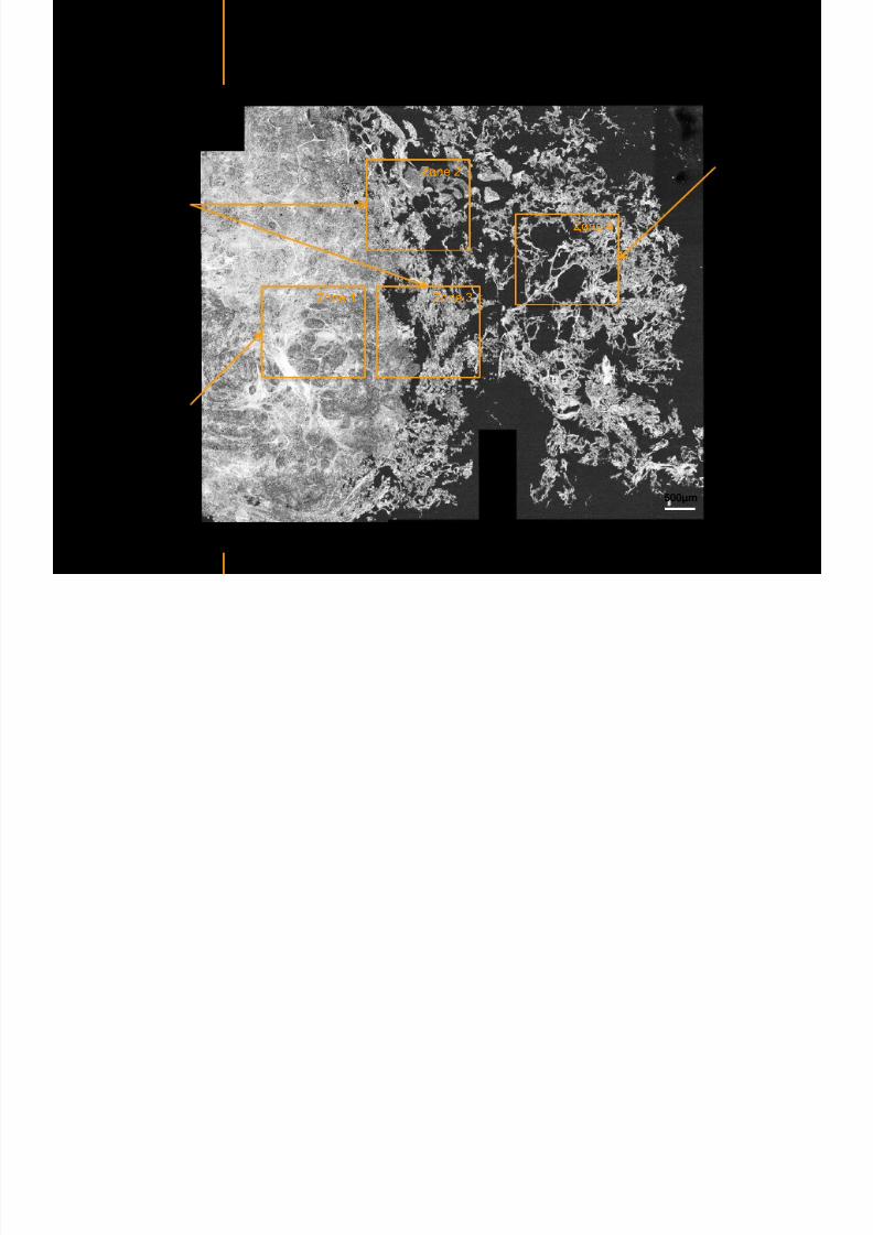
7292019 Atlas of Light-CT images
httpslidepdfcomreaderfullatlas-of-light-ct-images 59118
copy LLTech 2012 ndash wwwlltechimagingcom - No copy without authorization 59
Zone 1
Zone 2
Zone 3
Zone 4
Zone with
cancerous tissue
Limit of cancerous
stroma
alveoli with
macrophages
Alveoles
Human Lung
500microm
copy LLTech 2012 Courtesy of Hocircpital Hotel-Dieu Paris France
H L Z 1 Z t
7292019 Atlas of Light-CT images
httpslidepdfcomreaderfullatlas-of-light-ct-images 60118
copy LLTech 2012 ndash wwwlltechimagingcom - No copy without authorization 60
Stroma
Human Lung ndash Zone 1 ndash Zoom on cancerous stroma
Cancerous
area
200microm
copy LLTech 2012 Courtesy of Hocircpital Hotel-Dieu Paris France
Human Lung ndash Zone 2 ndash Limit cancerous stroma alveoli
7292019 Atlas of Light-CT images
httpslidepdfcomreaderfullatlas-of-light-ct-images 61118
copy LLTech 2012 ndash wwwlltechimagingcom - No copy without authorization 61
g
with visible macrophages and vessel
Alveoli
Macrophages
Vessel
copy LLTech 2012 Courtesy of Hocircpital Hotel-Dieu Paris France
Human Lung ndash Zone 3 ndash Limit cancerous stroma alveoli
7292019 Atlas of Light-CT images
httpslidepdfcomreaderfullatlas-of-light-ct-images 62118
copy LLTech 2012 ndash wwwlltechimagingcom - No copy without authorization 62
g
with visible macrophages
Macrophages
100microm
copy LLTech 2012 Courtesy of Hocircpital Hotel-Dieu Paris France
Human Lung ndash Zone 4 ndash Alveoli
7292019 Atlas of Light-CT images
httpslidepdfcomreaderfullatlas-of-light-ct-images 63118
copy LLTech 2012 ndash wwwlltechimagingcom - No copy without authorization 63
Human Lung Zone 4 Alveoli
Alveoli
200microm
copy LLTech 2012 Courtesy of Hocircpital Hotel-Dieu Paris France
7292019 Atlas of Light-CT images
httpslidepdfcomreaderfullatlas-of-light-ct-images 64118
copy LLTech 2012 ndash wwwlltechimagingcom - No copy without authorization 64
GI IMAGING
Back to table of contents
7292019 Atlas of Light-CT images
httpslidepdfcomreaderfullatlas-of-light-ct-images 65118
Colonic carcinoma
7292019 Atlas of Light-CT images
httpslidepdfcomreaderfullatlas-of-light-ct-images 66118
copy LLTech 2012 ndash wwwlltechimagingcom - No copy without authorization 66
Colonic Carcinoma
Colonic carcinoma
copy LLTech 2012 Courtesy of MD Anderson Houston USA
Intestines ndash Crohnrsquos disease
7292019 Atlas of Light-CT images
httpslidepdfcomreaderfullatlas-of-light-ct-images 67118
copy LLTech 2012 ndash wwwlltechimagingcom - No copy without authorization 67
Crohnrsquos disease structure not
visible from the surface
Fat cells
200 microm
Intestines Crohn s disease
copy LLTech 2012 Courtesy of Institut Curie Paris France
Rat stomach
7292019 Atlas of Light-CT images
httpslidepdfcomreaderfullatlas-of-light-ct-images 68118
copy LLTech 2012 ndash wwwlltechimagingcom - No copy without authorization 68
Rat stomach
Mucosa withgastric pits
Submucosa with
bright collagen
fibers
Muscularis
propria with
muscle bundles
copy LLTech 2012 Courtesy of Weill Cornell Medical College NY USA
200 microm
7292019 Atlas of Light-CT images
httpslidepdfcomreaderfullatlas-of-light-ct-images 69118
copy LLTech 2012 ndash wwwlltechimagingcom - No copy without authorization 69 copy LLTECH 2011
UROGENITAL IMAGING
Back to table of contents
Rat bladder
7292019 Atlas of Light-CT images
httpslidepdfcomreaderfullatlas-of-light-ct-images 70118
copy LLTech 2012 ndash wwwlltechimagingcom - No copy without authorization 70
Rat bladder
Stratifiedurothelium
Lamina propria
500 microm
Muscular
bundles of the
lamina propria
Serosa
copy LLTech 2012 Courtesy of Weill Cornell Medical College NY USA
100 microm
100 microm
Human bladder low grade papillary urothelial carcinoma
7292019 Atlas of Light-CT images
httpslidepdfcomreaderfullatlas-of-light-ct-images 71118
copy LLTech 2012 ndash wwwlltechimagingcom - No copy without authorization 71
Lesion with fibrovascular cores (arrow) and superficial umbrella cells (arrowheads)
Human bladder low grade papillary urothelial carcinoma
copy LLTech 2012 Courtesy of Weill Cornell Medical College NY USA
100 microm
7292019 Atlas of Light-CT images
httpslidepdfcomreaderfullatlas-of-light-ct-images 72118
Rat prostate
7292019 Atlas of Light-CT images
httpslidepdfcomreaderfullatlas-of-light-ct-images 73118
copy LLTech 2012 ndash wwwlltechimagingcom - No copy without authorization 73
p
copy LLTech 2012 Courtesy of Weill Cornell Medical College NY USA
Papillary folds of the
acinar glands
Prostatic acini
Fibromuscular stromaPeriprostatic fat Blood vessel
1 mm
200 microm
Mouse kidney
7292019 Atlas of Light-CT images
httpslidepdfcomreaderfullatlas-of-light-ct-images 74118
copy LLTech 2012 ndash wwwlltechimagingcom - No copy without authorization 74
1 mm
y
copy LLTech 2012 Courtesy of Institut Biomeacutedical de Bicecirctre Paris France
Renal cortex
Renal medulla
Renal sinus
7292019 Atlas of Light-CT images
httpslidepdfcomreaderfullatlas-of-light-ct-images 75118
copy LLTech 2012 ndash wwwlltechimagingcom - No copy without authorization 75
Mouse kidney ndash zoom on highlighted section of previous slide
800microm
copy LLTech 2012 Courtesy of Hocircpital Kremlin Bicecirctre Paris France
7292019 Atlas of Light-CT images
httpslidepdfcomreaderfullatlas-of-light-ct-images 76118
7292019 Atlas of Light-CT images
httpslidepdfcomreaderfullatlas-of-light-ct-images 77118
7292019 Atlas of Light-CT images
httpslidepdfcomreaderfullatlas-of-light-ct-images 78118
copy LLTech 2012 ndash wwwlltechimagingcom - No copy without authorization 78 copy LLTECH 2011
CORE NEEDLE BIOPSIES
Back to table of contents
Kidney
7292019 Atlas of Light-CT images
httpslidepdfcomreaderfullatlas-of-light-ct-images 79118
copy LLTech 2012 ndash wwwlltechimagingcom - No copy without authorization 79
100microm
50microm
Renal tubules
Vessel
500microm
copy LLTech 2012 Courtesy of Institut Curie Paris France
Breast Invasive lobular carcinoma
7292019 Atlas of Light-CT images
httpslidepdfcomreaderfullatlas-of-light-ct-images 80118
copy LLTech 2012 ndash wwwlltechimagingcom - No copy without authorization 80
500microm
stroma
adipocytes
copy LLTech 2012 Courtesy of Institut Curie Paris France
7292019 Atlas of Light-CT images
httpslidepdfcomreaderfullatlas-of-light-ct-images 81118
Breast Ductal carcinoma in situ
7292019 Atlas of Light-CT images
httpslidepdfcomreaderfullatlas-of-light-ct-images 82118
copy LLTech 2012 ndash wwwlltechimagingcom - No copy without authorization 82
500microm
copy LLTech 2012 Courtesy of Institut Curie Paris France
7292019 Atlas of Light-CT images
httpslidepdfcomreaderfullatlas-of-light-ct-images 83118
copy LLTech 2012 ndash wwwlltechimagingcom - No copy without authorization 83 copy LLTECH 2011
THYROID IMAGING
Back to table of contents
7292019 Atlas of Light-CT images
httpslidepdfcomreaderfullatlas-of-light-ct-images 84118
copy LLTech 2012 ndash wwwlltechimagingcom - No copy without authorization 84
Thyroid
800 microm
200 microm
copy LLTech 2012 Courtesy of Hocircpital Tenon Paris France
7292019 Atlas of Light-CT images
httpslidepdfcomreaderfullatlas-of-light-ct-images 85118
copy LLTech 2012 ndash wwwlltechimagingcom - No copy without authorization 85
Thyroid
800 microm
200 microm
copy LLTech 2012 Courtesy of Hocircpital Tenon Paris France
7292019 Atlas of Light-CT images
httpslidepdfcomreaderfullatlas-of-light-ct-images 86118
copy LLTech 2012 ndash wwwlltechimagingcom - No copy without authorization 86 copy LLTECH 2011
FAT CELLS IMAGING
Back to table of contents
7292019 Atlas of Light-CT images
httpslidepdfcomreaderfullatlas-of-light-ct-images 87118
copy LLTech 2012 ndash wwwlltechimagingcom - No copy without authorization 87
Brown fat cells - adipocytes
100 microm 100 microm
200 microm
10um depth
Adipocytes
45um depth
En face
slices
Reconstructed
depth slice
copy LLTech 2012 Courtesy of Centre de Recherche des Cordeliers Paris France
7292019 Atlas of Light-CT images
httpslidepdfcomreaderfullatlas-of-light-ct-images 88118
copy LLTech 2012 ndash wwwlltechimagingcom - No copy without authorization 88 copy LLTECH 2011
CORNEA AND RETINA
Back to table of contents
Human cornea reconstructed depth slice
7292019 Atlas of Light-CT images
httpslidepdfcomreaderfullatlas-of-light-ct-images 89118
copy LLTech 2012 ndash wwwlltechimagingcom - No copy without authorization 89
50 um
wwwlltechimagingcom
7292019 Atlas of Light-CT images
httpslidepdfcomreaderfullatlas-of-light-ct-images 90118
Large field en face view in stroma
7292019 Atlas of Light-CT images
httpslidepdfcomreaderfullatlas-of-light-ct-images 91118
copy LLTech 2012 ndash wwwlltechimagingcom - No copy without authorization 91
200 um
wwwlltechimagingcom
7292019 Atlas of Light-CT images
httpslidepdfcomreaderfullatlas-of-light-ct-images 92118
Rabbit cornea after LASIK reflectivity profiles reveal
i d tt i d thi k d ith li l l
7292019 Atlas of Light-CT images
httpslidepdfcomreaderfullatlas-of-light-ct-images 93118
copy LLTech 2012 ndash wwwlltechimagingcom - No copy without authorization 93
increased scattering and thickened epithelial layer
LASIK LASIK CONTROL CONTROL
copy LLTech 2012
Newborn mouse anterior segment
7292019 Atlas of Light-CT images
httpslidepdfcomreaderfullatlas-of-light-ct-images 94118
copy LLTech 2012 ndash wwwlltechimagingcom - No copy without authorization 94
Corneal epithelial cells
Basal membrane
Stroma
Descemetrsquos membrane
Aqueous humor
Lens capsule
Lens epithelial cells
Cortical fibers
copy LLTech 2012 Courtesy of ESPCI Paris Francecopy LLTech 2012
Rat retina comparison with histology
7292019 Atlas of Light-CT images
httpslidepdfcomreaderfullatlas-of-light-ct-images 95118
copy LLTech 2012 ndash wwwlltechimagingcom - No copy without authorization 95
Figure 1 Right Ex vivo rat retinaimaged using the f ull field OCT
system of ESPCI Left Histology
for comparison
Figure 1 Right Ex vivo rat retinaimaged using the f ull field OCT
system of ESPCI Left Histology
for comparison
Figure 1 Right Ex vivo rat retinaimaged using the f ull field OCT
system of ESPCI Left Histology
for comparisonField
230x300microm
RPE
PRL
ONL
OPL
INL
IPL
GCL
NFL
Invest Ophthalmol Vis Sci 2004 Nov45(11)4126-31
Grieve K Paques M Dubois A Sahel J Boccara C Le Gargasson JF copy LLTech 2012
Rat retina
7292019 Atlas of Light-CT images
httpslidepdfcomreaderfullatlas-of-light-ct-images 96118
copy LLTech 2012 ndash wwwlltechimagingcom - No copy without authorization 96 copy LLTech 2012 Courtesy of ESPCI Paris France
Nerve fiber layer
Retina
Invest Ophthalmol Vis Sci 2004 Nov45(11)4126-31
Grieve K Paques M Dubois A Sahel J Boccara C Le Gargasson JF
Transscleral image
7292019 Atlas of Light-CT images
httpslidepdfcomreaderfullatlas-of-light-ct-images 97118
7292019 Atlas of Light-CT images
httpslidepdfcomreaderfullatlas-of-light-ct-images 98118
7292019 Atlas of Light-CT images
httpslidepdfcomreaderfullatlas-of-light-ct-images 99118
In vivo imaging of drosophila melanogaster
7292019 Atlas of Light-CT images
httpslidepdfcomreaderfullatlas-of-light-ct-images 100118
copy LLTech 2012 ndash wwwlltechimagingcom - No copy without authorization 100 copy LLTech 2012 Courtesy of ESPCI Paris France
In vivo imaging of drosophila melanogaster ndash four stages of pupation over 72 hours
7292019 Atlas of Light-CT images
httpslidepdfcomreaderfullatlas-of-light-ct-images 101118
copy LLTech 2012 ndash wwwlltechimagingcom - No copy without authorization 101
Prepupa (0-2 h) Transition topupal stage (24 h)
Pupal stage (48 h) Advanced pupal phase
before eclosion (72 h)
copy LLTech 2012 Courtesy of ESPCI Paris France
Pupa 118 4 days 30um depth
100
7292019 Atlas of Light-CT images
httpslidepdfcomreaderfullatlas-of-light-ct-images 102118
copy LLTech 2012 ndash wwwlltechimagingcom - No copy without authorization 102
100 um
Drosophila
7292019 Atlas of Light-CT images
httpslidepdfcomreaderfullatlas-of-light-ct-images 103118
copy LLTech 2012 ndash wwwlltechimagingcom - No copy without authorization 103
200 microm
copy LLTech 2012 Courtesy of ESPCI Paris France
C Elegans ndash Fast identification of anatomy
7292019 Atlas of Light-CT images
httpslidepdfcomreaderfullatlas-of-light-ct-images 104118
copy LLTech 2012 ndash wwwlltechimagingcom - No copy without authorization 104
Body wall - cuticle
gonad arm
30 microm depth
uterus
ovocytes
intestine
100 microm 100 microm
15 microm depth
copy LLTech 2012 Courtesy of ENS Paris France
C elegans ndash Images at higher magnification 40X
7292019 Atlas of Light-CT images
httpslidepdfcomreaderfullatlas-of-light-ct-images 105118
copy LLTech 2012 ndash wwwlltechimagingcom - No copy without authorization 105
3D reconstruction 50 microm
100 microm
copy LLTech 2012 Courtesy of ENS Paris France
Zebra fish eye
7292019 Atlas of Light-CT images
httpslidepdfcomreaderfullatlas-of-light-ct-images 106118
copy LLTech 2012 ndash wwwlltechimagingcom - No copy without authorization 106
Zebra fish eye
100 microm
100 microm
En face slices
Reconstructed depth slice
copy LLTech 2012 Courtesy of Inserm Paris France
10um depth 20um depth 45um depth
7292019 Atlas of Light-CT images
httpslidepdfcomreaderfullatlas-of-light-ct-images 107118
Xenopus Laevis backtail
7292019 Atlas of Light-CT images
httpslidepdfcomreaderfullatlas-of-light-ct-images 108118
copy LLTech 2012 ndash wwwlltechimagingcom - No copy without authorization 108
100 um30um depth 60um depth
7292019 Atlas of Light-CT images
httpslidepdfcomreaderfullatlas-of-light-ct-images 109118
Mouse embryo in vitro 6 5 days old ndash
7292019 Atlas of Light-CT images
httpslidepdfcomreaderfullatlas-of-light-ct-images 110118
copy LLTech 2012 ndash wwwlltechimagingcom - No copy without authorization 110
Mouse embryo in vitro 65 days old
copy LLTech 2012 Courtesy of Ellvire Guiot and nstitut Jacques Monod Paris France
3D imaging of internal cavity
Sabellaria alveolata mouth palps
7292019 Atlas of Light-CT images
httpslidepdfcomreaderfullatlas-of-light-ct-images 111118
copy LLTech 2012 ndash wwwlltechimagingcom - No copy without authorization 111
500 microm
100 microm
Mouth palps
copy LLTech 2012 Courtesy of Museacutee National drsquoHistoire Naturelle Paris France
7292019 Atlas of Light-CT images
httpslidepdfcomreaderfullatlas-of-light-ct-images 112118
copy LLTech 2012 ndash wwwlltechimagingcom - No copy without authorization 112 copy LLTECH 2011
PLANT IMAGING
Back to table of contents
Mouse ear leaf epidermis
7292019 Atlas of Light-CT images
httpslidepdfcomreaderfullatlas-of-light-ct-images 113118
copy LLTech 2012 ndash wwwlltechimagingcom - No copy without authorization 113
100 microm
Epidermal cell
copy LLTech 2012
Stoma
Leaf veins
7292019 Atlas of Light-CT images
httpslidepdfcomreaderfullatlas-of-light-ct-images 114118
copy LLTech 2012 ndash wwwlltechimagingcom - No copy without authorization 114
114copy LLTech 2012
Fibers
Veins
100 microm
500 microm
Apple
7292019 Atlas of Light-CT images
httpslidepdfcomreaderfullatlas-of-light-ct-images 115118
copy LLTech 2012 ndash wwwlltechimagingcom - No copy without authorization 115
115copy LLTech 2012
100 microm
Wax
Skin
Flesh
500 microm
Water
Effect of harpin infiltration on tobacco leaf
7292019 Atlas of Light-CT images
httpslidepdfcomreaderfullatlas-of-light-ct-images 116118
copy LLTech 2012 ndash wwwlltechimagingcom - No copy without authorization 116
116copy LLTech 2012 Courtesy of UPMC Paris France
50 microm
50 microm
50 microm
50 microm
Upper epidermis
10 min after harpin infiltration 30 min after harpin infiltration
Palisade parenchyma
Epidermis cell
Chloroplast
7292019 Atlas of Light-CT images
httpslidepdfcomreaderfullatlas-of-light-ct-images 117118
copy LLTech 2012 ndash wwwlltechimagingcom - No copy without authorization 117
bull Light-CTtrade does not require tissue preparation nor staining of any kind
bull Creates images within minutes using a non-destructive process
bull Offers a 1 microm cellular resolution in 3D
bull Reveals structural and cellular information
bull Mosaicing allows fast visualisation at various scale
bull En-face high-resolution imaging allows vertical and 3D reconstruction
bull In-vivo images show good promise for in-vivo system
bull Clinical and research applications in cancer detection cosmetics biobanking small animal
developmental biology botanyhellip
Contact Details
7292019 Atlas of Light-CT images
httpslidepdfcomreaderfullatlas-of-light-ct-images 118118
USA
LLTech Inc
103 Carnegie Center DriveSuite 300
Princeton NJ 08540
USA
wwwlltechimagingcom
contactlltechfr
Phone + 1 609 955 3506
LLTech SAS
29 rue du Faubourg St-JacquesPeacutepiniegravere Paris Santeacute Cochin
75014
Paris - France
wwwlltechimagingcom
contactlltechfr
Phone +33 1 82 72 61 25
Europe
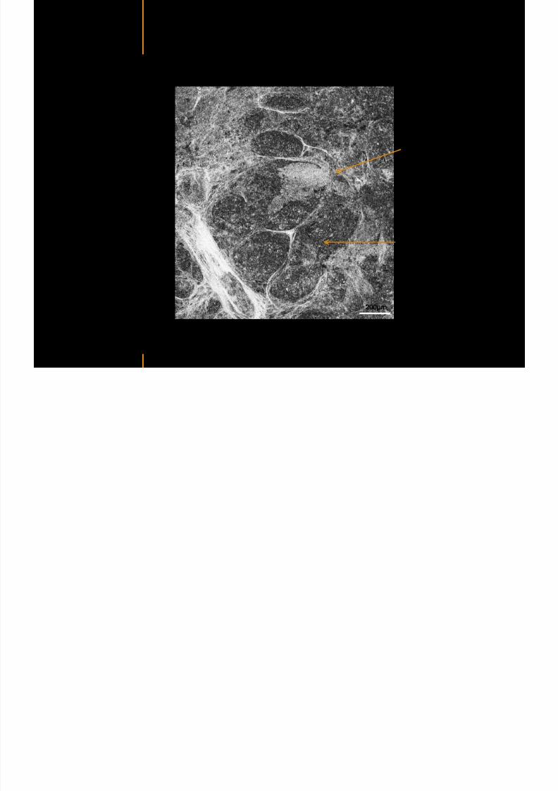
7292019 Atlas of Light-CT images
httpslidepdfcomreaderfullatlas-of-light-ct-images 61118
copy LLTech 2012 ndash wwwlltechimagingcom - No copy without authorization 61
g
with visible macrophages and vessel
Alveoli
Macrophages
Vessel
copy LLTech 2012 Courtesy of Hocircpital Hotel-Dieu Paris France
Human Lung ndash Zone 3 ndash Limit cancerous stroma alveoli
7292019 Atlas of Light-CT images
httpslidepdfcomreaderfullatlas-of-light-ct-images 62118
copy LLTech 2012 ndash wwwlltechimagingcom - No copy without authorization 62
g
with visible macrophages
Macrophages
100microm
copy LLTech 2012 Courtesy of Hocircpital Hotel-Dieu Paris France
Human Lung ndash Zone 4 ndash Alveoli
7292019 Atlas of Light-CT images
httpslidepdfcomreaderfullatlas-of-light-ct-images 63118
copy LLTech 2012 ndash wwwlltechimagingcom - No copy without authorization 63
Human Lung Zone 4 Alveoli
Alveoli
200microm
copy LLTech 2012 Courtesy of Hocircpital Hotel-Dieu Paris France
7292019 Atlas of Light-CT images
httpslidepdfcomreaderfullatlas-of-light-ct-images 64118
copy LLTech 2012 ndash wwwlltechimagingcom - No copy without authorization 64
GI IMAGING
Back to table of contents
7292019 Atlas of Light-CT images
httpslidepdfcomreaderfullatlas-of-light-ct-images 65118
Colonic carcinoma
7292019 Atlas of Light-CT images
httpslidepdfcomreaderfullatlas-of-light-ct-images 66118
copy LLTech 2012 ndash wwwlltechimagingcom - No copy without authorization 66
Colonic Carcinoma
Colonic carcinoma
copy LLTech 2012 Courtesy of MD Anderson Houston USA
Intestines ndash Crohnrsquos disease
7292019 Atlas of Light-CT images
httpslidepdfcomreaderfullatlas-of-light-ct-images 67118
copy LLTech 2012 ndash wwwlltechimagingcom - No copy without authorization 67
Crohnrsquos disease structure not
visible from the surface
Fat cells
200 microm
Intestines Crohn s disease
copy LLTech 2012 Courtesy of Institut Curie Paris France
Rat stomach
7292019 Atlas of Light-CT images
httpslidepdfcomreaderfullatlas-of-light-ct-images 68118
copy LLTech 2012 ndash wwwlltechimagingcom - No copy without authorization 68
Rat stomach
Mucosa withgastric pits
Submucosa with
bright collagen
fibers
Muscularis
propria with
muscle bundles
copy LLTech 2012 Courtesy of Weill Cornell Medical College NY USA
200 microm
7292019 Atlas of Light-CT images
httpslidepdfcomreaderfullatlas-of-light-ct-images 69118
copy LLTech 2012 ndash wwwlltechimagingcom - No copy without authorization 69 copy LLTECH 2011
UROGENITAL IMAGING
Back to table of contents
Rat bladder
7292019 Atlas of Light-CT images
httpslidepdfcomreaderfullatlas-of-light-ct-images 70118
copy LLTech 2012 ndash wwwlltechimagingcom - No copy without authorization 70
Rat bladder
Stratifiedurothelium
Lamina propria
500 microm
Muscular
bundles of the
lamina propria
Serosa
copy LLTech 2012 Courtesy of Weill Cornell Medical College NY USA
100 microm
100 microm
Human bladder low grade papillary urothelial carcinoma
7292019 Atlas of Light-CT images
httpslidepdfcomreaderfullatlas-of-light-ct-images 71118
copy LLTech 2012 ndash wwwlltechimagingcom - No copy without authorization 71
Lesion with fibrovascular cores (arrow) and superficial umbrella cells (arrowheads)
Human bladder low grade papillary urothelial carcinoma
copy LLTech 2012 Courtesy of Weill Cornell Medical College NY USA
100 microm
7292019 Atlas of Light-CT images
httpslidepdfcomreaderfullatlas-of-light-ct-images 72118
Rat prostate
7292019 Atlas of Light-CT images
httpslidepdfcomreaderfullatlas-of-light-ct-images 73118
copy LLTech 2012 ndash wwwlltechimagingcom - No copy without authorization 73
p
copy LLTech 2012 Courtesy of Weill Cornell Medical College NY USA
Papillary folds of the
acinar glands
Prostatic acini
Fibromuscular stromaPeriprostatic fat Blood vessel
1 mm
200 microm
Mouse kidney
7292019 Atlas of Light-CT images
httpslidepdfcomreaderfullatlas-of-light-ct-images 74118
copy LLTech 2012 ndash wwwlltechimagingcom - No copy without authorization 74
1 mm
y
copy LLTech 2012 Courtesy of Institut Biomeacutedical de Bicecirctre Paris France
Renal cortex
Renal medulla
Renal sinus
7292019 Atlas of Light-CT images
httpslidepdfcomreaderfullatlas-of-light-ct-images 75118
copy LLTech 2012 ndash wwwlltechimagingcom - No copy without authorization 75
Mouse kidney ndash zoom on highlighted section of previous slide
800microm
copy LLTech 2012 Courtesy of Hocircpital Kremlin Bicecirctre Paris France
7292019 Atlas of Light-CT images
httpslidepdfcomreaderfullatlas-of-light-ct-images 76118
7292019 Atlas of Light-CT images
httpslidepdfcomreaderfullatlas-of-light-ct-images 77118
7292019 Atlas of Light-CT images
httpslidepdfcomreaderfullatlas-of-light-ct-images 78118
copy LLTech 2012 ndash wwwlltechimagingcom - No copy without authorization 78 copy LLTECH 2011
CORE NEEDLE BIOPSIES
Back to table of contents
Kidney
7292019 Atlas of Light-CT images
httpslidepdfcomreaderfullatlas-of-light-ct-images 79118
copy LLTech 2012 ndash wwwlltechimagingcom - No copy without authorization 79
100microm
50microm
Renal tubules
Vessel
500microm
copy LLTech 2012 Courtesy of Institut Curie Paris France
Breast Invasive lobular carcinoma
7292019 Atlas of Light-CT images
httpslidepdfcomreaderfullatlas-of-light-ct-images 80118
copy LLTech 2012 ndash wwwlltechimagingcom - No copy without authorization 80
500microm
stroma
adipocytes
copy LLTech 2012 Courtesy of Institut Curie Paris France
7292019 Atlas of Light-CT images
httpslidepdfcomreaderfullatlas-of-light-ct-images 81118
Breast Ductal carcinoma in situ
7292019 Atlas of Light-CT images
httpslidepdfcomreaderfullatlas-of-light-ct-images 82118
copy LLTech 2012 ndash wwwlltechimagingcom - No copy without authorization 82
500microm
copy LLTech 2012 Courtesy of Institut Curie Paris France
7292019 Atlas of Light-CT images
httpslidepdfcomreaderfullatlas-of-light-ct-images 83118
copy LLTech 2012 ndash wwwlltechimagingcom - No copy without authorization 83 copy LLTECH 2011
THYROID IMAGING
Back to table of contents
7292019 Atlas of Light-CT images
httpslidepdfcomreaderfullatlas-of-light-ct-images 84118
copy LLTech 2012 ndash wwwlltechimagingcom - No copy without authorization 84
Thyroid
800 microm
200 microm
copy LLTech 2012 Courtesy of Hocircpital Tenon Paris France
7292019 Atlas of Light-CT images
httpslidepdfcomreaderfullatlas-of-light-ct-images 85118
copy LLTech 2012 ndash wwwlltechimagingcom - No copy without authorization 85
Thyroid
800 microm
200 microm
copy LLTech 2012 Courtesy of Hocircpital Tenon Paris France
7292019 Atlas of Light-CT images
httpslidepdfcomreaderfullatlas-of-light-ct-images 86118
copy LLTech 2012 ndash wwwlltechimagingcom - No copy without authorization 86 copy LLTECH 2011
FAT CELLS IMAGING
Back to table of contents
7292019 Atlas of Light-CT images
httpslidepdfcomreaderfullatlas-of-light-ct-images 87118
copy LLTech 2012 ndash wwwlltechimagingcom - No copy without authorization 87
Brown fat cells - adipocytes
100 microm 100 microm
200 microm
10um depth
Adipocytes
45um depth
En face
slices
Reconstructed
depth slice
copy LLTech 2012 Courtesy of Centre de Recherche des Cordeliers Paris France
7292019 Atlas of Light-CT images
httpslidepdfcomreaderfullatlas-of-light-ct-images 88118
copy LLTech 2012 ndash wwwlltechimagingcom - No copy without authorization 88 copy LLTECH 2011
CORNEA AND RETINA
Back to table of contents
Human cornea reconstructed depth slice
7292019 Atlas of Light-CT images
httpslidepdfcomreaderfullatlas-of-light-ct-images 89118
copy LLTech 2012 ndash wwwlltechimagingcom - No copy without authorization 89
50 um
wwwlltechimagingcom
7292019 Atlas of Light-CT images
httpslidepdfcomreaderfullatlas-of-light-ct-images 90118
Large field en face view in stroma
7292019 Atlas of Light-CT images
httpslidepdfcomreaderfullatlas-of-light-ct-images 91118
copy LLTech 2012 ndash wwwlltechimagingcom - No copy without authorization 91
200 um
wwwlltechimagingcom
7292019 Atlas of Light-CT images
httpslidepdfcomreaderfullatlas-of-light-ct-images 92118
Rabbit cornea after LASIK reflectivity profiles reveal
i d tt i d thi k d ith li l l
7292019 Atlas of Light-CT images
httpslidepdfcomreaderfullatlas-of-light-ct-images 93118
copy LLTech 2012 ndash wwwlltechimagingcom - No copy without authorization 93
increased scattering and thickened epithelial layer
LASIK LASIK CONTROL CONTROL
copy LLTech 2012
Newborn mouse anterior segment
7292019 Atlas of Light-CT images
httpslidepdfcomreaderfullatlas-of-light-ct-images 94118
copy LLTech 2012 ndash wwwlltechimagingcom - No copy without authorization 94
Corneal epithelial cells
Basal membrane
Stroma
Descemetrsquos membrane
Aqueous humor
Lens capsule
Lens epithelial cells
Cortical fibers
copy LLTech 2012 Courtesy of ESPCI Paris Francecopy LLTech 2012
Rat retina comparison with histology
7292019 Atlas of Light-CT images
httpslidepdfcomreaderfullatlas-of-light-ct-images 95118
copy LLTech 2012 ndash wwwlltechimagingcom - No copy without authorization 95
Figure 1 Right Ex vivo rat retinaimaged using the f ull field OCT
system of ESPCI Left Histology
for comparison
Figure 1 Right Ex vivo rat retinaimaged using the f ull field OCT
system of ESPCI Left Histology
for comparison
Figure 1 Right Ex vivo rat retinaimaged using the f ull field OCT
system of ESPCI Left Histology
for comparisonField
230x300microm
RPE
PRL
ONL
OPL
INL
IPL
GCL
NFL
Invest Ophthalmol Vis Sci 2004 Nov45(11)4126-31
Grieve K Paques M Dubois A Sahel J Boccara C Le Gargasson JF copy LLTech 2012
Rat retina
7292019 Atlas of Light-CT images
httpslidepdfcomreaderfullatlas-of-light-ct-images 96118
copy LLTech 2012 ndash wwwlltechimagingcom - No copy without authorization 96 copy LLTech 2012 Courtesy of ESPCI Paris France
Nerve fiber layer
Retina
Invest Ophthalmol Vis Sci 2004 Nov45(11)4126-31
Grieve K Paques M Dubois A Sahel J Boccara C Le Gargasson JF
Transscleral image
7292019 Atlas of Light-CT images
httpslidepdfcomreaderfullatlas-of-light-ct-images 97118
7292019 Atlas of Light-CT images
httpslidepdfcomreaderfullatlas-of-light-ct-images 98118
7292019 Atlas of Light-CT images
httpslidepdfcomreaderfullatlas-of-light-ct-images 99118
In vivo imaging of drosophila melanogaster
7292019 Atlas of Light-CT images
httpslidepdfcomreaderfullatlas-of-light-ct-images 100118
copy LLTech 2012 ndash wwwlltechimagingcom - No copy without authorization 100 copy LLTech 2012 Courtesy of ESPCI Paris France
In vivo imaging of drosophila melanogaster ndash four stages of pupation over 72 hours
7292019 Atlas of Light-CT images
httpslidepdfcomreaderfullatlas-of-light-ct-images 101118
copy LLTech 2012 ndash wwwlltechimagingcom - No copy without authorization 101
Prepupa (0-2 h) Transition topupal stage (24 h)
Pupal stage (48 h) Advanced pupal phase
before eclosion (72 h)
copy LLTech 2012 Courtesy of ESPCI Paris France
Pupa 118 4 days 30um depth
100
7292019 Atlas of Light-CT images
httpslidepdfcomreaderfullatlas-of-light-ct-images 102118
copy LLTech 2012 ndash wwwlltechimagingcom - No copy without authorization 102
100 um
Drosophila
7292019 Atlas of Light-CT images
httpslidepdfcomreaderfullatlas-of-light-ct-images 103118
copy LLTech 2012 ndash wwwlltechimagingcom - No copy without authorization 103
200 microm
copy LLTech 2012 Courtesy of ESPCI Paris France
C Elegans ndash Fast identification of anatomy
7292019 Atlas of Light-CT images
httpslidepdfcomreaderfullatlas-of-light-ct-images 104118
copy LLTech 2012 ndash wwwlltechimagingcom - No copy without authorization 104
Body wall - cuticle
gonad arm
30 microm depth
uterus
ovocytes
intestine
100 microm 100 microm
15 microm depth
copy LLTech 2012 Courtesy of ENS Paris France
C elegans ndash Images at higher magnification 40X
7292019 Atlas of Light-CT images
httpslidepdfcomreaderfullatlas-of-light-ct-images 105118
copy LLTech 2012 ndash wwwlltechimagingcom - No copy without authorization 105
3D reconstruction 50 microm
100 microm
copy LLTech 2012 Courtesy of ENS Paris France
Zebra fish eye
7292019 Atlas of Light-CT images
httpslidepdfcomreaderfullatlas-of-light-ct-images 106118
copy LLTech 2012 ndash wwwlltechimagingcom - No copy without authorization 106
Zebra fish eye
100 microm
100 microm
En face slices
Reconstructed depth slice
copy LLTech 2012 Courtesy of Inserm Paris France
10um depth 20um depth 45um depth
7292019 Atlas of Light-CT images
httpslidepdfcomreaderfullatlas-of-light-ct-images 107118
Xenopus Laevis backtail
7292019 Atlas of Light-CT images
httpslidepdfcomreaderfullatlas-of-light-ct-images 108118
copy LLTech 2012 ndash wwwlltechimagingcom - No copy without authorization 108
100 um30um depth 60um depth
7292019 Atlas of Light-CT images
httpslidepdfcomreaderfullatlas-of-light-ct-images 109118
Mouse embryo in vitro 6 5 days old ndash
7292019 Atlas of Light-CT images
httpslidepdfcomreaderfullatlas-of-light-ct-images 110118
copy LLTech 2012 ndash wwwlltechimagingcom - No copy without authorization 110
Mouse embryo in vitro 65 days old
copy LLTech 2012 Courtesy of Ellvire Guiot and nstitut Jacques Monod Paris France
3D imaging of internal cavity
Sabellaria alveolata mouth palps
7292019 Atlas of Light-CT images
httpslidepdfcomreaderfullatlas-of-light-ct-images 111118
copy LLTech 2012 ndash wwwlltechimagingcom - No copy without authorization 111
500 microm
100 microm
Mouth palps
copy LLTech 2012 Courtesy of Museacutee National drsquoHistoire Naturelle Paris France
7292019 Atlas of Light-CT images
httpslidepdfcomreaderfullatlas-of-light-ct-images 112118
copy LLTech 2012 ndash wwwlltechimagingcom - No copy without authorization 112 copy LLTECH 2011
PLANT IMAGING
Back to table of contents
Mouse ear leaf epidermis
7292019 Atlas of Light-CT images
httpslidepdfcomreaderfullatlas-of-light-ct-images 113118
copy LLTech 2012 ndash wwwlltechimagingcom - No copy without authorization 113
100 microm
Epidermal cell
copy LLTech 2012
Stoma
Leaf veins
7292019 Atlas of Light-CT images
httpslidepdfcomreaderfullatlas-of-light-ct-images 114118
copy LLTech 2012 ndash wwwlltechimagingcom - No copy without authorization 114
114copy LLTech 2012
Fibers
Veins
100 microm
500 microm
Apple
7292019 Atlas of Light-CT images
httpslidepdfcomreaderfullatlas-of-light-ct-images 115118
copy LLTech 2012 ndash wwwlltechimagingcom - No copy without authorization 115
115copy LLTech 2012
100 microm
Wax
Skin
Flesh
500 microm
Water
Effect of harpin infiltration on tobacco leaf
7292019 Atlas of Light-CT images
httpslidepdfcomreaderfullatlas-of-light-ct-images 116118
copy LLTech 2012 ndash wwwlltechimagingcom - No copy without authorization 116
116copy LLTech 2012 Courtesy of UPMC Paris France
50 microm
50 microm
50 microm
50 microm
Upper epidermis
10 min after harpin infiltration 30 min after harpin infiltration
Palisade parenchyma
Epidermis cell
Chloroplast
7292019 Atlas of Light-CT images
httpslidepdfcomreaderfullatlas-of-light-ct-images 117118
copy LLTech 2012 ndash wwwlltechimagingcom - No copy without authorization 117
bull Light-CTtrade does not require tissue preparation nor staining of any kind
bull Creates images within minutes using a non-destructive process
bull Offers a 1 microm cellular resolution in 3D
bull Reveals structural and cellular information
bull Mosaicing allows fast visualisation at various scale
bull En-face high-resolution imaging allows vertical and 3D reconstruction
bull In-vivo images show good promise for in-vivo system
bull Clinical and research applications in cancer detection cosmetics biobanking small animal
developmental biology botanyhellip
Contact Details
7292019 Atlas of Light-CT images
httpslidepdfcomreaderfullatlas-of-light-ct-images 118118
USA
LLTech Inc
103 Carnegie Center DriveSuite 300
Princeton NJ 08540
USA
wwwlltechimagingcom
contactlltechfr
Phone + 1 609 955 3506
LLTech SAS
29 rue du Faubourg St-JacquesPeacutepiniegravere Paris Santeacute Cochin
75014
Paris - France
wwwlltechimagingcom
contactlltechfr
Phone +33 1 82 72 61 25
Europe
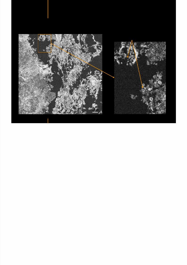
7292019 Atlas of Light-CT images
httpslidepdfcomreaderfullatlas-of-light-ct-images 63118
copy LLTech 2012 ndash wwwlltechimagingcom - No copy without authorization 63
Human Lung Zone 4 Alveoli
Alveoli
200microm
copy LLTech 2012 Courtesy of Hocircpital Hotel-Dieu Paris France
7292019 Atlas of Light-CT images
httpslidepdfcomreaderfullatlas-of-light-ct-images 64118
copy LLTech 2012 ndash wwwlltechimagingcom - No copy without authorization 64
GI IMAGING
Back to table of contents
7292019 Atlas of Light-CT images
httpslidepdfcomreaderfullatlas-of-light-ct-images 65118
Colonic carcinoma
7292019 Atlas of Light-CT images
httpslidepdfcomreaderfullatlas-of-light-ct-images 66118
copy LLTech 2012 ndash wwwlltechimagingcom - No copy without authorization 66
Colonic Carcinoma
Colonic carcinoma
copy LLTech 2012 Courtesy of MD Anderson Houston USA
Intestines ndash Crohnrsquos disease
7292019 Atlas of Light-CT images
httpslidepdfcomreaderfullatlas-of-light-ct-images 67118
copy LLTech 2012 ndash wwwlltechimagingcom - No copy without authorization 67
Crohnrsquos disease structure not
visible from the surface
Fat cells
200 microm
Intestines Crohn s disease
copy LLTech 2012 Courtesy of Institut Curie Paris France
Rat stomach
7292019 Atlas of Light-CT images
httpslidepdfcomreaderfullatlas-of-light-ct-images 68118
copy LLTech 2012 ndash wwwlltechimagingcom - No copy without authorization 68
Rat stomach
Mucosa withgastric pits
Submucosa with
bright collagen
fibers
Muscularis
propria with
muscle bundles
copy LLTech 2012 Courtesy of Weill Cornell Medical College NY USA
200 microm
7292019 Atlas of Light-CT images
httpslidepdfcomreaderfullatlas-of-light-ct-images 69118
copy LLTech 2012 ndash wwwlltechimagingcom - No copy without authorization 69 copy LLTECH 2011
UROGENITAL IMAGING
Back to table of contents
Rat bladder
7292019 Atlas of Light-CT images
httpslidepdfcomreaderfullatlas-of-light-ct-images 70118
copy LLTech 2012 ndash wwwlltechimagingcom - No copy without authorization 70
Rat bladder
Stratifiedurothelium
Lamina propria
500 microm
Muscular
bundles of the
lamina propria
Serosa
copy LLTech 2012 Courtesy of Weill Cornell Medical College NY USA
100 microm
100 microm
Human bladder low grade papillary urothelial carcinoma
7292019 Atlas of Light-CT images
httpslidepdfcomreaderfullatlas-of-light-ct-images 71118
copy LLTech 2012 ndash wwwlltechimagingcom - No copy without authorization 71
Lesion with fibrovascular cores (arrow) and superficial umbrella cells (arrowheads)
Human bladder low grade papillary urothelial carcinoma
copy LLTech 2012 Courtesy of Weill Cornell Medical College NY USA
100 microm
7292019 Atlas of Light-CT images
httpslidepdfcomreaderfullatlas-of-light-ct-images 72118
Rat prostate
7292019 Atlas of Light-CT images
httpslidepdfcomreaderfullatlas-of-light-ct-images 73118
copy LLTech 2012 ndash wwwlltechimagingcom - No copy without authorization 73
p
copy LLTech 2012 Courtesy of Weill Cornell Medical College NY USA
Papillary folds of the
acinar glands
Prostatic acini
Fibromuscular stromaPeriprostatic fat Blood vessel
1 mm
200 microm
Mouse kidney
7292019 Atlas of Light-CT images
httpslidepdfcomreaderfullatlas-of-light-ct-images 74118
copy LLTech 2012 ndash wwwlltechimagingcom - No copy without authorization 74
1 mm
y
copy LLTech 2012 Courtesy of Institut Biomeacutedical de Bicecirctre Paris France
Renal cortex
Renal medulla
Renal sinus
7292019 Atlas of Light-CT images
httpslidepdfcomreaderfullatlas-of-light-ct-images 75118
copy LLTech 2012 ndash wwwlltechimagingcom - No copy without authorization 75
Mouse kidney ndash zoom on highlighted section of previous slide
800microm
copy LLTech 2012 Courtesy of Hocircpital Kremlin Bicecirctre Paris France
7292019 Atlas of Light-CT images
httpslidepdfcomreaderfullatlas-of-light-ct-images 76118
7292019 Atlas of Light-CT images
httpslidepdfcomreaderfullatlas-of-light-ct-images 77118
7292019 Atlas of Light-CT images
httpslidepdfcomreaderfullatlas-of-light-ct-images 78118
copy LLTech 2012 ndash wwwlltechimagingcom - No copy without authorization 78 copy LLTECH 2011
CORE NEEDLE BIOPSIES
Back to table of contents
Kidney
7292019 Atlas of Light-CT images
httpslidepdfcomreaderfullatlas-of-light-ct-images 79118
copy LLTech 2012 ndash wwwlltechimagingcom - No copy without authorization 79
100microm
50microm
Renal tubules
Vessel
500microm
copy LLTech 2012 Courtesy of Institut Curie Paris France
Breast Invasive lobular carcinoma
7292019 Atlas of Light-CT images
httpslidepdfcomreaderfullatlas-of-light-ct-images 80118
copy LLTech 2012 ndash wwwlltechimagingcom - No copy without authorization 80
500microm
stroma
adipocytes
copy LLTech 2012 Courtesy of Institut Curie Paris France
7292019 Atlas of Light-CT images
httpslidepdfcomreaderfullatlas-of-light-ct-images 81118
Breast Ductal carcinoma in situ
7292019 Atlas of Light-CT images
httpslidepdfcomreaderfullatlas-of-light-ct-images 82118
copy LLTech 2012 ndash wwwlltechimagingcom - No copy without authorization 82
500microm
copy LLTech 2012 Courtesy of Institut Curie Paris France
7292019 Atlas of Light-CT images
httpslidepdfcomreaderfullatlas-of-light-ct-images 83118
copy LLTech 2012 ndash wwwlltechimagingcom - No copy without authorization 83 copy LLTECH 2011
THYROID IMAGING
Back to table of contents
7292019 Atlas of Light-CT images
httpslidepdfcomreaderfullatlas-of-light-ct-images 84118
copy LLTech 2012 ndash wwwlltechimagingcom - No copy without authorization 84
Thyroid
800 microm
200 microm
copy LLTech 2012 Courtesy of Hocircpital Tenon Paris France
7292019 Atlas of Light-CT images
httpslidepdfcomreaderfullatlas-of-light-ct-images 85118
copy LLTech 2012 ndash wwwlltechimagingcom - No copy without authorization 85
Thyroid
800 microm
200 microm
copy LLTech 2012 Courtesy of Hocircpital Tenon Paris France
7292019 Atlas of Light-CT images
httpslidepdfcomreaderfullatlas-of-light-ct-images 86118
copy LLTech 2012 ndash wwwlltechimagingcom - No copy without authorization 86 copy LLTECH 2011
FAT CELLS IMAGING
Back to table of contents
7292019 Atlas of Light-CT images
httpslidepdfcomreaderfullatlas-of-light-ct-images 87118
copy LLTech 2012 ndash wwwlltechimagingcom - No copy without authorization 87
Brown fat cells - adipocytes
100 microm 100 microm
200 microm
10um depth
Adipocytes
45um depth
En face
slices
Reconstructed
depth slice
copy LLTech 2012 Courtesy of Centre de Recherche des Cordeliers Paris France
7292019 Atlas of Light-CT images
httpslidepdfcomreaderfullatlas-of-light-ct-images 88118
copy LLTech 2012 ndash wwwlltechimagingcom - No copy without authorization 88 copy LLTECH 2011
CORNEA AND RETINA
Back to table of contents
Human cornea reconstructed depth slice
7292019 Atlas of Light-CT images
httpslidepdfcomreaderfullatlas-of-light-ct-images 89118
copy LLTech 2012 ndash wwwlltechimagingcom - No copy without authorization 89
50 um
wwwlltechimagingcom
7292019 Atlas of Light-CT images
httpslidepdfcomreaderfullatlas-of-light-ct-images 90118
Large field en face view in stroma
7292019 Atlas of Light-CT images
httpslidepdfcomreaderfullatlas-of-light-ct-images 91118
copy LLTech 2012 ndash wwwlltechimagingcom - No copy without authorization 91
200 um
wwwlltechimagingcom
7292019 Atlas of Light-CT images
httpslidepdfcomreaderfullatlas-of-light-ct-images 92118
Rabbit cornea after LASIK reflectivity profiles reveal
i d tt i d thi k d ith li l l
7292019 Atlas of Light-CT images
httpslidepdfcomreaderfullatlas-of-light-ct-images 93118
copy LLTech 2012 ndash wwwlltechimagingcom - No copy without authorization 93
increased scattering and thickened epithelial layer
LASIK LASIK CONTROL CONTROL
copy LLTech 2012
Newborn mouse anterior segment
7292019 Atlas of Light-CT images
httpslidepdfcomreaderfullatlas-of-light-ct-images 94118
copy LLTech 2012 ndash wwwlltechimagingcom - No copy without authorization 94
Corneal epithelial cells
Basal membrane
Stroma
Descemetrsquos membrane
Aqueous humor
Lens capsule
Lens epithelial cells
Cortical fibers
copy LLTech 2012 Courtesy of ESPCI Paris Francecopy LLTech 2012
Rat retina comparison with histology
7292019 Atlas of Light-CT images
httpslidepdfcomreaderfullatlas-of-light-ct-images 95118
copy LLTech 2012 ndash wwwlltechimagingcom - No copy without authorization 95
Figure 1 Right Ex vivo rat retinaimaged using the f ull field OCT
system of ESPCI Left Histology
for comparison
Figure 1 Right Ex vivo rat retinaimaged using the f ull field OCT
system of ESPCI Left Histology
for comparison
Figure 1 Right Ex vivo rat retinaimaged using the f ull field OCT
system of ESPCI Left Histology
for comparisonField
230x300microm
RPE
PRL
ONL
OPL
INL
IPL
GCL
NFL
Invest Ophthalmol Vis Sci 2004 Nov45(11)4126-31
Grieve K Paques M Dubois A Sahel J Boccara C Le Gargasson JF copy LLTech 2012
Rat retina
7292019 Atlas of Light-CT images
httpslidepdfcomreaderfullatlas-of-light-ct-images 96118
copy LLTech 2012 ndash wwwlltechimagingcom - No copy without authorization 96 copy LLTech 2012 Courtesy of ESPCI Paris France
Nerve fiber layer
Retina
Invest Ophthalmol Vis Sci 2004 Nov45(11)4126-31
Grieve K Paques M Dubois A Sahel J Boccara C Le Gargasson JF
Transscleral image
7292019 Atlas of Light-CT images
httpslidepdfcomreaderfullatlas-of-light-ct-images 97118
7292019 Atlas of Light-CT images
httpslidepdfcomreaderfullatlas-of-light-ct-images 98118
7292019 Atlas of Light-CT images
httpslidepdfcomreaderfullatlas-of-light-ct-images 99118
In vivo imaging of drosophila melanogaster
7292019 Atlas of Light-CT images
httpslidepdfcomreaderfullatlas-of-light-ct-images 100118
copy LLTech 2012 ndash wwwlltechimagingcom - No copy without authorization 100 copy LLTech 2012 Courtesy of ESPCI Paris France
In vivo imaging of drosophila melanogaster ndash four stages of pupation over 72 hours
7292019 Atlas of Light-CT images
httpslidepdfcomreaderfullatlas-of-light-ct-images 101118
copy LLTech 2012 ndash wwwlltechimagingcom - No copy without authorization 101
Prepupa (0-2 h) Transition topupal stage (24 h)
Pupal stage (48 h) Advanced pupal phase
before eclosion (72 h)
copy LLTech 2012 Courtesy of ESPCI Paris France
Pupa 118 4 days 30um depth
100
7292019 Atlas of Light-CT images
httpslidepdfcomreaderfullatlas-of-light-ct-images 102118
copy LLTech 2012 ndash wwwlltechimagingcom - No copy without authorization 102
100 um
Drosophila
7292019 Atlas of Light-CT images
httpslidepdfcomreaderfullatlas-of-light-ct-images 103118
copy LLTech 2012 ndash wwwlltechimagingcom - No copy without authorization 103
200 microm
copy LLTech 2012 Courtesy of ESPCI Paris France
C Elegans ndash Fast identification of anatomy
7292019 Atlas of Light-CT images
httpslidepdfcomreaderfullatlas-of-light-ct-images 104118
copy LLTech 2012 ndash wwwlltechimagingcom - No copy without authorization 104
Body wall - cuticle
gonad arm
30 microm depth
uterus
ovocytes
intestine
100 microm 100 microm
15 microm depth
copy LLTech 2012 Courtesy of ENS Paris France
C elegans ndash Images at higher magnification 40X
7292019 Atlas of Light-CT images
httpslidepdfcomreaderfullatlas-of-light-ct-images 105118
copy LLTech 2012 ndash wwwlltechimagingcom - No copy without authorization 105
3D reconstruction 50 microm
100 microm
copy LLTech 2012 Courtesy of ENS Paris France
Zebra fish eye
7292019 Atlas of Light-CT images
httpslidepdfcomreaderfullatlas-of-light-ct-images 106118
copy LLTech 2012 ndash wwwlltechimagingcom - No copy without authorization 106
Zebra fish eye
100 microm
100 microm
En face slices
Reconstructed depth slice
copy LLTech 2012 Courtesy of Inserm Paris France
10um depth 20um depth 45um depth
7292019 Atlas of Light-CT images
httpslidepdfcomreaderfullatlas-of-light-ct-images 107118
Xenopus Laevis backtail
7292019 Atlas of Light-CT images
httpslidepdfcomreaderfullatlas-of-light-ct-images 108118
copy LLTech 2012 ndash wwwlltechimagingcom - No copy without authorization 108
100 um30um depth 60um depth
7292019 Atlas of Light-CT images
httpslidepdfcomreaderfullatlas-of-light-ct-images 109118
Mouse embryo in vitro 6 5 days old ndash
7292019 Atlas of Light-CT images
httpslidepdfcomreaderfullatlas-of-light-ct-images 110118
copy LLTech 2012 ndash wwwlltechimagingcom - No copy without authorization 110
Mouse embryo in vitro 65 days old
copy LLTech 2012 Courtesy of Ellvire Guiot and nstitut Jacques Monod Paris France
3D imaging of internal cavity
Sabellaria alveolata mouth palps
7292019 Atlas of Light-CT images
httpslidepdfcomreaderfullatlas-of-light-ct-images 111118
copy LLTech 2012 ndash wwwlltechimagingcom - No copy without authorization 111
500 microm
100 microm
Mouth palps
copy LLTech 2012 Courtesy of Museacutee National drsquoHistoire Naturelle Paris France
7292019 Atlas of Light-CT images
httpslidepdfcomreaderfullatlas-of-light-ct-images 112118
copy LLTech 2012 ndash wwwlltechimagingcom - No copy without authorization 112 copy LLTECH 2011
PLANT IMAGING
Back to table of contents
Mouse ear leaf epidermis
7292019 Atlas of Light-CT images
httpslidepdfcomreaderfullatlas-of-light-ct-images 113118
copy LLTech 2012 ndash wwwlltechimagingcom - No copy without authorization 113
100 microm
Epidermal cell
copy LLTech 2012
Stoma
Leaf veins
7292019 Atlas of Light-CT images
httpslidepdfcomreaderfullatlas-of-light-ct-images 114118
copy LLTech 2012 ndash wwwlltechimagingcom - No copy without authorization 114
114copy LLTech 2012
Fibers
Veins
100 microm
500 microm
Apple
7292019 Atlas of Light-CT images
httpslidepdfcomreaderfullatlas-of-light-ct-images 115118
copy LLTech 2012 ndash wwwlltechimagingcom - No copy without authorization 115
115copy LLTech 2012
100 microm
Wax
Skin
Flesh
500 microm
Water
Effect of harpin infiltration on tobacco leaf
7292019 Atlas of Light-CT images
httpslidepdfcomreaderfullatlas-of-light-ct-images 116118
copy LLTech 2012 ndash wwwlltechimagingcom - No copy without authorization 116
116copy LLTech 2012 Courtesy of UPMC Paris France
50 microm
50 microm
50 microm
50 microm
Upper epidermis
10 min after harpin infiltration 30 min after harpin infiltration
Palisade parenchyma
Epidermis cell
Chloroplast
7292019 Atlas of Light-CT images
httpslidepdfcomreaderfullatlas-of-light-ct-images 117118
copy LLTech 2012 ndash wwwlltechimagingcom - No copy without authorization 117
bull Light-CTtrade does not require tissue preparation nor staining of any kind
bull Creates images within minutes using a non-destructive process
bull Offers a 1 microm cellular resolution in 3D
bull Reveals structural and cellular information
bull Mosaicing allows fast visualisation at various scale
bull En-face high-resolution imaging allows vertical and 3D reconstruction
bull In-vivo images show good promise for in-vivo system
bull Clinical and research applications in cancer detection cosmetics biobanking small animal
developmental biology botanyhellip
Contact Details
7292019 Atlas of Light-CT images
httpslidepdfcomreaderfullatlas-of-light-ct-images 118118
USA
LLTech Inc
103 Carnegie Center DriveSuite 300
Princeton NJ 08540
USA
wwwlltechimagingcom
contactlltechfr
Phone + 1 609 955 3506
LLTech SAS
29 rue du Faubourg St-JacquesPeacutepiniegravere Paris Santeacute Cochin
75014
Paris - France
wwwlltechimagingcom
contactlltechfr
Phone +33 1 82 72 61 25
Europe
7292019 Atlas of Light-CT images
httpslidepdfcomreaderfullatlas-of-light-ct-images 64118
copy LLTech 2012 ndash wwwlltechimagingcom - No copy without authorization 64
GI IMAGING
Back to table of contents
7292019 Atlas of Light-CT images
httpslidepdfcomreaderfullatlas-of-light-ct-images 65118
Colonic carcinoma
7292019 Atlas of Light-CT images
httpslidepdfcomreaderfullatlas-of-light-ct-images 66118
copy LLTech 2012 ndash wwwlltechimagingcom - No copy without authorization 66
Colonic Carcinoma
Colonic carcinoma
copy LLTech 2012 Courtesy of MD Anderson Houston USA
Intestines ndash Crohnrsquos disease
7292019 Atlas of Light-CT images
httpslidepdfcomreaderfullatlas-of-light-ct-images 67118
copy LLTech 2012 ndash wwwlltechimagingcom - No copy without authorization 67
Crohnrsquos disease structure not
visible from the surface
Fat cells
200 microm
Intestines Crohn s disease
copy LLTech 2012 Courtesy of Institut Curie Paris France
Rat stomach
7292019 Atlas of Light-CT images
httpslidepdfcomreaderfullatlas-of-light-ct-images 68118
copy LLTech 2012 ndash wwwlltechimagingcom - No copy without authorization 68
Rat stomach
Mucosa withgastric pits
Submucosa with
bright collagen
fibers
Muscularis
propria with
muscle bundles
copy LLTech 2012 Courtesy of Weill Cornell Medical College NY USA
200 microm
7292019 Atlas of Light-CT images
httpslidepdfcomreaderfullatlas-of-light-ct-images 69118
copy LLTech 2012 ndash wwwlltechimagingcom - No copy without authorization 69 copy LLTECH 2011
UROGENITAL IMAGING
Back to table of contents
Rat bladder
7292019 Atlas of Light-CT images
httpslidepdfcomreaderfullatlas-of-light-ct-images 70118
copy LLTech 2012 ndash wwwlltechimagingcom - No copy without authorization 70
Rat bladder
Stratifiedurothelium
Lamina propria
500 microm
Muscular
bundles of the
lamina propria
Serosa
copy LLTech 2012 Courtesy of Weill Cornell Medical College NY USA
100 microm
100 microm
Human bladder low grade papillary urothelial carcinoma
7292019 Atlas of Light-CT images
httpslidepdfcomreaderfullatlas-of-light-ct-images 71118
copy LLTech 2012 ndash wwwlltechimagingcom - No copy without authorization 71
Lesion with fibrovascular cores (arrow) and superficial umbrella cells (arrowheads)
Human bladder low grade papillary urothelial carcinoma
copy LLTech 2012 Courtesy of Weill Cornell Medical College NY USA
100 microm
7292019 Atlas of Light-CT images
httpslidepdfcomreaderfullatlas-of-light-ct-images 72118
Rat prostate
7292019 Atlas of Light-CT images
httpslidepdfcomreaderfullatlas-of-light-ct-images 73118
copy LLTech 2012 ndash wwwlltechimagingcom - No copy without authorization 73
p
copy LLTech 2012 Courtesy of Weill Cornell Medical College NY USA
Papillary folds of the
acinar glands
Prostatic acini
Fibromuscular stromaPeriprostatic fat Blood vessel
1 mm
200 microm
Mouse kidney
7292019 Atlas of Light-CT images
httpslidepdfcomreaderfullatlas-of-light-ct-images 74118
copy LLTech 2012 ndash wwwlltechimagingcom - No copy without authorization 74
1 mm
y
copy LLTech 2012 Courtesy of Institut Biomeacutedical de Bicecirctre Paris France
Renal cortex
Renal medulla
Renal sinus
7292019 Atlas of Light-CT images
httpslidepdfcomreaderfullatlas-of-light-ct-images 75118
copy LLTech 2012 ndash wwwlltechimagingcom - No copy without authorization 75
Mouse kidney ndash zoom on highlighted section of previous slide
800microm
copy LLTech 2012 Courtesy of Hocircpital Kremlin Bicecirctre Paris France
7292019 Atlas of Light-CT images
httpslidepdfcomreaderfullatlas-of-light-ct-images 76118
7292019 Atlas of Light-CT images
httpslidepdfcomreaderfullatlas-of-light-ct-images 77118
7292019 Atlas of Light-CT images
httpslidepdfcomreaderfullatlas-of-light-ct-images 78118
copy LLTech 2012 ndash wwwlltechimagingcom - No copy without authorization 78 copy LLTECH 2011
CORE NEEDLE BIOPSIES
Back to table of contents
Kidney
7292019 Atlas of Light-CT images
httpslidepdfcomreaderfullatlas-of-light-ct-images 79118
copy LLTech 2012 ndash wwwlltechimagingcom - No copy without authorization 79
100microm
50microm
Renal tubules
Vessel
500microm
copy LLTech 2012 Courtesy of Institut Curie Paris France
Breast Invasive lobular carcinoma
7292019 Atlas of Light-CT images
httpslidepdfcomreaderfullatlas-of-light-ct-images 80118
copy LLTech 2012 ndash wwwlltechimagingcom - No copy without authorization 80
500microm
stroma
adipocytes
copy LLTech 2012 Courtesy of Institut Curie Paris France
7292019 Atlas of Light-CT images
httpslidepdfcomreaderfullatlas-of-light-ct-images 81118
Breast Ductal carcinoma in situ
7292019 Atlas of Light-CT images
httpslidepdfcomreaderfullatlas-of-light-ct-images 82118
copy LLTech 2012 ndash wwwlltechimagingcom - No copy without authorization 82
500microm
copy LLTech 2012 Courtesy of Institut Curie Paris France
7292019 Atlas of Light-CT images
httpslidepdfcomreaderfullatlas-of-light-ct-images 83118
copy LLTech 2012 ndash wwwlltechimagingcom - No copy without authorization 83 copy LLTECH 2011
THYROID IMAGING
Back to table of contents
7292019 Atlas of Light-CT images
httpslidepdfcomreaderfullatlas-of-light-ct-images 84118
copy LLTech 2012 ndash wwwlltechimagingcom - No copy without authorization 84
Thyroid
800 microm
200 microm
copy LLTech 2012 Courtesy of Hocircpital Tenon Paris France
7292019 Atlas of Light-CT images
httpslidepdfcomreaderfullatlas-of-light-ct-images 85118
copy LLTech 2012 ndash wwwlltechimagingcom - No copy without authorization 85
Thyroid
800 microm
200 microm
copy LLTech 2012 Courtesy of Hocircpital Tenon Paris France
7292019 Atlas of Light-CT images
httpslidepdfcomreaderfullatlas-of-light-ct-images 86118
copy LLTech 2012 ndash wwwlltechimagingcom - No copy without authorization 86 copy LLTECH 2011
FAT CELLS IMAGING
Back to table of contents
7292019 Atlas of Light-CT images
httpslidepdfcomreaderfullatlas-of-light-ct-images 87118
copy LLTech 2012 ndash wwwlltechimagingcom - No copy without authorization 87
Brown fat cells - adipocytes
100 microm 100 microm
200 microm
10um depth
Adipocytes
45um depth
En face
slices
Reconstructed
depth slice
copy LLTech 2012 Courtesy of Centre de Recherche des Cordeliers Paris France
7292019 Atlas of Light-CT images
httpslidepdfcomreaderfullatlas-of-light-ct-images 88118
copy LLTech 2012 ndash wwwlltechimagingcom - No copy without authorization 88 copy LLTECH 2011
CORNEA AND RETINA
Back to table of contents
Human cornea reconstructed depth slice
7292019 Atlas of Light-CT images
httpslidepdfcomreaderfullatlas-of-light-ct-images 89118
copy LLTech 2012 ndash wwwlltechimagingcom - No copy without authorization 89
50 um
wwwlltechimagingcom
7292019 Atlas of Light-CT images
httpslidepdfcomreaderfullatlas-of-light-ct-images 90118
Large field en face view in stroma
7292019 Atlas of Light-CT images
httpslidepdfcomreaderfullatlas-of-light-ct-images 91118
copy LLTech 2012 ndash wwwlltechimagingcom - No copy without authorization 91
200 um
wwwlltechimagingcom
7292019 Atlas of Light-CT images
httpslidepdfcomreaderfullatlas-of-light-ct-images 92118
Rabbit cornea after LASIK reflectivity profiles reveal
i d tt i d thi k d ith li l l
7292019 Atlas of Light-CT images
httpslidepdfcomreaderfullatlas-of-light-ct-images 93118
copy LLTech 2012 ndash wwwlltechimagingcom - No copy without authorization 93
increased scattering and thickened epithelial layer
LASIK LASIK CONTROL CONTROL
copy LLTech 2012
Newborn mouse anterior segment
7292019 Atlas of Light-CT images
httpslidepdfcomreaderfullatlas-of-light-ct-images 94118
copy LLTech 2012 ndash wwwlltechimagingcom - No copy without authorization 94
Corneal epithelial cells
Basal membrane
Stroma
Descemetrsquos membrane
Aqueous humor
Lens capsule
Lens epithelial cells
Cortical fibers
copy LLTech 2012 Courtesy of ESPCI Paris Francecopy LLTech 2012
Rat retina comparison with histology
7292019 Atlas of Light-CT images
httpslidepdfcomreaderfullatlas-of-light-ct-images 95118
copy LLTech 2012 ndash wwwlltechimagingcom - No copy without authorization 95
Figure 1 Right Ex vivo rat retinaimaged using the f ull field OCT
system of ESPCI Left Histology
for comparison
Figure 1 Right Ex vivo rat retinaimaged using the f ull field OCT
system of ESPCI Left Histology
for comparison
Figure 1 Right Ex vivo rat retinaimaged using the f ull field OCT
system of ESPCI Left Histology
for comparisonField
230x300microm
RPE
PRL
ONL
OPL
INL
IPL
GCL
NFL
Invest Ophthalmol Vis Sci 2004 Nov45(11)4126-31
Grieve K Paques M Dubois A Sahel J Boccara C Le Gargasson JF copy LLTech 2012
Rat retina
7292019 Atlas of Light-CT images
httpslidepdfcomreaderfullatlas-of-light-ct-images 96118
copy LLTech 2012 ndash wwwlltechimagingcom - No copy without authorization 96 copy LLTech 2012 Courtesy of ESPCI Paris France
Nerve fiber layer
Retina
Invest Ophthalmol Vis Sci 2004 Nov45(11)4126-31
Grieve K Paques M Dubois A Sahel J Boccara C Le Gargasson JF
Transscleral image
7292019 Atlas of Light-CT images
httpslidepdfcomreaderfullatlas-of-light-ct-images 97118
7292019 Atlas of Light-CT images
httpslidepdfcomreaderfullatlas-of-light-ct-images 98118
7292019 Atlas of Light-CT images
httpslidepdfcomreaderfullatlas-of-light-ct-images 99118
In vivo imaging of drosophila melanogaster
7292019 Atlas of Light-CT images
httpslidepdfcomreaderfullatlas-of-light-ct-images 100118
copy LLTech 2012 ndash wwwlltechimagingcom - No copy without authorization 100 copy LLTech 2012 Courtesy of ESPCI Paris France
In vivo imaging of drosophila melanogaster ndash four stages of pupation over 72 hours
7292019 Atlas of Light-CT images
httpslidepdfcomreaderfullatlas-of-light-ct-images 101118
copy LLTech 2012 ndash wwwlltechimagingcom - No copy without authorization 101
Prepupa (0-2 h) Transition topupal stage (24 h)
Pupal stage (48 h) Advanced pupal phase
before eclosion (72 h)
copy LLTech 2012 Courtesy of ESPCI Paris France
Pupa 118 4 days 30um depth
100
7292019 Atlas of Light-CT images
httpslidepdfcomreaderfullatlas-of-light-ct-images 102118
copy LLTech 2012 ndash wwwlltechimagingcom - No copy without authorization 102
100 um
Drosophila
7292019 Atlas of Light-CT images
httpslidepdfcomreaderfullatlas-of-light-ct-images 103118
copy LLTech 2012 ndash wwwlltechimagingcom - No copy without authorization 103
200 microm
copy LLTech 2012 Courtesy of ESPCI Paris France
C Elegans ndash Fast identification of anatomy
7292019 Atlas of Light-CT images
httpslidepdfcomreaderfullatlas-of-light-ct-images 104118
copy LLTech 2012 ndash wwwlltechimagingcom - No copy without authorization 104
Body wall - cuticle
gonad arm
30 microm depth
uterus
ovocytes
intestine
100 microm 100 microm
15 microm depth
copy LLTech 2012 Courtesy of ENS Paris France
C elegans ndash Images at higher magnification 40X
7292019 Atlas of Light-CT images
httpslidepdfcomreaderfullatlas-of-light-ct-images 105118
copy LLTech 2012 ndash wwwlltechimagingcom - No copy without authorization 105
3D reconstruction 50 microm
100 microm
copy LLTech 2012 Courtesy of ENS Paris France
Zebra fish eye
7292019 Atlas of Light-CT images
httpslidepdfcomreaderfullatlas-of-light-ct-images 106118
copy LLTech 2012 ndash wwwlltechimagingcom - No copy without authorization 106
Zebra fish eye
100 microm
100 microm
En face slices
Reconstructed depth slice
copy LLTech 2012 Courtesy of Inserm Paris France
10um depth 20um depth 45um depth
7292019 Atlas of Light-CT images
httpslidepdfcomreaderfullatlas-of-light-ct-images 107118
Xenopus Laevis backtail
7292019 Atlas of Light-CT images
httpslidepdfcomreaderfullatlas-of-light-ct-images 108118
copy LLTech 2012 ndash wwwlltechimagingcom - No copy without authorization 108
100 um30um depth 60um depth
7292019 Atlas of Light-CT images
httpslidepdfcomreaderfullatlas-of-light-ct-images 109118
Mouse embryo in vitro 6 5 days old ndash
7292019 Atlas of Light-CT images
httpslidepdfcomreaderfullatlas-of-light-ct-images 110118
copy LLTech 2012 ndash wwwlltechimagingcom - No copy without authorization 110
Mouse embryo in vitro 65 days old
copy LLTech 2012 Courtesy of Ellvire Guiot and nstitut Jacques Monod Paris France
3D imaging of internal cavity
Sabellaria alveolata mouth palps
7292019 Atlas of Light-CT images
httpslidepdfcomreaderfullatlas-of-light-ct-images 111118
copy LLTech 2012 ndash wwwlltechimagingcom - No copy without authorization 111
500 microm
100 microm
Mouth palps
copy LLTech 2012 Courtesy of Museacutee National drsquoHistoire Naturelle Paris France
7292019 Atlas of Light-CT images
httpslidepdfcomreaderfullatlas-of-light-ct-images 112118
copy LLTech 2012 ndash wwwlltechimagingcom - No copy without authorization 112 copy LLTECH 2011
PLANT IMAGING
Back to table of contents
Mouse ear leaf epidermis
7292019 Atlas of Light-CT images
httpslidepdfcomreaderfullatlas-of-light-ct-images 113118
copy LLTech 2012 ndash wwwlltechimagingcom - No copy without authorization 113
100 microm
Epidermal cell
copy LLTech 2012
Stoma
Leaf veins
7292019 Atlas of Light-CT images
httpslidepdfcomreaderfullatlas-of-light-ct-images 114118
copy LLTech 2012 ndash wwwlltechimagingcom - No copy without authorization 114
114copy LLTech 2012
Fibers
Veins
100 microm
500 microm
Apple
7292019 Atlas of Light-CT images
httpslidepdfcomreaderfullatlas-of-light-ct-images 115118
copy LLTech 2012 ndash wwwlltechimagingcom - No copy without authorization 115
115copy LLTech 2012
100 microm
Wax
Skin
Flesh
500 microm
Water
Effect of harpin infiltration on tobacco leaf
7292019 Atlas of Light-CT images
httpslidepdfcomreaderfullatlas-of-light-ct-images 116118
copy LLTech 2012 ndash wwwlltechimagingcom - No copy without authorization 116
116copy LLTech 2012 Courtesy of UPMC Paris France
50 microm
50 microm
50 microm
50 microm
Upper epidermis
10 min after harpin infiltration 30 min after harpin infiltration
Palisade parenchyma
Epidermis cell
Chloroplast
7292019 Atlas of Light-CT images
httpslidepdfcomreaderfullatlas-of-light-ct-images 117118
copy LLTech 2012 ndash wwwlltechimagingcom - No copy without authorization 117
bull Light-CTtrade does not require tissue preparation nor staining of any kind
bull Creates images within minutes using a non-destructive process
bull Offers a 1 microm cellular resolution in 3D
bull Reveals structural and cellular information
bull Mosaicing allows fast visualisation at various scale
bull En-face high-resolution imaging allows vertical and 3D reconstruction
bull In-vivo images show good promise for in-vivo system
bull Clinical and research applications in cancer detection cosmetics biobanking small animal
developmental biology botanyhellip
Contact Details
7292019 Atlas of Light-CT images
httpslidepdfcomreaderfullatlas-of-light-ct-images 118118
USA
LLTech Inc
103 Carnegie Center DriveSuite 300
Princeton NJ 08540
USA
wwwlltechimagingcom
contactlltechfr
Phone + 1 609 955 3506
LLTech SAS
29 rue du Faubourg St-JacquesPeacutepiniegravere Paris Santeacute Cochin
75014
Paris - France
wwwlltechimagingcom
contactlltechfr
Phone +33 1 82 72 61 25
Europe

7292019 Atlas of Light-CT images
httpslidepdfcomreaderfullatlas-of-light-ct-images 67118
copy LLTech 2012 ndash wwwlltechimagingcom - No copy without authorization 67
Crohnrsquos disease structure not
visible from the surface
Fat cells
200 microm
Intestines Crohn s disease
copy LLTech 2012 Courtesy of Institut Curie Paris France
Rat stomach
7292019 Atlas of Light-CT images
httpslidepdfcomreaderfullatlas-of-light-ct-images 68118
copy LLTech 2012 ndash wwwlltechimagingcom - No copy without authorization 68
Rat stomach
Mucosa withgastric pits
Submucosa with
bright collagen
fibers
Muscularis
propria with
muscle bundles
copy LLTech 2012 Courtesy of Weill Cornell Medical College NY USA
200 microm
7292019 Atlas of Light-CT images
httpslidepdfcomreaderfullatlas-of-light-ct-images 69118
copy LLTech 2012 ndash wwwlltechimagingcom - No copy without authorization 69 copy LLTECH 2011
UROGENITAL IMAGING
Back to table of contents
Rat bladder
7292019 Atlas of Light-CT images
httpslidepdfcomreaderfullatlas-of-light-ct-images 70118
copy LLTech 2012 ndash wwwlltechimagingcom - No copy without authorization 70
Rat bladder
Stratifiedurothelium
Lamina propria
500 microm
Muscular
bundles of the
lamina propria
Serosa
copy LLTech 2012 Courtesy of Weill Cornell Medical College NY USA
100 microm
100 microm
Human bladder low grade papillary urothelial carcinoma
7292019 Atlas of Light-CT images
httpslidepdfcomreaderfullatlas-of-light-ct-images 71118
copy LLTech 2012 ndash wwwlltechimagingcom - No copy without authorization 71
Lesion with fibrovascular cores (arrow) and superficial umbrella cells (arrowheads)
Human bladder low grade papillary urothelial carcinoma
copy LLTech 2012 Courtesy of Weill Cornell Medical College NY USA
100 microm
7292019 Atlas of Light-CT images
httpslidepdfcomreaderfullatlas-of-light-ct-images 72118
Rat prostate
7292019 Atlas of Light-CT images
httpslidepdfcomreaderfullatlas-of-light-ct-images 73118
copy LLTech 2012 ndash wwwlltechimagingcom - No copy without authorization 73
p
copy LLTech 2012 Courtesy of Weill Cornell Medical College NY USA
Papillary folds of the
acinar glands
Prostatic acini
Fibromuscular stromaPeriprostatic fat Blood vessel
1 mm
200 microm
Mouse kidney
7292019 Atlas of Light-CT images
httpslidepdfcomreaderfullatlas-of-light-ct-images 74118
copy LLTech 2012 ndash wwwlltechimagingcom - No copy without authorization 74
1 mm
y
copy LLTech 2012 Courtesy of Institut Biomeacutedical de Bicecirctre Paris France
Renal cortex
Renal medulla
Renal sinus
7292019 Atlas of Light-CT images
httpslidepdfcomreaderfullatlas-of-light-ct-images 75118
copy LLTech 2012 ndash wwwlltechimagingcom - No copy without authorization 75
Mouse kidney ndash zoom on highlighted section of previous slide
800microm
copy LLTech 2012 Courtesy of Hocircpital Kremlin Bicecirctre Paris France
7292019 Atlas of Light-CT images
httpslidepdfcomreaderfullatlas-of-light-ct-images 76118
7292019 Atlas of Light-CT images
httpslidepdfcomreaderfullatlas-of-light-ct-images 77118
7292019 Atlas of Light-CT images
httpslidepdfcomreaderfullatlas-of-light-ct-images 78118
copy LLTech 2012 ndash wwwlltechimagingcom - No copy without authorization 78 copy LLTECH 2011
CORE NEEDLE BIOPSIES
Back to table of contents
Kidney
7292019 Atlas of Light-CT images
httpslidepdfcomreaderfullatlas-of-light-ct-images 79118
copy LLTech 2012 ndash wwwlltechimagingcom - No copy without authorization 79
100microm
50microm
Renal tubules
Vessel
500microm
copy LLTech 2012 Courtesy of Institut Curie Paris France
Breast Invasive lobular carcinoma
7292019 Atlas of Light-CT images
httpslidepdfcomreaderfullatlas-of-light-ct-images 80118
copy LLTech 2012 ndash wwwlltechimagingcom - No copy without authorization 80
500microm
stroma
adipocytes
copy LLTech 2012 Courtesy of Institut Curie Paris France
7292019 Atlas of Light-CT images
httpslidepdfcomreaderfullatlas-of-light-ct-images 81118
Breast Ductal carcinoma in situ
7292019 Atlas of Light-CT images
httpslidepdfcomreaderfullatlas-of-light-ct-images 82118
copy LLTech 2012 ndash wwwlltechimagingcom - No copy without authorization 82
500microm
copy LLTech 2012 Courtesy of Institut Curie Paris France
7292019 Atlas of Light-CT images
httpslidepdfcomreaderfullatlas-of-light-ct-images 83118
copy LLTech 2012 ndash wwwlltechimagingcom - No copy without authorization 83 copy LLTECH 2011
THYROID IMAGING
Back to table of contents
7292019 Atlas of Light-CT images
httpslidepdfcomreaderfullatlas-of-light-ct-images 84118
copy LLTech 2012 ndash wwwlltechimagingcom - No copy without authorization 84
Thyroid
800 microm
200 microm
copy LLTech 2012 Courtesy of Hocircpital Tenon Paris France
7292019 Atlas of Light-CT images
httpslidepdfcomreaderfullatlas-of-light-ct-images 85118
copy LLTech 2012 ndash wwwlltechimagingcom - No copy without authorization 85
Thyroid
800 microm
200 microm
copy LLTech 2012 Courtesy of Hocircpital Tenon Paris France
7292019 Atlas of Light-CT images
httpslidepdfcomreaderfullatlas-of-light-ct-images 86118
copy LLTech 2012 ndash wwwlltechimagingcom - No copy without authorization 86 copy LLTECH 2011
FAT CELLS IMAGING
Back to table of contents
7292019 Atlas of Light-CT images
httpslidepdfcomreaderfullatlas-of-light-ct-images 87118
copy LLTech 2012 ndash wwwlltechimagingcom - No copy without authorization 87
Brown fat cells - adipocytes
100 microm 100 microm
200 microm
10um depth
Adipocytes
45um depth
En face
slices
Reconstructed
depth slice
copy LLTech 2012 Courtesy of Centre de Recherche des Cordeliers Paris France
7292019 Atlas of Light-CT images
httpslidepdfcomreaderfullatlas-of-light-ct-images 88118
copy LLTech 2012 ndash wwwlltechimagingcom - No copy without authorization 88 copy LLTECH 2011
CORNEA AND RETINA
Back to table of contents
Human cornea reconstructed depth slice
7292019 Atlas of Light-CT images
httpslidepdfcomreaderfullatlas-of-light-ct-images 89118
copy LLTech 2012 ndash wwwlltechimagingcom - No copy without authorization 89
50 um
wwwlltechimagingcom
7292019 Atlas of Light-CT images
httpslidepdfcomreaderfullatlas-of-light-ct-images 90118
Large field en face view in stroma
7292019 Atlas of Light-CT images
httpslidepdfcomreaderfullatlas-of-light-ct-images 91118
copy LLTech 2012 ndash wwwlltechimagingcom - No copy without authorization 91
200 um
wwwlltechimagingcom
7292019 Atlas of Light-CT images
httpslidepdfcomreaderfullatlas-of-light-ct-images 92118
Rabbit cornea after LASIK reflectivity profiles reveal
i d tt i d thi k d ith li l l
7292019 Atlas of Light-CT images
httpslidepdfcomreaderfullatlas-of-light-ct-images 93118
copy LLTech 2012 ndash wwwlltechimagingcom - No copy without authorization 93
increased scattering and thickened epithelial layer
LASIK LASIK CONTROL CONTROL
copy LLTech 2012
Newborn mouse anterior segment
7292019 Atlas of Light-CT images
httpslidepdfcomreaderfullatlas-of-light-ct-images 94118
copy LLTech 2012 ndash wwwlltechimagingcom - No copy without authorization 94
Corneal epithelial cells
Basal membrane
Stroma
Descemetrsquos membrane
Aqueous humor
Lens capsule
Lens epithelial cells
Cortical fibers
copy LLTech 2012 Courtesy of ESPCI Paris Francecopy LLTech 2012
Rat retina comparison with histology
7292019 Atlas of Light-CT images
httpslidepdfcomreaderfullatlas-of-light-ct-images 95118
copy LLTech 2012 ndash wwwlltechimagingcom - No copy without authorization 95
Figure 1 Right Ex vivo rat retinaimaged using the f ull field OCT
system of ESPCI Left Histology
for comparison
Figure 1 Right Ex vivo rat retinaimaged using the f ull field OCT
system of ESPCI Left Histology
for comparison
Figure 1 Right Ex vivo rat retinaimaged using the f ull field OCT
system of ESPCI Left Histology
for comparisonField
230x300microm
RPE
PRL
ONL
OPL
INL
IPL
GCL
NFL
Invest Ophthalmol Vis Sci 2004 Nov45(11)4126-31
Grieve K Paques M Dubois A Sahel J Boccara C Le Gargasson JF copy LLTech 2012
Rat retina
7292019 Atlas of Light-CT images
httpslidepdfcomreaderfullatlas-of-light-ct-images 96118
copy LLTech 2012 ndash wwwlltechimagingcom - No copy without authorization 96 copy LLTech 2012 Courtesy of ESPCI Paris France
Nerve fiber layer
Retina
Invest Ophthalmol Vis Sci 2004 Nov45(11)4126-31
Grieve K Paques M Dubois A Sahel J Boccara C Le Gargasson JF
Transscleral image
7292019 Atlas of Light-CT images
httpslidepdfcomreaderfullatlas-of-light-ct-images 97118
7292019 Atlas of Light-CT images
httpslidepdfcomreaderfullatlas-of-light-ct-images 98118
7292019 Atlas of Light-CT images
httpslidepdfcomreaderfullatlas-of-light-ct-images 99118
In vivo imaging of drosophila melanogaster
7292019 Atlas of Light-CT images
httpslidepdfcomreaderfullatlas-of-light-ct-images 100118
copy LLTech 2012 ndash wwwlltechimagingcom - No copy without authorization 100 copy LLTech 2012 Courtesy of ESPCI Paris France
In vivo imaging of drosophila melanogaster ndash four stages of pupation over 72 hours
7292019 Atlas of Light-CT images
httpslidepdfcomreaderfullatlas-of-light-ct-images 101118
copy LLTech 2012 ndash wwwlltechimagingcom - No copy without authorization 101
Prepupa (0-2 h) Transition topupal stage (24 h)
Pupal stage (48 h) Advanced pupal phase
before eclosion (72 h)
copy LLTech 2012 Courtesy of ESPCI Paris France
Pupa 118 4 days 30um depth
100
7292019 Atlas of Light-CT images
httpslidepdfcomreaderfullatlas-of-light-ct-images 102118
copy LLTech 2012 ndash wwwlltechimagingcom - No copy without authorization 102
100 um
Drosophila
7292019 Atlas of Light-CT images
httpslidepdfcomreaderfullatlas-of-light-ct-images 103118
copy LLTech 2012 ndash wwwlltechimagingcom - No copy without authorization 103
200 microm
copy LLTech 2012 Courtesy of ESPCI Paris France
C Elegans ndash Fast identification of anatomy
7292019 Atlas of Light-CT images
httpslidepdfcomreaderfullatlas-of-light-ct-images 104118
copy LLTech 2012 ndash wwwlltechimagingcom - No copy without authorization 104
Body wall - cuticle
gonad arm
30 microm depth
uterus
ovocytes
intestine
100 microm 100 microm
15 microm depth
copy LLTech 2012 Courtesy of ENS Paris France
C elegans ndash Images at higher magnification 40X
7292019 Atlas of Light-CT images
httpslidepdfcomreaderfullatlas-of-light-ct-images 105118
copy LLTech 2012 ndash wwwlltechimagingcom - No copy without authorization 105
3D reconstruction 50 microm
100 microm
copy LLTech 2012 Courtesy of ENS Paris France
Zebra fish eye
7292019 Atlas of Light-CT images
httpslidepdfcomreaderfullatlas-of-light-ct-images 106118
copy LLTech 2012 ndash wwwlltechimagingcom - No copy without authorization 106
Zebra fish eye
100 microm
100 microm
En face slices
Reconstructed depth slice
copy LLTech 2012 Courtesy of Inserm Paris France
10um depth 20um depth 45um depth
7292019 Atlas of Light-CT images
httpslidepdfcomreaderfullatlas-of-light-ct-images 107118
Xenopus Laevis backtail
7292019 Atlas of Light-CT images
httpslidepdfcomreaderfullatlas-of-light-ct-images 108118
copy LLTech 2012 ndash wwwlltechimagingcom - No copy without authorization 108
100 um30um depth 60um depth
7292019 Atlas of Light-CT images
httpslidepdfcomreaderfullatlas-of-light-ct-images 109118
Mouse embryo in vitro 6 5 days old ndash
7292019 Atlas of Light-CT images
httpslidepdfcomreaderfullatlas-of-light-ct-images 110118
copy LLTech 2012 ndash wwwlltechimagingcom - No copy without authorization 110
Mouse embryo in vitro 65 days old
copy LLTech 2012 Courtesy of Ellvire Guiot and nstitut Jacques Monod Paris France
3D imaging of internal cavity
Sabellaria alveolata mouth palps
7292019 Atlas of Light-CT images
httpslidepdfcomreaderfullatlas-of-light-ct-images 111118
copy LLTech 2012 ndash wwwlltechimagingcom - No copy without authorization 111
500 microm
100 microm
Mouth palps
copy LLTech 2012 Courtesy of Museacutee National drsquoHistoire Naturelle Paris France
7292019 Atlas of Light-CT images
httpslidepdfcomreaderfullatlas-of-light-ct-images 112118
copy LLTech 2012 ndash wwwlltechimagingcom - No copy without authorization 112 copy LLTECH 2011
PLANT IMAGING
Back to table of contents
Mouse ear leaf epidermis
7292019 Atlas of Light-CT images
httpslidepdfcomreaderfullatlas-of-light-ct-images 113118
copy LLTech 2012 ndash wwwlltechimagingcom - No copy without authorization 113
100 microm
Epidermal cell
copy LLTech 2012
Stoma
Leaf veins
7292019 Atlas of Light-CT images
httpslidepdfcomreaderfullatlas-of-light-ct-images 114118
copy LLTech 2012 ndash wwwlltechimagingcom - No copy without authorization 114
114copy LLTech 2012
Fibers
Veins
100 microm
500 microm
Apple
7292019 Atlas of Light-CT images
httpslidepdfcomreaderfullatlas-of-light-ct-images 115118
copy LLTech 2012 ndash wwwlltechimagingcom - No copy without authorization 115
115copy LLTech 2012
100 microm
Wax
Skin
Flesh
500 microm
Water
Effect of harpin infiltration on tobacco leaf
7292019 Atlas of Light-CT images
httpslidepdfcomreaderfullatlas-of-light-ct-images 116118
copy LLTech 2012 ndash wwwlltechimagingcom - No copy without authorization 116
116copy LLTech 2012 Courtesy of UPMC Paris France
50 microm
50 microm
50 microm
50 microm
Upper epidermis
10 min after harpin infiltration 30 min after harpin infiltration
Palisade parenchyma
Epidermis cell
Chloroplast
7292019 Atlas of Light-CT images
httpslidepdfcomreaderfullatlas-of-light-ct-images 117118
copy LLTech 2012 ndash wwwlltechimagingcom - No copy without authorization 117
bull Light-CTtrade does not require tissue preparation nor staining of any kind
bull Creates images within minutes using a non-destructive process
bull Offers a 1 microm cellular resolution in 3D
bull Reveals structural and cellular information
bull Mosaicing allows fast visualisation at various scale
bull En-face high-resolution imaging allows vertical and 3D reconstruction
bull In-vivo images show good promise for in-vivo system
bull Clinical and research applications in cancer detection cosmetics biobanking small animal
developmental biology botanyhellip
Contact Details
7292019 Atlas of Light-CT images
httpslidepdfcomreaderfullatlas-of-light-ct-images 118118
USA
LLTech Inc
103 Carnegie Center DriveSuite 300
Princeton NJ 08540
USA
wwwlltechimagingcom
contactlltechfr
Phone + 1 609 955 3506
LLTech SAS
29 rue du Faubourg St-JacquesPeacutepiniegravere Paris Santeacute Cochin
75014
Paris - France
wwwlltechimagingcom
contactlltechfr
Phone +33 1 82 72 61 25
Europe
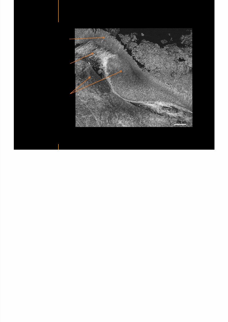
7292019 Atlas of Light-CT images
httpslidepdfcomreaderfullatlas-of-light-ct-images 70118
copy LLTech 2012 ndash wwwlltechimagingcom - No copy without authorization 70
Rat bladder
Stratifiedurothelium
Lamina propria
500 microm
Muscular
bundles of the
lamina propria
Serosa
copy LLTech 2012 Courtesy of Weill Cornell Medical College NY USA
100 microm
100 microm
Human bladder low grade papillary urothelial carcinoma
7292019 Atlas of Light-CT images
httpslidepdfcomreaderfullatlas-of-light-ct-images 71118
copy LLTech 2012 ndash wwwlltechimagingcom - No copy without authorization 71
Lesion with fibrovascular cores (arrow) and superficial umbrella cells (arrowheads)
Human bladder low grade papillary urothelial carcinoma
copy LLTech 2012 Courtesy of Weill Cornell Medical College NY USA
100 microm
7292019 Atlas of Light-CT images
httpslidepdfcomreaderfullatlas-of-light-ct-images 72118
Rat prostate
7292019 Atlas of Light-CT images
httpslidepdfcomreaderfullatlas-of-light-ct-images 73118
copy LLTech 2012 ndash wwwlltechimagingcom - No copy without authorization 73
p
copy LLTech 2012 Courtesy of Weill Cornell Medical College NY USA
Papillary folds of the
acinar glands
Prostatic acini
Fibromuscular stromaPeriprostatic fat Blood vessel
1 mm
200 microm
Mouse kidney
7292019 Atlas of Light-CT images
httpslidepdfcomreaderfullatlas-of-light-ct-images 74118
copy LLTech 2012 ndash wwwlltechimagingcom - No copy without authorization 74
1 mm
y
copy LLTech 2012 Courtesy of Institut Biomeacutedical de Bicecirctre Paris France
Renal cortex
Renal medulla
Renal sinus
7292019 Atlas of Light-CT images
httpslidepdfcomreaderfullatlas-of-light-ct-images 75118
copy LLTech 2012 ndash wwwlltechimagingcom - No copy without authorization 75
Mouse kidney ndash zoom on highlighted section of previous slide
800microm
copy LLTech 2012 Courtesy of Hocircpital Kremlin Bicecirctre Paris France
7292019 Atlas of Light-CT images
httpslidepdfcomreaderfullatlas-of-light-ct-images 76118
7292019 Atlas of Light-CT images
httpslidepdfcomreaderfullatlas-of-light-ct-images 77118
7292019 Atlas of Light-CT images
httpslidepdfcomreaderfullatlas-of-light-ct-images 78118
copy LLTech 2012 ndash wwwlltechimagingcom - No copy without authorization 78 copy LLTECH 2011
CORE NEEDLE BIOPSIES
Back to table of contents
Kidney
7292019 Atlas of Light-CT images
httpslidepdfcomreaderfullatlas-of-light-ct-images 79118
copy LLTech 2012 ndash wwwlltechimagingcom - No copy without authorization 79
100microm
50microm
Renal tubules
Vessel
500microm
copy LLTech 2012 Courtesy of Institut Curie Paris France
Breast Invasive lobular carcinoma
7292019 Atlas of Light-CT images
httpslidepdfcomreaderfullatlas-of-light-ct-images 80118
copy LLTech 2012 ndash wwwlltechimagingcom - No copy without authorization 80
500microm
stroma
adipocytes
copy LLTech 2012 Courtesy of Institut Curie Paris France
7292019 Atlas of Light-CT images
httpslidepdfcomreaderfullatlas-of-light-ct-images 81118
Breast Ductal carcinoma in situ
7292019 Atlas of Light-CT images
httpslidepdfcomreaderfullatlas-of-light-ct-images 82118
copy LLTech 2012 ndash wwwlltechimagingcom - No copy without authorization 82
500microm
copy LLTech 2012 Courtesy of Institut Curie Paris France
7292019 Atlas of Light-CT images
httpslidepdfcomreaderfullatlas-of-light-ct-images 83118
copy LLTech 2012 ndash wwwlltechimagingcom - No copy without authorization 83 copy LLTECH 2011
THYROID IMAGING
Back to table of contents
7292019 Atlas of Light-CT images
httpslidepdfcomreaderfullatlas-of-light-ct-images 84118
copy LLTech 2012 ndash wwwlltechimagingcom - No copy without authorization 84
Thyroid
800 microm
200 microm
copy LLTech 2012 Courtesy of Hocircpital Tenon Paris France
7292019 Atlas of Light-CT images
httpslidepdfcomreaderfullatlas-of-light-ct-images 85118
copy LLTech 2012 ndash wwwlltechimagingcom - No copy without authorization 85
Thyroid
800 microm
200 microm
copy LLTech 2012 Courtesy of Hocircpital Tenon Paris France
7292019 Atlas of Light-CT images
httpslidepdfcomreaderfullatlas-of-light-ct-images 86118
copy LLTech 2012 ndash wwwlltechimagingcom - No copy without authorization 86 copy LLTECH 2011
FAT CELLS IMAGING
Back to table of contents
7292019 Atlas of Light-CT images
httpslidepdfcomreaderfullatlas-of-light-ct-images 87118
copy LLTech 2012 ndash wwwlltechimagingcom - No copy without authorization 87
Brown fat cells - adipocytes
100 microm 100 microm
200 microm
10um depth
Adipocytes
45um depth
En face
slices
Reconstructed
depth slice
copy LLTech 2012 Courtesy of Centre de Recherche des Cordeliers Paris France
7292019 Atlas of Light-CT images
httpslidepdfcomreaderfullatlas-of-light-ct-images 88118
copy LLTech 2012 ndash wwwlltechimagingcom - No copy without authorization 88 copy LLTECH 2011
CORNEA AND RETINA
Back to table of contents
Human cornea reconstructed depth slice
7292019 Atlas of Light-CT images
httpslidepdfcomreaderfullatlas-of-light-ct-images 89118
copy LLTech 2012 ndash wwwlltechimagingcom - No copy without authorization 89
50 um
wwwlltechimagingcom
7292019 Atlas of Light-CT images
httpslidepdfcomreaderfullatlas-of-light-ct-images 90118
Large field en face view in stroma
7292019 Atlas of Light-CT images
httpslidepdfcomreaderfullatlas-of-light-ct-images 91118
copy LLTech 2012 ndash wwwlltechimagingcom - No copy without authorization 91
200 um
wwwlltechimagingcom
7292019 Atlas of Light-CT images
httpslidepdfcomreaderfullatlas-of-light-ct-images 92118
Rabbit cornea after LASIK reflectivity profiles reveal
i d tt i d thi k d ith li l l
7292019 Atlas of Light-CT images
httpslidepdfcomreaderfullatlas-of-light-ct-images 93118
copy LLTech 2012 ndash wwwlltechimagingcom - No copy without authorization 93
increased scattering and thickened epithelial layer
LASIK LASIK CONTROL CONTROL
copy LLTech 2012
Newborn mouse anterior segment
7292019 Atlas of Light-CT images
httpslidepdfcomreaderfullatlas-of-light-ct-images 94118
copy LLTech 2012 ndash wwwlltechimagingcom - No copy without authorization 94
Corneal epithelial cells
Basal membrane
Stroma
Descemetrsquos membrane
Aqueous humor
Lens capsule
Lens epithelial cells
Cortical fibers
copy LLTech 2012 Courtesy of ESPCI Paris Francecopy LLTech 2012
Rat retina comparison with histology
7292019 Atlas of Light-CT images
httpslidepdfcomreaderfullatlas-of-light-ct-images 95118
copy LLTech 2012 ndash wwwlltechimagingcom - No copy without authorization 95
Figure 1 Right Ex vivo rat retinaimaged using the f ull field OCT
system of ESPCI Left Histology
for comparison
Figure 1 Right Ex vivo rat retinaimaged using the f ull field OCT
system of ESPCI Left Histology
for comparison
Figure 1 Right Ex vivo rat retinaimaged using the f ull field OCT
system of ESPCI Left Histology
for comparisonField
230x300microm
RPE
PRL
ONL
OPL
INL
IPL
GCL
NFL
Invest Ophthalmol Vis Sci 2004 Nov45(11)4126-31
Grieve K Paques M Dubois A Sahel J Boccara C Le Gargasson JF copy LLTech 2012
Rat retina
7292019 Atlas of Light-CT images
httpslidepdfcomreaderfullatlas-of-light-ct-images 96118
copy LLTech 2012 ndash wwwlltechimagingcom - No copy without authorization 96 copy LLTech 2012 Courtesy of ESPCI Paris France
Nerve fiber layer
Retina
Invest Ophthalmol Vis Sci 2004 Nov45(11)4126-31
Grieve K Paques M Dubois A Sahel J Boccara C Le Gargasson JF
Transscleral image
7292019 Atlas of Light-CT images
httpslidepdfcomreaderfullatlas-of-light-ct-images 97118
7292019 Atlas of Light-CT images
httpslidepdfcomreaderfullatlas-of-light-ct-images 98118
7292019 Atlas of Light-CT images
httpslidepdfcomreaderfullatlas-of-light-ct-images 99118
In vivo imaging of drosophila melanogaster
7292019 Atlas of Light-CT images
httpslidepdfcomreaderfullatlas-of-light-ct-images 100118
copy LLTech 2012 ndash wwwlltechimagingcom - No copy without authorization 100 copy LLTech 2012 Courtesy of ESPCI Paris France
In vivo imaging of drosophila melanogaster ndash four stages of pupation over 72 hours
7292019 Atlas of Light-CT images
httpslidepdfcomreaderfullatlas-of-light-ct-images 101118
copy LLTech 2012 ndash wwwlltechimagingcom - No copy without authorization 101
Prepupa (0-2 h) Transition topupal stage (24 h)
Pupal stage (48 h) Advanced pupal phase
before eclosion (72 h)
copy LLTech 2012 Courtesy of ESPCI Paris France
Pupa 118 4 days 30um depth
100
7292019 Atlas of Light-CT images
httpslidepdfcomreaderfullatlas-of-light-ct-images 102118
copy LLTech 2012 ndash wwwlltechimagingcom - No copy without authorization 102
100 um
Drosophila
7292019 Atlas of Light-CT images
httpslidepdfcomreaderfullatlas-of-light-ct-images 103118
copy LLTech 2012 ndash wwwlltechimagingcom - No copy without authorization 103
200 microm
copy LLTech 2012 Courtesy of ESPCI Paris France
C Elegans ndash Fast identification of anatomy
7292019 Atlas of Light-CT images
httpslidepdfcomreaderfullatlas-of-light-ct-images 104118
copy LLTech 2012 ndash wwwlltechimagingcom - No copy without authorization 104
Body wall - cuticle
gonad arm
30 microm depth
uterus
ovocytes
intestine
100 microm 100 microm
15 microm depth
copy LLTech 2012 Courtesy of ENS Paris France
C elegans ndash Images at higher magnification 40X
7292019 Atlas of Light-CT images
httpslidepdfcomreaderfullatlas-of-light-ct-images 105118
copy LLTech 2012 ndash wwwlltechimagingcom - No copy without authorization 105
3D reconstruction 50 microm
100 microm
copy LLTech 2012 Courtesy of ENS Paris France
Zebra fish eye
7292019 Atlas of Light-CT images
httpslidepdfcomreaderfullatlas-of-light-ct-images 106118
copy LLTech 2012 ndash wwwlltechimagingcom - No copy without authorization 106
Zebra fish eye
100 microm
100 microm
En face slices
Reconstructed depth slice
copy LLTech 2012 Courtesy of Inserm Paris France
10um depth 20um depth 45um depth
7292019 Atlas of Light-CT images
httpslidepdfcomreaderfullatlas-of-light-ct-images 107118
Xenopus Laevis backtail
7292019 Atlas of Light-CT images
httpslidepdfcomreaderfullatlas-of-light-ct-images 108118
copy LLTech 2012 ndash wwwlltechimagingcom - No copy without authorization 108
100 um30um depth 60um depth
7292019 Atlas of Light-CT images
httpslidepdfcomreaderfullatlas-of-light-ct-images 109118
Mouse embryo in vitro 6 5 days old ndash
7292019 Atlas of Light-CT images
httpslidepdfcomreaderfullatlas-of-light-ct-images 110118
copy LLTech 2012 ndash wwwlltechimagingcom - No copy without authorization 110
Mouse embryo in vitro 65 days old
copy LLTech 2012 Courtesy of Ellvire Guiot and nstitut Jacques Monod Paris France
3D imaging of internal cavity
Sabellaria alveolata mouth palps
7292019 Atlas of Light-CT images
httpslidepdfcomreaderfullatlas-of-light-ct-images 111118
copy LLTech 2012 ndash wwwlltechimagingcom - No copy without authorization 111
500 microm
100 microm
Mouth palps
copy LLTech 2012 Courtesy of Museacutee National drsquoHistoire Naturelle Paris France
7292019 Atlas of Light-CT images
httpslidepdfcomreaderfullatlas-of-light-ct-images 112118
copy LLTech 2012 ndash wwwlltechimagingcom - No copy without authorization 112 copy LLTECH 2011
PLANT IMAGING
Back to table of contents
Mouse ear leaf epidermis
7292019 Atlas of Light-CT images
httpslidepdfcomreaderfullatlas-of-light-ct-images 113118
copy LLTech 2012 ndash wwwlltechimagingcom - No copy without authorization 113
100 microm
Epidermal cell
copy LLTech 2012
Stoma
Leaf veins
7292019 Atlas of Light-CT images
httpslidepdfcomreaderfullatlas-of-light-ct-images 114118
copy LLTech 2012 ndash wwwlltechimagingcom - No copy without authorization 114
114copy LLTech 2012
Fibers
Veins
100 microm
500 microm
Apple
7292019 Atlas of Light-CT images
httpslidepdfcomreaderfullatlas-of-light-ct-images 115118
copy LLTech 2012 ndash wwwlltechimagingcom - No copy without authorization 115
115copy LLTech 2012
100 microm
Wax
Skin
Flesh
500 microm
Water
Effect of harpin infiltration on tobacco leaf
7292019 Atlas of Light-CT images
httpslidepdfcomreaderfullatlas-of-light-ct-images 116118
copy LLTech 2012 ndash wwwlltechimagingcom - No copy without authorization 116
116copy LLTech 2012 Courtesy of UPMC Paris France
50 microm
50 microm
50 microm
50 microm
Upper epidermis
10 min after harpin infiltration 30 min after harpin infiltration
Palisade parenchyma
Epidermis cell
Chloroplast
7292019 Atlas of Light-CT images
httpslidepdfcomreaderfullatlas-of-light-ct-images 117118
copy LLTech 2012 ndash wwwlltechimagingcom - No copy without authorization 117
bull Light-CTtrade does not require tissue preparation nor staining of any kind
bull Creates images within minutes using a non-destructive process
bull Offers a 1 microm cellular resolution in 3D
bull Reveals structural and cellular information
bull Mosaicing allows fast visualisation at various scale
bull En-face high-resolution imaging allows vertical and 3D reconstruction
bull In-vivo images show good promise for in-vivo system
bull Clinical and research applications in cancer detection cosmetics biobanking small animal
developmental biology botanyhellip
Contact Details
7292019 Atlas of Light-CT images
httpslidepdfcomreaderfullatlas-of-light-ct-images 118118
USA
LLTech Inc
103 Carnegie Center DriveSuite 300
Princeton NJ 08540
USA
wwwlltechimagingcom
contactlltechfr
Phone + 1 609 955 3506
LLTech SAS
29 rue du Faubourg St-JacquesPeacutepiniegravere Paris Santeacute Cochin
75014
Paris - France
wwwlltechimagingcom
contactlltechfr
Phone +33 1 82 72 61 25
Europe
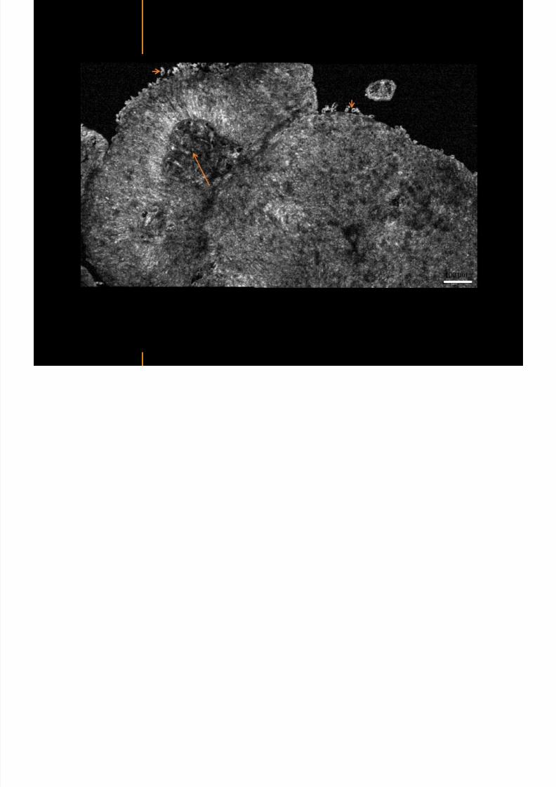
7292019 Atlas of Light-CT images
httpslidepdfcomreaderfullatlas-of-light-ct-images 74118
copy LLTech 2012 ndash wwwlltechimagingcom - No copy without authorization 74
1 mm
y
copy LLTech 2012 Courtesy of Institut Biomeacutedical de Bicecirctre Paris France
Renal cortex
Renal medulla
Renal sinus
7292019 Atlas of Light-CT images
httpslidepdfcomreaderfullatlas-of-light-ct-images 75118
copy LLTech 2012 ndash wwwlltechimagingcom - No copy without authorization 75
Mouse kidney ndash zoom on highlighted section of previous slide
800microm
copy LLTech 2012 Courtesy of Hocircpital Kremlin Bicecirctre Paris France
7292019 Atlas of Light-CT images
httpslidepdfcomreaderfullatlas-of-light-ct-images 76118
7292019 Atlas of Light-CT images
httpslidepdfcomreaderfullatlas-of-light-ct-images 77118
7292019 Atlas of Light-CT images
httpslidepdfcomreaderfullatlas-of-light-ct-images 78118
copy LLTech 2012 ndash wwwlltechimagingcom - No copy without authorization 78 copy LLTECH 2011
CORE NEEDLE BIOPSIES
Back to table of contents
Kidney
7292019 Atlas of Light-CT images
httpslidepdfcomreaderfullatlas-of-light-ct-images 79118
copy LLTech 2012 ndash wwwlltechimagingcom - No copy without authorization 79
100microm
50microm
Renal tubules
Vessel
500microm
copy LLTech 2012 Courtesy of Institut Curie Paris France
Breast Invasive lobular carcinoma
7292019 Atlas of Light-CT images
httpslidepdfcomreaderfullatlas-of-light-ct-images 80118
copy LLTech 2012 ndash wwwlltechimagingcom - No copy without authorization 80
500microm
stroma
adipocytes
copy LLTech 2012 Courtesy of Institut Curie Paris France
7292019 Atlas of Light-CT images
httpslidepdfcomreaderfullatlas-of-light-ct-images 81118
Breast Ductal carcinoma in situ
7292019 Atlas of Light-CT images
httpslidepdfcomreaderfullatlas-of-light-ct-images 82118
copy LLTech 2012 ndash wwwlltechimagingcom - No copy without authorization 82
500microm
copy LLTech 2012 Courtesy of Institut Curie Paris France
7292019 Atlas of Light-CT images
httpslidepdfcomreaderfullatlas-of-light-ct-images 83118
copy LLTech 2012 ndash wwwlltechimagingcom - No copy without authorization 83 copy LLTECH 2011
THYROID IMAGING
Back to table of contents
7292019 Atlas of Light-CT images
httpslidepdfcomreaderfullatlas-of-light-ct-images 84118
copy LLTech 2012 ndash wwwlltechimagingcom - No copy without authorization 84
Thyroid
800 microm
200 microm
copy LLTech 2012 Courtesy of Hocircpital Tenon Paris France
7292019 Atlas of Light-CT images
httpslidepdfcomreaderfullatlas-of-light-ct-images 85118
copy LLTech 2012 ndash wwwlltechimagingcom - No copy without authorization 85
Thyroid
800 microm
200 microm
copy LLTech 2012 Courtesy of Hocircpital Tenon Paris France
7292019 Atlas of Light-CT images
httpslidepdfcomreaderfullatlas-of-light-ct-images 86118
copy LLTech 2012 ndash wwwlltechimagingcom - No copy without authorization 86 copy LLTECH 2011
FAT CELLS IMAGING
Back to table of contents
7292019 Atlas of Light-CT images
httpslidepdfcomreaderfullatlas-of-light-ct-images 87118
copy LLTech 2012 ndash wwwlltechimagingcom - No copy without authorization 87
Brown fat cells - adipocytes
100 microm 100 microm
200 microm
10um depth
Adipocytes
45um depth
En face
slices
Reconstructed
depth slice
copy LLTech 2012 Courtesy of Centre de Recherche des Cordeliers Paris France
7292019 Atlas of Light-CT images
httpslidepdfcomreaderfullatlas-of-light-ct-images 88118
copy LLTech 2012 ndash wwwlltechimagingcom - No copy without authorization 88 copy LLTECH 2011
CORNEA AND RETINA
Back to table of contents
Human cornea reconstructed depth slice
7292019 Atlas of Light-CT images
httpslidepdfcomreaderfullatlas-of-light-ct-images 89118
copy LLTech 2012 ndash wwwlltechimagingcom - No copy without authorization 89
50 um
wwwlltechimagingcom
7292019 Atlas of Light-CT images
httpslidepdfcomreaderfullatlas-of-light-ct-images 90118
Large field en face view in stroma
7292019 Atlas of Light-CT images
httpslidepdfcomreaderfullatlas-of-light-ct-images 91118
copy LLTech 2012 ndash wwwlltechimagingcom - No copy without authorization 91
200 um
wwwlltechimagingcom
7292019 Atlas of Light-CT images
httpslidepdfcomreaderfullatlas-of-light-ct-images 92118
Rabbit cornea after LASIK reflectivity profiles reveal
i d tt i d thi k d ith li l l
7292019 Atlas of Light-CT images
httpslidepdfcomreaderfullatlas-of-light-ct-images 93118
copy LLTech 2012 ndash wwwlltechimagingcom - No copy without authorization 93
increased scattering and thickened epithelial layer
LASIK LASIK CONTROL CONTROL
copy LLTech 2012
Newborn mouse anterior segment
7292019 Atlas of Light-CT images
httpslidepdfcomreaderfullatlas-of-light-ct-images 94118
copy LLTech 2012 ndash wwwlltechimagingcom - No copy without authorization 94
Corneal epithelial cells
Basal membrane
Stroma
Descemetrsquos membrane
Aqueous humor
Lens capsule
Lens epithelial cells
Cortical fibers
copy LLTech 2012 Courtesy of ESPCI Paris Francecopy LLTech 2012
Rat retina comparison with histology
7292019 Atlas of Light-CT images
httpslidepdfcomreaderfullatlas-of-light-ct-images 95118
copy LLTech 2012 ndash wwwlltechimagingcom - No copy without authorization 95
Figure 1 Right Ex vivo rat retinaimaged using the f ull field OCT
system of ESPCI Left Histology
for comparison
Figure 1 Right Ex vivo rat retinaimaged using the f ull field OCT
system of ESPCI Left Histology
for comparison
Figure 1 Right Ex vivo rat retinaimaged using the f ull field OCT
system of ESPCI Left Histology
for comparisonField
230x300microm
RPE
PRL
ONL
OPL
INL
IPL
GCL
NFL
Invest Ophthalmol Vis Sci 2004 Nov45(11)4126-31
Grieve K Paques M Dubois A Sahel J Boccara C Le Gargasson JF copy LLTech 2012
Rat retina
7292019 Atlas of Light-CT images
httpslidepdfcomreaderfullatlas-of-light-ct-images 96118
copy LLTech 2012 ndash wwwlltechimagingcom - No copy without authorization 96 copy LLTech 2012 Courtesy of ESPCI Paris France
Nerve fiber layer
Retina
Invest Ophthalmol Vis Sci 2004 Nov45(11)4126-31
Grieve K Paques M Dubois A Sahel J Boccara C Le Gargasson JF
Transscleral image
7292019 Atlas of Light-CT images
httpslidepdfcomreaderfullatlas-of-light-ct-images 97118
7292019 Atlas of Light-CT images
httpslidepdfcomreaderfullatlas-of-light-ct-images 98118
7292019 Atlas of Light-CT images
httpslidepdfcomreaderfullatlas-of-light-ct-images 99118
In vivo imaging of drosophila melanogaster
7292019 Atlas of Light-CT images
httpslidepdfcomreaderfullatlas-of-light-ct-images 100118
copy LLTech 2012 ndash wwwlltechimagingcom - No copy without authorization 100 copy LLTech 2012 Courtesy of ESPCI Paris France
In vivo imaging of drosophila melanogaster ndash four stages of pupation over 72 hours
7292019 Atlas of Light-CT images
httpslidepdfcomreaderfullatlas-of-light-ct-images 101118
copy LLTech 2012 ndash wwwlltechimagingcom - No copy without authorization 101
Prepupa (0-2 h) Transition topupal stage (24 h)
Pupal stage (48 h) Advanced pupal phase
before eclosion (72 h)
copy LLTech 2012 Courtesy of ESPCI Paris France
Pupa 118 4 days 30um depth
100
7292019 Atlas of Light-CT images
httpslidepdfcomreaderfullatlas-of-light-ct-images 102118
copy LLTech 2012 ndash wwwlltechimagingcom - No copy without authorization 102
100 um
Drosophila
7292019 Atlas of Light-CT images
httpslidepdfcomreaderfullatlas-of-light-ct-images 103118
copy LLTech 2012 ndash wwwlltechimagingcom - No copy without authorization 103
200 microm
copy LLTech 2012 Courtesy of ESPCI Paris France
C Elegans ndash Fast identification of anatomy
7292019 Atlas of Light-CT images
httpslidepdfcomreaderfullatlas-of-light-ct-images 104118
copy LLTech 2012 ndash wwwlltechimagingcom - No copy without authorization 104
Body wall - cuticle
gonad arm
30 microm depth
uterus
ovocytes
intestine
100 microm 100 microm
15 microm depth
copy LLTech 2012 Courtesy of ENS Paris France
C elegans ndash Images at higher magnification 40X
7292019 Atlas of Light-CT images
httpslidepdfcomreaderfullatlas-of-light-ct-images 105118
copy LLTech 2012 ndash wwwlltechimagingcom - No copy without authorization 105
3D reconstruction 50 microm
100 microm
copy LLTech 2012 Courtesy of ENS Paris France
Zebra fish eye
7292019 Atlas of Light-CT images
httpslidepdfcomreaderfullatlas-of-light-ct-images 106118
copy LLTech 2012 ndash wwwlltechimagingcom - No copy without authorization 106
Zebra fish eye
100 microm
100 microm
En face slices
Reconstructed depth slice
copy LLTech 2012 Courtesy of Inserm Paris France
10um depth 20um depth 45um depth
7292019 Atlas of Light-CT images
httpslidepdfcomreaderfullatlas-of-light-ct-images 107118
Xenopus Laevis backtail
7292019 Atlas of Light-CT images
httpslidepdfcomreaderfullatlas-of-light-ct-images 108118
copy LLTech 2012 ndash wwwlltechimagingcom - No copy without authorization 108
100 um30um depth 60um depth
7292019 Atlas of Light-CT images
httpslidepdfcomreaderfullatlas-of-light-ct-images 109118
Mouse embryo in vitro 6 5 days old ndash
7292019 Atlas of Light-CT images
httpslidepdfcomreaderfullatlas-of-light-ct-images 110118
copy LLTech 2012 ndash wwwlltechimagingcom - No copy without authorization 110
Mouse embryo in vitro 65 days old
copy LLTech 2012 Courtesy of Ellvire Guiot and nstitut Jacques Monod Paris France
3D imaging of internal cavity
Sabellaria alveolata mouth palps
7292019 Atlas of Light-CT images
httpslidepdfcomreaderfullatlas-of-light-ct-images 111118
copy LLTech 2012 ndash wwwlltechimagingcom - No copy without authorization 111
500 microm
100 microm
Mouth palps
copy LLTech 2012 Courtesy of Museacutee National drsquoHistoire Naturelle Paris France
7292019 Atlas of Light-CT images
httpslidepdfcomreaderfullatlas-of-light-ct-images 112118
copy LLTech 2012 ndash wwwlltechimagingcom - No copy without authorization 112 copy LLTECH 2011
PLANT IMAGING
Back to table of contents
Mouse ear leaf epidermis
7292019 Atlas of Light-CT images
httpslidepdfcomreaderfullatlas-of-light-ct-images 113118
copy LLTech 2012 ndash wwwlltechimagingcom - No copy without authorization 113
100 microm
Epidermal cell
copy LLTech 2012
Stoma
Leaf veins
7292019 Atlas of Light-CT images
httpslidepdfcomreaderfullatlas-of-light-ct-images 114118
copy LLTech 2012 ndash wwwlltechimagingcom - No copy without authorization 114
114copy LLTech 2012
Fibers
Veins
100 microm
500 microm
Apple
7292019 Atlas of Light-CT images
httpslidepdfcomreaderfullatlas-of-light-ct-images 115118
copy LLTech 2012 ndash wwwlltechimagingcom - No copy without authorization 115
115copy LLTech 2012
100 microm
Wax
Skin
Flesh
500 microm
Water
Effect of harpin infiltration on tobacco leaf
7292019 Atlas of Light-CT images
httpslidepdfcomreaderfullatlas-of-light-ct-images 116118
copy LLTech 2012 ndash wwwlltechimagingcom - No copy without authorization 116
116copy LLTech 2012 Courtesy of UPMC Paris France
50 microm
50 microm
50 microm
50 microm
Upper epidermis
10 min after harpin infiltration 30 min after harpin infiltration
Palisade parenchyma
Epidermis cell
Chloroplast
7292019 Atlas of Light-CT images
httpslidepdfcomreaderfullatlas-of-light-ct-images 117118
copy LLTech 2012 ndash wwwlltechimagingcom - No copy without authorization 117
bull Light-CTtrade does not require tissue preparation nor staining of any kind
bull Creates images within minutes using a non-destructive process
bull Offers a 1 microm cellular resolution in 3D
bull Reveals structural and cellular information
bull Mosaicing allows fast visualisation at various scale
bull En-face high-resolution imaging allows vertical and 3D reconstruction
bull In-vivo images show good promise for in-vivo system
bull Clinical and research applications in cancer detection cosmetics biobanking small animal
developmental biology botanyhellip
Contact Details
7292019 Atlas of Light-CT images
httpslidepdfcomreaderfullatlas-of-light-ct-images 118118
USA
LLTech Inc
103 Carnegie Center DriveSuite 300
Princeton NJ 08540
USA
wwwlltechimagingcom
contactlltechfr
Phone + 1 609 955 3506
LLTech SAS
29 rue du Faubourg St-JacquesPeacutepiniegravere Paris Santeacute Cochin
75014
Paris - France
wwwlltechimagingcom
contactlltechfr
Phone +33 1 82 72 61 25
Europe
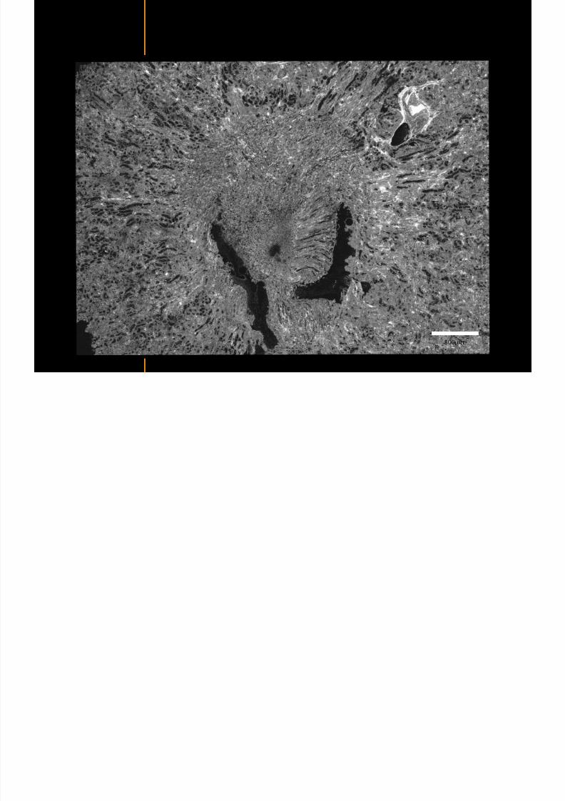
7292019 Atlas of Light-CT images
httpslidepdfcomreaderfullatlas-of-light-ct-images 75118
copy LLTech 2012 ndash wwwlltechimagingcom - No copy without authorization 75
Mouse kidney ndash zoom on highlighted section of previous slide
800microm
copy LLTech 2012 Courtesy of Hocircpital Kremlin Bicecirctre Paris France
7292019 Atlas of Light-CT images
httpslidepdfcomreaderfullatlas-of-light-ct-images 76118
7292019 Atlas of Light-CT images
httpslidepdfcomreaderfullatlas-of-light-ct-images 77118
7292019 Atlas of Light-CT images
httpslidepdfcomreaderfullatlas-of-light-ct-images 78118
copy LLTech 2012 ndash wwwlltechimagingcom - No copy without authorization 78 copy LLTECH 2011
CORE NEEDLE BIOPSIES
Back to table of contents
Kidney
7292019 Atlas of Light-CT images
httpslidepdfcomreaderfullatlas-of-light-ct-images 79118
copy LLTech 2012 ndash wwwlltechimagingcom - No copy without authorization 79
100microm
50microm
Renal tubules
Vessel
500microm
copy LLTech 2012 Courtesy of Institut Curie Paris France
Breast Invasive lobular carcinoma
7292019 Atlas of Light-CT images
httpslidepdfcomreaderfullatlas-of-light-ct-images 80118
copy LLTech 2012 ndash wwwlltechimagingcom - No copy without authorization 80
500microm
stroma
adipocytes
copy LLTech 2012 Courtesy of Institut Curie Paris France
7292019 Atlas of Light-CT images
httpslidepdfcomreaderfullatlas-of-light-ct-images 81118
Breast Ductal carcinoma in situ
7292019 Atlas of Light-CT images
httpslidepdfcomreaderfullatlas-of-light-ct-images 82118
copy LLTech 2012 ndash wwwlltechimagingcom - No copy without authorization 82
500microm
copy LLTech 2012 Courtesy of Institut Curie Paris France
7292019 Atlas of Light-CT images
httpslidepdfcomreaderfullatlas-of-light-ct-images 83118
copy LLTech 2012 ndash wwwlltechimagingcom - No copy without authorization 83 copy LLTECH 2011
THYROID IMAGING
Back to table of contents
7292019 Atlas of Light-CT images
httpslidepdfcomreaderfullatlas-of-light-ct-images 84118
copy LLTech 2012 ndash wwwlltechimagingcom - No copy without authorization 84
Thyroid
800 microm
200 microm
copy LLTech 2012 Courtesy of Hocircpital Tenon Paris France
7292019 Atlas of Light-CT images
httpslidepdfcomreaderfullatlas-of-light-ct-images 85118
copy LLTech 2012 ndash wwwlltechimagingcom - No copy without authorization 85
Thyroid
800 microm
200 microm
copy LLTech 2012 Courtesy of Hocircpital Tenon Paris France
7292019 Atlas of Light-CT images
httpslidepdfcomreaderfullatlas-of-light-ct-images 86118
copy LLTech 2012 ndash wwwlltechimagingcom - No copy without authorization 86 copy LLTECH 2011
FAT CELLS IMAGING
Back to table of contents
7292019 Atlas of Light-CT images
httpslidepdfcomreaderfullatlas-of-light-ct-images 87118
copy LLTech 2012 ndash wwwlltechimagingcom - No copy without authorization 87
Brown fat cells - adipocytes
100 microm 100 microm
200 microm
10um depth
Adipocytes
45um depth
En face
slices
Reconstructed
depth slice
copy LLTech 2012 Courtesy of Centre de Recherche des Cordeliers Paris France
7292019 Atlas of Light-CT images
httpslidepdfcomreaderfullatlas-of-light-ct-images 88118
copy LLTech 2012 ndash wwwlltechimagingcom - No copy without authorization 88 copy LLTECH 2011
CORNEA AND RETINA
Back to table of contents
Human cornea reconstructed depth slice
7292019 Atlas of Light-CT images
httpslidepdfcomreaderfullatlas-of-light-ct-images 89118
copy LLTech 2012 ndash wwwlltechimagingcom - No copy without authorization 89
50 um
wwwlltechimagingcom
7292019 Atlas of Light-CT images
httpslidepdfcomreaderfullatlas-of-light-ct-images 90118
Large field en face view in stroma
7292019 Atlas of Light-CT images
httpslidepdfcomreaderfullatlas-of-light-ct-images 91118
copy LLTech 2012 ndash wwwlltechimagingcom - No copy without authorization 91
200 um
wwwlltechimagingcom
7292019 Atlas of Light-CT images
httpslidepdfcomreaderfullatlas-of-light-ct-images 92118
Rabbit cornea after LASIK reflectivity profiles reveal
i d tt i d thi k d ith li l l
7292019 Atlas of Light-CT images
httpslidepdfcomreaderfullatlas-of-light-ct-images 93118
copy LLTech 2012 ndash wwwlltechimagingcom - No copy without authorization 93
increased scattering and thickened epithelial layer
LASIK LASIK CONTROL CONTROL
copy LLTech 2012
Newborn mouse anterior segment
7292019 Atlas of Light-CT images
httpslidepdfcomreaderfullatlas-of-light-ct-images 94118
copy LLTech 2012 ndash wwwlltechimagingcom - No copy without authorization 94
Corneal epithelial cells
Basal membrane
Stroma
Descemetrsquos membrane
Aqueous humor
Lens capsule
Lens epithelial cells
Cortical fibers
copy LLTech 2012 Courtesy of ESPCI Paris Francecopy LLTech 2012
Rat retina comparison with histology
7292019 Atlas of Light-CT images
httpslidepdfcomreaderfullatlas-of-light-ct-images 95118
copy LLTech 2012 ndash wwwlltechimagingcom - No copy without authorization 95
Figure 1 Right Ex vivo rat retinaimaged using the f ull field OCT
system of ESPCI Left Histology
for comparison
Figure 1 Right Ex vivo rat retinaimaged using the f ull field OCT
system of ESPCI Left Histology
for comparison
Figure 1 Right Ex vivo rat retinaimaged using the f ull field OCT
system of ESPCI Left Histology
for comparisonField
230x300microm
RPE
PRL
ONL
OPL
INL
IPL
GCL
NFL
Invest Ophthalmol Vis Sci 2004 Nov45(11)4126-31
Grieve K Paques M Dubois A Sahel J Boccara C Le Gargasson JF copy LLTech 2012
Rat retina
7292019 Atlas of Light-CT images
httpslidepdfcomreaderfullatlas-of-light-ct-images 96118
copy LLTech 2012 ndash wwwlltechimagingcom - No copy without authorization 96 copy LLTech 2012 Courtesy of ESPCI Paris France
Nerve fiber layer
Retina
Invest Ophthalmol Vis Sci 2004 Nov45(11)4126-31
Grieve K Paques M Dubois A Sahel J Boccara C Le Gargasson JF
Transscleral image
7292019 Atlas of Light-CT images
httpslidepdfcomreaderfullatlas-of-light-ct-images 97118
7292019 Atlas of Light-CT images
httpslidepdfcomreaderfullatlas-of-light-ct-images 98118
7292019 Atlas of Light-CT images
httpslidepdfcomreaderfullatlas-of-light-ct-images 99118
In vivo imaging of drosophila melanogaster
7292019 Atlas of Light-CT images
httpslidepdfcomreaderfullatlas-of-light-ct-images 100118
copy LLTech 2012 ndash wwwlltechimagingcom - No copy without authorization 100 copy LLTech 2012 Courtesy of ESPCI Paris France
In vivo imaging of drosophila melanogaster ndash four stages of pupation over 72 hours
7292019 Atlas of Light-CT images
httpslidepdfcomreaderfullatlas-of-light-ct-images 101118
copy LLTech 2012 ndash wwwlltechimagingcom - No copy without authorization 101
Prepupa (0-2 h) Transition topupal stage (24 h)
Pupal stage (48 h) Advanced pupal phase
before eclosion (72 h)
copy LLTech 2012 Courtesy of ESPCI Paris France
Pupa 118 4 days 30um depth
100
7292019 Atlas of Light-CT images
httpslidepdfcomreaderfullatlas-of-light-ct-images 102118
copy LLTech 2012 ndash wwwlltechimagingcom - No copy without authorization 102
100 um
Drosophila
7292019 Atlas of Light-CT images
httpslidepdfcomreaderfullatlas-of-light-ct-images 103118
copy LLTech 2012 ndash wwwlltechimagingcom - No copy without authorization 103
200 microm
copy LLTech 2012 Courtesy of ESPCI Paris France
C Elegans ndash Fast identification of anatomy
7292019 Atlas of Light-CT images
httpslidepdfcomreaderfullatlas-of-light-ct-images 104118
copy LLTech 2012 ndash wwwlltechimagingcom - No copy without authorization 104
Body wall - cuticle
gonad arm
30 microm depth
uterus
ovocytes
intestine
100 microm 100 microm
15 microm depth
copy LLTech 2012 Courtesy of ENS Paris France
C elegans ndash Images at higher magnification 40X
7292019 Atlas of Light-CT images
httpslidepdfcomreaderfullatlas-of-light-ct-images 105118
copy LLTech 2012 ndash wwwlltechimagingcom - No copy without authorization 105
3D reconstruction 50 microm
100 microm
copy LLTech 2012 Courtesy of ENS Paris France
Zebra fish eye
7292019 Atlas of Light-CT images
httpslidepdfcomreaderfullatlas-of-light-ct-images 106118
copy LLTech 2012 ndash wwwlltechimagingcom - No copy without authorization 106
Zebra fish eye
100 microm
100 microm
En face slices
Reconstructed depth slice
copy LLTech 2012 Courtesy of Inserm Paris France
10um depth 20um depth 45um depth
7292019 Atlas of Light-CT images
httpslidepdfcomreaderfullatlas-of-light-ct-images 107118
Xenopus Laevis backtail
7292019 Atlas of Light-CT images
httpslidepdfcomreaderfullatlas-of-light-ct-images 108118
copy LLTech 2012 ndash wwwlltechimagingcom - No copy without authorization 108
100 um30um depth 60um depth
7292019 Atlas of Light-CT images
httpslidepdfcomreaderfullatlas-of-light-ct-images 109118
Mouse embryo in vitro 6 5 days old ndash
7292019 Atlas of Light-CT images
httpslidepdfcomreaderfullatlas-of-light-ct-images 110118
copy LLTech 2012 ndash wwwlltechimagingcom - No copy without authorization 110
Mouse embryo in vitro 65 days old
copy LLTech 2012 Courtesy of Ellvire Guiot and nstitut Jacques Monod Paris France
3D imaging of internal cavity
Sabellaria alveolata mouth palps
7292019 Atlas of Light-CT images
httpslidepdfcomreaderfullatlas-of-light-ct-images 111118
copy LLTech 2012 ndash wwwlltechimagingcom - No copy without authorization 111
500 microm
100 microm
Mouth palps
copy LLTech 2012 Courtesy of Museacutee National drsquoHistoire Naturelle Paris France
7292019 Atlas of Light-CT images
httpslidepdfcomreaderfullatlas-of-light-ct-images 112118
copy LLTech 2012 ndash wwwlltechimagingcom - No copy without authorization 112 copy LLTECH 2011
PLANT IMAGING
Back to table of contents
Mouse ear leaf epidermis
7292019 Atlas of Light-CT images
httpslidepdfcomreaderfullatlas-of-light-ct-images 113118
copy LLTech 2012 ndash wwwlltechimagingcom - No copy without authorization 113
100 microm
Epidermal cell
copy LLTech 2012
Stoma
Leaf veins
7292019 Atlas of Light-CT images
httpslidepdfcomreaderfullatlas-of-light-ct-images 114118
copy LLTech 2012 ndash wwwlltechimagingcom - No copy without authorization 114
114copy LLTech 2012
Fibers
Veins
100 microm
500 microm
Apple
7292019 Atlas of Light-CT images
httpslidepdfcomreaderfullatlas-of-light-ct-images 115118
copy LLTech 2012 ndash wwwlltechimagingcom - No copy without authorization 115
115copy LLTech 2012
100 microm
Wax
Skin
Flesh
500 microm
Water
Effect of harpin infiltration on tobacco leaf
7292019 Atlas of Light-CT images
httpslidepdfcomreaderfullatlas-of-light-ct-images 116118
copy LLTech 2012 ndash wwwlltechimagingcom - No copy without authorization 116
116copy LLTech 2012 Courtesy of UPMC Paris France
50 microm
50 microm
50 microm
50 microm
Upper epidermis
10 min after harpin infiltration 30 min after harpin infiltration
Palisade parenchyma
Epidermis cell
Chloroplast
7292019 Atlas of Light-CT images
httpslidepdfcomreaderfullatlas-of-light-ct-images 117118
copy LLTech 2012 ndash wwwlltechimagingcom - No copy without authorization 117
bull Light-CTtrade does not require tissue preparation nor staining of any kind
bull Creates images within minutes using a non-destructive process
bull Offers a 1 microm cellular resolution in 3D
bull Reveals structural and cellular information
bull Mosaicing allows fast visualisation at various scale
bull En-face high-resolution imaging allows vertical and 3D reconstruction
bull In-vivo images show good promise for in-vivo system
bull Clinical and research applications in cancer detection cosmetics biobanking small animal
developmental biology botanyhellip
Contact Details
7292019 Atlas of Light-CT images
httpslidepdfcomreaderfullatlas-of-light-ct-images 118118
USA
LLTech Inc
103 Carnegie Center DriveSuite 300
Princeton NJ 08540
USA
wwwlltechimagingcom
contactlltechfr
Phone + 1 609 955 3506
LLTech SAS
29 rue du Faubourg St-JacquesPeacutepiniegravere Paris Santeacute Cochin
75014
Paris - France
wwwlltechimagingcom
contactlltechfr
Phone +33 1 82 72 61 25
Europe
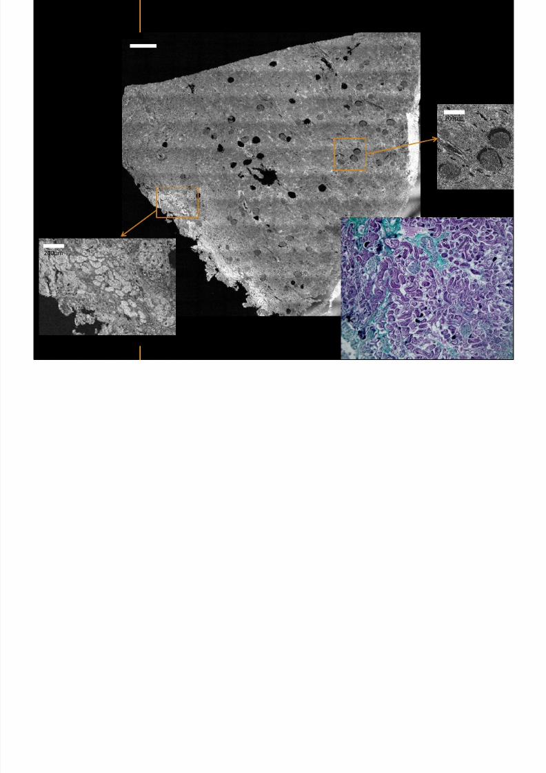
7292019 Atlas of Light-CT images
httpslidepdfcomreaderfullatlas-of-light-ct-images 77118
7292019 Atlas of Light-CT images
httpslidepdfcomreaderfullatlas-of-light-ct-images 78118
copy LLTech 2012 ndash wwwlltechimagingcom - No copy without authorization 78 copy LLTECH 2011
CORE NEEDLE BIOPSIES
Back to table of contents
Kidney
7292019 Atlas of Light-CT images
httpslidepdfcomreaderfullatlas-of-light-ct-images 79118
copy LLTech 2012 ndash wwwlltechimagingcom - No copy without authorization 79
100microm
50microm
Renal tubules
Vessel
500microm
copy LLTech 2012 Courtesy of Institut Curie Paris France
Breast Invasive lobular carcinoma
7292019 Atlas of Light-CT images
httpslidepdfcomreaderfullatlas-of-light-ct-images 80118
copy LLTech 2012 ndash wwwlltechimagingcom - No copy without authorization 80
500microm
stroma
adipocytes
copy LLTech 2012 Courtesy of Institut Curie Paris France
7292019 Atlas of Light-CT images
httpslidepdfcomreaderfullatlas-of-light-ct-images 81118
Breast Ductal carcinoma in situ
7292019 Atlas of Light-CT images
httpslidepdfcomreaderfullatlas-of-light-ct-images 82118
copy LLTech 2012 ndash wwwlltechimagingcom - No copy without authorization 82
500microm
copy LLTech 2012 Courtesy of Institut Curie Paris France
7292019 Atlas of Light-CT images
httpslidepdfcomreaderfullatlas-of-light-ct-images 83118
copy LLTech 2012 ndash wwwlltechimagingcom - No copy without authorization 83 copy LLTECH 2011
THYROID IMAGING
Back to table of contents
7292019 Atlas of Light-CT images
httpslidepdfcomreaderfullatlas-of-light-ct-images 84118
copy LLTech 2012 ndash wwwlltechimagingcom - No copy without authorization 84
Thyroid
800 microm
200 microm
copy LLTech 2012 Courtesy of Hocircpital Tenon Paris France
7292019 Atlas of Light-CT images
httpslidepdfcomreaderfullatlas-of-light-ct-images 85118
copy LLTech 2012 ndash wwwlltechimagingcom - No copy without authorization 85
Thyroid
800 microm
200 microm
copy LLTech 2012 Courtesy of Hocircpital Tenon Paris France
7292019 Atlas of Light-CT images
httpslidepdfcomreaderfullatlas-of-light-ct-images 86118
copy LLTech 2012 ndash wwwlltechimagingcom - No copy without authorization 86 copy LLTECH 2011
FAT CELLS IMAGING
Back to table of contents
7292019 Atlas of Light-CT images
httpslidepdfcomreaderfullatlas-of-light-ct-images 87118
copy LLTech 2012 ndash wwwlltechimagingcom - No copy without authorization 87
Brown fat cells - adipocytes
100 microm 100 microm
200 microm
10um depth
Adipocytes
45um depth
En face
slices
Reconstructed
depth slice
copy LLTech 2012 Courtesy of Centre de Recherche des Cordeliers Paris France
7292019 Atlas of Light-CT images
httpslidepdfcomreaderfullatlas-of-light-ct-images 88118
copy LLTech 2012 ndash wwwlltechimagingcom - No copy without authorization 88 copy LLTECH 2011
CORNEA AND RETINA
Back to table of contents
Human cornea reconstructed depth slice
7292019 Atlas of Light-CT images
httpslidepdfcomreaderfullatlas-of-light-ct-images 89118
copy LLTech 2012 ndash wwwlltechimagingcom - No copy without authorization 89
50 um
wwwlltechimagingcom
7292019 Atlas of Light-CT images
httpslidepdfcomreaderfullatlas-of-light-ct-images 90118
Large field en face view in stroma
7292019 Atlas of Light-CT images
httpslidepdfcomreaderfullatlas-of-light-ct-images 91118
copy LLTech 2012 ndash wwwlltechimagingcom - No copy without authorization 91
200 um
wwwlltechimagingcom
7292019 Atlas of Light-CT images
httpslidepdfcomreaderfullatlas-of-light-ct-images 92118
Rabbit cornea after LASIK reflectivity profiles reveal
i d tt i d thi k d ith li l l
7292019 Atlas of Light-CT images
httpslidepdfcomreaderfullatlas-of-light-ct-images 93118
copy LLTech 2012 ndash wwwlltechimagingcom - No copy without authorization 93
increased scattering and thickened epithelial layer
LASIK LASIK CONTROL CONTROL
copy LLTech 2012
Newborn mouse anterior segment
7292019 Atlas of Light-CT images
httpslidepdfcomreaderfullatlas-of-light-ct-images 94118
copy LLTech 2012 ndash wwwlltechimagingcom - No copy without authorization 94
Corneal epithelial cells
Basal membrane
Stroma
Descemetrsquos membrane
Aqueous humor
Lens capsule
Lens epithelial cells
Cortical fibers
copy LLTech 2012 Courtesy of ESPCI Paris Francecopy LLTech 2012
Rat retina comparison with histology
7292019 Atlas of Light-CT images
httpslidepdfcomreaderfullatlas-of-light-ct-images 95118
copy LLTech 2012 ndash wwwlltechimagingcom - No copy without authorization 95
Figure 1 Right Ex vivo rat retinaimaged using the f ull field OCT
system of ESPCI Left Histology
for comparison
Figure 1 Right Ex vivo rat retinaimaged using the f ull field OCT
system of ESPCI Left Histology
for comparison
Figure 1 Right Ex vivo rat retinaimaged using the f ull field OCT
system of ESPCI Left Histology
for comparisonField
230x300microm
RPE
PRL
ONL
OPL
INL
IPL
GCL
NFL
Invest Ophthalmol Vis Sci 2004 Nov45(11)4126-31
Grieve K Paques M Dubois A Sahel J Boccara C Le Gargasson JF copy LLTech 2012
Rat retina
7292019 Atlas of Light-CT images
httpslidepdfcomreaderfullatlas-of-light-ct-images 96118
copy LLTech 2012 ndash wwwlltechimagingcom - No copy without authorization 96 copy LLTech 2012 Courtesy of ESPCI Paris France
Nerve fiber layer
Retina
Invest Ophthalmol Vis Sci 2004 Nov45(11)4126-31
Grieve K Paques M Dubois A Sahel J Boccara C Le Gargasson JF
Transscleral image
7292019 Atlas of Light-CT images
httpslidepdfcomreaderfullatlas-of-light-ct-images 97118
7292019 Atlas of Light-CT images
httpslidepdfcomreaderfullatlas-of-light-ct-images 98118
7292019 Atlas of Light-CT images
httpslidepdfcomreaderfullatlas-of-light-ct-images 99118
In vivo imaging of drosophila melanogaster
7292019 Atlas of Light-CT images
httpslidepdfcomreaderfullatlas-of-light-ct-images 100118
copy LLTech 2012 ndash wwwlltechimagingcom - No copy without authorization 100 copy LLTech 2012 Courtesy of ESPCI Paris France
In vivo imaging of drosophila melanogaster ndash four stages of pupation over 72 hours
7292019 Atlas of Light-CT images
httpslidepdfcomreaderfullatlas-of-light-ct-images 101118
copy LLTech 2012 ndash wwwlltechimagingcom - No copy without authorization 101
Prepupa (0-2 h) Transition topupal stage (24 h)
Pupal stage (48 h) Advanced pupal phase
before eclosion (72 h)
copy LLTech 2012 Courtesy of ESPCI Paris France
Pupa 118 4 days 30um depth
100
7292019 Atlas of Light-CT images
httpslidepdfcomreaderfullatlas-of-light-ct-images 102118
copy LLTech 2012 ndash wwwlltechimagingcom - No copy without authorization 102
100 um
Drosophila
7292019 Atlas of Light-CT images
httpslidepdfcomreaderfullatlas-of-light-ct-images 103118
copy LLTech 2012 ndash wwwlltechimagingcom - No copy without authorization 103
200 microm
copy LLTech 2012 Courtesy of ESPCI Paris France
C Elegans ndash Fast identification of anatomy
7292019 Atlas of Light-CT images
httpslidepdfcomreaderfullatlas-of-light-ct-images 104118
copy LLTech 2012 ndash wwwlltechimagingcom - No copy without authorization 104
Body wall - cuticle
gonad arm
30 microm depth
uterus
ovocytes
intestine
100 microm 100 microm
15 microm depth
copy LLTech 2012 Courtesy of ENS Paris France
C elegans ndash Images at higher magnification 40X
7292019 Atlas of Light-CT images
httpslidepdfcomreaderfullatlas-of-light-ct-images 105118
copy LLTech 2012 ndash wwwlltechimagingcom - No copy without authorization 105
3D reconstruction 50 microm
100 microm
copy LLTech 2012 Courtesy of ENS Paris France
Zebra fish eye
7292019 Atlas of Light-CT images
httpslidepdfcomreaderfullatlas-of-light-ct-images 106118
copy LLTech 2012 ndash wwwlltechimagingcom - No copy without authorization 106
Zebra fish eye
100 microm
100 microm
En face slices
Reconstructed depth slice
copy LLTech 2012 Courtesy of Inserm Paris France
10um depth 20um depth 45um depth
7292019 Atlas of Light-CT images
httpslidepdfcomreaderfullatlas-of-light-ct-images 107118
Xenopus Laevis backtail
7292019 Atlas of Light-CT images
httpslidepdfcomreaderfullatlas-of-light-ct-images 108118
copy LLTech 2012 ndash wwwlltechimagingcom - No copy without authorization 108
100 um30um depth 60um depth
7292019 Atlas of Light-CT images
httpslidepdfcomreaderfullatlas-of-light-ct-images 109118
Mouse embryo in vitro 6 5 days old ndash
7292019 Atlas of Light-CT images
httpslidepdfcomreaderfullatlas-of-light-ct-images 110118
copy LLTech 2012 ndash wwwlltechimagingcom - No copy without authorization 110
Mouse embryo in vitro 65 days old
copy LLTech 2012 Courtesy of Ellvire Guiot and nstitut Jacques Monod Paris France
3D imaging of internal cavity
Sabellaria alveolata mouth palps
7292019 Atlas of Light-CT images
httpslidepdfcomreaderfullatlas-of-light-ct-images 111118
copy LLTech 2012 ndash wwwlltechimagingcom - No copy without authorization 111
500 microm
100 microm
Mouth palps
copy LLTech 2012 Courtesy of Museacutee National drsquoHistoire Naturelle Paris France
7292019 Atlas of Light-CT images
httpslidepdfcomreaderfullatlas-of-light-ct-images 112118
copy LLTech 2012 ndash wwwlltechimagingcom - No copy without authorization 112 copy LLTECH 2011
PLANT IMAGING
Back to table of contents
Mouse ear leaf epidermis
7292019 Atlas of Light-CT images
httpslidepdfcomreaderfullatlas-of-light-ct-images 113118
copy LLTech 2012 ndash wwwlltechimagingcom - No copy without authorization 113
100 microm
Epidermal cell
copy LLTech 2012
Stoma
Leaf veins
7292019 Atlas of Light-CT images
httpslidepdfcomreaderfullatlas-of-light-ct-images 114118
copy LLTech 2012 ndash wwwlltechimagingcom - No copy without authorization 114
114copy LLTech 2012
Fibers
Veins
100 microm
500 microm
Apple
7292019 Atlas of Light-CT images
httpslidepdfcomreaderfullatlas-of-light-ct-images 115118
copy LLTech 2012 ndash wwwlltechimagingcom - No copy without authorization 115
115copy LLTech 2012
100 microm
Wax
Skin
Flesh
500 microm
Water
Effect of harpin infiltration on tobacco leaf
7292019 Atlas of Light-CT images
httpslidepdfcomreaderfullatlas-of-light-ct-images 116118
copy LLTech 2012 ndash wwwlltechimagingcom - No copy without authorization 116
116copy LLTech 2012 Courtesy of UPMC Paris France
50 microm
50 microm
50 microm
50 microm
Upper epidermis
10 min after harpin infiltration 30 min after harpin infiltration
Palisade parenchyma
Epidermis cell
Chloroplast
7292019 Atlas of Light-CT images
httpslidepdfcomreaderfullatlas-of-light-ct-images 117118
copy LLTech 2012 ndash wwwlltechimagingcom - No copy without authorization 117
bull Light-CTtrade does not require tissue preparation nor staining of any kind
bull Creates images within minutes using a non-destructive process
bull Offers a 1 microm cellular resolution in 3D
bull Reveals structural and cellular information
bull Mosaicing allows fast visualisation at various scale
bull En-face high-resolution imaging allows vertical and 3D reconstruction
bull In-vivo images show good promise for in-vivo system
bull Clinical and research applications in cancer detection cosmetics biobanking small animal
developmental biology botanyhellip
Contact Details
7292019 Atlas of Light-CT images
httpslidepdfcomreaderfullatlas-of-light-ct-images 118118
USA
LLTech Inc
103 Carnegie Center DriveSuite 300
Princeton NJ 08540
USA
wwwlltechimagingcom
contactlltechfr
Phone + 1 609 955 3506
LLTech SAS
29 rue du Faubourg St-JacquesPeacutepiniegravere Paris Santeacute Cochin
75014
Paris - France
wwwlltechimagingcom
contactlltechfr
Phone +33 1 82 72 61 25
Europe
7292019 Atlas of Light-CT images
httpslidepdfcomreaderfullatlas-of-light-ct-images 78118
copy LLTech 2012 ndash wwwlltechimagingcom - No copy without authorization 78 copy LLTECH 2011
CORE NEEDLE BIOPSIES
Back to table of contents
Kidney
7292019 Atlas of Light-CT images
httpslidepdfcomreaderfullatlas-of-light-ct-images 79118
copy LLTech 2012 ndash wwwlltechimagingcom - No copy without authorization 79
100microm
50microm
Renal tubules
Vessel
500microm
copy LLTech 2012 Courtesy of Institut Curie Paris France
Breast Invasive lobular carcinoma
7292019 Atlas of Light-CT images
httpslidepdfcomreaderfullatlas-of-light-ct-images 80118
copy LLTech 2012 ndash wwwlltechimagingcom - No copy without authorization 80
500microm
stroma
adipocytes
copy LLTech 2012 Courtesy of Institut Curie Paris France
7292019 Atlas of Light-CT images
httpslidepdfcomreaderfullatlas-of-light-ct-images 81118
Breast Ductal carcinoma in situ
7292019 Atlas of Light-CT images
httpslidepdfcomreaderfullatlas-of-light-ct-images 82118
copy LLTech 2012 ndash wwwlltechimagingcom - No copy without authorization 82
500microm
copy LLTech 2012 Courtesy of Institut Curie Paris France
7292019 Atlas of Light-CT images
httpslidepdfcomreaderfullatlas-of-light-ct-images 83118
copy LLTech 2012 ndash wwwlltechimagingcom - No copy without authorization 83 copy LLTECH 2011
THYROID IMAGING
Back to table of contents
7292019 Atlas of Light-CT images
httpslidepdfcomreaderfullatlas-of-light-ct-images 84118
copy LLTech 2012 ndash wwwlltechimagingcom - No copy without authorization 84
Thyroid
800 microm
200 microm
copy LLTech 2012 Courtesy of Hocircpital Tenon Paris France
7292019 Atlas of Light-CT images
httpslidepdfcomreaderfullatlas-of-light-ct-images 85118
copy LLTech 2012 ndash wwwlltechimagingcom - No copy without authorization 85
Thyroid
800 microm
200 microm
copy LLTech 2012 Courtesy of Hocircpital Tenon Paris France
7292019 Atlas of Light-CT images
httpslidepdfcomreaderfullatlas-of-light-ct-images 86118
copy LLTech 2012 ndash wwwlltechimagingcom - No copy without authorization 86 copy LLTECH 2011
FAT CELLS IMAGING
Back to table of contents
7292019 Atlas of Light-CT images
httpslidepdfcomreaderfullatlas-of-light-ct-images 87118
copy LLTech 2012 ndash wwwlltechimagingcom - No copy without authorization 87
Brown fat cells - adipocytes
100 microm 100 microm
200 microm
10um depth
Adipocytes
45um depth
En face
slices
Reconstructed
depth slice
copy LLTech 2012 Courtesy of Centre de Recherche des Cordeliers Paris France
7292019 Atlas of Light-CT images
httpslidepdfcomreaderfullatlas-of-light-ct-images 88118
copy LLTech 2012 ndash wwwlltechimagingcom - No copy without authorization 88 copy LLTECH 2011
CORNEA AND RETINA
Back to table of contents
Human cornea reconstructed depth slice
7292019 Atlas of Light-CT images
httpslidepdfcomreaderfullatlas-of-light-ct-images 89118
copy LLTech 2012 ndash wwwlltechimagingcom - No copy without authorization 89
50 um
wwwlltechimagingcom
7292019 Atlas of Light-CT images
httpslidepdfcomreaderfullatlas-of-light-ct-images 90118
Large field en face view in stroma
7292019 Atlas of Light-CT images
httpslidepdfcomreaderfullatlas-of-light-ct-images 91118
copy LLTech 2012 ndash wwwlltechimagingcom - No copy without authorization 91
200 um
wwwlltechimagingcom
7292019 Atlas of Light-CT images
httpslidepdfcomreaderfullatlas-of-light-ct-images 92118
Rabbit cornea after LASIK reflectivity profiles reveal
i d tt i d thi k d ith li l l
7292019 Atlas of Light-CT images
httpslidepdfcomreaderfullatlas-of-light-ct-images 93118
copy LLTech 2012 ndash wwwlltechimagingcom - No copy without authorization 93
increased scattering and thickened epithelial layer
LASIK LASIK CONTROL CONTROL
copy LLTech 2012
Newborn mouse anterior segment
7292019 Atlas of Light-CT images
httpslidepdfcomreaderfullatlas-of-light-ct-images 94118
copy LLTech 2012 ndash wwwlltechimagingcom - No copy without authorization 94
Corneal epithelial cells
Basal membrane
Stroma
Descemetrsquos membrane
Aqueous humor
Lens capsule
Lens epithelial cells
Cortical fibers
copy LLTech 2012 Courtesy of ESPCI Paris Francecopy LLTech 2012
Rat retina comparison with histology
7292019 Atlas of Light-CT images
httpslidepdfcomreaderfullatlas-of-light-ct-images 95118
copy LLTech 2012 ndash wwwlltechimagingcom - No copy without authorization 95
Figure 1 Right Ex vivo rat retinaimaged using the f ull field OCT
system of ESPCI Left Histology
for comparison
Figure 1 Right Ex vivo rat retinaimaged using the f ull field OCT
system of ESPCI Left Histology
for comparison
Figure 1 Right Ex vivo rat retinaimaged using the f ull field OCT
system of ESPCI Left Histology
for comparisonField
230x300microm
RPE
PRL
ONL
OPL
INL
IPL
GCL
NFL
Invest Ophthalmol Vis Sci 2004 Nov45(11)4126-31
Grieve K Paques M Dubois A Sahel J Boccara C Le Gargasson JF copy LLTech 2012
Rat retina
7292019 Atlas of Light-CT images
httpslidepdfcomreaderfullatlas-of-light-ct-images 96118
copy LLTech 2012 ndash wwwlltechimagingcom - No copy without authorization 96 copy LLTech 2012 Courtesy of ESPCI Paris France
Nerve fiber layer
Retina
Invest Ophthalmol Vis Sci 2004 Nov45(11)4126-31
Grieve K Paques M Dubois A Sahel J Boccara C Le Gargasson JF
Transscleral image
7292019 Atlas of Light-CT images
httpslidepdfcomreaderfullatlas-of-light-ct-images 97118
7292019 Atlas of Light-CT images
httpslidepdfcomreaderfullatlas-of-light-ct-images 98118
7292019 Atlas of Light-CT images
httpslidepdfcomreaderfullatlas-of-light-ct-images 99118
In vivo imaging of drosophila melanogaster
7292019 Atlas of Light-CT images
httpslidepdfcomreaderfullatlas-of-light-ct-images 100118
copy LLTech 2012 ndash wwwlltechimagingcom - No copy without authorization 100 copy LLTech 2012 Courtesy of ESPCI Paris France
In vivo imaging of drosophila melanogaster ndash four stages of pupation over 72 hours
7292019 Atlas of Light-CT images
httpslidepdfcomreaderfullatlas-of-light-ct-images 101118
copy LLTech 2012 ndash wwwlltechimagingcom - No copy without authorization 101
Prepupa (0-2 h) Transition topupal stage (24 h)
Pupal stage (48 h) Advanced pupal phase
before eclosion (72 h)
copy LLTech 2012 Courtesy of ESPCI Paris France
Pupa 118 4 days 30um depth
100
7292019 Atlas of Light-CT images
httpslidepdfcomreaderfullatlas-of-light-ct-images 102118
copy LLTech 2012 ndash wwwlltechimagingcom - No copy without authorization 102
100 um
Drosophila
7292019 Atlas of Light-CT images
httpslidepdfcomreaderfullatlas-of-light-ct-images 103118
copy LLTech 2012 ndash wwwlltechimagingcom - No copy without authorization 103
200 microm
copy LLTech 2012 Courtesy of ESPCI Paris France
C Elegans ndash Fast identification of anatomy
7292019 Atlas of Light-CT images
httpslidepdfcomreaderfullatlas-of-light-ct-images 104118
copy LLTech 2012 ndash wwwlltechimagingcom - No copy without authorization 104
Body wall - cuticle
gonad arm
30 microm depth
uterus
ovocytes
intestine
100 microm 100 microm
15 microm depth
copy LLTech 2012 Courtesy of ENS Paris France
C elegans ndash Images at higher magnification 40X
7292019 Atlas of Light-CT images
httpslidepdfcomreaderfullatlas-of-light-ct-images 105118
copy LLTech 2012 ndash wwwlltechimagingcom - No copy without authorization 105
3D reconstruction 50 microm
100 microm
copy LLTech 2012 Courtesy of ENS Paris France
Zebra fish eye
7292019 Atlas of Light-CT images
httpslidepdfcomreaderfullatlas-of-light-ct-images 106118
copy LLTech 2012 ndash wwwlltechimagingcom - No copy without authorization 106
Zebra fish eye
100 microm
100 microm
En face slices
Reconstructed depth slice
copy LLTech 2012 Courtesy of Inserm Paris France
10um depth 20um depth 45um depth
7292019 Atlas of Light-CT images
httpslidepdfcomreaderfullatlas-of-light-ct-images 107118
Xenopus Laevis backtail
7292019 Atlas of Light-CT images
httpslidepdfcomreaderfullatlas-of-light-ct-images 108118
copy LLTech 2012 ndash wwwlltechimagingcom - No copy without authorization 108
100 um30um depth 60um depth
7292019 Atlas of Light-CT images
httpslidepdfcomreaderfullatlas-of-light-ct-images 109118
Mouse embryo in vitro 6 5 days old ndash
7292019 Atlas of Light-CT images
httpslidepdfcomreaderfullatlas-of-light-ct-images 110118
copy LLTech 2012 ndash wwwlltechimagingcom - No copy without authorization 110
Mouse embryo in vitro 65 days old
copy LLTech 2012 Courtesy of Ellvire Guiot and nstitut Jacques Monod Paris France
3D imaging of internal cavity
Sabellaria alveolata mouth palps
7292019 Atlas of Light-CT images
httpslidepdfcomreaderfullatlas-of-light-ct-images 111118
copy LLTech 2012 ndash wwwlltechimagingcom - No copy without authorization 111
500 microm
100 microm
Mouth palps
copy LLTech 2012 Courtesy of Museacutee National drsquoHistoire Naturelle Paris France
7292019 Atlas of Light-CT images
httpslidepdfcomreaderfullatlas-of-light-ct-images 112118
copy LLTech 2012 ndash wwwlltechimagingcom - No copy without authorization 112 copy LLTECH 2011
PLANT IMAGING
Back to table of contents
Mouse ear leaf epidermis
7292019 Atlas of Light-CT images
httpslidepdfcomreaderfullatlas-of-light-ct-images 113118
copy LLTech 2012 ndash wwwlltechimagingcom - No copy without authorization 113
100 microm
Epidermal cell
copy LLTech 2012
Stoma
Leaf veins
7292019 Atlas of Light-CT images
httpslidepdfcomreaderfullatlas-of-light-ct-images 114118
copy LLTech 2012 ndash wwwlltechimagingcom - No copy without authorization 114
114copy LLTech 2012
Fibers
Veins
100 microm
500 microm
Apple
7292019 Atlas of Light-CT images
httpslidepdfcomreaderfullatlas-of-light-ct-images 115118
copy LLTech 2012 ndash wwwlltechimagingcom - No copy without authorization 115
115copy LLTech 2012
100 microm
Wax
Skin
Flesh
500 microm
Water
Effect of harpin infiltration on tobacco leaf
7292019 Atlas of Light-CT images
httpslidepdfcomreaderfullatlas-of-light-ct-images 116118
copy LLTech 2012 ndash wwwlltechimagingcom - No copy without authorization 116
116copy LLTech 2012 Courtesy of UPMC Paris France
50 microm
50 microm
50 microm
50 microm
Upper epidermis
10 min after harpin infiltration 30 min after harpin infiltration
Palisade parenchyma
Epidermis cell
Chloroplast
7292019 Atlas of Light-CT images
httpslidepdfcomreaderfullatlas-of-light-ct-images 117118
copy LLTech 2012 ndash wwwlltechimagingcom - No copy without authorization 117
bull Light-CTtrade does not require tissue preparation nor staining of any kind
bull Creates images within minutes using a non-destructive process
bull Offers a 1 microm cellular resolution in 3D
bull Reveals structural and cellular information
bull Mosaicing allows fast visualisation at various scale
bull En-face high-resolution imaging allows vertical and 3D reconstruction
bull In-vivo images show good promise for in-vivo system
bull Clinical and research applications in cancer detection cosmetics biobanking small animal
developmental biology botanyhellip
Contact Details
7292019 Atlas of Light-CT images
httpslidepdfcomreaderfullatlas-of-light-ct-images 118118
USA
LLTech Inc
103 Carnegie Center DriveSuite 300
Princeton NJ 08540
USA
wwwlltechimagingcom
contactlltechfr
Phone + 1 609 955 3506
LLTech SAS
29 rue du Faubourg St-JacquesPeacutepiniegravere Paris Santeacute Cochin
75014
Paris - France
wwwlltechimagingcom
contactlltechfr
Phone +33 1 82 72 61 25
Europe

7292019 Atlas of Light-CT images
httpslidepdfcomreaderfullatlas-of-light-ct-images 79118
copy LLTech 2012 ndash wwwlltechimagingcom - No copy without authorization 79
100microm
50microm
Renal tubules
Vessel
500microm
copy LLTech 2012 Courtesy of Institut Curie Paris France
Breast Invasive lobular carcinoma
7292019 Atlas of Light-CT images
httpslidepdfcomreaderfullatlas-of-light-ct-images 80118
copy LLTech 2012 ndash wwwlltechimagingcom - No copy without authorization 80
500microm
stroma
adipocytes
copy LLTech 2012 Courtesy of Institut Curie Paris France
7292019 Atlas of Light-CT images
httpslidepdfcomreaderfullatlas-of-light-ct-images 81118
Breast Ductal carcinoma in situ
7292019 Atlas of Light-CT images
httpslidepdfcomreaderfullatlas-of-light-ct-images 82118
copy LLTech 2012 ndash wwwlltechimagingcom - No copy without authorization 82
500microm
copy LLTech 2012 Courtesy of Institut Curie Paris France
7292019 Atlas of Light-CT images
httpslidepdfcomreaderfullatlas-of-light-ct-images 83118
copy LLTech 2012 ndash wwwlltechimagingcom - No copy without authorization 83 copy LLTECH 2011
THYROID IMAGING
Back to table of contents
7292019 Atlas of Light-CT images
httpslidepdfcomreaderfullatlas-of-light-ct-images 84118
copy LLTech 2012 ndash wwwlltechimagingcom - No copy without authorization 84
Thyroid
800 microm
200 microm
copy LLTech 2012 Courtesy of Hocircpital Tenon Paris France
7292019 Atlas of Light-CT images
httpslidepdfcomreaderfullatlas-of-light-ct-images 85118
copy LLTech 2012 ndash wwwlltechimagingcom - No copy without authorization 85
Thyroid
800 microm
200 microm
copy LLTech 2012 Courtesy of Hocircpital Tenon Paris France
7292019 Atlas of Light-CT images
httpslidepdfcomreaderfullatlas-of-light-ct-images 86118
copy LLTech 2012 ndash wwwlltechimagingcom - No copy without authorization 86 copy LLTECH 2011
FAT CELLS IMAGING
Back to table of contents
7292019 Atlas of Light-CT images
httpslidepdfcomreaderfullatlas-of-light-ct-images 87118
copy LLTech 2012 ndash wwwlltechimagingcom - No copy without authorization 87
Brown fat cells - adipocytes
100 microm 100 microm
200 microm
10um depth
Adipocytes
45um depth
En face
slices
Reconstructed
depth slice
copy LLTech 2012 Courtesy of Centre de Recherche des Cordeliers Paris France
7292019 Atlas of Light-CT images
httpslidepdfcomreaderfullatlas-of-light-ct-images 88118
copy LLTech 2012 ndash wwwlltechimagingcom - No copy without authorization 88 copy LLTECH 2011
CORNEA AND RETINA
Back to table of contents
Human cornea reconstructed depth slice
7292019 Atlas of Light-CT images
httpslidepdfcomreaderfullatlas-of-light-ct-images 89118
copy LLTech 2012 ndash wwwlltechimagingcom - No copy without authorization 89
50 um
wwwlltechimagingcom
7292019 Atlas of Light-CT images
httpslidepdfcomreaderfullatlas-of-light-ct-images 90118
Large field en face view in stroma
7292019 Atlas of Light-CT images
httpslidepdfcomreaderfullatlas-of-light-ct-images 91118
copy LLTech 2012 ndash wwwlltechimagingcom - No copy without authorization 91
200 um
wwwlltechimagingcom
7292019 Atlas of Light-CT images
httpslidepdfcomreaderfullatlas-of-light-ct-images 92118
Rabbit cornea after LASIK reflectivity profiles reveal
i d tt i d thi k d ith li l l
7292019 Atlas of Light-CT images
httpslidepdfcomreaderfullatlas-of-light-ct-images 93118
copy LLTech 2012 ndash wwwlltechimagingcom - No copy without authorization 93
increased scattering and thickened epithelial layer
LASIK LASIK CONTROL CONTROL
copy LLTech 2012
Newborn mouse anterior segment
7292019 Atlas of Light-CT images
httpslidepdfcomreaderfullatlas-of-light-ct-images 94118
copy LLTech 2012 ndash wwwlltechimagingcom - No copy without authorization 94
Corneal epithelial cells
Basal membrane
Stroma
Descemetrsquos membrane
Aqueous humor
Lens capsule
Lens epithelial cells
Cortical fibers
copy LLTech 2012 Courtesy of ESPCI Paris Francecopy LLTech 2012
Rat retina comparison with histology
7292019 Atlas of Light-CT images
httpslidepdfcomreaderfullatlas-of-light-ct-images 95118
copy LLTech 2012 ndash wwwlltechimagingcom - No copy without authorization 95
Figure 1 Right Ex vivo rat retinaimaged using the f ull field OCT
system of ESPCI Left Histology
for comparison
Figure 1 Right Ex vivo rat retinaimaged using the f ull field OCT
system of ESPCI Left Histology
for comparison
Figure 1 Right Ex vivo rat retinaimaged using the f ull field OCT
system of ESPCI Left Histology
for comparisonField
230x300microm
RPE
PRL
ONL
OPL
INL
IPL
GCL
NFL
Invest Ophthalmol Vis Sci 2004 Nov45(11)4126-31
Grieve K Paques M Dubois A Sahel J Boccara C Le Gargasson JF copy LLTech 2012
Rat retina
7292019 Atlas of Light-CT images
httpslidepdfcomreaderfullatlas-of-light-ct-images 96118
copy LLTech 2012 ndash wwwlltechimagingcom - No copy without authorization 96 copy LLTech 2012 Courtesy of ESPCI Paris France
Nerve fiber layer
Retina
Invest Ophthalmol Vis Sci 2004 Nov45(11)4126-31
Grieve K Paques M Dubois A Sahel J Boccara C Le Gargasson JF
Transscleral image
7292019 Atlas of Light-CT images
httpslidepdfcomreaderfullatlas-of-light-ct-images 97118
7292019 Atlas of Light-CT images
httpslidepdfcomreaderfullatlas-of-light-ct-images 98118
7292019 Atlas of Light-CT images
httpslidepdfcomreaderfullatlas-of-light-ct-images 99118
In vivo imaging of drosophila melanogaster
7292019 Atlas of Light-CT images
httpslidepdfcomreaderfullatlas-of-light-ct-images 100118
copy LLTech 2012 ndash wwwlltechimagingcom - No copy without authorization 100 copy LLTech 2012 Courtesy of ESPCI Paris France
In vivo imaging of drosophila melanogaster ndash four stages of pupation over 72 hours
7292019 Atlas of Light-CT images
httpslidepdfcomreaderfullatlas-of-light-ct-images 101118
copy LLTech 2012 ndash wwwlltechimagingcom - No copy without authorization 101
Prepupa (0-2 h) Transition topupal stage (24 h)
Pupal stage (48 h) Advanced pupal phase
before eclosion (72 h)
copy LLTech 2012 Courtesy of ESPCI Paris France
Pupa 118 4 days 30um depth
100
7292019 Atlas of Light-CT images
httpslidepdfcomreaderfullatlas-of-light-ct-images 102118
copy LLTech 2012 ndash wwwlltechimagingcom - No copy without authorization 102
100 um
Drosophila
7292019 Atlas of Light-CT images
httpslidepdfcomreaderfullatlas-of-light-ct-images 103118
copy LLTech 2012 ndash wwwlltechimagingcom - No copy without authorization 103
200 microm
copy LLTech 2012 Courtesy of ESPCI Paris France
C Elegans ndash Fast identification of anatomy
7292019 Atlas of Light-CT images
httpslidepdfcomreaderfullatlas-of-light-ct-images 104118
copy LLTech 2012 ndash wwwlltechimagingcom - No copy without authorization 104
Body wall - cuticle
gonad arm
30 microm depth
uterus
ovocytes
intestine
100 microm 100 microm
15 microm depth
copy LLTech 2012 Courtesy of ENS Paris France
C elegans ndash Images at higher magnification 40X
7292019 Atlas of Light-CT images
httpslidepdfcomreaderfullatlas-of-light-ct-images 105118
copy LLTech 2012 ndash wwwlltechimagingcom - No copy without authorization 105
3D reconstruction 50 microm
100 microm
copy LLTech 2012 Courtesy of ENS Paris France
Zebra fish eye
7292019 Atlas of Light-CT images
httpslidepdfcomreaderfullatlas-of-light-ct-images 106118
copy LLTech 2012 ndash wwwlltechimagingcom - No copy without authorization 106
Zebra fish eye
100 microm
100 microm
En face slices
Reconstructed depth slice
copy LLTech 2012 Courtesy of Inserm Paris France
10um depth 20um depth 45um depth
7292019 Atlas of Light-CT images
httpslidepdfcomreaderfullatlas-of-light-ct-images 107118
Xenopus Laevis backtail
7292019 Atlas of Light-CT images
httpslidepdfcomreaderfullatlas-of-light-ct-images 108118
copy LLTech 2012 ndash wwwlltechimagingcom - No copy without authorization 108
100 um30um depth 60um depth
7292019 Atlas of Light-CT images
httpslidepdfcomreaderfullatlas-of-light-ct-images 109118
Mouse embryo in vitro 6 5 days old ndash
7292019 Atlas of Light-CT images
httpslidepdfcomreaderfullatlas-of-light-ct-images 110118
copy LLTech 2012 ndash wwwlltechimagingcom - No copy without authorization 110
Mouse embryo in vitro 65 days old
copy LLTech 2012 Courtesy of Ellvire Guiot and nstitut Jacques Monod Paris France
3D imaging of internal cavity
Sabellaria alveolata mouth palps
7292019 Atlas of Light-CT images
httpslidepdfcomreaderfullatlas-of-light-ct-images 111118
copy LLTech 2012 ndash wwwlltechimagingcom - No copy without authorization 111
500 microm
100 microm
Mouth palps
copy LLTech 2012 Courtesy of Museacutee National drsquoHistoire Naturelle Paris France
7292019 Atlas of Light-CT images
httpslidepdfcomreaderfullatlas-of-light-ct-images 112118
copy LLTech 2012 ndash wwwlltechimagingcom - No copy without authorization 112 copy LLTECH 2011
PLANT IMAGING
Back to table of contents
Mouse ear leaf epidermis
7292019 Atlas of Light-CT images
httpslidepdfcomreaderfullatlas-of-light-ct-images 113118
copy LLTech 2012 ndash wwwlltechimagingcom - No copy without authorization 113
100 microm
Epidermal cell
copy LLTech 2012
Stoma
Leaf veins
7292019 Atlas of Light-CT images
httpslidepdfcomreaderfullatlas-of-light-ct-images 114118
copy LLTech 2012 ndash wwwlltechimagingcom - No copy without authorization 114
114copy LLTech 2012
Fibers
Veins
100 microm
500 microm
Apple
7292019 Atlas of Light-CT images
httpslidepdfcomreaderfullatlas-of-light-ct-images 115118
copy LLTech 2012 ndash wwwlltechimagingcom - No copy without authorization 115
115copy LLTech 2012
100 microm
Wax
Skin
Flesh
500 microm
Water
Effect of harpin infiltration on tobacco leaf
7292019 Atlas of Light-CT images
httpslidepdfcomreaderfullatlas-of-light-ct-images 116118
copy LLTech 2012 ndash wwwlltechimagingcom - No copy without authorization 116
116copy LLTech 2012 Courtesy of UPMC Paris France
50 microm
50 microm
50 microm
50 microm
Upper epidermis
10 min after harpin infiltration 30 min after harpin infiltration
Palisade parenchyma
Epidermis cell
Chloroplast
7292019 Atlas of Light-CT images
httpslidepdfcomreaderfullatlas-of-light-ct-images 117118
copy LLTech 2012 ndash wwwlltechimagingcom - No copy without authorization 117
bull Light-CTtrade does not require tissue preparation nor staining of any kind
bull Creates images within minutes using a non-destructive process
bull Offers a 1 microm cellular resolution in 3D
bull Reveals structural and cellular information
bull Mosaicing allows fast visualisation at various scale
bull En-face high-resolution imaging allows vertical and 3D reconstruction
bull In-vivo images show good promise for in-vivo system
bull Clinical and research applications in cancer detection cosmetics biobanking small animal
developmental biology botanyhellip
Contact Details
7292019 Atlas of Light-CT images
httpslidepdfcomreaderfullatlas-of-light-ct-images 118118
USA
LLTech Inc
103 Carnegie Center DriveSuite 300
Princeton NJ 08540
USA
wwwlltechimagingcom
contactlltechfr
Phone + 1 609 955 3506
LLTech SAS
29 rue du Faubourg St-JacquesPeacutepiniegravere Paris Santeacute Cochin
75014
Paris - France
wwwlltechimagingcom
contactlltechfr
Phone +33 1 82 72 61 25
Europe
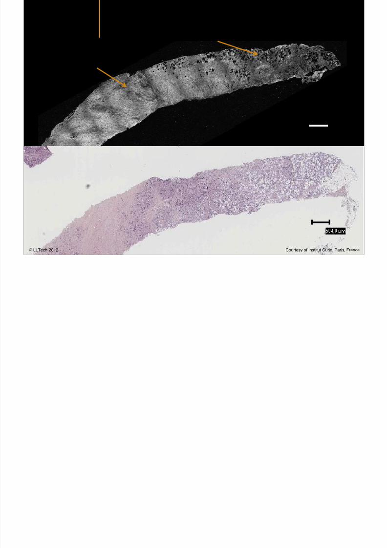
7292019 Atlas of Light-CT images
httpslidepdfcomreaderfullatlas-of-light-ct-images 80118
copy LLTech 2012 ndash wwwlltechimagingcom - No copy without authorization 80
500microm
stroma
adipocytes
copy LLTech 2012 Courtesy of Institut Curie Paris France
7292019 Atlas of Light-CT images
httpslidepdfcomreaderfullatlas-of-light-ct-images 81118
Breast Ductal carcinoma in situ
7292019 Atlas of Light-CT images
httpslidepdfcomreaderfullatlas-of-light-ct-images 82118
copy LLTech 2012 ndash wwwlltechimagingcom - No copy without authorization 82
500microm
copy LLTech 2012 Courtesy of Institut Curie Paris France
7292019 Atlas of Light-CT images
httpslidepdfcomreaderfullatlas-of-light-ct-images 83118
copy LLTech 2012 ndash wwwlltechimagingcom - No copy without authorization 83 copy LLTECH 2011
THYROID IMAGING
Back to table of contents
7292019 Atlas of Light-CT images
httpslidepdfcomreaderfullatlas-of-light-ct-images 84118
copy LLTech 2012 ndash wwwlltechimagingcom - No copy without authorization 84
Thyroid
800 microm
200 microm
copy LLTech 2012 Courtesy of Hocircpital Tenon Paris France
7292019 Atlas of Light-CT images
httpslidepdfcomreaderfullatlas-of-light-ct-images 85118
copy LLTech 2012 ndash wwwlltechimagingcom - No copy without authorization 85
Thyroid
800 microm
200 microm
copy LLTech 2012 Courtesy of Hocircpital Tenon Paris France
7292019 Atlas of Light-CT images
httpslidepdfcomreaderfullatlas-of-light-ct-images 86118
copy LLTech 2012 ndash wwwlltechimagingcom - No copy without authorization 86 copy LLTECH 2011
FAT CELLS IMAGING
Back to table of contents
7292019 Atlas of Light-CT images
httpslidepdfcomreaderfullatlas-of-light-ct-images 87118
copy LLTech 2012 ndash wwwlltechimagingcom - No copy without authorization 87
Brown fat cells - adipocytes
100 microm 100 microm
200 microm
10um depth
Adipocytes
45um depth
En face
slices
Reconstructed
depth slice
copy LLTech 2012 Courtesy of Centre de Recherche des Cordeliers Paris France
7292019 Atlas of Light-CT images
httpslidepdfcomreaderfullatlas-of-light-ct-images 88118
copy LLTech 2012 ndash wwwlltechimagingcom - No copy without authorization 88 copy LLTECH 2011
CORNEA AND RETINA
Back to table of contents
Human cornea reconstructed depth slice
7292019 Atlas of Light-CT images
httpslidepdfcomreaderfullatlas-of-light-ct-images 89118
copy LLTech 2012 ndash wwwlltechimagingcom - No copy without authorization 89
50 um
wwwlltechimagingcom
7292019 Atlas of Light-CT images
httpslidepdfcomreaderfullatlas-of-light-ct-images 90118
Large field en face view in stroma
7292019 Atlas of Light-CT images
httpslidepdfcomreaderfullatlas-of-light-ct-images 91118
copy LLTech 2012 ndash wwwlltechimagingcom - No copy without authorization 91
200 um
wwwlltechimagingcom
7292019 Atlas of Light-CT images
httpslidepdfcomreaderfullatlas-of-light-ct-images 92118
Rabbit cornea after LASIK reflectivity profiles reveal
i d tt i d thi k d ith li l l
7292019 Atlas of Light-CT images
httpslidepdfcomreaderfullatlas-of-light-ct-images 93118
copy LLTech 2012 ndash wwwlltechimagingcom - No copy without authorization 93
increased scattering and thickened epithelial layer
LASIK LASIK CONTROL CONTROL
copy LLTech 2012
Newborn mouse anterior segment
7292019 Atlas of Light-CT images
httpslidepdfcomreaderfullatlas-of-light-ct-images 94118
copy LLTech 2012 ndash wwwlltechimagingcom - No copy without authorization 94
Corneal epithelial cells
Basal membrane
Stroma
Descemetrsquos membrane
Aqueous humor
Lens capsule
Lens epithelial cells
Cortical fibers
copy LLTech 2012 Courtesy of ESPCI Paris Francecopy LLTech 2012
Rat retina comparison with histology
7292019 Atlas of Light-CT images
httpslidepdfcomreaderfullatlas-of-light-ct-images 95118
copy LLTech 2012 ndash wwwlltechimagingcom - No copy without authorization 95
Figure 1 Right Ex vivo rat retinaimaged using the f ull field OCT
system of ESPCI Left Histology
for comparison
Figure 1 Right Ex vivo rat retinaimaged using the f ull field OCT
system of ESPCI Left Histology
for comparison
Figure 1 Right Ex vivo rat retinaimaged using the f ull field OCT
system of ESPCI Left Histology
for comparisonField
230x300microm
RPE
PRL
ONL
OPL
INL
IPL
GCL
NFL
Invest Ophthalmol Vis Sci 2004 Nov45(11)4126-31
Grieve K Paques M Dubois A Sahel J Boccara C Le Gargasson JF copy LLTech 2012
Rat retina
7292019 Atlas of Light-CT images
httpslidepdfcomreaderfullatlas-of-light-ct-images 96118
copy LLTech 2012 ndash wwwlltechimagingcom - No copy without authorization 96 copy LLTech 2012 Courtesy of ESPCI Paris France
Nerve fiber layer
Retina
Invest Ophthalmol Vis Sci 2004 Nov45(11)4126-31
Grieve K Paques M Dubois A Sahel J Boccara C Le Gargasson JF
Transscleral image
7292019 Atlas of Light-CT images
httpslidepdfcomreaderfullatlas-of-light-ct-images 97118
7292019 Atlas of Light-CT images
httpslidepdfcomreaderfullatlas-of-light-ct-images 98118
7292019 Atlas of Light-CT images
httpslidepdfcomreaderfullatlas-of-light-ct-images 99118
In vivo imaging of drosophila melanogaster
7292019 Atlas of Light-CT images
httpslidepdfcomreaderfullatlas-of-light-ct-images 100118
copy LLTech 2012 ndash wwwlltechimagingcom - No copy without authorization 100 copy LLTech 2012 Courtesy of ESPCI Paris France
In vivo imaging of drosophila melanogaster ndash four stages of pupation over 72 hours
7292019 Atlas of Light-CT images
httpslidepdfcomreaderfullatlas-of-light-ct-images 101118
copy LLTech 2012 ndash wwwlltechimagingcom - No copy without authorization 101
Prepupa (0-2 h) Transition topupal stage (24 h)
Pupal stage (48 h) Advanced pupal phase
before eclosion (72 h)
copy LLTech 2012 Courtesy of ESPCI Paris France
Pupa 118 4 days 30um depth
100
7292019 Atlas of Light-CT images
httpslidepdfcomreaderfullatlas-of-light-ct-images 102118
copy LLTech 2012 ndash wwwlltechimagingcom - No copy without authorization 102
100 um
Drosophila
7292019 Atlas of Light-CT images
httpslidepdfcomreaderfullatlas-of-light-ct-images 103118
copy LLTech 2012 ndash wwwlltechimagingcom - No copy without authorization 103
200 microm
copy LLTech 2012 Courtesy of ESPCI Paris France
C Elegans ndash Fast identification of anatomy
7292019 Atlas of Light-CT images
httpslidepdfcomreaderfullatlas-of-light-ct-images 104118
copy LLTech 2012 ndash wwwlltechimagingcom - No copy without authorization 104
Body wall - cuticle
gonad arm
30 microm depth
uterus
ovocytes
intestine
100 microm 100 microm
15 microm depth
copy LLTech 2012 Courtesy of ENS Paris France
C elegans ndash Images at higher magnification 40X
7292019 Atlas of Light-CT images
httpslidepdfcomreaderfullatlas-of-light-ct-images 105118
copy LLTech 2012 ndash wwwlltechimagingcom - No copy without authorization 105
3D reconstruction 50 microm
100 microm
copy LLTech 2012 Courtesy of ENS Paris France
Zebra fish eye
7292019 Atlas of Light-CT images
httpslidepdfcomreaderfullatlas-of-light-ct-images 106118
copy LLTech 2012 ndash wwwlltechimagingcom - No copy without authorization 106
Zebra fish eye
100 microm
100 microm
En face slices
Reconstructed depth slice
copy LLTech 2012 Courtesy of Inserm Paris France
10um depth 20um depth 45um depth
7292019 Atlas of Light-CT images
httpslidepdfcomreaderfullatlas-of-light-ct-images 107118
Xenopus Laevis backtail
7292019 Atlas of Light-CT images
httpslidepdfcomreaderfullatlas-of-light-ct-images 108118
copy LLTech 2012 ndash wwwlltechimagingcom - No copy without authorization 108
100 um30um depth 60um depth
7292019 Atlas of Light-CT images
httpslidepdfcomreaderfullatlas-of-light-ct-images 109118
Mouse embryo in vitro 6 5 days old ndash
7292019 Atlas of Light-CT images
httpslidepdfcomreaderfullatlas-of-light-ct-images 110118
copy LLTech 2012 ndash wwwlltechimagingcom - No copy without authorization 110
Mouse embryo in vitro 65 days old
copy LLTech 2012 Courtesy of Ellvire Guiot and nstitut Jacques Monod Paris France
3D imaging of internal cavity
Sabellaria alveolata mouth palps
7292019 Atlas of Light-CT images
httpslidepdfcomreaderfullatlas-of-light-ct-images 111118
copy LLTech 2012 ndash wwwlltechimagingcom - No copy without authorization 111
500 microm
100 microm
Mouth palps
copy LLTech 2012 Courtesy of Museacutee National drsquoHistoire Naturelle Paris France
7292019 Atlas of Light-CT images
httpslidepdfcomreaderfullatlas-of-light-ct-images 112118
copy LLTech 2012 ndash wwwlltechimagingcom - No copy without authorization 112 copy LLTECH 2011
PLANT IMAGING
Back to table of contents
Mouse ear leaf epidermis
7292019 Atlas of Light-CT images
httpslidepdfcomreaderfullatlas-of-light-ct-images 113118
copy LLTech 2012 ndash wwwlltechimagingcom - No copy without authorization 113
100 microm
Epidermal cell
copy LLTech 2012
Stoma
Leaf veins
7292019 Atlas of Light-CT images
httpslidepdfcomreaderfullatlas-of-light-ct-images 114118
copy LLTech 2012 ndash wwwlltechimagingcom - No copy without authorization 114
114copy LLTech 2012
Fibers
Veins
100 microm
500 microm
Apple
7292019 Atlas of Light-CT images
httpslidepdfcomreaderfullatlas-of-light-ct-images 115118
copy LLTech 2012 ndash wwwlltechimagingcom - No copy without authorization 115
115copy LLTech 2012
100 microm
Wax
Skin
Flesh
500 microm
Water
Effect of harpin infiltration on tobacco leaf
7292019 Atlas of Light-CT images
httpslidepdfcomreaderfullatlas-of-light-ct-images 116118
copy LLTech 2012 ndash wwwlltechimagingcom - No copy without authorization 116
116copy LLTech 2012 Courtesy of UPMC Paris France
50 microm
50 microm
50 microm
50 microm
Upper epidermis
10 min after harpin infiltration 30 min after harpin infiltration
Palisade parenchyma
Epidermis cell
Chloroplast
7292019 Atlas of Light-CT images
httpslidepdfcomreaderfullatlas-of-light-ct-images 117118
copy LLTech 2012 ndash wwwlltechimagingcom - No copy without authorization 117
bull Light-CTtrade does not require tissue preparation nor staining of any kind
bull Creates images within minutes using a non-destructive process
bull Offers a 1 microm cellular resolution in 3D
bull Reveals structural and cellular information
bull Mosaicing allows fast visualisation at various scale
bull En-face high-resolution imaging allows vertical and 3D reconstruction
bull In-vivo images show good promise for in-vivo system
bull Clinical and research applications in cancer detection cosmetics biobanking small animal
developmental biology botanyhellip
Contact Details
7292019 Atlas of Light-CT images
httpslidepdfcomreaderfullatlas-of-light-ct-images 118118
USA
LLTech Inc
103 Carnegie Center DriveSuite 300
Princeton NJ 08540
USA
wwwlltechimagingcom
contactlltechfr
Phone + 1 609 955 3506
LLTech SAS
29 rue du Faubourg St-JacquesPeacutepiniegravere Paris Santeacute Cochin
75014
Paris - France
wwwlltechimagingcom
contactlltechfr
Phone +33 1 82 72 61 25
Europe
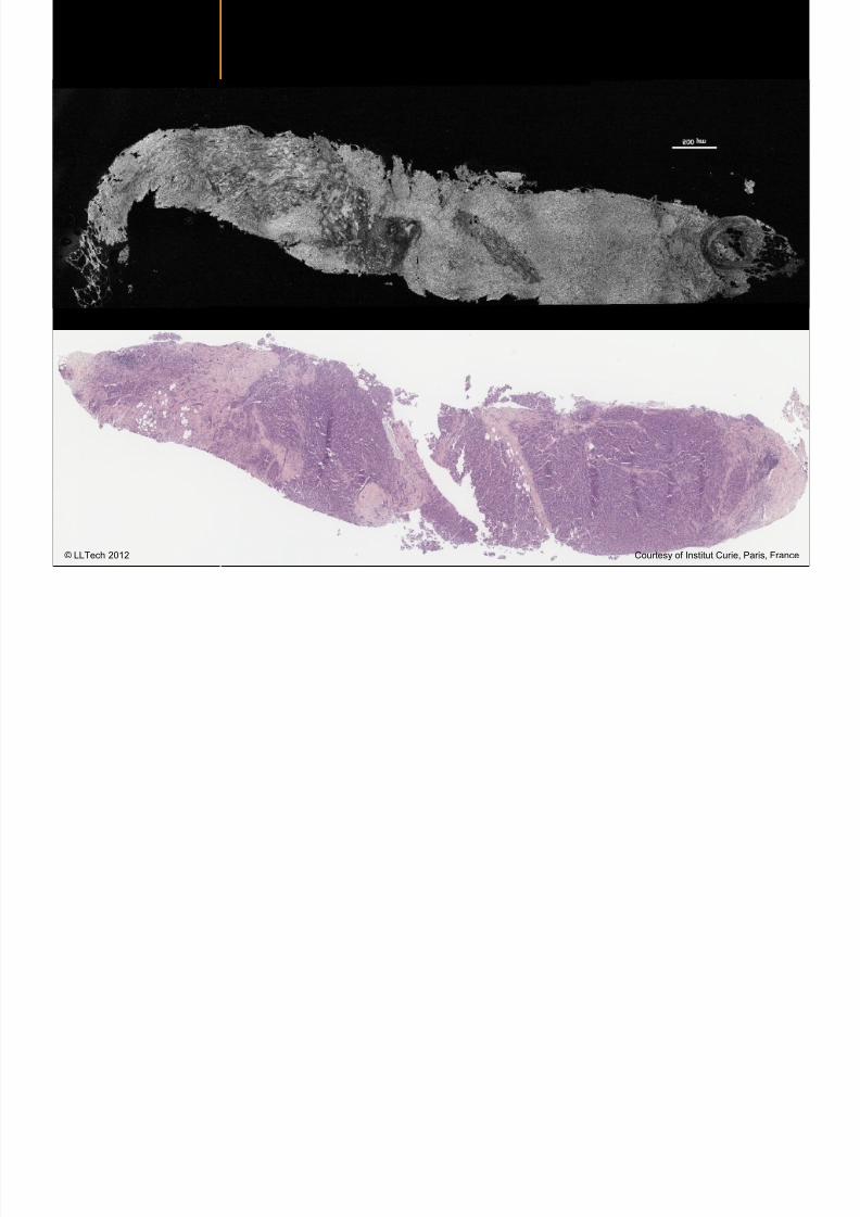
7292019 Atlas of Light-CT images
httpslidepdfcomreaderfullatlas-of-light-ct-images 89118
copy LLTech 2012 ndash wwwlltechimagingcom - No copy without authorization 89
50 um
wwwlltechimagingcom
7292019 Atlas of Light-CT images
httpslidepdfcomreaderfullatlas-of-light-ct-images 90118
Large field en face view in stroma
7292019 Atlas of Light-CT images
httpslidepdfcomreaderfullatlas-of-light-ct-images 91118
copy LLTech 2012 ndash wwwlltechimagingcom - No copy without authorization 91
200 um
wwwlltechimagingcom
7292019 Atlas of Light-CT images
httpslidepdfcomreaderfullatlas-of-light-ct-images 92118
Rabbit cornea after LASIK reflectivity profiles reveal
i d tt i d thi k d ith li l l
7292019 Atlas of Light-CT images
httpslidepdfcomreaderfullatlas-of-light-ct-images 93118
copy LLTech 2012 ndash wwwlltechimagingcom - No copy without authorization 93
increased scattering and thickened epithelial layer
LASIK LASIK CONTROL CONTROL
copy LLTech 2012
Newborn mouse anterior segment
7292019 Atlas of Light-CT images
httpslidepdfcomreaderfullatlas-of-light-ct-images 94118
copy LLTech 2012 ndash wwwlltechimagingcom - No copy without authorization 94
Corneal epithelial cells
Basal membrane
Stroma
Descemetrsquos membrane
Aqueous humor
Lens capsule
Lens epithelial cells
Cortical fibers
copy LLTech 2012 Courtesy of ESPCI Paris Francecopy LLTech 2012
Rat retina comparison with histology
7292019 Atlas of Light-CT images
httpslidepdfcomreaderfullatlas-of-light-ct-images 95118
copy LLTech 2012 ndash wwwlltechimagingcom - No copy without authorization 95
Figure 1 Right Ex vivo rat retinaimaged using the f ull field OCT
system of ESPCI Left Histology
for comparison
Figure 1 Right Ex vivo rat retinaimaged using the f ull field OCT
system of ESPCI Left Histology
for comparison
Figure 1 Right Ex vivo rat retinaimaged using the f ull field OCT
system of ESPCI Left Histology
for comparisonField
230x300microm
RPE
PRL
ONL
OPL
INL
IPL
GCL
NFL
Invest Ophthalmol Vis Sci 2004 Nov45(11)4126-31
Grieve K Paques M Dubois A Sahel J Boccara C Le Gargasson JF copy LLTech 2012
Rat retina
7292019 Atlas of Light-CT images
httpslidepdfcomreaderfullatlas-of-light-ct-images 96118
copy LLTech 2012 ndash wwwlltechimagingcom - No copy without authorization 96 copy LLTech 2012 Courtesy of ESPCI Paris France
Nerve fiber layer
Retina
Invest Ophthalmol Vis Sci 2004 Nov45(11)4126-31
Grieve K Paques M Dubois A Sahel J Boccara C Le Gargasson JF
Transscleral image
7292019 Atlas of Light-CT images
httpslidepdfcomreaderfullatlas-of-light-ct-images 97118
7292019 Atlas of Light-CT images
httpslidepdfcomreaderfullatlas-of-light-ct-images 98118
7292019 Atlas of Light-CT images
httpslidepdfcomreaderfullatlas-of-light-ct-images 99118
In vivo imaging of drosophila melanogaster
7292019 Atlas of Light-CT images
httpslidepdfcomreaderfullatlas-of-light-ct-images 100118
copy LLTech 2012 ndash wwwlltechimagingcom - No copy without authorization 100 copy LLTech 2012 Courtesy of ESPCI Paris France
In vivo imaging of drosophila melanogaster ndash four stages of pupation over 72 hours
7292019 Atlas of Light-CT images
httpslidepdfcomreaderfullatlas-of-light-ct-images 101118
copy LLTech 2012 ndash wwwlltechimagingcom - No copy without authorization 101
Prepupa (0-2 h) Transition topupal stage (24 h)
Pupal stage (48 h) Advanced pupal phase
before eclosion (72 h)
copy LLTech 2012 Courtesy of ESPCI Paris France
Pupa 118 4 days 30um depth
100
7292019 Atlas of Light-CT images
httpslidepdfcomreaderfullatlas-of-light-ct-images 102118
copy LLTech 2012 ndash wwwlltechimagingcom - No copy without authorization 102
100 um
Drosophila
7292019 Atlas of Light-CT images
httpslidepdfcomreaderfullatlas-of-light-ct-images 103118
copy LLTech 2012 ndash wwwlltechimagingcom - No copy without authorization 103
200 microm
copy LLTech 2012 Courtesy of ESPCI Paris France
C Elegans ndash Fast identification of anatomy
7292019 Atlas of Light-CT images
httpslidepdfcomreaderfullatlas-of-light-ct-images 104118
copy LLTech 2012 ndash wwwlltechimagingcom - No copy without authorization 104
Body wall - cuticle
gonad arm
30 microm depth
uterus
ovocytes
intestine
100 microm 100 microm
15 microm depth
copy LLTech 2012 Courtesy of ENS Paris France
C elegans ndash Images at higher magnification 40X
7292019 Atlas of Light-CT images
httpslidepdfcomreaderfullatlas-of-light-ct-images 105118
copy LLTech 2012 ndash wwwlltechimagingcom - No copy without authorization 105
3D reconstruction 50 microm
100 microm
copy LLTech 2012 Courtesy of ENS Paris France
Zebra fish eye
7292019 Atlas of Light-CT images
httpslidepdfcomreaderfullatlas-of-light-ct-images 106118
copy LLTech 2012 ndash wwwlltechimagingcom - No copy without authorization 106
Zebra fish eye
100 microm
100 microm
En face slices
Reconstructed depth slice
copy LLTech 2012 Courtesy of Inserm Paris France
10um depth 20um depth 45um depth
7292019 Atlas of Light-CT images
httpslidepdfcomreaderfullatlas-of-light-ct-images 107118
Xenopus Laevis backtail
7292019 Atlas of Light-CT images
httpslidepdfcomreaderfullatlas-of-light-ct-images 108118
copy LLTech 2012 ndash wwwlltechimagingcom - No copy without authorization 108
100 um30um depth 60um depth
7292019 Atlas of Light-CT images
httpslidepdfcomreaderfullatlas-of-light-ct-images 109118
Mouse embryo in vitro 6 5 days old ndash
7292019 Atlas of Light-CT images
httpslidepdfcomreaderfullatlas-of-light-ct-images 110118
copy LLTech 2012 ndash wwwlltechimagingcom - No copy without authorization 110
Mouse embryo in vitro 65 days old
copy LLTech 2012 Courtesy of Ellvire Guiot and nstitut Jacques Monod Paris France
3D imaging of internal cavity
Sabellaria alveolata mouth palps
7292019 Atlas of Light-CT images
httpslidepdfcomreaderfullatlas-of-light-ct-images 111118
copy LLTech 2012 ndash wwwlltechimagingcom - No copy without authorization 111
500 microm
100 microm
Mouth palps
copy LLTech 2012 Courtesy of Museacutee National drsquoHistoire Naturelle Paris France
7292019 Atlas of Light-CT images
httpslidepdfcomreaderfullatlas-of-light-ct-images 112118
copy LLTech 2012 ndash wwwlltechimagingcom - No copy without authorization 112 copy LLTECH 2011
PLANT IMAGING
Back to table of contents
Mouse ear leaf epidermis
7292019 Atlas of Light-CT images
httpslidepdfcomreaderfullatlas-of-light-ct-images 113118
copy LLTech 2012 ndash wwwlltechimagingcom - No copy without authorization 113
100 microm
Epidermal cell
copy LLTech 2012
Stoma
Leaf veins
7292019 Atlas of Light-CT images
httpslidepdfcomreaderfullatlas-of-light-ct-images 114118
copy LLTech 2012 ndash wwwlltechimagingcom - No copy without authorization 114
114copy LLTech 2012
Fibers
Veins
100 microm
500 microm
Apple
7292019 Atlas of Light-CT images
httpslidepdfcomreaderfullatlas-of-light-ct-images 115118
copy LLTech 2012 ndash wwwlltechimagingcom - No copy without authorization 115
115copy LLTech 2012
100 microm
Wax
Skin
Flesh
500 microm
Water
Effect of harpin infiltration on tobacco leaf
7292019 Atlas of Light-CT images
httpslidepdfcomreaderfullatlas-of-light-ct-images 116118
copy LLTech 2012 ndash wwwlltechimagingcom - No copy without authorization 116
116copy LLTech 2012 Courtesy of UPMC Paris France
50 microm
50 microm
50 microm
50 microm
Upper epidermis
10 min after harpin infiltration 30 min after harpin infiltration
Palisade parenchyma
Epidermis cell
Chloroplast
7292019 Atlas of Light-CT images
httpslidepdfcomreaderfullatlas-of-light-ct-images 117118
copy LLTech 2012 ndash wwwlltechimagingcom - No copy without authorization 117
bull Light-CTtrade does not require tissue preparation nor staining of any kind
bull Creates images within minutes using a non-destructive process
bull Offers a 1 microm cellular resolution in 3D
bull Reveals structural and cellular information
bull Mosaicing allows fast visualisation at various scale
bull En-face high-resolution imaging allows vertical and 3D reconstruction
bull In-vivo images show good promise for in-vivo system
bull Clinical and research applications in cancer detection cosmetics biobanking small animal
developmental biology botanyhellip
Contact Details
7292019 Atlas of Light-CT images
httpslidepdfcomreaderfullatlas-of-light-ct-images 118118
USA
LLTech Inc
103 Carnegie Center DriveSuite 300
Princeton NJ 08540
USA
wwwlltechimagingcom
contactlltechfr
Phone + 1 609 955 3506
LLTech SAS
29 rue du Faubourg St-JacquesPeacutepiniegravere Paris Santeacute Cochin
75014
Paris - France
wwwlltechimagingcom
contactlltechfr
Phone +33 1 82 72 61 25
Europe
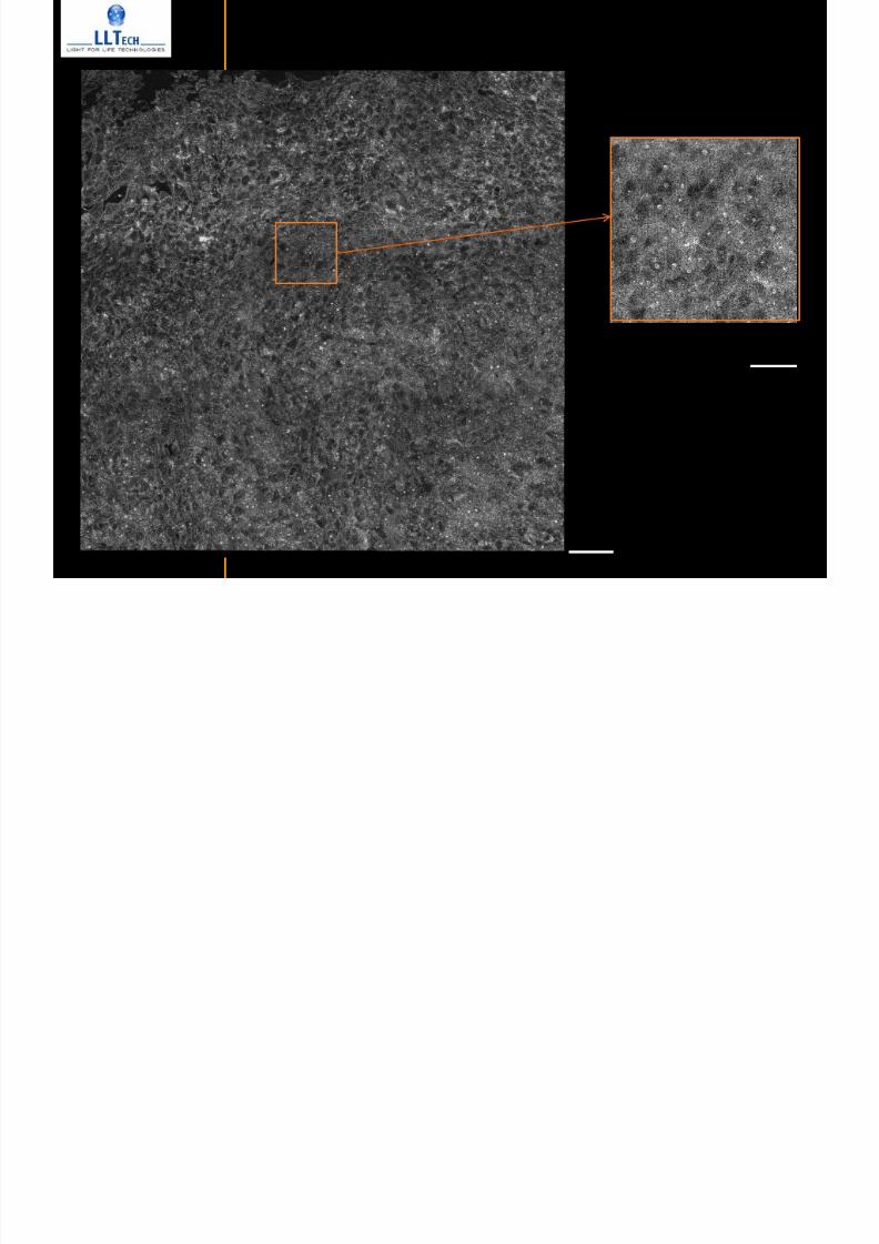
7292019 Atlas of Light-CT images
httpslidepdfcomreaderfullatlas-of-light-ct-images 91118
copy LLTech 2012 ndash wwwlltechimagingcom - No copy without authorization 91
200 um
wwwlltechimagingcom
7292019 Atlas of Light-CT images
httpslidepdfcomreaderfullatlas-of-light-ct-images 92118
Rabbit cornea after LASIK reflectivity profiles reveal
i d tt i d thi k d ith li l l
7292019 Atlas of Light-CT images
httpslidepdfcomreaderfullatlas-of-light-ct-images 93118
copy LLTech 2012 ndash wwwlltechimagingcom - No copy without authorization 93
increased scattering and thickened epithelial layer
LASIK LASIK CONTROL CONTROL
copy LLTech 2012
Newborn mouse anterior segment
7292019 Atlas of Light-CT images
httpslidepdfcomreaderfullatlas-of-light-ct-images 94118
copy LLTech 2012 ndash wwwlltechimagingcom - No copy without authorization 94
Corneal epithelial cells
Basal membrane
Stroma
Descemetrsquos membrane
Aqueous humor
Lens capsule
Lens epithelial cells
Cortical fibers
copy LLTech 2012 Courtesy of ESPCI Paris Francecopy LLTech 2012
Rat retina comparison with histology
7292019 Atlas of Light-CT images
httpslidepdfcomreaderfullatlas-of-light-ct-images 95118
copy LLTech 2012 ndash wwwlltechimagingcom - No copy without authorization 95
Figure 1 Right Ex vivo rat retinaimaged using the f ull field OCT
system of ESPCI Left Histology
for comparison
Figure 1 Right Ex vivo rat retinaimaged using the f ull field OCT
system of ESPCI Left Histology
for comparison
Figure 1 Right Ex vivo rat retinaimaged using the f ull field OCT
system of ESPCI Left Histology
for comparisonField
230x300microm
RPE
PRL
ONL
OPL
INL
IPL
GCL
NFL
Invest Ophthalmol Vis Sci 2004 Nov45(11)4126-31
Grieve K Paques M Dubois A Sahel J Boccara C Le Gargasson JF copy LLTech 2012
Rat retina
7292019 Atlas of Light-CT images
httpslidepdfcomreaderfullatlas-of-light-ct-images 96118
copy LLTech 2012 ndash wwwlltechimagingcom - No copy without authorization 96 copy LLTech 2012 Courtesy of ESPCI Paris France
Nerve fiber layer
Retina
Invest Ophthalmol Vis Sci 2004 Nov45(11)4126-31
Grieve K Paques M Dubois A Sahel J Boccara C Le Gargasson JF
Transscleral image
7292019 Atlas of Light-CT images
httpslidepdfcomreaderfullatlas-of-light-ct-images 97118
7292019 Atlas of Light-CT images
httpslidepdfcomreaderfullatlas-of-light-ct-images 98118
7292019 Atlas of Light-CT images
httpslidepdfcomreaderfullatlas-of-light-ct-images 99118
In vivo imaging of drosophila melanogaster
7292019 Atlas of Light-CT images
httpslidepdfcomreaderfullatlas-of-light-ct-images 100118
copy LLTech 2012 ndash wwwlltechimagingcom - No copy without authorization 100 copy LLTech 2012 Courtesy of ESPCI Paris France
In vivo imaging of drosophila melanogaster ndash four stages of pupation over 72 hours
7292019 Atlas of Light-CT images
httpslidepdfcomreaderfullatlas-of-light-ct-images 101118
copy LLTech 2012 ndash wwwlltechimagingcom - No copy without authorization 101
Prepupa (0-2 h) Transition topupal stage (24 h)
Pupal stage (48 h) Advanced pupal phase
before eclosion (72 h)
copy LLTech 2012 Courtesy of ESPCI Paris France
Pupa 118 4 days 30um depth
100
7292019 Atlas of Light-CT images
httpslidepdfcomreaderfullatlas-of-light-ct-images 102118
copy LLTech 2012 ndash wwwlltechimagingcom - No copy without authorization 102
100 um
Drosophila
7292019 Atlas of Light-CT images
httpslidepdfcomreaderfullatlas-of-light-ct-images 103118
copy LLTech 2012 ndash wwwlltechimagingcom - No copy without authorization 103
200 microm
copy LLTech 2012 Courtesy of ESPCI Paris France
C Elegans ndash Fast identification of anatomy
7292019 Atlas of Light-CT images
httpslidepdfcomreaderfullatlas-of-light-ct-images 104118
copy LLTech 2012 ndash wwwlltechimagingcom - No copy without authorization 104
Body wall - cuticle
gonad arm
30 microm depth
uterus
ovocytes
intestine
100 microm 100 microm
15 microm depth
copy LLTech 2012 Courtesy of ENS Paris France
C elegans ndash Images at higher magnification 40X
7292019 Atlas of Light-CT images
httpslidepdfcomreaderfullatlas-of-light-ct-images 105118
copy LLTech 2012 ndash wwwlltechimagingcom - No copy without authorization 105
3D reconstruction 50 microm
100 microm
copy LLTech 2012 Courtesy of ENS Paris France
Zebra fish eye
7292019 Atlas of Light-CT images
httpslidepdfcomreaderfullatlas-of-light-ct-images 106118
copy LLTech 2012 ndash wwwlltechimagingcom - No copy without authorization 106
Zebra fish eye
100 microm
100 microm
En face slices
Reconstructed depth slice
copy LLTech 2012 Courtesy of Inserm Paris France
10um depth 20um depth 45um depth
7292019 Atlas of Light-CT images
httpslidepdfcomreaderfullatlas-of-light-ct-images 107118
Xenopus Laevis backtail
7292019 Atlas of Light-CT images
httpslidepdfcomreaderfullatlas-of-light-ct-images 108118
copy LLTech 2012 ndash wwwlltechimagingcom - No copy without authorization 108
100 um30um depth 60um depth
7292019 Atlas of Light-CT images
httpslidepdfcomreaderfullatlas-of-light-ct-images 109118
Mouse embryo in vitro 6 5 days old ndash
7292019 Atlas of Light-CT images
httpslidepdfcomreaderfullatlas-of-light-ct-images 110118
copy LLTech 2012 ndash wwwlltechimagingcom - No copy without authorization 110
Mouse embryo in vitro 65 days old
copy LLTech 2012 Courtesy of Ellvire Guiot and nstitut Jacques Monod Paris France
3D imaging of internal cavity
Sabellaria alveolata mouth palps
7292019 Atlas of Light-CT images
httpslidepdfcomreaderfullatlas-of-light-ct-images 111118
copy LLTech 2012 ndash wwwlltechimagingcom - No copy without authorization 111
500 microm
100 microm
Mouth palps
copy LLTech 2012 Courtesy of Museacutee National drsquoHistoire Naturelle Paris France
7292019 Atlas of Light-CT images
httpslidepdfcomreaderfullatlas-of-light-ct-images 112118
copy LLTech 2012 ndash wwwlltechimagingcom - No copy without authorization 112 copy LLTECH 2011
PLANT IMAGING
Back to table of contents
Mouse ear leaf epidermis
7292019 Atlas of Light-CT images
httpslidepdfcomreaderfullatlas-of-light-ct-images 113118
copy LLTech 2012 ndash wwwlltechimagingcom - No copy without authorization 113
100 microm
Epidermal cell
copy LLTech 2012
Stoma
Leaf veins
7292019 Atlas of Light-CT images
httpslidepdfcomreaderfullatlas-of-light-ct-images 114118
copy LLTech 2012 ndash wwwlltechimagingcom - No copy without authorization 114
114copy LLTech 2012
Fibers
Veins
100 microm
500 microm
Apple
7292019 Atlas of Light-CT images
httpslidepdfcomreaderfullatlas-of-light-ct-images 115118
copy LLTech 2012 ndash wwwlltechimagingcom - No copy without authorization 115
115copy LLTech 2012
100 microm
Wax
Skin
Flesh
500 microm
Water
Effect of harpin infiltration on tobacco leaf
7292019 Atlas of Light-CT images
httpslidepdfcomreaderfullatlas-of-light-ct-images 116118
copy LLTech 2012 ndash wwwlltechimagingcom - No copy without authorization 116
116copy LLTech 2012 Courtesy of UPMC Paris France
50 microm
50 microm
50 microm
50 microm
Upper epidermis
10 min after harpin infiltration 30 min after harpin infiltration
Palisade parenchyma
Epidermis cell
Chloroplast
7292019 Atlas of Light-CT images
httpslidepdfcomreaderfullatlas-of-light-ct-images 117118
copy LLTech 2012 ndash wwwlltechimagingcom - No copy without authorization 117
bull Light-CTtrade does not require tissue preparation nor staining of any kind
bull Creates images within minutes using a non-destructive process
bull Offers a 1 microm cellular resolution in 3D
bull Reveals structural and cellular information
bull Mosaicing allows fast visualisation at various scale
bull En-face high-resolution imaging allows vertical and 3D reconstruction
bull In-vivo images show good promise for in-vivo system
bull Clinical and research applications in cancer detection cosmetics biobanking small animal
developmental biology botanyhellip
Contact Details
7292019 Atlas of Light-CT images
httpslidepdfcomreaderfullatlas-of-light-ct-images 118118
USA
LLTech Inc
103 Carnegie Center DriveSuite 300
Princeton NJ 08540
USA
wwwlltechimagingcom
contactlltechfr
Phone + 1 609 955 3506
LLTech SAS
29 rue du Faubourg St-JacquesPeacutepiniegravere Paris Santeacute Cochin
75014
Paris - France
wwwlltechimagingcom
contactlltechfr
Phone +33 1 82 72 61 25
Europe
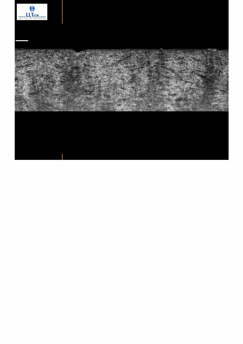
7292019 Atlas of Light-CT images
httpslidepdfcomreaderfullatlas-of-light-ct-images 93118
copy LLTech 2012 ndash wwwlltechimagingcom - No copy without authorization 93
increased scattering and thickened epithelial layer
LASIK LASIK CONTROL CONTROL
copy LLTech 2012
Newborn mouse anterior segment
7292019 Atlas of Light-CT images
httpslidepdfcomreaderfullatlas-of-light-ct-images 94118
copy LLTech 2012 ndash wwwlltechimagingcom - No copy without authorization 94
Corneal epithelial cells
Basal membrane
Stroma
Descemetrsquos membrane
Aqueous humor
Lens capsule
Lens epithelial cells
Cortical fibers
copy LLTech 2012 Courtesy of ESPCI Paris Francecopy LLTech 2012
Rat retina comparison with histology
7292019 Atlas of Light-CT images
httpslidepdfcomreaderfullatlas-of-light-ct-images 95118
copy LLTech 2012 ndash wwwlltechimagingcom - No copy without authorization 95
Figure 1 Right Ex vivo rat retinaimaged using the f ull field OCT
system of ESPCI Left Histology
for comparison
Figure 1 Right Ex vivo rat retinaimaged using the f ull field OCT
system of ESPCI Left Histology
for comparison
Figure 1 Right Ex vivo rat retinaimaged using the f ull field OCT
system of ESPCI Left Histology
for comparisonField
230x300microm
RPE
PRL
ONL
OPL
INL
IPL
GCL
NFL
Invest Ophthalmol Vis Sci 2004 Nov45(11)4126-31
Grieve K Paques M Dubois A Sahel J Boccara C Le Gargasson JF copy LLTech 2012
Rat retina
7292019 Atlas of Light-CT images
httpslidepdfcomreaderfullatlas-of-light-ct-images 96118
copy LLTech 2012 ndash wwwlltechimagingcom - No copy without authorization 96 copy LLTech 2012 Courtesy of ESPCI Paris France
Nerve fiber layer
Retina
Invest Ophthalmol Vis Sci 2004 Nov45(11)4126-31
Grieve K Paques M Dubois A Sahel J Boccara C Le Gargasson JF
Transscleral image
7292019 Atlas of Light-CT images
httpslidepdfcomreaderfullatlas-of-light-ct-images 97118
7292019 Atlas of Light-CT images
httpslidepdfcomreaderfullatlas-of-light-ct-images 98118
7292019 Atlas of Light-CT images
httpslidepdfcomreaderfullatlas-of-light-ct-images 99118
In vivo imaging of drosophila melanogaster
7292019 Atlas of Light-CT images
httpslidepdfcomreaderfullatlas-of-light-ct-images 100118
copy LLTech 2012 ndash wwwlltechimagingcom - No copy without authorization 100 copy LLTech 2012 Courtesy of ESPCI Paris France
In vivo imaging of drosophila melanogaster ndash four stages of pupation over 72 hours
7292019 Atlas of Light-CT images
httpslidepdfcomreaderfullatlas-of-light-ct-images 101118
copy LLTech 2012 ndash wwwlltechimagingcom - No copy without authorization 101
Prepupa (0-2 h) Transition topupal stage (24 h)
Pupal stage (48 h) Advanced pupal phase
before eclosion (72 h)
copy LLTech 2012 Courtesy of ESPCI Paris France
Pupa 118 4 days 30um depth
100
7292019 Atlas of Light-CT images
httpslidepdfcomreaderfullatlas-of-light-ct-images 102118
copy LLTech 2012 ndash wwwlltechimagingcom - No copy without authorization 102
100 um
Drosophila
7292019 Atlas of Light-CT images
httpslidepdfcomreaderfullatlas-of-light-ct-images 103118
copy LLTech 2012 ndash wwwlltechimagingcom - No copy without authorization 103
200 microm
copy LLTech 2012 Courtesy of ESPCI Paris France
C Elegans ndash Fast identification of anatomy
7292019 Atlas of Light-CT images
httpslidepdfcomreaderfullatlas-of-light-ct-images 104118
copy LLTech 2012 ndash wwwlltechimagingcom - No copy without authorization 104
Body wall - cuticle
gonad arm
30 microm depth
uterus
ovocytes
intestine
100 microm 100 microm
15 microm depth
copy LLTech 2012 Courtesy of ENS Paris France
C elegans ndash Images at higher magnification 40X
7292019 Atlas of Light-CT images
httpslidepdfcomreaderfullatlas-of-light-ct-images 105118
copy LLTech 2012 ndash wwwlltechimagingcom - No copy without authorization 105
3D reconstruction 50 microm
100 microm
copy LLTech 2012 Courtesy of ENS Paris France
Zebra fish eye
7292019 Atlas of Light-CT images
httpslidepdfcomreaderfullatlas-of-light-ct-images 106118
copy LLTech 2012 ndash wwwlltechimagingcom - No copy without authorization 106
Zebra fish eye
100 microm
100 microm
En face slices
Reconstructed depth slice
copy LLTech 2012 Courtesy of Inserm Paris France
10um depth 20um depth 45um depth
7292019 Atlas of Light-CT images
httpslidepdfcomreaderfullatlas-of-light-ct-images 107118
Xenopus Laevis backtail
7292019 Atlas of Light-CT images
httpslidepdfcomreaderfullatlas-of-light-ct-images 108118
copy LLTech 2012 ndash wwwlltechimagingcom - No copy without authorization 108
100 um30um depth 60um depth
7292019 Atlas of Light-CT images
httpslidepdfcomreaderfullatlas-of-light-ct-images 109118
Mouse embryo in vitro 6 5 days old ndash
7292019 Atlas of Light-CT images
httpslidepdfcomreaderfullatlas-of-light-ct-images 110118
copy LLTech 2012 ndash wwwlltechimagingcom - No copy without authorization 110
Mouse embryo in vitro 65 days old
copy LLTech 2012 Courtesy of Ellvire Guiot and nstitut Jacques Monod Paris France
3D imaging of internal cavity
Sabellaria alveolata mouth palps
7292019 Atlas of Light-CT images
httpslidepdfcomreaderfullatlas-of-light-ct-images 111118
copy LLTech 2012 ndash wwwlltechimagingcom - No copy without authorization 111
500 microm
100 microm
Mouth palps
copy LLTech 2012 Courtesy of Museacutee National drsquoHistoire Naturelle Paris France
7292019 Atlas of Light-CT images
httpslidepdfcomreaderfullatlas-of-light-ct-images 112118
copy LLTech 2012 ndash wwwlltechimagingcom - No copy without authorization 112 copy LLTECH 2011
PLANT IMAGING
Back to table of contents
Mouse ear leaf epidermis
7292019 Atlas of Light-CT images
httpslidepdfcomreaderfullatlas-of-light-ct-images 113118
copy LLTech 2012 ndash wwwlltechimagingcom - No copy without authorization 113
100 microm
Epidermal cell
copy LLTech 2012
Stoma
Leaf veins
7292019 Atlas of Light-CT images
httpslidepdfcomreaderfullatlas-of-light-ct-images 114118
copy LLTech 2012 ndash wwwlltechimagingcom - No copy without authorization 114
114copy LLTech 2012
Fibers
Veins
100 microm
500 microm
Apple
7292019 Atlas of Light-CT images
httpslidepdfcomreaderfullatlas-of-light-ct-images 115118
copy LLTech 2012 ndash wwwlltechimagingcom - No copy without authorization 115
115copy LLTech 2012
100 microm
Wax
Skin
Flesh
500 microm
Water
Effect of harpin infiltration on tobacco leaf
7292019 Atlas of Light-CT images
httpslidepdfcomreaderfullatlas-of-light-ct-images 116118
copy LLTech 2012 ndash wwwlltechimagingcom - No copy without authorization 116
116copy LLTech 2012 Courtesy of UPMC Paris France
50 microm
50 microm
50 microm
50 microm
Upper epidermis
10 min after harpin infiltration 30 min after harpin infiltration
Palisade parenchyma
Epidermis cell
Chloroplast
7292019 Atlas of Light-CT images
httpslidepdfcomreaderfullatlas-of-light-ct-images 117118
copy LLTech 2012 ndash wwwlltechimagingcom - No copy without authorization 117
bull Light-CTtrade does not require tissue preparation nor staining of any kind
bull Creates images within minutes using a non-destructive process
bull Offers a 1 microm cellular resolution in 3D
bull Reveals structural and cellular information
bull Mosaicing allows fast visualisation at various scale
bull En-face high-resolution imaging allows vertical and 3D reconstruction
bull In-vivo images show good promise for in-vivo system
bull Clinical and research applications in cancer detection cosmetics biobanking small animal
developmental biology botanyhellip
Contact Details
7292019 Atlas of Light-CT images
httpslidepdfcomreaderfullatlas-of-light-ct-images 118118
USA
LLTech Inc
103 Carnegie Center DriveSuite 300
Princeton NJ 08540
USA
wwwlltechimagingcom
contactlltechfr
Phone + 1 609 955 3506
LLTech SAS
29 rue du Faubourg St-JacquesPeacutepiniegravere Paris Santeacute Cochin
75014
Paris - France
wwwlltechimagingcom
contactlltechfr
Phone +33 1 82 72 61 25
Europe
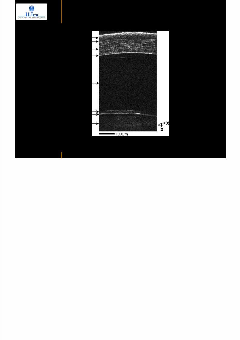
7292019 Atlas of Light-CT images
httpslidepdfcomreaderfullatlas-of-light-ct-images 95118
copy LLTech 2012 ndash wwwlltechimagingcom - No copy without authorization 95
Figure 1 Right Ex vivo rat retinaimaged using the f ull field OCT
system of ESPCI Left Histology
for comparison
Figure 1 Right Ex vivo rat retinaimaged using the f ull field OCT
system of ESPCI Left Histology
for comparison
Figure 1 Right Ex vivo rat retinaimaged using the f ull field OCT
system of ESPCI Left Histology
for comparisonField
230x300microm
RPE
PRL
ONL
OPL
INL
IPL
GCL
NFL
Invest Ophthalmol Vis Sci 2004 Nov45(11)4126-31
Grieve K Paques M Dubois A Sahel J Boccara C Le Gargasson JF copy LLTech 2012
Rat retina
7292019 Atlas of Light-CT images
httpslidepdfcomreaderfullatlas-of-light-ct-images 96118
copy LLTech 2012 ndash wwwlltechimagingcom - No copy without authorization 96 copy LLTech 2012 Courtesy of ESPCI Paris France
Nerve fiber layer
Retina
Invest Ophthalmol Vis Sci 2004 Nov45(11)4126-31
Grieve K Paques M Dubois A Sahel J Boccara C Le Gargasson JF
Transscleral image
7292019 Atlas of Light-CT images
httpslidepdfcomreaderfullatlas-of-light-ct-images 97118
7292019 Atlas of Light-CT images
httpslidepdfcomreaderfullatlas-of-light-ct-images 98118
7292019 Atlas of Light-CT images
httpslidepdfcomreaderfullatlas-of-light-ct-images 99118
In vivo imaging of drosophila melanogaster
7292019 Atlas of Light-CT images
httpslidepdfcomreaderfullatlas-of-light-ct-images 100118
copy LLTech 2012 ndash wwwlltechimagingcom - No copy without authorization 100 copy LLTech 2012 Courtesy of ESPCI Paris France
In vivo imaging of drosophila melanogaster ndash four stages of pupation over 72 hours
7292019 Atlas of Light-CT images
httpslidepdfcomreaderfullatlas-of-light-ct-images 101118
copy LLTech 2012 ndash wwwlltechimagingcom - No copy without authorization 101
Prepupa (0-2 h) Transition topupal stage (24 h)
Pupal stage (48 h) Advanced pupal phase
before eclosion (72 h)
copy LLTech 2012 Courtesy of ESPCI Paris France
Pupa 118 4 days 30um depth
100
7292019 Atlas of Light-CT images
httpslidepdfcomreaderfullatlas-of-light-ct-images 102118
copy LLTech 2012 ndash wwwlltechimagingcom - No copy without authorization 102
100 um
Drosophila
7292019 Atlas of Light-CT images
httpslidepdfcomreaderfullatlas-of-light-ct-images 103118
copy LLTech 2012 ndash wwwlltechimagingcom - No copy without authorization 103
200 microm
copy LLTech 2012 Courtesy of ESPCI Paris France
C Elegans ndash Fast identification of anatomy
7292019 Atlas of Light-CT images
httpslidepdfcomreaderfullatlas-of-light-ct-images 104118
copy LLTech 2012 ndash wwwlltechimagingcom - No copy without authorization 104
Body wall - cuticle
gonad arm
30 microm depth
uterus
ovocytes
intestine
100 microm 100 microm
15 microm depth
copy LLTech 2012 Courtesy of ENS Paris France
C elegans ndash Images at higher magnification 40X
7292019 Atlas of Light-CT images
httpslidepdfcomreaderfullatlas-of-light-ct-images 105118
copy LLTech 2012 ndash wwwlltechimagingcom - No copy without authorization 105
3D reconstruction 50 microm
100 microm
copy LLTech 2012 Courtesy of ENS Paris France
Zebra fish eye
7292019 Atlas of Light-CT images
httpslidepdfcomreaderfullatlas-of-light-ct-images 106118
copy LLTech 2012 ndash wwwlltechimagingcom - No copy without authorization 106
Zebra fish eye
100 microm
100 microm
En face slices
Reconstructed depth slice
copy LLTech 2012 Courtesy of Inserm Paris France
10um depth 20um depth 45um depth
7292019 Atlas of Light-CT images
httpslidepdfcomreaderfullatlas-of-light-ct-images 107118
Xenopus Laevis backtail
7292019 Atlas of Light-CT images
httpslidepdfcomreaderfullatlas-of-light-ct-images 108118
copy LLTech 2012 ndash wwwlltechimagingcom - No copy without authorization 108
100 um30um depth 60um depth
7292019 Atlas of Light-CT images
httpslidepdfcomreaderfullatlas-of-light-ct-images 109118
Mouse embryo in vitro 6 5 days old ndash
7292019 Atlas of Light-CT images
httpslidepdfcomreaderfullatlas-of-light-ct-images 110118
copy LLTech 2012 ndash wwwlltechimagingcom - No copy without authorization 110
Mouse embryo in vitro 65 days old
copy LLTech 2012 Courtesy of Ellvire Guiot and nstitut Jacques Monod Paris France
3D imaging of internal cavity
Sabellaria alveolata mouth palps
7292019 Atlas of Light-CT images
httpslidepdfcomreaderfullatlas-of-light-ct-images 111118
copy LLTech 2012 ndash wwwlltechimagingcom - No copy without authorization 111
500 microm
100 microm
Mouth palps
copy LLTech 2012 Courtesy of Museacutee National drsquoHistoire Naturelle Paris France
7292019 Atlas of Light-CT images
httpslidepdfcomreaderfullatlas-of-light-ct-images 112118
copy LLTech 2012 ndash wwwlltechimagingcom - No copy without authorization 112 copy LLTECH 2011
PLANT IMAGING
Back to table of contents
Mouse ear leaf epidermis
7292019 Atlas of Light-CT images
httpslidepdfcomreaderfullatlas-of-light-ct-images 113118
copy LLTech 2012 ndash wwwlltechimagingcom - No copy without authorization 113
100 microm
Epidermal cell
copy LLTech 2012
Stoma
Leaf veins
7292019 Atlas of Light-CT images
httpslidepdfcomreaderfullatlas-of-light-ct-images 114118
copy LLTech 2012 ndash wwwlltechimagingcom - No copy without authorization 114
114copy LLTech 2012
Fibers
Veins
100 microm
500 microm
Apple
7292019 Atlas of Light-CT images
httpslidepdfcomreaderfullatlas-of-light-ct-images 115118
copy LLTech 2012 ndash wwwlltechimagingcom - No copy without authorization 115
115copy LLTech 2012
100 microm
Wax
Skin
Flesh
500 microm
Water
Effect of harpin infiltration on tobacco leaf
7292019 Atlas of Light-CT images
httpslidepdfcomreaderfullatlas-of-light-ct-images 116118
copy LLTech 2012 ndash wwwlltechimagingcom - No copy without authorization 116
116copy LLTech 2012 Courtesy of UPMC Paris France
50 microm
50 microm
50 microm
50 microm
Upper epidermis
10 min after harpin infiltration 30 min after harpin infiltration
Palisade parenchyma
Epidermis cell
Chloroplast
7292019 Atlas of Light-CT images
httpslidepdfcomreaderfullatlas-of-light-ct-images 117118
copy LLTech 2012 ndash wwwlltechimagingcom - No copy without authorization 117
bull Light-CTtrade does not require tissue preparation nor staining of any kind
bull Creates images within minutes using a non-destructive process
bull Offers a 1 microm cellular resolution in 3D
bull Reveals structural and cellular information
bull Mosaicing allows fast visualisation at various scale
bull En-face high-resolution imaging allows vertical and 3D reconstruction
bull In-vivo images show good promise for in-vivo system
bull Clinical and research applications in cancer detection cosmetics biobanking small animal
developmental biology botanyhellip
Contact Details
7292019 Atlas of Light-CT images
httpslidepdfcomreaderfullatlas-of-light-ct-images 118118
USA
LLTech Inc
103 Carnegie Center DriveSuite 300
Princeton NJ 08540
USA
wwwlltechimagingcom
contactlltechfr
Phone + 1 609 955 3506
LLTech SAS
29 rue du Faubourg St-JacquesPeacutepiniegravere Paris Santeacute Cochin
75014
Paris - France
wwwlltechimagingcom
contactlltechfr
Phone +33 1 82 72 61 25
Europe
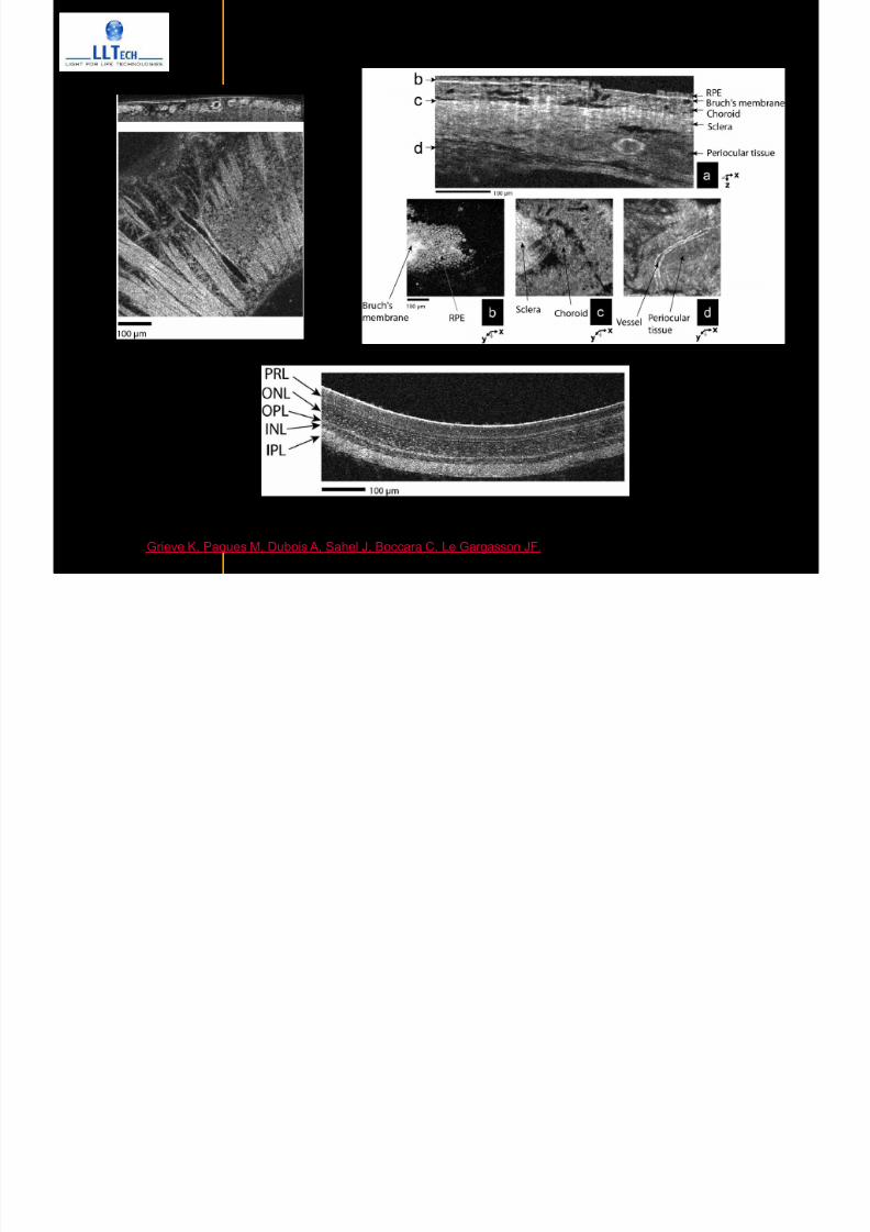
7292019 Atlas of Light-CT images
httpslidepdfcomreaderfullatlas-of-light-ct-images 97118
7292019 Atlas of Light-CT images
httpslidepdfcomreaderfullatlas-of-light-ct-images 98118
7292019 Atlas of Light-CT images
httpslidepdfcomreaderfullatlas-of-light-ct-images 99118
In vivo imaging of drosophila melanogaster
7292019 Atlas of Light-CT images
httpslidepdfcomreaderfullatlas-of-light-ct-images 100118
copy LLTech 2012 ndash wwwlltechimagingcom - No copy without authorization 100 copy LLTech 2012 Courtesy of ESPCI Paris France
In vivo imaging of drosophila melanogaster ndash four stages of pupation over 72 hours
7292019 Atlas of Light-CT images
httpslidepdfcomreaderfullatlas-of-light-ct-images 101118
copy LLTech 2012 ndash wwwlltechimagingcom - No copy without authorization 101
Prepupa (0-2 h) Transition topupal stage (24 h)
Pupal stage (48 h) Advanced pupal phase
before eclosion (72 h)
copy LLTech 2012 Courtesy of ESPCI Paris France
Pupa 118 4 days 30um depth
100
7292019 Atlas of Light-CT images
httpslidepdfcomreaderfullatlas-of-light-ct-images 102118
copy LLTech 2012 ndash wwwlltechimagingcom - No copy without authorization 102
100 um
Drosophila
7292019 Atlas of Light-CT images
httpslidepdfcomreaderfullatlas-of-light-ct-images 103118
copy LLTech 2012 ndash wwwlltechimagingcom - No copy without authorization 103
200 microm
copy LLTech 2012 Courtesy of ESPCI Paris France
C Elegans ndash Fast identification of anatomy
7292019 Atlas of Light-CT images
httpslidepdfcomreaderfullatlas-of-light-ct-images 104118
copy LLTech 2012 ndash wwwlltechimagingcom - No copy without authorization 104
Body wall - cuticle
gonad arm
30 microm depth
uterus
ovocytes
intestine
100 microm 100 microm
15 microm depth
copy LLTech 2012 Courtesy of ENS Paris France
C elegans ndash Images at higher magnification 40X
7292019 Atlas of Light-CT images
httpslidepdfcomreaderfullatlas-of-light-ct-images 105118
copy LLTech 2012 ndash wwwlltechimagingcom - No copy without authorization 105
3D reconstruction 50 microm
100 microm
copy LLTech 2012 Courtesy of ENS Paris France
Zebra fish eye
7292019 Atlas of Light-CT images
httpslidepdfcomreaderfullatlas-of-light-ct-images 106118
copy LLTech 2012 ndash wwwlltechimagingcom - No copy without authorization 106
Zebra fish eye
100 microm
100 microm
En face slices
Reconstructed depth slice
copy LLTech 2012 Courtesy of Inserm Paris France
10um depth 20um depth 45um depth
7292019 Atlas of Light-CT images
httpslidepdfcomreaderfullatlas-of-light-ct-images 107118
Xenopus Laevis backtail
7292019 Atlas of Light-CT images
httpslidepdfcomreaderfullatlas-of-light-ct-images 108118
copy LLTech 2012 ndash wwwlltechimagingcom - No copy without authorization 108
100 um30um depth 60um depth
7292019 Atlas of Light-CT images
httpslidepdfcomreaderfullatlas-of-light-ct-images 109118
Mouse embryo in vitro 6 5 days old ndash
7292019 Atlas of Light-CT images
httpslidepdfcomreaderfullatlas-of-light-ct-images 110118
copy LLTech 2012 ndash wwwlltechimagingcom - No copy without authorization 110
Mouse embryo in vitro 65 days old
copy LLTech 2012 Courtesy of Ellvire Guiot and nstitut Jacques Monod Paris France
3D imaging of internal cavity
Sabellaria alveolata mouth palps
7292019 Atlas of Light-CT images
httpslidepdfcomreaderfullatlas-of-light-ct-images 111118
copy LLTech 2012 ndash wwwlltechimagingcom - No copy without authorization 111
500 microm
100 microm
Mouth palps
copy LLTech 2012 Courtesy of Museacutee National drsquoHistoire Naturelle Paris France
7292019 Atlas of Light-CT images
httpslidepdfcomreaderfullatlas-of-light-ct-images 112118
copy LLTech 2012 ndash wwwlltechimagingcom - No copy without authorization 112 copy LLTECH 2011
PLANT IMAGING
Back to table of contents
Mouse ear leaf epidermis
7292019 Atlas of Light-CT images
httpslidepdfcomreaderfullatlas-of-light-ct-images 113118
copy LLTech 2012 ndash wwwlltechimagingcom - No copy without authorization 113
100 microm
Epidermal cell
copy LLTech 2012
Stoma
Leaf veins
7292019 Atlas of Light-CT images
httpslidepdfcomreaderfullatlas-of-light-ct-images 114118
copy LLTech 2012 ndash wwwlltechimagingcom - No copy without authorization 114
114copy LLTech 2012
Fibers
Veins
100 microm
500 microm
Apple
7292019 Atlas of Light-CT images
httpslidepdfcomreaderfullatlas-of-light-ct-images 115118
copy LLTech 2012 ndash wwwlltechimagingcom - No copy without authorization 115
115copy LLTech 2012
100 microm
Wax
Skin
Flesh
500 microm
Water
Effect of harpin infiltration on tobacco leaf
7292019 Atlas of Light-CT images
httpslidepdfcomreaderfullatlas-of-light-ct-images 116118
copy LLTech 2012 ndash wwwlltechimagingcom - No copy without authorization 116
116copy LLTech 2012 Courtesy of UPMC Paris France
50 microm
50 microm
50 microm
50 microm
Upper epidermis
10 min after harpin infiltration 30 min after harpin infiltration
Palisade parenchyma
Epidermis cell
Chloroplast
7292019 Atlas of Light-CT images
httpslidepdfcomreaderfullatlas-of-light-ct-images 117118
copy LLTech 2012 ndash wwwlltechimagingcom - No copy without authorization 117
bull Light-CTtrade does not require tissue preparation nor staining of any kind
bull Creates images within minutes using a non-destructive process
bull Offers a 1 microm cellular resolution in 3D
bull Reveals structural and cellular information
bull Mosaicing allows fast visualisation at various scale
bull En-face high-resolution imaging allows vertical and 3D reconstruction
bull In-vivo images show good promise for in-vivo system
bull Clinical and research applications in cancer detection cosmetics biobanking small animal
developmental biology botanyhellip
Contact Details
7292019 Atlas of Light-CT images
httpslidepdfcomreaderfullatlas-of-light-ct-images 118118
USA
LLTech Inc
103 Carnegie Center DriveSuite 300
Princeton NJ 08540
USA
wwwlltechimagingcom
contactlltechfr
Phone + 1 609 955 3506
LLTech SAS
29 rue du Faubourg St-JacquesPeacutepiniegravere Paris Santeacute Cochin
75014
Paris - France
wwwlltechimagingcom
contactlltechfr
Phone +33 1 82 72 61 25
Europe
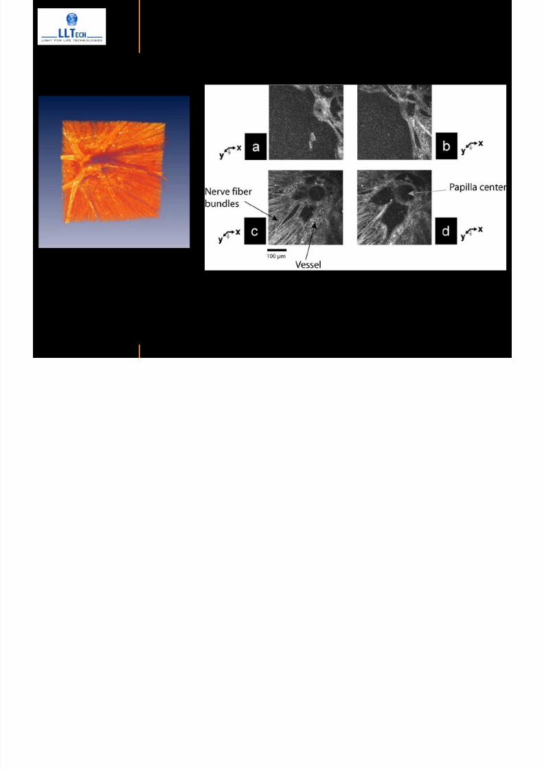
7292019 Atlas of Light-CT images
httpslidepdfcomreaderfullatlas-of-light-ct-images 99118
In vivo imaging of drosophila melanogaster
7292019 Atlas of Light-CT images
httpslidepdfcomreaderfullatlas-of-light-ct-images 100118
copy LLTech 2012 ndash wwwlltechimagingcom - No copy without authorization 100 copy LLTech 2012 Courtesy of ESPCI Paris France
In vivo imaging of drosophila melanogaster ndash four stages of pupation over 72 hours
7292019 Atlas of Light-CT images
httpslidepdfcomreaderfullatlas-of-light-ct-images 101118
copy LLTech 2012 ndash wwwlltechimagingcom - No copy without authorization 101
Prepupa (0-2 h) Transition topupal stage (24 h)
Pupal stage (48 h) Advanced pupal phase
before eclosion (72 h)
copy LLTech 2012 Courtesy of ESPCI Paris France
Pupa 118 4 days 30um depth
100
7292019 Atlas of Light-CT images
httpslidepdfcomreaderfullatlas-of-light-ct-images 102118
copy LLTech 2012 ndash wwwlltechimagingcom - No copy without authorization 102
100 um
Drosophila
7292019 Atlas of Light-CT images
httpslidepdfcomreaderfullatlas-of-light-ct-images 103118
copy LLTech 2012 ndash wwwlltechimagingcom - No copy without authorization 103
200 microm
copy LLTech 2012 Courtesy of ESPCI Paris France
C Elegans ndash Fast identification of anatomy
7292019 Atlas of Light-CT images
httpslidepdfcomreaderfullatlas-of-light-ct-images 104118
copy LLTech 2012 ndash wwwlltechimagingcom - No copy without authorization 104
Body wall - cuticle
gonad arm
30 microm depth
uterus
ovocytes
intestine
100 microm 100 microm
15 microm depth
copy LLTech 2012 Courtesy of ENS Paris France
C elegans ndash Images at higher magnification 40X
7292019 Atlas of Light-CT images
httpslidepdfcomreaderfullatlas-of-light-ct-images 105118
copy LLTech 2012 ndash wwwlltechimagingcom - No copy without authorization 105
3D reconstruction 50 microm
100 microm
copy LLTech 2012 Courtesy of ENS Paris France
Zebra fish eye
7292019 Atlas of Light-CT images
httpslidepdfcomreaderfullatlas-of-light-ct-images 106118
copy LLTech 2012 ndash wwwlltechimagingcom - No copy without authorization 106
Zebra fish eye
100 microm
100 microm
En face slices
Reconstructed depth slice
copy LLTech 2012 Courtesy of Inserm Paris France
10um depth 20um depth 45um depth
7292019 Atlas of Light-CT images
httpslidepdfcomreaderfullatlas-of-light-ct-images 107118
Xenopus Laevis backtail
7292019 Atlas of Light-CT images
httpslidepdfcomreaderfullatlas-of-light-ct-images 108118
copy LLTech 2012 ndash wwwlltechimagingcom - No copy without authorization 108
100 um30um depth 60um depth
7292019 Atlas of Light-CT images
httpslidepdfcomreaderfullatlas-of-light-ct-images 109118
Mouse embryo in vitro 6 5 days old ndash
7292019 Atlas of Light-CT images
httpslidepdfcomreaderfullatlas-of-light-ct-images 110118
copy LLTech 2012 ndash wwwlltechimagingcom - No copy without authorization 110
Mouse embryo in vitro 65 days old
copy LLTech 2012 Courtesy of Ellvire Guiot and nstitut Jacques Monod Paris France
3D imaging of internal cavity
Sabellaria alveolata mouth palps
7292019 Atlas of Light-CT images
httpslidepdfcomreaderfullatlas-of-light-ct-images 111118
copy LLTech 2012 ndash wwwlltechimagingcom - No copy without authorization 111
500 microm
100 microm
Mouth palps
copy LLTech 2012 Courtesy of Museacutee National drsquoHistoire Naturelle Paris France
7292019 Atlas of Light-CT images
httpslidepdfcomreaderfullatlas-of-light-ct-images 112118
copy LLTech 2012 ndash wwwlltechimagingcom - No copy without authorization 112 copy LLTECH 2011
PLANT IMAGING
Back to table of contents
Mouse ear leaf epidermis
7292019 Atlas of Light-CT images
httpslidepdfcomreaderfullatlas-of-light-ct-images 113118
copy LLTech 2012 ndash wwwlltechimagingcom - No copy without authorization 113
100 microm
Epidermal cell
copy LLTech 2012
Stoma
Leaf veins
7292019 Atlas of Light-CT images
httpslidepdfcomreaderfullatlas-of-light-ct-images 114118
copy LLTech 2012 ndash wwwlltechimagingcom - No copy without authorization 114
114copy LLTech 2012
Fibers
Veins
100 microm
500 microm
Apple
7292019 Atlas of Light-CT images
httpslidepdfcomreaderfullatlas-of-light-ct-images 115118
copy LLTech 2012 ndash wwwlltechimagingcom - No copy without authorization 115
115copy LLTech 2012
100 microm
Wax
Skin
Flesh
500 microm
Water
Effect of harpin infiltration on tobacco leaf
7292019 Atlas of Light-CT images
httpslidepdfcomreaderfullatlas-of-light-ct-images 116118
copy LLTech 2012 ndash wwwlltechimagingcom - No copy without authorization 116
116copy LLTech 2012 Courtesy of UPMC Paris France
50 microm
50 microm
50 microm
50 microm
Upper epidermis
10 min after harpin infiltration 30 min after harpin infiltration
Palisade parenchyma
Epidermis cell
Chloroplast
7292019 Atlas of Light-CT images
httpslidepdfcomreaderfullatlas-of-light-ct-images 117118
copy LLTech 2012 ndash wwwlltechimagingcom - No copy without authorization 117
bull Light-CTtrade does not require tissue preparation nor staining of any kind
bull Creates images within minutes using a non-destructive process
bull Offers a 1 microm cellular resolution in 3D
bull Reveals structural and cellular information
bull Mosaicing allows fast visualisation at various scale
bull En-face high-resolution imaging allows vertical and 3D reconstruction
bull In-vivo images show good promise for in-vivo system
bull Clinical and research applications in cancer detection cosmetics biobanking small animal
developmental biology botanyhellip
Contact Details
7292019 Atlas of Light-CT images
httpslidepdfcomreaderfullatlas-of-light-ct-images 118118
USA
LLTech Inc
103 Carnegie Center DriveSuite 300
Princeton NJ 08540
USA
wwwlltechimagingcom
contactlltechfr
Phone + 1 609 955 3506
LLTech SAS
29 rue du Faubourg St-JacquesPeacutepiniegravere Paris Santeacute Cochin
75014
Paris - France
wwwlltechimagingcom
contactlltechfr
Phone +33 1 82 72 61 25
Europe
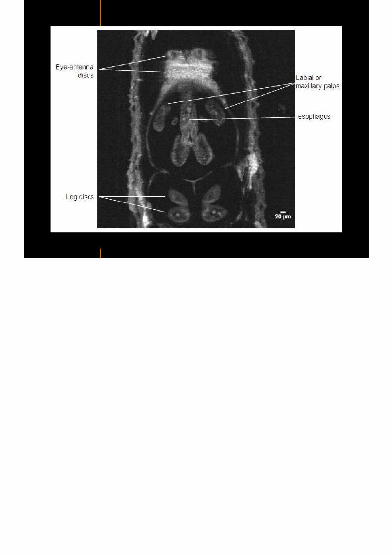
7292019 Atlas of Light-CT images
httpslidepdfcomreaderfullatlas-of-light-ct-images 100118
copy LLTech 2012 ndash wwwlltechimagingcom - No copy without authorization 100 copy LLTech 2012 Courtesy of ESPCI Paris France
In vivo imaging of drosophila melanogaster ndash four stages of pupation over 72 hours
7292019 Atlas of Light-CT images
httpslidepdfcomreaderfullatlas-of-light-ct-images 101118
copy LLTech 2012 ndash wwwlltechimagingcom - No copy without authorization 101
Prepupa (0-2 h) Transition topupal stage (24 h)
Pupal stage (48 h) Advanced pupal phase
before eclosion (72 h)
copy LLTech 2012 Courtesy of ESPCI Paris France
Pupa 118 4 days 30um depth
100
7292019 Atlas of Light-CT images
httpslidepdfcomreaderfullatlas-of-light-ct-images 102118
copy LLTech 2012 ndash wwwlltechimagingcom - No copy without authorization 102
100 um
Drosophila
7292019 Atlas of Light-CT images
httpslidepdfcomreaderfullatlas-of-light-ct-images 103118
copy LLTech 2012 ndash wwwlltechimagingcom - No copy without authorization 103
200 microm
copy LLTech 2012 Courtesy of ESPCI Paris France
C Elegans ndash Fast identification of anatomy
7292019 Atlas of Light-CT images
httpslidepdfcomreaderfullatlas-of-light-ct-images 104118
copy LLTech 2012 ndash wwwlltechimagingcom - No copy without authorization 104
Body wall - cuticle
gonad arm
30 microm depth
uterus
ovocytes
intestine
100 microm 100 microm
15 microm depth
copy LLTech 2012 Courtesy of ENS Paris France
C elegans ndash Images at higher magnification 40X
7292019 Atlas of Light-CT images
httpslidepdfcomreaderfullatlas-of-light-ct-images 105118
copy LLTech 2012 ndash wwwlltechimagingcom - No copy without authorization 105
3D reconstruction 50 microm
100 microm
copy LLTech 2012 Courtesy of ENS Paris France
Zebra fish eye
7292019 Atlas of Light-CT images
httpslidepdfcomreaderfullatlas-of-light-ct-images 106118
copy LLTech 2012 ndash wwwlltechimagingcom - No copy without authorization 106
Zebra fish eye
100 microm
100 microm
En face slices
Reconstructed depth slice
copy LLTech 2012 Courtesy of Inserm Paris France
10um depth 20um depth 45um depth
7292019 Atlas of Light-CT images
httpslidepdfcomreaderfullatlas-of-light-ct-images 107118
Xenopus Laevis backtail
7292019 Atlas of Light-CT images
httpslidepdfcomreaderfullatlas-of-light-ct-images 108118
copy LLTech 2012 ndash wwwlltechimagingcom - No copy without authorization 108
100 um30um depth 60um depth
7292019 Atlas of Light-CT images
httpslidepdfcomreaderfullatlas-of-light-ct-images 109118
Mouse embryo in vitro 6 5 days old ndash
7292019 Atlas of Light-CT images
httpslidepdfcomreaderfullatlas-of-light-ct-images 110118
copy LLTech 2012 ndash wwwlltechimagingcom - No copy without authorization 110
Mouse embryo in vitro 65 days old
copy LLTech 2012 Courtesy of Ellvire Guiot and nstitut Jacques Monod Paris France
3D imaging of internal cavity
Sabellaria alveolata mouth palps
7292019 Atlas of Light-CT images
httpslidepdfcomreaderfullatlas-of-light-ct-images 111118
copy LLTech 2012 ndash wwwlltechimagingcom - No copy without authorization 111
500 microm
100 microm
Mouth palps
copy LLTech 2012 Courtesy of Museacutee National drsquoHistoire Naturelle Paris France
7292019 Atlas of Light-CT images
httpslidepdfcomreaderfullatlas-of-light-ct-images 112118
copy LLTech 2012 ndash wwwlltechimagingcom - No copy without authorization 112 copy LLTECH 2011
PLANT IMAGING
Back to table of contents
Mouse ear leaf epidermis
7292019 Atlas of Light-CT images
httpslidepdfcomreaderfullatlas-of-light-ct-images 113118
copy LLTech 2012 ndash wwwlltechimagingcom - No copy without authorization 113
100 microm
Epidermal cell
copy LLTech 2012
Stoma
Leaf veins
7292019 Atlas of Light-CT images
httpslidepdfcomreaderfullatlas-of-light-ct-images 114118
copy LLTech 2012 ndash wwwlltechimagingcom - No copy without authorization 114
114copy LLTech 2012
Fibers
Veins
100 microm
500 microm
Apple
7292019 Atlas of Light-CT images
httpslidepdfcomreaderfullatlas-of-light-ct-images 115118
copy LLTech 2012 ndash wwwlltechimagingcom - No copy without authorization 115
115copy LLTech 2012
100 microm
Wax
Skin
Flesh
500 microm
Water
Effect of harpin infiltration on tobacco leaf
7292019 Atlas of Light-CT images
httpslidepdfcomreaderfullatlas-of-light-ct-images 116118
copy LLTech 2012 ndash wwwlltechimagingcom - No copy without authorization 116
116copy LLTech 2012 Courtesy of UPMC Paris France
50 microm
50 microm
50 microm
50 microm
Upper epidermis
10 min after harpin infiltration 30 min after harpin infiltration
Palisade parenchyma
Epidermis cell
Chloroplast
7292019 Atlas of Light-CT images
httpslidepdfcomreaderfullatlas-of-light-ct-images 117118
copy LLTech 2012 ndash wwwlltechimagingcom - No copy without authorization 117
bull Light-CTtrade does not require tissue preparation nor staining of any kind
bull Creates images within minutes using a non-destructive process
bull Offers a 1 microm cellular resolution in 3D
bull Reveals structural and cellular information
bull Mosaicing allows fast visualisation at various scale
bull En-face high-resolution imaging allows vertical and 3D reconstruction
bull In-vivo images show good promise for in-vivo system
bull Clinical and research applications in cancer detection cosmetics biobanking small animal
developmental biology botanyhellip
Contact Details
7292019 Atlas of Light-CT images
httpslidepdfcomreaderfullatlas-of-light-ct-images 118118
USA
LLTech Inc
103 Carnegie Center DriveSuite 300
Princeton NJ 08540
USA
wwwlltechimagingcom
contactlltechfr
Phone + 1 609 955 3506
LLTech SAS
29 rue du Faubourg St-JacquesPeacutepiniegravere Paris Santeacute Cochin
75014
Paris - France
wwwlltechimagingcom
contactlltechfr
Phone +33 1 82 72 61 25
Europe
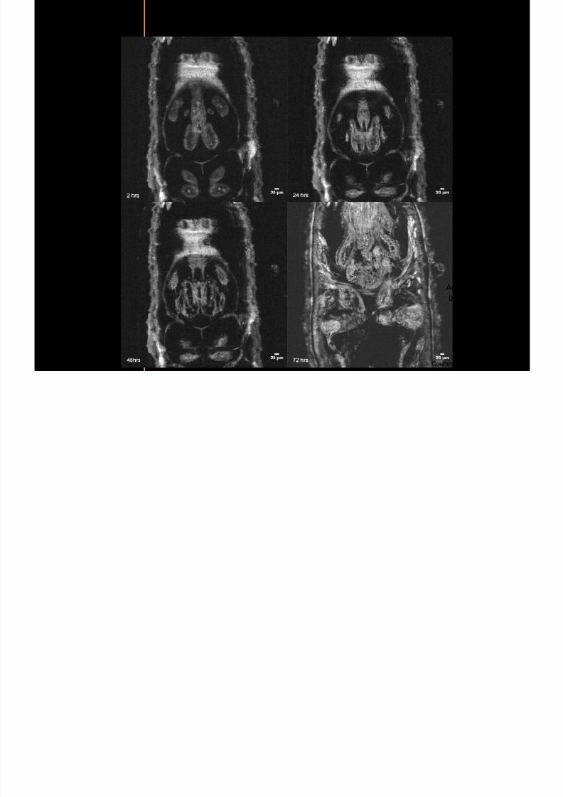
7292019 Atlas of Light-CT images
httpslidepdfcomreaderfullatlas-of-light-ct-images 102118
copy LLTech 2012 ndash wwwlltechimagingcom - No copy without authorization 102
100 um
Drosophila
7292019 Atlas of Light-CT images
httpslidepdfcomreaderfullatlas-of-light-ct-images 103118
copy LLTech 2012 ndash wwwlltechimagingcom - No copy without authorization 103
200 microm
copy LLTech 2012 Courtesy of ESPCI Paris France
C Elegans ndash Fast identification of anatomy
7292019 Atlas of Light-CT images
httpslidepdfcomreaderfullatlas-of-light-ct-images 104118
copy LLTech 2012 ndash wwwlltechimagingcom - No copy without authorization 104
Body wall - cuticle
gonad arm
30 microm depth
uterus
ovocytes
intestine
100 microm 100 microm
15 microm depth
copy LLTech 2012 Courtesy of ENS Paris France
C elegans ndash Images at higher magnification 40X
7292019 Atlas of Light-CT images
httpslidepdfcomreaderfullatlas-of-light-ct-images 105118
copy LLTech 2012 ndash wwwlltechimagingcom - No copy without authorization 105
3D reconstruction 50 microm
100 microm
copy LLTech 2012 Courtesy of ENS Paris France
Zebra fish eye
7292019 Atlas of Light-CT images
httpslidepdfcomreaderfullatlas-of-light-ct-images 106118
copy LLTech 2012 ndash wwwlltechimagingcom - No copy without authorization 106
Zebra fish eye
100 microm
100 microm
En face slices
Reconstructed depth slice
copy LLTech 2012 Courtesy of Inserm Paris France
10um depth 20um depth 45um depth
7292019 Atlas of Light-CT images
httpslidepdfcomreaderfullatlas-of-light-ct-images 107118
Xenopus Laevis backtail
7292019 Atlas of Light-CT images
httpslidepdfcomreaderfullatlas-of-light-ct-images 108118
copy LLTech 2012 ndash wwwlltechimagingcom - No copy without authorization 108
100 um30um depth 60um depth
7292019 Atlas of Light-CT images
httpslidepdfcomreaderfullatlas-of-light-ct-images 109118
Mouse embryo in vitro 6 5 days old ndash
7292019 Atlas of Light-CT images
httpslidepdfcomreaderfullatlas-of-light-ct-images 110118
copy LLTech 2012 ndash wwwlltechimagingcom - No copy without authorization 110
Mouse embryo in vitro 65 days old
copy LLTech 2012 Courtesy of Ellvire Guiot and nstitut Jacques Monod Paris France
3D imaging of internal cavity
Sabellaria alveolata mouth palps
7292019 Atlas of Light-CT images
httpslidepdfcomreaderfullatlas-of-light-ct-images 111118
copy LLTech 2012 ndash wwwlltechimagingcom - No copy without authorization 111
500 microm
100 microm
Mouth palps
copy LLTech 2012 Courtesy of Museacutee National drsquoHistoire Naturelle Paris France
7292019 Atlas of Light-CT images
httpslidepdfcomreaderfullatlas-of-light-ct-images 112118
copy LLTech 2012 ndash wwwlltechimagingcom - No copy without authorization 112 copy LLTECH 2011
PLANT IMAGING
Back to table of contents
Mouse ear leaf epidermis
7292019 Atlas of Light-CT images
httpslidepdfcomreaderfullatlas-of-light-ct-images 113118
copy LLTech 2012 ndash wwwlltechimagingcom - No copy without authorization 113
100 microm
Epidermal cell
copy LLTech 2012
Stoma
Leaf veins
7292019 Atlas of Light-CT images
httpslidepdfcomreaderfullatlas-of-light-ct-images 114118
copy LLTech 2012 ndash wwwlltechimagingcom - No copy without authorization 114
114copy LLTech 2012
Fibers
Veins
100 microm
500 microm
Apple
7292019 Atlas of Light-CT images
httpslidepdfcomreaderfullatlas-of-light-ct-images 115118
copy LLTech 2012 ndash wwwlltechimagingcom - No copy without authorization 115
115copy LLTech 2012
100 microm
Wax
Skin
Flesh
500 microm
Water
Effect of harpin infiltration on tobacco leaf
7292019 Atlas of Light-CT images
httpslidepdfcomreaderfullatlas-of-light-ct-images 116118
copy LLTech 2012 ndash wwwlltechimagingcom - No copy without authorization 116
116copy LLTech 2012 Courtesy of UPMC Paris France
50 microm
50 microm
50 microm
50 microm
Upper epidermis
10 min after harpin infiltration 30 min after harpin infiltration
Palisade parenchyma
Epidermis cell
Chloroplast
7292019 Atlas of Light-CT images
httpslidepdfcomreaderfullatlas-of-light-ct-images 117118
copy LLTech 2012 ndash wwwlltechimagingcom - No copy without authorization 117
bull Light-CTtrade does not require tissue preparation nor staining of any kind
bull Creates images within minutes using a non-destructive process
bull Offers a 1 microm cellular resolution in 3D
bull Reveals structural and cellular information
bull Mosaicing allows fast visualisation at various scale
bull En-face high-resolution imaging allows vertical and 3D reconstruction
bull In-vivo images show good promise for in-vivo system
bull Clinical and research applications in cancer detection cosmetics biobanking small animal
developmental biology botanyhellip
Contact Details
7292019 Atlas of Light-CT images
httpslidepdfcomreaderfullatlas-of-light-ct-images 118118
USA
LLTech Inc
103 Carnegie Center DriveSuite 300
Princeton NJ 08540
USA
wwwlltechimagingcom
contactlltechfr
Phone + 1 609 955 3506
LLTech SAS
29 rue du Faubourg St-JacquesPeacutepiniegravere Paris Santeacute Cochin
75014
Paris - France
wwwlltechimagingcom
contactlltechfr
Phone +33 1 82 72 61 25
Europe
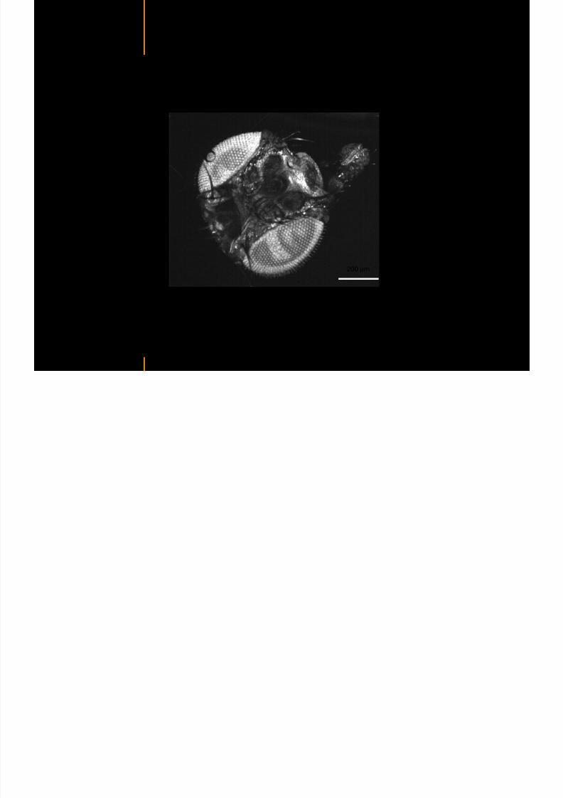
7292019 Atlas of Light-CT images
httpslidepdfcomreaderfullatlas-of-light-ct-images 103118
copy LLTech 2012 ndash wwwlltechimagingcom - No copy without authorization 103
200 microm
copy LLTech 2012 Courtesy of ESPCI Paris France
C Elegans ndash Fast identification of anatomy
7292019 Atlas of Light-CT images
httpslidepdfcomreaderfullatlas-of-light-ct-images 104118
copy LLTech 2012 ndash wwwlltechimagingcom - No copy without authorization 104
Body wall - cuticle
gonad arm
30 microm depth
uterus
ovocytes
intestine
100 microm 100 microm
15 microm depth
copy LLTech 2012 Courtesy of ENS Paris France
C elegans ndash Images at higher magnification 40X
7292019 Atlas of Light-CT images
httpslidepdfcomreaderfullatlas-of-light-ct-images 105118
copy LLTech 2012 ndash wwwlltechimagingcom - No copy without authorization 105
3D reconstruction 50 microm
100 microm
copy LLTech 2012 Courtesy of ENS Paris France
Zebra fish eye
7292019 Atlas of Light-CT images
httpslidepdfcomreaderfullatlas-of-light-ct-images 106118
copy LLTech 2012 ndash wwwlltechimagingcom - No copy without authorization 106
Zebra fish eye
100 microm
100 microm
En face slices
Reconstructed depth slice
copy LLTech 2012 Courtesy of Inserm Paris France
10um depth 20um depth 45um depth
7292019 Atlas of Light-CT images
httpslidepdfcomreaderfullatlas-of-light-ct-images 107118
Xenopus Laevis backtail
7292019 Atlas of Light-CT images
httpslidepdfcomreaderfullatlas-of-light-ct-images 108118
copy LLTech 2012 ndash wwwlltechimagingcom - No copy without authorization 108
100 um30um depth 60um depth
7292019 Atlas of Light-CT images
httpslidepdfcomreaderfullatlas-of-light-ct-images 109118
Mouse embryo in vitro 6 5 days old ndash
7292019 Atlas of Light-CT images
httpslidepdfcomreaderfullatlas-of-light-ct-images 110118
copy LLTech 2012 ndash wwwlltechimagingcom - No copy without authorization 110
Mouse embryo in vitro 65 days old
copy LLTech 2012 Courtesy of Ellvire Guiot and nstitut Jacques Monod Paris France
3D imaging of internal cavity
Sabellaria alveolata mouth palps
7292019 Atlas of Light-CT images
httpslidepdfcomreaderfullatlas-of-light-ct-images 111118
copy LLTech 2012 ndash wwwlltechimagingcom - No copy without authorization 111
500 microm
100 microm
Mouth palps
copy LLTech 2012 Courtesy of Museacutee National drsquoHistoire Naturelle Paris France
7292019 Atlas of Light-CT images
httpslidepdfcomreaderfullatlas-of-light-ct-images 112118
copy LLTech 2012 ndash wwwlltechimagingcom - No copy without authorization 112 copy LLTECH 2011
PLANT IMAGING
Back to table of contents
Mouse ear leaf epidermis
7292019 Atlas of Light-CT images
httpslidepdfcomreaderfullatlas-of-light-ct-images 113118
copy LLTech 2012 ndash wwwlltechimagingcom - No copy without authorization 113
100 microm
Epidermal cell
copy LLTech 2012
Stoma
Leaf veins
7292019 Atlas of Light-CT images
httpslidepdfcomreaderfullatlas-of-light-ct-images 114118
copy LLTech 2012 ndash wwwlltechimagingcom - No copy without authorization 114
114copy LLTech 2012
Fibers
Veins
100 microm
500 microm
Apple
7292019 Atlas of Light-CT images
httpslidepdfcomreaderfullatlas-of-light-ct-images 115118
copy LLTech 2012 ndash wwwlltechimagingcom - No copy without authorization 115
115copy LLTech 2012
100 microm
Wax
Skin
Flesh
500 microm
Water
Effect of harpin infiltration on tobacco leaf
7292019 Atlas of Light-CT images
httpslidepdfcomreaderfullatlas-of-light-ct-images 116118
copy LLTech 2012 ndash wwwlltechimagingcom - No copy without authorization 116
116copy LLTech 2012 Courtesy of UPMC Paris France
50 microm
50 microm
50 microm
50 microm
Upper epidermis
10 min after harpin infiltration 30 min after harpin infiltration
Palisade parenchyma
Epidermis cell
Chloroplast
7292019 Atlas of Light-CT images
httpslidepdfcomreaderfullatlas-of-light-ct-images 117118
copy LLTech 2012 ndash wwwlltechimagingcom - No copy without authorization 117
bull Light-CTtrade does not require tissue preparation nor staining of any kind
bull Creates images within minutes using a non-destructive process
bull Offers a 1 microm cellular resolution in 3D
bull Reveals structural and cellular information
bull Mosaicing allows fast visualisation at various scale
bull En-face high-resolution imaging allows vertical and 3D reconstruction
bull In-vivo images show good promise for in-vivo system
bull Clinical and research applications in cancer detection cosmetics biobanking small animal
developmental biology botanyhellip
Contact Details
7292019 Atlas of Light-CT images
httpslidepdfcomreaderfullatlas-of-light-ct-images 118118
USA
LLTech Inc
103 Carnegie Center DriveSuite 300
Princeton NJ 08540
USA
wwwlltechimagingcom
contactlltechfr
Phone + 1 609 955 3506
LLTech SAS
29 rue du Faubourg St-JacquesPeacutepiniegravere Paris Santeacute Cochin
75014
Paris - France
wwwlltechimagingcom
contactlltechfr
Phone +33 1 82 72 61 25
Europe
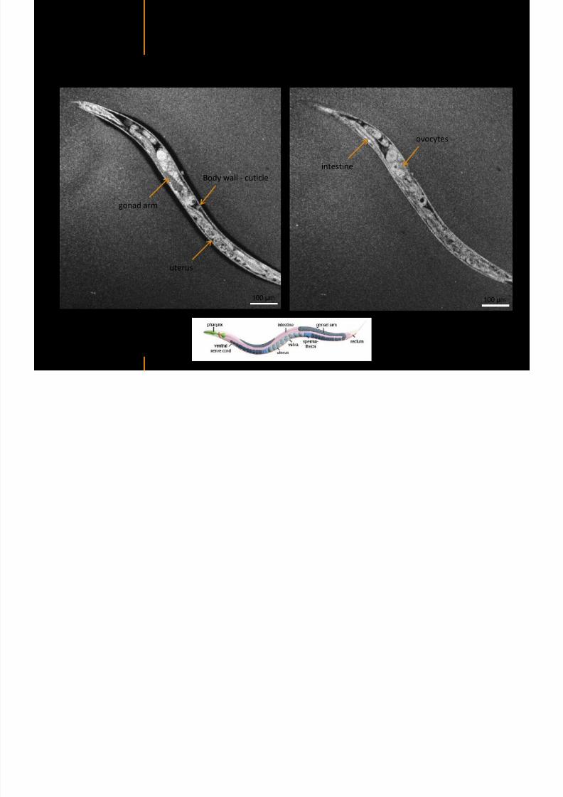
7292019 Atlas of Light-CT images
httpslidepdfcomreaderfullatlas-of-light-ct-images 104118
copy LLTech 2012 ndash wwwlltechimagingcom - No copy without authorization 104
Body wall - cuticle
gonad arm
30 microm depth
uterus
ovocytes
intestine
100 microm 100 microm
15 microm depth
copy LLTech 2012 Courtesy of ENS Paris France
C elegans ndash Images at higher magnification 40X
7292019 Atlas of Light-CT images
httpslidepdfcomreaderfullatlas-of-light-ct-images 105118
copy LLTech 2012 ndash wwwlltechimagingcom - No copy without authorization 105
3D reconstruction 50 microm
100 microm
copy LLTech 2012 Courtesy of ENS Paris France
Zebra fish eye
7292019 Atlas of Light-CT images
httpslidepdfcomreaderfullatlas-of-light-ct-images 106118
copy LLTech 2012 ndash wwwlltechimagingcom - No copy without authorization 106
Zebra fish eye
100 microm
100 microm
En face slices
Reconstructed depth slice
copy LLTech 2012 Courtesy of Inserm Paris France
10um depth 20um depth 45um depth
7292019 Atlas of Light-CT images
httpslidepdfcomreaderfullatlas-of-light-ct-images 107118
Xenopus Laevis backtail
7292019 Atlas of Light-CT images
httpslidepdfcomreaderfullatlas-of-light-ct-images 108118
copy LLTech 2012 ndash wwwlltechimagingcom - No copy without authorization 108
100 um30um depth 60um depth
7292019 Atlas of Light-CT images
httpslidepdfcomreaderfullatlas-of-light-ct-images 109118
Mouse embryo in vitro 6 5 days old ndash
7292019 Atlas of Light-CT images
httpslidepdfcomreaderfullatlas-of-light-ct-images 110118
copy LLTech 2012 ndash wwwlltechimagingcom - No copy without authorization 110
Mouse embryo in vitro 65 days old
copy LLTech 2012 Courtesy of Ellvire Guiot and nstitut Jacques Monod Paris France
3D imaging of internal cavity
Sabellaria alveolata mouth palps
7292019 Atlas of Light-CT images
httpslidepdfcomreaderfullatlas-of-light-ct-images 111118
copy LLTech 2012 ndash wwwlltechimagingcom - No copy without authorization 111
500 microm
100 microm
Mouth palps
copy LLTech 2012 Courtesy of Museacutee National drsquoHistoire Naturelle Paris France
7292019 Atlas of Light-CT images
httpslidepdfcomreaderfullatlas-of-light-ct-images 112118
copy LLTech 2012 ndash wwwlltechimagingcom - No copy without authorization 112 copy LLTECH 2011
PLANT IMAGING
Back to table of contents
Mouse ear leaf epidermis
7292019 Atlas of Light-CT images
httpslidepdfcomreaderfullatlas-of-light-ct-images 113118
copy LLTech 2012 ndash wwwlltechimagingcom - No copy without authorization 113
100 microm
Epidermal cell
copy LLTech 2012
Stoma
Leaf veins
7292019 Atlas of Light-CT images
httpslidepdfcomreaderfullatlas-of-light-ct-images 114118
copy LLTech 2012 ndash wwwlltechimagingcom - No copy without authorization 114
114copy LLTech 2012
Fibers
Veins
100 microm
500 microm
Apple
7292019 Atlas of Light-CT images
httpslidepdfcomreaderfullatlas-of-light-ct-images 115118
copy LLTech 2012 ndash wwwlltechimagingcom - No copy without authorization 115
115copy LLTech 2012
100 microm
Wax
Skin
Flesh
500 microm
Water
Effect of harpin infiltration on tobacco leaf
7292019 Atlas of Light-CT images
httpslidepdfcomreaderfullatlas-of-light-ct-images 116118
copy LLTech 2012 ndash wwwlltechimagingcom - No copy without authorization 116
116copy LLTech 2012 Courtesy of UPMC Paris France
50 microm
50 microm
50 microm
50 microm
Upper epidermis
10 min after harpin infiltration 30 min after harpin infiltration
Palisade parenchyma
Epidermis cell
Chloroplast
7292019 Atlas of Light-CT images
httpslidepdfcomreaderfullatlas-of-light-ct-images 117118
copy LLTech 2012 ndash wwwlltechimagingcom - No copy without authorization 117
bull Light-CTtrade does not require tissue preparation nor staining of any kind
bull Creates images within minutes using a non-destructive process
bull Offers a 1 microm cellular resolution in 3D
bull Reveals structural and cellular information
bull Mosaicing allows fast visualisation at various scale
bull En-face high-resolution imaging allows vertical and 3D reconstruction
bull In-vivo images show good promise for in-vivo system
bull Clinical and research applications in cancer detection cosmetics biobanking small animal
developmental biology botanyhellip
Contact Details
7292019 Atlas of Light-CT images
httpslidepdfcomreaderfullatlas-of-light-ct-images 118118
USA
LLTech Inc
103 Carnegie Center DriveSuite 300
Princeton NJ 08540
USA
wwwlltechimagingcom
contactlltechfr
Phone + 1 609 955 3506
LLTech SAS
29 rue du Faubourg St-JacquesPeacutepiniegravere Paris Santeacute Cochin
75014
Paris - France
wwwlltechimagingcom
contactlltechfr
Phone +33 1 82 72 61 25
Europe
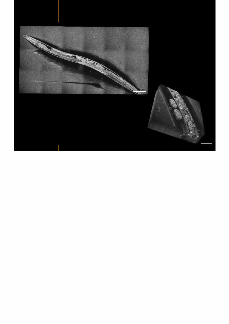
7292019 Atlas of Light-CT images
httpslidepdfcomreaderfullatlas-of-light-ct-images 105118
copy LLTech 2012 ndash wwwlltechimagingcom - No copy without authorization 105
3D reconstruction 50 microm
100 microm
copy LLTech 2012 Courtesy of ENS Paris France
Zebra fish eye
7292019 Atlas of Light-CT images
httpslidepdfcomreaderfullatlas-of-light-ct-images 106118
copy LLTech 2012 ndash wwwlltechimagingcom - No copy without authorization 106
Zebra fish eye
100 microm
100 microm
En face slices
Reconstructed depth slice
copy LLTech 2012 Courtesy of Inserm Paris France
10um depth 20um depth 45um depth
7292019 Atlas of Light-CT images
httpslidepdfcomreaderfullatlas-of-light-ct-images 107118
Xenopus Laevis backtail
7292019 Atlas of Light-CT images
httpslidepdfcomreaderfullatlas-of-light-ct-images 108118
copy LLTech 2012 ndash wwwlltechimagingcom - No copy without authorization 108
100 um30um depth 60um depth
7292019 Atlas of Light-CT images
httpslidepdfcomreaderfullatlas-of-light-ct-images 109118
Mouse embryo in vitro 6 5 days old ndash
7292019 Atlas of Light-CT images
httpslidepdfcomreaderfullatlas-of-light-ct-images 110118
copy LLTech 2012 ndash wwwlltechimagingcom - No copy without authorization 110
Mouse embryo in vitro 65 days old
copy LLTech 2012 Courtesy of Ellvire Guiot and nstitut Jacques Monod Paris France
3D imaging of internal cavity
Sabellaria alveolata mouth palps
7292019 Atlas of Light-CT images
httpslidepdfcomreaderfullatlas-of-light-ct-images 111118
copy LLTech 2012 ndash wwwlltechimagingcom - No copy without authorization 111
500 microm
100 microm
Mouth palps
copy LLTech 2012 Courtesy of Museacutee National drsquoHistoire Naturelle Paris France
7292019 Atlas of Light-CT images
httpslidepdfcomreaderfullatlas-of-light-ct-images 112118
copy LLTech 2012 ndash wwwlltechimagingcom - No copy without authorization 112 copy LLTECH 2011
PLANT IMAGING
Back to table of contents
Mouse ear leaf epidermis
7292019 Atlas of Light-CT images
httpslidepdfcomreaderfullatlas-of-light-ct-images 113118
copy LLTech 2012 ndash wwwlltechimagingcom - No copy without authorization 113
100 microm
Epidermal cell
copy LLTech 2012
Stoma
Leaf veins
7292019 Atlas of Light-CT images
httpslidepdfcomreaderfullatlas-of-light-ct-images 114118
copy LLTech 2012 ndash wwwlltechimagingcom - No copy without authorization 114
114copy LLTech 2012
Fibers
Veins
100 microm
500 microm
Apple
7292019 Atlas of Light-CT images
httpslidepdfcomreaderfullatlas-of-light-ct-images 115118
copy LLTech 2012 ndash wwwlltechimagingcom - No copy without authorization 115
115copy LLTech 2012
100 microm
Wax
Skin
Flesh
500 microm
Water
Effect of harpin infiltration on tobacco leaf
7292019 Atlas of Light-CT images
httpslidepdfcomreaderfullatlas-of-light-ct-images 116118
copy LLTech 2012 ndash wwwlltechimagingcom - No copy without authorization 116
116copy LLTech 2012 Courtesy of UPMC Paris France
50 microm
50 microm
50 microm
50 microm
Upper epidermis
10 min after harpin infiltration 30 min after harpin infiltration
Palisade parenchyma
Epidermis cell
Chloroplast
7292019 Atlas of Light-CT images
httpslidepdfcomreaderfullatlas-of-light-ct-images 117118
copy LLTech 2012 ndash wwwlltechimagingcom - No copy without authorization 117
bull Light-CTtrade does not require tissue preparation nor staining of any kind
bull Creates images within minutes using a non-destructive process
bull Offers a 1 microm cellular resolution in 3D
bull Reveals structural and cellular information
bull Mosaicing allows fast visualisation at various scale
bull En-face high-resolution imaging allows vertical and 3D reconstruction
bull In-vivo images show good promise for in-vivo system
bull Clinical and research applications in cancer detection cosmetics biobanking small animal
developmental biology botanyhellip
Contact Details
7292019 Atlas of Light-CT images
httpslidepdfcomreaderfullatlas-of-light-ct-images 118118
USA
LLTech Inc
103 Carnegie Center DriveSuite 300
Princeton NJ 08540
USA
wwwlltechimagingcom
contactlltechfr
Phone + 1 609 955 3506
LLTech SAS
29 rue du Faubourg St-JacquesPeacutepiniegravere Paris Santeacute Cochin
75014
Paris - France
wwwlltechimagingcom
contactlltechfr
Phone +33 1 82 72 61 25
Europe
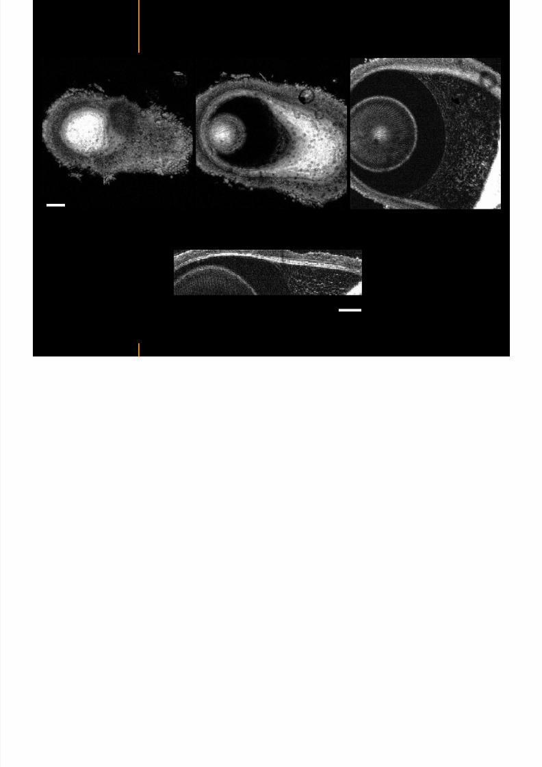
7292019 Atlas of Light-CT images
httpslidepdfcomreaderfullatlas-of-light-ct-images 107118
Xenopus Laevis backtail
7292019 Atlas of Light-CT images
httpslidepdfcomreaderfullatlas-of-light-ct-images 108118
copy LLTech 2012 ndash wwwlltechimagingcom - No copy without authorization 108
100 um30um depth 60um depth
7292019 Atlas of Light-CT images
httpslidepdfcomreaderfullatlas-of-light-ct-images 109118
Mouse embryo in vitro 6 5 days old ndash
7292019 Atlas of Light-CT images
httpslidepdfcomreaderfullatlas-of-light-ct-images 110118
copy LLTech 2012 ndash wwwlltechimagingcom - No copy without authorization 110
Mouse embryo in vitro 65 days old
copy LLTech 2012 Courtesy of Ellvire Guiot and nstitut Jacques Monod Paris France
3D imaging of internal cavity
Sabellaria alveolata mouth palps
7292019 Atlas of Light-CT images
httpslidepdfcomreaderfullatlas-of-light-ct-images 111118
copy LLTech 2012 ndash wwwlltechimagingcom - No copy without authorization 111
500 microm
100 microm
Mouth palps
copy LLTech 2012 Courtesy of Museacutee National drsquoHistoire Naturelle Paris France
7292019 Atlas of Light-CT images
httpslidepdfcomreaderfullatlas-of-light-ct-images 112118
copy LLTech 2012 ndash wwwlltechimagingcom - No copy without authorization 112 copy LLTECH 2011
PLANT IMAGING
Back to table of contents
Mouse ear leaf epidermis
7292019 Atlas of Light-CT images
httpslidepdfcomreaderfullatlas-of-light-ct-images 113118
copy LLTech 2012 ndash wwwlltechimagingcom - No copy without authorization 113
100 microm
Epidermal cell
copy LLTech 2012
Stoma
Leaf veins
7292019 Atlas of Light-CT images
httpslidepdfcomreaderfullatlas-of-light-ct-images 114118
copy LLTech 2012 ndash wwwlltechimagingcom - No copy without authorization 114
114copy LLTech 2012
Fibers
Veins
100 microm
500 microm
Apple
7292019 Atlas of Light-CT images
httpslidepdfcomreaderfullatlas-of-light-ct-images 115118
copy LLTech 2012 ndash wwwlltechimagingcom - No copy without authorization 115
115copy LLTech 2012
100 microm
Wax
Skin
Flesh
500 microm
Water
Effect of harpin infiltration on tobacco leaf
7292019 Atlas of Light-CT images
httpslidepdfcomreaderfullatlas-of-light-ct-images 116118
copy LLTech 2012 ndash wwwlltechimagingcom - No copy without authorization 116
116copy LLTech 2012 Courtesy of UPMC Paris France
50 microm
50 microm
50 microm
50 microm
Upper epidermis
10 min after harpin infiltration 30 min after harpin infiltration
Palisade parenchyma
Epidermis cell
Chloroplast
7292019 Atlas of Light-CT images
httpslidepdfcomreaderfullatlas-of-light-ct-images 117118
copy LLTech 2012 ndash wwwlltechimagingcom - No copy without authorization 117
bull Light-CTtrade does not require tissue preparation nor staining of any kind
bull Creates images within minutes using a non-destructive process
bull Offers a 1 microm cellular resolution in 3D
bull Reveals structural and cellular information
bull Mosaicing allows fast visualisation at various scale
bull En-face high-resolution imaging allows vertical and 3D reconstruction
bull In-vivo images show good promise for in-vivo system
bull Clinical and research applications in cancer detection cosmetics biobanking small animal
developmental biology botanyhellip
Contact Details
7292019 Atlas of Light-CT images
httpslidepdfcomreaderfullatlas-of-light-ct-images 118118
USA
LLTech Inc
103 Carnegie Center DriveSuite 300
Princeton NJ 08540
USA
wwwlltechimagingcom
contactlltechfr
Phone + 1 609 955 3506
LLTech SAS
29 rue du Faubourg St-JacquesPeacutepiniegravere Paris Santeacute Cochin
75014
Paris - France
wwwlltechimagingcom
contactlltechfr
Phone +33 1 82 72 61 25
Europe
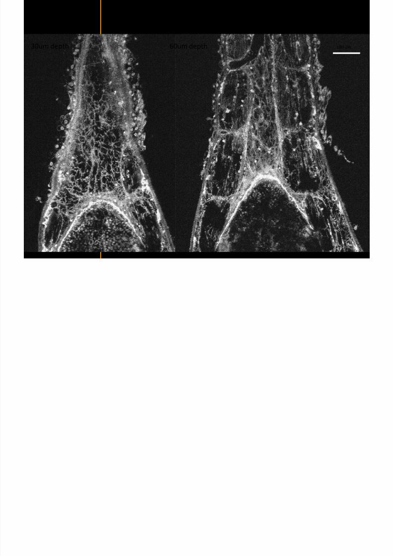
7292019 Atlas of Light-CT images
httpslidepdfcomreaderfullatlas-of-light-ct-images 108118
copy LLTech 2012 ndash wwwlltechimagingcom - No copy without authorization 108
100 um30um depth 60um depth
7292019 Atlas of Light-CT images
httpslidepdfcomreaderfullatlas-of-light-ct-images 109118
Mouse embryo in vitro 6 5 days old ndash
7292019 Atlas of Light-CT images
httpslidepdfcomreaderfullatlas-of-light-ct-images 110118
copy LLTech 2012 ndash wwwlltechimagingcom - No copy without authorization 110
Mouse embryo in vitro 65 days old
copy LLTech 2012 Courtesy of Ellvire Guiot and nstitut Jacques Monod Paris France
3D imaging of internal cavity
Sabellaria alveolata mouth palps
7292019 Atlas of Light-CT images
httpslidepdfcomreaderfullatlas-of-light-ct-images 111118
copy LLTech 2012 ndash wwwlltechimagingcom - No copy without authorization 111
500 microm
100 microm
Mouth palps
copy LLTech 2012 Courtesy of Museacutee National drsquoHistoire Naturelle Paris France
7292019 Atlas of Light-CT images
httpslidepdfcomreaderfullatlas-of-light-ct-images 112118
copy LLTech 2012 ndash wwwlltechimagingcom - No copy without authorization 112 copy LLTECH 2011
PLANT IMAGING
Back to table of contents
Mouse ear leaf epidermis
7292019 Atlas of Light-CT images
httpslidepdfcomreaderfullatlas-of-light-ct-images 113118
copy LLTech 2012 ndash wwwlltechimagingcom - No copy without authorization 113
100 microm
Epidermal cell
copy LLTech 2012
Stoma
Leaf veins
7292019 Atlas of Light-CT images
httpslidepdfcomreaderfullatlas-of-light-ct-images 114118
copy LLTech 2012 ndash wwwlltechimagingcom - No copy without authorization 114
114copy LLTech 2012
Fibers
Veins
100 microm
500 microm
Apple
7292019 Atlas of Light-CT images
httpslidepdfcomreaderfullatlas-of-light-ct-images 115118
copy LLTech 2012 ndash wwwlltechimagingcom - No copy without authorization 115
115copy LLTech 2012
100 microm
Wax
Skin
Flesh
500 microm
Water
Effect of harpin infiltration on tobacco leaf
7292019 Atlas of Light-CT images
httpslidepdfcomreaderfullatlas-of-light-ct-images 116118
copy LLTech 2012 ndash wwwlltechimagingcom - No copy without authorization 116
116copy LLTech 2012 Courtesy of UPMC Paris France
50 microm
50 microm
50 microm
50 microm
Upper epidermis
10 min after harpin infiltration 30 min after harpin infiltration
Palisade parenchyma
Epidermis cell
Chloroplast
7292019 Atlas of Light-CT images
httpslidepdfcomreaderfullatlas-of-light-ct-images 117118
copy LLTech 2012 ndash wwwlltechimagingcom - No copy without authorization 117
bull Light-CTtrade does not require tissue preparation nor staining of any kind
bull Creates images within minutes using a non-destructive process
bull Offers a 1 microm cellular resolution in 3D
bull Reveals structural and cellular information
bull Mosaicing allows fast visualisation at various scale
bull En-face high-resolution imaging allows vertical and 3D reconstruction
bull In-vivo images show good promise for in-vivo system
bull Clinical and research applications in cancer detection cosmetics biobanking small animal
developmental biology botanyhellip
Contact Details
7292019 Atlas of Light-CT images
httpslidepdfcomreaderfullatlas-of-light-ct-images 118118
USA
LLTech Inc
103 Carnegie Center DriveSuite 300
Princeton NJ 08540
USA
wwwlltechimagingcom
contactlltechfr
Phone + 1 609 955 3506
LLTech SAS
29 rue du Faubourg St-JacquesPeacutepiniegravere Paris Santeacute Cochin
75014
Paris - France
wwwlltechimagingcom
contactlltechfr
Phone +33 1 82 72 61 25
Europe
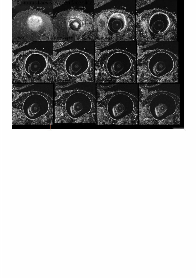
7292019 Atlas of Light-CT images
httpslidepdfcomreaderfullatlas-of-light-ct-images 109118
Mouse embryo in vitro 6 5 days old ndash
7292019 Atlas of Light-CT images
httpslidepdfcomreaderfullatlas-of-light-ct-images 110118
copy LLTech 2012 ndash wwwlltechimagingcom - No copy without authorization 110
Mouse embryo in vitro 65 days old
copy LLTech 2012 Courtesy of Ellvire Guiot and nstitut Jacques Monod Paris France
3D imaging of internal cavity
Sabellaria alveolata mouth palps
7292019 Atlas of Light-CT images
httpslidepdfcomreaderfullatlas-of-light-ct-images 111118
copy LLTech 2012 ndash wwwlltechimagingcom - No copy without authorization 111
500 microm
100 microm
Mouth palps
copy LLTech 2012 Courtesy of Museacutee National drsquoHistoire Naturelle Paris France
7292019 Atlas of Light-CT images
httpslidepdfcomreaderfullatlas-of-light-ct-images 112118
copy LLTech 2012 ndash wwwlltechimagingcom - No copy without authorization 112 copy LLTECH 2011
PLANT IMAGING
Back to table of contents
Mouse ear leaf epidermis
7292019 Atlas of Light-CT images
httpslidepdfcomreaderfullatlas-of-light-ct-images 113118
copy LLTech 2012 ndash wwwlltechimagingcom - No copy without authorization 113
100 microm
Epidermal cell
copy LLTech 2012
Stoma
Leaf veins
7292019 Atlas of Light-CT images
httpslidepdfcomreaderfullatlas-of-light-ct-images 114118
copy LLTech 2012 ndash wwwlltechimagingcom - No copy without authorization 114
114copy LLTech 2012
Fibers
Veins
100 microm
500 microm
Apple
7292019 Atlas of Light-CT images
httpslidepdfcomreaderfullatlas-of-light-ct-images 115118
copy LLTech 2012 ndash wwwlltechimagingcom - No copy without authorization 115
115copy LLTech 2012
100 microm
Wax
Skin
Flesh
500 microm
Water
Effect of harpin infiltration on tobacco leaf
7292019 Atlas of Light-CT images
httpslidepdfcomreaderfullatlas-of-light-ct-images 116118
copy LLTech 2012 ndash wwwlltechimagingcom - No copy without authorization 116
116copy LLTech 2012 Courtesy of UPMC Paris France
50 microm
50 microm
50 microm
50 microm
Upper epidermis
10 min after harpin infiltration 30 min after harpin infiltration
Palisade parenchyma
Epidermis cell
Chloroplast
7292019 Atlas of Light-CT images
httpslidepdfcomreaderfullatlas-of-light-ct-images 117118
copy LLTech 2012 ndash wwwlltechimagingcom - No copy without authorization 117
bull Light-CTtrade does not require tissue preparation nor staining of any kind
bull Creates images within minutes using a non-destructive process
bull Offers a 1 microm cellular resolution in 3D
bull Reveals structural and cellular information
bull Mosaicing allows fast visualisation at various scale
bull En-face high-resolution imaging allows vertical and 3D reconstruction
bull In-vivo images show good promise for in-vivo system
bull Clinical and research applications in cancer detection cosmetics biobanking small animal
developmental biology botanyhellip
Contact Details
7292019 Atlas of Light-CT images
httpslidepdfcomreaderfullatlas-of-light-ct-images 118118
USA
LLTech Inc
103 Carnegie Center DriveSuite 300
Princeton NJ 08540
USA
wwwlltechimagingcom
contactlltechfr
Phone + 1 609 955 3506
LLTech SAS
29 rue du Faubourg St-JacquesPeacutepiniegravere Paris Santeacute Cochin
75014
Paris - France
wwwlltechimagingcom
contactlltechfr
Phone +33 1 82 72 61 25
Europe
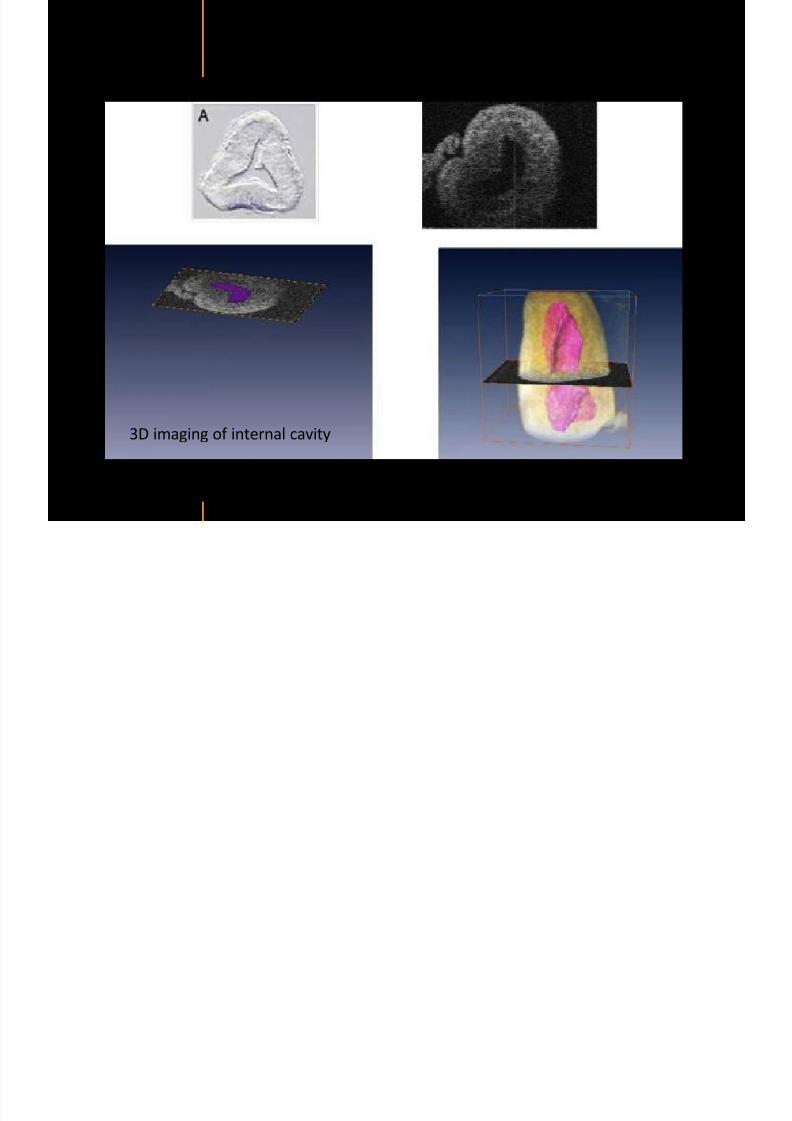
7292019 Atlas of Light-CT images
httpslidepdfcomreaderfullatlas-of-light-ct-images 112118
copy LLTech 2012 ndash wwwlltechimagingcom - No copy without authorization 112 copy LLTECH 2011
PLANT IMAGING
Back to table of contents
Mouse ear leaf epidermis
7292019 Atlas of Light-CT images
httpslidepdfcomreaderfullatlas-of-light-ct-images 113118
copy LLTech 2012 ndash wwwlltechimagingcom - No copy without authorization 113
100 microm
Epidermal cell
copy LLTech 2012
Stoma
Leaf veins
7292019 Atlas of Light-CT images
httpslidepdfcomreaderfullatlas-of-light-ct-images 114118
copy LLTech 2012 ndash wwwlltechimagingcom - No copy without authorization 114
114copy LLTech 2012
Fibers
Veins
100 microm
500 microm
Apple
7292019 Atlas of Light-CT images
httpslidepdfcomreaderfullatlas-of-light-ct-images 115118
copy LLTech 2012 ndash wwwlltechimagingcom - No copy without authorization 115
115copy LLTech 2012
100 microm
Wax
Skin
Flesh
500 microm
Water
Effect of harpin infiltration on tobacco leaf
7292019 Atlas of Light-CT images
httpslidepdfcomreaderfullatlas-of-light-ct-images 116118
copy LLTech 2012 ndash wwwlltechimagingcom - No copy without authorization 116
116copy LLTech 2012 Courtesy of UPMC Paris France
50 microm
50 microm
50 microm
50 microm
Upper epidermis
10 min after harpin infiltration 30 min after harpin infiltration
Palisade parenchyma
Epidermis cell
Chloroplast
7292019 Atlas of Light-CT images
httpslidepdfcomreaderfullatlas-of-light-ct-images 117118
copy LLTech 2012 ndash wwwlltechimagingcom - No copy without authorization 117
bull Light-CTtrade does not require tissue preparation nor staining of any kind
bull Creates images within minutes using a non-destructive process
bull Offers a 1 microm cellular resolution in 3D
bull Reveals structural and cellular information
bull Mosaicing allows fast visualisation at various scale
bull En-face high-resolution imaging allows vertical and 3D reconstruction
bull In-vivo images show good promise for in-vivo system
bull Clinical and research applications in cancer detection cosmetics biobanking small animal
developmental biology botanyhellip
Contact Details
7292019 Atlas of Light-CT images
httpslidepdfcomreaderfullatlas-of-light-ct-images 118118
USA
LLTech Inc
103 Carnegie Center DriveSuite 300
Princeton NJ 08540
USA
wwwlltechimagingcom
contactlltechfr
Phone + 1 609 955 3506
LLTech SAS
29 rue du Faubourg St-JacquesPeacutepiniegravere Paris Santeacute Cochin
75014
Paris - France
wwwlltechimagingcom
contactlltechfr
Phone +33 1 82 72 61 25
Europe
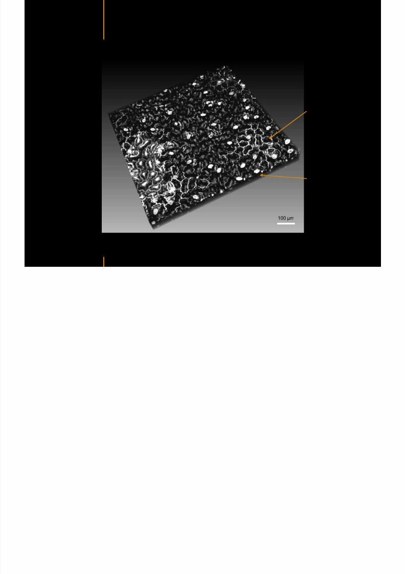
7292019 Atlas of Light-CT images
httpslidepdfcomreaderfullatlas-of-light-ct-images 116118
copy LLTech 2012 ndash wwwlltechimagingcom - No copy without authorization 116
116copy LLTech 2012 Courtesy of UPMC Paris France
50 microm
50 microm
50 microm
50 microm
Upper epidermis
10 min after harpin infiltration 30 min after harpin infiltration
Palisade parenchyma
Epidermis cell
Chloroplast
7292019 Atlas of Light-CT images
httpslidepdfcomreaderfullatlas-of-light-ct-images 117118
copy LLTech 2012 ndash wwwlltechimagingcom - No copy without authorization 117
bull Light-CTtrade does not require tissue preparation nor staining of any kind
bull Creates images within minutes using a non-destructive process
bull Offers a 1 microm cellular resolution in 3D
bull Reveals structural and cellular information
bull Mosaicing allows fast visualisation at various scale
bull En-face high-resolution imaging allows vertical and 3D reconstruction
bull In-vivo images show good promise for in-vivo system
bull Clinical and research applications in cancer detection cosmetics biobanking small animal
developmental biology botanyhellip
Contact Details
7292019 Atlas of Light-CT images
httpslidepdfcomreaderfullatlas-of-light-ct-images 118118
USA
LLTech Inc
103 Carnegie Center DriveSuite 300
Princeton NJ 08540
USA
wwwlltechimagingcom
contactlltechfr
Phone + 1 609 955 3506
LLTech SAS
29 rue du Faubourg St-JacquesPeacutepiniegravere Paris Santeacute Cochin
75014
Paris - France
wwwlltechimagingcom
contactlltechfr
Phone +33 1 82 72 61 25
Europe
7292019 Atlas of Light-CT images
httpslidepdfcomreaderfullatlas-of-light-ct-images 118118
USA
LLTech Inc
103 Carnegie Center DriveSuite 300
Princeton NJ 08540
USA
wwwlltechimagingcom
contactlltechfr
Phone + 1 609 955 3506
LLTech SAS
29 rue du Faubourg St-JacquesPeacutepiniegravere Paris Santeacute Cochin
75014
Paris - France
wwwlltechimagingcom
contactlltechfr
Phone +33 1 82 72 61 25
Europe