associative representational plasticity in the auditory
TRANSCRIPT

Associative representational plasticity in theauditory cortex: A synthesis of two disciplinesNorman M. WeinbergerCenter for the Neurobiology of Learning and Memory, and Department of Neurobiology and Behavior, University of California,Irvine, California 92697-3800, USA
Historically, sensory systems have been largely ignored as potential loci of information storage in the neurobiologyof learning and memory. They continued to be relegated to the role of “sensory analyzers” despite consistentfindings of associatively induced enhancement of responses in primary sensory cortices to behaviorally importantsignal stimuli, such as conditioned stimuli (CS), during classical conditioning. This disregard may have beenpromoted by the fact that the brain was interrogated using only one or two stimuli, e.g., a CS+ sometimes with aCS–, providing little insight into the specificity of neural plasticity. This review describes a novel approach thatsynthesizes the basic experimental designs of the experimental psychology of learning with that of sensoryneurophysiology. By probing the brain with a large stimulus set before and after learning, this unified method hasrevealed that associative processes produce highly specific changes in the receptive fields of cells in the primaryauditory cortex (A1). This associative representational plasticity (ARP) selectively facilitates responses to tonal CSs atthe expense of other frequencies, producing tuning shifts toward and to the CS and expanded representation of CSfrequencies in the tonotopic map of A1. ARPs have the major characteristics of associative memory: They are highlyspecific, discriminative, rapidly acquired, exhibit consolidation over hours and days, and can be retained indefinitely.Evidence to date suggests that ARPs encode the level of acquired behavioral importance of stimuli. The nucleusbasalis cholinergic system is sufficient both for the induction of ARPs and the induction of specific auditorymemory. Investigation of ARPs has attracted workers with diverse backgrounds, often resulting in behavioralapproaches that yield data that are difficult to interpret. The advantages of studying associative representationalplasticity are emphasized, as is the need for greater behavioral sophistication.
This review presents an account of research on neurophysiologi-cal plasticity in the primary auditory cortex (A1), focusing on theissues of associativity and specificity. Related reviews of greaterbreadth and different emphasis are available (Palmer et al. 1998;Edeline 2003; Ohl et al. 2003; Rauschecker 2003; Weinberger2004a). Consideration of perceptual learning, which involvesprolonged, increasingly difficult frequency discrimination train-ing to reduce the difference limen, is beyond the scope of thisreview (see, e.g., Goldstone et al. 1997; Gilbert et al. 2001; Mooreet al. 2003; Pleger et al. 2003; Brown et al. 2004; Hawkey et al.2004; Polley et al. 2006). However, we consider associative learn-ing to be fundamental to perceptual learning because, at a mini-mum, subjects must first learn an association between an acous-tic stimulus and a reinforcer; and second, they must learn anassociation between an instrumental response and a reinforcer,contingent on the acoustic stimulus.
A central goal of this paper is to demonstrate how a synthe-sis of approaches from the traditionally separate disciplines ofsensory neurophysiology and the experimental psychology oflearning can illuminate fundamental issues in the neurobiologyof learning and memory in general, and the neural substrates ofassociative processes in particular. Chief among these is the ques-tion of neural representations of sensory/perceptual events, suchrepresentations being widely thought to constitute core elementsthat become associated in learning.
It may seem strange to some readers that sensory neuro-physiology could play a critical role in attacking central issues inbrain substrates of learning/memory. After all, research in thisfield has been dominated and remarkable progress has been
made by concentrating on structures such as the cerebellum,amygdala, striatum, hippocampus, medial temporal lobe, fron-tal, cingulate, and other nonsensory cortical regions. Moreover,most basic knowledge of the workings of sensory systems, par-ticularly the auditory, somatosensory, and visual systems, hasbeen garnered from animals under deep general anesthesia, thestate that is most inimical to new learning. Thus, it has beenconcluded, usually implicitly, that sensory systems, while verycomplex and perhaps even elegant, are limited to providing de-tailed information about the outside world, in a sense supplyingthe “raw materials” for learning and memory. But, they areviewed as neither critical sites of processes enabling learning northe loci of memory storage.
Research, especially during the past 15 yr, has directly chal-lenged traditional views that essentially excluded sensory sys-tems from an active and likely critical role in learning andmemory. Most relevant research comes from work on the audi-tory system, particularly on the primary auditory cortex, towhich we will now turn. However, importantly, much of thisvery recent and currently burgeoning area of inquiry emanatesfrom workers whose major training has been in sensory neuro-physiology rather than in learning and memory. Therefore, it isnecessary to encourage the use of accepted and valid conceptsand methods from the field of learning/memory. Of course, theconverse holds for researchers whose training has been in learn-ing; their use of appropriate methods from the field of sensoryphysiology is equally important. These issues will be discussedlater. But first, it will be helpful to consider why sensory systemsin general, and the auditory cortex in particular, have beenlargely ignored in the neurobiology of learning and memory. Infact, they are among the very last brain systems admitted to thisfield.
E-mail [email protected]; fax (949) 824-4576.Article published online in January 2007. Article and publication date are athttp://www.learnmem.org/cgi/doi/10.1101/lm.421807.
Review
14:1–16 ©2007 by Cold Spring Harbor Laboratory Press ISSN 1072-0502/07; www.learnmem.org Learning & Memory 1www.learnmem.org
Cold Spring Harbor Laboratory Press on November 18, 2021 - Published by learnmem.cshlp.orgDownloaded from

Background
The auditory cortex was delineated by ablation work in animalsduring the latter part of the 19th century (Diamond 1985). The“gold standard” was a demonstration of crudely assessed deaf-ness, understandable given the technical limitations of the times.With the advent of electrophysiology in the 1920s–1930s, it be-came possible to seek cortical regions that responded (evokedpotentials) to sound, and eventually to successfully demarcate A1and its cochleotopic and tonotopic frequency organization (e.g.,Tunturi 1944). However, in 1906, long before the physiologicalidentification of auditory cortical fields, Campbell had publisheda landmark study of cortical cytoarchitectonics of the humanand some animals. He sought structural–functional relationshipsand essentially asserted them on anatomical grounds. While per-haps not the first worker to use the terms “sensory” and “psy-chic” cortex, his influence was profound. Campbell labeled thecytoarchitectonic region now identified as primary auditory cor-tex (A1) “auditory sensory,” and called auditory regions nearby(now often called auditory “belt” areas [Kaas and Hackett 2000])as “auditory psychic.” In this, Campbell intended and made aclear distinction between cortical regions he considered to bepurely sensory from those he believed to concern the under-standing or comprehension of sounds. His implicit assumptionwas one of strictly hierarchical cortical functional architecture,with what are now referred to as “cognitive functions” depen-dent upon input from “sensory” structures. This conceptualschema still has strong resonance today. The late Irving Dia-mond, a major contributor to the anatomical and behavioralunderstanding of thalamic and cortical levels of the neuraxis,insightfully held that Campbell’s monograph was instrumentalin removing learning and memory from primary sensory cortices(Diamond 1985).
Following World War II and continuing to the present, au-ditory neurophysiology expanded greatly. The coding of acousticparameters by single neurons and groups of cells in many specieshas yielded the functional organization of auditory fields adja-cent to A1 and refinement of the latter’s tonotopic organizationas well as providing insights into other stimulus domains, e.g.,binaural interactions, bandwidth (sharpness of tuning), temporaland spectral modulations, etc.
However, during the 30-yr span from the mid-1950s to themid-1980s there was a little-noted parallel line of inquiry intoassociative learning and the auditory cortex. In 1956, Galamboset al. (1956) published the first electrophysiological study of theauditory cortex during learning. They found that classical con-ditioning (click–shock pairing) in the cat was accompanied bysignificant increases in the amplitude of A1-evoked potentials tothe conditioned stimulus (CS). Interestingly, these investigatorsincluded a “sensory physiology” control, i.e., showed that in-creased amplitude was not due to inadvertent increased CS soundlevel (intensity), due to relaxation of the middle-ear muscles orother factors, by the use of neuromuscular blockade. But they didnot use controls for nonassociative factors, such as sensitization.Subsequently, scores of similar studies were performed in a vari-ety of species (including humans), with the addition of nonas-sociative controls, the extension of evoked potential recordingsto clusters of neurons and single-cell recordings, the use of two-tone discrimination protocols and the employment of instru-mental conditioning tasks, and appetitive as well as aversive re-inforcements. Overall, the findings clearly established that asso-ciative processes were responsible for increased responses of A1 toCS and CS+; responses to CS– stimuli did not increase as much orat all, and often decreased (Weinberger and Diamond 1987).
In toto, such plasticity in A1 attracted scant attention, evenwithin the neurobiology of learning and memory. Without de-
tailed historical study, the reasons for this neglect can only be thesubject of speculation. Several factors come to mind. First, therewas the discovery that patient H.M., having received bilateralexcisions of medial temporal lobe structures, could not retainnew facts. In its understandably exuberant expansion, subse-quent research focused on structures that appeared to be essentialfor memory, such as the hippocampus. Second, there was noconceptual framework within which to incorporate the findingsof associative plasticity in A1, particularly as most models ofbrain and learning restricted sensory cortices to the status ofstimulus analyzers. (The same consideration applied to the occa-sional reports of similar findings in the primary somatosensoryand visual cortices.) Third, workers in learning and memory hadno vested interest in sensory systems, and therefore were notconcerned that the research initiated by Galambos and col-leagues disproved the view that sensory systems responded tosensory parameters, not “psychological” parameters such as theacquired behavioral significance of a stimulus.
Neglect from auditory neurophysiologists might seem par-ticularly unexpected. After all, a major implication of the factthat associative processes systematically modify the responses ofA1 to physically constant sounds is that such neural responsesappear to be inherently ambiguous. For example, an increase inresponse magnitude might be due either to an increase in stimu-lus level, an increase in its behavioral significance, or both. Myconjecture here is that the learning/memory findings seemed tohave little relevance for sensory physiology, because the auditorystimuli used were too limited to be of any interest. Thus, standardconditioning studies used a single acoustic CS and discrimina-tion studies used only two, a CS+ and a CS–. However, the con-cept of the receptive field is fundamental to sensory physiology;no sensory physiologist would attempt to describe the responseproperties of a cell with such a restricted stimulus set. Moreover,the conditioning studies did not even provide information onthe relationship between the tone used as the CS and the bestfrequency (peak of the spectral tuning curve) or the characteristicfrequency (frequency of response at threshold). Finally, single-unit conditioning studies in the 1970s and early 1980s invariablyreported that a certain proportion of A1 cells increased their dis-charges, while another proportion decreased their discharges.While these effects were shown to be associative, they made littlefunctional sense. Therefore, from the viewpoint of auditoryphysiology, the changes in cortical responses during learningwere essentially not interpretable.
Our own studies of single cells in both A1 and the littleunderstood adjacent “secondary field” (A2), while adding somenew wrinkles to the discourse on associative processes (Diamondand Weinberger 1984b; Weinberger et al. 1984b), found similarmixed effects and convinced us that the standard “neural corre-lates of conditioning” approach to sensory systems was futile.
Why study the auditory cortex duringclassical conditioning?At this point, it may have occurred to some readers to ask whythe auditory cortex would be of interest in classical conditioning.Indeed, a common question from workers in both the fields oflearning/memory and sensory physiology is “Do lesions of A1impair or prevent classical conditioning?” The answer is gener-ally “No”. It has long been known that simple (i.e., single-tone,nondiscriminative) classical conditioned responses can developafter ablation of A1 alone or as part of extensive cortical destruc-tion (e.g., DiCara et al. 1970; Norman et al. 1977; Berntson et al.1983; Teich et al. 1988; Romanski and LeDoux 1992).
However, this question seems to involve implicit assump-tions such that a negative answer, as given above, leads to the
Weinberger
2 Learning & Memorywww.learnmem.org
Cold Spring Harbor Laboratory Press on November 18, 2021 - Published by learnmem.cshlp.orgDownloaded from

conclusion that A1 cannot be of much interest. These assump-tions include: (1) neurophysiological studies of conditioningseek stimulus–response circuitry; (2) the memory trace formed islocalized; (3) neurophysiological plasticity that develops is thesubstrate of the CR; (4) destruction of the site of such plasticityshould disrupt the CR.
While all of these assumptions may be relevant to investi-gation of a single, specific CR (e.g., Christian and Thompson2005), they do not apply to all neurophysiological studies ofconditioning (see Ohl and Scheich 2004 vs. Weinberger 2004b).First, even simple classical conditioning transcends stimulus–response associations. Rather, conditioning involves many asso-ciations, including stimulus–stimulus (CS–US), stimulus–outcome, stimulus–context, etc. Second, neurophysiologicalstudies can therefore be applied to associations other than CS–CRassociations. Third, CS–US associations are known to developrapidly (e.g., in a few trials), preceding CS–CR associations, andtherefore may be involved in the development of CS–CR links(e.g., Schlosberg 1937; Mowrer 1947; Konorski 1967; Lennartzand Weinberger 1992a). Fourth, multiple behavioral CRs developduring rapid CS–US learning, e.g., changes in heart rate, respira-tion, blood pressure, pupillary diameter, skin resistance, behav-ioral freezing, etc., any one of which can be used to verify that aCS–US association has developed (of course, given appropriatenonassociative controls). Fifth, the total memory for a givenevent is distributed, although its components may be localized,as in the case of a specific, somatic CS–CR association (e.g., tone–eyeblink). Some of the associations are likely to include the rel-evant sensory systems. After all, auditory memories are not likelyto be stored exclusively outside of the auditory system. Othercomponents are likely to involve somewhat specialized systems,such as those processing nociception in the case of a shock US,emotional-processing networks, etc. Sixth, circuitry for a simpleCS–CR association can be completely subcortical, so that it sur-vives cortical lesions. However, cortical areas also can store CS–US (and other) information in parallel with subcortical systems.Seventh, cortically stored information could affect immediate be-haviors by control over subcortical circuits. Most important,memory traces in the cortex have access to a much greater rangeof information than sub cortex, and they may store informationfor the indefinite future, where it can be used in a highly flexiblemanner to subserve adaptive behaviors. For example, whilesimple auditory conditioning is not destroyed by lesions of A1, assoon as two-tone discrimination is demanded, A1 is required(Teich et al. 1988). A1 is also obligatory to achieve experimentalextinction (Teich et al. 1989).
Thus, in response to the question posed at the outset of thissection, A1 (and other primary sensory cortices) can be profitablystudied to determine the cortical “fate” of stimuli that enter intoassociations. In short, such studies of neurophysiological plastic-ity can be directed to issues of the representation of stimuli andthe transformations that they may develop during learning,without any reference to particular behaviors that index the es-tablishment of stimulus–stimulus associations.
Associative representational plasticitySensory physiology and the neurobiology of learning andmemory are the two fields within neuroscience that are con-cerned with the “fate” of sensory stimuli. The former includes asearch for the principles of neural coding, the issue of how thebrain represents the world. The latter includes a concern with thetransformation of a “neutral” stimulus into a “signal” stimulusthat gains new behavioral potential as it enters into associations.This involves another aspect of “representation,” viz., the mecha-nisms by which one stimulus gains some of the properties ofother stimuli or events. Although these two disciplines have de-
veloped along separate paths, they can be seen as complemen-tary.
Figure 1 points out that sensory stimuli have both physicaland psychological parameters. The former are assessed by appro-priate engineering devices that can measure, e.g., acoustic fre-quency (in kHz) and level (in dB SPL). The latter are assessedbehaviorally, there being no universal behavioral units compa-rable to kHz, dB, and the like. Sensory physiology varies thephysical parameters and traditionally holds the psychological pa-rameters constant by the use of general anesthesia in animals,thus preventing (inadvertent) learning. In the case of the study ofassociative processes, the actual sensory stimuli are held con-stant, but their relationships are varied, e.g., when a CS and USare paired.
A synthesis of the basic approaches of these fields can beaccomplished by performing both types of studies in a singleexperiment. The first step is to perform a sensory neurophysiol-ogy experiment by, e.g., determining the receptive field (i.e.,acoustic frequency tuning function of A1) before the learningexperience. Second, any learning protocol can be run; for thesake of exposition, we assume a tonal CS in simple classical con-ditioning. The conditioning part of the design need only involvean additional, nonstandard decision: Selection of the CS fre-quency must be based on knowledge of the “place” of the tonalCS in the receptive field. For example, if the CS is at the peak ofthe tuning curve (best frequency [BF]), then there is danger of aceiling effect. That is, learning-induced increased response mightnot be detectable if cells are already firing at their maximumbefore training. If the CS is selected to be another frequency, thenthere is the possibility of detecting a tuning shift from the origi-nal BF toward or to the CS frequency. Thus, the preferred strategyis to select a non-BF frequency. Third, and finally, step one isrepeated, i.e., post-training determination of the frequency tun-ing function. Short and long-term retention can be studied byobtaining further RFs at desired intervals following training.
Pre- and post-training context should be the same, so thatpost-tuning changes are attributable to training and are not con-founded by a change of context from pre- to post-training peri-ods (e.g., a change in arousal level). The subject’s state can bedirectly monitored (heart rate, EEG) or controlled (e.g., by ob-taining pre- and post-training tuning with animals anesthetized).In contrast, training should be in a different context from thedetermination of receptive fields, to avoid responses to the CSfrequency when it is given as one of many frequencies in thepost-period, and hence, could produce experimental extinction.Contexts can differ in many ways; they can be different acousti-
Figure 1. The fundamental paradigms of the disciplines of sensory neu-rophysiology and the experimental psychology of learning/memory maybe viewed as complementary. This entails recognition that stimulus pa-rameters have two basic properties: physical and psychological. Eachdiscipline usually manipulates one of these while holding the other con-stant.
Associative representational plasticity
Learning & Memory 3www.learnmem.org
Cold Spring Harbor Laboratory Press on November 18, 2021 - Published by learnmem.cshlp.orgDownloaded from

cally (e.g., different durations of tone pulses, rates of tone pre-sentation, and actual tonal frequencies presented), visually (e.g.,ambient illumination, light vs. dark), and spatially (e.g., differentexperimental rooms and chambers). We routinely change audi-tory, visual, and spatial contexts between training sessions andpre- and post-training determination of receptive fields.
This unified design yields the usual behavioral and neuro-physiological data for the conditioning phase, i.e., the develop-ment of conditioned responses and the development of neuralplasticity. But, importantly, it also reveals the effects of associa-tive processes on the detailed processing of conditioned andmany other stimuli, plasticity of sensory receptive fields (RFs).Thereby, both fields benefit. For example, sensory physiologycan learn the extent to which the encoding of stimuli is affectedby experience. The neurobiology of learning/memory can learnthe functional significance of associatively induced plasticity inthe sensory system of the CS; that is, it can track changes in therepresentation of a CS as it gains “associative power.”
Traditionally, a neurophysiological study of classical condi-tioning can, and must, determine the extent to which resultantneuronal changes are due to associative vs. nonassociative pro-cesses. However, a second step is possible when a sensory physi-ology approach is included. If the neural plasticity is associative,one can additionally determine the extent to which the plasticityis specific to the value of the CS vs. general. Standard approachesare limited to measuring neural activity during conditioning tri-als. As noted previously, such studies generally found that re-sponses to the CS are increased during pairing trials (Fig. 2A).However, increased response to the CS could be due to an in-creased response to all stimuli in the dimension of the CS, e.g.,tone frequency if the CS were auditory, angle of orientation if theCS were a visual line segment, or locus of touch on the bodysurface if the CS were somatosensory. Receptive field analysis,using many stimuli along a dimension within a modality, wouldshow increased responses across the dimension in question (Fig.2B). This would constitute general associative plasticity. On theother hand, increased responses to the CS could be due to aneffect that was specific to the CS value. In this case, RF analysiswould reveal maximal facilitation at the CS value with somecombination of smaller increases, no changes, or decreases atother stimuli along the dimension. In toto, the result could be ashift of tuning to the value of the CS (Fig. 2C). This would con-stitute CS-specific associative plasticity.
The importance of determining the degree of specificity ofassociative representational plasticity (ARP) is considerable. Fouravenues of research will be mentioned here.
First, a major advantage of receptive field analysis is that itprovides for the study of specificity of neuronal plasticity. That is,sensory/perceptual representations can be investigated directly,and thus, learning-induced changes in representations can bedetected. For example, does the neural representation of a CSactually change in coding (not merely in amplitude) when itacquires a given association or set of associations? Given onlyresponses to the CS before, during, and after training, it is im-possible to determine the actual types of plastic changes inducedby associative processes. RF (or similar) analysis resolves this is-sue, in much the same way that behavioral stimulus generaliza-tion gradients resolve the issue of the specificity of learning.
Second, the nature of a change in representation can pro-vide a means for investigating the relationships between repre-sentational plasticity and changes in behavior or behavior po-tential. If both post-training receptive fields and generalizationgradients were obtained within the same subjects, it would bepossible to determine the relationship between the specificity ofbrain and behavior during learning in all sensory systems and atevery level of the neuraxis (brainstem, cerebellum, thalamus, cor-
tex). Such analyses could be applied to future situations as well asimmediate behavior. For example, can the development of anARP predict subsequent behavior in cases of behaviorally silentlearning and in the future when past learning is used to solve anew problem?
Third, research is currently dominated by studies of the pro-cesses necessary for the acquisition and maintenance of memory.However, all memories are about something: They have content.The study of ARPs can be used to determine the contents of theneural substrates of memory. Associations are between andamong specific elements, be they particular stimuli, responses,outcomes, contexts, or whatever. For example, are ARPs involvedonly in the acquisition of associations or are they as enduring asbehaviorally validated memory? Do ARPs exhibit actual consoli-dation after initial acquisition, become stronger over time, andless resistant to disruption? Do the forms of ARPs encode thedetails of associations and memories of their elements?
Fourth, the study of ARPs can illuminate basic issues of as-sociative processes. For example, does an “occasion setter” nec-essarily develop representational plasticity in order to catalyzeassociations between and among other stimuli and events? If so,to what extent are the neural substrates of effective occasionsetters fundamentally the same or different from the associatedelements?
Receptive field and map plasticityThe unified “receptive field–conditioning” approach was firstused in the mid-1980s, but not in A1. Rather, two nonprimary
Figure 2. Receptive field analysis reveals whether learning-inducedplasticity is general to the dimension of a conditioned stimulus or specificto the value of the CS. (A). During training trials, one can determinewhether or not responses to the CS changed; in this case, they increased.However, both general and specific plasticity could produce this result.(B). A general change revealed by receptive field analysis before and afterconditioning. (C). A CS-specific instance of associative representationalplasticity, in which responses to many non-CS frequencies are reduced,producing a shift in tuning to the frequency of the CS.
Weinberger
4 Learning & Memorywww.learnmem.org
Cold Spring Harbor Laboratory Press on November 18, 2021 - Published by learnmem.cshlp.orgDownloaded from

fields (A2) and ventral ectosylvian (VE) were studied, because itwas assumed (wrongly as it turned out) that A1 would be lessplastic, based on dominant beliefs in auditory physiology. Catswere trained in fear conditioning and developed associative pu-pillary dilation conditioned responses. Frequency RFs (tuningfunctions) obtained before and after conditioning revealed CS-specific plasticity: The maximum changes in response were at theCS frequency. Both CS-specific increases and decreases werefound. This ARP was retained unless subjects underwent standardextinction training, in which case, tuning returned toward or tobaseline status (Weinberger et al. 1984a; Diamond and Wein-berger 1986). Importantly, the sign of plasticity that developedduring training trials was not necessarily the same as that whichwas evident in post-training RFs. This finding indicates that con-text can affect the expression of associative plasticity and hasbeen attributed to performance factors during actual training tri-als, such as arousal and motivational factors caused by the pres-ence of the unconditioned stimulus (US) (Diamond and Wein-berger 1989). Indeed, in the auditory cortex, even associativeprocesses can induce increased tonic arousal level with increasedrates of background (spontaneous), single-unit discharges, oreven decreased arousal and decreased background unit dis-charges at the time that subjects “solve” the CS–US contingency,as indexed by the development of CRs (Diamond and Wein-berger 1984a,b).
Although such state effects cannot be eliminated duringtraining, they can be eliminated when pre- and post-training RFsare obtained because, as explained above, RFs can be obtainedwith subjects in a planned, markedly different context that ame-liorates or eliminates generalization of, e.g., arousal effects. Gen-eralization also can be prevented altogether by training subjectsin the waking state but obtaining pre- and post-training RFs (orfrequency maps) with subjects under deep general anesthesia(Weinberger 2004a).
The discovery of ARPs in A2 and VE was largely ignored.This may have reflected the fact that little was known about theseauditory fields, which do not contain the fine-grain tonotopicorganization found in A1. Thus, an “anything is possible” beliefabout A2 and VE would tend to reduce attention to any findings,no matter how distinctive. Indeed, to the extent that these re-gions might have been considered auditory “psychic” cortex,learning effects would be expected.
Inquiry was thereafter directed to unit discharges in A1. Thefirst such study involved classical fear conditioning in the adultguinea pig (Bakin and Weinberger 1990). Subjects received asingle brief training session of tone paired with shock. The CSfrequency was selected to not be the pre-training best frequency,to allow for the detection of possible changes in frequency tun-ing. Subjects developed conditioned responses to the CS follow-ing a period of habituation to the CS presented alone. Figure 3presents typical behavioral data, which was used only to validateCS–US learning, not for the purpose of CR circuit tracing, asexplained previously in the section “Why study the auditory cor-tex during classical conditioning?”.
Immediately after training, neuronal RFs had shifted fromthe pre-training BF toward or all the way to the CS frequency sothat it could become the new best frequency (Fig. 4). Shifts werecaused by a simultaneous increase in response to the CS fre-quency, while responses to the pre-training BF and many otherfrequencies decreased. These tuning shifts were only toward theCS frequency, so were not due to random variation. “Tracking”tuning for weeks revealed only small random fluctuations in bestfrequency (Galvan et al. 2001). ARPs could be seen even in com-plex RFs, such as double-peaked tuning (Fig. 5A). Also, responsesto the CS frequency alone could develop even when a cell ap-peared to be nonresponsive to tones (Fig. 5B).
Subsequent studies revealed that ARP can develop very rap-idly (within five trials), is discriminative (increased responses tothe CS+, decreased responses to the CS– tone), exhibits consoli-dation (post-training increased strength over hours and dayswithout further training) (Fig. 6), and could last indefinitely(tracked to 8 wk post-training) (Weinberger 1998). It also devel-oped in avoidance learning (Bakin et al. 1996). Tuning shifts aregenerally assessed at stimulus levels used for training, i.e., wellabove threshold (e.g., 70 dB SPL), although even when trained atone level, they can develop across the range of stimulus levels(10–80 dB SPL) (Galvan and Weinberger 2002). Tuning shifts tothe CS frequency are not limited to aversive situations, as theyhave been found with rewarding brain stimulation as the US(Kisley and Gerstein 2001).
ARPs are highly specific, consistently exhibiting increasedresponses only at or near the CS frequency across subjects, withdecreased responses to lower and higher frequencies (Fig. 7A).CS-specific increased responses and tuning shifts are associative,as they require pairing. Random or unpaired presentation of toneand shock produce increased responses across the frequency spec-trum. In fact, general increased responses develop in A1 whethersensitization training involves a tone with random shock or aflashing light with random shock, demonstrating that generalincreased responses across the frequency RF are truly arousal de-pendent, not due to auditory processing per se (Fig. 7B) (Bakin etal. 1992). Habituation produces the opposite effect, i.e., a specificdecrease in response to the repeated stimulus, with little or nochange in response to other frequencies (Condon and Wein-berger 1991) (Fig. 7C).
It might be thought that arousal confounds during post-training testing are responsible for the specificity of increasedresponses to the CS frequency. That might occur if presentationof this frequency, alone among many other frequencies, pro-duced arousal due to prior conditioning. However, the acousticcontext is very different. Pairing involves presentation of randomand infrequent presentations of a CS (and US) (mean intertrialintervals not less than 40 sec), whereas RF determination con-sisted of the presentation of many frequencies (typically at least15), at many levels (typically 10–80 dB) at constant, brief inter-
Figure 3. Behavioral verification of associative learning in classical con-ditioning. Cardiac activity (changes in heart rate to a tone) are shown fortwo groups of guinea pigs. First, both groups received a tone unpairedwith shock for 10 trials (Sens), which resulted in an initial decrease inheart rate during the first block of five trials; this response was no longerpresent during the second block, perhaps indicating habituation to thetone. Subsequently, one group (Condit) received tone paired with shock,while the other (Sens) continued to receive tone and shock unpaired.Conditioning produced cardiac deceleration CRs as soon as the first blockof pairing, which continued to develop across trials. In contrast, the sen-sitization group showed no such growth of the CR. Such behavioral find-ings were used to validate the development of a CS–US association, notfor purposes of determining CS–CR circuitry (see text).
Associative representational plasticity
Learning & Memory 5www.learnmem.org
Cold Spring Harbor Laboratory Press on November 18, 2021 - Published by learnmem.cshlp.orgDownloaded from
vals (typically 1–2/sec). Direct measurement of the CR behaviorrevealed no response to the CS frequency when embedded inthe post-training stimulus set (e.g., Diamond and Weinberger1989). Also, the latency of neuronal response to any tone is onthe order of 10–50 msec, whereas the latency to detectable EEGactivation is well over 100 msec (Weinberger and Lindsley 1964).Moreover, if the CS caused arousal, then the tone following theCS within a few seconds should also show an effect, which wasnot the case. Finally, ARPs are found when post-training RFs aredetermined with subjects under general anesthesia (Lennartz andWeinberger 1992b; Weinberger et al. 1993; Rutkowski et al.2003).
Associative processes were predicted to increase the area ofrepresentation of the A1 octave band containing the CS fre-quency, because the tonotopic map is comprised of preferredfrequencies across cortical space, and tuning shifts should in-crease the number of loci at which the CS frequency is preferred
(Weinberger et al. 1990a,b). This hasbeen demonstrated in a study of the re-lationship between the level of CS-tonalimportance and representational area.Rats were trained to associate a 6.0 kHztone with the opportunity to bar pressfor water. The behavioral importance ofthis CS for different animals was con-trolled by varying motivational levels bydifferential amounts of supplementalwater, so that asymptotic performanceranged across subjects from 60% tomore than 90% correct. (As maps cannoteasily be obtained more than oncewithin subjects, the pre-post trainingdesign was modified to obtain post-training maps in different groups). Con-trols received the same schedule of tonepresentations, but were trained to barpress only in the presence of a visualstimulus. Maps of the ACx showed anexpanded representation for the fre-quency band centered on the CS. Fur-ther, the greater the level of behavioralimportance, as indexed by the level ofcorrect performance, the larger the per-cent of area tuned to the CS (Figs. 8, 9).
The control group failed to develop a change in tonotopic orga-nization, not differing from naive subjects. The findings indicatethat the amount of representational area induced during associa-tive learning might serve as a “memory code” for the level ofacquired behavioral significance of sound (Rutkowski and Wein-berger 2005).
In summary, the initial line of research that combines au-ditory physiological determination of frequency tuning withstandard conditioning training revealed that associative pro-cesses modify the representation of the conditioned stimulus.Specifically, they systematically alter A1 receptive fields such thatresponses to the CS are facilitated, while responses to other fre-quencies are generally decreased, enabling tuning shifts towardand to the CS frequency. This CS-specific tuning plasticity hasthe major characteristics of associative memory and the amountof increased representation of the CS is proportional to its degreeof motivational importance.
Controversies: The importanceof behavioral factorsFollowing the development of this lineof inquiry, several major controversiesarose. These include opposing claims re-garding the form of receptive field plas-ticity, the interpretation of its functionalsignificance, and its underlying neuralmechanisms. The position taken here isthat these controversies reflect “growingpains” in the field, due to failures to ap-propriately combine approaches fromthe disciplines of learning/memory withthose of auditory neurophysiology.More specifically, I will suggest that theunderlying problem is a failure to under-stand the critical importance of behav-ioral factors, including insufficient appre-ciation of the need to actually obtain be-havioral evidence of learning, inadequate
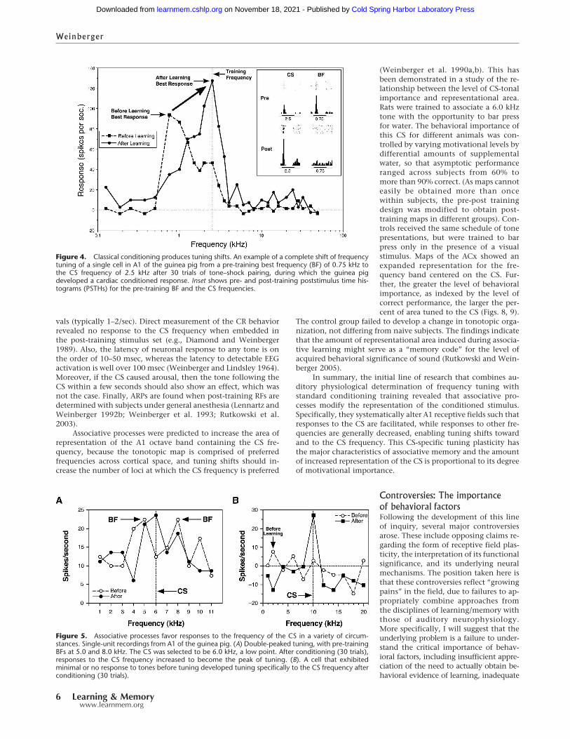
Figure 4. Classical conditioning produces tuning shifts. An example of a complete shift of frequencytuning of a single cell in A1 of the guinea pig from a pre-training best frequency (BF) of 0.75 kHz tothe CS frequency of 2.5 kHz after 30 trials of tone–shock pairing, during which the guinea pigdeveloped a cardiac conditioned response. Inset shows pre- and post-training poststimulus time his-tograms (PSTHs) for the pre-training BF and the CS frequencies.
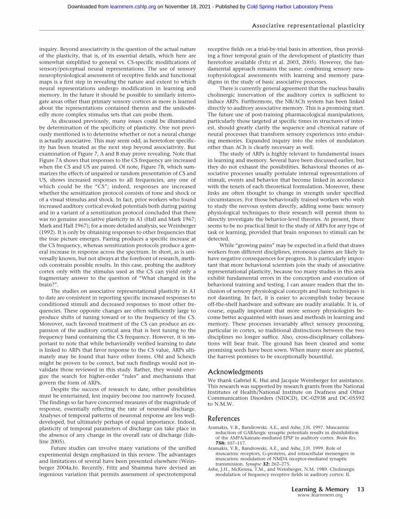
Figure 5. Associative processes favor responses to the frequency of the CS in a variety of circum-stances. Single-unit recordings from A1 of the guinea pig. (A) Double-peaked tuning, with pre-trainingBFs at 5.0 and 8.0 kHz. The CS was selected to be 6.0 kHz, a low point. After conditioning (30 trials),responses to the CS frequency increased to become the peak of tuning. (B). A cell that exhibitedminimal or no response to tones before tuning developed tuning specifically to the CS frequency afterconditioning (30 trials).
Weinberger
6 Learning & Memorywww.learnmem.org
Cold Spring Harbor Laboratory Press on November 18, 2021 - Published by learnmem.cshlp.orgDownloaded from

experimental designs, and lack of knowledge of the relevant behav-ioral literature.
The form of associative receptive field plasticityOhl and Scheich (1996, 1997) have claimed that associativelearning in the gerbil induces decreased responses at the CS fre-quency, i.e., the opposite direction of change found previously.
They propose that this provides for “contrast enhancement,” notincreased stimulus importance. However, their data were ob-tained from a behavioral paradigm that has two shortcomings.First, they used a classical aversive discrimination protocol inwhich a single CS+ (paired with shock) was intermixed with 11–30 different CS– (no shock) frequencies in a single training ses-sion. However, there is no prior evidence that animals can learn
Figure 6. Representation of neuronal responses in A1 (A) before, (B) immediately after, and (C) 1 h after two-tone discrimination training. The guineapig received 30 each CS+ (22.0 kHz) and CS– (39 kHz) intermixed trials. Displayed are rates of discharge (y-axis) as a function of tonal frequency (x-axis)and level of testing stimuli (y-axis, 10–70 dB). Note that conditioning changed the “topography” of neuronal response. The pre-training best frequencyof 27.0 kHz suffered a reduction in response as did the CS– frequency. In contrast, responses to the CS+ frequency increased. Strikingly, consolidation,in the form of a continued development of these changes is evident. After a period of 1 h of silence, the only excitatory response is at the CS+ frequency.
Figure 7. Summary of the effects of (A) conditioning, (B) sensitization, and (C) habituation on frequency receptive fields in the primary auditory cortexof the guinea pig. Data are normalized to octave distance from the CS frequency (A), the presensitization best frequency (B) or the repeated frequency(C). Note that conditioning produces a CS-specific increased response, whereas sensitization (tone–shock or light–shock unpaired) produces generalincreases across the spectrum. Habituation produces frequency-specific decreased response.
Associative representational plasticity
Learning & Memory 7www.learnmem.org
Cold Spring Harbor Laboratory Press on November 18, 2021 - Published by learnmem.cshlp.orgDownloaded from

this highly complex discrimination, particularly in a single ses-sion. The investigators provided no behavioral evidence thattheir subjects had learned the discrimination. Heart rate was re-corded but not related to any particular CS+ or CS– stimuli. In-deed, such data may not have been attainable because they usedextreme massing of intertrial intervals, which varied from 0.25 to3 sec. Second, they compared RFs obtained in a pre-training stateof quiet with those from a different post-training state, one in-volving expectation of shock and possibly new learning. The
post-training state must have been different from pre-training,because Ohl and Scheich used a design in which the same acous-tic context (i.e., the identical tone stimulus set) was presentedbefore, during, and after tone–shock pairings. As shock waspaired with a CS+ that was intermixed with numerous othertones, presentation of the same stimuli after pairing probablyinvolved an expectation of shock, at least until subjects may havelearned that shock had been deleted. The latter would constitutesome sort of extinction, i.e., new learning. That the pre- andpost-training periods differed in expectation, probable state ofarousal, or in other ways, precludes attributing post-trainingchanges in neural responses to (alleged) discrimination learning.
Ohl and Scheich not only argue that conditioning producesCS-specific decreased responses, but that prior observations ofdecreased responses have been ignored by this investigator inorder to promote our claim of increased responses to the CS (Ohland Scheich 1996, 1997, 2004, 2005). They are correct that de-creased unit responses to the CS can be found (e.g., Diamond andWeinberger 1984b, Weinberger et al. 1984b), but these findingsare irrelevant to the issue of associative representational plastic-ity, as they occurred during training trials. Ohl and Scheich makeno distinction between plasticity that is observed during trainingtrials from that which is observed after training trials. But, acentral point of the unified design presented in this article is thatit reduces or eliminates performance factors, such as statechanges, that must occur during training trials when a reinforceris introduced. It does so by use of pre- and post-training RF de-terminations and in a context different from the context of train-ing trials. It is well known that performance factors severely limit
Figure 9. Evidence of a “memory code” for the acquired behavioralimportance of sound. Level of tone importance was controlled by theamount of water deprivation; asymptotic performance was significantlycorrelated with level of deprivation (for details, see Rutkowski and Wein-berger 2005). The area of representation of the frequency band contain-ing the 6.0 kHz tone signal increases as a direct function of the level ofbehavioral importance of the tone, as operationally indexed by the levelof correct performance.
Figure 8. Effect of learning tone-contingent bar-press for water on tonotopic map in A1. Trained rats received water reward for bar-presses in thepresence of a 6.0 kHz tone. Illustrations show tonotopic maps and quantifications of percent of total area (octave frequency bands) for a naïve rat (left)and a rat that attained over 90% correct performance (right). Note that training greatly increased the area of representation for the frequency bandcontaining the 6.0 kHz tone signal.
Weinberger
8 Learning & Memorywww.learnmem.org
Cold Spring Harbor Laboratory Press on November 18, 2021 - Published by learnmem.cshlp.orgDownloaded from

the interpretation of behavioral data obtained during trainingtrials, as opposed to after training trials (Rescorla 1988a,b). Thesame strictures are no less important for determining the neuralcorrelates of associative processes during training trials. A strikingexample of the Ohl and Scheich fallacy is that the sign of plas-ticity (increase or decrease in response to the CS) is often differ-ent during training from after training, as noted above (Diamondand Weinberger 1989).
The failure of Ohl and Scheich to appreciate the essentialrequirement that behavioral validation of learning is necessary tosupport a claim that learning has occurred does not invalidatethe possibility of learning-induced decreased responses to the CS.There well may be circumstances under which such decreases dodevelop. But neither should the lack of behavioral evidence beignored, as the investigators continue to do (Ohl and Scheich2005). At this time, CS-specific increases, CS-directed tuningshifts, and CS-expansions of area in the tonotopic map remainthe only behaviorally validated ARPs in the primary auditorycortex. The Ohl and Scheich findings of CS-specific decreases inA1 do not challenge this conclusion because of the absence ofbehavioral verification of associative learning. If this is forthcom-ing, then the domain of associative representational plasticity inthe primary auditory cortex will have been enlarged, and a nextstep would be to discover the superordinate rules that governwhich types of ARPs are invoked by associative processes andtheir respective functional consequences.
Consequences of failure to understand fundamental aspectsof conditioningInterestingly, studies in the big brown bat (Eptesicus fuscus) havereported CS-specific tuning shifts during tone–shock pairing(Suga and Ma 2003). However, they also lack behavioral valida-tion of association, although, in this case, behavioral data weresaid to have been obtained. The investigators presented sessionsof 60 trials of tone–shock pairing and reported that limb flexion-conditioned responses developed starting at trial 50 (Gao andSuga 1998). However, there is reason to suspect the claims ofbehavioral learning. First, these investigators have consistentlyfailed to publish individual flexion records, learning functions,or any statistical report of the behavioral data. Second, their pre-vious study showed a failure of acquisition of flexion CRs withtone CSs and required many days of training to obtain consistentCRs using an effective white noise CS (Riquimaroux et al. 1992).Third, the animals are under whole body restraint while beingtrained with shock, and access to the brain is obtained by drillinga hole in the skull while they are awake. These procedures arelikely to cause considerable stress and might increase limb move-ment during a session. Konorski (1967) has emphasized the prob-lem of generalized body movement in such circumstances, whichcan produce apparent flexion CRs. Fourth, intertrial intervalswere fixed at 30 sec. Therefore, any genuine CRs could have beencaused by temporal conditioning, without any control by thetonal CS itself. Fifth, the investigators treated extinction as “era-sure,” assuming that repeated training of subjects after extinctionwas equivalent to training naive subjects. Only data from the firstsession would be clearly interpretable, but data from all sessionsand subjects were combined. There are also neurobiological rea-sons for caution. Suga and coworkers have relied on findingsfrom cortical inactivations using muscimol. Interpretation of theclaimed highly focal effects of this drug depend upon assurancesthat its diffusion was limited to ∼1 mm, due to the proximity ofprimary somatosensory and auditory cortices. However, radio-tracing studies of muscimol diffusion, using smaller doses thanused in these studies, has revealed a considerably more extensivespread (Edeline et al. 2002). Finally, Suga and colleagues found
that repeated presentation of a tone alone produced increasedresponse to and tuning shifts toward the repeated frequency(Gao and Suga 1998). This is in direct opposition to the extensiveliterature on the effects of repeated auditory stimulation, whichproduces response decrements (habituation) in the auditory cor-tices in other taxa (e.g., Marsh and Worden 1964; Ellinwood et al.1968; Wickelgren 1968; Weber 1970; Oleson et al. 1975; Wein-berger et al. 1975; Westenberg and Weinberger 1976; Condonand Weinberger 1991).
Thus, despite the temptation to assume that associative tun-ing shifts develop in the big brown bat during the developmentof conditioned responses, the claims of Suga and colleagues mustremain suspect in the absence of appropriate behavioral and neu-ral controls. Perhaps as an echolocating animal, the principles ofauditory system plasticity are specialized in the big brown bat,and thus, it is not a good model for general mammalian auditoryassociative plasticity.
Mechanisms and models of ARP andassociative memoryI conclude with a consideration of mechanisms that may be re-sponsible for the development of associative representationalplasticity, focusing on the cholinergic system because it has gar-nered most attention. There is little controversy about the im-portance of acetylcholine (ACh) in auditory cortical (and othercortical) plasticity. The same cannot be said for the two extantmodels of ARP, which are in direct conflict on all issues exceptthe role of acetylcholine.
The nucleus basalis, acetylcholine, and associativerepresentational plasticityNeuromodulators have profound effects on neuronal functions.With respect to the auditory cortex, norepinephrine can alterfrequency tuning (Manunta and Edeline 1997, 1999), dopaminecan increase the area of cortical representation of a tone withwhich it is paired (Bao et al. 2001), and serotonin levels canincrease during initial stages of avoidance training (Stark andScheich 1997). Acetylcholine has long been implicated in asso-ciative learning, (e.g., Deutsch 1971; Introini-Collison and Mc-Gaugh 1988) and also the auditory cortex. For example, ACh,and stimulation of the nucleus basalis (NB), its cortical source(Mesulam et al. 1983), can modulate the responses of A1 tosounds (Ashe et al. 1989; McKenna et al. 1989; Metherate et al.1990; Metherate and Ashe 1993), regulate intracortical process-ing (Hsieh et al. 2000), synaptic plasticity (Metherate and Ashe1995; Aramakis et al. 1997, 1999; Bandrowski et al. 2001), andlearning-induced auditory cortical plasticity in animals (Mether-ate and Weinberger 1989; Shulz et al. 1997; Kudoh et al. 2004;Ma and Suga 2005) and humans (Thiel et al. 2002a,b); see also(Edeline 2003; Weinberger 2003; Metherate et al. 2005).
While the direct application of cholinergic agonists and an-tagonists to the brain (both in vitro or in vivo) has providedimportant insights into the roles of ACh in the auditory system,plasticity, and learning, this approach is necessarily limiting withrespect to associative processes in behaving subjects. A majorobstacle is the difficulty of achieving precise timing when it isdesirable to use a cholinergic agent as a US following presenta-tion of an acoustic CS because of the uncontrolled time lag indiffusion of drugs applied to the brain. Electrical stimulation ofthe NB does permit precise timing and such stimulation is knownto release ACh in the cortex (Rasmusson et al. 1994; Detari et al.1997a; Duque et al. 2000). Moreover, NB stimulation appears tobe motivationally neutral (Pennartz 1995; Miasnikov et al. 2004),thus avoiding potential confounds due to reward or punishment.
In the first study of this type, a tone was paired with NBstimulation as a substitute for the US in one group of waking
Associative representational plasticity
Learning & Memory 9www.learnmem.org
Cold Spring Harbor Laboratory Press on November 18, 2021 - Published by learnmem.cshlp.orgDownloaded from

guinea pigs or presented unpaired in another group. Frequency-tuning curves were obtained before and after a single trainingsession of only 40 trials (Bakin et al. 1996). Pairing induced ARP,i.e., responses to the CS frequency were augmented, while thoseto the pre-training BF and many other frequencies decreased.Subsequent similar studies demonstrated that CS-specific tuningplasticity induced by using NB stimulation as the US was specificto a CS+ vs. a CS– in two-tone discrimination training (Dimyanand Weinberger 1999) and that it exhibits consolidation over a24-h period of retention, i.e., the effects become larger in theabsence of further training (Bjordahl et al. 1998). The cholinergicnature of associative NB-induced CS-specific tuning plasticity isevidenced by the fact that it requires NB induction of EEG acti-vation (Bjordahl et al. 1998), which is known to involve therelease of ACh in the cortex (Metherate et al. 1992; Detari et al.1997b; Cape and Jones 2000; Duque et al. 2000) and the engage-ment of muscarinic receptors in A1 (Miasnikov et al. 2001). Insummary, ARPs induced by nucleus basalis/cholinergic activa-tion have the same characteristics as ARPs that develop duringassociative learning.
A series of related studies replicated and extended CS-specific frequency tuning plasticity (Kilgard and Merzenich1998a) and also demonstrated that pairing NB stimulation withother acoustic stimuli, e.g., frequency-modulated sweeps, soundspresented at particular pulse rates, and sequences of differentacoustic stimuli (tones and noise) was sufficient to produce CS-specific facilitation (Kilgard and Merzenich 1998b, 2002; Kilgardet al. 2001; Moucha et al. 2005; Pandya et al. 2005). However, allof these experiments presented hundreds of trials per day forperiods of weeks, thus involving from about 6000 to 12,500 tri-als. Further, some studies lack controls for nonassociative pro-cesses (Kilgard and Merzenich 1998a,b). This training approachcontrasts with the studies summarized above that mimic stan-dard behavioral conditioning protocols, e.g., 40 trials for single-tone conditioning and 30 trials each of a CS+ and CS– for dis-crimination training, all in a single session (Bakin and Wein-berger 1996; Bjordahl et al. 1998; Dimyan and Weinberger1999). Therefore, the extent to which massed pairing of tone andNB stimulation sheds light on basic associative processes is un-clear.
Induction of specific, associative behavioral memoryby stimulation of the NBRecent experiments indicate that pairing a tone with NB stimu-lation induces not only ARPs in A1, but also CS-specific behav-ioral memory. Inferences that such pairing produces specific, as-sociative memory have to meet the dual criteria of associativityand specificity. The first criterion simply requires a nonpairedcontrol group. The criterion of specificity can be examined byobtaining stimulus generalization gradients, obtained when asubject is trained with one stimulus (CS) and later tested withmany stimuli. If NB stimulation paired with a tone induces spe-cific memory about that tone, then this CS frequency shouldlater elicit the largest behavioral responses to all tones tested, i.e.,occupy the peak of the frequency generalization gradient.
The first study involved overtraining using 200 daily trialsfor 15 d (3000 trials). This is the same order of magnitude used byMerzenich and colleagues in studies of the induction of neuronalplasticity by stimulation of the NB (above). However, the induc-tion of specific associative behavioral memory by NB stimulationhad never been demonstrated, and we assumed (wrongly as itturned out) that as NB stimulation was motivationally neutral,overtraining might be necessary to induce behavioral memory.The behavioral measures were changes in heart rate and in res-piration. This protocol did produce CS-specific memory (McLin
III et al. 2002); generalization gradients for the paired group ex-hibited peak response at the CS frequency, in contrast to thecontrol group (Fig. 10). This appears to be the first description ofthe induction of specific associative memory by direct stimula-tion of the brain. Overtraining proved unnecessary; a single ses-sion of 200 trials has proven to be sufficient (Miasnikov et al.2006). The minimal amount of training has not yet been deter-mined. The frequency generalization gradients are indistinguish-able from those that develop in standard learning protocols usingstandard motivational reinforcers (Mackintosh 1974). Thus, thebehavior meets the same criteria as used to assess learning undernormal circumstances.
NB stimulation not only can induce specific, associativememory, but actually control the amount of detail in thatmemory. In a very recent study using a 2�2 design, rats receivedtone and NB stimulation either paired or randomly with eitherlow (∼45 µA) or moderate (∼65 µA) levels of NB stimulation.Previous studies of CS-specific memory induction (above) hadused the moderate level. As in the past, the moderate pairedgroup developed associative CS-specific memory; the only sig-nificant increased response was at the spectral range containingthe CS frequency (6.0 kHz). In contrast, the weak stimulationgroup developed only associative memory without any fre-quency specificity (Fig. 11). Additional experiments are neededto further characterize the contents or detail in such memories.The current results are consistent with the conclusion that theweak paired group learned that tone (or sound per se) had be-come important, whereas the moderate paired group had learned
Figure 10. Respiration responses to post-training tone and generaliza-tion gradients, showing the induction of CS-specific memory after tonepaired with stimulation of the nucleus basalis. (A) Examples of individualrespiration records (with value of respiration change index, RCI) to threefrequencies (2, 6, and 12 kHz) for one animal each from the paired andunpaired groups. The largest response was at the CS frequency of 6 kHzfor the paired animal (RCI = 0.50). Horizontal bar indicates tone duration.(B, left) Group mean (� SEM) change in respiration to all tones for bothgroups. The maximal response (left) was at 6 kHz for the paired group,but not for the unpaired group. The generalization gradient for only thepaired group was significantly quadratic (P < 0.01), with responses to 6kHz being of greatest magnitude. The group difference function (right)shows a high degree of specificity of respiratory responses to 6 kHz.
Weinberger
10 Learning & Memorywww.learnmem.org
Cold Spring Harbor Laboratory Press on November 18, 2021 - Published by learnmem.cshlp.orgDownloaded from

that the CS frequency (and its neighbors) had become important.In the absence of a normal US, neither group would have learnedwhy these stimuli were important (Weinberger et al. 2006).
In summary, these findings supportthe view that the NB is normally en-gaged during associative S–S condition-ing, promoting the establishment ofspecific cortical plasticity that itself maybe a substrate for specific behavioralmemory. The use of NB stimulation toinduce memory may not only elucidatethe mechanisms of memory inductionbut also allow “dissection” of memoryinto its components of sensory contentand level of behavioral importance. Dur-ing normal motivated learning, both as-pects of memory may be stored when astandard reward or punishment is pres-ent and the NB is activated. In the pres-ent studies lacking a standard US, behav-ioral importance can be induced in theabsence of a full CS–US association.
Neural models of associative receptive fieldplasticity and behaviorA preliminary model of CS-specific re-ceptive field plasticity was proposed atthe outset of this line of research (Wein-berger et al. 1990a,b). Its goal was to out-line the minimum circuitry sufficientto account for both the cortical plasticityand behavioral (autonomic) fear condi-tioning (Fig. 12). The key ideas were(1) CS–US convergence occurs first in the(nonlemniscal) magnocellular medialgeniculate nucleus (MGm), where the re-sultant plasticity results in shifts of tun-ing that favors the CS frequency; (2) pro-jection of this MGm plasticity to theamygdala; (3) projection of the effects ofthe plasticity (i.e., increased response tothe CS) from the amygdala to both thecholinergic nucleus basalis and to brain-stem nuclei controlling autonomic be-havioral responses (e.g., heart rate);(4) convergence of MGm and auditorylemniscal tonal information in the pri-mary auditory cortex to promote short-term plasticity; and (5) convergence ofNB/ACh and auditory information in A1to promote long-term plasticity. Thismodel incorporated many establishedfindings (such as the convergence of au-ditory and nociceptive somatosensoryinformation in the MGm (Wepsic 1966;Love and Scott 1969), the necessity for anintact MGm region for auditory fear con-ditioning (LeDoux et al. 1983, 1986a; Jar-rell et al. 1986a) and the ability of NB/cholinergic influences to facilitate corticalresponses in sensory cortex (Metherateet al. 1987; Ashe et al. 1989).
This model was soon shown to beincorrect. Thus, while it hypothesizedthat the lemniscal ventral medial ge-niculate (MGv) was not plastic and sent
only the “physical parameters” of sound to A1, in fact, the MGvdevelops CS-specific tuning plasticity, although it is transient,disappearing by 1-h post-training (Edeline and Weinberger
Figure 11. Level of NB stimulation controls specificity of induced memory. Pre-training and post-training responses to test tones in the Moderate and Weak NB stimulation (NBs) groups. (A) Pre-training responses for subjects later trained with either paired or unpaired CS tone and NB stimulation.There were no differences between the groups. (B) Post-training responses for the Moderate NBsgroups. Note the significant difference between the paired and unpaired groups, confined to theCS-band frequencies. This indicates that training with a moderate level of NBs produced memory thatwas both associative and CS specific. (C) Comparisons of changes within the Moderate group (post-minus pre-training responses to test tones). Note that the paired group had developed a significantincrease to the CS-band frequencies only, while the unpaired group had developed a significantdecrease, probably indicating frequency-specific habituation due to lack of pairing with NB stimula-tion. (D) Post-training responses for the Weak NBs groups. In contrast to the Moderate NBs group,pairing produced a significant difference in response across all test frequencies compared with itsunpaired controls. This indicates that training with weak NBs was sufficient to produce associativememory but insufficient to produce memory for frequency detail, i.e., memory that the frequency ofthe CS was paired with NBs. (E) Comparisons of changes within the Weak NBs groups showed that thepaired group did not develop absolute increased responses, but that the unpaired group did developsignificant decreases in responses across the spectrum of test frequencies. Thus, pairing the CS withweak NBs apparently prevented a habituatory decrement in the Weak paired group, which is evidentin the Weak unpaired group. *P < 0.05; **P < 0.01; ***P < 0.005.
Associative representational plasticity
Learning & Memory 11www.learnmem.org
Cold Spring Harbor Laboratory Press on November 18, 2021 - Published by learnmem.cshlp.orgDownloaded from

1991). We have not been able to formulate a reasonable revisionof the model that explains both the source of the brief CS-specificplasticity in the MGv and the functional implications of thisplasticity. Regarding sources, it is certainly possible that cortico-geniculate connections could be responsible, and it is intriguingthat cortical stimulation appears to facilitate MGv cells whileinhibiting nonlemniscal medial geniculate neurons (Yu et al.2004). But, this in itself would not explain the specificity of CS-tuning plasticity in the MGv. Thus, even reasonable specula-tion needs more detailed knowledge of cortico-fugal trans-actions in the MGv (He 2003). As for the functional implications,the transient plasticity in the MGv could be responsible in partfor setting up the enduring plasticity in the cortex. But, as forthe model itself, two time scales would have to be reconciled,that of less than an hour in the MGv and that of months in theauditory cortex. Mere postulation of dual temporal mechanismsin the cortex would be little more than an expression of cur-rent ignorance. Thus, while several avenues of speculationare possible, rather than construct a tenuous and untestablemodification of the model, we await further empirical develop-ments.
However, the model has been successful in several respects.For example, it predicted CS-specific tuning plasticity in theMGm (Edeline and Weinberger 1992), the ability of electricalstimulation of the MGm to substitute for the shock US in fear(cardiac) conditioning (Cruikshank et al. 1992), CS/tone associa-tive plasticity in the NB that precedes associative tuning shifts inA1 (Maho et al. 1995), and the ability of electrical stimulation ofthe NB to substitute for the US to induce CS-specific tuning plas-ticity in A1 (Bakin and Weinberger 1996; Kilgard and Merzenich1998a) that is dependent on muscarinic receptors in A1 (Miasni-kov et al. 2001). It also explains why lesions of the MGm blockboth associative plasticity that develops in the amygdala andelsewhere in the brain, and also impairs conditioning (Porembaand Gabriel 1997). Additionally, imaging studies in humans haveobtained results predicted by the model. For example, aversivetwo-tone discriminative conditioning produces CS-specific plas-ticity in the primary auditory cortex and associative changes onlyin the medial geniculate, amygdala, basal forebrain, and orbito-frontal cortex (Morris et al. 1998). These structures, except theorbitofrontal cortex, are the core of the model.
Suga and colleagues have proposed an alternative model
(Suga and Ma 2003). It ignores the MGm completely but ratherholds that the CS and US ascend to the auditory and somatosen-sory cortices, respectively, and are projected to association cor-tex, which is said to be the locus of CS–US convergence, and thento the amygdala. This model also includes the descending (cor-ticofugal) auditory system and asserts that plasticity that devel-ops in the association cortex is relayed (via the amygdala) to theinferior colliculus, which in turn promotes CS-specific plasticityin A1. Finally, cortico-collicular projections are thought to in-duce CS-specific response increments in the inferior colliculus,thus forming a cortico-colliculo-cortico positive feedback loop.The investigators suggest that this positive feedback loop isturned off by the thalamic reticular nucleus (TRN), which pre-vents auditory information from reaching A1. Suga and col-leagues do not specify how or when the TRN is engaged. Theirformulation also seems to ignore the fact that the auditory cortexof conditioned animals continues to receive perfectly fine audi-tory information. The Suga model accepts the components of theWeinberger et al. model that (1) link the amygdala to the initia-tion of behavioral CRs, (2) hypothesize that the amygdala en-gages the nucleus basalis, and (3) suggests that the cholinergicprojections of the nucleus basalis to A1 promote long term CS-specific tuning shifts therein.
Why Suga and colleagues ignore the MGm is baffling. Itprojects to Layer I and the apical dendrites of pyramidal cells inA1 and in all other auditory cortical fields (Winer and Morest1983) and does so via giant axons that provide the fastestthalamo-cortical transmission (Huang and Winer 2000). In addi-tion to findings listed above that implicate the MGm (and itsrelated posterior intralaminar nucleus [PIN]) in associative learn-ing, this structure is well known to develop associative plasticityduring conditioning (Gabriel et al. 1975, 1976; Disterhoft andStuart 1976; Birt et al. 1979; Birt and Olds 1981; Weinberger1982; Jarrell et al. 1986b; LeDoux et al. 1986b; Edeline et al. 1988,1990; Edeline 1990; McEchron et al. 1995, 1996; Hennevin et al.1998). Moreover, its stimulation induces in the auditory cortexheterosynaptic (Weinberger et al. 1995) long-term potentiation(LTP), which has been implicated in learning (see also Parsons etal. 2006).
Another inexplicable feature of the Suga model is their claimthat CS–US convergence occurs in the cerebral cortex. This isincompatible with an extensive literature which shows that nei-ther the auditory cortex, nor indeed the cerebral cortex as awhole, are necessary for simple classical conditioning (DiCara etal. 1970; Norman et al. 1977; Berntson et al. 1983; Teich et al.1988; Romanski and LeDoux 1992). In summary, the Suga modelincludes the descending auditory system, which is a potentialadvantage, as its function has been obscure. However, othernovel features of this model ignore or are directly contradicted byprior neural and behavioral findings.
Conclusions and future directionsThe major goal of this review has been to explain how a synthesisof the fundamental experimental approaches of two disciplines,sensory neurophysiology and the experimental psychology oflearning/memory, can provide a new level of understanding ofthe neurobiology of associative processes. In particular, the as-sessment of sensory representations preceding and followingstandard associative learning tasks (exemplified by classical con-ditioning) reveals the extent to which resultant neural plasticityis general or specific to signal stimuli, i.e., stimuli that acquirepredictive value through learning. The issue of specificity is keyto the construct of representation. It is of course important tofind that neural plasticity during learning is associative. This typeof conclusion has been with us since the dawn of neurophysi-ological studies of learning and is the starting point for further
Figure 12. The model of Weinberger and colleagues. This model hy-pothesizes the minimal circuitry that would be sufficient to account forshort- and long-term associative representational plasticity and rapidlyacquired conditioned autonomic responses. See text for details.
Weinberger
12 Learning & Memorywww.learnmem.org
Cold Spring Harbor Laboratory Press on November 18, 2021 - Published by learnmem.cshlp.orgDownloaded from
inquiry. Beyond associativity is the question of the actual natureof the plasticity, that is, of its essential details, which here aresomewhat simplified to general vs. CS-specific modifications ofsensory/perceptual neural representations. The use of sensoryneurophysiological assessment of receptive fields and functionalmaps is a first step in revealing the nature and extent to whichneural representations undergo modification in learning andmemory. In the future it should be possible to similarly interro-gate areas other than primary sensory cortices as more is learnedabout the representations contained therein and the undoubt-edly more complex stimulus sets that can probe them.
As discussed previously, many issues could be illuminatedby determination of the specificity of plasticity. One not previ-ously mentioned is to determine whether or not a neural changeis actually associative. This may seem odd, as heretofore specific-ity has been treated as the next step beyond associativity. Butexamination of Figure 7, A and B may prove revealing. Note thatFigure 7A shows that responses to the CS frequency are increasedwhen the CS and US are paired. Of note, Figure 7B, which sum-marizes the effects of unpaired or random presentation of CS andUS, shows increased responses to all frequencies, any one ofwhich could be the “CS”; indeed, responses are increasedwhether the sensitization protocol consists of tone and shock orof a visual stimulus and shock. In fact, prior workers who foundincreased auditory cortical evoked potentials both during pairingand in a variant of a sensitization protocol concluded that therewas no genuine associative plasticity in A1 (Hall and Mark 1967;Mark and Hall 1967); for a more detailed analysis, see Weinberger(1992). It is only by obtaining responses to other frequencies thatthe true picture emerges. Pairing produces a specific increase atthe CS frequency, whereas sensitization protocols produce a gen-eral increase in response across the spectrum. In short, as is uni-versally known, but not always at the forefront of research, meth-ods constrain possible results. In this case, probing the auditorycortex only with the stimulus used as the CS can yield only afragmentary answer to the question of “What changed in thebrain?”.
The studies on associative representational plasticity in A1to date are consistent in reporting specific increased responses toconditioned stimuli and decreased responses to most other fre-quencies. These opposite changes are often sufficiently large toproduce shifts of tuning toward or to the frequency of the CS.Moreover, such favored treatment of the CS can produce an ex-pansion of the auditory cortical area that is best tuning to thefrequency band containing the CS frequency. However, it is im-portant to note that while behaviorally verified learning to dateis linked to ARPs that favor response to the CS value, ARPs ulti-mately may be found that have other forms. Ohl and Scheichmight be proven to be correct, but such findings would not in-validate those reviewed in this study. Rather, they would ener-gize the search for higher-order “rules” and mechanisms thatgovern the form of ARPs.
Despite the success of research to date, other possibilitiesmust be entertained, lest inquiry become too narrowly focused.The findings so far have concerned measures of the magnitude ofresponse, essentially reflecting the rate of neuronal discharge.Analyses of temporal patterns of neuronal response are less well-developed, but ultimately perhaps of equal importance. Indeed,plasticity of temporal parameters of discharge can take place inthe absence of any change in the overall rate of discharge (Ede-line 2005).
Future studies can involve many variations of the unifiedexperimental design emphasized in this review. The advantagesand limitations of several have been presented elsewhere (Wein-berger 2004a,b). Recently, Fritz and Shamma have devised aningenious variation that permits assessment of spectrotemporal
receptive fields on a trial-by-trial basis in attention, thus provid-ing a finer temporal grain of the development of plasticity thanheretofore available (Fritz et al. 2003, 2005). However, the fun-damental approach remains the same: combining sensory neu-rophysiological assessments with learning and memory para-digms in the study of basic associative processes.
There is currently general agreement that the nucleus basalischolinergic innervation of the auditory cortex is sufficient toinduce ARPs. Furthermore, the NB/ACh system has been linkeddirectly to auditory associative memory. This is a promising start.The future use of post-training pharmacological manipulations,particularly those targeted at specific times in structures of inter-est, should greatly clarify the sequence and chemical nature ofneural processes that transform sensory experiences into endur-ing memories. Expanded inquiry into the roles of modulatorsother than ACh is clearly necessary as well.
The study of ARPs is highly relevant to fundamental issuesin learning and memory. Several have been discussed earlier, butthey do not exhaust the possibilities. Behavioral theories of as-sociative processes usually postulate internal representations ofstimuli, events and behavior that become linked in accordancewith the tenets of each theoretical formulation. Moreover, theselinks are often thought to change in strength under specifiedcircumstances. For those behaviorally trained workers who wishto study the nervous system directly, adding some basic sensoryphysiological techniques to their research will permit them todirectly investigate the behavior-level theories. At present, thereseems to be no practical limit to the study of ARPs for any type oftask or learning, provided that brain responses to stimuli can bedetected.
While “growing pains” may be expected in a field that drawsworkers from different disciplines, erroneous claims are likely tohave negative consequences for progress. It is particularly impor-tant that more behavioral scientists join the study of associativerepresentational plasticity, because too many studies in this areaexhibit fundamental errors in the conception and execution ofbehavioral training and testing. I can assure readers that the in-clusion of sensory physiological concepts and basic techniques isnot daunting. In fact, it is easier to accomplish today becauseoff-the-shelf hardware and software are readily available. It is, ofcourse, equally important that more sensory physiologists be-come better acquainted with issues and methods in learning andmemory. These processes invariably affect sensory processing,particular in cortex, so traditional distinctions between the twodisciplines no longer suffice. Also, cross-disciplinary collabora-tions will bear fruit. The ground has been cleared and somepromising seeds have been sown. When many more are planted,the harvest promises to be exceptionally bountiful.
AcknowledgmentsWe thank Gabriel K. Hui and Jacquie Weinberger for assistance.This research was supported by research grants from the NationalInstitutes of Health/National Institute on Deafness and OtherCommunication Disorders (NIDCD), DC-02938 and DC-05592to N.M.W.
ReferencesAramakis, V.B., Bandrowski, A.E., and Ashe, J.H. 1997. Muscarinic
reduction of GABAergic synaptic potentials results in disinhibitionof the AMPA/kainate-mediated EPSP in auditory cortex. Brain Res.758: 107–117.
Aramakis, V.B., Bandrowski, A.E., and Ashe, J.H. 1999. Role ofmuscarinic receptors, G-proteins, and intracellular messengers inmuscarinic modulation of NMDA receptor-mediated synaptictransmission. Synapse 32: 262–275.
Ashe, J.H., McKenna, T.M., and Weinberger, N.M. 1989. Cholinergicmodulation of frequency receptive fields in auditory cortex: II.
Associative representational plasticity
Learning & Memory 13www.learnmem.org
Cold Spring Harbor Laboratory Press on November 18, 2021 - Published by learnmem.cshlp.orgDownloaded from

Frequency-specific effects of anticholinesterases provide evidence fora modulatory action of endogenous ACh. Synapse 4: 44–54.
Bakin, J.S. and Weinberger, N.M. 1990. Classical conditioning inducesCS-specific receptive field plasticity in the auditory cortex of theguinea pig. Brain Res. 536: 271–286.
Bakin, J.S. and Weinberger, N.M. 1996. Induction of a physiologicalmemory in the cerebral cortex by stimulation of the nucleus basalis.Proc. Natl. Acad. Sci. 93: 11219–11224.
Bakin, J.S., Lepan, B., and Weinberger, N.M. 1992. Sensitization inducedreceptive field plasticity in the auditory cortex is independent ofCS-modality. Brain Res. 577: 226–235.
Bakin, J.S., South, D.A., and Weinberger, N.M. 1996. Induction ofreceptive field plasticity in the auditory cortex of the guinea pigduring instrumental avoidance conditioning. Behav. Neurosci.110: 905–913.
Bandrowski, A.E., Moore, S.L., and Ashe, J.H. 2001. Cholinergic synapticpotentials in the supragranular layers of auditory cortex. Synapse41: 118–130.
Bao, S., Chan, V.T., and Merzenich, M.M. 2001. Cortical remodellinginduced by activity of ventral tegmental dopamine neurons. Nature412: 79–83.
Berntson, G.G., Tuber, D.S., Ronca, A.E., and Bachman, D.S. 1983. Thedecerebrate human: Associative learning. Exp. Neurol. 81: 77–88.
Birt, D. and Olds, M. 1981. Associative response changes in lateralmidbrain tegmentum and medial geniculate during differentialappetitive conditioning. J. Neurophysiol. 46: 1039–1055.
Birt, D., Nienhuis, R., and Olds, M. 1979. Separation of associative fromnon-associative short latency changes in medial geniculate andinferior colliculus during differential conditioning and reversal inrats. Brain Res. 167: 129–138.
Bjordahl, T.S., Dimyan, M.A., and Weinberger, N.M. 1998. Induction oflong-term receptive field plasticity in the auditory cortex of thewaking guinea pig by stimulation of the nucleus basalis. Behav.Neurosci. 112: 467–479.
Brown, M., Irvine, D.R., and Park, V.N. 2004. Perceptual learning on anauditory frequency discrimination task by cats: Association withchanges in primary auditory cortex. Cereb. Cortex 14: 952–965.
Cape, E.G. and Jones, B.E. 2000. Effects of glutamate agonist versusprocaine microinjections into the basal forebrain cholinergic cellarea upon � and � EEG activity and sleep–wake state. Eur. J. Neurosci.12: 2166–2184.
Christian, K.M. and Thompson, R.F. 2005. Long-term storage of anassociative memory trace in the cerebellum. Behav. Neurosci.119: 526–537.
Condon, C.D. and Weinberger, N.M. 1991. Habituation producesfrequency-specific plasticity of receptive fields in the auditory cortex.Behav. Neurosci. 105: 416–430.
Cruikshank, S.J., Edeline, J.-M., and Weinberger, N.M. 1992. Stimulationat a site of auditory–somatosensory convergence in the medialgeniculate nucleus is an effective unconditioned stimulus for fearconditioning. Behav. Neurosci. 106: 471–483.
Detari, L., Rasmusson, D.D., and Semba, K. 1997a. Phasic relationshipbetween the activity of basal forebrain neurons and cortical EEG inurethane-anesthetized rat. Brain Res. 759: 112–121.
Detari, L., Semba, K., and Rasmusson, D.D. 1997b. Responses of corticalEEG-related basal forebrain neurons to brainstem and sensorystimulation in urethane-anaesthetized rats. Eur. J. Neurosci. 9: 1153–1161.
Deutsch, J.A. 1971. The cholinergic synapse and the site of memory.Science 174: 788–794.
Diamond, I.T. 1985. A history of the study of the cortex: Changes in theconcept of the sensory pathway. In Topics in the history of psychology,(eds. G.A. Kimble and K. Schlesinger), Vol. 1, pp. 305–387. LawrenceErlbaum Associates, Hillsdale, N.J.
Diamond, D.M. and Weinberger, N.M. 1984a. Physiological plasticity ofsingle neurons in auditory cortex of the cat during acquisition of thepupillary dilation conditioned response: II. Secondary field (AII).Behav. Neurosci. 98: 189–210.
Diamond, D.M. and Weinberger, N.M. 1984b. Physiological plasticity ofsingle neurons in auditory cortex of the cat during acquisition of thepupillary conditioned response: II. Secondary field (AII). Behav.Neurosci. 98: 189–210.
Diamond, D.M. and Weinberger, N.M. 1986. Classical conditioningrapidly induces specific changes in frequency receptive fields ofsingle neurons in secondary and ventral ectosylvian auditorycortical fields. Brain Res. 372: 357–360.
Diamond, D.M. and Weinberger, N.M. 1989. Role of context in theexpression of learning-induced plasticity of single neurons inauditory cortex. Behav. Neurosci. 103: 471–494.
DiCara, L.V., Braun, J.J., and Pappas, B.A. 1970. Classical conditioningand instrumental learning of cardiac and gastrointestinal responsesfollowing removal of neocortex in the rat. J. Comp. Physiol. Psychol.
73: 208–216.Dimyan, M.A. and Weinberger, N.M. 1999. Basal forebrain stimulation
induces discriminative receptive field plasticity in the auditorycortex. Behav. Neurosci. 113: 691–702.
Disterhoft, J.F. and Stuart, D.K. 1976. Trial sequence of changed unitactivity in auditory system of alert rat during conditioned responseacquisition and extinction. J. Neurophysiol. 39: 266–281.
Duque, A., Balatoni, B., Detari, L., and Zaborszky, L. 2000. EEGcorrelation of the discharge properties of identified neurons in thebasal forebrain. J. Neurophysiol. 84: 1627–1635.
Edeline, J.-M. 1990. Frequency-specific plasticity of single unitdischarges in the rat medial geniculate body. Brain Res. 529: 109–119.
Edeline, J.-M. 2003. The thalamo-cortical auditory receptive fields:Regulation by the states of vigilance, learning and theneuromodulatory systems. Exp. Brain Res. 153: 554–572.
Edeline, J.-M. 2005. Learning-induced plasticity in the thalamo-corticalauditory system: Should we move from a rate code description totemporal code description? In The auditory cortex: A synthesis ofhuman and animal research (eds. R. König et al.), pp. 365–382.Lawrence Erlbaum Associates, Mahwah, N.J.
Edeline, J.-M. and Weinberger, N.M. 1991. Thalamic short-termplasticity in the auditory system: Associative returning of receptivefields in the ventral medial geniculate body. Behav. Neurosci.105: 618–639.
Edeline, J.-M. and Weinberger, N.M. 1992. Associative retuning in thethalamic source of input to the amygdala and auditory cortex:Receptive field plasticity in the medial division of the medialgeniculate body. Behav. Neurosci. 106: 81–105.
Edeline, J.-M., Dutrieux, G., and Neuenschwander-el Massioui, N. 1988.Multiunit changes in hippocampus and medial geniculate body infree-behaving rats during acquisition and retention of a conditionedresponse to a tone. Behav. Neural Biol. 50: 61–79.
Edeline, J.-M., Neuenschwander-el Massioui, N., and Dutrieux, G. 1990.Discriminative long-term retention of rapidly induced multiunitchanges in the hippocampus, medial geniculate and auditory cortex.Behav. Brain Res. 39: 145–155.
Edeline, J.-M., Hars, B., Hennevin, E., and Cotillon, N. 2002. Muscimoldiffusion after intracerebral microinjections: A reevaluation based onelectrophysiological and autoradiographic quantifications. Neurobiol.Learn. Mem. 78: 100–124.
Ellinwood, E.H., Cook, J.D., and Wiilson, W.P. 1968. Habituation ofevoked response to uniaural clicks. Brain Res. 7: 306–309.
Fritz, J., Shamma, S., Elhilali, M., and Klein, D. 2003. Rapid task-relatedplasticity of spectrotemporal receptive fields in primary auditorycortex. Nat. Neurosci. 6: 1216–1223.
Fritz, J., Elhilali, M., and Shamma, S. 2005. Active listening:Task-dependent plasticity of spectrotemporal receptive fields inprimary auditory cortex. Hear. Res. 206: 159–176.
Gabriel, M., Saltwick, S.E., and Miller, J.D. 1975. Conditioning andreversal of short-latency multiple-unit responses in the rabbit medialgeniculate nucleus. Science 189: 1108–1109.
Gabriel, M., Miller, J.D., and Saltwick, S.E. 1976. Multiple unit activityof the rabbit medial geniculate nucleus in conditioning, extinction,and reversal. Physiol. Psychol. 4: 124–134.
Galambos, R., Sheatz, G., and Vernier, V. 1956. Electrophysiologicalcorrelates of a conditioned response in cats. Science 123: 376–377.
Galvan, V.V. and Weinberger, N.M. 2002. Long-term consolidation andretention of learning-induced tuning plasticity in the auditorycortex of the guinea pig. Neurobiol. Learn. Mem. 77: 78–108.
Galvan, V.V., Chen, J., and Weinberger, N.M. 2001. Long-termfrequency tuning of local field potentials in the auditory cortex ofthe waking guinea pig. J. Assoc. Res. Otolaryngol. 2: 199–215.
Gao, E. and Suga, N. 1998. Experience-dependent corticofugaladjustment of midbrain frequency map in bat auditory system. Proc.Natl. Acad. Sci. 95: 12663–12670.
Gilbert, C.D., Sigman, M., and Crist, R.E. 2001. The neural basis ofperceptual learning. Neuron 31: 681–697.
Goldstone, R.L., Schyns, P.G., and Medin, D.L. 1997. Perceptual learning.Academic Press, San Diego, CA.
Hall, R.D. and Mark, R.G. 1967. Fear and the modification ofacoustically evoked potentials during conditioning. J. Neurophysiol.30: 893–910.
Hawkey, D.J., Amitay, S., and Moore, D.R. 2004. Early and rapidperceptual learning. Nat. Neurosci. 7: 1055–1056.
He, J. 2003. Corticofugal modulation of the auditory thalamus. Exp.Brain Res. 153: 579–590.
Hennevin, E., Maho, C., and Hars, B. 1998. Neuronal plasticity inducedby fear conditioning is expressed during paradoxical sleep: Evidencefrom simultaneous recordings in the lateral amygdala and themedial geniculate in rats. Behav. Neurosci. 112: 839–862.
Hsieh, C.Y., Cruikshank, S.J., and Metherate, R. 2000. Differential
Weinberger
14 Learning & Memorywww.learnmem.org
Cold Spring Harbor Laboratory Press on November 18, 2021 - Published by learnmem.cshlp.orgDownloaded from

modulation of auditory thalamocortical and intracortical synaptictransmission by cholinergic agonist. Brain Res. 880: 51–64.
Huang, C.L. and Winer, J.A. 2000. Auditory thalamocortical projectionsin the cat: Laminar and areal patterns of input. J. Comp. Neurol.427: 302–331.
Introini-Collison, I.B. and McGaugh, J.L. 1988. Modulation of memoryby post-training epinephrine: Involvement of cholinergicmechanisms. Psychopharmacology 94: 379–385.
Jarrell, T.W., Gentile, C.G., McCabe, P.M., and Schneiderman, N. 1986a.The role of the medial geniculate region in differential Pavlovianconditioning of bradycardia in rabbits. Brain Res. 374: 126–136.
Jarrell, T.W., Romanski, L.M., Gentile, C.G., McCabe, P.M., andSchneiderman, N. 1986b. Ibotenic acid lesions in the medialgeniculate region prevent the acquisition of differential Pavlovianconditioning of bradycardia to acoustic stimuli in rabbits. Brain Res.382: 199–203.
Kaas, J.H. and Hackett, T.A. 2000. Subdivisions of auditory cortex andprocessing streams in primates. Proc. Natl. Acad. Sci. 97: 11793–11799.
Kilgard, M.P. and Merzenich, M.M. 1998a. Cortical map reorganizationenabled by nucleus basalis activity. Science 279: 1714–1718.
Kilgard, M.P. and Merzenich, M.M. 1998b. Plasticity of temporalinformation processing in the primary auditory cortex. Nat. Neurosci.1: 727–731.
Kilgard, M.P. and Merzenich, M.M. 2002. Order-sensitive plasticity inadult primary auditory cortex. Proc. Natl. Acad. Sci. 99: 3205–3209.
Kilgard, M.P., Pandya, P.K., Vazquez, J., Gehi, A., Schreiner, C.E., andMerzenich, M.M. 2001. Sensory input directs spatial and temporalplasticity in primary auditory cortex. J. Neurophysiol. 86: 326–338.
Kisley, M.A. and Gerstein, G.L. 2001. Daily variation and appetitiveconditioning-induced plasticity of auditory cortex receptive fields.Eur. J. Neurosci. 13: 1993–2003.
Konorski, J. 1967. Integrative activity of the brain: An interdisciplinaryapproach. University of Chicago Press, Chicago, IL.
Kudoh, M., Seki, K., and Shibuki, K. 2004. Sound sequencediscrimination learning is dependent on cholinergic inputs to therat auditory cortex. Neurosci. Res. 50: 113–123.
LeDoux, J.E., Sakaguchi, A., and Reis, D.J. 1983. Subcortical efferentprojections of the medial geniculate emotional responsesconditioned to acoustic stimuli. J. Neurosci. 4: 683–698.
LeDoux, J.E., Iwata, J., Pearl, D., and Reis, D.J. 1986a. Disruption ofauditory but not visual learning by destruction of intrinsic neuronsin the medial geniculate body of the rat. Brain Res. 371: 395–399.
LeDoux, J.E., Iwata, J., Pearl, D., and Reis, D.J. 1986b. Disruption ofauditory but not visual learning by destruction of intrinsic neuronsin the rat medial geniculate body. Brain Res. 371: 395–399.
Lennartz, R.C. and Weinberger, N.M. 1992a. Analysis of responsesystems in Pavlovian conditioning reveals rapidly vs slowly acquiredconditioned responses: Support for two factors and implications forneurobiology. Psychobiology 20: 93–119.
Lennartz, R.C. and Weinberger, N.M. 1992b. Frequency-specificreceptive field plasticity in the medial geniculate body induced byPavlovian fear conditioning is expressed in the anesthetized brain.Behav. Neurosci. 106: 484–497.
Love, J.A. and Scott, J.W. 1969. Some response characteristics of cells ofthe magnocellular division of the medial geniculate body of the cat.Can. J. Physiol. Pharmacol. 47: 881–888.
Ma, X. and Suga, N. 2005. Long-term cortical plasticity evoked byelectric stimulation and acetylcholine applied to the auditory cortex.Proc. Natl. Acad. Sci. 102: 9335–9340.
Mackintosh, N.J. 1974. The psychology of animal learning. Academic Press,New York.
Maho, C., Hars, B., Edeline, J.-M., and Hennevin, E. 1995. Conditionedchanges in the basal forebrain: Relations with learning-inducedcortical plasticity. Psychobiology 23: 10–25.
Manunta, Y. and Edeline, J.-M. 1997. Effects of noradrenaline onfrequency tuning of rat auditory cortex neurons. Eur. J. Neurosci.9: 833–847.
Manunta, Y. and Edeline, J.-M. 1999. Effects of noradrenaline onfrequency tuning of auditory cortex neurons during wakefulness andslow-wave sleep. Eur. J. Neurosci. 11: 2134–2150.
Mark, R.G. and Hall, R.D. 1967. Acoustically evoked potentials in thecat during conditioning. J. Neurophysiol. 30: 875–892.
Marsh, J.T. and Worden, F.G. 1964. Auditory potential during acoustichabituation: Cochlear nucleus, cerebellum, and auditory cortex.Electroencephalogr. Clin. Neurophysiol. 17: 685–692.
McEchron, M.D., McCabe, P.M., Green, E.J., Llabre, M.M., andSchneiderman, N. 1995. Simultaneous single unit recording in themedial nucleus of the medial geniculate nucleus and amygdaloidcentral nucleus throughout habituation, acquisition, and extinctionof the rabbit’s classically conditioned heart rate. Brain Res.682: 157–166.
McEchron, M.D., Green, E.J., Winters, R.W., Nolen, T.G., Schneiderman,N., and McCabe, P.M. 1996. Changes of synaptic efficacy in themedial geniculate nucleus as a result of auditory classicalconditioning. J. Neurosci. 16: 1273–1283.
McKenna, T.M., Ashe, J.H., and Weinberger, N.M. 1989. Cholinergicmodulation of frequency receptive fields in auditory cortex: I.Frequency-specific effects of muscarinic agonists. Synapse 4: 30–43.
McLin III, D.E., Miasnikov, A.A., and Weinberger, N.M. 2002. Inductionof behavioral associative memory by stimulation of the nucleusbasalis. Proc. Natl. Acad. Sci. 99: 4002–4007.
Mesulam, M.-M., Mufson, E.J., Levey, A.I., and Wainer, B.H. 1983.Cholinergic innervation of cortex by the basal forebrain:Cytochemistry and cortical connections of the septal area diagonalband nuclei, nucleus basalia (substantia innominata), andhypothalamus in the rhesus monkey. J. Comp. Neurol. 214: 170–197.
Metherate, R. and Ashe, J.H. 1993. Nucleus basalis stimulation facilitatesthalamocortical synaptic transmission in the rat auditory cortex.Synapse 14: 132–143.
Metherate, R. and Ashe, J.H. 1995. Synaptic interactions involvingacetylcholine, glutamate, and GABA in rat auditory cortex. Exp.Brain Res. 107: 59–72.
Metherate, R. and Weinberger, N.M. 1989. Acetylcholine producesstimulus-specific receptive field alterations in cat auditory cortex.Brain Res. 480: 372–377.
Metherate, R., Tremblay, N., and Dykes, R.W. 1987. Acetylcholinepermits long-term enhancement of neuronal responsiveness in catprimary somatosensory cortex. Neuroscience 22: 78–81.
Metherate, R., Ashe, J.H., and Weinberger, N.M. 1990. Acetylcholinemodifies neuronal acoustic rate-level functions in guinea pigauditory cortex by an action at muscarinic receptors. Synapse6: 364–368.
Metherate, R., Cox, C.L., and Ashe, J.H. 1992. Cellular bases ofneocortical activation: Modulation of neural oscillations by thenucleus basalis and endogenous acetylcholine. J. Neurosci.12: 4701–4711.
Metherate, R., Kaur, S., Kawai, H., Lazar, R., Liang, K., and Rose, H.J.2005. Spectral integration in auditory cortex: Mechanisms andmodulation. Hear. Res. 206: 146–158.
Miasnikov, A.A., McLin III, D., and Weinberger, N.M. 2001. Muscarinicdependence of nucleus basalis induced conditioned receptive fieldplasticity. Neuroreport 12: 1537–1542.
Miasnikov, A.A., Poytress, B.S., Chen, J.C., and Weinberger, N.M. 2004.Stimulation of the nucleus basalis that induces behavioral memoryin the rat is motivationally neutral. Abstract Viewer/ItineraryPlanner, Program No. 207.6. Society for Neuroscience, Washington,D.C.
Miasnikov, A.A., Chen, J.C., and Weinberger, N.M. 2006. Rapidinduction of specific associative behavioral memory by stimulationof the nucleus basalis in the rat. Neurobiol. Learn. Mem. 86: 47–65.
Moore, D.R., Amitay, S., and Hawkey, D.J. 2003. Auditory perceptuallearning. Learn. Mem. 10: 83–85.
Morris, J.S., Friston, K.J., and Dolan, R.J. 1998. Experience-dependentmodulation of tonotopic neural responses in human auditory cortex.Proc. R. Soc. Lond. B. Biol. Sci. 265: 649–657.
Moucha, R., Pandya, P.K., Engineer, N.D., Rathbun, D.L., and Kilgard,M.P. 2005. Background sounds contribute to spectrotemporalplasticity in primary auditory cortex. Exp. Brain Res. 162: 417–427.
Mowrer, O.H. 1947. On the dual nature of learning: A re-interpretationof “conditioning” and “problem-solving”. Harv. Educ. Rev.17: 102–148.
Norman, R.J., Buchwald, J.S., and Villablanca, J.R. 1977. Classicalconditioning with auditory discrimination of the eye blink indecerebrate cats. Science 196: 551–553.
Ohl, F.W. and Scheich, H. 1996. Differential frequency conditioningenhances spectral contrast sensitivity of units in auditory cortex(field Al) of the alert Mongolian gerbil. Eur. J. Neurosci. 8: 1001–1017.
Ohl, F.W. and Scheich, H. 1997. Learning-induced dynamic receptivefield changes in primary auditory cortex of the unanaesthetizedMongolian gerbil. J. Comp. Physiol. [A] 181: 685–696.
Ohl, F.W. and Scheich, H. 2004. Online correspondence: Fallacies inbehavioural interpretation of auditory cortex plasticity. Nature Rev.Neuro. http://www.nature.com/nrn/journal/v5/n4/corres/nrn1366_fs.html.
Ohl, F.W. and Scheich, H. 2005. Learning-induced plasticity in animaland human auditory cortex. Curr. Opin. Neurobiol. 15: 470–477.
Ohl, F.W., Deliano, M., Scheich, H., and Freeman, W.J. 2003. Analysisof evoked and emergent patterns of stimulus-related auditorycortical activity. Rev. Neurosci. 14: 35–42.
Oleson, T.D., Ashe, J.H., and Weinberger, N.M. 1975. Modification ofauditory and somatosensory system activity during pupillaryconditioning in the paralyzed cat. J. Neurophysiol. 38: 1114–1139.
Associative representational plasticity
Learning & Memory 15www.learnmem.org
Cold Spring Harbor Laboratory Press on November 18, 2021 - Published by learnmem.cshlp.orgDownloaded from
Palmer, C.V., Nelson, C.T., and Lindley, G.A.T. 1998. The functionallyand physiologically plastic adult auditory system. J. Acoust. Soc. Am.103: 1705–1721.
Pandya, P.K., Moucha, R., Engineer, N.D., Rathbun, D.L., Vazquez, J.,and Kilgard, M.P. 2005. Asynchronous inputs alter excitability, spiketiming, and topography in primary auditory cortex. Hear. Res.203: 10–20.
Parsons, R.G., Riedner, B.A., Gafford, G.M., and Helmstetter, F.J. 2006.The formation of auditory fear memory requires the synthesis ofprotein and mRNA in the auditory thalamus. Neuroscience141: 1163–1170.
Pennartz, C.M. 1995. The ascending neuromodulatory systems inlearning by reinforcement: Comparing computational conjectureswith experimental findings. Brain Res. Brain Res. Rev. 21: 219–245.
Pleger, B., Foerster, A.F., Ragert, P., Dinse, H.R., Schwenkreis, P., Malin,J.P., Nicolas, V., and Tegenthoff, M. 2003. Functional imaging ofperceptual learning in human primary and secondary somatosensorycortex. Neuron 40: 643–653.
Polley, D.B., Steinberg, E.E., and Merzenich, M.M. 2006. Perceptuallearning directs auditory cortical map reorganization throughtop-down influences. J. Neurosci. 26: 4970–4982.
Poremba, A. and Gabriel, M. 1997. Medial geniculate lesions blockamygdalar and cingulothalamic learning-related neuronal activity.J. Neurosci. 17: 8645–8655.
Rasmusson, D.D., Clow, K., and Szerb, J.C. 1994. Modification ofneocortical acetylcholine release and electroencephalogramdesynchronization due to brainstem stimulation by drugs applied tothe basal forebrain. Neuroscience 60: 665–677.
Rauschecker, J.P. 2003. Functional organization and plasticity ofauditory cortex. In The cognitive neuroscience of music (ed. I. Peretz),pp. 357–365. Oxford University Press, London.
Rescorla, R.A. 1988a. Behavioral studies of Pavlovian conditioning.Annu. Rev. Neurosci. 11: 329–352.
Rescorla, R.A. 1988b. Pavlovian conditioning. Am. Psychol. 43: 151–160.Riquimaroux, H., Gaioni, S.J., and Suga, N. 1992. Inactivation of the
DSCF area of the auditory cortex with muscimol disrupts frequencydiscrimination in the mustached bat. J. Neurophysiol. 68: 1613–1623.
Romanski, L.M. and LeDoux, J.E. 1992. Equipotentiality ofthalamo-amygdala and thalamo-cortico-amygdala circuits inauditory fear conditioning. J. Neurosci. 12: 4501–4509.
Rutkowski, R.G. and Weinberger, N.M. 2005. Encoding of learnedimportance of sound by magnitude of representational area inprimary auditory cortex. Proc. Natl. Acad. Sci. 102: 13664–13669.
Rutkowski, R.G., Miasnikov, A.A., and Weinberger, N.M. 2003.Characterisation of multiple physiological fields within theanatomical core of rat auditory cortex. Hear. Res. 181: 116–130.
Schlosberg, H. 1937. The relationship between success and the laws ofconditioning. Psychol. Rev. 44: 379–394.
Shulz, D.E., Cohen, S., Haidarliu, S., and Ahissar, E. 1997. Differentialeffects of acetylcholine on neuronal activity and interactions in theauditory cortex of the guinea-pig. Eur. J. Neurosci. 9: 396–409.
Stark, H. and Scheich, H. 1997. Dopaminergic and serotonergicneurotransmission systems are differentially involved in auditorycortex learning: A long-term microdialysis study of metabolites.J. Neurochem. 68: 691–697.
Suga, N. and Ma, X. 2003. Multiparametric corticofugal modulation andplasticity in the auditory system. Nat. Rev. Neurosci. 4: 783–794.
Teich, A.H., McCabe, P.M., Gentile, C.G., Jarrell, T.W., Winters, R.W.,Liskowsky, D.R., and Schneiderman, N. 1988. Role of auditory cortexin the acquisition of differential heart rate conditioning. Physiol.Behav. 44: 405–412.
Teich, A.H., McCabe, P.M., Gentile, C.C., Schneiderman, L.S., Winters,R.W., Liskowsky, D.R., and Schneiderman, N. 1989. Auditory cortexlesions prevent the extinction of Pavlovian differential heart rateconditioning to tonal stimuli in rabbits. Brain Res. 480: 210–218.
Thiel, C.M., Bentley, P., and Dolan, R.J. 2002a. Effects of cholinergicenhancement on conditioning-related responses in human auditorycortex. Eur. J. Neurosci. 16: 2199–2206.
Thiel, C.M., Friston, K.J., and Dolan, R.J. 2002b. Cholinergic modulationof experience-dependent plasticity in human auditory cortex. Neuron35: 567–574.
Tunturi, A.R. 1944. Audio frequency localization in the acoustic cortexof the dog. Am. J. Physiol. 141: 397–403.
Weber, B.A. 1970. Habituation and dishabituation of the averagedauditory evoked response. J. Speech Hear. Res. 13: 387–394.
Weinberger, N.M. 1982. Sensory plasticity and learning: Themagnocellular medial geniculate nucleus of the auditory system.
In Conditioning: Representation of involved neural function (ed. C.D.Woody), pp. 697–710. Plenum Publishing, New York.
Weinberger, N.M. 1992. Beyond neuronal excitability: Receptive fieldanalysis reveals that association specifically modifies therepresentation of auditory information. In Neuropsychology ofmemory, 2nd ed. (eds. L. Squire and N. Butters), pp. 496–509.Guilford Press, New York.
Weinberger, N.M. 1998. Physiological memory in primary auditorycortex: Characteristics and mechanisms. Neurobiol. Learn. Mem.70: 226–251.
Weinberger, N.M. 2003. The nucleus basalis and memory codes:Auditory cortical plasticity and the induction of specific, associativebehavioral memory. Neurobiol. Learn. Mem. 80: 268–284.
Weinberger, N.M. 2004a. Experience-dependent response plasticity inthe auditory cortex: Issues, characteristics, mechanisms andfunctions. In Plasticity of the auditory system (eds. T.N. Parks,E. Rubel, and A. Popper), pp. 173–228. Springer–Verlag,New York.
Weinberger, N.M. 2004b. Specific long-term memory traces in primaryauditory cortex. Nat. Rev. Neurosci. 5: 279–290.
Weinberger, N.M. and Diamond, D.M. 1987. Physiological plasticity inauditory cortex: Rapid induction by learning. Prog. Neurobiol.29: 1–55.
Weinberger, N.M. and Lindsley, D.B. 1964. Behavioral andelectroencephalographic arousal to contrasting novel stimulation.Science 144: 1355–1357.
Weinberger, N.M., Oleson, T.D., and Ashe, J.H. 1975. Sensory systemneural activity during habituation of the pupillary orienting reflex.Behav. Biol. 15: 283–301.
Weinberger, N.M., Diamond, D.M., and McKenna, T.M. 1984a. Initialevents in conditioning: Plasticity in the pupillomotor and auditorysystems. In: Neurobiology of learning and memory (eds. G. Lynch, J.L.McGaugh, and N.M. Weinberger), chap. 12, pp. 197–227. GuilfordPress, New York.
Weinberger, N.M., Hopkins, W., and Diamond, D.M. 1984b.Physiological plasticity of single neurons in auditory cortex of thecat during acquisition of the pupillary conditioned response: I.Primary field (AI). Behav. Neurosci. 98: 171–188.
Weinberger, N.M., Ashe, J.H., Metherate, R., McKenna, T.M., Diamond,D.M., and Bakin, J.S. 1990a. Retuning auditory cortex by learning: Apreliminary model of receptive field plasticity. Concepts Neurosci.1: 91–131.
Weinberger, N.M., Ashe, J.H., Metherate, R., McKenna, T.M., Diamond,D.M., Bakin, J.S., Lennartz, R.C., and Cassady, J.M. 1990b. Neuraladaptive information processing: A preliminary model of receptivefield plasticity in auditory cortex during Pavlovian conditioning. InNeurocomputation and learning: Foundations of adaptive networks (eds.M. Gabriel and J. Moore), pp. 91–138. Bradford Books/MIT Press,Cambridge, MA.
Weinberger, N.M., Javid, R., and Lepan, B. 1993. Long-term retention oflearning-induced receptive-field plasticity in the auditory cortex.Proc. Natl. Acad. Sci. 90: 2394–2398.
Weinberger, N.M., Javid, R., and Lepan, B. 1995. Heterosynapticlong-term facilitation of sensory-evoked responses in the auditorycortex by stimulation of the magnocellular medial geniculate bodyin guinea pigs. Behav. Neurosci. 109: 10–17.
Weinberger, N.M., Miasnikov, A.A., and Chen, J.C. 2006. The level ofcholinergic nucleus basalis activation controls the specificity ofauditory associative memory. Neurobiol. Learn. Mem. 86: 270–285.
Wepsic, J.G. 1966. Multimodal sensory activation of cells in themagnocellular medial geniculate nucleus. Exp. Neurol. 15: 299–318.
Westenberg, I.S. and Weinberger, N.M. 1976. Evoked potentialdecrements in auditory cortex. II: Critical test for habituation.Electroencephalogr. Clin. Neurophysiol. 40: 356–369.
Wickelgren, W.O. 1968. Effect of acoustic habituation on click-evokedresponses in cats. J. Neurophysiol. 31: 777–785.
Winer, J.A. and Morest, D.K. 1983. The medial division of the medialgeniculate body of the cat: Implications for thalamic organization.J. Neurosci. 3: 2629–2651.
Yu, Y.Q., Xiong, Y., Chan, Y.S., and He, J. 2004. Corticofugal gating ofauditory information in the thalamus: An in vivo intracellularrecording study. J. Neurosci. 24: 3060–3069.
Received August 31, 2006; accepted in revised form October 20, 2006.
Weinberger
16 Learning & Memorywww.learnmem.org
Cold Spring Harbor Laboratory Press on November 18, 2021 - Published by learnmem.cshlp.orgDownloaded from

10.1101/lm.421807Access the most recent version at doi: originally published online January 3, 200714:2007, Learn. Mem.
Norman M. Weinberger synthesis of two disciplinesAssociative representational plasticity in the auditory cortex: A
References
http://learnmem.cshlp.org/content/14/1-2/1.full.html#ref-list-1
This article cites 139 articles, 22 of which can be accessed free at:
License
ServiceEmail Alerting
click here.top right corner of the article or
Receive free email alerts when new articles cite this article - sign up in the box at the
Copyright © 2007, Cold Spring Harbor Laboratory Press
Cold Spring Harbor Laboratory Press on November 18, 2021 - Published by learnmem.cshlp.orgDownloaded from