assessment of early damage of endometrium after artificial
TRANSCRIPT
ORIGINAL ARTICLE Open Access
Assessment of early damage ofendometrium after artificial abortion byshear wave elastographyYan Jiao1,2, Nianyu Xue3, Chunpeng Zou4, Xujuan Shui2, Hongqing Wang5 and Chunhong Hu1*
Abstract
Objectives: This study aimed to investigate the application of shear wave elastography (SWE) in the early damagedetection through assessing the endometrial elasticity after artificial abortion.
Methods: A total of nulliparous women (20–30 years) who received ultrasonography in our hospital were recruitedbetween January 2017 and December 2017. These women were divided into normal control group (NC; n = 65),after once artificial abortion group (AOAA; n = 68), after twice artificial abortion group (ATAA; n = 61), and after threetimes or more (range, 3–6) artificial abortion group (ATTMAA; n = 60). SWE was performed to evaluate theendometrium; Young’s modulus of the endometrium was determined and then the endometrial thickness wasmeasured.
Results: Young’s modulus of the endometrium increased in the order of NC group, AOAA group, ATAA group, andATTMAA group, and Young’s modulus increased with the increase in the number of artificial abortions (p < 0.05).The endometrial thickness in the ATTMAA group was significantly lower than in the NC group, AOAA group, andATAA group (p < 0.05), but there was no marked difference among the NC group, AOAA group, and ATAA group(p > 0.05).
Conclusions: SWE increases with increasing number of abortions, which may indicate the damage that is done tothe endometrium earlier than measurement of the endometrial thickness do.
Keywords: Shear wave elastography, Endometrium, Elasticity, Artificial abortion, Young’s modulus
Key points
� SWE is effective to detect the endometrial elasticity.� Assessment of endometrial elasticity by SWE may be
a better indication in detecting early endometrialdamage than measurement of the endometrialthickness.
IntroductionArtificial abortion is common due to the no pregnancyintention, limited medical resources, and poor repro-ductive health education [1–3]. In recent years, artificialabortion still has a high prevalence and has become an
important public health problem [4–6]. Artificial abor-tion may cause mechanical damage to the endometrium,which increases the risk for some complications such asreproductive tract infection, intrauterine adhesions, andsecondary infertility. This significantly affects the mentalhealth and quality of life of women [7–9]. The endomet-rial recovery after artificial abortion is of great import-ance. In available studies, some clinical characteristics(such as the time of sustained vaginal bleeding, men-strual blood loss, and time to menstrual cycle regularity)and endometrial thickness are employed to evaluate andmonitor the endometrial recovery [10, 11]. However,these factors that indirectly reflect the endometrial re-covery and the endometrial thickness may not reflect theendometrial elasticity, which limit their applications inclinical practice.
© The Author(s). 2020 Open Access This article is distributed under the terms of the Creative Commons Attribution 4.0International License (http://creativecommons.org/licenses/by/4.0/), which permits unrestricted use, distribution, andreproduction in any medium, provided you give appropriate credit to the original author(s) and the source, provide a link tothe Creative Commons license, and indicate if changes were made.
* Correspondence: [email protected] of Radiology, The First Affiliated Hospital of SoochowUniversity, No. 188 Shizi Street, Suzhou 215006, ChinaFull list of author information is available at the end of the article
Insights into ImagingJiao et al. Insights into Imaging (2020) 11:28 https://doi.org/10.1186/s13244-020-0841-4
Shear wave elastography (SWE) is a technique used toevaluate the elasticity of living tissue. It is a real-time,non-invasive, and quantitative technique. In SWE, theprobe emits safe acoustic radiation pulses, which canfocus on tissues at different depths and continuously in-duce the tissue particles to vibrate and produce trans-verse shear wave which is then accurately measured[12]. SWE has been used to evaluate the elasticity of avariety of normal and/or injured tissues [13–15].Manchanda et al. applied SWE to study the elasticity
of normal uterus, and they found no significant differ-ence in the mean endometrial elasticity among womenin different menstrual stages or different age groups andno age-related difference in the mean cervical elasticity[16]. Ono et al. revealed that the cervical hardness dur-ing pregnancy was negatively related to the gestationalage [17]. Zhang et al. found that the myometrial hard-ness in patients with adenomyosis and leiomyomas wasgreater than that of the normal myometrium [18]. Cur-rently, little is known about the use of SWE in the detec-tion of elasticity of diseased endometrium. This study
aimed to investigate the application of SWE in theassessment of endometrial elasticity after artificialabortion.
Materials and methodsSubjectsA total of 254 nulliparous women (20–30 years) who re-ceived gynecological ultrasonography in our hospitalwere reviewed between January 2017 and December2017. Their menstrual cycle ranged from 26 to 30 days.According to the number of artificial abortion, thesewomen were divided into normal control group (NC;n = 65; no abortion; mean age 24.39 ± 4.26 years), afteronce artificial abortion group (AOAA; n = 68; mean age,26.01 ± 3.85 years), after twice artificial abortion group(ATAA; n = 61; mean age 25.56 ± 4.53 years), and afterthree times or more (range 3–6) artificial abortion group(ATTMAA; n = 60; mean age, 24.91 ± 4.11 years). Exclu-sion criteria were as follows: (1) pathology of the uterus,ovary, and fallopian tube; (2) use of drugs that may affectthe endometrium (such as estrogen and progesterone)
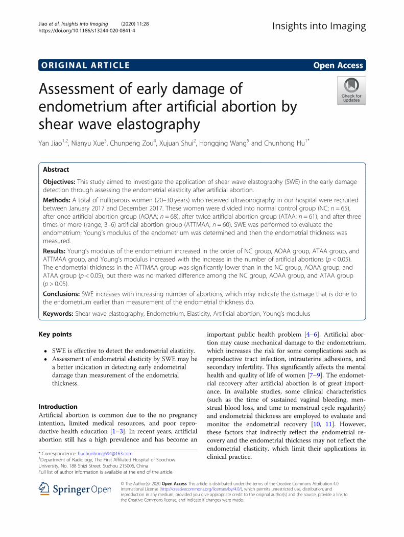
Fig. 1 Sites of endometrium for the measurement of endometrial elasticity by SWE. Notes: Line ae and uterine cavity line are parallel and have the samelength; the three dotted lines are vertical to the line ae and endometrial line; point c is the middle point between point a and point e; point b localizes0.5 cm away from the point c (close to a); point d localizes 0.5 cm away from the point c (close to e). Red dots represent the six sampling sites for SWE.
Table 1 Parameters in different groups
Group n Age (years) Estradiol (ng/ml) Progesterone (pg/ml) Endometrial thickness (mm) Young’s modulus (kPa)
NC 65 24.39 ± 4.26 8.13 ± 2.42 295.89 ± 38.24 11.25 ± 2.81 10.17 ± 3.81
AOAA 68 26.01 ± 3.85 8.35 ± 2.31 306.16 ± 36.37 12.03 ± 2.59 17.01 ± 4.63*
ATAA 61 25.56 ± 4.53 7.80 ± 2.55 298.35 ± 37.66 11.57 ± 2.75 23.35 ± 4.92*#
ATTMAA 60 24.91 ± 4.11 7.98 ± 2.29 293.13 ± 40.08 8.25 ± 2.06*#Δ 32.17 ± 5.38*#Δ
PAOAA vs NC 0.07 0.59 0.11 0.10 0.00
PATAA vs NC 0.14 0.46 0.72 0.52 0.00
PATTMAA vs NC 0.49 0.72 0.69 0.00 0.00
PATAA vs AOAA 0.55 0.20 0.23 0.33 0.00
PATTMAA vs AOAA 0.12 0.37 0.06 0.00 0.00
PATTMAA vs ATAA 0.41 0.68 0.46 0.00 0.00*p < 0.05 vs NC group; #p < 0.05 vs AOAA group; Δp < 0.05 vs ATAA group
Jiao et al. Insights into Imaging (2020) 11:28 Page 2 of 7

within 1month before ultrasonography; (3) presence ofuterine effusion, intrauterine adhesions, and intrauterineresidual or normal menstrual cycle within 3months afterthe last artificial abortion. The study was conducted in ac-cordance with the Committee for Human Research at ourinstitution and followed all regulations. Informed consentwas obtained before the study.
Instruments and methodsSupersonic Imagine AixPlorer ultrasonic instrumentwith SWE (Shear WaveTM) was used for the detection ofendometrial elasticity. The SE12-3 probe (frequency 3–12MHz) applicable for SWE was used.For subjects, SWE of the endometrium was per-
formed by the same sonographer. Meanwhile theexamination was performed 3 months after the lastabortion. The endometrial elasticity was evaluated inthe ovulation stage (days 14–16 of menstrual cycle;maximal diameter of follicles, 18–22 mm). In brief,women lied in a supine position, and routine abdom-inal ultrasonography was performed to explore thepelvic organs. Then, transvaginal two-dimensionalultrasonography was done to observe the shape,
outline, and internal structures of the uterus afteremptying the bladder. Thereafter, transvaginal ultra-sonography was done at multiple views to evaluatethe symmetry of the uterus, possible presence ofuterine effusion and other lesions, and assess theendometrial echoes and thickness as well as the bor-derline between the endometrium and myometrium.The endometrial thickness was measured at the max-imal longitudinal section and recorded. Measurementwas done at the middle point between the fundus ofuterus and cervix. Then, SWE of the endometriumwas performed with the SWE mode. The red, green,and blue colors represent high, intermediate, andlow Young’s modulus, respectively. When the imagesbecame stable, the images were frozen, and quantifi-cation was done with the Q-BOX system. Young’smodulus of the anterior and posterior endometriumwas measured. The diameter of region of interest(ROI) was 2 mm, and the distance between ROI andprobe was 2–4 cm. The ROI was set at three sites ofthe anterior endometrium and three sites of theposterior endometrium (middle point between thefundus of uterus and cervix, 0.5 cm away from the
Fig. 2 Age, progesterone, and estradiol in the NC group, AOAA group, ATAA group, and ATTMAA group. a age; b progesterone; c estradiol
Fig. 3 Representative images of a woman in the NC group. a Young’s modulus at the three sites of the anterior endometrial wall was 5.5, 10.2,and 8.9 kPa, respectively; b Young’s modulus at the three sites of the posterior endometrial wall was 5.8, 10.6, and 11.6 kPa, respectively. Themean Young’s modulus of the six sampling sites was 8.8 kPa
Jiao et al. Insights into Imaging (2020) 11:28 Page 3 of 7
middle point [close to the fundus of uterus] and 0.5cm away from the middle point [close to the cervix])(Fig. 1). All these ROIs localized at the middlepoints between endometrium-myometrium boundaryline and uterine cavity line. The mean, maximal, andminimal Young’s moduli were automatically calcu-lated by the Q-BOX system. The mean Young’smodulus of six sites was determined. At the sametime, blood was collected on the day of ultrasonog-raphy, and the serum levels of estradiol and proges-terone were detected and recorded.
Statistical analysisData were expressed as mean ± standard deviation (SD).Statistical analyses were performed using the statisticalpackage for social software version 22 (SPSS Inc., Chi-cago, IL, USA). Comparisons of quantitative data weredone using one-way analysis of variance (ANOVA)
followed by post hoc least significant difference (LSD)test. A value of p < 0.05 was considered statisticallysignificant.
ResultsThere were no marked differences in age and serumlevels of progesterone and estradiol among the NCgroup, AOAA group, ATAA group, and ATTMAAgroup (P > 0.05) (Table 1; Fig. 2).In the NC group, AOAA group, ATAA group, and
ATTMAA group, the endometrial thickness was 11.25 ±2.81 mm, 12.03 ± 2.59 mm, 11.57 ± 2.75 mm, and 8.25 ±2.06 mm, respectively. The endometrial thickness in theATTMAA group was significantly lower than in the NCgroup, AOAA group, and ATAA group (p < 0.05), butthere was no marked difference among the NC group,AOAA group, and ATAA group (p > 0.05). In the NCgroup, AOAA group, ATAA group, and ATTMAA
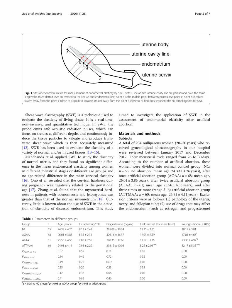
Fig. 4 Representative images of a woman in the AOAA group. a Young’s modulus at the three sites of the anterior endometrial wall was 6.9,14.7, and 16.2 kPa, respectively; b Young’s modulus at the three sites of the posterior endometrial wall was 20.1, 12.3, and 10.8 kPa, respectively.The mean Young’s modulus of the six sampling sites was 13.5 kPa
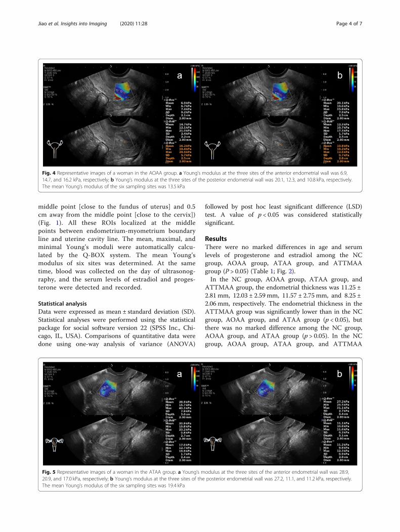
Fig. 5 Representative images of a woman in the ATAA group. a Young’s modulus at the three sites of the anterior endometrial wall was 28.9,20.9, and 17.0 kPa, respectively; b Young’s modulus at the three sites of the posterior endometrial wall was 27.2, 11.1, and 11.2 kPa, respectively.The mean Young’s modulus of the six sampling sites was 19.4 kPa
Jiao et al. Insights into Imaging (2020) 11:28 Page 4 of 7
group, Young’s modulus was 10.17 ± 3.81 kPa, 17.01 ±4.63 kPa, 23.35 ± 4.92 kPa, and 32.17 ± 5.38 kPa, respect-ively. Young’s modulus was significantly different be-tween any two groups (p < 0.05) (Figs. 3, 4, 5 and 6).Young’s modulus increased with the increase in thenumber of artificial abortion (Table 1; Fig. 7).
DiscussionIn the present study, the endometrial thickness and elas-ticity were assessed in post-abortion patients and normalsubjects. Results showed that endometrial thickness inthe ATTMAA group was significantly lower than in theNC group, AOAA group, and ATAA group, but therewas no marked difference among the AOAA group,ATAA group, and NC group. In addition, Young’smodulus increased significantly in the order of NCgroup, AOAA group, ATAA group, and ATTMAAgroup. These findings indicate that the endometrial elas-ticity reduces after repeated artificial abortion in theATTMAA group; although the endometrial thickness in
the AOAA group and ATAA group remains unchanged,the physical characteristics of the endometrium changesignificantly (elasticity reduces and stiffness increases),which was more evident in the TAA group than in theAOAA group.Some investigators found the mechanical damage to
the endometrium could reduce the endometrial glandu-lar epithelium and increase the collagen fibers in animalmodel of artificial abortion [19], leading to the increasedproportion of collagen fibers in the endometrium. Thereis evidence showing that the increased collagen fibers inthe organ may significantly compromise the elasticity ofthis organ and increases its stiffness [20, 21]. Thus, onthe basis of our findings, we speculate that the mechan-ical damage to the endometrium after artificial abortionmay cause hyperplasia of collagen fibers in the endomet-rium and thereafter increase their amount, which leadsto the increase in the endometrial stiffness [22–24].Moreover, the endometrial stiffness increases with theincrease in the number of artificial abortion. The
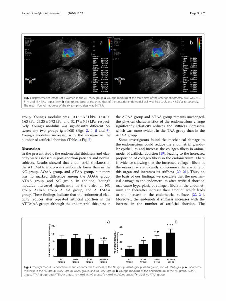
Fig. 6 Representative images of a woman in the ATTMAA group. a Young’s modulus at the three sites of the anterior endometrial wall was 25.9,31.4, and 43.4 kPa, respectively; b Young’s modulus at the three sites of the posterior endometrial wall was 30.3, 34.8, and 42.5 kPa, respectively.The mean Young’s modulus of the six sampling sites was 34.7 kPa
Fig. 7 Young’s modulus endometrium and endometrial thickness in the NC group, AOAA group, ATAA group, and ATTMAA group. a Endometrialthickness in the NC group, AOAA group, ATAA group, and ATTMAA group. b Young’s modulus of the endometrium in the NC group, AOAAgroup, ATAA group, and ATTMAA group. *p < 0.05 vs NC group. #p < 0.05 vs AOAA group. Δp < 0.05 vs ATAA group
Jiao et al. Insights into Imaging (2020) 11:28 Page 5 of 7

pathological changes in the endometrium are consistentwith the change in the endometrial stiffness. Recentstudies have shown that SWE can be used to evaluatethe elasticity of normal myometrium, intima, and cervixand that SWE is a promising tool for the uterine assess-ment and helpful for the diagnosis of various uterine dis-eases [16]. Available findings together with our resultsindicate that SWE may be used to display the early dam-age to the endometrium when it is invisible on conven-tional ultrasonography and 2D measurements. Takingthis research as an opportunity, we can carry out moresystematic studies on the application of SWE in theassessment of endometrium after artificial abortion. Thisis a direction of our future investigations.There were limitations in this study. The uterus is
three-dimensional, and current techniques cannot sam-ple the endometrium at different sites simultaneously.Thus, the middle points of the anterior and posteriorwalls of the uterus were selected. We believe that thedevelopment of medical technology will improve themeasurement of endometrial elasticity. However, ourfindings provide valuable information for the clinicalassessment of endometrial damage after artificial abortion,which is helpful for the diagnosis and management ofendometrial complications after abortion.Taken together, SWE is effective to detect the endo-
metrial elasticity after artificial abortion, which seems toindicate the assessment of physical characteristics andpathological changes of the endometrium. The changesin the endometrium after artificial abortion may occurpreceding the clinical manifestations and the alterationof endometrial thickness. Thus, the endometrial elasticitydetermined by SWE may provide important informationfor the assessment of endometrial recovery.
AbbreviationsAOAA: After once artificial abortion; ATAA: After twice artificial abortion;ATTMAA: After three times or more artificial abortion; ROI: Region of interest;SD: Standard deviation; SWE: Shear wave elastography
Authors’ contributionsYJ and CH designed this study. YJ, NX, CZ, XS, HW, and CH collected andinterpreted the patients’ data. YJ analyzed the data. YJ was the majorcontributor to the writing of the manuscript. All authors read and approvedthe final manuscript.
FundingThis study was supported by the Basic and Public Studies of ZhejiangProvince (No. LGF18H180003) and the Health and Family PlanningCommission of Zhejiang Province, China (Nos. 2018ZD043 and 2019KY665).
Availability of data and materialsThe datasets used and/or analyzed in the present study are available fromthe corresponding author on reasonable request.
Ethics approval and consent participateThe study was conducted in accordance with the Committee for HumanResearch at our institution and followed all regulations. Informed consentwas obtained before study.
Consent for publicationA consent to publication has been signed by each participant.
Competing interestsThe authors declare that they have no competing interests.
Author details1Department of Radiology, The First Affiliated Hospital of SoochowUniversity, No. 188 Shizi Street, Suzhou 215006, China. 2Obstetrics andGynecology Ultrasonic Department, Wenzhou Peoples’ Hospital, Wenzhou325000, China. 3Department of Diagnostic Ultrasonography, Ningbo FirstHospital, Ningbo 315010, China. 4Department of Diagnostic Ultrasonography,The Second Affiliated Hospital and Yuying Children’s Hospital of WenzhouMedical University, Wenzhou 325027, China. 5Department of Radiology, TheFirst Affiliated Hospital of Wenzhou Medical University, Wenzhou 325000,China.
Received: 9 September 2019 Accepted: 30 January 2020
References1. Faundes A, Shah IH (2015) Evidence supporting broader access to safe legal
abortion. Int J Gynaecol Obstet 131(Suppl 1):S56–S592. Maina BW, Mutua MM, Sidze EM (2015) Factors associated with repeat
induced abortion in Kenya. BMC Public Health 15:10483. Pereira J, Pires R, Canavarro MC (2017) Psychosocial adjustment after
induced abortion and its explanatory factors among adolescent and adultwomen. J Reprod Infant Psychol 35:119–136
4. Erdman JN (2012) Harm reduction, human rights, and access to informationon safer abortion. Int J Gynaecol Obstet 118:83–86
5. Labandera A, Gorgoroso M, Briozzo L (2016) Implementation of the risk andharm reduction strategy against unsafe abortion in Uruguay: from auniversity hospital to the entire country. Int J Gynaecol Obstet 134:S7–s11
6. Sedgh G, Singh S, Shah IH, Ahman E, Henshaw SK, Bankole A (2012)Induced abortion: incidence and trends worldwide from 1995 to 2008.Lancet 379:625–632
7. Gerdts C, Hudaya I (2016) Quality of care in a safe-abortion hotline inIndonesia: beyond harm reduction. Am J Public Health 106:2071–2075
8. Russo JA, Achilles S, DePineres T, Gil L (2013) Controversies in familyplanning: postabortal pelvic inflammatory disease. Contraception87:497–503
9. Xiao S, Wan Y, Xue M et al (2014) Etiology, treatment, and reproductiveprognosis of women with moderate-to-severe intrauterine adhesions. Int JGynaecol Obstet 125:121–124
10. Madeiro AP, Rufino AC (2017) Maltreatment and discrimination in inducedabortion care: perception of women in Teresina, State of Piaui, Brazil. CienSaude Colet 22:2771–2780
11. Rottenstreich A, Amsalem H, Kleinstern G, Kalish Y (2017) Outcomes ofthreatened abortions after anticoagulation treatment to prevent recurrentpregnancy loss. Reprod Biomed Online 35:461–467
12. Taljanovic MS, Gimber LH, Becker GW et al (2017) Shear-wave elastography:basic physics and musculoskeletal applications. Radiographics 37:855–870
13. Barr RG (2018) Shear wave liver elastography. Abdom Radiol (NY)43:800–807
14. Hu X, Liu Y, Qian L (2017) Diagnostic potential of real-time elastography(RTE) and shear wave elastography (SWE) to differentiate benign andmalignant thyroid nodules: a systematic review and meta-analysis. Medicine(Baltimore) 96:e8282
15. Liu H, Wan J, Xu G et al (2019) Conventional US and 2-D shear waveelastography of virtual touch tissue imaging quantification: correlation withimmunohistochemical subtypes of breast cancer. Ultrasound Med Biol45:2612–2622
16. Manchanda S, Vora Z, Sharma R et al (2019) Quantitative sonoelastographicassessment of the normal uterus using shear wave elastography: an initialexperience. J Ultrasound Med 38:3183–3189
17. Ono T, Katsura D, Yamada K et al (2017) Use of ultrasound shear-waveelastography to evaluate change in cervical stiffness during pregnancy.J Obstet Gynaecol Res 43:1405–1410
18. Zhang M, Wasnik AP, Masch WR et al (2019) Transvaginal ultrasound shearwave elastography for the evaluation of benign uterine pathologies: aprospective pilot study. J Ultrasound Med 38:149–155
Jiao et al. Insights into Imaging (2020) 11:28 Page 6 of 7
19. Huberlant S, Fernandez H, Vieille P et al (2015) Application of a hyaluronicacid gel after intrauterine surgery may improve spontaneous fertility: arandomized controlled trial in New Zealand White rabbits. PLoS One 10:e0125610
20. Lunde IG, Herum KM, Carlson CC, Christensen G (2016) Syndecans in heartfibrosis. Cell Tissue Res 365:539–552
21. Voorhees AP, DeLeon-Pennell KY, Ma Y et al (2015) Building a betterinfarct: modulation of collagen cross-linking to increase infarct stiffnessand reduce left ventricular dilation post-myocardial infarction. J Mol CellCardiol 85:229–239
22. Brabrand A, Kariuki II, Engstrom MJ et al (2015) Alterations in collagen fibrepatterns in breast cancer. A premise for tumour invasiveness? APMIS 123:1–8
23. Liu H, Zhao LX, Xu G et al (2015) Diagnostic value of virtual touch tissueimaging quantification for benign and malignant breast lesions withdifferent sizes. Int J Clin Exp Med 8:13118–13126
24. Wang ZL, Li JL, Li M, Huang Y, Wan WB, Tang J (2013) Study of quantitativeelastography with supersonic shear imaging in the diagnosis of breasttumours. Radiol Med 118:583–590
Publisher’s NoteSpringer Nature remains neutral with regard to jurisdictional claims inpublished maps and institutional affiliations.
Jiao et al. Insights into Imaging (2020) 11:28 Page 7 of 7