assessment of collateral status using multiphasic …
TRANSCRIPT

A
ASSESSMENT OF COLLATERAL STATUS USING MULTIPHASIC CT ANGIOGRAPHY IN THE ACUTE ANTERIOR CIRCULATION ISCHEMIC
STROKE: ITS CORRELATION WITH CLINICAL AND RADIOLOGIC OUTCOMES
THESIS SUBMITTED IN PARTIAL FULFILLMENT FOR DEGREE OF
DM (NEUROIMAGING AND INTERVENTIONAL NEURORADIOLOGY)
(2016- 2018)
OF THE SREE CHITRA TIRUNAL INSTITUTE FOR MEDICAL SCIENCES
AND TECHNOLOGY
TRIVANDRUM, INDIA
Dr. ADHITHYAN R
DEPARTMENT OF IMAGING SCIENCES AND INTERVENTIONAL RADIOLOGY,
SREE CHITRA TIRUNAL INSTITUTE FOR MEDICAL SCIENCES AND
TECHNOLOGY, TRIVANDRUM, INDIA


E
CONTENTS
PAGE NO
1. INTRODUCTION 1
2. AIMS & OBJECTIVES 4
3. REVIEW OF LITERATURE 6
4. MATERIALS AND METHODS 17
5. RESULTS 29
6. REPRESENTATIVE CASES 45
7. DISCUSSION 56
8. CONCLUSION 66
9. REFERENCES 68
10. ANNEXURES 76

1
Introduction

2
INTRODUCTION:
Computed tomography Angiography (CT) remains the initial imaging modality of
choice in hyper-acute stroke. Non-contrast CT (NCCT) excludes intracranial
hemorrhage and tumor and detects early signs of infarction. CT angiography
(CTA) can rapidly provide useful information that may influence management and
may indicate the extent of vessel occlusion, all of which can influence clinical
outcome and recanalization.
Recently, leptomeningeal collaterals in acute stroke have been shown to have a
significant impact upon the clinical and radiological outcome of the patients.
Collaterals can be studied by various imaging modalities including CT, MRI,
catheter angiography and transcranial Doppler. However, in the majority of the
stroke centers throughout the world, plain CT followed by CT angiography is the
initial and in many cases sole imaging modality to assess the acute stroke patients
for major vessel occlusion as well as triage for intravenous thrombolysis or
mechanical thrombectomy. Infarct has shown to evolve from a few hours to days
and it varies depending upon the intracranial collaterals of the patient. In almost
the majority of the studies, collateral status was evaluated by single-phase CT
angiography.

3
Recently, the concept of multiphase CT angiography is evolving and few centers
have adopted this as an initial imaging modality for acute stroke patients. The
benefits of multiphase CT angiography over single-phase CT angiography has to
be validated for large-scale implementation.
We hypothesized that multiphase CT angiography collaterals due to its dynamicity
would predict the clinical as well as radiologic outcomes of the patients better than
the static single-phase CT angiography. It would also a good prognostic marker for
the risk of major hemorrhagic transformation.
So we designed this study as a comparison of multiphase CTA collateral scores
with the single-phase collateral scoring system in acute stroke patients and its
correlation with early clinical outcome (NIHSS at discharge), late clinical outcome
(mRS 90 days), radiologic outcomes (ASPECTS 24 hours, hemorrhagic
transformation).

4
Aims Objectives

5
AIMS AND OBJECTIVES
“Comparison of the multiphasic computed tomographic (CT) angiography
collateral scoring system with already available single-phase CT scoring system,
in assessing intracranial vascular collateral status, in patients with acute anterior
circulation ischemic stroke and its ability in determining clinical as well as
radiological-outcomes”

6
Review of Literature

7
REVIEW OF LITERATURE
1.INTRODUCTION:
Acute stroke is the second most common cause of death worldwide after
coronary artery disease. Globally approximately 6.3 million deaths (1) were
reported due to acute stroke in 2015. Acute stroke may be ischemic or
hemorrhagic. Atherosclerosis and embolism are major causes of acute ischemic
stroke. In the acute ischemic stroke, arterial recanalization by either intravenous
thrombolysis or mechanical thrombectomy remains the principal treatment.
Cerebral neuronal damage is not uniform in the initial few hours after stroke
onset. Depending upon intra cranial collaterals, infarct may not be complete for
hours or even days (2). In some patients infarct gets completely developed in a
few hours and other the ischemic tissue remains viable for few days, depending
upon the degree of leptomeningeal collaterals which have a significant protective
role. (3)
2.ANATOMY OF COLLATERAL CIRCULATION: (4)
Collateral types Vessels
Large-artery communications
between the extracranial and
intracranial circulations
Ophthalmic artery, maxillary artery,
superficial temporal artery, occipital artery.
Mainly in chronic occlusion.
Circle of Willis Anterior and posterior communicating
artery. 50% - complete.
Leptomeningeal collaterals Important routes in acute occlusion. Small
arteriolar communications allowing
retrograde perfusion. Between ACA and
MCA. Between MCA and PCA.

8
3.FACTORS AFFECTING COLLATERALS DEVELOPMENT:
History of hypertension is associated with poor intracranial collaterals (5). Use of
statins prior to stroke was linked to better collaterals (6). Potential of collaterals
developing decreases with age. Hyperglycemia, dehydration, hyperthermia,
cardiac failure, pulmonary compromise, renal failure, electrolyte disturbance,
diffuse atherosclerosis were also shown to reduce the development of
collaterals.
Incomplete circle of Willis also leads to reduced collateral blood flow. Rapidness
of occlusion also has an impact on collaterals. Gradual onset major vessel
occlusion like moya-moya disease have good collaterals development than acute
occlusion.
4.IMAGING OF COLLATERALS:
Conventional digital subtraction angiography (DSA), CT angiography, MR
angiography, Trans cranial Doppler (TCD) are the imaging methods available to
evaluate intracranial collaterals
4.1DSA:
The gold standard method allowing assessment of all three major type of
collaterals.

9
A. Kucinski et al method (7):
Good if >= three MCA branches up to M2 segment, retrograde filling. Poor < three
MCA branches retrograde filling.
B. American Society of Interventional and Therapeutic Neuroradiology and
the Society of Interventional Radiology (ASTIN/SIR) (8): 5-point scale.
0 No collaterals visible to the ischemic site
1 Slow collateral to the periphery
Persistence of the defect
2 Rapid filling of collaterals to the periphery
Persistence of the defect
3 Slow collaterals to the periphery
Complete angiographic blood flow
4 Rapid filling of collaterals to the periphery
Complete angiographic blood flow

10
4.2 CT ANGIOGRAPHY EVALUATION OF COLLATERALS:
Advantages of CT angiography for collateral assessment:
Widely available.
Rapid assessment.
Good spatial resolution for assessment of leptomeningeal collaterals.
CT perfusion, multiphasic angiograms, cervical vessel imaging can be done
simultaneously.
Single-phase CT angiography collateral scoring methods for anterior
circulation ischemic stroke:
A.Tan scale(8):
0- absent collateral supply in the occluded MCA territory.
1- Collateral supply filling <= 50% but >0% of the occluded MCA territory.
2 - Collateral supply filling > 50% but < 100% of the occluded MCA territory.
3 - 100% collateral supply of the occluded MCA territory.
Patients with high TAN collateral score had lower pretreatment perfusion defects,
smaller final infarct and also final functional outcome, as well as recanalization
rates.
B.Modified Tan Scale:
Simplest and rapid system, with less interobserver variation.
Good- >= 50% of the MCA territory.
Poor - <50% of the MCA territory.

11
C.Miteff System :
3-point score grading of middle cerebral artery collateral branches with respect to
the Sylvain fissure. One study showed miteff is more reliable predictor of good
outcome than other single-phase scoring systems(11). Patients with good
collaterals have lower NIHSS score (3), small infarct size and better prognosis.
3 - vessels reconstituted distal to the occlusion.
2-vessels seen at the Sylvian fissure.
1-vessels seen only in the distal superficial branches.
D.Maas System (9):
5-point score comparing collaterals on the affected hemisphere against those on
the unaffected side.
5 -exuberant.
4-more than the contralateral side.
3-equal to the contralateral side.
2-less than the contralateral side.
1-no vessel seen.
E.Alberta Stroke Program Early CT Score (ASPECTS) 20 Point Grading
Scale(10):
Collaterals are scored in each of the 10 regions corresponding to the ASPECTS
system, to form a score from 0 to 20
0- artery not seen.
1- less prominent.
2- equal or more prominent

12
4.3 MRI EVALUATION OF COLLATERALS: A.T2 FLAIR:
In patients with MCA occlusion, prominent distal T2 FLAIR hyperintense signals
(12,13) is associated with smaller initial ischemic infarct, larger diffusion-perfusion
mismatch, better NIHSS score and smaller subacute ischemic lesion volume.
Although definitely not proved, distal T2 FLAIR hyperintense signal may be said to
be related to good collateral flow distal to the occlusion site (14) (15).
B.Arterial spin labelling (ASL):
Territorial ASL gave information regarding intracranial collaterals, comparable to
DSA in steno occlusive patients. (16)
3D pseudo-continuous ASL with multiple post label delays had been used to
estimate antegrade and collateral flow in unilateral MCA stenosis. (17)
ASL collaterals detected in acute stroke were showed to correlate with
neurological outcome of mRS at discharge. (18,19)
C.Susceptibility weighted imaging (SWI)
-SWI was shown to accurately predict intracranial collateral circulation in acute
infarcts and can be used for prognostication and curative evaluation.(20)
-SWI-diffusion mismatch was also proved to identify patients who benefit from
reperfusion therapy (21).

13
4.4TRANS CRANIAL DOPPLER:
High velocity, low resistance flow pattern (22,23) in ipsilateral ACA or PCA in
patients with MCA occlusion was associated with leptomeningeal collaterals and is
termed flow diversion(FD). 30% difference of flow velocity between right and left
ACA is associated with leptomeningeal collaterals. FD is more commonly seen
with ACA than PCA. FD was shown associated with CT angiography collaterals,
admission infarct volume, and 24-hour infarct volume. FD can be used as bedside
prognostic tool in anterior circulation stroke.
4.5 LATEST IMAGING-MULTIPHASIC CT COLLATERAL SCORING (24): Multiphase CT angiography is the latest modification of the CT angiography
protocol for better evaluation of intra cranial collaterals. This imaging modality is
still not widely used. This was first used in the ESCAPE trial (25). In this type of
angiography, time-resolved angiograms from the skull base to the vertex would be
taken in three phases at peak arterial, peak venous and late venous phases.
Radiation dose and acquisition time is lower than CT perfusion. Unlike CT
perfusion, post-processing and additional contrast are not required.
Multiphasic CT is using a six-point scale (ALBERTA stroke program) for assessing
pial collaterals (later discussed in materials and methods), by comparing with the
normal hemisphere in analyzing the number of phases delay as well as the extent,
prominence or absence of vessels in the ischemic territory. Multiphasic CT
Collateral scales are finally graded as, 0 and 1-poor, 2 and 3- intermediate, 4 and
5- good.

14
5. COLLATERALS AND STROKE CLINICAL, RADIOLOGICAL OUTCOMES:
CT collaterals (26) were shown to correlate well with CT perfusion imaging
data. In patients of IMS III trial (27), better collaterals were proved to have
small infarct core and larger mismatch. Some stroke trials used CT
perfusion and Ct angiography collaterals as an imaging tool for inclusion
criteria of patients for endovascular management.
In stroke trials like Extend IA (28), SWIFT prime (29) CT perfusion was
used. While CT collaterals were used in the ESCAPE trial. Ct perfusion has
various disadvantages like post-processing time, variations in CTP
acquisition and post-processing software.
Good base line collaterals at sylvian fissure were associated with good
functional recovery (30) and reduced mRS at 90 days. This was
mediated through reduced final infarct volume.
Poor intracranial collaterals were associated with a high incidence of
hemorrhagic transformation (31,32). In one study (31), the rate of
significant hemorrhage (> 25ml) after intra arterial therapy, was seen in
25% of patients with poor collateral, while it was only 2.7% in patients with
poor collaterals. Hence, intra arterial therapy was cautioned in patients with
very poor collaterals.
Good collaterals in angiography were associated with successful
recanalization (33,35) in Solitaire FR With the Intention for Thrombectomy
(SWIFT) study (34).

15
ASA revised guidelines 2018 (36) for acute stroke management, mentioned
that “it may be reasonable to include collateral status into clinical decision
making in some patients to include for mechanical thrombectomy”
with a level of evidence C (limited data) and class 2b class of
recommendation.
In patients with good collaterals, there would be a better dissolution of
thrombus with IV-tPA (Tissue plasminogen activator) by transporting
fibrinolytic to either side of the thrombus. In systematic review and meta -
analysis (37,38) of the impact of collateral status in functional outcomes
after thrombolysis showed good collateral were associated with better
recanalization and reperfusion rate as well as good long term functional
outcome.
6. POSTERIOR CIRCULATION COLLATERALS ASSESSMENT:
-One study (39) used a collateral staging method 0 to 2. 0=no pcom, 1=1 pcom
and 2 both pcom present. Presence of pretreatment bilateral pcom is
associated with favorable outcome after endovascular therapy.
-Another CT angiography collateral scoring method (10 point scoring) was
provided by Erik et al(40). In this method, each PICA, AICA, SCA if visible, is
given a score of 1. Each Pcom is given a score if diameter less than PCA and
2 if more than PCA. Score 6 to 10 is good.

16
-Qureshi at al (41) gave a posterior circulation angiographic collateral scoring
method-Grades I and II - retrograde filling of the basilar artery through PCA
with or without filling of the superior cerebellar artery, respectively. Grades III
and IV - bilateral or unilateral anastomoses of cerebellar arteries or PCAs,
respectively.
7. COLLATERALS THERAPEUTICS FOR ACUTE ISCHEMIC STROKE:
Experimental collateral therapeutics like inhaled nitric oxide(42), high dose
albumin(43), sphenopalatine ganglion stimulation (44), transient aortic
occlusion(45) had been showed to improve collaterals in animal models.
However, before the implementation of these methods, further studies
regarding collaterals pathophysiology and its implications have to be evaluated
in large-scale studies.

17
Materials Methods
18
MATERIALS AND METHODS
Institutional Ethics Committee (IEC) approval was obtained vide letter No.
SCT/IEC/987/OCT-2016 (Annexures). This was a prospective study
conducted in department of IS&IR, SCTIMST, between October 2016 and
May 2018. Consecutive patients who were evaluated for inclusion into the
study.
Informed written consent was obtained from each and every
patient/guardian after explanation of all aspects of the study as per consent
form and details were noted as per proforma.(Annexures)
All patients with acute stroke who underwent both plain CT followed by
multiphase CT angiography evaluation were included in the study. No
pregnant woman, prisoner, staff, student or healthy volunteer was inducted
into this study.
INCLUSION CRITERIA:
1. Patient presenting to the SCTIMST stroke or emergency department with
symptoms consistent with the acute ischemic stroke.
2. Age > 18 yrs.
3. Pre-treatment multi-phase CTA done within 8 hours of stroke symptom
onset.
4. Pre-treatment NIHSS score >=5
5. Patients consenting for their inclusion in the study.

19
EXCLUSION CRITERIA:
1. Intracranial haemorrhage (ICH) identified on baseline CT.
2. Previous moderate to large stroke in the ipsilesional hemisphere.
3. Unable to have CT-angiography performed due to contrast allergy, known
chronic/acute kidney failure history, pregnancy or other reasons.
4. Patients aged less than or equal to eighteen years.
STUDY DESIGN
1. IEC approval was obtained vide letter SCT/IEC/987/OCT-2016
2. Written informed consent of all eligible patients was obtained.
3. Patients’ history and other clinical details were documented as per
Performa.
4. Patients underwent plain CT followed by multiphase CT angiography,
as per existing hospital protocol. In Plain CT, ASPECTS (0 to 10)
details were noted.
5. In multiphase CT angiography, major vessel occlusion was noted.
Only MCA (M1, M2) or ICA occlusion or combined MCA ICA
occlusion cases who presented within 8 hours of symptom onset
were included.
6. Then multiphase CT collateral grading was assessed (0 to 5). Then
single phase CT grading 0 to 3 as per most widely used TAN grading
system was assessed.
7. Base line NIHSS, mRS of the patient were assessed and recorded
by certified neurologist. Age, sex, risk factors (hypertension,
diabetes, coronary heart disease, atrial fibrillation, smoking,

20
hyperlipidemia) of the patients were recorded.
8. Patients treatment history details- either conservative, iv
thrombolysis, mechanical thrombectomy were included in the study.
9. In patients who had undergone mechanical thrombectomy, time to
groin, onset to reperfusion and mTICI grading were also included in
the study.
10. All patients underwent 24 hrs plain CT as per our institutional
protocol and details of CT ASPECTS (0 to 10), hemorrhagic
transformation (ECASS grading) were assessed.
11. All patients were examined for NIHSS at discharge. Those patients
who were alive at discharge were followed after 90 days to assess
mRS scale of functional outcome. Good and poor functional
outcomes were defined by mRS scores of 0–2 and 3–6, respectively.
12. Categorical and continuous variables were tabled. Statistical
differences of these variables in single and multiphase CT collateral
groups were assessed by student T test (for continuous variables) or
Fisher exact test (for categorical variables). Multivariate logistic was
done for variables with significant difference. Statistical analysis of
the data was performed and sensitivity, specificity, positive, negative
predictive values and accuracy of both the single and multiphase CT
angiography collaterals in predicting 3 months functional outcome
(mRS) was assessed. Receiver operator characteristic (ROC) curve
analysis of single and multiphase CT angiography for predicting
functional outcome along with area under curve (AUC), Youden’s J,
test efficiency individually for both collateral grading as assessed.

21
FLOWCHART OF STUDY DESIGN:
Acute stroke patients who within 8 hours of symptom onset between Oct 2016 and May 2018
(n=56)
Base line NIHSS (>5), mRS, Age, sex, risk factors (hypertension, diabetes, coronary heart disease, atrial
fibrillation, smoking, hyperlipidemia)
Plain CT -ASPECTS (0 to 10)
CT angiography-
MCA (M1,M2) occlusion–42 cases
ICA occlusion or combined MCA ICA occlusion-14 cases
Multiphase CT collateral grading mCT grading (0 to 5)
Single-phase CT grading 0 to 3 TAN grading system
Patient treatment history - either conservative, IV thrombolysis, mechanical thrombectomy.
If Mechanical thrombectomy, time to groin, onset to reperfusion and TICI grading.
24 hrs plain CT - 24 hrs ASPECTS (0 to 10), hemorrhagic transformation (ECASS grading).
NIHSS at discharge (n=56 cases)
90 days mRS scale of functional outcome (n=34 cases)
Student T test (for continuous variables) or Fisher exact test (for categorical variables).
Multivariate logistic analysis.
Sensitivity, specificity, positive, negative predictive values and accuracy of collaterals in predicting 3
months functional outcome.
Receiver operator characteristic (ROC) curve analysis of single and multiphase CT angiography for
predicting functional outcome.

22
STUDY PROTOCOLS:
HEAD PLAIN CT:
Examinations ware performed in the 256 slice CT scanner (Philips). All patients
who fulfilled the inclusion criteria, undergone standard unenhanced CT with
0.625-mm section thickness. It was reformed into 3mm thick images to see for
ASPECTS at admission.
MULTIPHASE CT ANGIOGRAPHY:
Scanning was triggered by using bolus tracking, with the region of interest placed
in the posterior aortic arch with the trigger threshold set at 150 HU. Three phases
of cerebral angiograms of brain vasculature ware acquired.
The first phase covers the aortic arch to vertex region during peak arterial phase.
Subsequent two phases cover the skull base to the vertex in peak venous and late
venous phase respectively. Images were acquired at an interval of 8 seconds
apart.
A total of 50 mL of contrast material (Iohexol, Omnipaque/ Iodixanal, Visipaque)
was be injected at a rate of 5 mL/sec and followed by 30 ml normal saline chase.
Source images were reformatted into 3-mm-thick axial, coronal, and sagittal
projections.

23
DEFINITIONS OF PARAMETERS:
Plain CT ASPECTS:
Alberta stroke programme early CT score (ASPECTS) (46,47) is a 10-point scale
to assess early ischemic changes in acute middle cerebral artery stroke in plain
CT. Two levels of the brain are read, one at the level of basal ganglia and another
at the level of ventricles just above basal ganglia. One point is deducted from the
initial score of 10 if any of the regions involved. Caudate, internal capsule,
lentiform nucleus, insula, M1 to M3 (at the level of basal ganglia), M4 to M6 (at the
level of ventricles). Plain CT ASPECTS was calculated in our study at the time of
admission as well as in 24 hours follow up CT, to assess the extent of stroke.
NIHSS:
NIHSS (48,49) is a 42-point quantitative measure to evaluate the neurological
deficit in stroke patients. It can be performed quickly and has good interrater
agreement. 11 items (level of consciousness, horizontal eye movement, visual
field, facial palsy, motor arm, motor leg, limb ataxia, sensory, language, speech,
extinction and inattention) were evaluated in NIHSS, and each item has scores 0
to 4 depending upon patient ability. Maximum score is 42 and minimum score is 0.

24
Patients with scores less than 5 were considered minor stroke and excluded in our
study. Patients with MCA M1/ ICA occlusion with NIHSS 5 to 25 were taken for
mechanical thrombectomy.
mRS:
mRS is a 6 point clinical outcome score (50, 51). It is the widely used disability/
dependency assessment scale of daily activities of people after acute stroke. Its
scale ranges from 0 to 6, with 0 being no symptoms and 6 being dead. In our
study, mRS of the patients at the time of admission, at discharge and 90 days
follow up were calculated. mRS at 90 days follow up with scores <=2 were
considered good functional outcome, while scores>2 were considered bad
outcome.

25
SINGLE PHASE CT COLLATERAL GRADING:
In our study we used the most widely used single-phase intracranial collateral
scoring method –TAN system. TAN system scoring is a 3-point scoring system
done in traditionally followed single-phase CT angiography. We have applied this
system in the first phase of the multiphase CT angiography. Scores 0 and 1 were
considered poor collaterals. 2 and 3 scores were considered good collaterals.
TAN COLLATERAL SCORE FINDINGS IN THE FIRST PHASE OF CT ANGIOGRAPHY
0 No collateral supply in the occluded MCA territory.
1 <=50% collateral supply in occluded MCA territory
2 >50 to <100% collateral supply in occluded MCA territory
3 >=100% collateral supply in occluded MCA territory.
MULTIPHASE CT COLLATERAL GRADING :
We used the multiphase CT angiography scoring developed by ALBERTA stroke
program (25). It is based upon scoring intracranial collaterals in the affected MCA
ischemic territory, in three individual subsequent phases of multiphase
intracranial CT angiography. Scores 0 to 3 were considered poor collaterals.
Scores 4 and 5 were considered good collaterals.

26
MULTIPHASE CT ANGIOGRAPHY COLLATERAL SCORE
FINDINGS IN CT ANGIOGRAPHY
0 Compared to asymptomatic contralateral hemisphere there are
no vessels in any phase within the occluded vascular territory
1 Compared to asymptomatic contralateral hemisphere there are
just a few vessels visible in any phase within the occluded
vascular territory
2 Compared to asymptomatic contralateral hemisphere there is a
delay of two phases in filling in of peripheral vessels and
decreased prominence and extent or a one-phase delay and
some regions with no vessels in some part of the territory
occluded
3 Compared to asymptomatic contralateral hemisphere there is a
delay of two phases in filling in of peripheral vessels but
prominence and extent is the same or there is a one phase
delay and decreased prominence /reduced number of vessels
in some part of the territory occluded
4 Compared to asymptomatic contralateral hemisphere there is a
delay of one phase in filling in of peripheral vessels, but
prominence and extent is the same
5 Compared to asymptomatic contralateral hemisphere, there is
no delay and normal/increased prominence of peripheral
vessels/normal extent within the occluded arteries territory
within the symptomatic hemisphere

27
ECASS II HEMORRHAGIC TRANSFORMATION SCALE:
We used the European Cooperative Acute Stroke Study (ECASS II) hemorrhagic
transformation scale to look for any hemorrhagic transformation in the follow up 24
hours CT. ECASS II PH2 was considered poor radiological outcome in the follow
up CT (52,53).
ECASS II HEMORRHAGIC
TRANSFORMATION SCALE
BLEED STATUS IN 24 HRS FOLLOW
UP CT
Haemorrhagic infarction type 1 (HI1) Petechial haemorrhages at infarct
margins
Haemorrhagic infarction type 2 (HI2) Petechial haemorrhages throughout
the infarct no mass-effect due to bleed
Parenchymal hematoma type 1 (PH1)
≤30% of the infarcted area
Minor mass effect due to bleed
Parenchymal hematoma type 2 (PH2) >30% of infarct zone, substantial
mass effect attributable to the
haematoma
MODIFIED TREATMENT IN CEREBRAL ISCHEMIA (mTICI) SCALE.(54)
mTICI Grades
Definitions
Grade 0 No perfusion
Grade 1 Antegrade reperfusion past the initial occlusion, but limited distal branch filling with little or slow distal reperfusion
Grade 2a Antegrade reperfusion of less than half of the occluded target artery previously ischemic territory
Grade 2b Antegrade reperfusion of more than half of the previously occluded target artery ischemic territory
Grade 3 Complete antegrade reperfusion of the previously occluded target artery ischemic territory

28
STATISTICS The patient demographic data, risk factors, base line plain CT, CT
angiography collateral status, treatment history, follow up radiological and
clinical outcomes were tabulated in Microsoft Excel format.
Categorical and continuous variables were tabled.
1. Statistical differences of these variables in single and multiphase CT
collateral groups were assessed by student T test (for continuous
variables) or Fisher exact test (for categorical variables).
2. Multivariate logistic regression and linear regression analysis
were done for variables with significant difference in student T/ Fisher
exact test, to assess the odds ratio, p value and linear regression
coefficient value of the variables.
3. Sensitivity, specificity, positive, negative predictive values,
likelihood ratios and accuracy of both the single and multiphase CT
angiography collaterals in predicting 3 months functional outcome
(mRS) was assessed.
4. Receiver operator characteristic (ROC) curve analysis of single
and multiphase CT angiography for predicting functional outcome
along with area under curve (AUC), youden’s J, test efficiency were
calculated individually for both collateral grading were assessed.
5. Spearman coefficient was done to assess the correlation of single
and multiphase CT angiography with variables.

29
Results

30
RESULTS
DEMOGRAPHIC DETAILS
A total of 56 consecutive patients with anterior circulation acute stroke with
major vessel (M1, M2 or ICA or combined), who presented to the institute
between October 2016 and 30 June 2018 were included in this prospective
study.
The age of patients in the study ranged from 37 to 86 years, with the mean age of 63 years.
FIGURE 1: AGE IN YEARS

31
There were 35 males and 21 females in the study group. The male to
female sex ratio within the cohort was 5:3.
FIGURE 2: SEX DISTRIBUTION
There were 42 MCA occlusion and 14 cases of ICA/ combined ICA-MCA
occlusion cases in this study.
FIGURE 3: VESSEL OCCLUDED
0
5
10
15
20
25
30
35
40
NUMBER
SEX DISTRIBUTION
MALES FEMALES
0
5
10
15
20
25
30
35
40
45
NUMBER
VESSEL OCCULUDED
MCA (MA/M2) ICA/ COMBINED

32
RISK FACTORS:
The commonest risk factor of our patients was hypertension (n=25),
diabetes (n=21) followed by CAD (11 cases), AF (11 cases) and smoking
(10 cases). 9 patients had no risk factors.
FIGURE 4: RISK FACTORS DISTRIBUTION
0
5
10
15
20
25
30
HYPERTENSION DIABETES CAD AF SMOKING NO RISK FACTORS
RISK FACTORS DISTRIBUTION
NUMBER OF PATIENTS
33
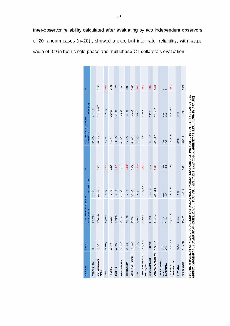
Inter-observor reliability calculated after evaluating by two independent observors
of 20 random cases (n=20) , showed a excellant inter rater reliability, with kappa
vaule of 0.9 in both single phase and multiphase CT collaterals evaluation.

34
Mean NIHSS score of the 56 patients was 16 (+/-6). Mean ASPECTS at
admission was 6 (+/2). Mean time from symptom onset to CT was 3 hours 20
minutes. 29 patients (52%) undergone mechanical thrombectomy and stent
retrievers were used in all cases.
Mechanical thrombectomy was done as per universally followed guidelines
(ASPECTS>=6, NIHSS 5 to 25, M1/ T occlusion). Mean door to groin puncture
was 51 (+/-22) minutes. 9 patients received both intravenous thrombolysis and
mechanical thrombectomy, while 4 cases (7%) received only intravenous
thrombolysis. TICI 2B or 3 recanalization was attained in 76% (22 out of 29)
patients. Mean time from stroke onset to recanalization was 263 +/-80 minutes.
24 hours follow up mean CT ASPECTS score was 4. Of the 56 patients, 6 patients
had PH2 ECASS2 type of haemorrhagic transformation and 3 patients had PH1
bleed. 7 patients had died in hospital while on treatment. Mean NIHSS at
discharge of the 49 alive patients was 8. 34 patients came for follow up after 90
days and 48% (17) patients attained good functional outcome of mRS <=2.
Patients with good collaterals (in both sCT and mCT) had a younger age (65 vs 61
years), but not statistically significant. Males had statistically poor collaterals
compared to females. Among the risk factors only patients with coronary heart
disease had statistically significant poor multiphasic CT collaterals (25% vs
8%p=0.002). Patients with hypertension, atrial fibrillation, diabetes,
hyperlipidaemia had poor multiphasic collaterals, but not statistically significant.
While in single phase CT collaterals assessment, patients with coronary heart
disease, diabetes and hypertension had poor collaterals. Among patients with
good collaterals in mCT, 62% undergone thrombectomy and while among patient
with poor collaterals in mCT, 43% undergone thrombectomy (p-0.01). There is no

35
difference in time to groin between patients with good or bad collaterals in both
sCT and mCT.
FIGURE 6: MULTIVARIATE ANALYSIS OF PREDICTORS OF GOOD FUNCTIONAL OUTCOME (RED COLOR-SIGNIFICANT VARIABLES) In multivariate logistic regression model (Table 6) adjusted for sex, coronary heart
disease, NIHSS at admission, mRS at admission, CT ASPECTS at admission,
mechanical thrombectomy, Single phase collateral score, multiphase collateral
score and 24 hours followup CT ASPECTS, only NIHSS at admission, ASPECTS
at admission, mRS at admission, mechanical thrombectomy, multiphase collateral
score and 24 hrs admission were found to have independent predictors of
favourable outcomes. Odd’s ratio of multiphase collateral score is 15.1(p=0.001),
and is only 2.2 for single phase collateral score (p=0.2). Mechanical thrombectomy
showed odd’s ratio of 3.4 and next most important variable is 24 hours CT volume
(ASPECTS).

36
FFIIGGUURREE 77 MMUULLTTIIPPLLEE LLIINNEEAARRRREEGGRREESSSSIIOONN AANNAALLYYSSIISS OOFFPPRREEDDIICCTTOORRSS OOFF
FFUUNNCCTTIIOONNAALL OOUUTTCCOOMMEE
Y (GOOD FUNCTIONAL OUTCOME) = 0.15(FEMALE) + 0.03(NO CAD)-0.02(NIHSS SCORE)-0.20 (mRS SCORE) +0.006 (ASPECTS SCORE)+0.25 (MECHANICAL THROMBECTOMY)+0.43(GOOD MULTIPHASE CT SCORE)+0.02(ASPECTS)-0.45 R2=0.51. RSS=4.1. Similarly, linear regression analysis of variables (figure 7) was done and
regression coefficient of variables was attained. Regression coefficient of
multiphase collateral score is 0.43.

37
FFIIGGUURREE 88:: CCLLIINNIICCAALL,, FFUUNNCCTTIIOONNAALL AANNDD RRAADDIIOOLLOOGGIICCAALL OOUUTTCCOOMMEESS
AACCCCOORRDDIINNGG TTOO CCOOLLLLAATTEERRAALL CCIIRRCCUULLAATTIIOONN SSTTAATTUUSS IINN BBOOTTHH TTHHEE ssCCTTAA
AANNDD mmCCTTAA GGRROOUUPPSS (RED COLOR-SIGNIFICANT BASED UPON BY P VALUE) Patients with good collateral scoring were found to be functional independent
(mRS<=2), when analyzed after 90 days. 82% of the patients with good mCT
collateral score were functional independent (p=0.001). Early radiological
outcome (ASPECTS 24 hrs, 5.2 vs 3.0, p=0.002) and clinical outcome (NIHSS at
discharge, 11.6 vs 5.5, p=0.003) were also significantly better in patients with mCT
collateral score. None of the patients with good mCT collateral score patients had
significant haemorrhagic transformation (PH2, p<0.001). There is no difference in
time to recanalization and TICI scoring between patients with good and poor mCT
collaterals.
Patients with good single-phase collateral score were found to have good mRS at
90days, NIHSS at discharge and ASPECTS at 24 hours, but none reach statistical
significance.

38
FIGURE 9: SPECIFICITY, PREDICTIVE, LIKELIHOOD AND ACCURACY VALUES OF GOOD FUNCTIONAL OUTCOME BY COLLATERAL SCORES.
Patients with good mCT collateral scores have 78% sensitivity, 81 specificity and
80% accurate in predicting good functional outcome. In contrast, single phase CT
had only 54% accuracy. Positive predictive value was 0.82 and negative predictive
value was 0.76 for mCT scoring. mCT collateral scoring method has high positive
likelihood ratio of 4.2, compared to sCT positive likelihood ratio of 1.15.

39
FIGURE 10: SPEARMAN RANK CORRELATION OF SINGLE PHASE AND MULTIPHASE COLLATERAL SCORING Multiphase CT scoring was found to have statistically better spearman rank
correlation (p<0.05) “R” value than single phase collateral scoring system, with
NIHSS at admission, ASPECTS at admission, NIHSS at discharge, ASPECTS at
discharge and mRS at 90 days.
FIGURE 11: RATE OF THROMBECTOMY BY MULTIPHASE COLLATERAL SCORE
40
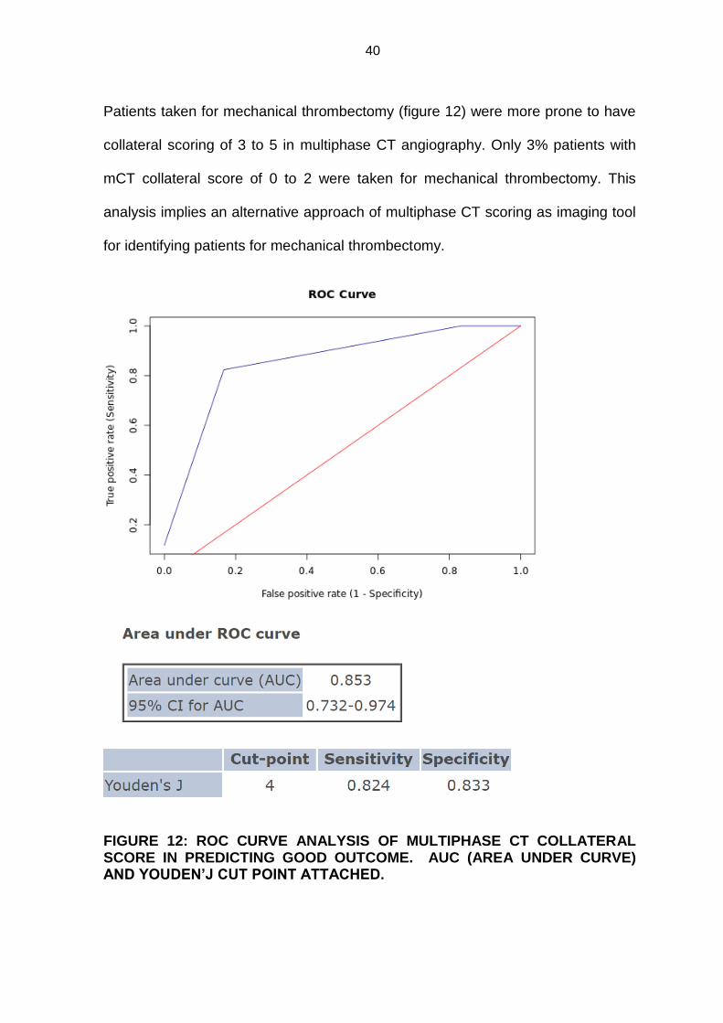
Patients taken for mechanical thrombectomy (figure 12) were more prone to have
collateral scoring of 3 to 5 in multiphase CT angiography. Only 3% patients with
mCT collateral score of 0 to 2 were taken for mechanical thrombectomy. This
analysis implies an alternative approach of multiphase CT scoring as imaging tool
for identifying patients for mechanical thrombectomy.
FIGURE 12: ROC CURVE ANALYSIS OF MULTIPHASE CT COLLATERAL SCORE IN PREDICTING GOOD OUTCOME. AUC (AREA UNDER CURVE) AND YOUDEN’J CUT POINT ATTACHED.

41
Finally, ROC curve analaysis was done separately for mCT and sCT collateral
scoring for predicting good functional outcome. Area of curve was 0.853 (C.I=0.73
to 0.97) for mCT collateral scoring (figure 13), indicating mCT is good indicator
(0.8 to 0.9) of predicting long-term functional independence. By Youden’s J point,
mCT collateral score of 4 is found to be optimum cut off point in predicting good
functional outcome, with sensitivity of 82% and specificity of 83%.
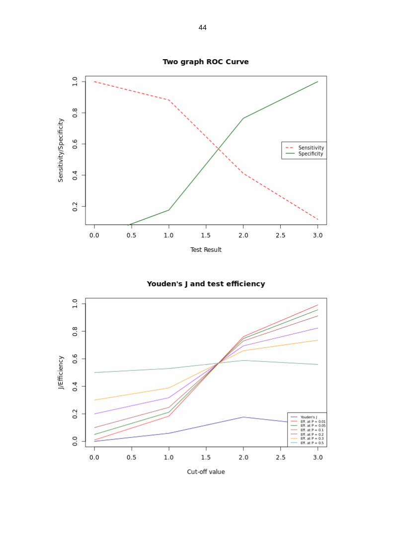
While sCT scoring (figure 14) had AUC value of 0.609 (C.I=0.43 to 0.78),
indicating it as poor predictor of functional independence. Youden’s J point for
sCT collateral scoring shown that score of 2 is the optimum cut off point with
sensitivity of 41% and specificity of 76%.

42
mRS<=2
Box plot results of good outcome by m CT score
Strip chart results of good outcome by m CT score
mRS >2

43
FIGURE 13: ROC CURVE ANALYSIS OF SINGLEPHASE CT COLLATERAL
SCORE IN PREDICTING GOOD OUT COME. AUC (AREA UNDER CURVE)
AND YOUDEN’J CUT POINT ATTACHED.
44

45
Representative Cases
46
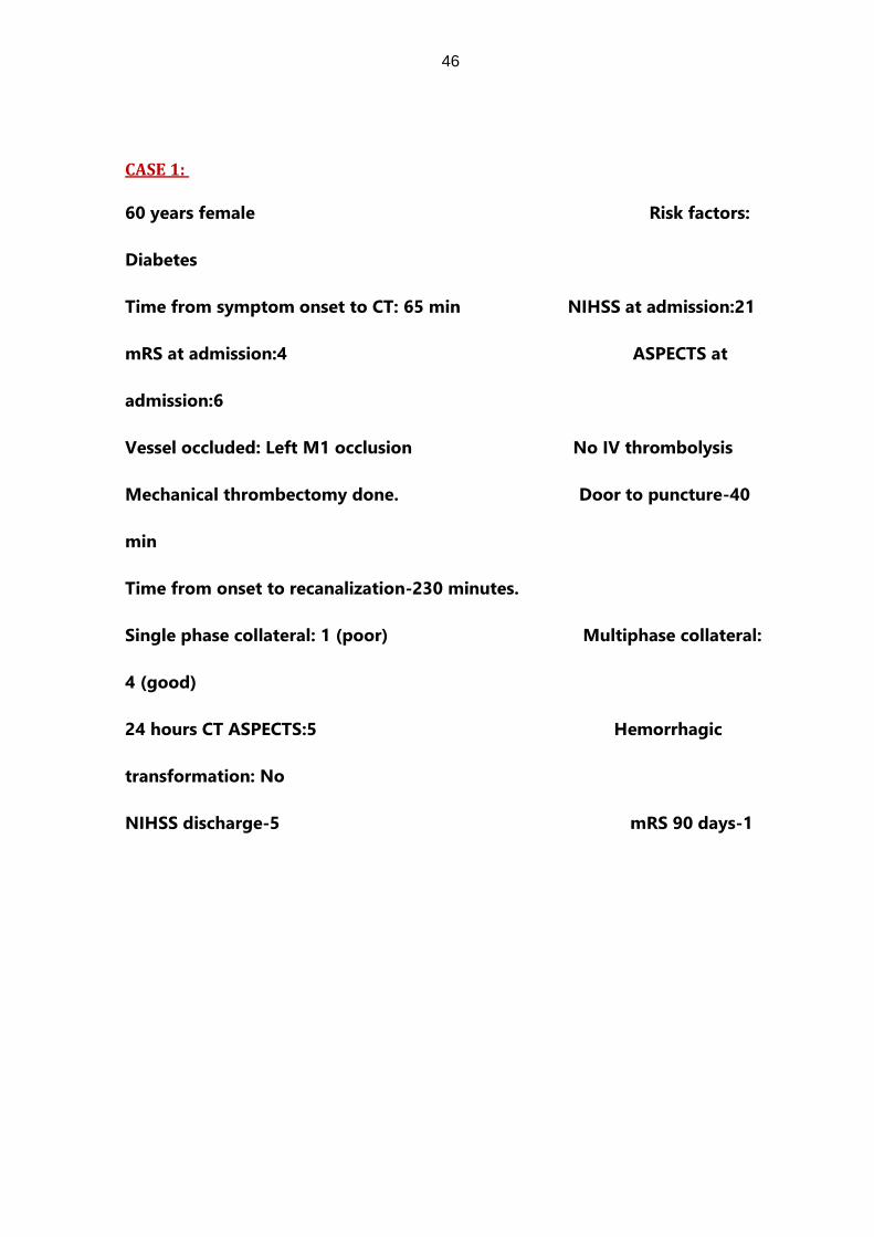
CASE 1:
60 years female Risk factors:
Diabetes
Time from symptom onset to CT: 65 min NIHSS at admission:21
mRS at admission:4 ASPECTS at
admission:6
Vessel occluded: Left M1 occlusion No IV thrombolysis
Mechanical thrombectomy done. Door to puncture-40
min
Time from onset to recanalization-230 minutes.
Single phase collateral: 1 (poor) Multiphase collateral:
4 (good)
24 hours CT ASPECTS:5 Hemorrhagic
transformation: No
NIHSS discharge-5 mRS 90 days-1

47
PLAIN CT AT ADMISSION: ASPECTS 6. CT ANGIOGRAPHY: LEFT M1 OCCULSION
SINGLE PHASE CT ANGIOGRAPHY DSA: LEFT M1 OCCLUSION. (TAN): GRADE 1 (POOR)

48
MULTIPHASE CT ANGIOGRAPHY: GRADE 4(GOOD)
24 HOURS CT: ASPECTS 5 CASE 2:
51 years male Risk factors:
Hypertension
Time from symptom onset to CT: 4 hours 15min. NIHSS at admission:24
mRS at admission:4 ASPECTS at
admission:7
Vessel occluded: Left M1 occlusion No IV thrombolysis
Mechanical thrombectomy done. Door to puncture-35 min

49
Time from onset to recanalization-310 minutes.
Single phase collateral: 1 (poor) Multiphase collateral: 4
(good)
24 hours CT ASPECTS:6 Hemorrhagic
transformation: No
NIHSS discharge-0 mRS 90 days-1
PLAIN CT AT ADMISSION: ASPECTS 7 CT ANGIOGRAPHY: LEFT M1 OCCULSION

50
SINGLE PHASE CT ANGIOGRAPHY DSA: LEFT M1 OCCULSION (TAN): GRADE 1. (POOR)
MULTIPHASE CT ANGIOGRAPHY: GRADE 4(GOOD)
24 HOURS CT: NO HEMORRHAGE TRANSFORMATION AND ASPECTS 6

51
CASE 3:
69 years female Risk factors: Atrial
fibrillation
Time from symptom onset to CT: 8 hours NIHSS at admission:25
mRS at admission:5 ASPECTS at admission:4
Vessel occluded: Left M1 occlusion No intravenous or intra-
arterial therapy
Single phase collateral: 2 (good) Multiphase collateral: 3
(poor)
24 hours CT ASPECTS:3 Hemorrhagic
transformation: PH2
On discharge: died/mRS 6
PLAIN CT AT ADMISSION: ASPECTS 4 CT ANGIOGRAPHY: LEFT M1 OCCULSION

52
SINGLE PHASE CT ANGIOGRAPHY (TAN): GRADE 2 (GOOD)
MULTIPHASE CT ANGIOGRAPHY: GRADE 2 (POOR)
24 HOURS CT: PH2 HEMORRHAGE TRANSFORMATION/ ASPECTS 3

53
CASE 4:
40 years female Risk
factors: Atrial fibrillation
Time from symptom onset to CT: 2 hours 50 min NIHSS at admission:8
mRS at admission:4 ASPECTS at
admission:7
Vessel occluded: Right M1 occlusion No intravenous
thrombolysis
Mechanical thrombectomy –attempted but failed. Door to puncture -35 min.
Single phase collateral: 3 (good) Multiphase
collateral: 5 (good)
24 hours CT ASPECTS:7 Hemorrhagic
transformation: No
NIHSS at discharge: 0 mRS 90
days:0

54
PLAIN CT AT ADMISSION: CT ANGIOGRAPHY: ASPECTS 7 RIGHT M1 OCCULSION
SINGLE PHASE CT ANGIOGRAPHY (TAN): GRADE 3 (GOOD)

55
MULTIPHASE CT ANGIOGRAPHY: GRADE 5 (GOOD)
24 HOURS CT: NO HEMORRHAGE TRANSFORMATION AND CT ASPECTS 7 CASE5:
60 years male Risk factors:
Diabetes, hyperlipidemia
Time from symptom onset to CT: 5 hours 30 min NIHSS at admission:8
mRS at admission:4 ASPECTS at
admission:8

56
Vessel occluded: Right M1 occlusion No intravenous therapy
Single-phase collateral: 1 (poor) Multiphase collateral: 4
(good)
24 hours CT ASPECTS: 8 Hemorrhagic
transformation: No
NIHSS at discharge: 2 mRS at 90 days:2
PLAIN CT AT ADMISSION: ASPECTS 8 CT ANGIOGRAPHY: RIGHT M1 OCCULSION

57
SINGLE PHASE CT ANGIOGRAPHY. DSA: RIGHT M1 OCCULSION (TAN): GRADE 1 (POOR)
MULTIPHASE CT ANGIOGRAPHY: GRADE 4 (GOOD)
24 HOURS CT: NO HEMORRHAGE TRANSFORMATION/ ASPECTS 8

58
Discussion

59
Risk factors and collaterals:
Among risk factors, we have shown only coronary heart disease (CHD) as
statistically significant variable prone to have poor multiphase CT collaterals. This
may be due to reduced ejection fraction in patients with CHD, secondarily leading
to poor intra cranial blood flow and collateral formation.
Patients with hypertension and hyperglycemia were shown to have poor mCT
collaterals, but not statistically significant. Prior studies with angiography also
showed patients with hyperglycemia had poor collaterals, but did not achieve
independent predictor status in multivariate analysis (55). There is no significant
relationship of atrial fibrillation and smoking to intracranial collaterals in both single
and multiphase angiography in our study as in Multicenter Randomized Clinical
Trial of Endovascular Treatment of Acute Ischemic Stroke in the Netherlands
(MRCLEAN) (56). Among sex, males were shown to have less collateral formation
compared to females.
Collaterals and intracranial hemorrhages:
Previously, few studies have shown patients with good collaterals have been less
prone to have symptomatic intra cerebral hemorrhagic transformation (31,32,56).
In our study, none of the patients with good mCT collateral score had ECASS2
PH2 type of hemorrhagic transformation, while 6 patients (18%) of patients with
poor collaterals had PH2 type of bleeding. By this, multiphase CT is 100% specific
in predicting patients who are prone to significant hemorrhagic transformation.
This is a new prognostic indicator found by our study.

60
Collaterals and mechanical thrombectomy:
Patients who had undergone mechanical thrombectomy were shown to have
better mCT collaterals, although there is no difference in single-phase CT
collaterals. However there is no significant difference in time to groin, time to
recanalization and TICI scoring between patients with good and poor single as
well as multiphase CT collaterals.
Among patients who were taken for mechanical thrombectomy, most of them
(97%) had mCT collaterals of 3 to 5. Since mechanical thrombectomy was done in
our cases based upon the universally accepted guidelines and not based upon
mCT collateral scoring, we can now retrospectively assume mCT collateral scoring
would be one of the best imaging tools to predict patients who may benefit from
mechanical thrombectomy.
In our study, we have achieved good recanalization (TICI 2B OR 3) in 76 %
patients, on par to stroke trials. Mean time to recanalization was 263 minutes.
Collaterals and clinical-radiological scores at admission:
Both patients with good single phase and mCT collateral scoring have good initial
NIHSS score (13 vs 17 for multiphase, 17 vs 12 in single phase). This was in
agreement with prior studies involving single-phase collateral systems.
Radiologically also, patients with good collaterals have good initial CT ASPECTS
(6.6 vs 4.7 in multiphase). This is because of the sustained perfusion of the
penumbra by the good leptomeningeal collaterals.

61
Collaterals and radiological outcomes:
Multiphase CT scoring was shown to better correlate with 24 hrs CT infarct
volume, while sCT scoring shows no statistically significant correlation. In patients
with good mCT collaterals (mean ASPECTS 5 vs 3 in better vs poor collaterals),
24 hrs ASPECTS was shown to be significantly better than patients with good sCT
collaterals (mean ASPECTS 4.5 vs 3.7 in better vs poor collaterals).
Collaterals and early clinical outcomes:
Unlike single-phase collaterals, multiphase collaterals were shown to better and
statistically significant correlate and predict the early clinical outcome i.e NIHSS at
discharge. Patients with good mCT collaterals had lower mean NIHSS at
discharge. Mean NIHSS was 5 in patients with good mCT collaterals, while mean
NIHSS was 11 in patients with poor mCT collaterals.
Collaterals and long-term clinical outcome:
Patients with good mCT were shown to have better clinical outcome than sCT
collaterals scoring method. 86 % of the patients with good mCT collaterals are
functionally independent, compared to 54% in sCT collaterals system. Only 22%
of the patients with poor mCT collaterals, had good mRS<=2. Also mCT was
shown to have very high spearman correlation of -0.74 with 90 days mRS.

62
Cut off value of collateral scoring by ROC:
There were no studies, which predicted the cutoff of collateral scoring methods to
predict good functional outcome. In this study, we analyzed scoring methods with
ROC and Youden for mRS 90 days. Cutoff scoring value of 4 and above in mCT
scoring, was shown to predict good functional independency with good sensitivity
and specificity. For sCT scoring method of 0 to 3, Youden’s cut off of 2 and above
was shown to better predict good clinical outcome, even though sensitivity and
specificity was less than < 50%.
Multivariate analysis of variables:
Since multiple variables may predict the functional outcome, we did multivariate
logistic regression analysis, to look into the odd ratio of independent variables.
mCT collateral scoring method have the highest odds ratio of around 15, when
compared to next important variable of mechanical thrombectomy of 3.4.
Single-phase CT collaterals had an odds ratio of 2.2 but not in significant level.
Other variables like NIHSS at admission, ASPECTS at admission and discharge
were also shown to have significant p value on logistic regression analysis in
addition to multiphase CT collaterals and mechanical thrombectomy.
63
Studies involving multiphase CT collateral scoring:
1. Multiphase CT collateral scoring was first introduced by BK Menon et al
(24) and is published as a new tool for imaging triage of acute stroke
patients. In this study, they used information of the PRoveIT (Precise and
Rapid assessment of collaterals using multi-phase CTA in the triage of
patients with acute ischemic stroke for IA Therapy) study of 147 patients.
These study inclusion criteria used acute stroke within 12 hours, unlike our
study, which included only patients within 8 hours which may be more
informative to assess the response to interventional procedures.
Of the 147 patients, 3 are PCA occlusion, 24 are distal vessel occlusion
and 33 are no occlusion. These distal occlusion and no occlusion cases
may not informative. So in our study we included only major vessel anterior
circulation occlusions.
Totally 31 patients had undergone intra-arterial therapy in this study which
is almost similar (29 patients) to our study. In this study, ROC curve
analysis was done which showed a C statistic value of 0.6 for mCT
collateral score in predicting mRS at 90 days and is better than single
phase CT collateral scoring. In our study, C statistic for mCT collateral
scoring was much higher at 0.85 and is significantly better than single
phase CT collateral scoring. Radiation dose of multiphase CT angiography
was less than the CT perfusion in their institute.

64
2. Alan flores at al, (32) showed Poor collateral circulation in multiphase
Ct angiography predicts malignant MCA infarction progression. Totally
82 patients of proximal MCA or terminal ICA occlusion were included. Mean
NIHSS at admission (16 NIHSS) and age (63years) are similar to our study.
15 patients had malignant infarct transformation.
In univariate analysis, patients with malignant stroke were associated with
lower multiphasic collateral scores (2.2 vs 3.7). On multivariate analysis,
mCT collateral was an only independent predictor of malignant stroke. This
is in concordance with our results of multiphase CT collateral better
correlating with 24 hours infarct volume in terms of CT ASPECTS
(ASPECTS of 5 in good mCT collateral vs 3 ASPECTS in poor mCT
collaterals, p=0.002 and good spearman correlation of +0.52).
3. Recently a study by Alvaro et al (33) including BK Menon, evaluated
multiphase CT angiography in acute stroke patients treated with
endovascular reperfusion therapies.
In this study, logistic regression analysis of age, NIHSS, ASPECTS, mCT
angiography collateral, single-phase CT angiography collaterals and
recanalization, only age and mCT angiography were shown to be
independent predictor of functional outcome.

65
In our study of multivariate logistic regression analysis including sex,
coronary heart disease, NIHSS at admission, mRS at admission,
ASPECTS at admission, use of mechanical thrombectomy, single phase
collateral, multiphase collaterals and 24 hours ASPECTS, only multiphase
CT collaterals, use of mechanical thrombectomy, NIHSS at admission,
mRS at admission, ASPECTS at admission and at 24 hours were found to
be significant independent predictors of 90 days mRS.
This result of the collateral score was similar to our study, but in our study
additionally told ASPECTS, NIHSS score also shown as independent
predictors of functional outcome.
Multiphase CT collateral scoring attained highest ODD ratio in their study
similar to our results but value is much higher in our study (Odd ratio of 15
in our study vs odd ratio of 5 in their study). Odd ratio of NIHSS, ASPECTS
was almost similar (0.86 our study vs 1.06 alvaro study for NIHSS, 1.49 our
study vs 1.13 in Álvaro study for ASPECTS for admission).
In this study even though the symptomatic hemorrhagic transformation is
less in patients with good mCT collaterals, it did not attain a statistical
significant level. However, in our study none of the patients with good mCT
collaterals developed PH2 type of bleeding, while 18% of the patients with
poor collaterals developed PH2 bleeding. So future studies must come to
prove our hypothesis of accuracy of mCT collateral score in predicting
hemorrhagic transformation.

66
4. Santos et al studied the use of multiphase CT angiography for
thrombus perviousness assessment by thrombus attenuation
increase (TAI) in arterial, venous, delayed and time invariant CT
angiography. They found an association of TAI with arterial phase CT
angiography was optimal and multiphase CT angiography gives no added
advantage in thrombus perviousness assessment.
5. Ondrej et al studied the single-phase vs multiphase CT angiography in
MCA clot detection and its benefits for less experienced radiologists
and neurologists. In this study 10 radiologist and10 neurologists (3
experienced and 7 less experienced) assessed 20 single phase and
multiphase CT angiography cases of M2 occlusion. This study showed that
multiphase CT angiography improves detection of M2 occlusion in less
experienced physicians.
6. ESCAPE trial of Randomized Assessment of Rapid Endovascular
Treatment of Ischemic Stroke (25), it was mentioned multiphasic CT
angiography was used in majority of patients. In this study large infarct core
or poor collaterals on multiphasic Ct angiography were excluded. However
they used moderate-to-good collaterals as filling of 50% or more of the
MCA pial arterial circulation on multiphase CTA, which is different from the
currently used multiphasic CT angiography collateral scoring.
67
LIMITATIONS:
Even though this was a prospective study with inclusion of consecutive cases
of acute anterior circulation ischemic stroke presenting to our institute there
were a few limitations in this study.
1. The number of patients included in the study was 56, which was
limited in number, even though this was one of the largest
prospective studies, which assessed intracranial collaterals.
2. Only anterior circulation ischemic stroke was included. Posterior
circulation strokes collaterals studies have to be studied in future
studies.
3. The inclusion of patients with conservative as well as mechanical
thrombectomy did cases. However to overcome this bias multivariate
logistic regression analysis was done.
4. The number of mechanical thrombectomy cases were limited (n=29).

68
Conclusion

69
CONCLUSION:
This study with prospective evaluation of 56 patients represents the one of
the largest available prospective studies of intracranial collaterals of
patients with anterior circulation major vessel occlusion in literature.
1. Multiphasic CT collateral grading system was significantly much
better than the traditional single-phase collateral grading system in
predicting early radiological and functional outcome as well as
long-term functional outcome.
2. Good multiphase CT collateral scoring was the strongest predictor
of long-term functional independence, irrespective of endovascular
management in our study.
3. Patients with good mCT collaterals would have better initial clinical
and radiological findings.
4. mCT collaterals system more accurately predict the hemorrhagic
transformation.
5. Good and intermediate mCT collaterals (3 to 5) can be used as an
alternate cut off value limit as a triage for mechanical thrombectomy.
6. Cut off value of mCT collateral scoring of 4 and 5 better predicts a
good functional outcome.
Hence, multiphasic CT angiography has to be added to the existing acute
stroke imaging protocols.

70
References

71
REFERENCES
1. Vos T, Barber RM, Bell B, Bertozzi-Villa A, Biryukov S, Bolliger I, Charlson F, Davis A,
Degenhardt L, Dicker D, Duan L. Global, regional, and national incidence, prevalence, and
years lived with disability for 301 acute and chronic diseases and injuries in 188 countries,
1990–2013: a systematic analysis for the Global Burden of Disease Study 2013. The
Lancet. 2015 Aug 22;386(9995):743-800.
2. Hammer MD, Schwamm L, Starkman S, Schellinger PD, Jovin T, Nogueira R, Burgin WS,
Sen S, Diener HC, Watson T, Michel P. Safety and feasibility of NeuroFlo use in eight‐to
24‐hour ischemic stroke patients. International journal of stroke. 2012 Dec;7(8):655-61.
3. Miteff F, Levi CR, Bateman GA, Spratt N, McElduff P, Parsons MW. The independent
predictive utility of computed tomography angiographic collateral status in acute ischaemic
stroke. Brain. 2009 Jun 9;132(8):2231-8.
4. Shuaib A, Butcher K, Mohammad AA, Saqqur M, Liebeskind DS. Collateral blood vessels
in acute ischaemic stroke: a potential therapeutic target. The Lancet Neurology. 2011 Oct
1;10(10):909-21.
5. Lima FO, Furie KL, Silva GS, Lev MH, Camargo ÉC, Singhal AB, Harris GJ, Halpern EF,
Koroshetz WJ, Smith WS, Yoo AJ. The pattern of leptomeningeal collaterals on CT
angiography is a strong predictor of long-term functional outcome in stroke patients with
large vessel intracranial occlusion. Stroke. 2010 Oct 1;41(10):2316-22.
6. Ovbiagele B, Saver JL, Starkman S, Kim D, Ali LK, Jahan R, Duckwiler GR, Vinuela F,
Pineda S, Liebeskind DS. Statin enhancement of collateralization in acute stroke.
Neurology. 2007 Jun 12;68(24):2129-31.
7. Kucinski T, Koch C, Eckert B, Becker V, Krömer H, Heesen C, Grzyska U, Freitag H,
Röther J, Zeumer H. Collateral circulation is an independent radiological predictor of
outcome after thrombolysis in acute ischaemic stroke. Neuroradiology. 2003 Jan
1;45(1):11-8.
8. Tan IY, Demchuk AM, Hopyan J, Zhang L, Gladstone D, Wong K, Martin M, Symons
SP, Fox AJ, Aviv RI. CT angiography clot burden score and collateral score: correlation
with clinical and radiologic outcomes in acute middle cerebral artery infarct. American
Journal of Neuroradiology. 2009 Mar 1;30(3):525-31.

72
9. Maas MB, Lev MH, Ay H, Singhal AB, Greer DM, Smith WS, Harris GJ, Halpern E,
Kemmling A, Koroshetz WJ, Furie KL. Collateral vessels on CT angiography predict
outcome in acute ischemic stroke. Stroke. 2009 Sep 1;40(9):3001-5.
10. Menon BK, Smith EE, Modi J, Patel SK, Bhatia R, Watson TW, Hill MD, Demchuk AM,
Goyal M. Regional leptomeningeal score on CT angiography predicts clinical and imaging
outcomes in patients with acute anterior circulation occlusions. American journal of
neuroradiology. 2011 Oct 1;32(9):1640-5.
11. Yeo LL, Paliwal P, Teoh HL, Seet RC, Chan BP, Ting E, Venketasubramanian N, Leow
WK, Wakerley B, Kusama Y, Rathakrishnan R. Assessment of intracranial collaterals on
CT angiography in anterior circulation acute ischemic stroke. American Journal of
Neuroradiology. 2014 Oct 16.
12. Lee KY, Latour LL, Luby M, Hsia AW, Merino JG, Warach S. Distal hyperintense vessels
on FLAIR An MRI marker for collateral circulation in acute stroke?. Neurology. 2009 Mar
31;72(13):1134-9.
13. Karadeli HH, Giurgiutiu DV, Cloonan L, Fitzpatrick K, Kanakis A, Ozcan ME, Schwamm
LH, Rost NS. FLAIR vascular hyperintensity is a surrogate of collateral flow and
leukoaraiosis in patients with acute stroke due to proximal artery occlusion. Journal of
Neuroimaging. 2016 Mar;26(2):219-23.
14. Mahdjoub E, Turc G, Legrand L, Benzakoun J, Edjlali M, Seners P, Charron S, Hassen
WB, Naggara O, Meder JF, Mas JL. Do Fluid-Attenuated Inversion Recovery Vascular
Hyperintensities Represent Good Collaterals before Reperfusion Therapy?. American
Journal of Neuroradiology. 2017 Oct 26.
15. Gawlitza M, Böhme J, Maros M, Lobsien D, Michalski D, Groden C, Hoffmann KT,
Förster A. FLAIR vascular hyperintensities and 4D MR angiograms for the estimation of
collateral blood flow in anterior cerebral artery ischemia. PloS one. 2017 Feb
24;12(2):e0172570.
16. Chng SM, Petersen ET, Zimine I, Sitoh YY, Lim CT, Golay X. Territorial arterial spin
labeling in the assessment of collateral circulation: comparison with digital subtraction
angiography. Stroke. 2008 Dec 1;39(12):3248-54.
17. Lyu J, Ma N, Liebeskind DS, Wang DJ, Ma L, Xu Y, Wang T, Miao Z, Lou X. Arterial
spin labeling magnetic resonance imaging estimation of antegrade and collateral flow in
unilateral middle cerebral artery stenosis. Stroke. 2016 Feb 1;47(2):428-33.

73
18. De Havenon A, Haynor DR, Tirschwell DL, Majersik JJ, Smith G, Cohen W, Andre JB.
Association of collateral blood vessels detected by arterial spin labeling magnetic resonance
imaging with neurological outcome after ischemic stroke. Jama neurology. 2017 Apr
1;74(4):453-8.
19. Lou X, Ma X, Liebeskind DS, Ma N, Tian C, Lyu J, Long X, Ma L, Wang DJ. Collateral
perfusion using arterial spin labeling in symptomatic versus asymptomatic middle cerebral
artery stenosis. Journal of Cerebral Blood Flow & Metabolism. 2017 Jan
1:0271678X17725212.
20. Hua T, Li L, Xu Y. Clinical application of magnetic susceptibility weighted imaging in
treatment of collateral circulation of cerebral infarction. Biomedical Research. 2018 Jan
23;28(21):9401-4.
21. Lou M, Chen Z, Wan J, Hu H, Cai X, Shi Z, Sun J. Susceptibility-diffusion mismatch
predicts thrombolytic outcomes: a retrospective cohort study. American Journal of
Neuroradiology. 2014 Jul 10.
22. Guan J, Zhang S, Zhou Q, Li C, Lu Z. Usefulness of transcranial Doppler ultrasound in
evaluating cervical-cranial collateral circulations. Interventional neurology. 2013;2(1):8-18.
23. Pan J, Zhou J, Liu P, Gu Y, Luo B. Use of Transcranial Doppler Ultrasound to Assess
Leptomeningeal Collateral Flow in Patients with Severe Occlusive Internal Carotid and
Middle Cerebral Artery Disorders: Correlation with Cerebral Angiography. Iranian Journal
of Radiology. 2017;14(2).
24. 31. Menon BK, d’Esterre CD, Qazi EM, Almekhlafi M, Hahn L, Demchuk AM, Goyal M.
Multiphase CT angiography: a new tool for the imaging triage of patients with acute
ischemic stroke. Radiology. 2015 Jan 29;275(2):510-20.
25. Goyal M, Demchuk AM, Menon BK, Eesa M, Rempel JL, Thornton J, Roy D, Jovin TG,
Willinsky RA, Sapkota BL, Dowlatshahi D. Randomized assessment of rapid endovascular
treatment of ischemic stroke. New England Journal of Medicine. 2015 Mar
12;372(11):1019-30.
26. Vagal A, Menon BK, Foster LD, Livorine A, Yeatts SD, Qazi E, d’Esterre C, Shi J,
Demchuk AM, Hill MD, Liebeskind DS. Association between CT angiogram collaterals
and CT perfusion in the Interventional Management of Stroke III Trial. Stroke. 2016 Feb
1;47(2):535-8.

74
27. Broderick JP, Palesch YY, Demchuk AM, Yeatts SD, Khatri P, Hill MD, Jauch EC, Jovin
TG, Yan B, Silver FL, Von Kummer R. Endovascular therapy after intravenous t-PA versus
t-PA alone for stroke. New England Journal of Medicine. 2013 Mar 7;368(10):893-903.
28. Campbell BC, Mitchell PJ, Kleinig TJ, Dewey HM, Churilov L, Yassi N, Yan B, Dowling
RJ, Parsons MW, Oxley TJ, Wu TY. Endovascular therapy for ischemic stroke with
perfusion-imaging selection. New England Journal of Medicine. 2015 Mar
12;372(11):1009-18.
29. Saver JL, Goyal M, Bonafe A, Diener HC, Levy EI, Pereira VM, Albers GW, Cognard C,
Cohen DJ, Hacke W, Jansen O. Stent-retriever thrombectomy after intravenous t-PA vs. t-
PA alone in stroke. New England Journal of Medicine. 2015 Jun 11;372(24):2285-95.
30. Kim BJ, Chung JW, Park HK, Kim JY, Yang MH, Han MK, Jeong C, Hwang G, Kwon
OK, Bae HJ. CT Angiography of Collateral Vessels and Outcomes in Endovascular-Treated
Acute Ischemic Stroke Patients. Journal of Clinical Neurology. 2017 Apr 1;13(2):121-8.
31. Christoforidis GA, Karakasis C, Mohammad Y, Caragine LP, Yang M, Slivka AP.
Predictors of hemorrhage following intra-arterial thrombolysis for acute ischemic stroke: the
role of pial collateral formation. American Journal of Neuroradiology. 2009 Jan
1;30(1):165-70.
32. Bang OY, Saver JL, Kim SJ, Kim GM, Chung CS, Ovbiagele B, Lee KH, Liebeskind DS.
Collateral flow averts hemorrhagic transformation after endovascular therapy for acute
ischemic stroke. Stroke. 2011 Aug 1;42(8):2235-9.
33. Liebeskind DS, Jahan R, Nogueira RG, Zaidat OO, Saver JL. Impact of collaterals on
successful revascularization in Solitaire FR with the intention for thrombectomy. Stroke.
2014 Jan 1:STROKEAHA-114.
34. Saver JL, Jahan R, Levy EI, Jovin TG, Baxter B, Nogueira RG, Clark W, Budzik R, Zaidat
OO. Solitaire flow restoration device versus the Merci Retriever in patients with acute
ischaemic stroke (SWIFT): a randomised, parallel-group, non-inferiority trial. The Lancet.
2012 Oct 6;380(9849):1241-9.
35. Bang OY, Saver JL, Kim SJ, Kim GM, Chung CS, Ovbiagele B, Lee KH, Liebeskind DS.
Collateral flow predicts response to endovascular therapy for acute ischemic stroke. Stroke.
2011 Jan 1:STROKEAHA-110.

75
36. Powers WJ, Rabinstein AA, Ackerson T, Adeoye OM, Bambakidis NC, Becker K, Biller J,
Brown M, Demaerschalk BM, Hoh B, Jauch EC. 2018 guidelines for the early management
of patients with acute ischemic stroke: a guideline for healthcare professionals from the
American Heart Association/American Stroke Association. Stroke. 2018 Mar 1;49(3):e46-
110.
37. Wufuer A, Wubuli A, Mijiti P, Zhou J, Tuerxun S, Cai J, Ma J, Zhang X. Impact of
collateral circulation status on favorable outcomes in thrombolysis treatment: A systematic
review and meta-analysis. Experimental and therapeutic medicine. 2018 Jan 1;15(1):707-
18.
38. Leng X, Lan L, Liu L, Leung TW, Wong KS. Good collateral circulation predicts favorable
outcomes in intravenous thrombolysis: a systematic review and meta‐analysis. European
journal of neurology. 2016 Dec;23(12):1738-49.
39. Goyal N, Tsivgoulis G, Nickele C, Doss VT, Hoit D, Alexandrov AV, Arthur A, Elijovich
L. Posterior circulation CT angiography collaterals predict outcome of endovascular acute
ischemic stroke therapy for basilar artery occlusion. Journal of neurointerventional surgery.
2015 Aug 17:neurintsurg-2015.
40. van der Hoeven EJ, McVerry F, Vos JA, Algra A, Puetz V, Kappelle LJ, Schonewille WJ,
BASICS registry investigators. Collateral flow predicts outcome after basilar artery
occlusion: the posterior circulation collateral score. International Journal of Stroke. 2016
Oct;11(7):768-75.
41. Alqadri S, Adil M, Watanabe M, Qureshi A. Patterns of Collateral Formation in Basilar
Artery Steno-Occlusive Diseases (P05. 214). Neurology. 2013 Feb 12;80(7
Supplement):P05-214.
42. Terpolilli NA, Moskowitz MA, Plesnila N. Nitric oxide: considerations for the treatment
of ischemic stroke. Journal of Cerebral Blood Flow & Metabolism. 2012 Jul;32(7):1332-
46.
43. Ginsberg MD, Palesch YY, Hill MD, Martin RH, Moy CS, Barsan WG, Waldman BD,
Tamariz D, Ryckborst KJ. High-dose albumin treatment for acute ischaemic stroke
(ALIAS) Part 2: a randomised, double-blind, phase 3, placebo-controlled trial. The Lancet
Neurology. 2013 Nov 1;12(11):1049-58.

76
44. Khurana D, Kaul S, Bornstein NM. Implant for augmentation of cerebral blood flow trial 1:
a pilot study evaluating the safety and effectiveness of the Ischaemic Stroke System for
treatment of acute ischaemic stroke. International journal of stroke. 2009 Dec;4(6):480-5.
45. Hammer M, Jovin T, Wahr JA, Heiss WD. Partial occlusion of the descending aorta
increases cerebral blood flow in a nonstroke porcine model. Cerebrovascular diseases.
2009;28(4):406-10.
46. Barber PA, Demchuk AM, Zhang J, Buchan AM, ASPECTS Study Group. Validity and
reliability of a quantitative computed tomography score in predicting outcome of
hyperacute stroke before thrombolytic therapy. The Lancet. 2000 May
13;355(9216):1670-4.
47. Pexman JW, Barber PA, Hill MD, Sevick RJ, Demchuk AM, Hudon ME, Hu WY, Buchan
AM. Use of the Alberta Stroke Program Early CT Score (ASPECTS) for assessing CT
scans in patients with acute stroke. American Journal of Neuroradiology. 2001 Sep
1;22(8):1534-42.
48. National Institute of Health, National Institute of Neurological Disorders and Stroke.
Stroke
Scale. https://www.ninds.nih.gov/sites/default/files/NIH_Stroke_Scale_Booklet.pdf
49. Lyden P. Using the National Institutes of Health Stroke Scale: a cautionary tale. Stroke.
2017 Feb 1;48(2):513-9.
50. Rankin J. Cerebral vascular accidents in patients over the age of 60: II. Prognosis. Scottish
medical journal. 1957 May;2(5):200-15.
51. Van Swieten JC, Koudstaal PJ, Visser MC, Schouten HJ, Van Gijn J. Interobserver
agreement for the assessment of handicap in stroke patients. Stroke. 1988 May
1;19(5):604-7.
52. Hacke W, Kaste M, Fieschi C, von Kummer R, Davalos A, Meier D, Larrue V, Bluhmki E,
Davis S, Donnan G, Schneider D. Randomised double-blind placebo-controlled trial of
thrombolytic therapy with intravenous alteplase in acute ischaemic stroke (ECASS II). The
Lancet. 1998 Oct 17;352(9136):1245-51
53. Sussman ES, Connolly Jr ES. Hemorrhagic transformation: a review of the rate of
hemorrhage in the major clinical trials of acute ischemic stroke. Frontiers in neurology.
2013 Jun 10;4:69.

77
54. Zaidat OO, Yoo AJ, Khatri P, Tomsick TA, Von Kummer R, Saver JL, Marks MP,
Prabhakaran S, Kallmes DF, Fitzsimmons BF, Mocco J. Recommendations on angiographic
revascularization grading standards for acute ischemic stroke: a consensus statement. Stroke.
2013 Jan 1:STROKEAHA-113.
55. Fanou EM, Knight J, Aviv RI, Hojjat SP, Symons SP, Zhang L, Wintermark M. Effect of
collaterals on clinical presentation, baseline imaging, complications, and outcome in acute
stroke. American Journal of Neuroradiology. 2015 Oct 15.
56. Berkhemer OA, Jansen IG, Beumer D, Fransen PS, Van Den Berg LA, Yoo AJ, Lingsma
HF, Sprengers ME, Jenniskens SF, à Nijeholt GJ, van Walderveen MA. Collateral status on
baseline computed tomographic angiography and intra-arterial treatment effect in patients
with proximal anterior circulation stroke. Stroke. 2016 Mar 1;47(3):768-76.
57. Flores A, Rubiera M, Ribó M, Pagola J, Rodriguez-Luna D, Muchada M, Boned S, Seró L,
Sanjuan E, Meler P, Carcámo D. Poor collateral circulation assessed by multiphase
computed tomographic angiography predicts malignant middle cerebral artery evolution
after reperfusion therapies. Stroke. 2015 Nov 1;46(11):3149-53.
58. García-Tornel A, Carvalho V, Boned S, Flores A, Rodríguez-Luna D, Pagola J, Muchada
M, Sanjuan E, Coscojuela P, Juega J, Rodriguez-Villatoro N. Improving the evaluation of
collateral circulation by multiphase computed tomography angiography in acute stroke
patients treated with endovascular reperfusion therapies. Interventional neurology.
2016;5(3-4):209-17.
59. Santos EM, d’Esterre CD, Treurniet KM, Niessen WJ, Najm M, Goyal M, Demchuk AM,
Majoie CB, Menon BK, Marquering HA. Added value of multiphase CTA imaging for
thrombus perviousness assessment. Neuroradiology. 2018 Jan 1;60(1):71-9.
60. Volny O, Cimflova P, Kadlecova P, Vanek P, Vanicek J, Menon BK, Mikulik R. Single-
Phase Versus Multiphase CT Angiography in Middle Cerebral Artery Clot Detection—
Benefits for Less Experienced Radiologists and Neurologists. Journal of Stroke and
Cerebrovascular Diseases. 2017 Jan 1;26(1):19-24.
78
Annexures

79
APPENDIX A
TITLE OF THE STUDY: ASSESSMENT OF COLLATERAL STATUS USING
MULTIPHASIC CT ANGIOGRAPHY IN THE ACUTE ANTERIOR CIRCULATION
ISCHEMIC STROKE: ITS CORRELATION WITH CLINICAL AND RADIOLOGIC
OUTCOMES
Study number: SCT/IEC/987/OCT-2016
There is a clinical suspicion that you have acute stroke, and it has to be evaluated
with CT angiography, for assessing your brain & neck vessel status and choosing
the best modality of management in your case. The CT angiography investigation
as a part of clinical evaluation helps to diagnose the acute stroke and plan the
treatment.
The CT angiography is a type of CT study done routinely in most acute stroke
patients, to assess the blood vessels of the brain and neck. It is helpful for the
diagnosis of the acute stroke, blood vessels affected and in choosing the best
modality of treatment to be undertaken in the patients.
This study is designed to evaluate the role of CT angiography findings to assess
the prognosis of acute stroke. This study has the advantage of likely providing
more accurate prognostic indices and therefore directing the better mode of
treatment.
You are being requested to participate in this study which is likely better in
assessing the prognosis of the acute stroke and therefore in better individual
patient management. Also, there is no additional risk anticipated as the
investigation involves the existing institute protocol.

80
What is CT ANGIOGRAPHY and does it have any harmful effects?
CT ANGIOGRAPHY is an advanced imaging technique which uses X ray radiation
and iodine based contrast to image body part. Some patients may rarely develop
allergic reactions to the contrast agent. This investigation is not to be done for with
kidney failure, pregnancy patients. This CT angiography is being done as a part of
clinical evaluation of your disease; however certain data from this study will be
used for research purpose as part of this study.
If you take part what will you have to do?
This study will only analyze the results of the routinely ordered imaging
investigations you will undergo for your illness. You will not be required to undergo
any other additional investigation modality for this study.
Can you withdraw from this study after it starts?
Your participation in this study is entirely voluntary and you are also free to decide
to withdraw permission to participate in this study. If you do so, this will not affect
your usual treatment at this hospital in any way.
What will happen if you develop any study related injury?
This study only analyzes the results of the CT angiography investigation which you
will be undergoing as part of your disease evaluation. Thus we do not expect any
injury to happen to you but if you do develop any side effects or problems due to
the study, these will be treated at this institute by the experienced team of medical
professionals.

81
‘
Will you have to pay for the study?
The study will only analyze the results of the investigations which you will undergo
in natural process of the management of your disease process at this institute. No
extra cost will be borne by you for this particular study.
What happens after the study is over?
If the study is found useful, the study will help better assess the prognosis of the
acute stroke patients and better assess the mode of treatment to be chosen for
the patients. The study may or may not be helpful to you directly.
Will your personal details be kept confidential?
The results of this study may be published in a medical journal but you will not be
identified by name in any publication or presentation of results. However, your
medical notes may be reviewed by people associated with the study, without your
additional permission, should you decide to participate in this study.
82
CONSENT FORM
Participant’s name: Date of Birth / Age (in years):
I_____________________________________________________________
___________, son/daughter of ___________________________________
(Please tick boxes)
1. I Declare that I have read the above information provide to me regarding
the study the collaterals score in acute stroke, have clarified any doubts
that I had. [ ]
2. I also understand that my participation in this study is entirely voluntary and
that I am free to withdraw permission to continue to participate at any time
without affecting my usual treatment or my legal rights [ ]
3. I understand that the study staff and institutional ethics committee members
will not need my permission to look at my health records even if I withdraw
from the trial. I agree to this access [ ]
4. I understand that my identity will not be revealed in any information
released to third parties or published [ ]
5. I voluntarily agree to take part in this study [ ]
6. I received a copy of this signed consent form [ ]
Name: Signature: Date: Name of witness: Relation to participant: Date:
(Person Obtaining Consent) I attest that the requirements for informed consent for
the medical research project described in this form have been satisfied. I have
discussed the research project with the participant and explained to him or her in
nontechnical terms all of the information contained in this informed consent form,
including any risks and adverse reactions that may reasonably be expected to
occur. I further certify that I encouraged the participant to ask questions and that
all questions asked were answered.
________________________________ ___________________
Name and Signature of Person Obtaining Consent

83
APPENDIX B:
SREE CHITRA TIRUNAL INSTITUTE FOR MEDICAL SCIENCES
& TECHNOLOGY
PROFORMA
General Instructions
Please fill in all the questions
Write Yes / No/NA wherever applicable
If the response is not
known please write UK If
additional info is available
please elaborate
1. PROFORMA SERIAL NUMBER: 2. AGE (YEARS) : 3. SEX (M/F) :
4. CLINICAL PRESENTATION:
TIME TO CT < 8 HRS : YES/ NO
5. EXAMINATION FINDINGS AT ADMISSION
NIHSS SCORE AD ADMISSION (0 TO 42) : mRS (0 TO 6) :
6. CLINICAL RISK FACTORS:
HYPERTENSION
DIABETES MELLITUS
DYSLIPIDAEMIA
ATRIAL FIBRILLATION
SMOKING
CORONARY ARTERY DISEASE
7. CT FINDINGS AT ADMISSION:
a. CT PLAIN (ASPECTS SCORE) :
b. CT ANGIOGRAPHY : BI) VESSEL TERRITORY (MCA M1/M2 OR MCA M1/M2+ICA OR ICA OCCULSION): BII}MULTIPHASE CT COLLATERAL SCORE (0 TO 5) : BIII)TAN SCORING SYSTEM(0 TO 3) :

84
8. TREATMENT DETAILS: CONSERVATIVE/IV THROMBOLYSIS/MECHANICAL THROMBECTOMY. IF MECHANICAL THROMBECTOMY -DOOR TO GROIN (IN MINUTES) : -ONSET TO REPERFUSION (IN MINUTES) :
9. POST TREATMENT IMAGING FINDINGS (24 HRS CT)
ASPECTS SCORE (0 TO 10) : HEMORRHAGE HI TYPE 1/ HI TYPE 2/PH TYPE 1/PH TYPE 2:
10. NIHSS AT DISCHARGE (0 TO 42) :
11. 90 DAYS mRS FUNCTIONAL OUTCOME(0 TO 6) :
PRINCIPAL INVESTIGATOR MASTER KEY PROFORMA
PROFORMA SERIAL NUMBER:
1.1 Name
1.2 Hospital No
1.3 Address
1.4 Phone number
1.5 Mobile
1.6 Email id
1.7 Date of admission
1.8 Date of discharge/death

85

83

83
APPENDIX C:
ABBREVIATIONS:
ASPECTS-Alberta Stroke Program Early CT Score
CT-computed tomography
CTA- Computed Tomography Angiography
MRI- Magnetic Resonance Imaging
DWI- Diffusion Weighted Imaging ADC- Apparent Diffusion Coefficient
HI- hemorrhagic infarction
ICA- Internal Carotid Artery
ICA-internal cerebral artery
ICH- Intra Cerebral Haemorrhage
IVTPA-intravenous thrombolysis
MCA- middle cerebral artery
mCT- multiphase CT
MRA- Magnetic Resonance Angiography
DSA- Digital Subtraction Angiography
SWI- Susceptibility Weighed Imaging
ASL- Arterial Spin labelling
MRI-magnetic resonance imaging
mRS- modified Rankin Scale
MT-mechanical thrombectomy
NCCT- Non Contrast Computed Tomography
NIHSS-National Institutes of Health Stroke Scale
PH-parenchymatous hematoma
sCT- single phase CT
SCTIMST- Sree Chitra Tirunal Institute for Medical Sciences and Technology
MDCT- Multi Detector Computed Tomography
TOF- Time of Flight

84
APPENDIX D:
EXCEL SHEET:
S/ NO Age Sex
Risk
factors
Time to
CT
NIHSS
at
admisss
ion (0 to
42)
mRS at
admissi
on (0 to
6)
ASPEC
TS plain
CT (0 to
10)
Vessel
ooculde
d
IV TPA
(N/ Yes)
Door to
punctur
e (in
min)
Groin to
recanali
zation
time
(min)
TICI
score
Single
phase
CT (0 to
3)
Multipha
se score
CT(o to
5)
Hemorh
agic
grading
24 hrs
CT
ASPEC
TS(0 to
10)
NIHSS
discharg
e(0 to
10)
mRS at
90
days(0
to 6)
1 63 M Sm
1 hr 35
min 16 4 6
R
M1+ICA N 29 174 2A 1 3 No 6 8 4
2 58 M H/D
4hr 20
min 16 4 ONE
R
M1+ICA N N N 1 3 HI2 1 11 4
3 69 F AF 8 hr 25 5 4 L M1 N N N 2 3 PH2 3 Died 6
4 81 M H/C
2 hrs 15
min 33 5 0
L
ICA+M1 N N N 0 0 HI1 0 Died 6
5 38 M N 1 hr 21 4 7
L ICA+L
M2 N 65 185 0 1 3 No 6 6 3
6 58 M
D/H/CA
D/AF/L
3 hrs 10
min 15 4 5 R M1. N 45 270 3 1 3 HI1 5 11 4
7 65 F H/D/L
3 hrs 20
min 24 5 5 L M1. N 30 Failed 0 2 3 No 0 Died 6
8 85 F AF
3 hrs 58
min 19 5 5
L
ICA+M1 N 47 Failed 0 2 3 No 1 21 NF
9 42 F N
2 hrs 40
minutes 23 4 2 L M1. N N 1 1 HI1 0 22 NF
10 48 F N
2 hr 20
min 26 5 8 RM1. yes 73 246 2B 1 3 HI1 2 died 6
11 61 M
D/H/L/C
AD
2 hr 40
min 21 4 5 LM1. yes 25 229 2A 0 1 PH2 3 died 6
12 74 M
D/H/CA
D. 6 hrs 16 4 5 RM1. N 35 2B 1 3 PH2 5 died 6
13 65 M D/CAD/L 2 hr 9 4 6 RM2 yes N 0 2 HI1 6 14 NF
14 74 M CAD. 3 hrs 14 4 6 RM1. N 80 318 2B 1 3 PH2 6 2 NF
15 62 F
D/H/L/A
F 7 hrs 23 4 2 LM2. N N 1 2 No 1 16 NF
16 54 M H/CAD
1 hr 35
min 21 4 9
LICA+M
1 yes 60 180 2B 0 3 No 5 10 2
17 65 F N
2 hrs
15min 24 5 3 RM1. N N 0 1 HI2 2 13 4
18 56 F N
5 hr 30
min 10 4 2
R
ICA+RM
1 N N 1 2 No 1 12 NF
19 65 M N
1 hr
35min 16 5 3 RM1. N N 1 2 No 2 14 4
20 60 F D/H/L
2 hr
10min 15 4 2
RICA+M
1 N N 1 2 HI1 0 died 6
21 76 M H/CAD 6 hr 10 4 5 RM1. N N 1 2 HI1 4 7 NF
22 80 M
H/D/CA
D
3 hr
30min 19 5 1 RM1. N N 1 2 HI1 1 20 5
23 85 M H/Sm
2
hr20min 23 5 8 LM2. yes N 1 3 PH1 3 20 NF
24 78 M D/H/Af
2hr45mi
n 16 5 8 RM1. N 40 253 3 1 3 HI1 6 7 NF
25 74 M CAD. 3hrs. 14 4 6 RM1. N 90 328 2b 1 3 PH2 6 2 2
26 76 M H/AF
2 hrs
45min 6 4 10 RM2. N N 2 3 PH2 7 4 2
27 54 M L/Sm
7 hrs
15min 18 4 5 LM1 N N 1 3 No 3 16 NF
28 64 F D/H
2 hrs
15min 22 4 4 LM1. N 105 283 3 1 3 No 5 7 NF
29 68 M H/sm
4 hr 5
min 7 4 4 RM1. N N 0 2 No 0 2 2
30 59 M H
1hr 50
min 16 4 5 RM1. yes 40 180 3 1 3 No 5 16 NF
31 54 M D/H/Sm/
5hr
30min 15 4 4 RM2 N N 0 1 HI2 3 14 NF
32 81 M H/D
3 hr
45min 18 5 7 RM1. Yes N 1 3 PH1 1 16 NF
33 49 F AF
4 hrs 45
min 6 3 8
T
occulsio
n N N N N 2 4 No 0 2 3
34 65 M D/H/C
1 hrs 10
min 23 4 7
L
ICA+LM
2 N(sugar) 30 Failed 0 1 4 HI1 4 20 NF
35 52 F AF
3 hrs 10
min 6 4 8 RM1. Yes N 2 4 No 8 0 NF
36 40 F N
2 hrs 50
min 8 4 7 R M1 Yes 35 Failed 0 3 5 No 7 0 0
37 67 F N
3 hrs 40
min 17 4 7
R
ICA+RM
1 N 65 345 2B 3 5 HI1 7 1 0
38 48 M Sm
2 hours
30
minutes 8 4 5 RM2 N N 1 4 No 5 2 2
39 42 M Sm
4 hrs 45
min 6 3 6 LM1 N 25 370 2B 2 4 HI1 6 1 0
40 60 F D
1 hr 5
min 21 4 5 L M1 N(INR) 40 105 3 1 4 PH1 5 5 1
41 51 M H
4 hr
15min 24 4 7 LM1. N 35 310 2B 1 4 No 6 0 1
42 63 M
D/H/L/C
AD.
2hr
45min 9 4 10 RICA. N N 2 4 No 10 1 1
43 84 M H/Sm
3hr
15min 14 4 5 LICA. N N 2 4 No 0 23 NF
44 75 F D
2 hr 40
min 23 4 6 LM1. yes 90 290 2B 1 4 No 6 2 NF
45 60 M D/L
5 h 30
min 10 4 8 RM1. no 60 410 3 1 4 No 8 0 2
46 67 M D/L/Sm
3hr 30
min 8 4 9 RM1. yes 60 330 2b 2 4 No 6 5 0
47 65 F D
2 hr
35min 14 4 6 RM1. yes 35 205 3 2 4 PH1 5 13 4
48 86 M H/AF
2 hrs 35
min 12 4 7 RM1. N 50 250 2B 2 4 No 6 4 NF
49 68 F H/L/CAD
4hr20mi
n 17 4 6 RM1. N 55 335 3 1 4 HI2 6 4 NF
50 46 F AF
3hr20mi
n 10 4 6 RM1. N(INR) 90 328 3 2 4 No 6 1 2
51 67 M D/H/AF 25min 16 4 10 RM1. yes 30 85 3 1 4 No 8 0 0
52 75 M H/AF 4 hr 13 4 9 LM2 N(INR) N 1 4 No 7 8 4
53 72 F D
2 hr
50min 21 5 4 LM2 N N 1 4 H12 3 15 NF
54 37 F N 2hr. 8 4 2 RM1. N N 1 4 HI1 1 4 0
55 51 M H
4 hrs
15min 24 5 7 LM1 N 35 310 2b 1 4 No 6 0 1
56 84 M H/sm
3hrs
15min 14 4 5 LICA. N N 2 4 No 0 23 NF

85
