article #2 ce renal tubular acidosis - amazon web...
TRANSCRIPT

There are few published discussions ofrenal tubular acidosis (RTA) in the veteri-nary literature despite the abundance of
reports of such disorders in humans. Although itis possible that the incidence of such conditionsin small animals is less than that in humans, it isalso plausible that tubular disorders are over-looked in veterinary patients (Table 1).
RTA typically causes metabolic acidosis withboth a normal anion gap and normal glomeru-lar filtration rate (GFR). In contrast, renal fail-ure is often associated with an increased aniongap due to the presence of phosphates, sulfates,and organic anions as well as a reduced GFR.Traditionally, RTA has been classified into fourtypes in human medicine: type 1 (i.e., distaltubular acidosis), type 2 (i.e., proximal tubularacidosis), type 3 (i.e., an ill-defined combina-tion of proximal and distal tubular acidosis),and type 4 (i.e., hyperkalemic RTA). Type 3
RTA is an obsolete termbecause it is no longer con-sidered a distinct form ofRTA. Type 4 RTA is associ-ated with hyperkalemia anddecreased renin and aldos-terone concentrations. In hu-
Article #2
ABSTRACT:
This article discusses the pathophysiology, causes, diagnosis, treatment, and prognosis ofrenal tubular acidosis (RTA) in veterinary patients. RTA is classified as a non–anion-gapmetabolic acidosis in the presence of a normal glomerular filtration rate. Proximal RTAoccurs because of a deficiency in bicarbonate resorption in the proximal tubule, whereasdistal RTA occurs because of decreased production of bicarbonate in the distal tubule.RTA can be transient or permanent and can occur secondary to other diseases.Therapyincludes bicarbonate supplementation with careful acid–base and electrolyte monitoringand treatment of underlying causes.
mans, this is most commonly recognized inpatients with concurrent diabetes mellitus andrenal insufficiency.1 Types 3 and 4 have notbeen reported in the veterinary literature andare not discussed in depth in this article.Because of the anatomic relationship of thetubules, numeric assignments for the varyingforms of RTA can be confusing. Therefore,numeric descriptions of the various types ofRTA are avoided in this article.
RENAL PHYSIOLOGYRTA cannot be adequately understood with-
out a good foundation in renal physiology. Theproximal and distal tubules play key roles in reg-ulating plasma bicarbonate, thus strongly affect-ing acid–base balance (Figure 1). The proximaltubule resorbs bicarbonate ions present in theglomerular filtrate, thereby preventing their lossin the urine. The distal tubule is responsible forproducing bicarbonate, which replaces that usedin buffering the daily acid load.
THE PROXIMAL TUBULEThe flow of urine through the proximal
tubule greatly alters the composition of glo-merular filtrate. Approximately 80% of filtered
July 2005 513 COMPENDIUM
Renal Tubular Acidosis
Laura Riordan, DVMMichael Schaer, DVM, DACVIM, DACVECCUniversity of Florida
Now Online: Full-Text Articles and CE Testing at CompendiumVet.com
Send comments/questions via [email protected] fax 800-556-3288.
Visit CompendiumVet.com for full-text articles, CE testing, and CE test answers.
CE

bicarbonate and about 65% of filtered sodium and waterare reabsorbed in the proximal tubule.2,3 In addition,nearly all filtered glucose, phosphate, and amino acidsare reclaimed at this site.
The proximal tubule cell maintains a low intracellularconcentration of sodium through the action of a sodiumion (Na+)/potassium ion (K+)–ATPase pump on thebasolateral membrane (Figure 2). In exchange for potas-sium, the proximal tubule cell pumps out sodiumthrough an active process driven by hydrolysis of ATP.Thus a gradient is established between the tubularlumen and proximal tubule cell so that sodium can pas-sively diffuse into the tubule cell down its concentrationgradient. This movement of sodium also providesenergy for transporting glucose, phosphate, and aminoacids into the tubule cell, effectively reabsorbing nearlyall that was filtered by the glomerulus.
Sodium reabsorption also facilitates the active ex-change of sodium and hydrogen between the proximaltubule cell and lumen (Figure 3). The hydrogen isderived from intracellular conversion of water to hydro-gen ion (H+) and hydroxide (OH-). The secreted hydro-
gen binds to bicarbonate in the filtrate toform carbonic acid (H2CO3), which rap-idly dissociates to form carbon dioxide(CO2) and water. CO2 then diffuses backinto the proximal tubule cell, where, cat-alyzed by carbonic anhydrase, it com-bines with the previously formed OH- toform H2CO3. Within the tubular cell,this H2CO3 dissociates into bicarbonateand H+, the latter of which is pumpedback into the tubular lumen in exchangefor sodium. By passive diffusion down anelectrical gradient, bicarbonate passesinto the plasma from the proximal tubulecell and effectively maintains theacid–base balance in the blood.
Bicarbonate absorption is an indirectprocess. The bicarbonate in glomerularfiltrate must combine with secreted H+ todiffuse into the proximal tubule cell asCO2 and be converted back into bicar-bonate intracellularly. Therefore, it isimportant to recognize that bicarbonatereabsorption has the same functionaleffect as H+ secretion into the proximaltubule lumen.4
THE DISTAL TUBULEDespite the nearly complete reabsorption of filtered
bicarbonate in the proximal tubule, there would be amarked deficit in plasma bicarbonate concentrationwithout its production by the distal tubule. This needfor bicarbonate synthesis is due to the use of bicarbonatein buffering the daily acid load. The metabolism ofdietary proteins and catabolism of dietary lipids result inproduction of sulfuric acid and phosphoric acid. Anestimated acid load of 50 to 100 mmol/day leads to 50to 100 mEq of hydrogen ion that requires buffering bybicarbonate each day.2 To maintain acid–base homeosta-sis, bicarbonate must be synthesized to compensate forits tremendous consumption. For simplicity’s sake, thesynthesis of bicarbonate can be divided into three steps:
• Sodium reabsorption by the distal tubule principal cells
• H+ secretion by the intercalated cells of the distaltubule
• Prevention of back diffusion of hydrogen ions intothe distal tubule cells by an impermeable distal tubu-lar wall
COMPENDIUM July 2005
Renal Tubular Acidosis514 CE
Table 1. Expected Biochemical and Clinical Findings in Dogs with Renal Tubular Acidosis3,11,12,26
Proximal RTA Distal RTA
Serum bicarbonate 15–18 mEq/L ≤10–12 mEq/L
Serum phosphorus Low or normal Low or normal
Serum potassium Low or normal Voltage dependent: HighClassic: LowLeaky membrane: Low
Urine glucose Frequently present Absent
Urine amino acids Frequently present Absent
Urine phosphate Increased Increased
Urine calcium Increased Increased
Urine citrate Normal Decreased
Urine pH during <6 >6acidosis
Bicarbonate required >10 mEq/kg/day <3 mEq/kg/dayfor treatment
Bone disease Rare Common
Nephrolithiasis Rare Commonor nephrocalcinosis

COMPENDIUM July 2005
Renal Tubular Acidosis516 CE
Sodium passively diffuses into the principal cell of thedistal tubule, thereby establishing an electrical gradientbetween the intracellular and tubular environments (Fig-ure 4). The negative charge in the tubular lumen aids inH+ secretion by the distal tubule intercalated cells. Thedissociation of water inside the intercalated cell allowsformation of H+ and OH-. Hydrogen is actively secretedinto the tubular lumen by an H+–ATPase pump. TheOH- combines with CO2 intracellularly to form newbicarbonate that diffuses into the bloodstream. The con-centration of H+ within the tubular lumen is significantlyhigher than that in the distal tubule cells. Therefore, thetubule wall must be impermeable to H+ to prevent thepassive diffusion of the ion down its concentration gradi-ent back into the tubular cells.
The excretion of hydrogen ions secreted into the distaltubule occurs in three forms: ammonium ion (NH4
+),titratable acids (HPO4
-, dihydrogen phosphate anion),
and free H+. Most of the hydrogen ions com-bine with ammonia (NH3) to be excreted asNH4
+. The ammonia is produced by glutaminein the tubular cells in proportion to the dailyacid load, where more ammonia is producedand more ammonium excreted in the presenceof an increased acid load. Anions filtered by theglomerulus also combine with hydrogen toform titratable acids. Because the amount ofanions filtered by the glomerulus is unchangedregardless of the daily acid load, the amount ofhydrogen excreted in the form of titratableacids is constant, leaving only a small amountof hydrogen to be excreted in the free ion form.
PROXIMAL RTAPathophysiology and BicarbonateHandling
Proximal RTA is characterized by reducedbicarbonate reabsorption in the proximal tubule.As with other solutes, there is a maximum ratefor bicarbonate reabsorption known as the trans-port maximum (Tm). This value describes themaximum plasma bicarbonate concentration atwhich all of the bicarbonate present in the fil-trate will be reabsorbed. At concentrationsgreater than this Tm, excess bicarbonate will belost in the urine. The normal Tm for bicarbonatein dogs is 24 to 26 mEq/L.3 Hence, in a normaldog, bicarbonate is not lost in the urine until theplasma bicarbonate concentration exceeds 26
mEq/L. In a dog with proximal RTA, however, the Tm forbicarbonate is reduced (i.e., usually 12 to 20 mEq/L).5
Essentially, this translates into urinary loss of bicarbonateat lower plasma bicarbonate concentrations compared withthose in normal animals.
Fortunately, bicarbonate loss is limited by the Tm forbicarbonate. Once the plasma bicarbonate level equals theTm, a new steady state is achieved in which the amount ofbicarbonate filtered by the glomerulus equals the reabsorp-tive capacity of the tubule5 (Figure 5). Thus in the steadystate, as long as distal tubular function is normal, bicar-bonate reabsorption and production occur normally. There-fore, H+ is excreted in the urine, producing acidic urine thatis appropriate in the presence of metabolic acidosis.
When the patient’s plasma bicarbonate is above theTm, a smaller percentage of bicarbonate is reabsorbed,leading to loss of bicarbonate in the urine. In this sce-nario, the urine is alkaline despite the presence of meta-
Figure 1. The proximal and distal tubules of the nephron play keyroles in regulating plasma bicarbonate and, subsequently, theacid–base balance. (Illustration by Felicia Paras)

bolic acidosis. This may be seen in animalsearly in the disease process before a steadystate has been reached. Conversely, if apatient’s plasma bicarbonate concentration isless than the Tm, the proximal tubule is capa-ble of reabsorbing all of the bicarbonate in theglomerular filtrate. However, distal tubuleproduction of bicarbonate is necessary toincrease the plasma bicarbonate to the steadystate concentration. Bicarbonate production inthe distal tubule is associated with secretion ofH+ and subsequent acidification of urine.
HypokalemiaIn addition to bicarbonate loss, proximal
RTA may be associated with potassium wast-ing for two reasons. For one, increased bicar-bonate in the proximal tubule creates anelectronegative luminal environment that pro-motes potassium secretion. In addition, just asthe reabsorption of bicarbonate and sodium inthe proximal tubule is linked, so too is the lossof K+. With increased loss of bicarbonate andsodium, there is a proportional increase inwater loss. This increased luminal water trans-lates to higher flow through the distal tubule.The secreted potassium in the distal tubulelumen is quickly flushed away because of thehigher flow, thereby reestablishing a concen-tration gradient that favors further potassiumsecretion. Aldosterone secretion is also stimu-lated by hypovolemia associated withincreased water loss. The action of aldosteronemediates the continued loss of potassium viaits action on the distal tubule.
Despite the physiologic tendency forhypokalemia, most humans have normal serumpotassium levels at the time of diagnosis. Intheory, potassium wasting could be potentiatedwith alkali therapy. Increased delivery ofsodium bicarbonate to the distal tubule wouldseem to increase the exchange of sodium andpotassium and subsequently increase urinarypotassium. However, this does not seem to bethe case in many humans.6 However, in dogswith Fanconi syndrome, which includes proxi-mal RTA among other proximal tubuledefects, hypokalemia can develop in the laterstages of the disease.7
COMPENDIUM July 2005
Renal Tubular Acidosis518 CE
Figure 2. Proximal tubule cells maintain a low intracellular Na+
concentration via a Na+/K+-ATPase pump. Subsequent flow of Na+ downits concentration gradient into the cells provides the energy for reabsorption ofglucose, phosphate, and amino acids as well as secretion of H+. (Illustration byFelicia Paras)
Figure 3. Sodium reabsorption (Figure 2) facilitates the activeexchange of sodium and hydrogen. Intracellular water converts to H+ andOH–. Secreted H+ binds to bicarbonate in the lumen to form H2CO3 thatdissociates to CO2 and water. CO2 diffuses back into the cell and combines withOH– via carbonic anhydrase to form H2CO3.This dissociates into H+ andbicarbonate, which passively diffuses into the plasma to maintain the acid–basebalance. (Illustration by Felicia Paras)

COMPENDIUM July 2005
Renal Tubular Acidosis520 CE
ComplicationsThe major complication of proximal RTA in humans
is bone destruction related in part to chronic metabolicacidosis. To buffer hydrogen, both bicarbonate and phos-phate are released from bone. This bony destruction isenhanced by an acquired vitamin D deficiency thatdevelops because of failure of the proximal tubule to con-vert vitamin 25-OH-D to vitamin 1,25-(OH)2-D. Thelatter is responsible for raising or sustaining plasma cal-cium concentrations by increasing calcium absorptionfrom the intestine and calcium reabsorption from bone.Subsequent development of hypocalcemia from vitaminD deficiency leads to increased production of parathy-roid hormone. In the hyperparathyroid state, both cal-cium and phosphate are released from the alreadycompromised bone. Because acidemia is mild in thesteady state of proximal RTA, bone disease such as rick-ets or osteomalacia is considerably less likely to occurcompared with patients with distal RTA. Reports ofproximal RTA in the veterinary literature suggest thatthis bone pathology occurs only rarely in dogs. In a casereport of two border terriers with renal dysplasia and
Fanconi syndrome, histologicevidence of rickets was foundand resolved with calcitriol andpotassium phosphate therapy.8
Unlike patients with distalRTA, patients with proximalRTA are also at little risk ofnephrolithiasis. The solubilityof calcium is increased by bothacidification of the urine bythe distal tubule and higherconcentrations of citrate andamino acids in the filtrate.
Fanconi SyndromeFanconi syndrome in dogs
is characterized by impair-ment of renal tubular reab-sorption of several moleculesand ions, including aminoacids, glucose, sodium, potas-sium, calcium, phosphate, bi-carbonate, and uric acid. Thecondition can be congenital,as in basenjis, or acquired as aresult of proximal tubule in-sult. The congenital form has
been well described in basenjis, along with isolatedreports in other breeds.9–11 In basenjis, the disorder ispostulated to be due to a defect in either sodium trans-port or amino acid leakage back into the tubularlumen.12 Patients with Fanconi syndrome often havesubstantial polyuria and polydipsia. Weight loss inthese patients has been associated with chronic, persis-tent acidosis rather than urinary loss of amino acids.13
These patients are more predisposed to bone densityloss and bone pain than those with isolated proximalRTA. Hypophosphatemia resulting from excessive uri-nary phosphate loss and hypercalciuria are the primarycauses of such bone pathology. As in patients with iso-lated RTA, hypokalemia can cause muscle weakness.
CausesSeveral causes of acquired proximal RTA, many of
which could lead to the disorder in small animals, havebeen reported in humans. The presence of proximalRTA without other signs of proximal tubule dysfunctionis rare. Urinary loss of glucose, uric acid, phosphate,and/or amino acids is often noted in conjunction with
Figure 4. Passive diffusion of sodium into the principal cell creates a negative chargein the lumen, which aids intercalculated cells in H+ excretion via an H+-ATPase pump.The tubular wall is impermeable to H+, thereby preventing its passive diffusion back into the cells.The dissociation of water within the intercalculated cell creates H+ (which is pumped into thelumen) and OH–. OH– combines with CO2 to form bicarbonate, which diffuses into the plasma.(Illustration by Felicia Paras)

July 2005 COMPENDIUM
Renal Tubular Acidosis 521CE
bicarbonate loss due to generalized damage or defects inthe proximal tubule.
Although the congenital form predominates in dogs,Fanconi syndrome has reportedly occurred secondary todrug administration and other disease processes. Experi-mental administration of maleic acid to dogs resulted inFanconi syndrome.14 Because gentamicin first disruptscellular transport mechanisms, proteinuria, amino-aciduria, and glycosuria may be the first signs of genta-micin nephrotoxicity.15,16 Thus although continuedgentamicin administration can result in a declined GFR,azotemia is preceded by Fanconi syndrome. AcquiredFanconi syndrome in dogs has also been reported inassociation with suspected ethylene glycol toxicosis.17
Although there are several reported causes of proximalRTA in humans, much of the pathophysiology remains tobe described. Some drugs, such as the chemotherapeuticdrug ifosfamide, are directly toxic to the proximal tubule.Cisplatin, streptozotocin, and expired tetracycline havealso been associated with proximal tubule damage andsubsequent RTA in humans.18 Streptozotocin, achemotherapeutic that has been used with limited successin treating canine insulinomas, causes proximal tubularcell necrosis. Renal toxicity is dose-related and cumula-tive.19 Dogs receiving intravenous streptozotocin devel-
oped euglycemic glucosuria and decreased inorganicphosphate clearance; however, the presence of RTA wasnot investigated.20 Under the influence of heat, moisture,and low pH, tetracycline degrades into several by-prod-ucts. One such metabolite, 4-epianhydrotetracycline, hasbeen identified as toxic to the renal tubules.21 Tubularcells undergo hydropic degeneration, which is more pro-nounced in the proximal tubules than the distal tubules.22
Acetazolamide, a diuretic used to treat glaucoma, can alsocause proximal RTA by inhibiting carbonic anhydrase,the intracellular enzyme that facilitates bicarbonateresorption. Fortunately, the tubular defect normalizesonce the drug is withheld.
Many heavy metals are also directly toxic to the renaltubule: Exposure to lead, mercury, and cadmium can alldisrupt proximal tubule transport. Hypocalcemic condi-tions such as hypoparathyroidism, vitamin D deficiency,and chronic renal failure can also lead to proximal RTA.Transient Fanconi syndrome was reported in a dog withhypoparathyroidism.23 This dog was also deficient in1,25-dihydroxycholecalciferol and, because vitamin Ddeficiency reportedly causes reversible Fanconi syn-drome in humans, this deficiency may have caused thetransient tubular defects.6 In addition, there are numer-ous reports of proximal RTA associated with multiple
Figure 5. In steady-state proximal RTA, the plasma HCO3– is equal to the Tm of HCO3
– in the proximal tubule. Becausethe distal tubule continues to produce and reabsorb HCO3
–, the urine is acidic.When plasma HCO3– is less than Tm, the distal tubule
increases bicarbonate production (increases H+ secretion) to bring the plasma HCO3– back to the steady state.When plasma HCO3
– isgreater than Tm, bicarbonate is lost in the urine, creating alkaline urine. (Illustration by Felicia Paras)

COMPENDIUM July 2005
Renal Tubular Acidosis522 CE
myeloma in humans thought to be due to the renal toxi-city of light chains.24
Although leptospirosis has not been described as acause of proximal RTA in dogs, a human with lepto-spirosis also reportedly had reversible proximal tubulardysfunction in the absence of renal failure.25 Hypo-kalemia, hypophosphatemia, and hypouricemia werenoted in conjunction with inappropriate excretion ofpotassium, phosphate, and uric acid. In addition, renalglycosuria was diagnosed with increased urinary glucoseexcretion in the presence of normoglycemia. Theseabnormalities resolved following treatment with peni-cillin. Although the patient was not acidotic, the sever-ity of proximal tubular damage suggests thatleptospirosis may be a potential cause of proximal RTAin both humans and animals. Further support comesfrom experimental Leptospira spp infection of guineapigs and fractional urinary clearance studies showingthat decreases in proximal tubule sodium and bicarbon-ate absorption occur as a result of proximal RTA.26
DiagnosisProximal RTA is often more difficult to diagnose than
is distal RTA because patients with proximal RTA lackmany clinical signs and have the ability to acidify urineonce in the steady state. Patients with proximal RTA inthe steady state characteristically have acidic urine witha urinary pH of usually 5.5 to 6, along with hyper-chloremic metabolic acidosis. This form of acidosis ischaracterized by increased bicarbonate loss rather thanincreased H+ production. If the anion gap is calculatedas (Na+ + K+) – (chloride anion [Cl-] + bicarbonateanion [HCO3
-]), the value should be in the normalrange (i.e., 12 to 25 mEq/L).12 To maintain a normalanion gap despite a reduced plasma bicarbonate concen-tration, the plasma chloride concentration increases.Hence, RTA is characterized as a hyperchloremic,non–anion-gap metabolic acidosis. By definition, thesedescribed changes occur in the presence of normalGFR. If these findings are present, a bicarbonate chal-lenge can be conducted to determine whether thepatient has proximal RTA. Bicarbonate is administeredvia constant-rate infusion so that the serum bicarbonateconcentration increases to 0.5 to 1 mEq/L/hr.27 Inpatients with proximal RTA, the urine pH increases togreater than 6 and the urinary fractional excretion ofbicarbonate increases to greater than 15% once theserum bicarbonate level is in the normal range.27 Be-cause this diagnostic procedure is not readily available
for most clinicians, response to empirical therapy withbicarbonate, as will be discussed, can serve as a means ofclinical diagnosis of the condition.
As previously indicated, proximal RTA rarely occursindependently from other proximal tubular abnormali-ties. Thus the discovery of glycosuria in a normo-glycemic patient may warrant investigation into otherproximal tubular abnormalities besides reduced tubularglucose resorption. Along with an evaluation for proxi-mal RTA, fractional urinary excretion of electrolytes andamino acids can be measured by collecting a 24-hoursample of urine. Samples can be sent to the MetabolicGenetic Disease Testing Laboratory of the School ofVeterinary Medicine at the University of Pennsylvania(PennGen) to screen for amino and lactic aciduria.
TreatmentTreatment of both proximal RTA and Fanconi syn-
drome is aimed at reducing the plasma bicarbonate deficit.As bicarbonate is supplemented, the steady state is lost.The plasma bicarbonate rises above the Tm, allowingbicarbonate loss in the urine. To correct acidosis, it is nec-essary to supplement enough bicarbonate to keep up withthe renal loss. Unfortunately, the large amounts of sodiumbicarbonate required to normalize both the pH and serumbicarbonate can aggravate potassium wasting. Therefore,alkali treatment can require concurrent therapy withpotassium gluconate.4,10 Because of the presence of hyper-chloremic metabolic acidosis, potassium chloride shouldbe avoided. Because the plasma potassium concentrationfails to reflect total body potassium, it can be difficult toaccurately determine the potassium deficit. A total dailydose of 1 mEq/kg can be given initially in divided doses.10
Patients with proximal RTA may require over 10mEq/kg/day of bicarbonate to correct serum bicarbonateand pH disturbances.28–30 Electrolytes should be moni-tored closely, especially until the plasma pH and bi-carbonate normalize. The greater the required dose ofbicarbonate, the greater the potential is for significanthypokalemia. Potassium gluconate elixir can be used inpatients with hypokalemia or in those that develop hypo-kalemia with alkali therapy. A dose of 1 mEq/kg/day POcan be given in divided doses.13 In addition, the reportedadverse effects of bicarbonate therapy in humans includemetabolic alkalosis, abdominal bloating, and increasedintestinal gas production.31 These effects have not beenreported in small animals; however, they may occur. Potas-sium citrate may be a better option for long-term alkalitreatment.12 In addition to correcting acidosis, potassium
citrate also provides a source ofpotassium that may be neededif a patient is hypokalemic. Therecommended dose in humanswith proximal RTA is 3 to 4mEq/kg/day. Although dosesfor treating small animals withRTA have not been published, apublished dose of potassiumcitrate for preventing calciumoxalate crystal formation indogs with hypocitraturia is 150mg/kg/day (approximately 1.35mEq/kg/day) divided into twodoses.32 This serves as a reason-able initial dose for dogs withproximal RTA, with subsequenttitration aimed at normalizingpotassium and reducing theseverity of acidosis.
A comparatively aggressiveform of bicarbonate treatmenthas been established for dogswith idiopathic Fanconi syn-drome and is included in theGonto protocol.33 In this regi-men, dogs are also supple-mented with vitamins, aminoacids, and minerals. The details of this protocol are pub-lished elsewhere, and its efficacy compared with othertreatment protocols remains to be established.33
Administering hydrochlorothiazide to treat proximalRTA has had variable results in humans.34 Hydro-chlorothiazide would be expected to increase bicarbo-nate resorption in the proximal tubule by mildlycontracting the plasma volume.6,34 Although the drughas been effective in correcting acidosis in some pa-tients, in others, acidosis persisted and therapy inducedsevere hypokalemia. Because some studies have foundthat high doses of bicarbonate can decrease the reab-sorptive capacity of the proximal tubule by lowering theTm, hydrochlorothiazide therapy could be considered incases requiring exceedingly high doses of bicarbonate tocorrect plasma bicarbonate.35 A starting dose of 1.5 to 2mg/kg/day is recommended in humans, and the doseshould be lowered when acidosis has been corrected.Potassium supplementation is initiated concurrently toavoid hypokalemia, which can be exacerbated withdiuretic therapy.36 Because there are no reports of such
diuretic therapy in dogs or cats with proximal RTA, it isunclear whether this treatment would be beneficial.
During initial alkali therapy, electrolyte concentra-tions should be monitored every 1 to 2 weeks. Venousblood gases can be monitored similarly to evaluatebicarbonate concentration and pH. Once these valueshave stabilized, quarterly rechecks should be sufficientin monitoring electrolytes, bicarbonate, and pH. Urinespecific gravity, serum creatinine, and blood urea nitro-gen should be evaluated for evidence of renal failure,especially in patients with Fanconi syndrome.
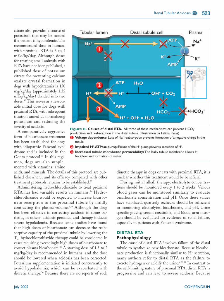
DISTAL RTAPathophysiology
The cause of distal RTA involves failure of the distaltubule to synthesize new bicarbonate. Because bicarbo-nate production is functionally similar to H+ secretion,many authors refer to distal RTA as the failure toexcrete hydrogen or acidify the urine.4,30,31 In contrast tothe self-limiting nature of proximal RTA, distal RTA isprogressive and can lead to severe acidosis. Because
July 2005 COMPENDIUM
Renal Tubular Acidosis 523CE
Figure 6. Causes of distal RTA. All three of these mechanisms can prevent HCO3–
production and reabsorption in the distal tubule. (Illustration by Felicia Paras)1 Voltage dependence:Loss of Na+ reabsorption prevents formation of a negative charge in the
tubule.2 Impaired H+-ATPase pump:Failure of the H+ pump prevents secretion of H+.3 Increased tubule membrane permeability: The leaky tubule membrane allows H+
backflow and formation of water.

bicarbonate production is a stepwise process, failure ofproduction can occur for a variety of reasons.
Voltage-dependent distal RTA occurs because ofimpaired sodium reabsorption4,37 (Figure 6). As previouslydiscussed, sodium is normally reabsorbed into the distaltubule cell without the simultaneous transfer of an anioninto the cell or a cation out of the cell. This type of trans-port creates a negatively charged tubular lumen that facil-itates the transport of H+ and K+ out of the cell. Retainingboth H+ and K+ inside the cell causes both metabolic aci-dosis and hyperkalemia.
The second step in bicarbonate production involvesthe action of the H+–ATPase pump. This condition,called classic distal RTA, can be either congenital oracquired. In contrast to voltage-dependent RTA, potas-sium levels in classic distal RTA are actually below nor-mal. The independent movement of sodium into thedistal tubule cell creates an electronegative charge in thetubule lumen. When H+ cannot be pumped out of thecell to neutralize this charge, the negative charge in thelumen remains relatively high. Because of the greaterpull from the increased electronegativity of the distaltubule, more potassium is lost from the distal tubule cellinto the tubular lumen.
The final cause of distal tubule RTA is related toincreased permeability of the distal tubule membrane. Inthe normal distal tubule, the concentration gradient forhydrogen can be quite large as the hydrogen pumpsecretes ions into the lumen and the tubular membraneprevents the flow of these ions back into the cell downtheir concentration gradient. If this tubular membrane iscompromised, H+ can pass back into the cell and com-bine with the hydroxide normally reserved for bicarbon-ate production. The leaky tubular membrane alsofacilitates the transfer of potassium into the tubularlumen and subsequently leads to potassium wasting.Hence, as with classic distal RTA, defects in membraneintegrity result in metabolic acidosis with hypokalemia.
Clinical SignsAs with proximal RTA, distal RTA can result in signs
associated with acidemia, including muscle weakness,inappetence, nausea, weight loss, stunted growth, andneurologic signs. In a 5-year-old mixed-breed dog diag-nosed with distal RTA, the only clinical sign wasanorexia.29 If distal RTA is associated with hypokalemia,owners may report polyuria and polydipsia associatedwith hypokalemic nephropathy as well as muscle weak-ness.4 Increased urinary calcium excretion can also cre-
ate hyposthenuric urine and subsequent polyuria andpolydipsia because urinary calcium can inhibit tubularsodium reabsorption, leading to sodium and water wast-ing. This sodium diuresis disrupts the normal counter-current mechanism that allows urine concentration. Inaddition, high urinary calcium can impair the water per-meability in the collecting duct membrane, thus inter-fering with the action of antidiuretic hormone.9 Whendistal RTA is associated with another clinical disease(i.e., pyelonephritis, multiple myeloma, lupus), clinicalsigns attributable to the primary disease also occur.
Chronic metabolic acidosis can lead to osteomalaciaby the same mechanism as described for bone bufferingin proximal RTA. In humans, such bone changes aresignificantly more common in distal RTA than in proxi-mal RTA; however, such pathology has not beenreported in dogs and cats with distal RTA.
Along with bone destruction, distal RTA can also causenephrolithiasis. As with proximal RTA, chronic meta-bolic acidosis associated with distal RTA causes leachingof calcium, phosphate, and bicarbonate from bone. How-ever, unlike in proximal RTA, the urine in distal RTA isalways alkaline and deficient in citrate. The reduced cit-rate decreases the solubility of calcium, while the highurine pH reduces the solubility of both calcium andphosphate. This reduced solubility combined with theincreased urinary concentration of calcium and phosphatepredisposes animals with distal RTA to stone formation.Although humans most frequently form calcium phos-phate stones, both calcium oxalate and struvite urolithscan occur. A 1-year-old Labrador retriever evaluated atthe University of Minnesota Veterinary Teaching Hospi-tal was diagnosed with multiple struvite urocystolithsassociated with distal RTA.29
DiagnosisAn initial minimum database should provide sufficient
information to warrant consideration of RTA. Hyper-chloremic non–anion-gap metabolic acidosis with a urinepH greater than 5.5 to 6 suggests distal RTA; however, aurine culture should be conducted to ensure the changesare not due to the presence of urease-producing bacte-ria.10,29,31,38 The inability of the distal tubules to secrete H+
leads to metabolic acidosis. Hyperchloremia results fromsecretion of the conjugate base of the metabolic acid as asodium salt, leading to volume contraction and subse-quent retention of sodium and chloride. The clinicianshould ensure that the patient has neither diarrhea norevidence of renal failure that could also cause normal
COMPENDIUM July 2005
Renal Tubular Acidosis524 CE

COMPENDIUM July 2005
Renal Tubular Acidosis526 CE
anion-gap acidosis. If these conditions are ruled out, further diagnostic testingis appropriate. Imaging modalities might reveal nephroliths, urocystoliths,and/or evidence of decreased bone density or, rarely, pathologic fractures. Uri-nalysis may reveal crystalluria, whereas a serum chemistry profile can showeither hypokalemia or hyperkalemia.
A bicarbonate challenge test can be conducted in patients with normalurine pH when distal RTA is still suspected. Sodium bicarbonate should begiven as previously described to diagnose proximal RTA, and urine CO2
concentrations should be measured. Confirmation can be made whenpatients exhibit no increase in urine CO2 levels despite the presence of largeamounts of bicarbonate in the filtrate.
The diagnosis of distal RTA can also be confirmed with the ammoniumchloride loading test. Because NH4
+ is an acid, when it is given orally, nor-mal kidneys excrete excess H+ in the urine. In distal RTA, the kidneys fail toacidify the urine, leading to alkaline urine. Patients should be fasted for 8hours and then given ammonium chloride at 0.1 g/kg PO. Two hours later,hourly urine sampling should begin and continue for 6 hours. Failure to col-lect urine on an hourly basis allows admixing of the urine and may lessenthe decrease in urine pH.16 The pH of each collected sample should bemeasured immediately and recorded. A pH less than 5.5 rules out the pres-ence of distal RTA. Failure of the urine pH to fall below 5.5 8 hours follow-ing administration of ammonium chloride confirms the diagnosis of distalRTA.16 In humans, the cut-off pH (range: 5.4 to 7) for determining thepresence of RTA is controversial.10 This suggests that urine pH should notbe looked at alone when confirming the diagnosis of distal RTA. Instead, itshould be considered in light of the concurrent metabolic acidosis and theinability of the pH to drop despite excess total body hydrogen.
CausesAlthough some of the causes of proximal and distal RTA are shared, there
are some specific causes of distal RTA. For example, amphotericin B destroysthe distal renal tubular membrane and is associated with the third type of dis-tal RTA described. Pyelonephritis is a reported cause of distal RTA in bothhumans and small animals. Watson et al37 reported distal RTA secondary toEscherichia coli pyelonephritis in a cat. A cat with chronic pyelonephritis wasalso found to have distal RTA in an earlier report by Drazner.38 Systemiclupus erythematosus is also a reported cause of distal RTA in humans but hasnot been reported in veterinary patients.4 As with proximal RTA, distal RTAcan also be associated with heavy metal toxicity, multiple myeloma, chronichypocalcemic conditions, and outdated tetracycline administration.5,6
TreatmentThe bicarbonate dose required to correct metabolic acidosis associated
with distal RTA is much less than that of proximal RTA. Empirical dosingof NaHCO3 with careful monitoring is the usual method of therapy becauseof varying individual requirements. The recommended initial dose is 1 to1.5 mEq/kg/day PO of sodium bicarbonate in divided doses.10 Acidosis isusually corrected with doses of 1 to 3 mEq/kg/day PO; however, higherdoses may be required.39 Sodium bicarbonate can be administered orally as

tablets, powder, or a solution. An 8-oz box of bakingsoda can be combined with 2.88 L of distilled water tocreate a 1 mEq/ml solution of bicarbonate.27 Such asolution can be kept refrigerated for 2 months ifcapped.40 As described for proximal RTA, if a patient ishypokalemic, potassium citrate might also be consideredan alternative to bicarbonate therapy. If bicarbonate isused to correct acidemia, potassium gluconate elixir canbe used as described for proximal RTA. The amount ofpotassium replacement should be adjusted according toindividual need. In patients with nephroliths, serumpotassium concentration should be closely monitoredbecause of potential renal failure and concurrent hyper-kalemia. Concurrent administration of vitamin D andcalcium is not recommended because this may increasethe tendency for urolithiasis. Alkali therapy usuallyallows fairly rapid correction of pH and serum bicar-bonate; however, osteomalacia in humans may take sev-eral months to resolve. Appropriate antibiotic therapyshould be administered for 4 to 6 weeks in patients with
pyelonephritis, with a follow-up urine culture to con-firm resolution of the infection.
Patients should be seen every 1 to 2 weeks during ini-tial alkali therapy to evaluate serum electrolytes, pH,and serum bicarbonate. The dose of bicarbonate orpotassium citrate should be increased to return pH andserum bicarbonate to normal.12 Once these values arestable, the patient should be reevaluated at least every 3months to assess its metabolic status.
PROGNOSISThe prognosis in patients with RTA is not well de-
fined because of the few reports of this condition in theveterinary literature. The prognosis in patients with dis-tal RTA is not as good as proximal RTA becausepatients with distal RTA are predisposed to urolithiasis.Although treatment and control of metabolic acidosismakes further urolithiasis and bone disease less likely,progressive renal damage from existing nephroliths orresistant infection can lead to renal failure. A recentreview of 60 dogs with idiopathic Fanconi syndrome,95% of which were basenjis, suggested that with theowners’ meticulous care, the disease did not significantly
reduce a dog’s life span compared with that of unaf-fected dogs.33 Furthermore, owners of these dogs be-lieved their dogs had a good to excellent quality of life.Despite this, a fraction of dogs with Fanconi syndromedevelop chronic renal failure, and, in this same study,this was the sole or predominant reason for euthanasiaor death in 41% of affected dogs.33 In addition, acuterenal failure can occur and was described in a report offatal uremia in a dog with Fanconi syndrome and resist-ant pyelonephritis.10
Even without complications such as urolithiasis orinfection, and unless the condition is transient, manage-ment of RTA is lifelong and owners should be awarethat frequent evaluation of their pet is necessary toassess its acid–base and electrolyte status.
CONCLUSIONRTA is an uncommon diagnosis in veterinary
patients. The condition is characterized by non–anion-gap metabolic acidosis with a normal GFR. Proximal
RTA is caused by decreased bicarbonate reabsorption inthe proximal tubule and most commonly occurs in asso-ciation with other proximal tubular defects. Distal RTAis associated with decreased production of bicarbonateby the distal tubule. Therapy for both conditionsincludes oral bicarbonate supplementation, although therequirement for patients with proximal RTA greatlyexceeds that of those with distal RTA. Both proximaland distal RTA can be transient or permanent, depend-ing on the underlying cause. Because of the acid–baseand electrolyte derangements associated with the dis-ease, routine biochemistry monitoring is recommended.
REFERENCES1. Defronzo FA: Hyperkalemia and hyporeninemic hypoaldosteronism. Kidney
Int 17:118, 1980.2. Berne RM, Levy MN: Physiology, ed 3. St. Louis, Mosby, 1998, pp 763–776.3. Pitts RF: Physiology of the Kidney and Body Fluids, ed 3. Chicago, Year Book
Medical Publishers, 1974.4. Faubel S, Topf J: The Electrolyte and Acid–Base Companion. Chelsea, MI,
Sheridan Books, 1999.5. Rose BD: Pathophysiology of Renal Diseases. New York, McGraw-Hill, 1981.6. Gonick HC: Renal Tubular Disorders: Pathophysiology, Diagnosis, and Manage-
ment. New York, Marcel Dekker, 1985.7. Easley JR, Breitschwerdt EB: Glucosuria associated with renal tubular dys-
July 2005 COMPENDIUM
Renal Tubular Acidosis 527CE
Distal RTA occurs because of decreased bicarbonate production by the distal tubule.

1. RTA is characterized bya. anion-gap metabolic acidosis.b. azotemia and non–anion-gap metabolic acidosis.c. non–anion-gap metabolic acidosis and normal GFR.d. isosthenuria and azotemia.
2. Proximal RTA occurs because ofa. failure of bicarbonate synthesis in the proximal tubule.b. decreased proximal tubular resorption of bicarbonate.c. increased acid production by the proximal tubule.d. increased acid load in the glomerular filtrate.
3. _____________ is(are) not a reported cause ofproximal RTA.a. Gentamicin c. Heavy metalsb. Streptozotocin d. Cushing’s disease
4. Fanconi syndromea. is always a congenital condition.b. occurs only in basenjis.c. is associated with proximal RTA.d. is associated with distal RTA.
5. The diagnosis of proximal RTA can be based ona. the response to intramuscular injection of bicarbonate.b. a urine pH of 5.5 to 6 and hypochloremic metabolic
acidosis.
function in three basenji dogs. JAVMA 168:938–943, 1976.8. Darrigrand-Haag RA, Center SA, Randolph JF, et al: Congenital Fanconi
syndrome associated with renal dysplasia in 2 border terriers. J Vet Intern Med20(6):412–419, 1996.
9. Bovee KC, Joyce T, Blazer-Yost B, et al: Characterization of renal defects indogs with a syndrome similar to the Fanconi syndrome in man. JAVMA 174:1094–1099, 1979.
10. Noonan CHB, Kay JM: Prevalence and geographic distribution of Fanconisyndrome in basenjis in the United States. JAVMA 197(3):345–349, 1990.
11. Bovée KC: Fanconi syndrome in the dog, in Kirk RW (ed): Current Veteri-nary Therapy VII. Philadelphia, WB Saunders, 1980, p 1075.
12. Dibartola SP: Fluid Therapy in Small Animal Practice, ed 2. Philadelphia, WBSaunders, 2000.
13. Thornhill JA: Renal tubular acidosis, in Kirk RW (ed): Current VeterinaryTherapy VI. Philadelphia, WB Saunders, 1977, pp 1087–1097.
14. Hilton JG: Effect of maleic acid on kidney function. Proc Soc Exp Biol Med75:791–794, 1950.
15. Cronin RE, Bulger RE, Heinrich WL: Natural history of aminoglycosidenephrotoxicity in the dog. J Lab Clin Med 95:467–474, 1980.
16. Schentag JJ: Aminoglycoside pharmacodynamics as a guide to therapy andtoxicology, in Whelton A, Weu HC (eds): The Aminoglycosides. New York,Marcel Dekker, 1982, pp 151–157.
17. Settles EL, Schmidt D: Fanconi syndrome in a Labrador retriever. J VetIntern Med 8:390–393, 1994.
18. Soriano JR: Renal tubular acidosis: The clinical entity. J Am Soc Nephrol13:2160–2170, 2002.
19. Moore AS, Kitchell BE: New chemotherapy agents in veterinary medicine, inKitchell B (ed): Vet Clin North Am Adv Med Oncol 33:629–649, 2003.
20. Keneko JJ, Mattheeuws D, Rottiers RP, Vermeulen A: Renal function, insulinsecretion, and glucose tolerance in mild streptozotocin diabetes in dogs. Am JVet Res 35(5):807–809, 1978.
21. Lowe MB, Obst D, Tapp E: Renal damage caused by anhydro-4-EPI-tetra-cycline. Arch Pathol 81:362–364, 1966.
22. Obek A, Petorak I, Eroglu L, Gurkan A: Effects of tetracycline in the dogkidney: A functional-ultrastructural study. Isr J Med Sci 10:765–771, 1974.
23. Freeman LM, Breitschwerdt EB, Keene BW, Hansen B: Fanconi’s syndromein a dog with primary hypoparathyroidism. J Vet Intern Med 8:349–354, 1994.
24. Lacy MQ, Gertz MA: Acquired Fanconi’s syndrome associated with mono-clonal gammopathies. Hematol Oncol Clin North Am 13(6):1273–1280, 1999.
25. Liberopoulos E, Bairaktari E, Elisaf M: Reversible proximal tubular dysfunc-tion in a patient with acute febrile illness, marked hyperbilirubinemia, andnormal renal function: Evidence of leptospirosis. Nephron 91:532–533, 2002.
26. Magaldi AJ, Yasuda PN, Kudo LH, et al: Renal involvement in leptospirosis:A pathophysiologic study. Nephron 62(3):332–339, 1992.
27. Polzin DJ, Osborne CA, Bell FW: Canine distal renal tubular acidosis andurolithiasis, in Osborne C (ed): Vet Clin Sm Anim Prac 16(2):241–250, 1986.
28. Dibartola SP, Leonard PO: Renal tubular acidosis in a dog. JAVMA180:70–73, 1982.
29. Mueller DL, Jergens AE: Renal tubular acidosis. Compend Contin Educ PractVet 13(3):435–444, 1991.
30. Edelmann Jr CM: Isolated proximal (type 2) renal tubular acidosis, inGonick HC, Buckalew VM (eds): Renal Tubular Disorders: Pathophysiology,Diagnosis, and Management. New York, Marcel Dekker, 1985, pp 261–279.
31. Gruskin AB, Patel MS, Linshaw M, et al: Renal function studies and kidneypyruvate carboxylase in subacute necrotizing encephalomyelopathy (Leigh’ssyndrome). Pediatr Res 7:832–841, 1973.
32. Plumb DC: Veterinary Drug Handbook, ed 4. Ames, Iowa State UniversityPress, 2002.
33. Yearley JH, Hancock DD, Mealey KL: Survival time, lifespan, and quality oflife in dogs with idiopathic Fanconi syndrome. JAVMA 225(3):377–383, 2004.
34. Unwin RJ, Capasso G: The renal tubular acidoses. J R Soc Med 94:221–225,2001.
COMPENDIUM July 2005
Renal Tubular Acidosis528 CE
ARTICLE #2 CE TESTThis article qualifies for 2 contact hours of continuingeducation credit from the Auburn University College ofVeterinary Medicine. Subscribers may purchase individualCE tests or sign up for our annual CE program.Those who wish to apply this credit to fulfill state relicensurerequirements should consult their respective stateauthorities regarding the applicability of this program.To participate, fill out the test form inserted at the end of this issue or take CE tests online and get real-time scores at CompendiumVet.com.
CE
35. Batlle DC: Renal tubular acidosis. Med Clin North Am 67(4):859–878, 1983.36. Simpson DP: Control of hydrogen ion homeostasis and renal acidosis. Medi-
cine (Baltimore) 50:503–541, 1971.37. Watson ADJ, Culvenor JA, Middleton DJ, Rothwell TLW: Distal renal
tubular acidosis in a cat with pyelonephritis. Vet Rec 119:65–68, 1986.38. Drazner FH: Distal renal tubular acidosis associated with chronic
pyelonephritis in a cat. Calif Vet 34:15, 1980.39. McSherry E, Morris Jr RE: Attainment and maintenance of normal stature
with alkali therapy in infants and children with classic renal tubular acidosis. JClin Invest 61:509, 1978.
40. Rose BD: Clinical Physiology of Acid–Base and Electrolyte Disorders, ed 2. NewYork, McGraw-Hill, 394–439, 1984.

July 2005 COMPENDIUM
Renal Tubular Acidosis 529CE
c. a urine pH greater than 6 following an intravenousconstant-rate infusion of bicarbonate.
d. metabolic acidosis in the absence of azotemia.
6. Bicarbonate therapy for proximal RTAa. requires very small amounts of bicarbonate.b. can lead to hyperkalemia.c. leads to further bicarbonate loss in the urine.d. does not require serum electrolyte monitoring.
7. Distal RTA isa. caused by decreased bicarbonate production by the
distal tubule.b. always associated with acidic urine.c. self-limiting.d. caused by increased acid production by the distal
tubule.
8. _____________ is not a reported cause of distalRTA.a. Amphotericin B c. Outdated tetracyclineb. Enrofloxacin d. Pyelonephritis
9. _______________ may be used as an alternative tooral sodium bicarbonate in treating RTA.a. Potassium citrate c. Calcium carbonateb. Famotidine d. Omeprazole
10. In general, dogs with Fanconi syndrome arethought to havea. a poor quality of life.b. a shorter life span compared with that of unaffected
dogs.c. only a very small chance of developing renal failure.d. a relatively good quality of life and normal life span.
Test answers now available at CompendiumVet.com