art end clasificacion
TRANSCRIPT
Australian Dental Journal Endodontic Supplement 2007;52:1. S17
A clinical classification of the status of the pulp and theroot canal system
PV Abbott,* C Yu*
AbstractMany different classification systems have beenadvocated for pulp diseases. However, most of themare based on histopathological findings rather thanclinical findings which leads to confusion since thereis little correlation between them. Most classificationsmix clinical and histological terms resulting inmisleading terminology and diagnoses. This in turnleads to further confusion and uncertainty in clinicalpractice when a rational treatment plan needs to beestablished in order to manage a specific pathologicalentity. A simple, yet practical classification of pulpdiseases which uses terminology related to clinicalfindings is proposed. This classification will helpclinicians understand the progressive nature of thepulp disease processes and direct them to the mostappropriate and conservative treatment strategy foreach condition. With a comprehensive knowledge ofthe pathophysiology of pain and inflammation in thepulp tissues, clinicians may accomplish this taskwith confidence.
Key words: Dental pulp, classification, pulp disease,inflammation, necrosis, infection.
INTRODUCTIONMany different classification systems (Table 1) have
been advocated for pulp diseases1-13 although most ofthem are based on histopathological findings. Manyauthors and clinicians have attempted to correlate thehistological conditions of the pulp to the clinical signsand symptoms.1-13 These classifications mix clinical andhistological terms resulting in many misleading termsand diagnoses for the same clinical condition (Table 1).This in turn leads to confusion and uncertainty inclinical practice when a rational treatment plan needsto be established in order to target a specificpathological entity. Confusion in diagnosis can alsoarise when using a classification with a wide selectionof overlapping conditions.
One of the primary purposes of establishing adiagnosis is to determine what clinical treatment isrequired. If an incorrect diagnosis is made, thenincorrect or inappropriate treatment may be instigatedand this may result in various consequences ranging
*School of Dentistry, The University of Western Australia.
Australian Dental Journal Supplement 2007;52:(1 Suppl):S17-S31
from mild to severe. Diseases of the pulp and periapicaltissues are, in general terms, either inflammatory innature or due to infections. An infection will also beassociated with inflammation of the adjacent tissues.Hence, if the condition is inflammatory then thetreatment regime adopted by the clinician should betargeted at removing the inflammatory stimuli andreducing the inflammation in the tissues – in such acase, an anti-inflammatory agent would be indicated aspart of the local treatment (e.g., within a root canalmedicament) or as part of any systemic medicationsused. On the other hand, an infection will require theuse of an anti-microbial agent (e.g., antibiotic or anti-septic) within the root canal medicament or, in certaincircumstances, systemically. Infected canals causeinflammation within the adjacent periapical tissues andtherefore an anti-inflammatory agent combined withthe anti-microbial agent in the root canal medicamentshould also be considered. Hence an accurate diagnosisis essential to ensue appropriate treatment is provided.
A further purpose of a classification of diseases is toenable communication between teachers, students,clinicians and researchers. Hence any classificationneeds to be useful, easily understood and readilyapplied to the clinical environment. The use ofterminology that is inappropriate or that cannot beapplied clinically is counter-productive to these aims.For example, if a histological examination is requiredsince histological terms are used, then this is impracticalin a clinical situation where an immediate diagnosis isrequired before treatment is instigated; in this scenario,a diagnosis is required in order to dictate whattreatment regime should be followed.
A clinician’s ability to diagnose accurately isdependent on having a thorough understanding of thedisease processes involved as well as having a thoroughunderstanding of the diagnostic procedures and testsbeing used and their limitations (see below).
The progression of disease in the dental pulp issimilar to changes in other connective tissues. Typically,the tissue and disease progresses through the followingstages: normal, inflammation (i.e., pulpitis), necrosis,infection and loss of pulp tissue (i.e., pulpless canals).Inflammatory changes may be acute, chronic, reversibleor irreversible, and chronic conditions may have acuteexacerbations at any time. The classification presented
below and in Table 2 has been proposed previously byAbbott.14-16 It is a simple yet comprehensive clinicaldiagnostic system that utilizes terminology relating tothe clinical findings (i.e., signs and symptoms) and it isbased on the progression of pulp diseases through thevarious stages shown in Fig 1. It also includes “normal”pulp tissue which is an entity that should be diagnosedand given recognition when there are no signs ofdisease.
Signs and symptoms of pulp and root canal conditionsDiseases of the pulp tissues are dynamic and
progressive in nature (Fig 1). Each disease condition may
progress to other conditions if left untreated. Hence, thesigns and symptoms will vary depending on the stage ofthe disease at the time the patient presents for treatment.In addition, the reaction to and the perception of painwill vary between individual patients and is influencedby the individual’s emotional status and the copingstrategies used to manage the pain. Many of the signsand symptoms overlap between the various pulpconditions due to the dynamic interactions and theprogressive nature of the disease process. Therefore, thefollowing descriptions of pulp diseases are based on thetypical presenting complaints and clinical findings. Whenmaking a diagnosis, clinicians should be aware of the
S18 Australian Dental Journal Endodontic Supplement 2007;52:1.
Table 1. Comparative terminology and classifications of pulp diseases used by various authors and organisations1-13
World Health Weine2 Ingle3 Seltzer & Bender4 Cohen & Burns5 Tronstad6
Organization1
(NOTE: Normal pulp (NOTE: Normal pulp Healthy pulp (NOTE: Normal pulp Within normal limits Healthy pulpnot mentioned) not mentioned) not mentioned) Normal pulp
Calcificmetamorphosis
Pulpitis Pulpitis Pulpitis Pulpitis Pulpitis PulpitisInitial (hyperaemia) Hyperalgesia Hyper-reactive Incipient form of Reversible AsymptomaticAcute (reversible pulpitis) pulpalgia chronic pulpitis Irreversible pulpitisSuppurative (pulpal Hypersensitive dentine Hypersensitivity Acute pulpitis Asymptomatic Symptomaticabscess) Hyperaemia Hyperaemia Chronic partial irreversible pulpitis pulpitis
Chronic Painful pulpitis Acute pulpalgia pulpitis with partial Hyperplastic pulpitisChronic ulcerative Acute pulpalgia Incipient necrosis Internal resorptionChronic hyperplastic (acute pulpitis) Moderate Chronic total pulpitis Canal calcification(pulpal polyp) Chronic pulpalgia Advanced with partial Symptomatic
Other unspecified (subacute pulpitis) Chronic pulpalgia liquefaction necrosis irreversible pulpitispulpitis Nonpainful pulpitis Hyperplastic Chronic partial pulpitis
Pulpitis unspecified Chronic ulcerative pulposis (hyperplastic form)pulpitis
Chronic pulpitis(no caries)
Chronic hyperplasticpulpitis (pulp polyp)
Necrosis of the pulp Pulp necrosis Pulp necrosis Pulp necrosis Necrosis Necrotic pulpLiquefaction PartialSicca Complete
Pulp degenerations Pulp degeneration Pulp degeneration Pulp degenerationDenticles Atrophy Atrophic pulposis Atrophic pulpPulpal calcification Dystrophic Calcific pulposis DystrophicPulpal stones calcification mineralizationAbnormal hard tissue Internal resorption Internal resorptionformation in pulp
Secondary or irregulardentine
American Association Walton &of Endodontists’ Harty8
Torabinejad9 Grossman10 Castellucci11 Stock12 Bergenholtz13
Glossary7
Normal pulp Normal pulp (NOTE: Normal pulp (NOTE: Normal Healthy pulp Normal pulp Pulpa sananot mentioned not mentioned) pulp not
mentioned)Pulpitis Pulpitis Pulpitis Hyperaemia Pulpitis Concussed pulp PulpitisReversible pulpitis Reversible Reversible pulpitis Pulpitides Hyperaemia Reversible pulpitisIrreversible pulpitis pulpitis Irreversible pulpitis Acute pulpitis Pulpitis Irreversible pulpitis
Irreversible Hyperplastic pulpitis Chronic irreversiblepulpitis ulcerative
pulpitisChronichyperplasticpulpitis
Pulp necrosis Necrosis Pulpal necrosis Necrosis Necrosis Pulpal necrosis Necrosis pulpaePulp calcification Pulp degeneration Internal resorptionInternal (intracanal) Calcificresorption Fibrous
AtrophicInternal resorption
Australian Dental Journal Endodontic Supplement 2007;52:1. S19
dynamic nature of the pulp disease process whilst alsotaking into consideration the limitations of assessing thetrue state of the pulp’s blood supply with current pulptesting and examination techniques.
Clinically normal pulpThe term “clinically normal pulp” is used to classify
a pulp that has no signs or symptoms to suggest thatany form of disease is occurring. The term “clinically”is used since such a pulp may not be histologicallynormal and/or may have some degree of fibrosis(scarring) as a result of previous injury or stimuli.
A clinically normal pulp is asymptomatic. Itproduces a mild and transient response to variousstimuli but the nature and severity of the response may
vary according to the age and state of the tooth. Aslong as there has been no calcification of the coronalpulp space, a clinically normal pulp will react to coldstimuli with mild pain that lasts for no more than 1–2seconds after the stimulus is removed. A clinicallynormal pulp does not respond to heat stimuli.Percussion and palpation tests will not elicit anytenderness. Radiographic examination will demonstratenormal appearance of the pulp chamber, root canalsand periapical tissues.
Dentine is usually sensitive when exposed to irritantshence dentine sensitivity should be distinguished frompulp inflammation.
Dentine sensitivityWhen pain occurs with thermal, chemical, tactile, or
osmotic stimuli associated with exposed dentine, thediagnosis is dentine sensitivity. The pain is consistentwith an exaggerated response of the normal pulpo-dentinal complex, and it is severe and sharp onapplication of the stimulus to the exposed dentine.Nonetheless, there is no lingering discomfort once thestimulus is removed.
When there is a specific factor (other than exposeddentine) causing the sensitivity, such as caries, fractures,open restoration margins or recent restorativetreatment, teeth may exhibit symptoms that areidentical to dentine sensitivity. When symptomsdevelop in these situations, a diagnosis of reversiblepulpitis is appropriate.
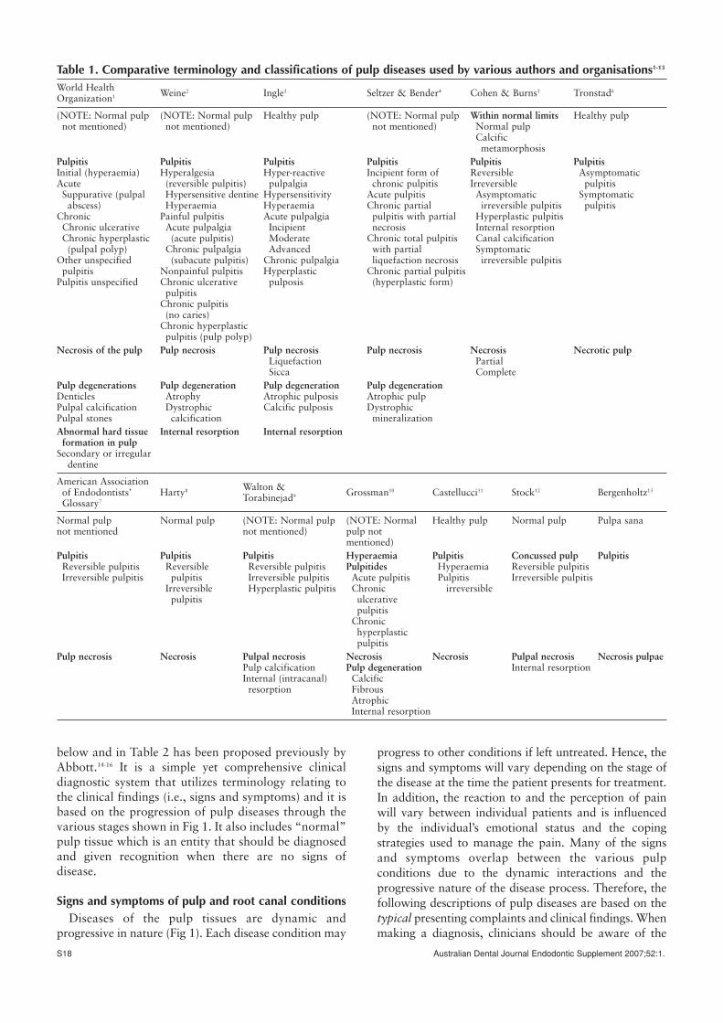
Table 2. Clinical classification of the status of thepulp and root canal conditions proposed by Abbott14,15
and used in the School of Dentistry at The Universityof Western Australia16
Clinically Normal Pulp (based on clinical examination and testresults)Reversible pulpitis – AcuteReversible Pulpitis – ChronicIrreversible pulpitis – AcuteIrreversible Pulpitis – ChronicNecrobiosis (Part of pulp necrotic & infected; the rest is irreversiblyinflamed)Pulp necrosis – No sign of infectionPulp necrosis – InfectedPulpless, infected root canal systemDegenerative changes• Atrophy• Pulpal canal calcification – partial• Pulpal canal calcification – total• Hyperplasia• Internal resorption – Surface• Internal resorption – Inflammatory• Internal resorption – ReplacementPrevious root canal treatment• No sign of infection• Infected• Technical standard (based on the radiographic appearance)
– Adequate– Inadequate
• Other problems - e.g. perforation, missed canals, fracturedinstrument, etc.
Fig 1. The progression and interaction of the various stages of pulpand root canal conditions and their relationships to apical
periodontitis.
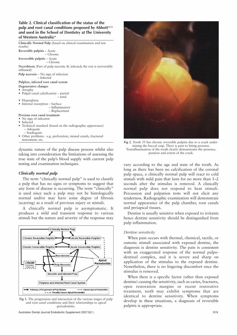
Fig 2. Tooth 35 has chronic reversible pulpitis due to a crack under-mining the buccal cusp. There is pain to biting pressure.
Transillumination of the tooth clearly demonstrates the presence,position and extent of the crack.

Reversible pulpitisA pulp with reversible pulpitis has mild inflammation
and it is capable of healing once the irritating stimulushas been removed. Pain is only felt when a stimulus(usually cold or sweet foods but sometimes heat) isapplied to the tooth, and the pain ceases within a fewseconds or immediately upon removal of the stimulus.The pain is short and sharp in nature but notspontaneous. There are no significant radiographicchanges evident in the periapical region, and the onlyradiographic findings of note may be the cause of theproblem, such as caries, a deep restoration, etc. Usuallymore extreme temperatures are required to induce thepain rather than mild changes (e.g., ice cream ratherthan tap water).
If there is pain to biting pressure as well as the abovesymptoms, then this may indicate a crack in the tooth(Fig 2) or restoration. While reversible pulpitis is usuallyacute, it may also be an acute exacerbation of a chroniccondition. Here the terms “acute” and “chronic” arenot used as histological terms but are based on theclinical symptoms: that is, acute means painful andchronic means no pain or only mild discomfort.Conservative pulp therapy in conjunction with theremoval of the cause and the pathway of the irritationwill result in resolution of the pulp inflammation andthe return of the pulp to a clinically normal state.
A diagnosis of reversible pulpitis should always beconsidered as a “provisional diagnosis” since it isimpossible to be completely certain about anyindividual pulp’s ability to recover. This will depend onmany factors – such as previous problems, previousinflammation, the degree of fibrosis, the true status ofthe pulp, etc. – and these are generally impossible toassess clinically. Hence once the diagnosis of reversiblepulpitis has been provisionally made, the tooth can bemanaged in a conservative manner (as above) and thenarrangements must be made to review the status of thepulp to determine whether it has actually returned to aclinically normal state. If it has, then it should be free
of symptoms, have no signs of pulp or periapicaldiseases and should respond normally to pulp sensibilitytests. The pulp status should be reviewed after severalweeks (assuming there are no postoperative symptoms)although a three-month interval is generally consideredto be more reliable as healing may take some time ornecrosis (if it occurs) may take some time to becomeevident. At the review appointment, the provisionaldiagnosis can be confirmed as reversible pulpitis if thepulp has returned to a clinically normal state. However,if symptoms have persisted or if pulp necrosis hasoccurred, then the original provisional diagnosis willneed to be altered to that of “irreversible pulpitis”.
Irreversible pulpitisOne of the classic symptoms of irreversible pulpitis is
lingering pain induced by thermal stimuli. Only mild
S20 Australian Dental Journal Endodontic Supplement 2007;52:1.
Fig 3. Tooth 46 has acute irreversible pulpitis with primary acuteapical periodontitis due to marginal breakdown of the composite
resin restoration.
Fig 4. (A) Tooth 47 has acute irreversible pulpitis with primaryacute apical periodontitis due to a vertical crack on the distal aspect
of the tooth. (B) The extent of the crack can be seen followinginvestigation of the tooth by removing the restoration – it extendsinto the tooth root and communicates with the pulp chamber and
root canals.
a
b

Australian Dental Journal Endodontic Supplement 2007;52:1. S21
temperature changes are required to induce the pain(e.g., tap water, breathing cold air). The initial reactionis a very sharp pain to hot or cold stimuli and it thenlingers for minutes to hours after the stimulus isremoved. The lingering pain is usually a dull ache or athrobbing pain. Spontaneous (unprovoked) pain, whichmay wake the patient at night and may become worsewhen lying down, is another hallmark feature ofirreversible pulpitis. Patients with irreversible pulpitisoften need strong analgesics and may have difficultylocating the precise tooth that is the source of the pain.They may even confuse the maxillary and mandibulararches (but not the left and right sides of the mouth)because of the extensive branching of dental nerveaxons17,18 and perhaps fewer proprioceptive fibres in thepulp.19 Pulps afflicted with irreversible pulpitis withoutperiapical pathosis may sometimes be difficult todiagnose; information provided by the patients and theresults of pulp sensibility tests become particularlyhelpful in these cases. When the periapical tissues areinvolved (Figs 3 and 4), the tooth will usually be tenderto pressure and/or percussion. Dynamic changes occurin the irreversibly inflamed pulp and it may move frombeing chronic and asymptomatic to having acute painwithin a matter of just a few hours.
Acute irreversible pulpitisAcute irreversible pulpitis usually has a sudden onset
that may wake the patient at night. The pain isspontaneous with moderate to very severe intensity,and it lingers in response to temperature changes (heatand/or cold). The pain may be intensified by posturechanges such as when lying down or bending over.Common analgesics are rarely effective in controllingthe pain. In most cases, radiographs are not useful indiagnosis but they can be helpful in identifying thepossible cause of the disease (e.g., deep caries, anextensive or fractured restoration, pins etc.). The toothmay be tender to biting pressure and/or percussion and,if present, this usually indicates spread of theinflammatory process to the periapical tissues (Figs 3and 4). In some cases, the biting or percussion pain mayindicate a crack in the tooth (Fig 4) and this isparticularly noticeable when the crack is undermining acusp.
Chronic irreversible pulpitisChronic irreversible pulpitis will have similar signs
and symptoms but they will be much less severe thanthose in the acute cases. Patients may complain ofmoderate pain, which is more intermittent rather thancontinuous and it may be controlled by commonanalgesics. In the early stages of the disease process, thediagnosis may be difficult because the tooth may notshow any demonstrable periapical change on theradiographs or a definitive sign of tenderness topercussion. However, information from the patient andpulp sensibility tests should be useful. As the diseaseprogresses to involve the periapical tissues, periapicalchanges are more likely to be evident radiographicallyand/or clinically.
NecrobiosisA tooth with necrobiosis has both inflamed and
necrotic (usually infected) pulp tissue.20 Many dentistsuse the term “partial necrosis” for this stage of thedisease process; however, “necrobiosis” was suggestedby Grossman20 because it more accurately indicates thecondition – the key factor to the spread of the diseaseprocess is the presence of bacteria within the necroticpart of the pulp rather than the necrosis itself of part ofthe pulp. The necrotic tissue may be in the coronalportion of the pulp (e.g., pulp chamber) with theinflamed tissue apically, or the different tissue statesmay exist in different canals of a multi-canal tooth.Teeth with this condition can be quite difficult todiagnose since they usually present with a mixture ofthe signs and symptoms of both pulpitis and necrosiswith infection. The symptoms may be mild withintermittent painful episodes over many weeks ormonths. Pulp sensibility test results are mixed andfrequently inconclusive or inconsistent with thepatient’s description of symptoms. Teeth withnecrobiosis may also have apical periodontitis withradiographic evidence of a widened periodontalligament space, which may be unexpected because thepatient has reported sensitivity to hot and/or cold stimuli.
Pulp necrosisA necrotic pulp should be suspected when the tooth
does not respond to pulp sensibility tests. However, thiswill not always be the case since teeth with pulp canalcalcification, previous root fillings or pulpotomies willalso not respond to pulp sensibility tests. Likewise,some teeth or patients just do not respond to such testsfor no apparent reason. When pulp necrosis is present,the history may reveal past trauma, previous episodesof pain or history of restorations and caries.Radiographically, a tooth with a necrotic pulp mayhave signs (such as untreated caries, an extensiverestoration, previous pulp capping) or there may be nosuch signs (e.g., following trauma). Trauma to a toothmay cause pulp necrosis as a result of severing theapical blood supply if the tooth has been displacedfrom its normal position (e.g., luxations, avulsion) or ifthere has been significant damage and inflammation tothe apical periodontal ligament (e.g., subluxation).
No significant radiographic changes are evident atthe root apex unless there is also periapical involve-ment, and this only occurs once the necrotic tissuebecomes infected. It is important to realize that anecrotic pulp per se does not cause apical periodontitisunless it is infected.21-23 Pain usually does not presentunless the periodontal ligament is affected. In thissituation, the canal is infected and leads to apicalperiodontitis.
Occasionally, patients will complain of a dullcontinuous ache that is exacerbated by heat butrelieved by cold water being held over the tooth. Thereason is unknown although it has been speculated thatmicro-organisms producing gases may infect the
necrotic pulp and these gases contract with applicationof the cold water which relieves pressure on the nerveendings that may still be functioning, particularly in theapical portion of the canal. However, the relief from thecold water is only temporary and the pain returns assoon as the tooth warms up again and the gasesexpand. This situation occurs since the apical part ofthe pulp nerves are the last part of the pulp to necrose.It is likely that the apical nerves maintain some viabilitythrough blood supply to their more medial portions fora short time after the rest of the pulp has died.
Once a necrotic pulp is invaded by bacteria, then thebacteria will spread throughout the entire root canalsystem. The necrotic pulp acts as a source of nutrientsfor the bacteria which ingest the necrotic tissue andrender the tooth pulpless. This may occur within 1–2months of the initial invasion by the bacteria.24
Pulpless and infected root canalsPulpless canals will always be infected. There will be
no pain from the tooth itself when it has a pulplesscanal, although some patients may give a history ofoccasional vague discomfort over a period of time.However, pain may arise from the periradicular tissuesthat become inflamed because of the presence ofbacteria in the pulp space. The only clinical signsuggesting the condition may be the lack of response topulp sensibility tests. No significant changes can bedetected on the radiograph in the early stages of thiscondition but within 2–10 months there will be aradiolucency suggesting periapical involvement.24 Asthere is the potential for erroneous pulp sensibility testresponses, corroborating the clinical findings, radio-graphs and patient information is necessary for adefinitive diagnosis.
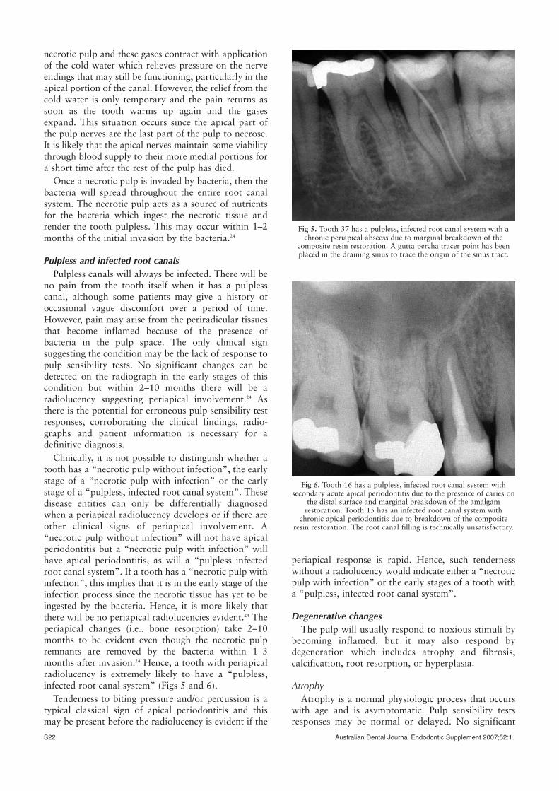
Clinically, it is not possible to distinguish whether atooth has a “necrotic pulp without infection”, the earlystage of a “necrotic pulp with infection” or the earlystage of a “pulpless, infected root canal system”. Thesedisease entities can only be differentially diagnosedwhen a periapical radiolucency develops or if there areother clinical signs of periapical involvement. A“necrotic pulp without infection” will not have apicalperiodontitis but a “necrotic pulp with infection” willhave apical periodontitis, as will a “pulpless infectedroot canal system”. If a tooth has a “necrotic pulp withinfection”, this implies that it is in the early stage of theinfection process since the necrotic tissue has yet to beingested by the bacteria. Hence, it is more likely thatthere will be no periapical radiolucencies evident.24 Theperiapical changes (i.e., bone resorption) take 2–10months to be evident even though the necrotic pulpremnants are removed by the bacteria within 1–3months after invasion.24 Hence, a tooth with periapicalradiolucency is extremely likely to have a “pulpless,infected root canal system” (Figs 5 and 6).
Tenderness to biting pressure and/or percussion is atypical classical sign of apical periodontitis and thismay be present before the radiolucency is evident if the
periapical response is rapid. Hence, such tendernesswithout a radiolucency would indicate either a “necroticpulp with infection” or the early stages of a tooth witha “pulpless, infected root canal system”.
Degenerative changesThe pulp will usually respond to noxious stimuli by
becoming inflamed, but it may also respond bydegeneration which includes atrophy and fibrosis,calcification, root resorption, or hyperplasia.
AtrophyAtrophy is a normal physiologic process that occurs
with age and is asymptomatic. Pulp sensibility testsresponses may be normal or delayed. No significant
S22 Australian Dental Journal Endodontic Supplement 2007;52:1.
Fig 5. Tooth 37 has a pulpless, infected root canal system with achronic periapical abscess due to marginal breakdown of the
composite resin restoration. A gutta percha tracer point has beenplaced in the draining sinus to trace the origin of the sinus tract.
Fig 6. Tooth 16 has a pulpless, infected root canal system withsecondary acute apical periodontitis due to the presence of caries on
the distal surface and marginal breakdown of the amalgamrestoration. Tooth 15 has an infected root canal system with
chronic apical periodontitis due to breakdown of the compositeresin restoration. The root canal filling is technically unsatisfactory.
Australian Dental Journal Endodontic Supplement 2007;52:1. S23
radiographic or clinical signs are present. As the pulpatrophies, there will also likely be fibrosis of the pulptissue and the extent of this will be largely determinedby the number of irritant episodes suffered by thatparticular pulp throughout its history. The size of thepulp chamber may be reduced. No treatment isrequired.
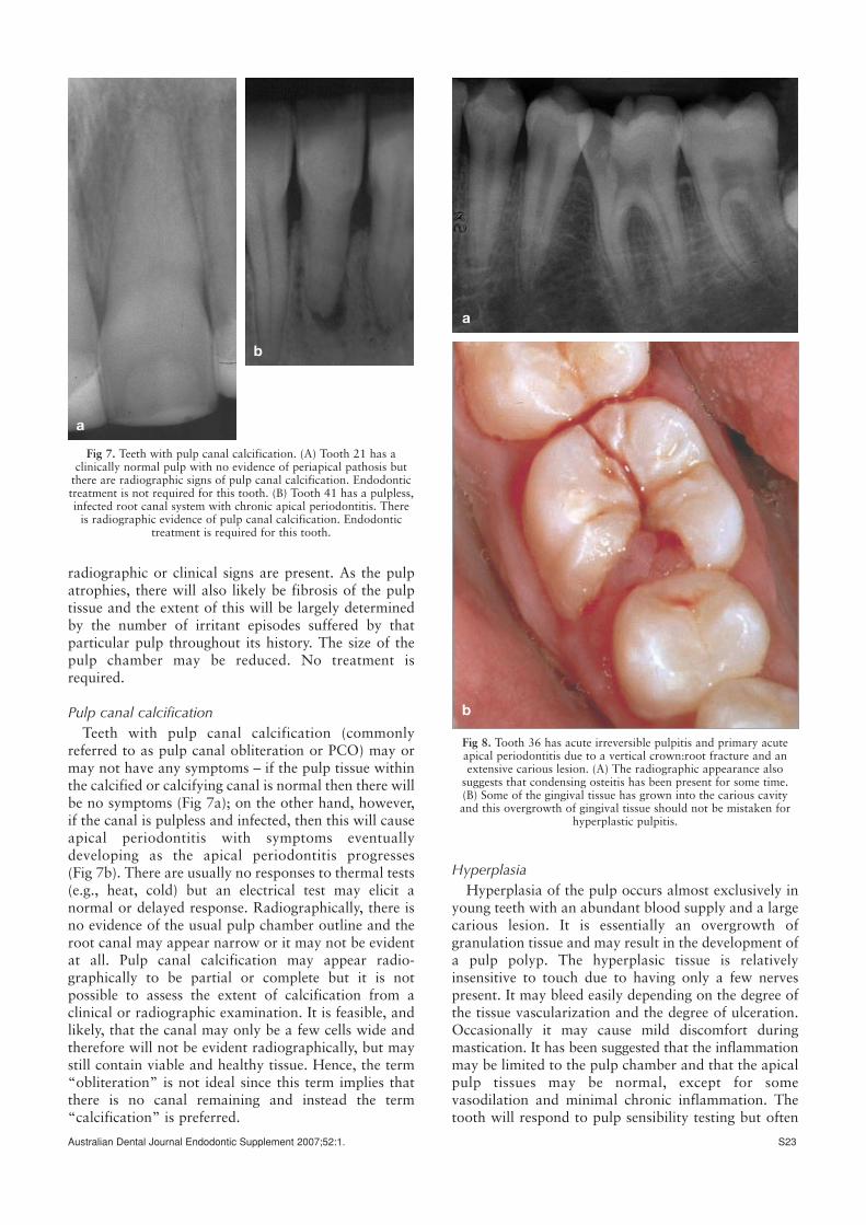
Pulp canal calcificationTeeth with pulp canal calcification (commonly
referred to as pulp canal obliteration or PCO) may ormay not have any symptoms – if the pulp tissue withinthe calcified or calcifying canal is normal then there willbe no symptoms (Fig 7a); on the other hand, however,if the canal is pulpless and infected, then this will causeapical periodontitis with symptoms eventuallydeveloping as the apical periodontitis progresses (Fig 7b). There are usually no responses to thermal tests(e.g., heat, cold) but an electrical test may elicit anormal or delayed response. Radiographically, there isno evidence of the usual pulp chamber outline and theroot canal may appear narrow or it may not be evidentat all. Pulp canal calcification may appear radio-graphically to be partial or complete but it is notpossible to assess the extent of calcification from aclinical or radiographic examination. It is feasible, andlikely, that the canal may only be a few cells wide andtherefore will not be evident radiographically, but maystill contain viable and healthy tissue. Hence, the term“obliteration” is not ideal since this term implies thatthere is no canal remaining and instead the term“calcification” is preferred.
HyperplasiaHyperplasia of the pulp occurs almost exclusively in
young teeth with an abundant blood supply and a largecarious lesion. It is essentially an overgrowth ofgranulation tissue and may result in the development ofa pulp polyp. The hyperplasic tissue is relativelyinsensitive to touch due to having only a few nervespresent. It may bleed easily depending on the degree ofthe tissue vascularization and the degree of ulceration.Occasionally it may cause mild discomfort duringmastication. It has been suggested that the inflammationmay be limited to the pulp chamber and that the apicalpulp tissues may be normal, except for somevasodilation and minimal chronic inflammation. Thetooth will respond to pulp sensibility testing but often
Fig 7. Teeth with pulp canal calcification. (A) Tooth 21 has aclinically normal pulp with no evidence of periapical pathosis but
there are radiographic signs of pulp canal calcification. Endodontictreatment is not required for this tooth. (B) Tooth 41 has a pulpless,infected root canal system with chronic apical periodontitis. There
is radiographic evidence of pulp canal calcification. Endodontictreatment is required for this tooth.
a
b
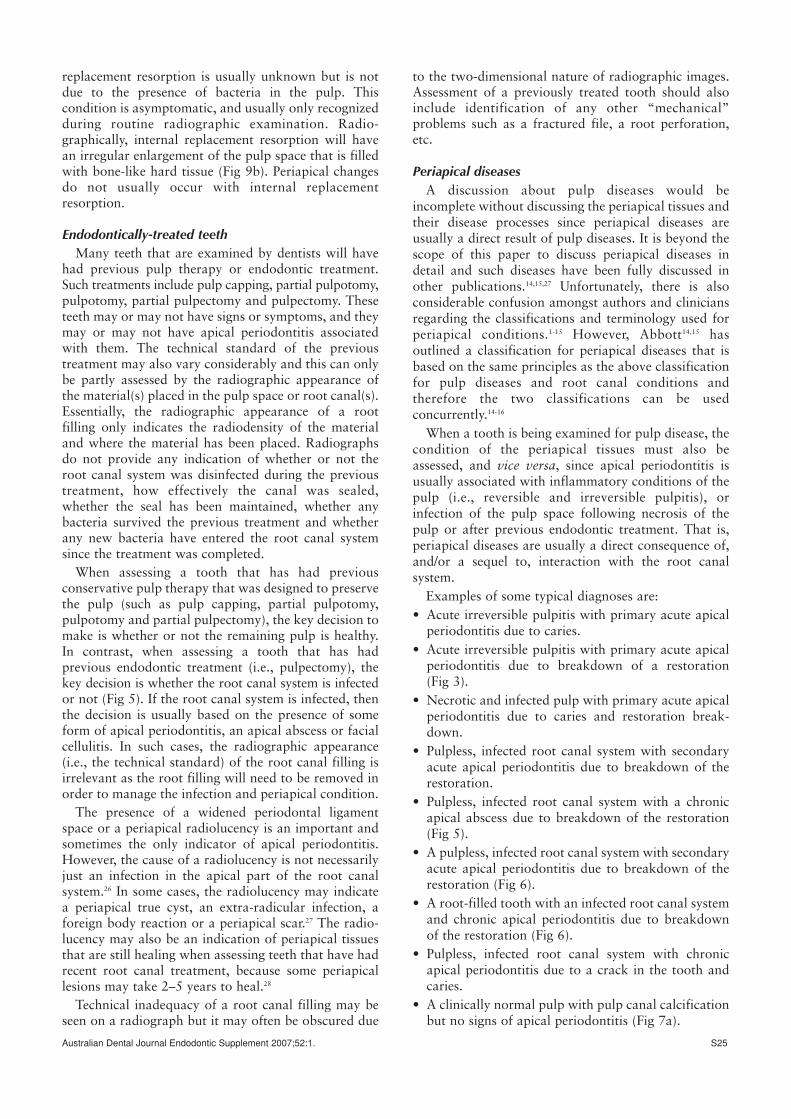
Fig 8. Tooth 36 has acute irreversible pulpitis and primary acuteapical periodontitis due to a vertical crown:root fracture and anextensive carious lesion. (A) The radiographic appearance also
suggests that condensing osteitis has been present for some time. (B) Some of the gingival tissue has grown into the carious cavityand this overgrowth of gingival tissue should not be mistaken for
hyperplastic pulpitis.
a
b

with an exaggerated response to cold tests. Nosignificant radiographic changes (except for the causeof the problem – for example, caries, fracturedrestoration, etc.) are evident unless there is alsoperiapical involvement presenting as a radiolucency orradiopacity.25
Sometimes, the gingival tissues may grow into thecarious lesion and this may be suggestive of a hyper-plasic pulpitis (Fig 8b). In these cases the distinctionmay be made by careful examination of the tissue mass,so as to determine if it is connected to the pulp or to thegingivae.
Internal resorptionThere are three forms of internal resorption –
surface, inflammatory and replacement. Internalsurface resorption is unlikely to ever be diagnosed sinceit is defined as minor areas of resorption of the surfaceof the root canal wall. It is unlikely to be noticedradiographically and there will be no clinical signs orsymptoms. No treatment is required, which is fortunatesince it cannot be clinically or radiographicallydiagnosed.
Internal inflammatory resorption can occur at anypoint along the length of the pulp space – that is,coronally within the pulp chamber or within the root
canal. It is believed to occur in teeth with necrobiosisand it is probably due to a metaplastic change oractivation of dentinoclasts within the inflamed pulptissue that is in contact with the coronal pulp which isnecrotic and infected. This condition is usuallyasymptomatic and often only recognized during aroutine radiographic examination. If symptoms are pre-sent, they are usually indicative of acute apicalperiodontitis due to an infected canal, or pain whenperforation of the crown occurs and the metaplastictissue is exposed to oral fluid and bacteria. Initially,part of the pulp (i.e., the apical portion) is alive and stillcontains nerve fibres so a response to pulp sensibilitytesting is possible. However, as the lesion progresses,the entire pulp becomes necrotic and infected, and overtime the canal will become pulpless and infected –hence there will be no response to pulp sensibility test-ing. Radiographically, internal inflammatory resorptionwill have an oval-shaped increase in the size of part ofthe root canal system. Periapical changes may be notedafter the canal has become infected and apicalperiodontitis develops (Fig 9a).
Internal replacement resorption is an uncommoncondition that occurs when the pulp undergoesmetaplastic changes and the dentine is resorbed andreplaced by bone-like hard tissue. The cause of internal
S24 Australian Dental Journal Endodontic Supplement 2007;52:1.
Fig 9. (A) Tooth 11 has an arrested lesion of internal inflammatory resorption. Originally, there would have been pulp necrobiosis thatstimulated the resorption but now the tooth has a pulpless, infected root canal system and chronic apical periodontitis, as evidenced by noresponses to thermal and electric pulp sensibility testing, a very slight widening of the apical periodontal ligament space and a “different”
sensation on percussion. (B) Tooth 25 has internal replacement resorption. The pulp has been replaced by bone-like hard tissue. The periradicular tissues are normal.
a
b
Australian Dental Journal Endodontic Supplement 2007;52:1. S25
replacement resorption is usually unknown but is notdue to the presence of bacteria in the pulp. Thiscondition is asymptomatic, and usually only recognizedduring routine radiographic examination. Radio-graphically, internal replacement resorption will havean irregular enlargement of the pulp space that is filledwith bone-like hard tissue (Fig 9b). Periapical changesdo not usually occur with internal replacementresorption.
Endodontically-treated teethMany teeth that are examined by dentists will have
had previous pulp therapy or endodontic treatment.Such treatments include pulp capping, partial pulpotomy,pulpotomy, partial pulpectomy and pulpectomy. Theseteeth may or may not have signs or symptoms, and theymay or may not have apical periodontitis associatedwith them. The technical standard of the previoustreatment may also vary considerably and this can onlybe partly assessed by the radiographic appearance ofthe material(s) placed in the pulp space or root canal(s).Essentially, the radiographic appearance of a rootfilling only indicates the radiodensity of the materialand where the material has been placed. Radiographsdo not provide any indication of whether or not theroot canal system was disinfected during the previoustreatment, how effectively the canal was sealed,whether the seal has been maintained, whether anybacteria survived the previous treatment and whetherany new bacteria have entered the root canal systemsince the treatment was completed.
When assessing a tooth that has had previousconservative pulp therapy that was designed to preservethe pulp (such as pulp capping, partial pulpotomy,pulpotomy and partial pulpectomy), the key decision tomake is whether or not the remaining pulp is healthy.In contrast, when assessing a tooth that has hadprevious endodontic treatment (i.e., pulpectomy), thekey decision is whether the root canal system is infectedor not (Fig 5). If the root canal system is infected, thenthe decision is usually based on the presence of someform of apical periodontitis, an apical abscess or facialcellulitis. In such cases, the radiographic appearance(i.e., the technical standard) of the root canal filling isirrelevant as the root filling will need to be removed inorder to manage the infection and periapical condition.
The presence of a widened periodontal ligamentspace or a periapical radiolucency is an important andsometimes the only indicator of apical periodontitis.However, the cause of a radiolucency is not necessarilyjust an infection in the apical part of the root canalsystem.26 In some cases, the radiolucency may indicatea periapical true cyst, an extra-radicular infection, aforeign body reaction or a periapical scar.27 The radio-lucency may also be an indication of periapical tissuesthat are still healing when assessing teeth that have hadrecent root canal treatment, because some periapicallesions may take 2–5 years to heal.28
Technical inadequacy of a root canal filling may beseen on a radiograph but it may often be obscured due
to the two-dimensional nature of radiographic images.Assessment of a previously treated tooth should alsoinclude identification of any other “mechanical”problems such as a fractured file, a root perforation,etc.
Periapical diseasesA discussion about pulp diseases would be
incomplete without discussing the periapical tissues andtheir disease processes since periapical diseases areusually a direct result of pulp diseases. It is beyond thescope of this paper to discuss periapical diseases indetail and such diseases have been fully discussed inother publications.14,15,27 Unfortunately, there is alsoconsiderable confusion amongst authors and cliniciansregarding the classifications and terminology used forperiapical conditions.1-15 However, Abbott14,15 has outlined a classification for periapical diseases that isbased on the same principles as the above classificationfor pulp diseases and root canal conditions andtherefore the two classifications can be usedconcurrently.14-16
When a tooth is being examined for pulp disease, thecondition of the periapical tissues must also beassessed, and vice versa, since apical periodontitis isusually associated with inflammatory conditions of thepulp (i.e., reversible and irreversible pulpitis), orinfection of the pulp space following necrosis of thepulp or after previous endodontic treatment. That is,periapical diseases are usually a direct consequence of,and/or a sequel to, interaction with the root canalsystem.
Examples of some typical diagnoses are:• Acute irreversible pulpitis with primary acute apical
periodontitis due to caries.• Acute irreversible pulpitis with primary acute apical
periodontitis due to breakdown of a restoration (Fig 3).
• Necrotic and infected pulp with primary acute apicalperiodontitis due to caries and restoration break-down.
• Pulpless, infected root canal system with secondaryacute apical periodontitis due to breakdown of therestoration.
• Pulpless, infected root canal system with a chronicapical abscess due to breakdown of the restoration(Fig 5).
• A pulpless, infected root canal system with secondaryacute apical periodontitis due to breakdown of therestoration (Fig 6).
• A root-filled tooth with an infected root canal systemand chronic apical periodontitis due to breakdownof the restoration (Fig 6).
• Pulpless, infected root canal system with chronicapical periodontitis due to a crack in the tooth andcaries.
• A clinically normal pulp with pulp canal calcificationbut no signs of apical periodontitis (Fig 7a).
Examination and diagnostic procedures to assess thestatus of the pulp and root canals
The importance of gathering all the relevantinformation for making a correct diagnosis cannot beover-emphasized. An accurate diagnosis is imperative inall cases so appropriate treatment can be provided in atimely manner.
A current medical and dental health history isimportant, not only for preventing health problemsduring treatment but also to help reach a thoroughdiagnosis.29 For example, pain medication taken within6–12 hours prior to examination may alter theresponses to pulp sensibility tests or other clinical tests.One tablet of pain medication may be sufficient toreduce the pulp or periapical inflammation, or theanalgesic may alter the patient’s perception of pain bylowering the pain threshold.30
Whilst taking the dental history, the clinician shouldbe formulating a provisional diagnosis in his/her mind.The provisional diagnosis should be based on thepatient’s chief complaint, their description of thesymptoms and a thorough history of any relevantproblems and any prior dental treatment (Table 3). Theprovisional diagnosis should be made prior toexamining the patient and his/her mouth and teeth. Thesubsequent clinical and radiographic examinationstogether with the appropriate clinical tests are used toconfirm (or change) the provisional diagnosis. Extra-oral, intra-oral soft tissue and dentition examinationsshould be performed to establish the patient’s overalloral health. Clinical tests include pulp sensibility testsand peri-radicular tests (e.g., percussion, palpation,mobility) that are aimed to reproduce the patient’ssymptoms. Some periradicular tests (e.g., periodontalprobing, mobility) also help to assess the integrity ofthe attachment apparatus of the tooth and the extent ofinflammation of the periodontal ligament resultingfrom periodontal disease and pulp inflammation. Pulp
sensibility tests measure the ability of the pulp’s nervefibres to respond to a stimulus. Among the testsavailable clinically, the cold pulp sensibility test is themost useful test and the dry ice (carbon dioxide) test isthe most reliable and reproducible of the various coldtests available commercially.31 “Cold sprays” and icehave been shown to be only about 40–60 per centaccurate; therefore they should be considered asunreliable31 and hence they are not recommended foruse. Severe and prolonged response to a cold stimulusis indicative of irreversible pulpitis, whereas noresponse could indicate pulp necrosis, a pulp confinedin an obliterated canal, a previous pulpotomy or apreviously filled root canal. Heat tests are not usedroutinely unless the major symptom reported by thepatient is sensitivity to a hot stimulus. An exaggeratedand lingering response to heat testing is indicative ofirreversible pulpitis. Electric pulp tests can give falsenegative results (e.g., in some obliterated canals) orfalse positive results (e.g., with pus in canals, necro-biosis or improper technique). Hence electric pulpsensibility tests are only useful in some cases of coronalpulp canal calcification when cold tests areinconclusive, or as an adjunct during the follow-up oftrauma to the teeth. When selecting the appropriateclinical test, one has to remember that no single test issufficient to make a firm diagnosis of reversible orirreversible pulpitis and therefore multiple tests arerequired. Periodontal examination of the suspect teethis essential in all cases because periodontal diseases canmimic endodontic problems and they are often inter-related. Periodontal probing depths also help todetermine the prognosis of the tooth. Transilluminationof the tooth is a particularly useful examinationprocedure that can be used to readily detect cracks inteeth (Fig 2), and it should be incorporated into theroutine examination of all teeth with pulp or periapicaldiseases, especially following trauma (Table 4).
S26 Australian Dental Journal Endodontic Supplement 2007;52:1.
Table 3. Summary of the examination and diagnostic processes for the assessment of pulp, root canal andperiapical conditions14,15
Stage Procedure Outcome
History Medical historyDental historyDescription of presenting complaintDetails of previous treatment of presenting complaint ➜ Provisional diagnosis of presenting condition
Clinical examination Extra-oral signsIntra-oral signsIndividual tooth assessment ➜ Identify possible cause(s)Restoration assessment Provisional diagnosis of tooth status
Clinical tests Pulp sensibility tests ➜ Provisional diagnosis of the status of thepulp and/or root canal system
Periradicular tests ➜ Provisional diagnosis of the periapical statusRadiographic examination Periapical radiograph(s) ➜ Provisional diagnosis of the periapical status
Bitewings radiograph (if needed) ➜ Assess/identify/confirm cause(s)Correlation of all above findings Combine the history, clinical examination, clinical ➜ DEFINITIVE DIAGNOSIS
tests and radiographic examination results Pulp, root canal and periapical status+ Cause(s) of the condition(s)
Management plan Investigation of tooth (i.e. remove restoration, ➜ Confirm the definitive diagnosis and cause(s)caries, cracks, etc)
Reassess amount of tooth structure and ➜ Finalise the management plan and continueoverall prognosis treatment

Australian Dental Journal Endodontic Supplement 2007;52:1. S27
In the above discussion, the term “pulp sensibilitytest” has been used. It should be noted by cliniciansthat the older, and sometimes more commonly usedterm “vitality tests” is no longer recognized as beingappropriate since thermal and electric tests do not testthe “vitality” of the tooth or the pulp. The term“vitality” refers to the presence of blood supply to thetissues whereas the term “sensibility” is defined as theability to respond to a stimulus. Hence, “sensibility” isthe appropriate term to use since thermal and electrictests are assessing whether the nerve fibres within thepulp are able to respond to the hot, cold or electricalstimulus. If the pulp’s nerve fibres do respond, then anassumption is made that there must be a viable bloodsupply to keep the nerve fibres alive and functioning,and therefore the rest of the pulp tissues and cells areprobably also alive and functioning. However, thesetests do not necessarily indicate the degree of health ordisease within the pulp. It is also important toremember that the nerve fibres are usually the last partof the pulp to undergo necrosis, especially in the apicalpart of the canal, and therefore a “false positive”response may be obtained from a pulp that is essentiallynecrotic.
The main functions of pulp sensibility tests are to,firstly, determine whether there is a nerve response and,secondly, to assess the nature of the response. If there isa response, then the pulp may be alive, whereas noresponse may indicate necrosis, pulp canal calcification,or a tooth that has had a pulpotomy or a root canalfilling, as discussed above. This is important when theprovisional diagnosis was pulp necrosis or a pulplesscanal as here the clinician is searching for the tooth thatdoes not respond. However, if the provisional diagnosiswas pulpitis (either reversible or irreversible), then theclinician is searching for the tooth that has anexaggerated response which replicates the painexperienced by the patient – that is, the degree ofsensitivity of the pulp (note: sensitivity is defined as an
exaggerated response and is different to sensibility).The severity can also help to distinguish betweenreversible and irreversible pulpitis but the time that thepain lingers after removing the stimulus is usually ofmore value to distinguish between these twoconditions. In this latter scenario of testing for pulpitis,the pulp sensibility tests could be considered as“sensitivity tests” as they are assessing which toothresponds more severely than the others.
Pulp sensibility tests cannot be interpreted withoutalso viewing an accurate and current periapicalradiograph of the tooth in question. The results of pulpsensibility tests should correlate with the radiographicappearance of the tooth and the periapical tissuesbefore making a definitive diagnosis. As an example, atooth that does not respond to a pulp sensibility testcould have a necrotic pulp (with or without infection),a pulpless, infected root canal system, or pulp canalcalcification. It may also have had a root canal filling,a pulpotomy or a partial pulpectomy, all of which mayor may not be infected. All of these conditions can onlybe fully assessed by viewing a periapical radiographand therefore the pulp test result could be misinter-preted if a radiograph is not viewed. The radiographalso needs to be a current radiograph since pulp andperiapical diseases are progressive in nature and willchange over time. Hence, an accurate diagnosis of thecurrent condition cannot be based on a radiographtaken several weeks, months or years earlier, and some-times even one taken just several days earlier.
Radiography may arguably be the most reliable of allthe diagnostic tests although it may not help in theassessment of teeth with pulpitis. However, radiographswill always provide valuable information to assist withdiagnosis and treatment.32,33 A routine radiograph maysometimes be the first indication of the presence ofpathosis, such as internal root resorption or chronicapical periodontitis. The cause of the pulp disease (deepcaries, deep restorations, open margins, etc.) may also
Table 4. Summary of the examination procedures that should be performed as part of any routine dentalexamination and diagnosis but particularly whenever pulp and/or periapical pathosis is suspected16
General medical history As required for all dental proceduresPresenting complaint Long and short term history, past and current symptoms, past and recent treatment,
medications being used (prescribed and self administered)Description of PAIN: Location, onset, nature, duration, stimuli, relief, referred
Clinical examination Puckering, indentation, draining sinus, facial asymmetry, caries, periodontal diseases,restorations, restoration margins, etc
Clinical tests Periradicular tests Percussion, palpation, mobility, periodontal probingClinical tests Pulp sensibility tests Cold (CO2 dry ice at -78ºC)
Electric (in some cases e.g. when pulp canal calcification)Heat (if required, if patient complains of sensitivity to heat)
Radiographic examination Periapical radiographs, tube shifts, bitewings, occlusal, panoramicOther tests Biting on individual cusps (e.g. with a ‘Tooth Slooth’)
Transillumination of the tooth with a fibre optic lightAnaesthetic (e.g. block, infiltration, intra-periodontal ligament, etc.)
Investigation Remove all restorations, caries, cracksTransillumination of the cavity, cusps, marginal ridges, etcAssess whether, and how, the tooth can be restored againAssess the need for any other treatment (e.g. periodontal)Assess the long term prognosis of the tooth (consider endodontic, periodontal and restorative
aspects)
be evident on radiographs. A widened periodontalligament space or a periapical radiolucency that cannotbe “moved” with tube-shift radiography may bediagnostic for periapical pathosis. However, radio-graphy is not a perfect diagnostic tool. Soft tissuediseases of the pulp are not visible on radiographs; onlyhard tissue changes can be seen on radiographs.Radiographs only provide a two-dimensionalrepresentation of three-dimensional structures.Periapical lesions may not be directly evident on theradiographs and their real extent and the spatialrelationships to anatomical structures may not bereadily visualized. Periapical lesions also take sometime (2–10 months) to develop to a size which will beevident on a radiograph.24
A pretreatment diagnosis is made after interpretationof all the collected information. This diagnosis should
include an assessment of the pulp or root canalcondition, the periapical status and the cause of thedisease(s).14,15 Ideally, there should be at least twodifferent signs and/or symptoms present to indicate andconfirm the disease(s).16 Only at this stage can themanagement of the patient’s problem(s) be planned.
It is not possible to diagnose the histological status ofthe pulp from the clinical signs and symptoms.34
Diagnosis of the level of pulp inflammation is difficultbut it is relatively easy to determine from clinical findingsthat a pulp is necrotic or the canal is pulpless. Severityand duration of pain appears to be related to the statusof the pulp. Severe lingering pulp pain usually indicatesthe presence of acute irreversible pulpitis with anincrease in pulp tissue pressure. When mild to moderatepain of very short duration is present with no previoushistory of pain in the tooth, reversible pulpitis may be
S28 Australian Dental Journal Endodontic Supplement 2007;52:1.
Table 5. Summary of endodontic treatment strategies for the various pulp and root canal conditions. (Note – recommendations for endodontic treatment and re-treatment are dependent on the tooth havingsufficient tooth structure remaining to enable it to be adequately restored again)Clinically normal pulp NIL treatment required
Dentine hypersensitivity Occlude the exposed dentine (e.g. with potassium oxalate), and/or hyperpolarize theintradental nerves (e.g. with potassium nitrate)
Reversible pulpitis Acute Conservative pulp therapy then reassess healing responseChronic Conservative pulp therapy then reassess healing response
Irreversible pulpitis Acute Routine endodontic treatment with anti-inflammatory medications then reassesshealing response, or extraction
Chronic Routine endodontic treatment with anti-inflammatory medications then reassesshealing response, or extraction
Necrobiosis Routine endodontic treatment with anti-inflammatory and anti-microbialmedications then reassess healing response, or extraction
Pulp necrosis No infection No treatment required but monitor and reassess periapical tissues for signs of periapicalinflammation (which would indicate an infected root canal)
Infected Routine endodontic treatment with anti-inflammatory and anti-microbialmedications then reassess healing response, or extraction
Pulpless, infected root canals Routine endodontic treatment with anti-inflammatory and anti-microbialmedications then reassess healing response, or extraction
Degenerative changes
Atrophy NIL treatment requiredPulpal canal calcification Partial NIL treatment required unless canal infected with apical periodontitis ➜ then do
endodontic treatment and reassess healing responseTotal NIL treatment required unless canal infected with apical periodontitis ➜ then do
endodontic treatment and reassess healing responseHyperplasia Routine endodontic treatment with anti-inflammatory medications then reassess
healing response, or extractionInternal root resorption Inflammatory Routine endodontic treatment with anti-inflammatory medications then reassess
healing response, or extractionReplacement Extraction (eventually) – can monitor initially
Previous root canal treatment
No sign of infection No treatment but monitor and reassess unless restoration being replaced ➜ then retreatand reassess healing response
Infected Orthograde endodontic re-treatment with anti-inflammatory and anti-microbialmedications then reassess the need for periapical surgery, or extraction, if the canal isstill infected or if there are signs of on-going periapical pathosis
Technical standard Inadequate No treatment but monitor unless the restoration is being replaced or the canal isinfected ➜ then retreat and reassess healing response
Adequate No treatment but monitor unless the restoration is being replaced or the canal isinfected ➜ then retreat and reassess healing response
Others Perforation Orthograde endodontic re-treatment and repair the perforation from an internalapproach if possible; then reassess the need for periapical or periodontal surgery, orextraction, if there are signs of on-going periapical and/or periodontal pathosis
Missed canal Orthograde endodontic re-treatment then reassess the need for periapical surgery, orextraction, if the canal is still infected or if there are signs of on-going periapicalpathosis
occurring. If the tooth is associated with a previoushistory of pain, irreversible pulpitis is more likely.35
Sometimes the clinical and radiographic examinationsare inconclusive yet the patient has pain. However, it isimportant not to initiate irreversible treatment withouta definite diagnosis. In these cases, the correctprocedure is to make a provisional diagnosis. Referralto an appropriate specialist should be considered insuch cases, although if not feasible then, guided mainlyby the radiographic findings, such as the deepestrestoration in the suspect quadrant or caries close to thepulp, the likely tooth can be identified and treated withzinc oxide-eugenol cement that has an excellent localanaesthetic and sedative effect. If necessary, one toothat a time is treated this way until a definitive diagnosisis established. It is most likely that only one tooth isresponsible for acute pain. However, if there is verylittle evidence that provides definitive information, theneither specialist referral or a conservative “wait andsee” approach may be necessary in order to allow thevague symptoms to localize to the specific tooth. It isimportant to understand, and remember, that pulp andperiapical diseases are progressive and therefore thesymptoms will vary over time, which usually assists thelocalization and the diagnosis of pain.
Complete patient care must include a discussion ofthe diagnosis with the patient. It is essential to disclosethe examination findings and for the patient to under-stand his/her presenting condition(s). Details of therecommended treatment, the prognosis and thealternatives to treatment should be discussed as well asthe consequences if no treatment is performed.
Management of pulp and root canal conditionsThe ultimate decision the practitioner must make is
whether to treat or not to treat the tooth, and if treat-ment is indicated, whether to treat the pulp or the rootcanal system. The alternative management is to extractthe tooth and then to consider a prosthesis to replace it.Accurate diagnosis and identification of the cause(s) ofthe problem(s) will lead to effective management of theoffending tooth. Table 5 summarizes the treatmentstrategies for the management of the various pulpdiseases.
Management strategies vary considerably for thevarious pulp conditions which emphasizes the need foran accurate diagnosis before considering any treatment.Conditions such as a pulp with atrophy or pulp canalcalcification do not require any treatment (unless thepulp has become necrotic and the canal has beeninfected). A conservative approach should be adoptedwhen dealing with conditions such as pulp necrosiswithout infection since pulp tests are not entirelyreliable and the periapical tissues only become inflamedonce the canal is infected, rather than as a response tothe necrosis itself. In cases of pulpitis, the key tofavourable treatment outcomes is to accurately diagnosethe status of the pulp and this means deciding whetherthe inflammation is reversible (in which case it may be
treated conservatively) or irreversible when the pulp (orthe tooth) must be removed.
The first principle of managing any diseases is toremove the cause of the problem.36 Since the presence ofmicro-organisms is the main cause of pulp diseases, anessential part of the diagnosis and assessment of thetooth is to identify how the bacteria have entered thetooth and the pulp space. In the case of pulp diseases,the cause is often a defective restoration, caries and/orcracks that have allowed bacteria to enter the rootcanal system. In recent years, Abbott36 has advocatedthat all restorations should be removed prior toendodontic treatment in order to remove the causes ofthe pulp and periapical diseases. A clinical study hasshown that signs of bacterial entry via restorationbreakdown, caries or cracks were found in only 40 percent of restored teeth with pulp and periapical diseasewhen they were examined prior to restoration removalbut these pathways for bacterial entry weresubsequently found in 99 per cent of the teeth followingremoval of the restorations.36
Removal of all the restorations, caries, cracks andany other factors that may be causing the pulp andperiapical diseases provides the clinician with an idealopportunity to fully assess whether the tooth is suitablefor further restoration following endodontic or otherpulp therapy. One of the key factors affecting thetreatment outcome and the long-term survival ofendodontically-treated teeth is the quality and longevityof the coronal restoration since breakdown of therestoration provides the most likely pathway of entryfor bacteria to enter the tooth again in the future, whichthen leads to a new lesion of apical periodontitis.Hence, it is essential to assess the amount and qualityof remaining tooth structure prior to continuing withendodontic and restorative dental treatment. Theremoval of the restorations, caries and cracks (Fig 4)can be considered as “investigation” of the tooth36 andit should be done routinely for every tooth that requiresa restoration (Table 3).
When treating teeth with reversible pulpitis, the aimis to preserve the pulp and return it to a clinicallynormal state. Measures to minimize iatrogenic pulpinjury should be incorporated into routine restorativetechniques. Since pulp inflammation occurs withpolymicrobial infection, it is prudent to avoid thecontamination of all cavities with microbial-rich saliva.The use of rubber dam during routine restorativedentistry will limit the number of bacteria in deepcavities to only the cariogenic bacteria, which are weakpathogens to the pulp.37
Irreversible pulpitis, pulp necrobiosis and theirsequelae (such as pulp necrosis with infection, pulplessinfected canals) require root canal therapy or toothextraction. Likewise, a tooth that has had previousendodontic therapy (such as pulpectomy, pulpotomy)but has remained infected or has become infected againwill require endodontic re-treatment or extraction.When root canal treatment (or re-treatment) is the
Australian Dental Journal Endodontic Supplement 2007;52:1. S29
treatment of choice, the pulp tissue and tissue breakdown products as well as the bacteria and theirby-products should be completely eliminated from theroot canal system, which is subsequently filled ascompletely as possible. Coronal restorations shouldprevent recurrent bacterial entry during and after rootcanal treatment.38 These procedures should also beperformed under rubber dam isolation. Occlusalreduction may be performed to reduce postoperativepain in patients whose teeth initially exhibit pulpinflammation, tenderness to percussion, and pre-operative pain.39 After root canal treatment, adequatehealing is evidenced clinically by resolution of symptomsand radiographically by bone filling in the radiolucentarea at the root apex if it was present prior totreatment. Thereby, a functioning tooth is establishedwith a healthy periodontium.
Internal root resorption is rare in permanent teethand it can sometimes mimic external invasive rootresorption radiographically. Management for internalinflammatory root resorption is usually via endodontictreatment in order to remove the blood supply to theresorbing cells through the apical foramina. Severaldressings of calcium hydroxide may be required if thelesion is active, to ensure complete removal of alldentinoclasts and to encourage hard tissue repair on theexternal surface of the root if there has been aperforation. Currently there is no treatment availableto prevent the disease progression of internal replace-ment resorption. Extraction of the affected tooth is thetreatment of choice. However, if the lesion is diagnosedearly enough, endodontic treatment with calciumhydroxide dressings may be attempted.
With all pulp diseases, post-treatment follow-up isextremely important due to the diagnostic uncertaintieswith some conditions and the possible need for furthertreatment such as periapical surgery when periapicalhealing is not evident.
CONCLUSIONSIt hurts everyone involved when accessing the pulp
space of the wrong tooth, accessing a tooth when painis of non-endodontic origin, or not intervening when adiseased pulp is causing severe pain. The significance ofa thorough diagnosis cannot be overstated. Theproposed classification of pulp diseases is simple, yetpractical and uses terminology related to clinicalfindings. This classification will help clinicians under-stand the progressive nature of the pulp diseaseprocesses and will direct them to the most appropriateand conservative treatment strategy for each condition.With a comprehensive knowledge of the patho-physiology of pain and inflammation in the pulptissues, clinicians may accomplish this task withconfidence.
REFERENCES1. World Health Organization. Application of the International
Classification of Diseases to Dentistry and Stomatology. 3rd edn.Geneva: WHO, 1995.
2. Smulsen MH, Sieraski SM. Histophysiology and diseases of thedental pulp. In: Weine FS, ed. Endodontic therapy. 4th edn. St.Louis: Mosby, 1989:128-150.
3. Olgilvie AL. Pulpal pathosis. In: Ingle J, ed. Endodontics.London: Kimpton, 1965:295-345.
4. Seltzer S, Bender IB. The dental pulp. 3rd edn. Philadelphia: JBLippincott, 1984:281.
5. Cohen S. Diagnostic procedures. In: Cohen S, Burns RC, eds.Pathways of the pulp. 7th edn. St. Louis: Mosby 1998;17-19.
6. Tronstad L. Clinical endodontics. A textbook. 2nd edn. Stuttgart:Thieme, 1991;76-83.
7. Glickman GN, Mickel AK, Levin LG, Fouad AF, Johnson WT.Glossary of endodontic terms. 7th edn. Chicago: AmericanAssociation of Endodontists, 2003.
8. Pitt Ford TR. The dental pulp. In: Harty FJ, ed. Endodontics inclinical practice. 3rd edn. London: Wright Butterworth, 1990:56-57.
9. Torabinejad M. Pulp and periradicular pathosis. In: Walton RE,Torabinejad M, eds. Principles and practice of endodontics. 3rdedn. Philadelphia: WB Saunders Co., 2002:34-37.
10. Grossman LI. Endodontic Practice. 9th edn. Philadelphia: Lea &Febiger, 1978:51-75.
11. Castellucci A. Pulpal pathology. In: Castellucci A. Endodontics.Vol 1. Florence: Il Tridente, 2004:139-153.
12. Stock CJR. Patient assessment. In: Stock C, Walker R, GulabivalaK. Endodontics, 3rd edn. Edinburgh: Mosby, 2004:67-76.
13. Reit C, Petersson K, Molven O. Diagnosis of pulpal andperiapical disease. In: Bergenholtz G, Hørsted-Bindslev P, Reit C,eds. Textbook of Endodontology. Oxford: BlackwellMunksgaard, 2003:9-18.
14. Abbott PV. The periapical space – a dynamic interface. Ann RAustralas Coll Dent Surg 2000;15:223-234.
15. Abbott PV. Classification, diagnosis and clinical manifestationsof apical periodontitis. Endod Topics 2004;8:36-54.
16. Abbott P. Endodontics and dental traumatology – An overview ofmodern endodontics. Teaching manual. Perth: The University ofWestern Australia, 1999:11-15.
17. Foster E, Robinson PP, Foster E, Robinson PP. The incidence anddistribution of branched pulpal axons in the adult ferret. ArchOral Biol 1993;38:965-970.
18. Lisney SJ, Matthews B. Branched afferent nerves supplying tooth-pulp in the cat. J Physiol 1978;279:509-517.
19. Glick DH. The interpretation of pain of dental origin. Dent ClinNorth Am 1967;535-548.
20. Grossman LI, Oliet S. Diagnosis and treatment of endodonticemergencies. Chicago: Quintessence Publishing Co., 1981:25-26.
21. Möller AJ, Fabricius L, Dahlén G, Öhman AE, Heyden G.Influence on periapical tissues of indigenous oral bacteria andnecrotic pulp tissue in monkeys. Scand J Dent Res 1981;89:475-484.
22. Sundqvist G. Associations between microbial species in dentalroot canal infections. Oral Microbiol Immunol 1992;7:257-262.
23. Korzen BH, Krakow AA, Green DB. Pulpal and periapical tissueresponses in conventional and monoinfected gnotobiotic rats.Oral Surg Oral Med Oral Pathol 1974;37:783-802.
24. Jansson L, Ehnevid H, Lindskog S, Blomlöf L. Development ofperiapical lesions. Swed Dent J 1993;17:85-93.
25. Çaliskan MK. Pulpotomy of carious vital teeth with periapicalinvolvement. Int Endod J 1995;28:172-176.
26. Nair PN, Sjögren U, Figdor D, Sundqvist G. Persistent periapicalradiolucencies of root-filled human teeth, failed endodontic treat-ments, and periapical scars. Oral Surg Oral Med Oral PatholOral Radiol Endod 1999;87:617-627.
27. Nair PN. Apical periodontitis: a dynamic encounter between rootcanal infection and host response. Periodontol 20001997;13:121-148.
28. Byström A, Happonen RP, Sjögren U, Sundqvist G. Healing ofperiapical lesions of pulpless teeth after endodontic treatmentwith controlled asepsis. Endod Dent Traumatol 1987;3:58-63.
S30 Australian Dental Journal Endodontic Supplement 2007;52:1.
29. Halpern IL. Patient's medical status – a factor in dental treat-ment. Oral Surg Oral Med Oral Pathol 1975;39:216-226.
30. Modaresi J, Dianat O, Mozayeni MA, Modaresi J, Dianat O,Mozayeni MA. The efficacy comparison of ibuprofen,acetaminophen-codeine, and placebo premedication therapy onthe depth of anesthesia during treatment of inflamed teeth. OralSurg Oral Med Oral Pathol Oral Radiol Endod 2006;102:399-403.
31. Fuss Z, Trowbridge H, Bender IB, et al. Assessment of reliabilityof electrical and thermal pulp testing agents. J Endod1986;12:301-305.
32. Weerheijm KL. Occlusal 'hidden caries'. Dent Update1997;24:182-184.
33. Rohlin M, Akerblom A. Individualized periapical radiographydetermined by clinical and panoramic examination.Dentomaxillofacial Radiol 1992;21:135-141.
34. Dummer PM, Hicks R, Huws D. Clinical signs and symptoms inpulp disease. Int Endod J 1980;13:27-35.
35. Bender IB. Reversible and irreversible painful pulpitides:diagnosis and treatment. Aust Endod J 2000;26:10-14.
36. Abbott PV. Assessing restored teeth with pulp and periapicaldiseases for the presence of cracks, caries and marginal break-down. Aust Dent J 2004;49:33-39.
37. Paterson RC, Pountney SK. Pulp response to Streptococcusmutans. Oral Surg Oral Med Oral Pathol 1987;64:339-347.
38. Cox CF. Evaluation and treatment of bacterial microleakage. AmJ Dent 1994;7:293-295.
39. Rosenberg PA, Babick PJ, Schertzer L, Leung A. The effect ofocclusal reduction on pain after endodontic instrumentation. JEndod 1998;24:492-496.
Address for correspondence/reprints: Professor Paul Abbott
School of DentistryThe University of Western Australia
17 Monash AveNedlands, Western Australia 6009Email: [email protected]
Australian Dental Journal Endodontic Supplement 2007;52:1. S31
