arpitha · arpitha parthasarathy, phd ... director research-translational and molecular biology...
TRANSCRIPT

10/1/2014
1
About OMICS GroupAbout OMICS GroupAbout OMICS GroupAbout OMICS Group
OMICS Group International is an amalgamation of Open Accesspublications and worldwide international science conferences and events.Established in the year 2007 with the sole aim of making the information onSciences and technology ‘Open Access’, OMICS Group publishes 400 onlineopen access scholarly journals in all aspects of Science, Engineering,Management and Technology journals. OMICS Group has been instrumentalin taking the knowledge on Science & technology to the doorsteps ofordinary men and women. Research Scholars, Students, Libraries,Educational Institutions, Research centers and the industry are mainstakeholders that benefitted greatly from this knowledge dissemination.OMICS Group also organizes 300 International conferences annually acrossthe globe, where knowledge transfer takes place through debates, roundtable discussions, poster presentations, workshops, symposia andexhibitions.
About OMICS Group ConferencesAbout OMICS Group ConferencesAbout OMICS Group ConferencesAbout OMICS Group Conferences
OMICS Group International is a pioneer and leading science eventorganizer, which publishes around 400 open access journals andconducts over 300 Medical, Clinical, Engineering, Life Sciences,Phrama scientific conferences all over the globe annually with thesupport of more than 1000 scientific associations and 30,000 editorialboard members and 3.5 million followers to its credit.
OMICS Group has organized 500 conferences, workshops and nationalsymposiums across the major cities including San Francisco, Las Vegas,San Antonio, Omaha, Orlando, Raleigh, Santa Clara, Chicago,Philadelphia, Baltimore, United Kingdom, Valencia, Dubai, Beijing,Hyderabad, Bengaluru and Mumbai.
A Coordinating Epithelial Cell Proliferation and
Migration in Corneal Wound Healing
ARPITHA PARTHASARATHY, PhDJerome Canady Research Institute for Advanced Biological and Technological Sciences
Plasma Medicine Life Sciences
Director Research-Translational and Molecular Biology
Takoma Park, MD
10/1/2014
2
Introduction
Migration of Epithelial a cells as a sheet
Stem Cells of the basal limbus migrate along the
epithelial sheet
Cdk5- Cyclin Dependent Kinase 5, a neuronal
protein involved in cell-cell adhesion
Does Epithelial Stem cells require CDK5 for cell
adhesion and migration?
A phase contrast image of 3-day limbal explant culture, showing small outgrowth
of rounded and flattened cells on the dish and clusters of pigmented cells (P)
migrating from the limbal explant along the cut edge.
Location of BrdU LRCs in cryosections of cultured limbal explant, which were pulse
labeled with BrdU for 5 days, followed by 21-day chase.
Epithelial stem cell migration
Distribution of LRCs in the outgrowth of limbal explant cultures.
Limbal explant cultures were pulse labeled with BrdU for 5 days, followed by 21-day chase
Epithelial cells in the outgrowth showing BrdU-positive (green) cells in 2 clusters
(arrowheads) of small cells and a few labeled large cells (arrow).
STEM CELLS IN THE TRANSPLANTABLE EPITHELIAL CULTURES

10/1/2014
3
Distribution of epithelial cells pulse labeled with BrdU on the limbal explant cultures. Limbal explant cultures were pulse labeled with BrdU for 5
days, cryosectioned, and then processed for immunostaining for BrdU. Confocal overlay images of BrdU (green) and PI (red) along with
transmitted light image are shown. Note the presence of 2–3 layers of epithelial cells on the stroma of limbal explant cultures, with
several BrdU-positive cells, which appeared yellow because of merging of green and red. Arrowheads indicate BrdU-positive pigmented cells in
the basal layer of cultured limbal explant; arrows indicate BrdU-positive limbal suprabasal cells. F, fibroblast;
St, PI-stained stromal cells.
STEM CELLS ON Explant after 21 Days
Buccal Epithelial Stem Cells Holoclones transplanted on to Human Eye
Cell-Cell Adhesion Complexes
p120
E-Cadherin
/ N-Cadherin
Rac
Rho
CDK5

10/1/2014
4
Cdk5-a serine/threonine kinase
Y15
p35 (or p39)
Cdk5
Phosphorylation
at Y15 increases
activity
Cdk5-a serine/threonine kinase
IB:
IP: E-Cad
98kDa38kDa
E-Cadherin
Red
Overlay
Cdk5(pY15)
Green
Cdk5(pY15)
Green +Olomoucin
E-cadherin and Cdk5 (pY15) co-localize at cell-cell adhesions
Interaction of E-cadherin and CDK5
ShCdk5
A B
Generation of Cdk5 Deficient Human Corneal Epithelial Cell Line
� Cdk5-lentiviral titers on the HCLE cells were selected using Blasticidin and
tested for the suppression of Cdk5
� Cdk5 was suppressed by 90% in the stable ShCdk5 cells
ShCdk5 Stable Cell line showing loss of Cdk5 expression
IB:
Cdk5
Tubulin
ShCdk5HCLE
Cdk5-green

10/1/2014
5
Aggregation of Human Corneal Epithelial
Cells is Dependent on the Presence of Cdk5
Absence of Cdk5 inhibits
junction formation in human corneal epithelial cells
HCLEHCLEHCLEHCLE
With CalciumWithout Calcium
HCLE
HCLE +
Olomoucine
ShCdk5
NN1003
Lens epithelial cells
NN1003+
Olomoucine
0
2
4
6
8
10
12
14
16
18
20
HCLE
Olo
mouci
ne
ShC
dk5
NN10
03
NN10
03+O
lom
oucin
e
Rati
o o
f P
art
icle
Siz
e i
n µ
m²
(Ca²
+/C
a²- )
Differential Activity of Cdk5
�Cdk5 is required for cell-cell junctions in the E-cadherin expressing cells
�Cdk5 is not required for cell-cell junctions in N-cadherin expressing
cells
MDA-MB-231 cells
olomoucineolomoucineolomoucine
GFP-N-Cad
transfectedUntransfected GFP only
GFP-E-Cad
transfected
Part
icle
siz
e i
n µ
m2
Ca
2+/C
a2-
0
2
4
6
olomoucineolomoucineolomoucine
olomoucine
Active Rho
Tubulin
Rho and Rac Activity in the Cdk5 Deficient Cells
Increased Rho and decreased Rac activity in the absence of Cdk5 is consistent
with destabilization of junctions in E-cadherin containing HCLE cells
Actin
Total Rac
Active Rac
Total Rho
0
2
4
6
Active Rho / Total Rho
0
0.5
1
1.5
2
Active Rac / Total Rac

10/1/2014
6
With Calcium
C3 transferase
Rho Inhibitor- C3 transferase
CDK5 Inhibitor-Olomoucine
C3 transferase
+Olomoucine
Rati
o o
f P
art
icle
Siz
e i
n µ
m2
(Ca
2+/C
a2- )
Increase in Rho activity caused by inhibiting
Cdk5 is responsible for the decrease in cell-
cell adhesion
Regulation of Cdk5 at Cell-Cell adhesion
Requires Rho Signaling
Without Calcium
HCLE
Olomoucine
Absence of Cdk5 exhibits weak cell-cell junctions due to Increased association of
IQGAP-1 with E-cadherin (ββββ-catenin) complex
Interaction of E-cadherin with IQGAP-1 and Cdk5
IQGAP-1
� a substrate for Rac-1
�binds to the E-cadherin junctional complex through ββββ- catenin and inhibits cell-cell adhesion
IB:IQGAP1
IB:E-Cadherin
IP: E Cadherin
0.1
0.3
0.5
0.7
0.9
1.1
1.3
1.5
HCLE ShCdk5
Am
ou
nt
of
IQG
AP
1/E
-Cad
he
rin
Cdk5 deficient cells-
� are capable of forming junctions containing E-cadherin and p120
� have increased endocytosis
� have p120 at junctions
E-Cadherin and p120 in the ShCdk5 Cells
Localization of E-Cadherin is significantly
reduced at the cell- cell borders of ShCdk5
cells
ShCdk5 0.347 ±±±± 0.145 0.576 ±±±± 0.027
HCLE 0.535 ±±±± 0.142 0.516 ±±±± 0.0291
Ratio of cell border to total intensity
E-Cadherin p120
HCLE ShCDK5
E-CadherinE-Cadherin
p120p120
0
10
20
30
40
50
60
70
80
Total
Surface Biotinylation of E-cadherin
% B
ioti
nyla
ted
E-c
ad
heri
n
10/1/2014
7
HCLE
ShCdk5
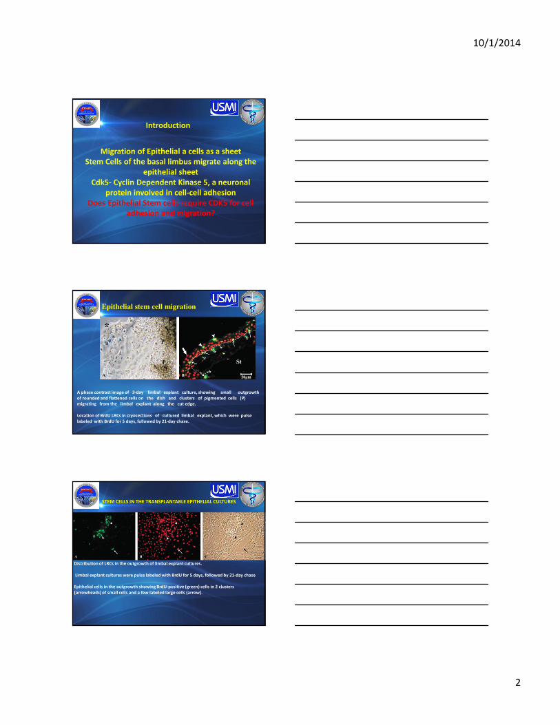
Total Internal Reflection Fluorescence (TIRF) Microscopic
Analysis
Of E-Cadherin Transfected ShCdk5 Cells
Long PathsParticle tracking of border
localized E-cadherin
showing long straight
paths indicating
internalization
Olomoucine
Total Internal Reflection Fluorescence (TIRF) Microscopic
Analysis of p120 Transfected ShCdk5Cells
HCLE ShCdk5
GFP-P120 appears punctate; individual particles may represent junctional
complexes or vesicles containing p120
TIRF Analysis of E-Cadherin and p120 at the
Cell-Cell Junctions
The length and tortuosity of paths by E-Cadherin and p120-containing particles (n>100) were tracked
by total internal reflectance fluorescence (TIRF) microscopy
E-Cadherin-containing particles in ShCdk5 cells moved fast and these long, straight paths
were internalized
p120-containing particles in ShCdk5 cells moved slowly and stayed
near the cell-cell boundary
Cdk5 stabilizes the junction by preventing E-Cadherin-containing
vesicles from being endocytosed
0
10
20
30
40
50
60
70
80
90
100
E-Cadherin p120
Distance ≥ 3mm; Tortuosity ≤ 1.5
HCLE ShCdk5 Olomoucine
Fast-moving particles traverse away from cell-cell boundaries
% o
f p
art
icle
s c
on
tain
ing
ju
ncti
on
al
pro
tein
10/1/2014
8
N-Cadherin Expression in Cdk5 Deficient Stable Cells
Inhibition of Cdk5 leads to internalization of E-cadherin and induction of
N-cadherin
p-120 at the cell-cell adhesions in the absence Cdk5 is associated with
N-cadherin
IP: N-CADHERIN
IB: p120
IB: N-Cadherin
IB- N-cadherin
Actin
Summary - Corneal Epithelial Cell-Cell Junctions are
stabilized by CDK5
In the absence of Cdk5 activity-
�p120 dissociates from E-cadherin
�E-cadherin trafficking is altered to a site 20µm or more
away from the surface suggesting increased degradation
of E-cadherin
+Cdk5
E-cadherin
p120
¯
Dissociation of p120
from E-cadherin
Increase in Rho and
Decrease in Rac E- Cadherin Endocytosed /
induction of N-cadherin
Cdk5
+ Cdk5
Cdk5 is required for –
� E-cadherin based cell-cell junction stabilization� regulation of Rho and Rac activity
About UsUS Medical Innovations, LLC (USMI), a subsidiary of US Patent Innovations (USPI), is a privately owned, FDA registered biomedical device company focused on the research, development and manufacturing of advanced, innovative and affordable plasma electrosurgical devices. USMI products are designed for ambulatory and inpatient endoscopy centers as well as for complex surgical operations.
MissionUSMI is dedicated to innovation in the fields of electrosurgery and plasma technology for the advancement of patient outcomes, the eradication of cancer and the improvement of human lives. Our core values of innovation, diversity, and community are key to our pursuit of this mission and to our ongoing success.
Company Overview
Confidential 24
10/1/2014
9
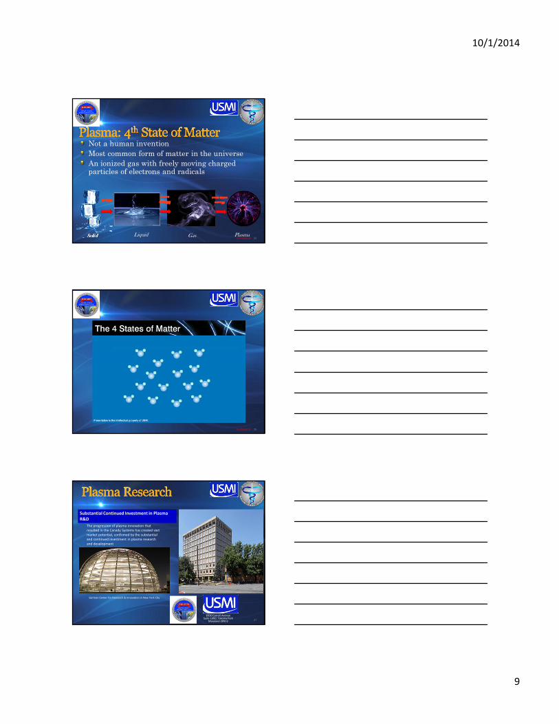
Not a human invention
Most common form of matter in the universe
An ionized gas with freely moving charged particles of electrons and radicals
PlasmaSolid GasLiquid
eeeeven more ven more ven more ven more
energyenergyenergyenergy
more more more more
energyenergyenergyenergyenergyenergyenergyenergy
Confidential 25
Confidential 26
27
Substantial Continued Investment in Plasma
R&D
German Center for Research & Innovation in New York City
The progression of plasma innovation that
resulted in the Canady Systems has created vast
market potential, confirmed by the substantial
and continued investment in plasma research
and development
6930 Carroll AvenueSuite 1000, Takoma Park
Maryland 20912
10/1/2014
10
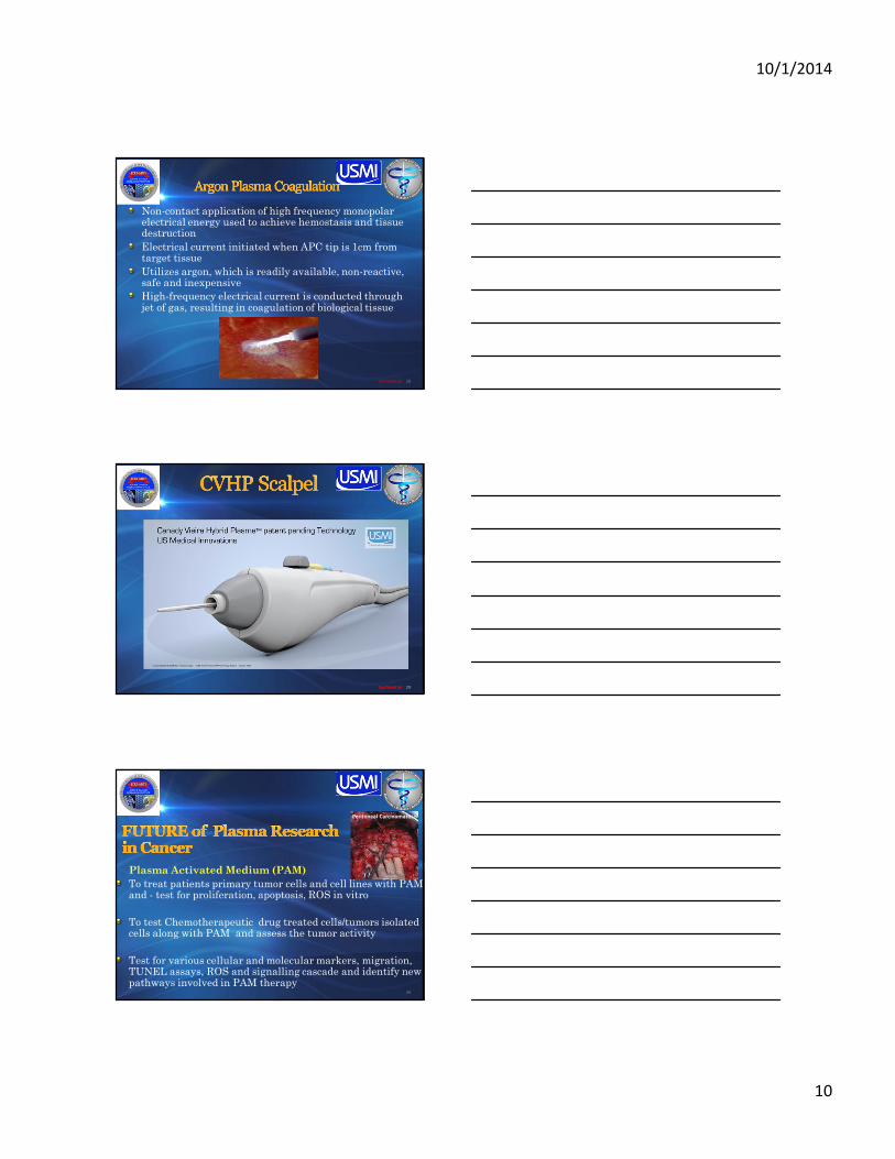
Non-contact application of high frequency monopolar electrical energy used to achieve hemostasis and tissue destruction
Electrical current initiated when APC tip is 1cm from target tissue
Utilizes argon, which is readily available, non-reactive, safe and inexpensive
High-frequency electrical current is conducted through jet of gas, resulting in coagulation of biological tissue
Confidential 28
Confidential 29Confidential 29
Plasma Activated Medium (PAM)
To treat patients primary tumor cells and cell lines with PAM and - test for proliferation, apoptosis, ROS in vitro
To test Chemotherapeutic drug treated cells/tumors isolated cells along with PAM and assess the tumor activity
Test for various cellular and molecular markers, migration, TUNEL assays, ROS and signalling cascade and identify new pathways involved in PAM therapy
30
Peritoneal Carcinomatosis

10/1/2014
11
Let Us Meet AgainLet Us Meet AgainLet Us Meet AgainLet Us Meet Again
We welcome you all to our future conferences of OMICS Group
International
Please Visit:
www.omicsgroup.com
www.conferenceseries.comhttp://ophthalmology.conferenceseries.com/