applying the paris system for reporting urinary cytology · applying the paris system for reporting...
TRANSCRIPT

Applying the Paris system for reporting Urinary Cytology
Eva M. Wojcik, MD
Chair and Professor of Pathology and Urology
Loyola University, Chicago, USA
And
Ashish Chandra
Lead Cytopathologist and Uropathologist
Guy’s ant St Thomas Hospitals
London, UK

Outlines
• What is the goal of urine cytology?
• Why to standardize, why Paris?
• What is the guiding principle?
• What are diagnostic categories?
• What are the criteria?
• What adjuvant studies?
• What are future clinical and research needs?
The main purpose of urine cytology
To detect bladder cancer
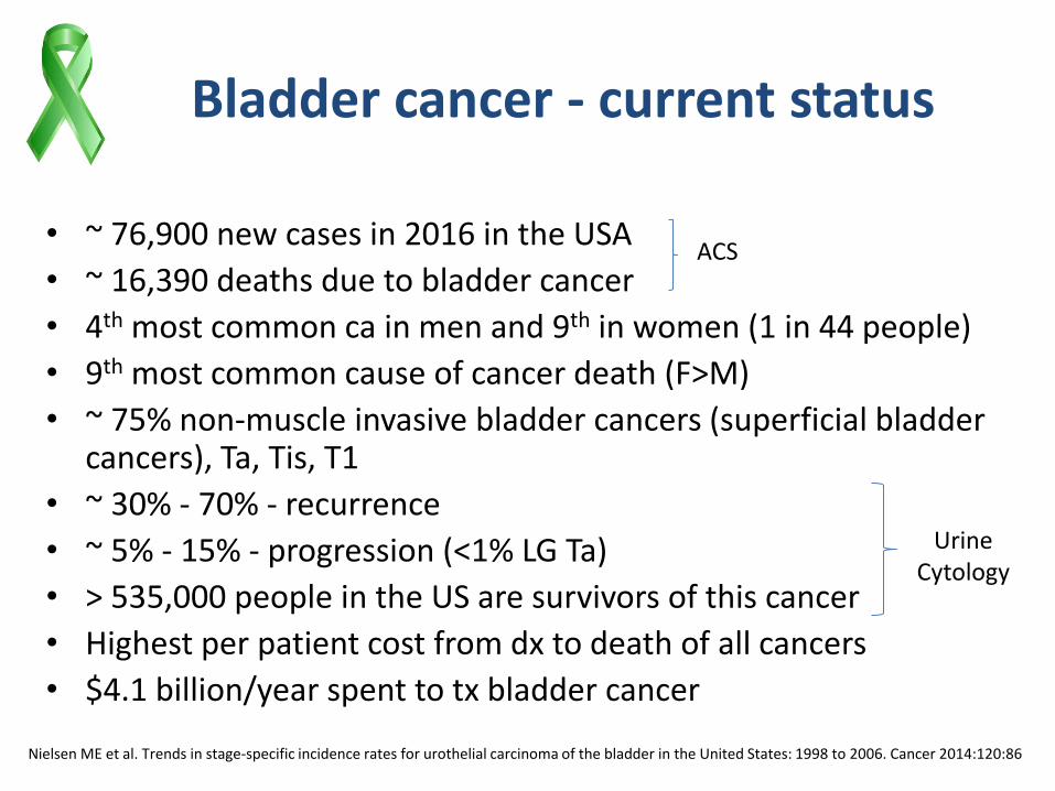
Bladder cancer - current status
• ~ 76,900 new cases in 2016 in the USA
• ~ 16,390 deaths due to bladder cancer
• 4th most common ca in men and 9th in women (1 in 44 people)
• 9th most common cause of cancer death (F>M)
• ~ 75% non-muscle invasive bladder cancers (superficial bladder cancers), Ta, Tis, T1
• ~ 30% - 70% - recurrence
• ~ 5% - 15% - progression (<1% LG Ta)
• > 535,000 people in the US are survivors of this cancer
• Highest per patient cost from dx to death of all cancers
• $4.1 billion/year spent to tx bladder cancer
Urine Cytology
Nielsen ME et al. Trends in stage-specific incidence rates for urothelial carcinoma of the bladder in the United States: 1998 to 2006. Cancer 2014:120:86
ACS
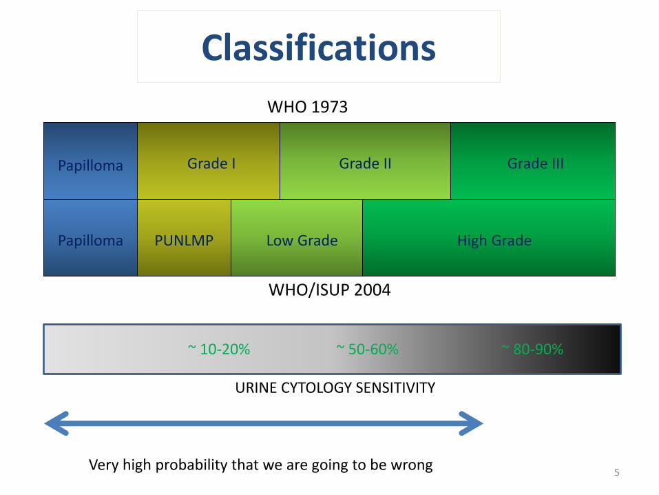
Classifications
WHO 1973
WHO/ISUP 2004
Papilloma
Papilloma
Grade I Grade III Grade II
Low Grade High Grade PUNLMP
~ 80-90% ~ 10-20% ~ 50-60%
URINE CYTOLOGY SENSITIVITY
5 Very high probability that we are going to be wrong

• Reproducibility
• Improvement of communication
• Atypical cells
– Wide intraobserver variability
• Nationally rates of atypical vary among institutions
– Range from 2% to 30% (51% atypical + suspicious)
Why to standardize reporting of urinary cytology?

Where did we start?
• 18th International Congress of Cytology, Paris, May, 2013
– “Paris Group” – all participants of two Urine Cytology Symposia
– Outline of the Paris System for Reporting Urinary Cytopathology
– Ultimate goal – detection of HGUC
• Sponsorship by the ASC and IAC
• Contract with Springer
• Numerous face-to-face meetings
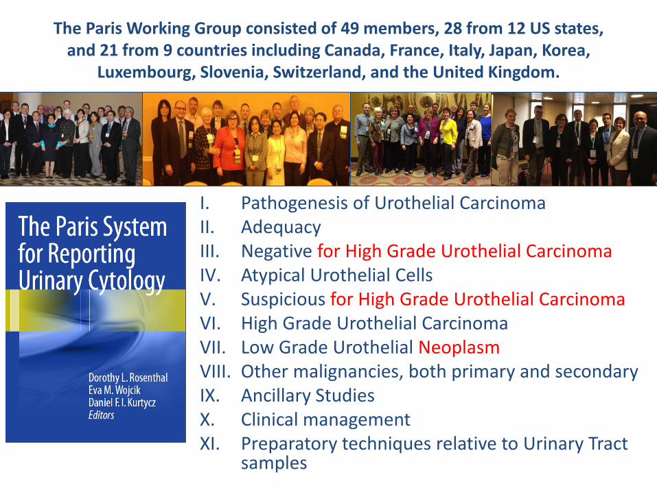
I. Pathogenesis of Urothelial Carcinoma II. Adequacy III. Negative for High Grade Urothelial Carcinoma IV. Atypical Urothelial Cells V. Suspicious for High Grade Urothelial Carcinoma VI. High Grade Urothelial Carcinoma VII. Low Grade Urothelial Neoplasm VIII. Other malignancies, both primary and secondary IX. Ancillary Studies X. Clinical management XI. Preparatory techniques relative to Urinary Tract
samples
The Paris Working Group consisted of 49 members, 28 from 12 US states, and 21 from 9 countries including Canada, France, Italy, Japan, Korea,
Luxembourg, Slovenia, Switzerland, and the United Kingdom.

System has to be build based on:
• Consensus • Evidence • Inclusion • Acceptance • Understanding
Urothelial Carcinoma

Normal Urothelium
Hyperplasia Dysplasia
Low Grade Carcinoma High Grade Carcinoma Carcinoma in situ
Invasive Carcinoma
9p-, 9q- p16
Genetically Stable FGFR3 (~85%)
Genetically Unstable p53 (~60%)
<10%
Recurrence Recurrence
RAS (?)
Pathogenesis of Urothelial Carcinoma
Eva M. Wojcik and Stefan E. Pambuccian
Normal Urothelium
Hyperplasia Dysplasia
High Grade Carcinoma Carcinoma in situ
Invasive Carcinoma
Papillary Pathway
80%
Non-Papillary Pathway
20% 9p-, 9q-
p16
Genetically Unstable p53 (~60%)
<10%
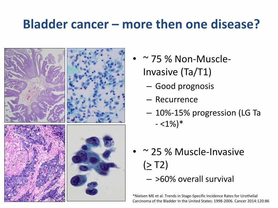
Bladder cancer – more then one disease?
• ~ 75 % Non-Muscle-Invasive (Ta/T1)
– Good prognosis
– Recurrence
– 10%-15% progression (LG Ta - <1%)*
• ~ 25 % Muscle-Invasive (> T2)
– >60% overall survival
*Nielsen ME et al. Trends in Stage-Specific Incidence Rates for Urothelial Carcinoma of the Bladder In the United States: 1998-2006. Cancer 2014:120:86

“Approximately 80% (of Ta bladder tumors) appear to follow a benign course without developing invasive tumors or dying of bladder cancer”

Question…. “Carcinoma”?
GU GI

CARCINOMA
ADENOMA
Question…. “Carcinoma”?

Mr. Smith - You have a bladder cancer
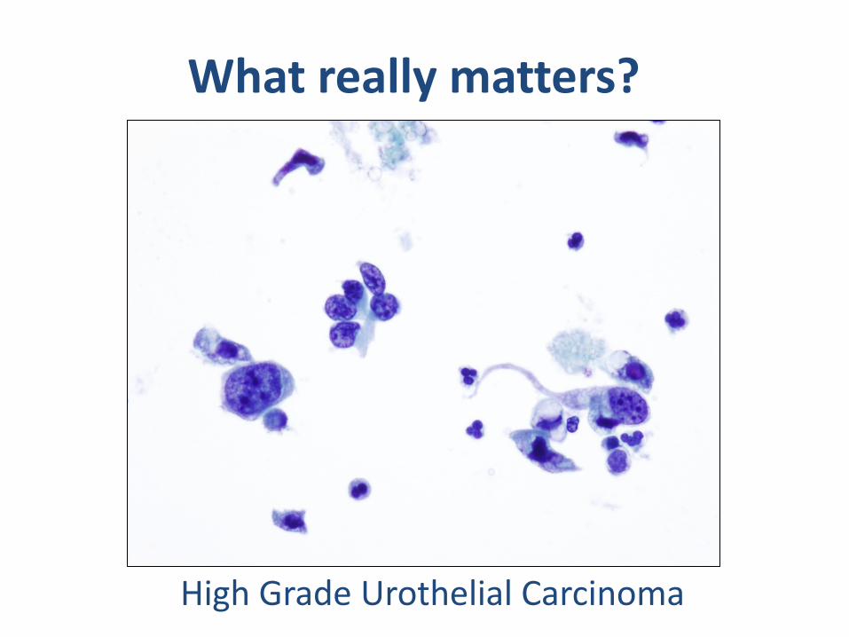
What really matters?
High Grade Urothelial Carcinoma

Diagnostic Categories
Positive Negative Atypical/Suspicious
HGUC Everything else
Hope
Reality
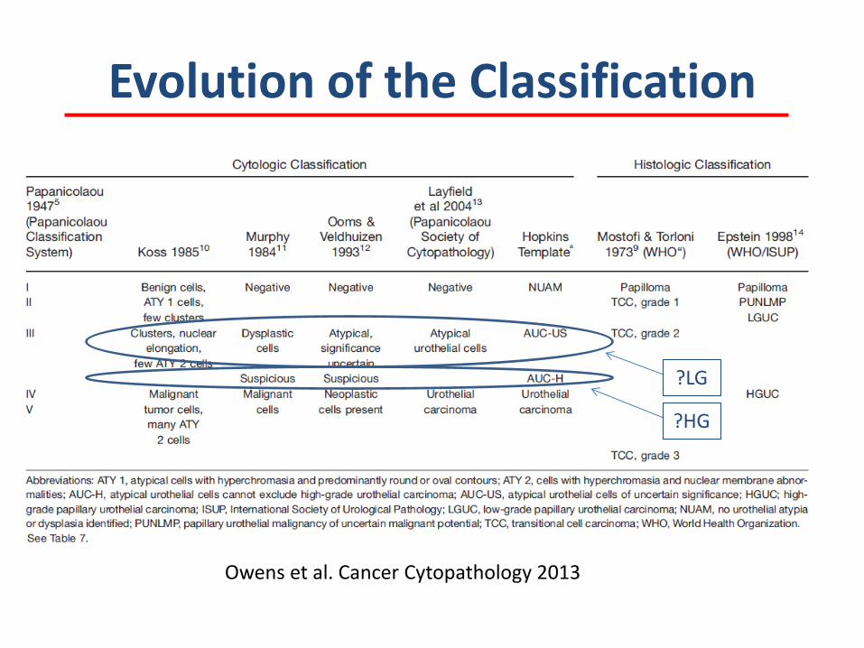
Evolution of the Classification
Owens et al. Cancer Cytopathology 2013
?LG
?HG
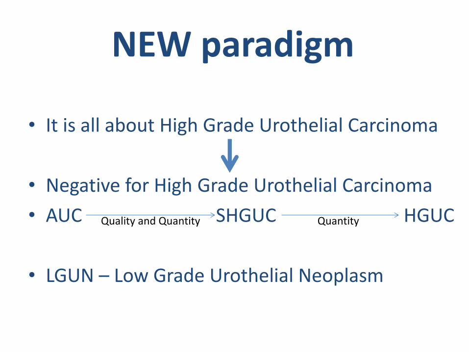
NEW paradigm
• It is all about High Grade Urothelial Carcinoma
• Negative for High Grade Urothelial Carcinoma
• AUC SHGUC HGUC
• LGUN – Low Grade Urothelial Neoplasm
Quality and Quantity Quantity
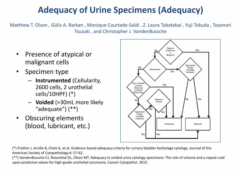
Adequacy of Urine Specimens (Adequacy)
Matthew T. Olson , Güliz A. Barkan , Monique Courtade-Saïdi , Z. Laura Tabatabai , Yuji Tokuda , Toyonori Tsuzuki , and Christopher J. VandenBussche
• Presence of atypical or malignant cells
• Specimen type – Instrumented (Cellularity,
2600 cells, 2 urothelial cells/10HPF) (*)
– Voided (>30mL more likely “adequate”) (**)
• Obscuring elements (blood, lubricant, etc.)
(*) Prather J, Arville B, Chatt G, et al. Evidence-based adequacy criteria for urinary bladder barbotage cytology. Journal of the American Society of Cytopathology.4: 57-62. (**) VandenBussche CJ, Rosenthal DL, Olson MT. Adequacy in voided urine cytology specimens: The role of volume and a repeat void upon predictive values for high-grade urothelial carcinoma. Cancer Cytopathol. 2015.
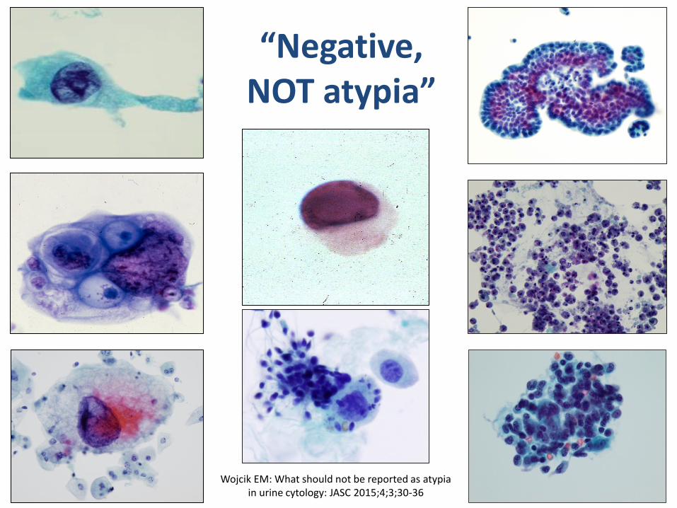
“Negative, NOT atypia”
Wojcik EM: What should not be reported as atypia in urine cytology: JASC 2015;4;3;30-36

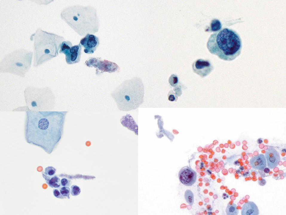
Negative for High-Grade Urothelial Carcinoma (Negative) Dorothy L. Rosenthal, Michael B. Cohen, Hui Guan, Christopher L. Owens, Yuji Tokuda, and Eva M. Wojcik
Definition of Negative for High-Grade Urothelial Carcinoma
• A sample of urine, either voided or instrumented, may be considered benign, i.e., NHGUC, if any of the following components are present in the specimen:
– Benign urothelial, glandular, and squamous cells
– Benign urothelial tissue fragments (BUTF) and urothelial sheets or clusters
– Changes associated with lithiasis
– Viral cytopathic effect; polyoma virus (BK virus—decoy cells)
– Post-therapy effect, including epithelial cells from urinary diversions

Benign Superficial (Umbrella) Urothelial Cells

“Atypical” Umbrella Cells
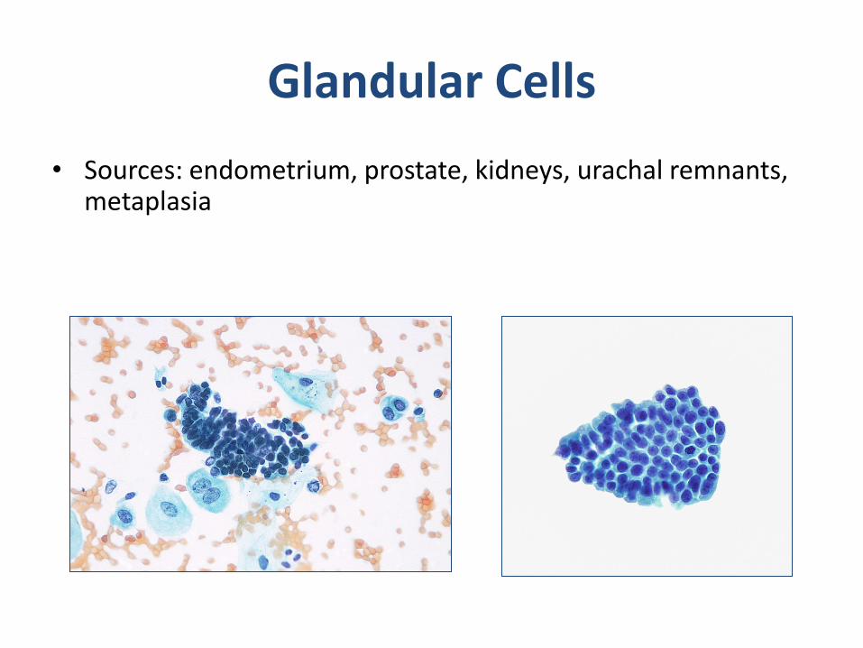
Glandular Cells
• Sources: endometrium, prostate, kidneys, urachal remnants, metaplasia
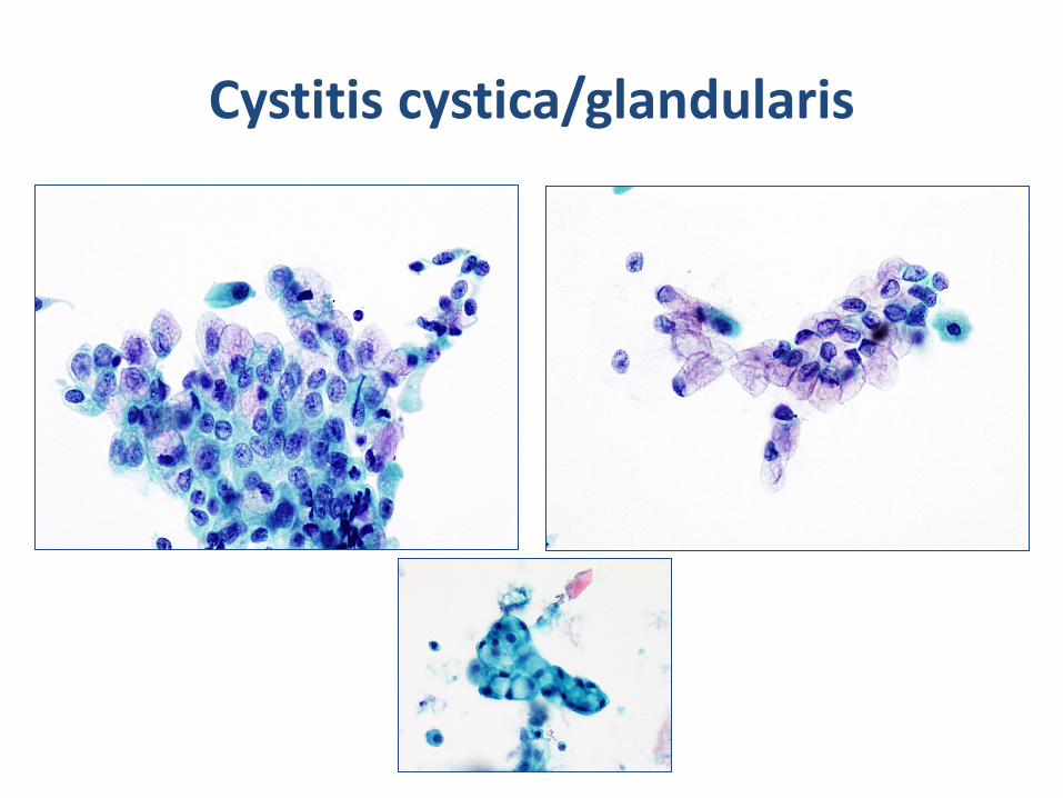
Cystitis cystica/glandularis
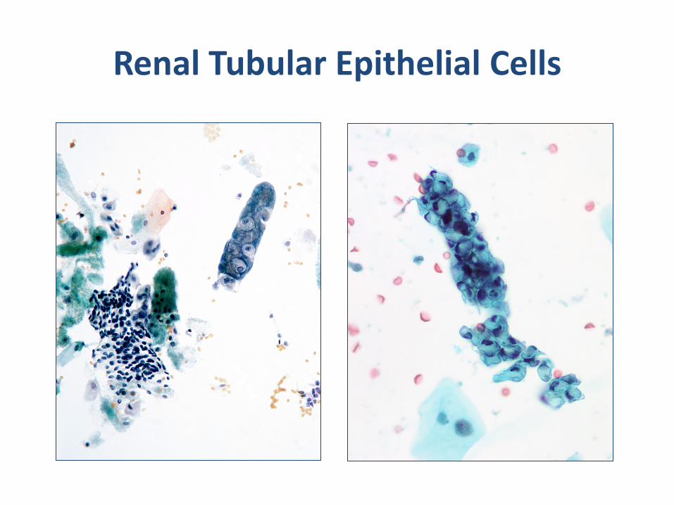
Renal Tubular Epithelial Cells

Benign Urothelial Tissue Fragments - BUTF
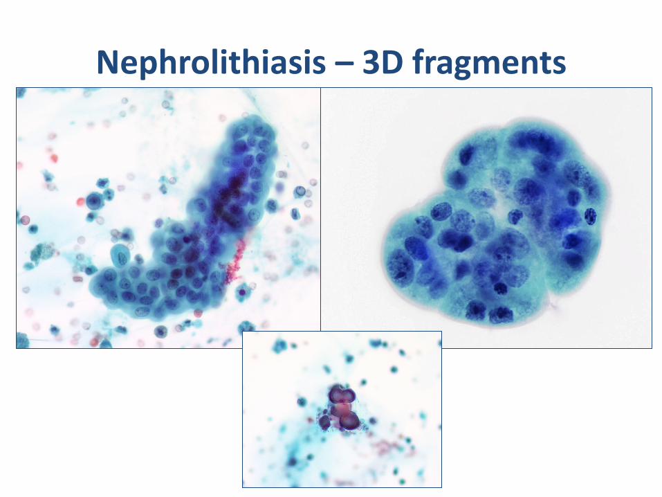
Nephrolithiasis – 3D fragments

Viral Cytopathic Effects
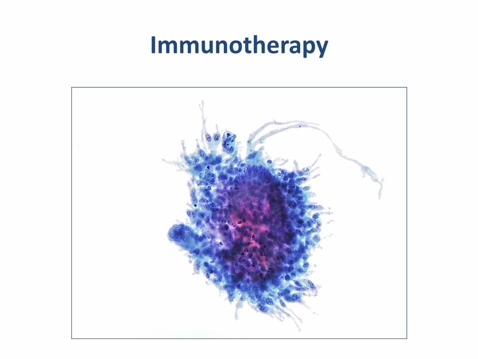
Immunotherapy

Seminal Vesicle Cells

Bladder Diversion Urine
Melamed – Wolinska body
Negative - Summary
• Negative for High Grade Urothelial Carcinoma
– This diagnostic category will include cases where “low grade urothelial carcinoma can not be excluded”
• If there is a cause for “atypia” i.e. urolithiasis, treatment related changes etc. – it is negative!
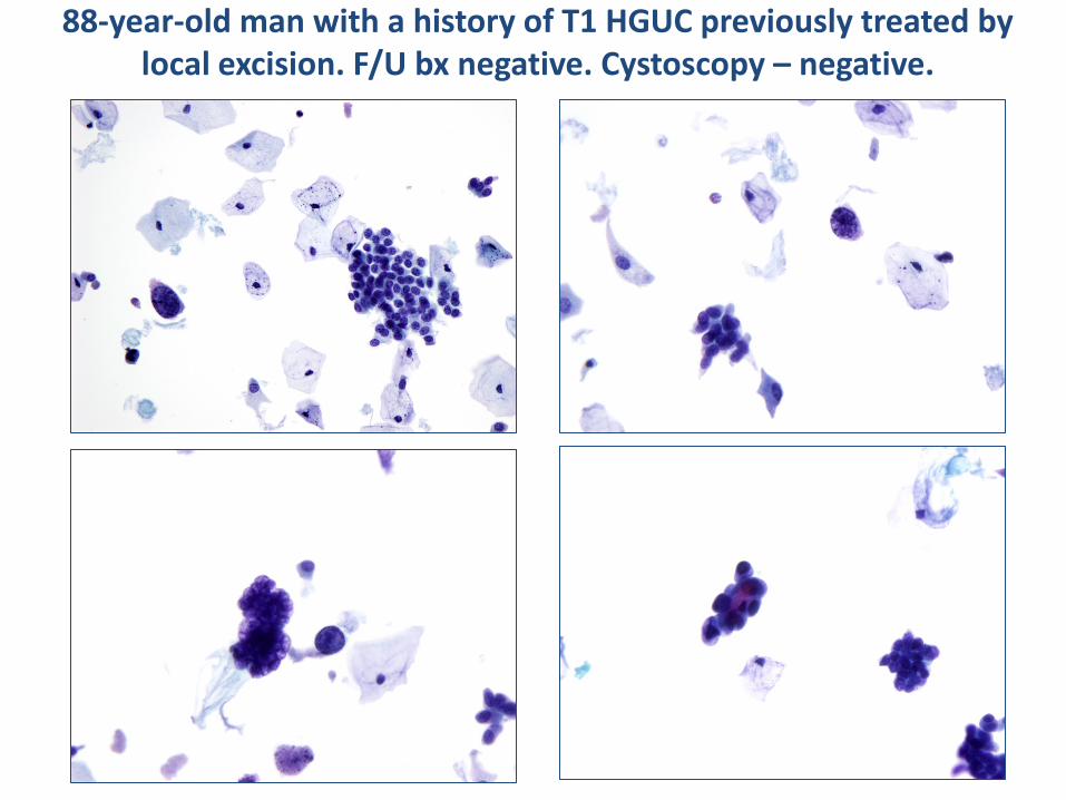
88-year-old man with a history of T1 HGUC previously treated by local excision. F/U bx negative. Cystoscopy – negative.
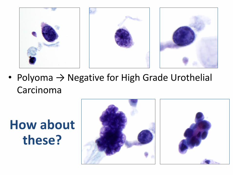
• Polyoma → Negative for High Grade Urothelial Carcinoma
How about these?

Positive Suspicious Atypical Negative
What is Atypia
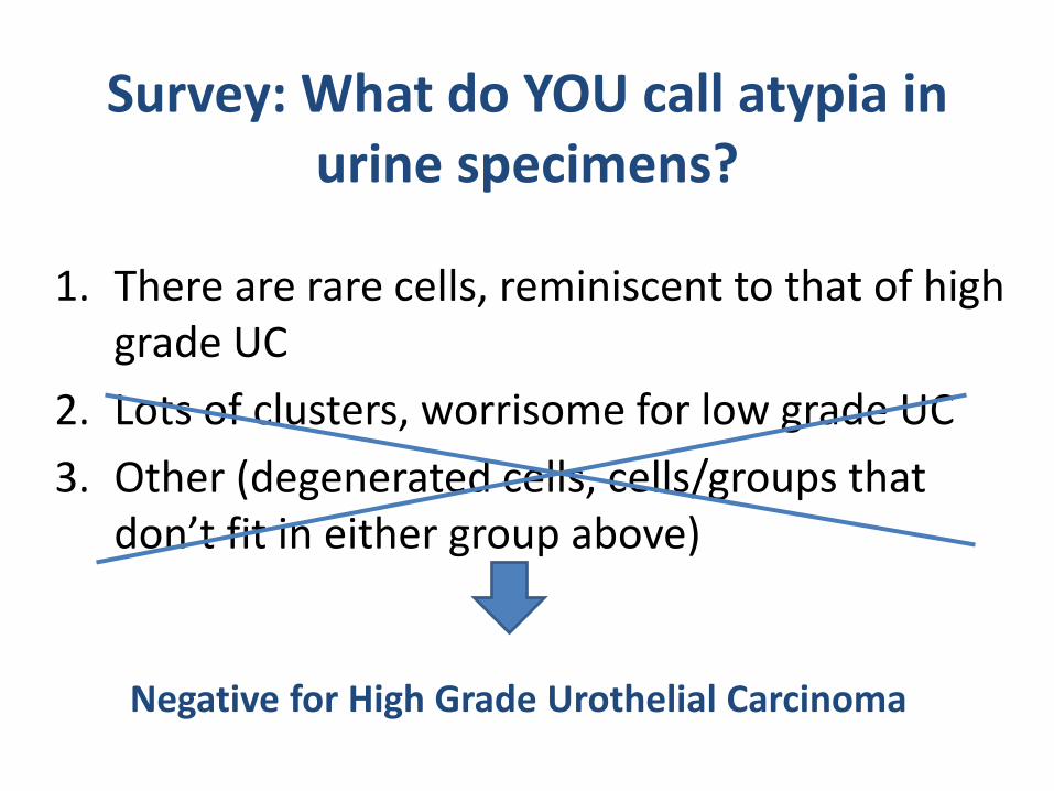
Survey: What do YOU call atypia in urine specimens?
1. There are rare cells, reminiscent to that of high grade UC
2. Lots of clusters, worrisome for low grade UC
3. Other (degenerated cells, cells/groups that don’t fit in either group above)
Negative for High Grade Urothelial Carcinoma
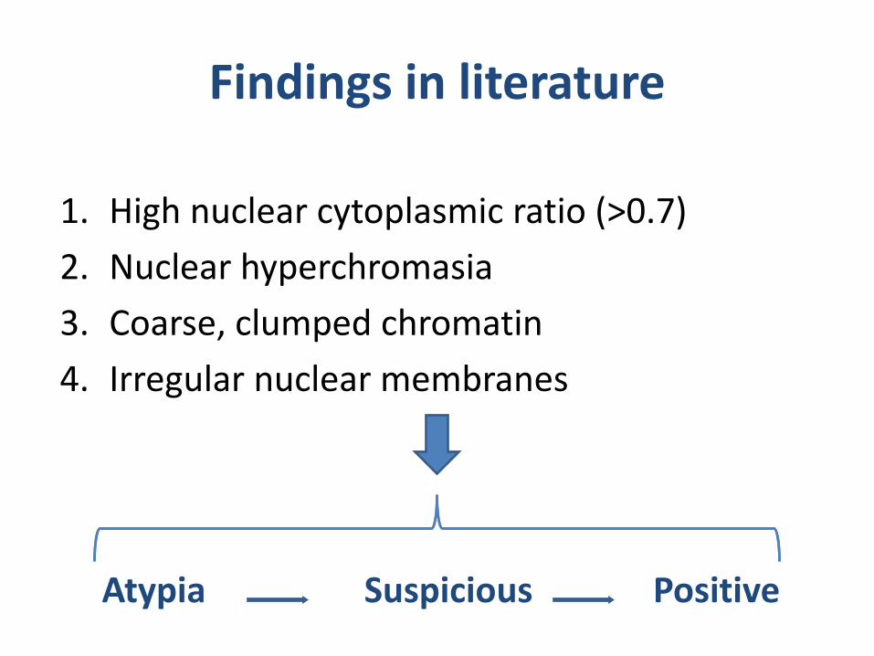
Findings in literature
1. High nuclear cytoplasmic ratio (>0.7)
2. Nuclear hyperchromasia
3. Coarse, clumped chromatin
4. Irregular nuclear membranes
Atypia Suspicious Positive
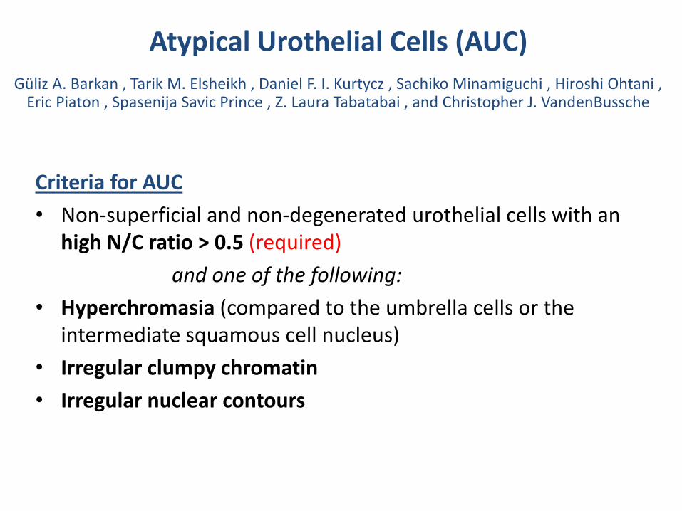
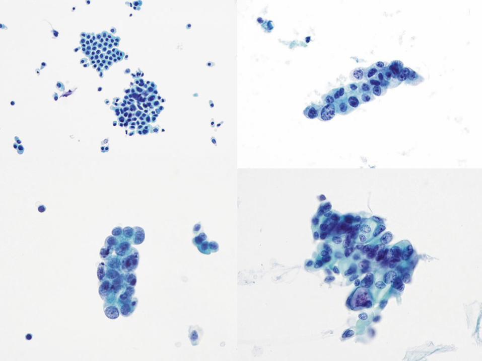
Criteria for AUC
• Non-superficial and non-degenerated urothelial cells with an high N/C ratio > 0.5 (required)
and one of the following:
• Hyperchromasia (compared to the umbrella cells or the intermediate squamous cell nucleus)
• Irregular clumpy chromatin
• Irregular nuclear contours
Atypical Urothelial Cells (AUC)
Güliz A. Barkan , Tarik M. Elsheikh , Daniel F. I. Kurtycz , Sachiko Minamiguchi , Hiroshi Ohtani , Eric Piaton , Spasenija Savic Prince , Z. Laura Tabatabai , and Christopher J. VandenBussche
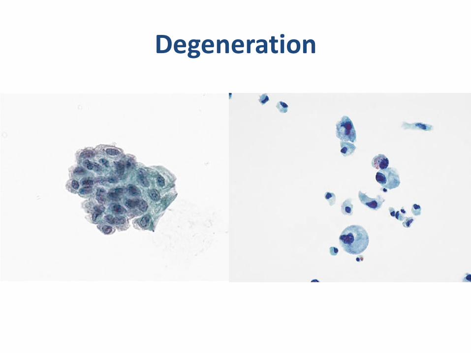
Degeneration
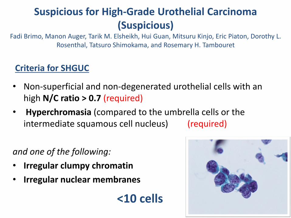
Suspicious for High-Grade Urothelial Carcinoma (Suspicious)
Fadi Brimo, Manon Auger, Tarik M. Elsheikh, Hui Guan, Mitsuru Kinjo, Eric Piaton, Dorothy L. Rosenthal, Tatsuro Shimokama, and Rosemary H. Tambouret
• Non-superficial and non-degenerated urothelial cells with an high N/C ratio > 0.7 (required)
• Hyperchromasia (compared to the umbrella cells or the intermediate squamous cell nucleus) (required)
and one of the following:
• Irregular clumpy chromatin
• Irregular nuclear membranes
<10 cells
Criteria for SHGUC


Suspicious for HGUC vs. Positive HGUC Quantity matters..
“The number of atypical urothelial cells is an important criterion to classify urine cytology specimens into the ‘positive’ or the ‘suspicious’ categories. A cut-off number of >10 cells to render a definitive diagnosis of HGUCA seems valid from the clinical standpoint .”
5 – 10 cells – gray zone, based on experience, history, individual threshold, etc
JASC 2015;4(4)232–238

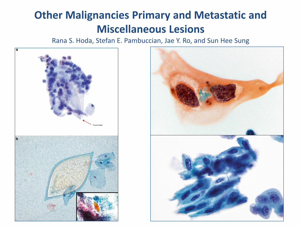
Other Malignancies Primary and Metastatic and Miscellaneous Lesions
Rana S. Hoda, Stefan E. Pambuccian, Jae Y. Ro, and Sun Hee Sung

Melanoma
ADC
Lymphoma
Clear cell adc bladder

Ancillary Studies in Urinary Cytology Lukas Bubendorf, Nancy P. Caraway, Andrew H. Fischer, Ruth L. Katz, Matthew T. Olson,
Fernando Schmitt, Margareta Strojan Fležar, Theodorus H. Van Der Kwast, Philippe Vielh

Nucle
ar
/ cyto
logic
aty
pia
Probability of high grade UC
low moderate/high certain
AUC/SHGUC
8%-30%
HGUC NFHG

Cytopreparatory Techniques Gary W. Gill, William N. Crabtree, and Deidra P. Kelly
• No generally accepted best materials and methods of collecting and processing urine to detect urothelial malignancies
How are UT specimens processed in your laboratory? n = 739
(Multiple responses allowed) No. %
ThinPrep 424 57.4
Cytospin 336 45.5
Cell block 202 27.3
Conventional smear 69 9.3
SurePath 49 6.6
Filter preparation 16 2.2
Other 11 1.5
2014 Supplemental Questionnaire of the College of American Pathologists (CAP) Cytopathology Interlaboratory Comparison Program (CICP), Barkan et al.
Clinical Management Marcus L. Quek, Trinity J. Bivalacqua, Ashish M. Kamat, and Mark P. Schoenberg
• From the standpoint of the urologist, the workup for AUC should be individualized based on the risk assessment of the patient
• From a practical standpoint, the clinical management of “suspicious for HGUC” is similar to a “positive for HGUC” diagnosis
• Transurethral resection establishes the histologic diagnosis and is therapeutic for most solitary low grade tumors
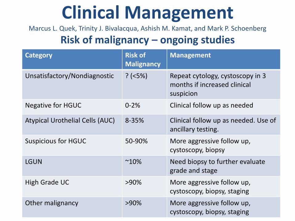
Clinical Management Marcus L. Quek, Trinity J. Bivalacqua, Ashish M. Kamat, and Mark P. Schoenberg
Risk of malignancy – ongoing studies Category Risk of
Malignancy Management
Unsatisfactory/Nondiagnostic ? (<5%) Repeat cytology, cystoscopy in 3 months if increased clinical suspicion
Negative for HGUC 0-2% Clinical follow up as needed
Atypical Urothelial Cells (AUC) 8-35% Clinical follow up as needed. Use of ancillary testing.
Suspicious for HGUC 50-90% More aggressive follow up, cystoscopy, biopsy
LGUN ~10% Need biopsy to further evaluate grade and stage
High Grade UC >90% More aggressive follow up, cystoscopy, biopsy, staging
Other malignancy >90% More aggressive follow up, cystoscopy, biopsy, staging
The opportunities: Future studies
• Using the new system does the atypia rate change?
• Does the cytology:histology correlation change?
• What is the outcome of ancillary tests in the atypical category (especially UroVysion FISH testing)?
• What are individual and laboratory AUC:HGUC ratios? Can it be used as a quality assurance tool?
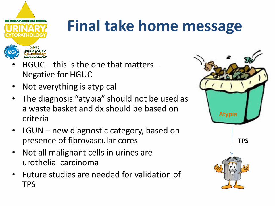
Final take home message
• HGUC – this is the one that matters – Negative for HGUC
• Not everything is atypical
• The diagnosis “atypia” should not be used as a waste basket and dx should be based on criteria
• LGUN – new diagnostic category, based on presence of fibrovascular cores
• Not all malignant cells in urines are urothelial carcinoma
• Future studies are needed for validation of TPS
TPS
Atypia

JOIN US NEXT YEAR!