application of proteomics and cytomics in human
TRANSCRIPT

1
Application of Proteomics and Cytomics in Human Neutrophils Functional Studies
Alexandre Rosa Campos Programa de Patologia Molecular, Universidade de Brasilia
Plataforma de Proteomica, Parc Cientific de Barcelona
Tutors: Wagner Fontes, University of Brasilia Eliandre de Oliveira, Science Park of Barcelona

2
Dedicated with Love to my wife, Yolanda Virgili
And my two mothers, Solange Rosa
and Carmem Villa Sola.
And in Memory of my Father, João Bezerra Campos Filho
This Dissertation would not have been possible without the timely cooperation from my colleagues, who I would like to take the opportunity to thank individually. They are: Carlos Garcia, Elaine Nascimento, Renan Rodrigues, Pedro Ivo, Nuno Domingos, David Billido, Antonia Odena and my two tutors Wagner Fontes and Eliandre de Oliveira.

3
Abstract
Biomedical research commonly starts by raising a hypothesis to solve a problem. In this context, scientists select the most appropriate method(s) to answer the question and solve a common dilemma. Over the past decade, we have witnessed a revolution of new technologies in molecular biology – the Omics Science. High-scale technologies such as metabolomics, cytomics, genomics and proteomics are changing the way we study complex biological systems. Current approaches to understanding the functional diversity of an organism preferentially strive for a systems biology approach whereby first the phenotypic classification of a specific cytome is achieved prior to an attempt to perform proteomic analysis. In this context, to better understand the features that involve neutrophil activation and programmed cell death in the pathological and healthy states, this study proposes the integration of cell biology approaches such as flow cytometry with a very robust proteomics platform in an attempt to integrate data at the molecular level with phenotypic data of neutrophils. The application of subcellular fractionation method using digitonin detergent extraction to enrich cytosolic proteins from neutrophils was found reproducible, simple to perform, and inexpensive.

4
Contents Page
1. Introduction 06 [1.1] The Neutrophil – an interesting biological puzzle 06
[1.2] Neutrophil Morphology a. Azurophil granules b. Peroxidase-negative granules
07
[1.3] Neutrophils in Action a. Neutrophil cytotoxic arsenal
10
[1.4] The Neutrophil Implication in Pathological Processes 14
[1.5] Proteomics Technologies – Advances and Caveats a. Protein Visualization on 2D gels – Staining Procedures
b. Differential Gel Electrophoresis (DIGE) c. Gleaning Biological Relevancy through Differential Statistical
Analysis d. Mass Spectrometry in Proteomics e. MALDI-TOF Mass Spectrometers f. Mass Spectrometry Proteomics Strategies g. Data Mining in Proteomics h. Neutrophil Proteomics – Lessons from a technical perspective
15
[1.6] Proteomics Meets Cell Biology a. Neutrophil Proteomics – Lessons from a biological perspective
b. Tackling the Proteome Complexity c. Subcellular Proteomics d. Subcellular Fractionation Methods e. Using Detergents to Fractionate Cellular Compartments
f. Neutrophil Cell Biology – Lessons from a technical perspective
25
2. Objectives 32
3. Experimental Methods 33
[3.1] Blood Sample Collection 33
[3.2] Neutrophil Purification from Blood by Isopycnic Centrifugation in Percoll Discontinuous Gradient.
33
[3.3] Neutrophil Priming and Activation 34
[3.4] Flow Cytometry Acquisition and Analysis 35

5
[3.4] Cytosolic and Membrane/Organelle Proteins Fractionation 35
[3.5] Preparation of Cytosolic Protein Samples for 2DE. 36
[3.6] Preparation of Cytosolic and Membrane/Organelle Protein Samples for 1DE.
36
[3.7] Preparation of Total Protein Samples for 4-7 gels. 36
[3.8] Preparation of Cytosolic and Membrane/Organelles Protein Fractions for 1DE.
36
[3.9] Isoelectric Focusing. 36
[3.10] Equilibration and 2nd Dimension PAGE. 37
[3.11] Gel Staining – 1D and 2D gels. 37
[3.12] Computer-assisted 2-D gel analysis 38
[3.13] In-gel Digestion of Proteins 39
[3.14] Protein Identification by Mass Spectrometry 39
[3.15] Confocal Laser Scanning Microscopy 40
4. Results and Discussion 41
[4.1] Cell Separation by Gradient Centrifugation 41
[4.2] Assessing Neutrophil Oxidative Burst in Primed and Activated Neutrophils by Flow Cytometry
43
[4.3] Two-dimensional Gel Electrophoresis (2-DE) Analysis – The ‘Divide-and-Conquer’ Workflow
46
[4.4] Subcellular Fractionation of Cytosolic and Plasma Membrane Compartments
56
[4.5] Optimizing Cytosolic Protein Enrichment in Neutrophils 59
5. Conclusion 65
6. Perspectives 66
Resume of dissertation in Portuguese 67
7. References 71

6
1. Introduction [1.1] The Neutrophil – an interesting biological puzzle The immune cells are classified into two distinct lineages with specialized cell types required for proper immune response (Fisher, A. 2002). The myeloid lineage of monocytes, macrophages, dendritic cells and neutrophils carries out the innate immune response. The lymphoid lineage of T cells, B cells and natural killer (NK) cells enables adaptive immunity by distinguishing self from non-self antigens and also providing a memory of foreign proteins seen before (Kindt, T. et al., 2002). Other immune cells, such as eosinophils, basophils and mast cells, also participate in the immune response. The human body contains about five liters of blood. Red blood cells constitute about 45% of this volume and white blood cells (WBC) around 1%, the rest being the liquid blood plasma (Diggs, L. 1985). The white blood cells, or leukocytes, can be sub-divided into: lymphocytes, monocytes and granulocytes (Kindt, T. et al., 2002). The former represents the largest group of leukocytes and can themselves be subdivided in neutrophils, eosinophils and basophils (table 1). Circulating neutrophils are terminally differentiated white blood cells presenting a short-life span and characterized by a multilobed nucleus and a granular cytoplasm (fig. 1 and 2e). The latter is due to the presence of specialized granules containing the host-defense molecules, which is rapidly mobilized following an infectious challenge (Faurschou, M. and Borregaard, N. 2003).
Fig. 1: Transmission Electron Micrograph of human neutrophil. Inset is an image taken from a neutrophil 20 s after the phagocytosis of latex particles opsonized with IgG (V, vacuole). The section was stained for myeloperoxidase (MPO) to reveal the

7
electron-dense product in the azurophil granules, some of which can be seen degranulating into the phagocytic vacuole (arrows). Bar = 1 µm. (Image from Segal, AW. et al., 1980)
Immunologists have long praised the essential role of polymorphonuclear
leukocytes, predominantly neutrophils, in the host defense against invading microorganisms. On a per-cell basis, neutrophils make fewer molecules of a given cytokine than do macrophages or lymphocytes, but neutrophils often outnumber mononuclear leukocytes at inflammatory sites by one to two orders of magnitude, and they can therefore be important sources of cytokines such as tumor necrosis factor α (TNFα) at the crucial juncture at which the decision is made to mount an immune response (Nathan, C. 2006). A normal adult produces more than 10 billion neutrophils per day, a figure that can increase as much as tenfold during a period of acute inflammation. The relative abundance of neutrophils, with approximately 50 billion circulating in the bloodstream, and its rapid recruitment to the site of infection make these cells a prominent early player of the innate immune system. Table 1: Cells in Normal Human Adult Peripheral Blood
Cells No. per cm3 Percent of WBC Diameter (µm)
Platelets 1-3 x 10⁸ ---- 2-3
Erythrocytes (RBC) 4-6 x 10⁹ ---- 6-8
Leukocytes (WBC)
• Neutrophils • Eosinophils
• Basophils
3-6 x 10⁶
2-4 x 10⁶
0.1-5 x 10⁵
0-0.1 x 10⁶
100%
50-70% 1-3%
0-1%
10-12 10-12
8-10
Lymphocytes 1-4 x 10⁶ 20-40% 6-12
Monocytes 0.2-1 x 10⁶ 1-6% 12-15
Source: Diggs LW, et al., (1970). The Morphology of Human Blood Cells, 5th edition. Abbott Laboratories, Abbott Park, IL.
[1.2] Neutrophil Morphology Cell biologists and immunologists have relied mainly on cell morphology to characterize the five different stages of neutrophil differentiation: myeloblast, promyelocyte, myelocyte, banded cells, and segmented cells (fig. 2a-e) (Bainton, D. 1971). Differentiation of myeloblasts into mature neutrophils does not result from a simple state transition; instead, neutrophil differentiation appears to be a multi-step process characterized by the sequential formation of azurophil, specific and gelatinase granules (granulopoiesis) and their constituent granule proteins (Faurschou, M. and Borregaard, N. 2003). The various subsets of granules contained within the neutrophil are key effectors of the neutrophil immune response.
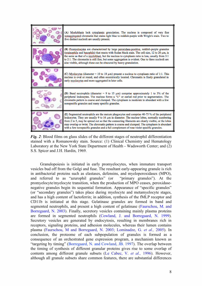
8
Fig. 2: Blood films on glass slides of the different stages of neutrophil differentiation stained with a Romanowsky stain. Source: (1) Clinical Chemistry and Hematology Laboratory at the New York State Department of Health - Wadsworth Center; and (2) S.S. Spicer and J.H. Hardin, 1969.
Granulopoiesis is initiated in early promyelocytes, when immature transport vesicles bud off from the Golgi and fuse. The resultant early-appearing granule is rich in antibacterial proteins such as elastases, defensins, and myeloperoxidases (MPO), and referred to as “azurophil granules” (or “primary granules”). At the promyelocyte/myelocyte transition, when the production of MPO ceases, peroxidase-negative granules begin its sequential formation. Appearance of “specific granules” (or “secondary granules”) takes place during myelocyte and metamyelocyte stages, and has a high content of lactoferrin; in addition, synthesis of the fMLP receptor and CD11b is initiated at this stage. Gelatinase granules are formed in band and segmented neutrophils, and present a high content of gelatinase (Faurschou, M. and Borregaard, N. 2003). Finally, secretory vesicles containing mainly plasma proteins are formed in segmented neutrophils (Cowland, J. and Borregaard, N. 1999). Secretory vesicles are generated by endocytosis, resulting in membranes rich in receptors, signaling proteins, and adhesion molecules, whereas their lumen contains plasma (Faurschou, M and Borregaard, N. 2003; Lominadze, G. et al., 2005). In conclusion, the proteome of each subpopulation of granules is formed as a consequence of an orchestrated gene expression program, a mechanism known as “targeting by timing” (Borregaard, N. and Cowland, JB. 1997). The overlap between the timing of synthesis of different granular proteins gives rise to some overlap of contents among different granule subsets (Le Cabec, V. et al., 1996). However, although all granule subsets share common features, there are substantial differences

9
with respect to protein content and the extent of protein release to phagosomes and extracellular environment.
Azurophil granules
Despite its early recognition as specialised lysosomes, azurophil granules are nowadays probably best regarded as regulated secretory granules (Cieutat, A. et al., 1998). Like lysosomes, azurophil granules contain granulophysin (CD63) in their membrane. However, in contrast to lysosomes, they do not express the lysosome-associated membrane proteins LAMP-1 and LAMP-2 (Cieutat, A. et al., 1998), and the sorting of proteins to azurophil granules does not involve the mannose-6-phosphate receptor system essential for lysosomal enzyme targeting (Dahms, N. et al., 1989; Nauseef, W. et al., 1992). Azurophilic granules are the largest of the human neutrophilic granules, measuring 0.3 µm in diameter (Nüsse, O. and Lindau, M. et al., 1988; Lollike, K. et al., 2002). They are also referred to as primary or peroxidase-positive granules. Each human neutrophil contains 1300 azurophilic granules based on morphological data (Nüsse, O. and Lindau, M. et al., 1988). Based on their time of appearance during myelopoiesis, azurophil granules are often divided into two major subsets, namely early- and late-appearing azurophil granules, which are formed from the beginning of the promyelocytic stage and near the promyeocyte/myelocyte transition, respectively (Sengelov, H. et al., 1993; Arnljots, K. et al., 1998).
Azurophil granules contain a highly packed matrix of acid mucopolysaccharide and highly cationic myeloperoxidase. These granules contain the largest number of luminal bactericidal proteins such as the α-defensins and myeloperoxidase (MPO, the iron-containing enzyme that colours pus green.), while there is a paucity of membrane, cytoskeletal, and GTP-binding proteins. Many proteins of azurophil granules are synthesized as proforms, which go through proteolytic trimming on arrival at the granule compartment. This seems to be important to prevent misfolding, autotoxicity or retention of the mature proteins during their passage through the biosynthetic machinery of the cell (Garwicz, D. et al., 1998; Liu, L. and Ganz, T. 1995). After removal of amino-terminal propeptides, the proteins are stored in their active conformations (Lindmark, A. et al., 1998; Garwicz, D. et al., 1998).
Peroxidase-negative granules
Historically, specific and gelatinase granules have been also designated peroxidase-negative granules (Kjeldsen, L. et al., 1992). Although widely employed, this nomenclature may be misleading since myeloperoxidase has been found in these granule subsets as well (Lominadze, G. et al., 2005). Recently, two independent approaches were used to determine MPO presence in the neutrophil granule subsets. Theilgaard-Monch et al., (2005) found that MPO expression is downregulated during transition from promyelocytes (formation of azurophil granules) to myelocytes/metamyelocytes (formation of specific granules). By peering the microarray data, one can note that MPO expression is not completely abrogated during this transition, whereas transition from myelocytes/metamyelocytes to bone marrow PMNs seems to correlate with complete abrogation of this gene. MPO protein expression in peripheral blood neutrophils has also been assessed by ELISA method (Lominadze, G. et al., 2005). This study showed that 73% of total MPO was present

10
in azurophil granules, 20% was in specific granules, and 7% was in gelatinase granules. Similar distribution has been also reported by Kjeldsen et al., (1994; 1999). However, these studies are not conclusive and absolute quantification has not been carried out to date. Furthermore, this supposedly MPO detection in specific and gelatinase granules may be a result of contamination during granule separation process. Gelatinase granule membranes contain a large number of membrane receptors and adhesion molecules. Therefore, the large amount of actin cytoskeleton and cytoskeletal regulatory proteins in these granules supports its role in enhancing the plasma membrane expression of molecules necessary for neutrophil adherence to and migration through inflamed vascular endothelium.
Specific granules contain large amounts of moderately cationic luminal proteins. The greater complexity of the specific granule lumen and the presence of a significant number of transmembrane and membrane-associated proteins suggest that these granules represent a transitional phase that can contribute to neutrophil activation through exocytosis or provide bactericidal proteins to phagosomes. Consistent with this notion, gelatinase and specific granules contain overlapping sets of proteins, which include lactoferrin, lipocalin, lysozyme, LL37, three metalloproteases (MMP8, MMP9 and MMP25), and several other antimicrobial proteins, matrix-degrading enzymes and membrane receptors (Lominadze, G. et al., 2005). From a total of 247 proteins identified from the three granule subsets in a recent study, Lominadze et al., (2005) reported an overlap of 79 proteins between gelatinase and specific granules, but only five proteins overlap between specific and azurophil granules. Moreover, a total of 86 proteins were identified only from gelatinase granules, 28 proteins only from specific granules, and 26 proteins only from azurophil granules. In contrast to azurophil granules, specific granules store proteins as intact proforms.
[1.3] Neutrophils in Action The remarkable capacity of detecting and eradicating invading pathogenic
microorganism without destroying self-tissues requires a well-orchestrated innate and adaptive immune response. Activation of immune response with subsequent eradication of microbial pathogens is a complex, multi-step process (fig. 3). As a first step, neutrophils are rapidly recruited from circulation and bone marrow reserves to sites of infection by host and pathogen-derived stimuli (chemotatic factors) such as leukotriene LTB4 (Freeland, H. et al., 1988) platelet-activating factor (PAF) (Mencia-Huerta, J. et al., 1983); tumor-necrosis factor α (TNFα) (Williams, C. and Coleman, J. 1995), interleukin-8 (IL-8) (Gordon, J. et al., 1990), N-formylmethionyl-leucyl-phenylalanine (fMLP) (Le, Y. et al., 2002) and lipopolysaccharide (LPS) (Fessler, M et al., 2002). In addition to recruiting neutrophils, many chemotactic factors, including lipopolysaccharide (LPS), tumor necrosis factor-alpha (TNFα), and platelet-activating factor (PAF), purportedly modulate neutrophil responses to subsequent stimuli, a process known as neutrophil priming. (Brazil, T. et al., 1998) During recruitment and migration to infection focus, neutrophils produce a number of factors to orientate their migration, ensure their survival in the hostile inflammatory milieu, recruit additional neutrophils and ‘inactivate’ their own toxic products (Botha, A. et al., 1995).

11
Fig. 3: Multi-step activation process of neutrophils
The migration of neutrophils from the blood to target tissues (extravasation) involves tethering and rolling of neutrophils on the blood-vessel wall, firm adhesion and crossing through the endothelial barrier (diapedesis) (Cinamon, G et al., 2001). Adherence of circulating leukocytes to the vascular endothelium is facilitated by the expression of several membrane proteins such as CEA-related cell-adhesion molecules (CEACAMs), selectins, immunoglobulin superfamily members, and integrins. In addition to these classical interactions, other cell-surface-expressed enzymes with catalytic domains outside the plasma membrane - ectoenzymes such as CD26, CD38, and vascular adhesion protein 1 - were recently reported to take part in the different steps of the extravasation cascade (Goding, J. 2000). The adhesive interactions of neutrophils are usually dynamic and transient. In fact, a high degree of flexibility in cell adhesion and mobility should be attained to allow neutrophils to circulate in the blood without pathologically adhering to the vascular wall. Several studies have underscored the importance of a small-scale respiratory burst and granule exocytosis to release certain neutrophil proteases and adhesion molecules as a precondition to neutrophil recruitment to inflammatory loci (Nathan, C et al., 1993; Raptis, SZ et al., 2005). The neutrophil–endothelial interactions have been shown to trigger mobilization of secretory vesicles from neutrophils (Borregaard, N et al., 1994). Secretory vesicles are mobilized at the earliest phases of the neutrophil-mediated inflammatory response in response to a wide variety of inflammatory stimuli (Faurschou, M and Borregaard, N. 2003; Lominadze, G et al., 2005). These vesicles constitute a reservoir of membrane-associated receptors including the β2-integrin CD11b/CD18 (Mac-1, CR3), the complement receptor 1 (CR1), receptors for formylated bacterial peptides (formylmethionyl-leucyl-phenylalanine (fMLP)-receptors), the LPS/lipoteichoic acid-receptor CD14, the FcγIII receptor CD16, and the metalloprotease leukolysin, all of which are incorporated in the plasma membrane after exocytosis. The disassembly of extracellular matrix is believed to be crucial for neutrophil extravasation and migration, and to facilitate the neutrophil-bacterial contact (Borregaard, N. and Cowland, J. 1997; Owen, C. and Campbell, E. 1999; Kang, T. et
12
al., 2001). The release of reactive oxygen species such as hydrogen peroxide, hypohalites and chloramines activates matrix metalloproteinases (MMPs), and inactivates protease inhibitors (Henson, P. and Johnston, R. 1987; Weiss, S. 1989). MMPs play fundamental roles in a wide range of mammalian physiological and pathological processes by degrading major structural components of the extracellular matrix including collagens, fibronectin, proteoglycans, laminin and gelatin.
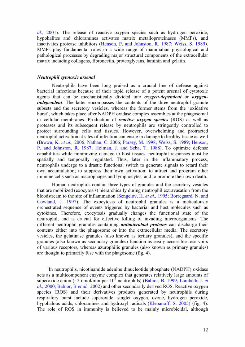
Neutrophil cytotoxic arsenal
Neutrophils have been long praised as a crucial line of defense against bacterial infections because of their rapid release of a potent arsenal of cytotoxic agents that can be mechanistically divided into oxygen-dependent or oxygen-independent. The latter encompasses the contents of the three neutrophil granule subsets and the secretory vesicles, whereas the former stems from the ‘oxidative burst’, which takes place after NADPH oxidase complex assembles at the phagosomal or cellular membranes. Production of reactive oxygen species (ROS) as well as proteases and its subsequent release by neutrophils are stringently controlled to protect surrounding cells and tissues. However, overwhelming and protracted neutrophil activation at sites of infection can ensue in damage to healthy tissue as well (Brown, K. et al., 2006; Nathan, C. 2006; Parsey, M. 1998; Weiss, S. 1989; Henson, P. and Johnston, R. 1987; Holman, J. and Seba, T. 1988). To optimize defense capabilities while minimizing damage to host tissues, neutrophil responses must be spatially and temporally regulated. Thus, later in the inflammatory process, neutrophils undergo to a drastic functional switch to generate signals to retard their own accumulation; to suppress their own activation; to attract and program other immune cells such as macrophages and lymphocytes; and to promote their own death.
Human neutrophils contain three types of granules and the secretory vesicles that are mobilized (exocytosis) hierarchically during neutrophil extravasation from the bloodstream to the site of inflammation (Sengeløv, H. et al., 1995; Borregaard, N. and Cowland, J. 1997). The exocytosis of neutrophil granules is a meticulously orchestrated sequence of events triggered by bacterial and host molecules such as cytokines. Therefore, exocytosis gradually changes the functional state of the neutrophil, and is crucial for effective killing of invading microorganisms. The different neutrophil granules containing antimicrobial proteins can discharge their contents either into the phagosome or into the extracellular media. The secretory vesicles, the gelatinase granules (also known as tertiary granules), and the specific granules (also known as secondary granules) function as easily accessible reservoirs of various receptors, whereas azurophilic granules (also known as primary granules) are thought to primarily fuse with the phagosome (fig. 4).
In neutrophils, nicotinamide adenine dinucleotide phosphate (NADPH) oxidase acts as a multicomponent enzyme complex that generates relatively large amounts of superoxide anion (~2 nmol/min per 106 neutrophils) (Babior, B. 1999; Lambeth, J. et al., 2000; Babior, B et al., 2002) and other secondarily derived ROS. Reactive oxygen species (ROS) and their derivatives products generated by neutrophils during respiratory burst include superoxide, singlet oxygen, ozone, hydrogen peroxide, hypohalous acids, chloramines and hydroxyl radicals (Klebanoff, S. 2005) (fig. 4). The role of ROS in immunity is believed to be mainly microbicidal, although

13
controversial reports have come into light in the last years (Reeves, E. et al., 2002). In contrast to many signal transduction systems in which agonist-induced transcription of genes initiates a vigorous biochemical cellular response, the NADPH oxidase is spatially regulated, whereby the dormant oxidase is maintained in an inactive state by partitioning the components into a cytoplasmic fraction, and a membrane-bound flavocytochrome b558. The latter is a stable heterodimer composed of the p22phox and gp91phox subunits, which is localized both in the plasma membrane (around 5%) and in the membranes of mobilizable intracellular granules (around 95%) in the rest neutrophils (Borregaard, N. et al., 1983; Sengeløv, H et al., 1992). The cytoplasmic phox components exist as a complex (Mr 250kDa) stabilized through multiple SH3-domain-mediated interactions of its factors, p47phox, p67phox, p40phox, the small regulatory GTPases Rac2 and Cdc42, and the newly identified p29 peroxiredoxin. Upon neutrophil stimulation, several serine residues within the polybasic region of p47phox become phosphorylated resulting in a conformational change in p47phox, unfolding the protein and exposing the now interactive SH3 domains of p47phox to eventually interact stably with proline-rich regions (PRRs) on the cytoplasmic tail of p22phox (Nagasawa, T. et al., 2003), thereby resulting in translocation to the membrane and activity of a functional oxidase (Shiose, A. and Sumimoto, H. 2000).
Fig. 4: Oxygen-dependent and oxygen-independent mechanisms of neutrophil defense.
The reactivity between ROS and proteins has been investigated in the past years leading to the postulation that ROS partake in several cell signaling events (Nathan, C. 2003). Hydrogen peroxide (H2O2) was shown to convert free L-tyrosine in the human blood plasma to tyrosyl radical, which may react with tyrosine residues of target proteins to produce o,o’-dityrosine bonds (Heinecke, J. et al., 1993; Marquez, L. and Dunford, H. 1995; Jacob, J. et al., 1996). Peroxynitrite and HOCl can also give rise to the formation of chlorotyrosine and dityrosine, respectively (Hazen, S. et al., 1997; Kettle, A. 1996; Pfeiffer, S. et al., 2000). These protein modifications have been associated with organ dysfunction in several diseases (Heinecke, J. 2002). A recent study showed that human neutrophils can moduate their proteome via a tyrosine oxidation pathway induced by pro-inflammatory mediators. In addition, upon neutrophil stimulation with TNFα, IFNγ or PMA, a limited number of membrane-associated proteins became tyrosylated (Avram, D. et al., 2004). ROS

14
has been also shown to mediate temporary inactivation of tyrosine phosphatases by reversible oxidation of their active-site cysteine sulphydryls (Tonks, N. 2005).
The ability of neutrophils to ingest and kill microorganisms is crucial to innate immunity and host defense. Phagocytosis is an inherently complex process that requires phagocyte-microbe contact and is accompanied by activation of signaling pathways that orchestrate rearrangement of the actin cytoskeleton, extension of the plasma membrane, and engulfment. Neutrophil-microbe contact is initiated via a plethora of membrane receptors ‘provided’ by the aforementioned fusion of secretory vesicles, gelatinase and specific granules with the plasma membrane during the exocytosis process. Azurophilic and specific granules fuse with the phagocytic vacuole after microbe uptake (Segal, A. et al., 1980). It has been estimated that the granule protein makes up about 40% of the vacuolar volume (Hampton, M. et al., 1998), which gives a concentration of about 0.5 g ml-1 (Reeves, E. et al., 2002). The phagosome volume depends on the size of the engulfed microbe, but will also increase dramatically over time. In vitro, the phagosome volume increases about 3-fold over 15 min in neutrophils (Reeves, E. et al., 2002), and this increase is mostly attributable to the addition of specific granule contents, which are already present in 50% of phagosomes within 30 seconds after ingestion of bacteria (Bainton, D. 1973), and later of azurophil granule contents. Fusion of specific granules adds 0.26 fl and azurophilic granules add another 0.92 fl to the phagosome, which together would triple its volume if it started out at 0.6 fl. These estimates are based on the dimensions and numbers of granules in neutrophils, 4600 specific granules 0.13 µm in diameter and 1300 azurophilic granules 0.3 µm in diameter (Nüsse, O. and Lindau, M. 1988), and the assumption that all granules fuse with the first 20 phagosomes, which seems a reasonable synthesis of several observations (Hampton, M. et al., 1998; Bainton, D. 1973). It is also important to note that multiple phagosomes may coalesce, forming much larger structures (Bainton, D. 1973).
[1.4] The Neutrophil Implication in Pathological Processes Overwhelming neutrophil activation has been suggested to play a major role in several inflammatory-related disease such as the pathogenesis of organ dysfunction in sepsis (Brown, K. et al., 2006). Such premise has been the subject of intense scrutiny in the last years, as researchers have sought to develop efficient therapies to control neutrophil-mediated healthy tissue damage in septic patients. Although limiting the inflammatory response attributable to neutrophils appears to represent a promising avenue for therapeutic intervention in sepsis (Abraham, E. 2003), neutrophils are essential components in the activation of innate immune response for detection and eradication of invading pathogenic microorganisms. Evidence to support a pivotal role of neutrophils in sepsis-induced organ dysfunction derives from both experimental models and clinical studies. Postmortem pathological examination of specimens from septic patients with multiple organ failure reveals neutrophil infiltrations in compromised organs (Nuytinck, H. et al., 1998; Thijs, A. and Thijs, L. 1998; Brealey, D. and Singer, M. 2000). The extent of neutrophil infiltrates correlates with impaired lung function and with high concentrations of neutrophil-derived proteolytic enzymes in the bronchoalveolar lavage from patients with acute respiratory distress syndrome (ARDS), a severe form of acute lung injury that may be associated with sepsis (Windsor, A. 1993; Ware, L. and Matthay, M. 2000; Steinberg, K. et al., 1994; Adams, J. et al., 2001).

15
Experimental interventions that deplete or inhibit the activity of neutrophils attenuated organ dysfunction and improved survival in animal models (Nathens, A. et al., 1997; Abraham, E. 2003). These findings concur with clinical observations in patients with systemic inflammatory response where removal of blood neutrophils (by leucodepletion filters) enhances respiratory and renal function (Treacher, D. et al., 2001). Moreover, neutrophils retrieved from both lungs and blood of patients with sepsis exhibit increased production of ROS (Baldwin, S. et al., 1986); delayed apoptosis (Matute-Bello, G. and Liles, W. 1997; Feterowski, C. et al., 2001); and increased predisposition to endothelial binding as evidenced by increased expression of β2 integrins (Brown, K. et al., 2001; Chollet-Martin, S. et al., 1996).
Neutrophil turnover is a healthy and critical mechanism to dampen local tissue destruction, diminish further inflammation and recruitment of immune cells, and facilitate termination of the innate immune response by limiting pro-inflammatory capacity (Matute-Bello, G. and Liles, W. 1997; Feterowski, C. et al., 2001).
[1.5] Proteomics Technologies – Advances and Caveats Proteomics defined in its most broad terms is the understanding of the protein expressed from biological compartments such as cell, tissue, or organisms in term of identification, quantify, post-translational modification, protein–protein interaction, sub-cellular localization activities and their biological functions. The introduction of novel protein separation techniques (Gorg, A. et al., 2004; Washburn, M. et al., 2001), the advent of mass spectrometers with higher mass accuracy, dynamic range and resolution (Shevchenko, A. et al., 2000; Medzihradszky, K. 2000; Wilm, M. and Mann, M. 1996; Wilm, M. et al., 1996; Syka, J. et al., 2004), the improvement of computational data analysis tools (Clauser, K. et al., 1999; Creasy, D. and Cottrell, J. 2002; Mann, M. and Wilm, M. 1994) and the proliferation of genomic and proteomic databases (Henzel, W. et al., 1989; Mann, M. et al., 1994; Pappin, D. et al., 1993; Yates, J. et al., 1993; Lander, E. et al., 2001) are likely the main reasons for the increase in number and size of proteomic datasets. However, it has become evident that proteins are much more complex, diverse and dynamic than was originally anticipated (Aerbersold, R. and Mann, M. 2003); and despite the technological advances, there are important biological and technological caveats in proteomic analysis of complex material such as biological samples. Several different protein prefractionation techniques can be combined with various analytical techniques. Gel-based methods such as one-dimensional and two-dimensional polyacrylamide gel electrophoresis (1D-PAGE and 2D-PAGE or 2-DE, respectively) are arguably the most popular and versatile methods of protein separation. Gel-independent approaches focus mainly on chromatographic separation techniques in combination with fast and sensitive MS. Two-dimensional polyacrylamide gel electrophoresis (2-DE) combines isoelectric focusing (IEF), which separates proteins according to their isoelectric point (pI), and sodium dodecyl sulfate-polyacrylamide gel electrophoresis (SDS-PAGE), which separates them further according to their molecular mass. In an electric field, a protein will migrate toward the pH region on the IPG strip in which the pH equals its isoelectric point. After the first dimension separation, the IPG strip is laid atop the second dimension polyacrylamide sodium dodecyl sulfate gel (SDS-PAGE), and the proteins are run in an orthogonal direction, to achieve separation by size of fully

16
unfolded, SDS coated protein molecules. The major steps of the 2-DE-based proteomics workflow include: (i) sample preparation and protein solubilization; (ii) protein separation by 2-DE; (iii) protein detection and quantitation; (iv) computer-assisted analysis of 2-DE patterns; and (v) protein identification usually by mass spectrometry (Gork, A. et al., 2004). Sample preparation for 2D gel electrophoresis can be divided into three basic steps, namely cell disruption, inactivation or removal of interfering substances, and solubilization of the proteins. Protein extraction is a crucial step in any proteomics strategy because of its influence in yield, biological activity, and the structural integrity of the specific target protein. Several techniques for cell disruption are available such as osmotic lysis, freeze- thaw cycling, detergent lysis, enzymatic lysis of the cell wall, sonication, grinding with (or without) liquid nitrogen, high pressure (e.g. French press), homogenization with glass beads and a bead beater, nitrogen cavitation, or a rotating blade homogenizer. Neutrophil cell membranes are weak and thus easily disrupted by gentle treatment with mild detergents. This is best accomplished by using lower concentrations of a nonionic detergent such as Triton X-100. Detergents also prevent hydrophobic interactions between the hydrophobic protein domains; hence, avoiding loss of proteins due to aggregation and precipitation. The most common interfering compounds in sample preparation are proteolytic enzymes, salts, lipids, nucleic acids, polysaccharides, and/ or highly abundant proteins. Salt ions may interfere with electrophoretic separation and should be removed if their concentration is too high (>100 mM). Salt increases the conductivity of the IEF gel, thereby prolonging the time required to reach the steady-state. High amounts of lipids may interact with detergent and thus reduce its effectiveness. Lipids can also form complexes with proteins, reducing its solubility. The presence of polysaccharides and nucleic acids can result in poor focusing in the acidic region of the IEF gel, and obstruction of the pores of the polyacrylamide gels due to an increase in the viscosity of the solutions. To assure protein solubilization, 2D electrophoresis sample buffer usually contain reducing agents and high concentrations of chaotropes. The chaotropes urea and thiourea are often used at concentrations of 7 M and 2M, respectively. The former is quite efficient in disrupting hydrogen bonds, leading to protein unfolding and denaturation. Nevertheless, it has been shown that hydrophobic interactions are more efficiently broken by thiourea (Rabilloud, T. et al., 1997). Due to its poor solubility in water, thiourea is used in concentrated urea solutions to increase its solubility. Reducing agents are necessary for cleavage of intra- and intermolecular disulfide bonds to achieve complete protein unfolding. The most commonly used reductants are DTT or dithioerythritol (DTE) are commonly used at concentrations ranging from 20 mM to 100 mM. A disadvantage of these agents is that in a pH between 8.5 and 9, they are ionized and behave as a weak acid; and therefore, run short in the alkaline gel area due to migration to the anode during IEF.
Last but not the least, many cell types, particularly the neutrophils, have a powerful arsenal of proteases stored in their organelles/granules. Hence, these proteases must be inactivated to prevent protein degradation that otherwise may result in artifactual spots and loss of high Mr proteins. To prevent proteolysis of sample proteins, protease inhibitors are usually added to lysis buffers.
Two-dimensional PAGE delivers a map of intact proteins, which reflects

17
changes in protein expression level, isoforms or posttranslational modifications (PTMs). Depending on the gel size and pH gradient used, 2-DE can theoretically resolve more than 5,000 proteins simultaneously (~1,000 proteins routinely). Nonetheless, there is well-justified concern about the overall reproducibility of 2-DE when it is used as the sole method to identify qualitative and quantitative differences in protein expression in samples of interest (Horgan, G. 2007; Campostrini, N. et al., 2005). In addition, 2DE-based approaches are biased against membrane proteins, low
abundance proteins, and proteins at the extremes of isoelectric point and molecular mass (Pedersen, S. 2002; Rabilloud, T. 2003; Helling, S. et al., 2006). For example, Lominadze et al., found that the presence of large amounts of basic proteins in neutrophil granules produced poor isoelectric focalization. To overcome these limitations, several strategies have been employed in the past years to increase the number and certainty of the protein identifications. One example of such proteomics platform is the multidimensional protein identification technology [MUDPIT] (Washburn, M. et al., 2001), which combines peptide separation by strong cation exchange and reverse phase two-dimensional chromatography, followed by identification by mass spectrometry. Protein Visualization on 2D gels – Staining Procedures Several detection methods are widely used for protein visualization in polyacrilamide gels that use labels that are fixed to the protein molecules. In general, visualization dyes and procedures can be divided into three major categories: (1) detection by light absorption of organinc dyes such as Coomassie Brilliant Blue; (2) detection by fluorescence of fluorescent probes such as Sypro dyes or CyDyes; and (3) metal ions permit protein visualization either by differential salt binding (e.g. reverse staining with zinc-imidazole) or by metal ion reduction such as silver stains (Simpson, R. 2003). The latter has gained wide popularity because of its excellent sensitivity (in the low nanogram range), flexibility (can be completed in a time range from 2 h to 1 d), and mass spectrometry compatibility. Silver staining was first introduced in 1979 by Switzer et al.,; however, issues related to background, dynamic range and reproducibility resulted in many improvements of the original protocol. By 1994, Rabilloud et al., (Rabilloud, T. et al., 1994) found more than 100 different silver staining protocols in the literature. The principle of silver staining is straightforward. Silver ions bind to proteins and, under appropriate conditions, are reduced to elementary Ag developing a visible image typically in brownish-grey-black color (Rabilloud, T. et al., 1994). Despite its wide use, silver staining is somewhat tricky. Unlike staining with organic dyes, silver staining goes against general thermodynamics. Proteins bind silver ions, and this binding decreases the reactivity of the ions, which leads to the formation of the commonly observed 'hollow' or 'doughnut' spots (Chevallet, M. 2006). Moreover, silver ions bind preferentially to basic amino acids of proteins; hence, it may cause a higher affinity of basic proteins for silver ions decreasing the staining linearity. Silver staining methods have been reported to be linear over a 40-fold range in protein concentration depending on certain physical-chemical properties of a given protein (Merril, C. et al., 1984).
In general, depending on the silver impregnation, the silver staining methods can be divided into simple silver nitrate or silver-ammonia. The former is relatively simple and compatible with mass spectrometry; in addition, it presents good

18
consistency. Basic proteins are less efficiently stained than acidic ones with this type of stain (Rabilloud, T. 1999). Silver-ammonia stains are more sensitive than silver nitrate stains, in the range of 2-4 ng (Simpson, R. 2003). In contrast to simple silver stains, ammonical silver stains basic proteins more efficiently than acidic ones (Rabilloud, T. 1999). Differential Gel Electrophoresis (DIGE) Many of the disadvantages and shortcomings of 2D electrophoresis (2DE) have been overcome by 2D difference gel electrophoresis (DIGE) (Unlu, M. et al., 1997). This technology uses three spectrally distinct fluorescent cyanine dyes (CyDyes - Cy2, Cy3, and Cy5) that match in charge and mass (Viswanathan, S. et al., 2006). Hence, up to three samples can be differentially labeled and then run together on a single 2D polyacrylamide gel. The generated protein map of each sample labeled with different CyDye DIGE fluors can then be visualized separately by imaging (scanning) the gel at CyDye-specific excitation wavelengths. A CyDye fluor generates a digital image of the proteomic pattern in an individual sample that is then compared to the other samples (CyDye images) in the same gel by DeCyder differential analysis software (from Amersham).
Each CyDye DIGE fluor minimal dye has an NHS-ester reactive group, which covalently attaches to protein amino-termini and the epsilon amino group of lysine residues in proteins via an amide linkage (Marouga, R. et al., 2005). The relatively high lysine content of most proteins makes this amino acid suitable for this labeling strategy, in which very small amounts of dye are used. In addition, it has been reported that only 3–5% of the total protein present in the sample is labeled. When bound to a lysine residue, the single positive charge on the CyDye DIGE fluor minimal dyes replaces the single positive charge present in lysine at neutral or acidic pH, thus ensuring that the pI of the protein is not significantly altered. In addition, the dyes are size-matched, adding approximately 500 Da to the labeled protein. Consequently, the same protein labeled with any of these dyes will migrate to the same position on a 2D gel. The CyDye fluor minimal dyes are highly sensitive – Cy2 = 0.075 ng, Cy3 = 0.025 ng, and Cy5 = 0.025 ng – than silver staining (~1 ng). In contrast to silver staining, CyDyes have a high dynamic range resulting in more reliable quantification of protein spots than that obtained using standard colorimetric staining methods.
Overall, the use of CyDye fluor dyes has the ability to substantially reduce the effects of gel-to-gel variation on the quantification of a protein spot on a gel, increasing therefore, the confidence that a difference in fluorescence intensity between two samples is due to biological rather than experimental variation (Marouga, R. et al., 2005; Gharbi, S. et al., 2002). This greater quantitative accuracy is enabled by the ability to multiplexing, and the possibility to use an internal standard. The latter consists in pooling equal amounts of each biological sample in the experiment, and labeling it with one of the CyDye DIGE fluor dyes, usually the Cy2 for minimal labeling. The internal standard is then run on every single gel along with each individual sample labeled. The advantages of using an internal pooled standard for 2D gel electrophoresis have been described in the literature (Alban, A. et al., 2003; Friedman, D. et al., 2004; Knowles, M. et al., 2003). Most notably, every protein from all samples will be represented in the internal standard. Hence, the abundance of each protein spot on a gel can be measured relatively to its corresponding spot in the internal standard present on the same gel. In addition to accurate quantitation, this

19
strategy enables partition of experimental variation from inherent biological variation.
Gleaning Biological Relevancy through Differential Statistical Analysis The aim of the differential analysis is to detect the proteins whose abundance differs according to the condition. The detection of differences in protein abundance in most cases relies on a statistical procedure that compares the differences of observed spot volumes. In statistical terms, this means to test simultaneously a large number of hypotheses – for each spot, we have to test the hypothesis that the spot volume does not differ according to the condition. In this context, the experimental design must properly estimate whether the variability in spot volume come from the biological and technical variation. Statistical analysis to detect differential expression among 2D gels usually begin with filtering out interesting protein spots whose volume difference between two conditions is significant with a type I error cutoff predefined at α = 5%. The statistical testing procedure consists of choosing a test statistic and deciding if the hypothesis is rejected or not. In general, two approaches may be employed – Spot-by-spot and Global approaches. The former approach (also referred to as Univariate analysis) is based on the data observed for the spot ‘s’ only, whereas the latter approach (also known as Multivariate analysis) is based on the results of an analysis of variance considering all the observations together. The spot by spot analysis is commonly available in several software packages used for 2D gel image analysis. Because univariate statistical analyses, such as the Student’s t-test and the analysis of variance test (ANOVA), treat each individual variable independently, they cannot easily capture information about correlated trends. In addition, a large number of replications is necessary since this type of analysis uses information coming from only one spot. In the spot by spot analysis, we assume that the volumes of the spots within an experiment are distributed as Gaussian independent variables with mean and variance. To test the variance of given spot, the mean of such spot within a given condition should be compared to the mean in other conditions.
Non-parametric tests such as the Mann–Whitney (or Kolmogorov) test can be quite handy in gel analysis since it does not need to assume Gaussian distribution. The Mann–Whitney test consists in testing the degree of ‘similarity’ of the distributions function among the observations of spot ‘s’ under a given condition. This test is based on the ranks of the observations rather than on the observations. As with the aforementioned Gaussian-dependent univariate tests, the Mann–Whitney test lacks power when the number of observations is small; thus, a minimum of seven replications by condition has been recommended (Meunier, B. et al., 2005).
Alternatively to the use of univariate analyses for 2DE analysis, the use of multivariate statistics and pattern recognitions techniques consider a group of variables together rather than focusing on only one variable at a time; in other words, it uses information from all the data for testing each hypothesis. The Multivariate approaches include dimension reduction methods, such as principal components analysis (PCA), partial least squares (PLS), hierarchical cluster analysis, linear discriminate analysis, multivariate analysis of variance (MANOVA), and machine learning methods, such as self-organizing maps including neural nets, genetic programming, and support vector machines. Multivariate techniques in general cope better with “ long and lean” data sets, where the number of experiments is very much

20
smaller than the number of variables (e.g., proteins). Such multivariate approaches are also robust to random variation and experimental error in the system, multi co-linearity (where variables may appear to be collinear as a result of the size of the matrix) and a degree of missing data. It is important to keep in mind that multivariate and machine learning methods can still have problems with long lean datasets, and thus any model produced must be tested by robust cross validation using a ‘train and test’ procedure. The past decade has brought stunning achievements in genome technology, and consequently data analysis; most notably, the partnership among bioinformaticians, statistician biologists to develop tools for gene expression analysis by microarrays. Although the microarray scientific community has achieved great advances in the field of differential analysis of gene regulation, proteomics researchers still struggle to analyze 2D gels. Albeit feasible, the use of statistical tools made available by the microarray community is not straightforward for 2DE analysis. One reason is that the 2DE data present a greater variability due to the complexity of the image analysis (Dowsey, A. et al., 2003). Moreover, the number of replication is generally small, while the number of missing data may be way too large, up to 50–60% (Roy, A. et al., 2003).
Successful application of microarray data analysis methods in 2DE data comes from the use of false discovery rate (FDR) methods such as significance analysis of microarray (SAM) to correct the observed significance levels (Meunier, B. et al., 2005). In another application, Kim Kultima and collaborators (Kultima, K. et al., 2006) have adapted a microarray-based permutation test (Subramanian, A. et al., 2005; Tian, L. et al., 2005) to produce cut-off levels for finding differentially expressed proteins, namely DEPPS (Differential Expression in Predefined Protein Sets). Mass Spectrometry in Proteomics During the past two decades, mass spectrometry (MS) has increasingly become the method of choice for analysis of complex protein samples, particularly for protein identification, peptide sequencing, identification and location of post-translational modifications of proteins (Aerbersold, R. and Mann, M. 2003). Mass spectrometry can provide information about specific protein structural modifications without a priori knowledge of the modification. The term MS encompasses a wide range of very different methods of analysis, each with its own unique characteristics. However, all mass spectrometers have certain features (components) in common: an ionization source, a mass analyzer and a detector (Aerbersold, R. and Mann, M. 2003). The ionization source is the region of the instrument in which the sample of interest is ionized, with a positive or negative charge, and then desorbed into the gas phase. The mass analyzer is where the gas phase ions created in the source region are guided through the instrument to the detector, where their mass-to-charge (m/z) ratio is measured (Mann, M. et al., 2001).
The primary function of a mass spectrometer is to separate charge-bearing molecules according to molecular mass and to measure their mass numbers. These measurements are carried out in the gas phase on ionized analytes rather than molecules because mass separation relies on the properties of charged particles moving under the influence of electric and magnetic fields, usually in high vacuum

21
where the mean free path is sufficient to ensure that they mostly travel without collisions. Ions may be positively or negatively charged, although most mass spectrometric experiments are performed on positive ions. Positive ions can be formed either by removal of one or more electrons from each molecule or by the addition of one or more cations (Gross, J. 2004). A charged species that is formed by the removal or attachment of an electron is referred to as a molecular ion, whereas ionization that is achieved by addition or removal of a charged atom or molecule such as H+ or NH4+ gives a species that is termed a quasi-molecular ion. (Todd, J. 1991). Most often, proteins are ionized by addition of a H+ and then analyzed by mass spectrometry.
As ions are actually analyzed in the vacuum of the mass spectrometer, arguably the most important reaction in MS is the one that converts analytes of interest into gas-phase ions. A fundamental problem in biological mass spectrometry was how to transfer highly polar, completely non-volatile molecules with a mass of tens of kDa into the gas phase without destroying them. This problem was solved by the advent of 'soft' ionization techniques such as matrix-assisted laser desorption/ionization (MALDI) (Karas, M. et al., 1988) and electrospray ionization (ESI) (reviewed in Fenn, J. 2003). These techniques have indeed revolutionized biological mass spectrometry and now account for virtually all applications of MS for the analysis of peptides and proteins (Yates, J. 1998), DNA and RNA (Gross, J. et al., 2000). Soft ionization methods transfer less vibrational energy and form ions having lower internal energies as compared to electron ionization.
MALDI is a solid-state sputtering/desorption method that produces ions by laser bombardment of crystals containing a small amount of analyte mixed with a large excess of ultraviolet-absorbing matrix, which is normally a low-molecular-weight aromatic acid (Gross, J. 2004). On irradiation with a pulsed laser beam of the appropriate wavelength, the excess matrix molecules sublime and transfer the embedded non-volatile analyte molecules into the gas phase. Most commercially available MALDI instruments employ a nitrogen laser that gives 3-ns length pulses at a frequency of 337.1 nm (Mann, M. et al., 2001). Whether the analyte is preionized in the solid state or becomes ionized in the plume, perhaps by excited state proton transfer, has not been yet established (Zenobi, R. and Knochenmuss, R. 1998).
Ions formed by MALDI have higher internal energies than those from ESI, and the desorption process helps free the analyte ions from undesirable impurities, just as it frees them from the matrix. When carried out in high vacuum and at high accelerating voltage, the pulsed nature of laser radiation produces ions in pulses that are well suited to Time-of-Flight (TOF) analysis (Baldwin, M. 2005). Compared with ESI, MALDI ionization appears to be relatively inefficient since ESI adds protons to all the basic sites in the analyte molecules, whereas the ions observed from MALDI are typically only singly charged. It is reasonable to conclude that either most analyte molecules remain uncharged or the ions become neutralized in MALDI and, therefore, do not contribute to the mass spectrum. However, in most applications, MALDI ions are formed in a high vacuum and with high kinetic energies, so they are transmitted through the mass spectrometer with high efficiency, whereas ESI ions are formed at atmospheric pressure and many are lost in the transfer to the vacuum system of the mass analyzer (Gross, J. 2004). In addition, MALDI is almost always used with TOF analysis, which maximizes the sensitivity of detection (Mann, M. et al., 2001).

22
A major advantage of MALDI is its high level of sensitivity, often providing data from sub-femtomole (< 1 x 10-15 moles) amounts of sampling loading, can be observed in the MALDI-TOF setting (Baldwin, M. 2005). Another significant advantage of using MALDI for limited quantities of sample is the ability to study the sample for as long as a sufficient portion remains on a target. Another favorable practical attribute of MALDI lies in its relatively high tolerance to salts and buffers. Unlike ESI, ions can be formed by MALDI from samples that contain physiological levels of salts (Baldwin, M. 2005). Although MALDI has significant advantages, it also has some drawbacks. The first caveat comes from the sample preparation strategies, which are quite heterogeneous. Thus, it is often necessary to identify a ‘‘sweet spot’’ among the crystals to obtain the best spectra. In addition, the presence of a matrix causes a large degree of chemical noise, which hurdles the analysis of samples with low molecular weights by MALDI (Karas, M. et al., 1988).
MALDI-TOF Mass Spectrometers Conceptually, the principle of linear time-of-flight (TOF) mass spectrometer is straightforward and probably the simplest compared to other mass analyzers. In TOF analysis, ions being pulsed into a TOF drift tube by an accelerating potential (for example, 1–20 kV) results in ion velocities that are inversely proportional to the square root of m/z (Gross, J. 2004). For a nitrogen laser, the pulses have a half-width of 3 ns, so all ions are formed within this short period. As all the ions with same charge obtain the same kinetic energy after acceleration, the lower m/z ions achieve higher velocities than the higher m/z ions. After the ions are accelerated, they travel through a fixed distance (typically 0.5–2.0 meters) before striking the detector. Typical flight times are tens of microseconds, so the length of the initial pulse is relatively insignificant. Thus, by measuring the time it takes to reach the detector after the ion is formed, the m/z of the ion can be determined. In practice, TOF offers mass resolution in the thousands and mass accuracies in the tens of parts per million (ppm) (Strupat, K. 2005).
A common issue frequently seen in the early MALDI-TOF instruments was the scattering and dissociation of many ions during acceleration, with consequent broadening of the mass spectral peaks, as these ions were accelerated as soon as they were desorbed by the laser pulse in the ion source. To overcome this problem, delayed ion extraction (DE) was introduced in the 1990s in order to compensate for the initial velocity distribution (Vestal, M. et al., 2000) of MALDI-produced ions (Juhasz et al.,, 1997). In delayed extraction, a grid is placed in front of the MALDI target, and by applying a high potential to both the target and the grid, an initial desorption occurs in a field-free region. In general, the ions of interest are heavy and slow-moving, whereas most of the neutrals are relatively light molecules formed by thermolysis of the matrix and can diffuse away rapidly. After a delay period of perhaps 100 ns, the grid voltage is instantaneously reduced, causing the ions to be accelerated into the drift tube of the mass spectrometer, experiencing fewer collisions (Strupat, K. 2005).
In a linear TOF, ions of a single mass accelerated through the same potential should all have the same flight time and should arrive at the detector simultaneously. In practice, the kinetic energy imparted by the accelerating voltage is superimposed upon a range of energies arising from the laser desorption process (Strupat, K. 2005). To improve performance of TOF instruments, a series of electrostatic mirrors, namely

23
reflectron placed at the far end of the drift tube is commonly used (Mann, M. et al., 2001). The reflectron provides a linear potential gradient from ground to just above the accelerating voltage, which slows the ions and then reverses their direction of flight, accelerating them back toward a detector situated at a slightly offset angle. The fastest ions penetrate further into the reflectron and travel a longer distance before reaching the detector than the slowest ions. Through a suitable choice of geometry, it is possible to have all ions of each unique mass arriving at the detector simultaneously, even though they have slightly different velocities. Therefore, the purpose of the reflectron is to compensate for small differences in the velocities of ions with the same m/z. These differences in velocity are caused by a number of different factors, but are inherent in the experiment. Therefore, the reflectron increases the resolution of TOF spectrometry. The reflectron is predominantly used to analyze smaller species such as peptides, for which increased resolution and more accurate mass measurements represent a substantial advantage (Clauser, K. et al., 1999).
The state-of-the-art MALDI–TOF instruments are normally equipped with both a linear and a reflector port, and with the possibility for delayed ion extraction in the MALDI source. In particular, with the combination of the DE source, calibration and the reflectron, modern high-performance MALDI-TOF instruments easily attain an resolution power of 10,000 or more for peptides in the mass range up to 5000 Da (Vestal, M. et al., 2005), and allows mass measurements to be made with an accuracy of 10 to 50 ppm (0.01 Da to 0.05 Da in 1000 Da) or better. Such a high mass accuracy dramatically increases the specificity of database interrogation, and identification of proteins can be achieved unambiguously if at least five peptide masses are determined with better than 50 ppm accuracy (Clauser, K. et al., 1999, Jensen, O. et al., 1996; Shevchenko, A. et al., 1996). In general, MALDI-TOF is used for the analysis of simple peptide mixtures, such as the peptide obtained from an interest of single spot that separated on 2DE.
Mass Spectrometry Proteomics Strategies There are two main approaches to characterizing proteins by mass spectrometry: bottom up and top down (Sze, S. et al., 2002). In the former approach, proteins present in biological samples are enzymatically digested, peptide fragments are detected by MS, and information about the protein identity and modifications is assembled from the peptide data. Although peptides can be easily analyzed by MS, digested samples are quite complex making bottom-up approaches limited by this complexity. In practice, few peptides are detected per protein identification in bottom-up approaches; hence, a large portion of the protein sequence remains undetected. On the other hand, the top-down approaches to protein analysis by MS assay native proteins. Although the collision‐induced dissociation (CID) techniques that readily give sequence information for peptide ions are generally not applicable to species as large as intact protein ions, the fragmentation mechanisms of electron‐capture dissociation (ECD) and electron‐transfer dissociation (ETD) make direct protein sequencing a realistic possibility (Horn, D. et al., 2000). Here, the attachment of a low thermal energy electron to one of the positive charges in a multiply charged ion with a recombination energy of about 6 eV gives a radical ion that will rapidly fragment before energy equilibration occurs. Such species are classified as distonic ions because the charge and radical sites are distinct from each other. As in the

24
well‐known radical cation reactions of species formed by electron impact, alpha‐cleavage occurs not at the site of the charge but one bond removed, giving predominant c‐ and z‐ions for cleavage between the amide nitrogen and the α-carbon. This differs from CID techniques normally used in bottom-down approaches that predominantly produce b‐ and y‐type ions. Data Mining in Proteomics Bioinformatics tools play a fundamental role in proteomics studies enabling the analyses of complex sample and organization of the data in a useful biological context. In the past decade or so, several websites and software have been created to help the proteomics community to better interpret, automate and organize proteomics data. Two dimensional gel databases allow researchers to examine into any region of 2D gel and seek a desired pattern with the corresponding MW and pI scale. Some examples of 2D gel databases include the SWISS-2DPAGE, WORLD-2DPAGE (http://www.expasy.ch/ch2d/2d-index.html) and 2DWG Image Meta-database (http://www-lecb.ncifcrf.gov/2dwgDB).
Protein identification by mass spectrometry relies very much on bioinformatics. Firstly, experimentally collected MS data must be interrogated against proteins and/or genes databases in order to generate the most likely protein identity. There are three different approaches to identify proteins using mass spectrometry data. In the first method, peptide mass fingerprint (PMF), the molecular weights of the peptides resulting from digestion of a protein by a specific enzyme are used to interrogate the database for possible matches (Wilkins, M. et al., 1998). In the second method, a sequence query is performed using MS data comprising of mass values combined with amino acid sequence. Last but not the least, MS/MS ion from one or more peptide is used to search the database and find matches (Clauser, K. et al., 1999). Some of the protein databases and existing software for protein identification by PMF, sequence query and MS/MS ions are liste on table 2. Neutrophil Proteomics – Lessons from a technical perspective As proteomics remains a rapidly developing field with many different experimental approaches and different ways of searching and interpreting the data, a large variability in protein identifications across studies is not a surprising feature given the lack of experimental reproducibility and standards in the field. Additionally, the proteomic databases mainly contain information on the most abundant, technique-biased separable proteins, whereas low abundance, difficult-to-separate proteins such as regulatory signaling proteins, hydrophobic transmembrane receptors and basic nuclear proteins are usually under-represented in most proteomic studies. These limitations pose one of the main hurdles in proteomics, namely impractical inter-laboratory data comparison. Given the enormous proteomic complexity, it is likely that no individual technology would have the capability to tackle the complexity of the human proteome single handedly. Different analytical platforms may target different protein and peptide subsets and thus integration of results from a diversity of platforms is more likely to give a full picture (Hanash, S. and Celis, J. 2002; Omenn, G. et al., 2005).

25
Table 2: Most commonly used software for protein identification by peptide mass fingerprint (PMF), sequence query and MS/MS ions
MS data Websites
PMF
Mascot http://www.matrixscience.com/search_form_select.html
MS-FIT prospector http://prospector.ucsf.edu/ucsfhtml4.0/msfit.htm
PeptideSearch http://www.mann.embl-heidelberg.de
ProFound http://bioinformatics.genomicsolutions.com/service/prowl/profound.html
PeptIdent http://us.expasy.org/tools/peptident.html
ProteinLynx http://www.waters.com
IonIQ http://www.proteomesystems.com
Sequence query
Mascot http://www.matrixscience.com/search_form_select.html
MS-Seq prospector http://prospector.ucsf.edu/ucsfhtml4.0/msseq.htm
Multident (TagIdent) http://www.expasy.or/tools/multildent/
PeptideSearch http://www.mann.embl-heidelberg.de
PepSea http://www.unb.br/cbsp/paginiciais/pepseaseqtag.htm
MS/MS ions
Mascot http://www.matrixscience.com/search_form_select.html
MS-Tag prospector http://prospector.ucsf.edu/ucsfhtml4.0/mstagfd.htm
Omssa http://pubchem.ncbi.nlm.nih.gov/omssa/index.htm
Phenyx http://www.phenyx-ms.com
X!Tandem http://thegpm.org/TANDEM/index.html
Sequest http://www.thermo.com
PepFrag (Prowl) http://prowl.rockefeller.edu/PROWL/pepfragch.html
GutenTag http://fields.scripps.edu/GutenTag/index.html

26
[1.6] Proteomics Meets Cell Biology Proteomics can be applied in a hypothesis-free fashion to generate data used to elucidate protein function and regulation in the cellular context. The translation from genome to proteome is highly desirable to provide insights into the functional level of the cytome (Human Cytome Project, www.cytomics.info); that is, the study of molecular single cell phenotypes that results from the genotype and the environmental condition. The driving force for integrating cytomics into proteomics is the need to generate pure populations of cells from highly heterogeneous biological mixtures. In addition, acquiring additional layers of information from the cells being analyzed by proteomics can provide a better understanding of protein function pos-data analysis.
Neutrophil Proteomics – Lessons from a biological perspective
Reports on immune cell proteomics are dated as early as 1982 when Gemmell and Anderson (Gemmell, M. and Anderson, NL. 1982) separated the three major components of the leukocyte population (lymphocytes, monocytes, and granulocytes) and subjected them to 2-D gel electrophoresis. At the same time Willard et al., (Willard, K. et al., 1982) undertook one of the first attempts to find disease biomarkers in patients with rheumatoid arthritis, and carried out a 2-D PAGE analysis of leukocyte proteins. Proteomic analysis of neutrophils has been reported in human, bovine and rat. Kasper et al., used 2-D PAGE to compare the proteomic profile of neutrophils from healthy donors and patients with severe chronic neutropenia. Using analysis tools available back then, the authors were able to identify by internal sequencing one protein that was overexpressed in disease, tropomyosin (Kasper, B. et al., 1997). By combining two-dimensional gel electrophoresis (2-DE) and mass spectrometry, Boussac and Garin identified a limited number of proteins secreted by neutrophils in a calcium-dependent manner (Boussac, M. and Garin, J. 2000). Piubelli et al., took a similar 2-DE-MS approach to identify 52 major protein spots in rat neutrophils including multiple post-translational modifications of the same protein (Piubelli, C. et al., 2002). The proteome of neutrophils was also analyzed following LPS challenge and compared with unstimulated neutrophils by 2-DE and proteins identified by MS (Fessler, M. et al., 2002). This study yielded a list of up-regulated modulators of inflammation, signaling molecules, and cytoskeletal proteins. The same approach was later on used to profile proteomic changes in neutrophils of rats chronically treated with nicotine (Piubelli, C. et al., 2005). A technically different approach, one-dimensional gel electrophoresis followed by LC-MS/MS, was employed by Lippolis and Reinhardt (2005) to identify over 250 proteins from bovine neutrophils. Recently, two groups have applied a subcellular fractionation approach to identify novel granule proteins in neutrophils (Lominadze, G. et al., 2005) and to better understand the dynamics of phagosome maturation in these cells (Burlak, C. et al., 2006). The former analyzed subcellular fractions of neutrophils enriched for azurophil, specific and gelatinase granules, separated in a Percoll discontinuous gradient. Using two independent proteomic platforms (2D PAGE-MS and 2D HLPC
ESI-MS/MS), these authors identified 286 proteins on the three granule subsets assigned to various functional gene categories, including kinases and phosphatases, transporters, structural proteins, signal transducers, host defense and enzymes.

27
Burlak and coworkers purified neutrophil phagosomes containing latex beads and identified more than 198 unique proteins (excluding protein isoforms and subunits translated from the same gene) associated with the phagosome by two-dimensional electrophoresis and mass spectrometry (Burlak, C. et al., 2006). This study reported the identification of many proteins not previously known to be associated with phagosomes, as well as many novel proteins. In a similar approach, macrophage phagosomes containing latex beads were purified and more than 140 proteins associated with the phagosome were identified by similar proteomic techniques (Desjardins et al., M. 2003). Tackling the Proteome Complexity One of the major hurdles associated with proteomic analysis of complex samples is the high dynamic range of proteins, which may be on the order of 106-1011 depending on the biological source (e.g., cells [Corthals, G. et al., 2000], plasma [Anderson, N.L. and Anderson, N.G. 2002], bronchoalveolar lavage fluid [Wattiez, R. and Falmagne, P. 2005] and cerebrospinal fluid [Hu, Y. et al., 2005]). These figures are at least three orders of magnitude beyond any proteomic analytical method (e.g., 2D gel, mass spectrometry, liquid chromatrography). The complexity becomes such that identification of low-abundance proteins are inevitably masked by those expressed in high abundance (Patterson, S.D. and Aebersold, R.H. 2003). Since most of the regulatory proteins such as kinases, GTPases and certain membrane receptors are present in low copy numbers, important layers of information are missing from studies of whole cell proteomics. Another important shortcoming in proteomic analysis is regarded to the size of the proteome. Despite a relatively modest number (~25,000) of putative protein-coding genes (Lander, E. et al., 2001) and the total number of gene products present in any given cell to be estimated around 10,000, the effective number is likely to be several fold higher owing to splice variants and the wide variety of possible post-translational modifications (Huber, L. 2003).
The combination of large-scale proteomics studies with traditional cell-biology techniques, a strategy known as Subcellular Fractionation (or Organelle Proteomics, or Subcellular Proteomics), has provided an interesting means to reduce complexity of cellular proteome and partially overcome the abovementioned resolution limitation of proteomic separation technologies (Wu, C. et al., 2004; Blonder, J. et al., 2002; Brunet, S. et al., 2003; Yates, J. et al., 2005). The underlying purpose of subcellular prefractionation is to isolate cellular compartments into distinguishable fractions to further analysis. After plasma membrane disruption, subcellular compartments can be fractionated using a variety of approaches including differential or gradient centrifugation, immunodepletion and free-flow electrophoresis methods. The total protein content of cells or biofluids can also be simplified by fractionation methods based on affinity procedures to isolate groups of proteins displaying similar features (thiol-based isolation of cysteinyl proteins, hydrazide-based isolation of glycoproteins, metal chelating solid phases or specific antibodies to isolate phosphoproteins, etc.). This strategy is commonly known as Subproteomic Enrichment (or Subproteomic Fractionation). Although information on protein localization is lost with this strategy, complexity can be greatly reduced at the protein or peptide level and number of identifications increased substantially including low-abundance proteins (Liu, T et al., 2006). In addition to reducing sample complexity,

28
subproteomic enrichment at the posttranslational modified proteins can provide insight into signaling transduction in cells. In general, subcellular and subproteomic analyses are helping to unravel novel concepts in cell biology, leading to a better understanding of complex molecular processes in health and disease.
Subcellular Proteomics Combining large-scale proteomics approaches with traditional cell-biology techniques is providing a strategy for mapping proteins in organelles (de Hoog, C. and Mann, M. 2004) and in biofluids (Liu, T. et al., 2006, Andersen, N.L. et al., 2002, Krapfenbauer, K. et al.,, 2003, Mootha, V et al.,, 2003, Nielsen, P. et al.,, 2005, Schirmer, E. et al.,, 2003 and Wu, C. et al.,, 2004b). Protein catalogs are now available for virtually all subcellular compartments (phagosome, nucleus, mitochondria, plasma membrane, etc) from a variety of cells (Wu, C. et al., 2004b; Kikuchi, M. 2004; Desjardins, M. 2003; Garin, J. 2001). This reductionist approach has also provided a valuable insight into signaling transduction and protein trafficking during many cellular functioning processes.
Subcellular Fractionation Methods Most subcellular fractionation procedures begin by disrupting cellular plasma membrane. In this case, one must achieve, reproducibly, the highest degree of cell “breakage” using the minimum of disruptive forces, but at the same time preserve the integrity of the other organelles. The most common strategy is to homogenize cells in the absence of detergents using a soft plasma membrane disruptive method. Currently, there is no gold standard methodology for such process. In addition, the cell membrane disruptive conditions for a given cell type should be pre-determined on a ‘case-by-case’ and ‘trial-and-error’ basis making reproducibility a common issue.
The use of osmotic pressure or detergent interaction to disrupt cell membranes has been efficiently employed for decades (Trautmann, M. et al., 1992; Carboni, L. et al., 2002; Henningsen, R. 2002; Rodgers, W. 2002; Han, D. et al., 2001). Following cell disruption, subcellular compartments can be enriched by a variety of methods including differential centrifugation, centrifugation in sucrose or percoll gradient, lectin affinity chromatography, silica beads or biotinylation and interaction with immobilized streptavidin (Zhang, W. et al., 2003). Centrifugation is arguably the most common method used in subcellular fractionation. It separates components on the basis of their size and density by using different time periods and velocities of centrifugation. The rate at which a given particle sediments in a centrifugal field will depend mainly on the particle’s size (d). In addition, this rate is also determined by the difference in density between that of the particle and that of the liquid (pp-pl) (equation 1). Equation 1
v=d2(pp-pl)g / 18µ
where v=velocity of sedimentation; d=diameter of particle; pp=density of particle; pl =density of liquid; µ=viscosity of liquid; and g=gravitational force.
29
In differential centrifugation strategies, sequential (increasing gravitational force) steps of centrifugation are applied to cell lysate (after membrane disruption). In the past years, differential centrifugation technique has been largely surpassed by centrifugal fractionation in gradient media such as sucrose and Percoll. The former, although widely used, may impart an important technical issue for subcellular fractionation. Due to its hypertonicity, sucrose causes subcellular organelles to shrink leading to alteration of their density and in lack of granule membrane integrity (Borregaard, N. et al., 1983). In contrast, Percoll gradient can be made iso-osmotic by addition of buffer, thus avoiding shrinkage of organelles during fractionation (Borregaard, N. et al., 1983). Another advantage of Percoll over sucrose gradients is that Percoll can be made as a self-generating density medium, resulting in a continuously increasing density towards the bottom of the tube. Alternatively, one can prepare discontinuous gradients to increase resolution in specific range(s) of density. A number of efficient Percoll density gradient protocols for neutrophil fractionation have been developed in the past two decades (Kjeldsen, L. et al., 1994, Lominadze, G. et al., 2005; Burlak, C. et al., 2006)
Using Detergents to Fractionate Cellular Compartments
Cell lysis with detergent is arguably the most common method for membrane disruption. Like the components of biological membranes, detergents have hydrophobic-associating properties as a result of their nonpolar tail groups; nevertheless, detergents are themselves water-soluble. Given these traits, detergents are thought to work by solubilizing proteins from their lipid-surrounding environment. The success of the purification by detergent relies greatly on the choice of detergents and their concentrations. The detergent(s) choice for a particular experiment (cell type, downstream applications, overall goal, etc) is very much a ‘trial-and-error’ procedure. A given protein that is solubilized by a certain detergent may not be solubilized by a different detergent. This premise is the basis of a well-established detergent-based method for sequential protein extraction – Differential Detergent Fractionation (DDF). DDF has been used as a rapid, practical method of partitioning subcellular compartments (Lenk, R. et al., 1977; Lenstra, J. and Bloemendal, H. 1983; Fey, E. et al., 1984; Ramsby, M. and Makowski, G. 2003). DDF is based on the use of different detergents to sequentially extract cellular proteins while preserving the cell architecture intact. Often, differential protein extraction is carried out in PIPES buffers containing first digitonin, then Triton, and finally Tween/deoxycholate. This procedure yields four biochemically and electrophoretically distinct fractions (fig. 5): (1) cytosolic proteins and extractable cytoskeletal elements; (2) membrane and organelle proteins; (3) nuclear membrane proteins and extractable nuclear proteins; and (4) detergent-resistent cytoskeletal filaments and nuclear matrix proteins.
In the past years, DDF, and variations therof, has been used in a number of gel- and LC-based proteomic studies (Ramsby, M. et al., 1994; Abdolzade-Bavil, A. et al., 2004; Ramsby M. and Makowski G. 2003, 112, 53-66; Blonder, J. et al., 2002; Lehner, I. et al., 2003; McCarthy, F. et al., 2005). Overall, DDF compares favorably with some of the methods for isolating membrane proteins (Blonder, J. et al., 2002; Lehner, I. et al., 2003; McCarthy, F. et al., 2005). Although membrane proteins are expected to be found almost exclusively in the Triton X-100 fraction, a recent LC-based proteomic study (LC-ESI-MS/MS) showed that a number of membrane

30
proteins are also present in other DDF fractions; yet there is a higher proportion of membrane proteins in the Triton X-100 fraction (e.g., at least a 4-fold increase in membrane proteins of Triton X-100 fractions compared with the digitonin fractions).
Supernatant
Cytosolic Proteins
Supernatant
Membrane-organelle Proteins
SupernatantNuclear Proteins
PelletDetergent-resistant Proteins
Tween/DOC Extraction
(Sonication)
Centrifugation
Pellet
Triton X-100 Extraction
(30 minutes)
Centrifugation
Pellet
Digitonin Extraction
(10 minutes)
Centrifugation
CellsNeutrophils
Fig. 5: Schematic for Differential Detergent Fractionation (DDF)
The first step in the DDF procedure makes use of the nonionic detergent
Digitonin (fig. 6), which binds specifically and rapidly to sterols and precipitates them out, allowing the formation of pores in the membranes and release of soluble cytoplasmic proteins (Ramsby M. and Makowski G. 2003; Mackall, J. et al., 1979). The use of digitonin at low concentrations (Fiskum, G. et al., 1980; Weigel, P. et al., 1983; Veitch, N. et al., 2004) and the large differences in sterol contents of the plasma membrane and the cell organelles (Colbeau, A. et al., 1971) and other organelles allow selective lysis of the sterol-rich plasma membrane while keeping more or less intact sterol-poor intracellular membranes such as endoplasmic reticulum, lysosomes (Schulz, I. 1990), and mitochondria (Bijleveld, C. and Geelen, M. J. 1987).
Another considerable advantage of digitonin extraction for proteomics is its capacity to preserve the cytoskeleton of the cell as observed in rat cells and in Leishmania (Fiskum, G. et al., 1980; Foucher, A. et al., 2006). This trait can be considered an advantage given the fact that a number of cytoskeleton proteins may be overrepresented in a variety of cells; hence the identification of the low-abundance proteins would be hindered (see the dynamic range issue in Tackling the Proteome Complexity).

31
Fig. 6: Chemical structure of digitonin. Neutrophil Cell Biology – Lessons from a technical perspective Flow cytometry is one of the most versatile techniques in many clinical and research applications in the fields of immunology, biology, genetics, and, more recently, proteomics (Krutzik, P. et al., 2004). Undoubtedly, flow cytometry has considerably improved our understanding of cell function by providing the possibility to reproducibly measure several parameters in individual cells in a short time using multiple fluorochromes. Although conventional techniques for measuring a given molecule such as spectophotometry, fluorimetry, Western blot, RT-PCR and ELISA have been vastly used to study cell signaling pathways, these methods usually fail to distinguish subsets in heterogeneous populations of cells. In contrast, flow cytometry has gained acceptance in the past years as a powerful multiparameter technique capable of measuring events at the single cell level as contrasted with average measurements of the abovementioned techniques. Flow cytometry has provided many new insights into the functional status of neutrophils (Rothe, G et al.,, 1991; Vowells, S et al.,, 1995; Antal, P. et al.,, 1995; Santos, J, et al.,, 1995; Bassøe, C. et al.,, 2000), particularly interesting is the method for assessing oxidative burst (a measure of NAPH oxidase activity) based on detection of rhodamine 123 derived from dihydrorhodamine 123 (DHR 123) oxidation by some ROS including H2O2. This method involves the free uptake of DHR 123 by neutrophils. Upon entry in the cell, the nonfluorescent compound DHR 123 is oxidized in presence of H2O2 (and possibly other ROS) to its fluorescent derivative, rhodamine 123, in activated neutrophils (O’Gorman, M. and Corrochano, V. 1995). In a comparative study (Vowells, S. et al., 1995) of three fluorescent probes (27-dichlorofluorescein diacetate, 5,6-carboxy-27-dichlorofluorescein diacetate, bis(acetoxymethyl) ester, and DHR), DHR was demonstrated to be the most sensitive probe to detect H2O2 production. In addition, flow cytometric assay for neutrophil respiratory burst with DHR was found to be more sensitive and reliable than conventional methods, such as chemiluminescence, cytochrome C reduction, or nitroblue tetrazolium reduction (Emmendorffer, A. et al., 1994).

32
2. Objectives The general objective of this Master Dissertation is to build a solid and robust platform to integrate basic proteomics research and clinical research. To this end, a series of sample processing optimization has been proposed to evaluate our capacity to generate high-level biological data from sampling. The main goals are as follows:
1. To implement an effective and low-cost neutrophil purification methodology in the Laboratory of Biochemistry and Protein Chemistry at University of Brasilia.
2. To set up a practical approach to evaluate the functional and morphological characteristics of the samples (neutrophils) being analyzed.
3. To determine the optimal conditions and strategies for proteomics studies of neutrophils. This goal was further divided into two ‘specific aims’:
a. To standardize the optimal conditions for 2D gel electrophoresis of whole neutrophil proteome.
b. To examine the feasibility of subcellular fractionation of neutrophils for proteomics analysis

33
3. Experimental Methods [3.1] Blood Sample Collection Peripheral heparinized blood samples were obtained from healthy adults, and processed immediately after sampling. Blood was collected either into Vacuntainer tubes (Becton Dickson, USA) coated with EDTA as anticoagulant agent, or heparin-coated syringes. The preferred anticoagulant, recommended by the International Council for Standardization in Haematology (ICSH), is K2EDTA in a final concentration of 1.5–2.2mg/ml.
[3.2] Neutrophil Purification from Blood by Isopycnic Centrifugation in Percoll Discontinuous Gradient. In this study, isolation of neutrophils from peripheral blood for all cellular and proteomic experiments was carried out by centrifugation on Percoll density gradients. Two different approaches, two- and four-layer Percoll discontinuous gradients, were assessed by flow cytometry for best purity, viability and yield of purified neutrophils. Detailed procedure for the two-layer separation is described bellow. To make a four-layer gradient, one should include the 63% and 72% solutions to the tube, reducing therefore the final volume of each solution to 2 mL.
Procedure 1. Prepare the 57% Percoll solution and the 67% Percoll solution (see table
bellow). Each separation tube takes 4 ml of each solution
2. Pipette 4 ml of the 57% solution into a 15-ml Falcon tube. 3. Underlay 4 ml of the 67% solution slowly using a bore-end needle.
4. Collect blood into Vacuntainer tubes and then mix the blood with PBS 1x in a 2:1 ratio.
5. Overlay 4 ml of blood-PBS mixture on top of the gradient. 6. Place the tubes with the same weight in a swing-out rotor and centrifuge for 30
min at 400xg. 7. After centrifugation, discard the top layers and collect the neutrophils in the
57%-67% interface. 8. Wash the cells twice with PBS 1x (5 volumes to 1 volume of cell suspension)
and pellet the cells for 10 min at 1500 rpm. 9. After the second centrifugation, decant the PBS leaving approximately 400 µL
of the buffer in the tube. Then, resuspend the pelleted cells. 10. Lyse the remaining red blood cells by adding 9 mL of MilliQ H2O and
homogenizing the solution gently for 20-25 sec. Then, reconstitute the osmolarity of the medium by adding 1 mL of PBS 10x.
11. Centrifuge the tube for 10 min at 1500 rpm and remove the supernatant. 12. Wash the cells with 5 mL of PBS 1x and centrifuge for 10 minutes at 1500
rpm. Repeat this step one more time. 13. Resuspend neutrophils at the desired concentration with the desired buffer.
34
Percoll Solutions
67% Percoll 4 mL (1tube)
Percoll 2.68 mL
PBS 10x 0.27 mL
PBS 1x 1.05 mL 57% Percoll 4 mL (1tube)
Percoll 2.28 mL
PBS 10x 0.23 mL
PBS 1x 1.49 mL Critical. One should take into account the effect of having, in the percoll solution, solid silica particles that obviously occupy a certain volume. The electrolytes in the stock solution have a higher effective concentration than in physiological salt solution, and the percoll solution made in this way will be hyperosmolal (Vincent and Nadeau 1984). Adding 9 parts (v/v) of Percoll to 1 part (v/v) of 10x concentrated phosphate buffered saline (PBS from Sigma) gives a stock hypertonic (354 mOsm) Percoll solution. To obtain a stock solution of ~ 320 mOsm, a quick procedure is to add 1 part of 10X concentrated PBS to 10 parts of Percoll.
[3.3] Neutrophil Priming and Activation All reagents for neutrophil priming and activation were obtained from Sigma Aldrich (USA) unless otherwise stated. Stock solutions of tumor necrosis factor α (TNFα), lipopolysacharide (LPS), phorbol 12-myristate 13-acetate (PMA), platelet-activating factor (PAF), N-formylmethionine-leucyl-phenylalanine (fMLF) were prepared and stored at -20oC. Dihydrorhodamine 123 (Molecular Probes, Eugene, OR, USA) stock solution was also prepared in dimethyl sulphoxide, and stored at -20oC protected from light.
Immediatelly after purification, neutrophils were washed with ice-cold phosphate-buffered saline (PBS) without Ca2+ and Mg2+, and then separated into seven aliquots of approximately 3x10⁶. The cell suspension were pellet again by centrifugation and each aliquot were resuspended with its respective stimulation solution (PBS with Ca2+ and Mg2+) containing either one of the following agents - TNFα at 10 ng/ml, LPS 10 ng/ml, PAF 20 ng/ml, fMLP 100 mM, or PMA at 100 ng/ml. The remaining two aliquots were resuspended as follows: the control sample was resuspended in PBS with Ca2+ and Mg2+ only, and the ‘priming sample’ was resuspend firstly in PAF solution (as above) and after 5 minutes fMLP was added to a final concetration of 10 ng/ml. Neutrophil priming and activation were induced by incubating each aliquot for 30 minutes. Twenty-five minutes following incubation, a

35
small sub-aliquot (~3x10⁵) was taken from each of the 7 aliquots for flow cytometry analysis, and dihydrorhodamine 123 (Molecular Probes, Eugene, OR, USA) was added to each of these subaliquots to a final concentration of 15µM. After 40 min of initiating the resuspension in the agent solution, each aliquot was analysed by flow cytometry as detailed bellow.
[3.4] Flow Cytometry Acquisition and Analysis All flow cytometry data in this study were collected on a FACSCalibur or FACSAria (Becton Dickinson, San Jose, CA), and analyzed with CellQuest software. The instrument was calibrated according to the manufacturer’s instructions. Before acquiring data the instrument was set up using the control sample. Herein, forward and side scatter signals were collected on a linear scale, and adjusted to ensure that the neutrophil population was clearly displayed. Fluorescence was detected on its respective channel using a logarithmic scale, and the photomultiplier gain was adjusted so that the fluorescence of the control sample (no activation) was confined to the first decade of the channel histogram display. Data were then acquired from all resting and stimulated tubes. Sample acquisition was performed at a low–medium rate (150 events/s), was stopped at 10,000-15,000 total events. The dot plot of forward scatter vs. side scatter was displayed, and neutrophil population was gated for further analysis. The gated region was then plotted to display rhodamine fluorescence FL1 vs. Forward Scatter or PI fluorescence FL3 vs. Forward Scatter. The percentage of activated cells (rhodamine-positive cells) was determined based on the number of ROS-producing cells present above the baseline (region) determined by control sample, whereas percentage of PI-positive cells were considered as the cells above the ‘zero’ baseline. The level of activation was measured in arbitrary units of the geometric mean fluorescence intensity (GMFI) in FL-1.
[3.4] Cytosolic and Membrane/Organelle Proteins Fractionation The use of the nonionic detergent digitonin at low concentrations has been reported as highly effective and selective for cytosolic proteins extraction (see Using Detergents to Fractionate Cellular Compartments in section [1.6]). In this study, digitonin extraction was carried out immediately after neutrophil separation. The procedure was performed at 4°C unless otherwise stated. Approximately 3x10⁶ neutrophils were washed twice in phosphate-buffered saline solution pH 7.4 by centrifugation at 1500 × g for 5 min. Digitonin fractions were obtained by resuspending the cells in 5 volumes of freshly prepared Digitonin extraction buffer (0.015% w/v digitonin, 300 mM sucrose, 100 mM NaCl, 3 mM MgCl2, 10 mM PIPES pH 7.4) containing inhibitors of proteases (1 mM PMSF, 1 µM pepstatin A and 5 mM EDTA) and phosphatases (2 mM of sodium orthovanadate, 10 mM β-glycerophosphate, 10 mM sodium fluoride and 5 mM sodium pyrophosphate). The solution was then incubated for 10 min at 4°C with gently shaking. The soluble fraction (cytosolic proteins) were then recovered by centrifugation 2000 × g for 5 min at 4°C. This procedure was repeated three more times, and each collected fraction was labeled 1 through 4. The insoluble fraction (centrifuged pellet after the fourth digitonin extraction) was resuspended in 5 volumes of freshly prepared Triton extraction buffer (0.5% v/v

36
triton x-100, 300 mM sucrose, 100 mM NaCl, 3 mM MgCl2, 10 mM PIPES pH 7.4) in the presence of the same set of protease and phosphatase inhibitors described for digitonin extraction buffer. After 30 min of incubation with shaking at 4°C, the suspension was centrifuged (5,000 × g, 4°C, 5 min), and the supernatant was recovered and saved as the membrane/organelles fraction.
[3.5] Preparation of Cytosolic Protein Samples for 2DE. The extracted cytosolic proteins were precipitated overnight with 3 volumes of ice-cold acetone and, after centrifugation at 10,000 rpm for 30 minutes, the pellet were resolubilized in IEF buffer (7M urea, 2 M thiourea, 2% CHAPS, 40 mM DTT, and 0.5% IPG buffer 3-10NL). The protein concentrations on each tube were determined using the 2D Quant Kit (Amersham) as recommended by the manufacturer.
[3.6] Preparation of Cytosolic and Membrane/Organelle Protein Samples for 1DE. The extracted cytosolic and membrane/organelle proteins were also precipitated overnight with 3 volumes of ice-cold acetone and, after centrifugation at 10,000 rpm for 30 minutes, the pellet were resolubilized in SDS-PAGE sample buffer (Tris-HCl pH, glycerol, SDS, DTT and bromophenol blue). The protein concentrations on each tube were determined using the 2D Quant Kit (Amersham) as recommended by the manufacturer.
[3.7] Preparation of Total Protein Samples for 4-7 gels. Immediately after purification by Percoll, neutrophils were briefly washed twice with cold PBS to remove debris from the red blood cell lysis process. Two aliquots containing approximately the same number of neutrophils (~3x10⁶) were treated respectively with TNFα or PBS. The treated neutrophils were then resuspended in 2D lysis buffer containing 7 M urea, 2 M thiourea, 2% Triton X-100, 0.5% IPG buffer 4-7, and protease inhibitor cocktail, and RNase mix. After 20 minutes, EDTA and DTT were added to a final concentration of 5 mM and 40mM, respectively. Lysis was carried out for 1 h at room temperature (~22°C) in a platform mixer with periodic vortexing. The protein concentrations were assayed by Bradford method (Bradford, M. 1976).
[3.8] Preparation of Cytosolic and Membrane/Organelles Protein Fractions for 1DE. Proteins from the cytosolic (10 and 50 µg) and the membrane/organelle fractions (10 µg) in SDS-PAGE sample buffer were heated at 95°C for 5 minutes and then separated by one dimensional SDS-PAGE. The gel was assembled using the Ettan II system from Amersham Pharmacia (size 20x25 cm2). The gel was buffered with 375 mM Tris (pH 8.8), and a 4% T, 3.6% C stacking gel (~5 mm tall) were cast on top of 12% T, 3.6% C separating gel. Electrophoresis was carried out at 20°C using tris-glycine-SDS electrode buffer with constant voltage (200V) and amperage around

37
18mA. After electrophoresis, the gel was stained with Colloidal Coomassie Brilliant Blue.
[3.9] Isoelectric Focusing. One hundred µg of cytosolic-fractionated protein was made up to a final volume of 450 µL in a 7 M urea, 2 M thiourea, DTT, 0.5% IPG buffer, 2% CHAPS sample buffer. The same protein extract was used to rehydrate the 24 cm IPG strips (3-10NL) for 12 h at 20°C and constant voltage of 30 V. Isoelectric focusing (IEF) was carried out at 200 V for 200 Vh, 500 V for 500 Vh, 1000 V for 1000 Vh and 8000 V for 40,000 Vh at 20°C and a maximum current setting 50 µA/ strip in an IPGphor isoelectric focusing unit (Amersham Pharmacia).
[3.10] Equilibration and 2nd Dimension PAGE. Following isoelectric focusing, IPG strips were equilibrated (reduced and alkylated) to allow the separated proteins to fully interact with SDS. Firstly, proteins were reduced by immersing IPG strips for 15 minutes in DTT equilibration buffer (50 mM Tris-HCl (pH 8.8), containing 2% w/v SDS, 1% w/v DTT, 6 M urea and 30% w/v glycerol). Urea and glycerol are used to reduce electroendosmotic effects improving protein transfer from the first to the second dimension. Following reduction, IPG strips were incubated for 15 minutes in IAA equilibration buffer (50 mM Tris-HCl (pH 8.8), containing 2% w/v SDS, 4% (w/v) iodoacetamide, 6 M urea and 30% w/v glycerol). Iodoacetamide alkylates sulfhydryl groups and prevents their reoxidation; in addition, it alkylates any free DTT, as otherwise it migrates through the second-dimension SDS-PAGE gel, resulting in an artifact.
Second-dimension of cytosolic protein extracts – The equilibrated IPG strip was laid atop of a 1-mm thick polyacrylamide sodium dodecyl sulfate gel (PAGE-SDS), and separation was carried out using the Ettan DALTtwelve system from Amersham (gel size 20x25 cm2). A 0.5% low melting agarose overlay, containing 0.1% SDS and 375 mM Tris (pH 8.8), was warmed to its melting point and used to seal the IPG to the surface of the stacking gel. The gels were buffered with 375 mM Tris (pH 8.8), and 5% T, 3.6% C stacking gels (~5 mm tall) were cast on top of 12.5% T, 3.6% C separating gels. Electrophoresis was carried out at 20°C with constant voltage (200V); to rapidly drive proteins out of the IPG and through the stacking gel, 5 mA per gel was applied, after which the amperage was increased to 20mA for completion of the separation. Second-dimension of total protein extracts – The equilibrated IPG strip was laid atop of a 1-mm thick polyacrylamide sodium dodecyl sulfate gel (PAGE-SDS), and separation was carried out using the Protean II XL Cell system from Biorad (gel size 18x20 cm2). A 0.5% low melting agarose overlay, containing 0.1% SDS and 375 mM Tris (pH 8.8), was warmed to its melting point and used to seal the IPG to the surface of the stacking gel. The gels were buffered with 375 mM Tris (pH 8.8), and 5% T, 3.6% C stacking gels (~5 mm tall) were cast on top of 12.5% T, 3.6% C separating gels. Electrophoresis was carried out at 20°C with constant voltage (200V); to rapidly drive proteins out of the IPG and through the stacking gel, 10 mA per gel was applied, after which the amperage was increased to 20mA for completion of the separation.

38
[3.11] Gel Staining – 1D and 2D gels. Colloidal Coomassie Staining of 1D gel - This protocol is a modification of the method of Neuhoff et al., (1988), and uses Coomassie Blue G250 that complexes with basic amino acids, such as arginine, tyrosine, lysine, and histidine, producing a low background and a detection range of 8 – 50 ng. After electrophoresis, the gel was fixed for 1h with 10% acetic acid, 40% methanol. Following three wash steps of 10 minutes each, the gel was stained overnight with Staining solution (8% ammonium sulfate, 0.5 % phosphoric acid (at 85%), 16% of Coomassie Blue G250 stock solution (at 5%). To remove the residual Coomassie after overnight staining, the gel was washed 5 times in MilliQ water at 45-50°C. Finally, the gel was stored with 5% glycerol solution until band excision.
Ammoniacal Silver Staining for 2D gels - After electrophoresis, the slab gels were fixed for 1h to overnight with 12% trichoroacetic acid, and 50% methanol. A second fixation step was carried out for 1h in 50% ethanol, 5% acetic acid. The gels were then washed twice in MilliQ water, and stained in ammoniacal silver solution (20mM sodium hydroxide, 0.3% ammonium hydroxide at 25-28%, and 0.89% silver nitrate). Following 20 min of staining, the gels were washed twice in MilliQ water for 5 min to remove the residual silver ions from staining step. Staining development was carried out by immersing the gels in developing solution (0.0175% citric acid and 0.236 formaldehyde), and after approximately 90 sec, the reaction was stoped in 30% ethanol, 7% acetic acid and 2.5% glycerol. Finally, the gels were stored in 5% glycerol solution until imaging and excision.
[3.12] Computer-assisted 2-D gel analysis Stained gels were imaged on an ImageScanner (Amersham Pharmacia) using the LabScan software (v.3) with 300 DPI of resolution, 16 bits per pixel and in a transmissive light path mode. Gel images were imported as TIFF files into the ImageMaster software (v.5) for automated spot detection under the following conditions: smooth 3 or 4, minimal area 65 to 80, and saliency 1,200 – 1,300. Following detection, the gels were imported into the ImageMaster Platinum version 6 for further analysis. Firstly, 10 user-defined spot landmarks were picked in each set of image to improve automatic matching. The landmarks were selected taking into consideration whether the spot appeared clearly in other gels. Then gels were matched with a reference gel. In the case of the three cytosolic protein gels, the reference gel was choosen the gel_2, while the TNF-activated gels were matched against the normal gel that runs together with the TNF-activated gels in the same 2DE experiment; that is, the same IEF and the same SDS-PAGE runs. Data analysis was performed using statistical functions from Microsoft Excel software, the ImageMaster Platinum analysis software version 6. Firstly, spot volumes from every match across all the gels were exported to Excel tables. The spot volumes were then normalized by dividing each spot volume ‘s’ by the average volume for all spots ‘a’ in each gel ‘g’. Alternatively, the spot %volume retrieved from the image analysis software report for each gel was used as the normalized value. The normalized volumes were then used to extract the ratio between two gels. In the case

39
of TNF vs. normal expression analysis, this ratio was then transformed to Logarithm base 2. The Pearson correlation coefficients (r) and standard deviations were also calculated using the following formulas: Pearson coefficient (r)
where x and y are the sample means AVERAGE (gel_1) and and AVERAGE(gel_2)
Standard Deviation
where x is the sample mean AVERAGE (%vol1, %vol2,…) and ‘n’ is the sample size.
[3.13] In-gel Digestion of Proteins The in-gel digestion protocol described here was modified from Shevchenko et al., (Shevchenko, A. et al., 1996). Briefly, protein spots of interest were excised from the 2D gel with a cutted pipette tip and washed once with acetonitrile (ACN). Then, proteins were reduced and alkylated, respectively, by treatment for 30 min with 10 mM DTT and 55 mM IAA. After washing with ACN, proteins were digested overnight at 37ºC with 0.27 nM trypsin. Tryptic peptides were extracted from the gel matrix with extraction buffer (1:2 v/v 5% formic acid/ACN) for 15 minutes at 37°C; the extracts were finally dried in a vacuum centrifuge.
[3.14] Protein Identification by Mass Spectrometry The digests were redissolved in 10 µL 0.1% TFA. Each digest sample was desalted on a homemade POROS R3 microcolumn, which was then washed with 20 µL 0.1% TFA. The peptides were eluted with 1µL of matrix solution (2 mg/mL α-ciano-4-hydroxycinnamic acid (CHCA) (Aldrich) directly onto the MALDI target plate. Tryptic digested peptide samples were analyzed by MALDI-TOF/TOF MS (4700 Proteomics Analyzer, Applied Biosystems). MS spectra were acquired in positive reflector mode (voltage of 20 kV in the source1 and laser intensity ranged from 5200 – 5800). Typically, 500 shots per spectrum were accumulated, and three major peaks were selected for further characterization by MS/MS analysis. MSMS spectra were acquired using CID with atmosferic air as the collision gas. A MS-MS 1kV positive mode was used. MS and MSMS spectra from the same spot were merged in a single mgf file prior to submission for database searching.
The resulting peptide mass maps and the associated fragmentation spectra were collectively used to interrogate sequences present in the Swiss-Prot and National Center for Biotechnology Information non-redundant (NCBInr) databases to generate

40
statistically significant candidate identifications using GPS Explorer software (Applied Biosystems) running the MASCOT search algorithm (Matrix Science). Searches were performed with complete carbamidomethylation of cysteine, with partial oxidation of methionine residues, and with one missed cleavage also allowed in the search parameters. Mass tolerance was set at 100 ppm for the masses of peptide precursors and at 0.25 Da for the masses of fragment ions.
Significant Molecular Weight Search (MOWSE) scores (p < 0.05), number of matched ions, number of matching ions with independent MS/MS matches, percent protein sequence coverage, and correlation of gel region with predicted molecular weight and pI were collectively considered for each protein identification (Table 6).
[3.15] Confocal Laser Scanning Microscopy Neutrophils were stained with rhodamine 123 and propidium iodide at final concentration of 15µM and 60µM, respectively. Rhodamine 123 was obtained from the oxidation of dihydrorhodamine 123. Cells were washed three times with PBS and mounted onto a coverslip slide, which was then analyzed with a Zeiss LSM510 confocal laser-scanning microscope coupled to an Axiovert 200M inverted microscope (Carl Zeiss, Inc., Thornwood, NY). Images were acquired using a 100 Plan-Apochromat oil immersion objective (1.4 numerical aperture) and a two-channel setup.

41
4. Results and Discussion [4.1] Cell Separation by Gradient Centrifugation
In the past decades, various gradient media have been developed for specific applications (e.g. Percoll [see bellow], sucrose, Ficoll [a synthetic polymer of sucrose], dextran, CsCl, K-tartrate and NaBr). Iodinated compounds developed for use as X-ray contrast media are also widely used as centrifugation media, e.g. Hypaque®, Urografin, Metrizoate, Isopaque, Metrizamide, Nycodenz®, Optiprep®, etc (Graham, J. 2003). Discontinuous gradients of Percoll are vastly used to isopycnically separate mononuclear cells, polymorphonuclear cells and erythrocytes. In general, Percoll performs better for isopycnic separations as compared to rate zonal separations. Several conditions were optimized in this study, including blood withdrawn, separation technique, activation conditions, and concentration of the chemicals used in the experiment. Percoll purification in a two-layer gradient of Percoll gives a highly pure population of neutrophils without affecting its original resting state. Neutrophils could be clearly separated from the whole blood by a two-layer gradient of Percoll at 57% and 67%. The degree of neutrophil purification using this approach ranges between 96% and 99% as measured by light scatter in flow cytometry in over 30 experiments performed between November/ 2005 and May/ 2006. Figure 7 shows the flow cytometric pattern of one such purification process, in which neutrophils were purified at a 99% level.
Fig. 7: Example of the flow cytometry analysis of a two-layer gradient Percoll purification of neutrophils. Approximately 99% of the cells appear on the region of the density dot plot assumed to contain polymorphonuclears.
To test whether neutrophil purification in Percoll gradient affects the original state of these cells, the degree of oxidative burst of neutrophils and the integrity of plasma membrane were assessed by flow cytometry assays using DHR and PI, respectively, as probes. To assess the oxidative burst of the purified neutrophils,

42
phorbol-12-myristate-13-acetate (PMA), an inducer of oxidative burst through activation of protein kinase C, was used as a positive control for the assay. Figure 8 shows that while non-activated purified neutrophils maintain a basal level of ROS production, PMA-activated purified neutrophils present a significant higher production of ROS. The ROS production level was also assessed in whole blood neutrophils and compared with Percoll purified neutrophils (data not shown). Purified neutrophils are extremely fragile cells, highly susceptible to membrane disruption. Purified neutrophil membrane integrity, an indirect measurement of cell viability, was assessed by the capacity to remain impermeable to propidium iodide. Overall, purification of neutrophils in an iso-osmotic Percoll medium maintains the integrity of cell membranes (viability ~98%). Importantly, neutrophil viability decreased with time, 2 h after purification, approximately 23% of the cells had undergone lysis (data not shown).
Fig. 8: (a) Rhodamine fluorescence as a measurement of the Oxidative Burst in Percoll-purified neutrophils before (a) and after (b) PMA activation.
The final step of almost any neutrophil purification technique based on centrifugation gradient is the lysis of remaining erythrocytes. Several techniques have been described in the literature including lysis with formaldehyde-containing reagents (such as Optilyse B from Immunotech, and FACS Lyse from BD bioscience), hypotonic shock lysis with H2O or lysis with ammonium chloride (NH4Cl) solutions (Vuorte, J. 2001). In this study, hypotonic lysis with H2O for ~20 seconds followed by osmolality reconstitution with 10x phosphate-buffered saline (or 10x NaCl) was found to perform better than lysis with ammonium chloride-containing solution in terms of not inducing oxidative burst of the neutrophils (figure 9a). In addition, the use of FACSLyse solution must be avoided since this reagent caused a strong activation of a subpopulation of neutrophils (figure 9b). Other methods or solutions for erythrocyte lysis were not tested in this study.
Gradient centrifugation in a defined medium is arguably the most common method for cell separation (as well as separation of subcellular compartments). Complete separation of components in a mixture can be achieved according to size (rate zonal centrifugation) or density (isopycnic centrifugation). In the former type, the size difference between particles affects the separation along with the density of the

43
particles. Large particles move faster through the gradient than small particles, and the density range is chosen so that the density of the particles is greater than the density of the medium at all points during the separation. The run is terminated before the separated zones reach the bottom of the tube (or their equilibrium positions). In isopycnic centrifugation, the density range of the gradient medium encompasses all densities of the sample particles. Here, each particle sediments to its equilibrium position in the gradient where the gradient density is equal to the density of the particle (isopycnic position). Thus, in this type of separation, the particles are separated solely on the basis of differences in density, irrespective of size. Separation of cells from biofluids for proteomic analysis requires highly purified preparations. In addition, the isolated cells must be unimpaired regarding viability and biological function after the separation process. It is important that the separation and purification procedure is as harmless as possible and that no interfering substances are introduced during the process.
Fig. 9: Comparison of different methods for erythrocytes lysis. Hypotonic lysis with (b) water did not affect the activation state of the neutrophils as measured by rhodamine fluorescence, whereas lysis with (c) BD FACSLyse solution strongly activates a subpopulation of neutrophils. Dot plot (a) represents the negative control (without DHR 123). Of note, the FL1-H parameter (y-axis) was set to a linear scale instead of logarithm for “better visualization” of the activated population.
[4.2] Assessing Neutrophil Oxidative Burst in Primed and Activated Neutrophils by Flow Cytometry The ability of neutrophil to kill pathogenic microorganisms depends on its microbicidal arsenal of proteases and reactive oxygen species (ROS). The latter plays roles in a myriad of physiological and pathophysiological events (see Introduction). Our understanding of how neutrophils progress from a circulating naïve cell to a fully activated microbicidal cell has greatly evolved during the past decade. Some immunologists have reasoned that neutrophil is not activated in an all-or-none fashion; instead the concept of a two-stage process - priming and activation - has been applied to explain the activation process of neutrophils. Swain and colleagues

44
(2002) have conceptualized neutrophil priming as follow: “Priming is when the neutrophil’s functional response (e.g., ROS production, chemotaxis) to a stimulus is amplified by previous exposure of the cell to a priming agent. The priming agent itself, however, does not normally cause a noticeable functional response, except possibly at very high concentrations of the priming agent.” Pro-inflamatory agents, in particular fMLP, evoke the generation of ROS in neutrophils. Nevertheless, the effects of priming with TNFα or PAF were not significantly different from the direct activation with a single agent. To better understand the role of certain inflammatory mediators on neutrophil oxidative burst, neutrophils were activated with either LPS, fMLP, TNF, PAF, PMA, TNF followed by LPS, or PAF followed by fMLP. Then, after incubation with DHR 123, rhodamine fluorescence was read on the FL1 channel of a flow cytometry as a measurement of the oxidative burst; that is, the generation of ROS. The level of activation for each agent is measured by ‘geometric mean fluorescence intensity’ (GMFI) and the percent of activated cells is calculated from a baseline (threshold) established from control measurements (figure 10). All agents showed a similar capacity to induce oxidative burst; except PMA and fMLP, which presented a significantly higher level of oxidative burst reaching its plateau within the first 15 minutes (data not shown). Interestingly, the priming/activation with PAF and fMLP, respectively, did not induce a high degree of activation comparable to or higher than PMA as has been reported by other authors (Botha, A. et al., 1995).
Neutrophil oxidative burst was also profiled in a time-series fashion; that is, after neutrophil incubation with a given agent, flow cytometry measurements were taken at different time points. In general, after incubation with the activating agent, a small degree of neutrophil activation was observed within the first 5 min, after which the activation level increased steadily within the 60 min to then reach a plateau (data not shown). At this point, the cell viability becomes a problem in this assay since a relatively large amount of neutrophils are lysed after ~60 minutes.
Perhaps, one of the most noteworthy results from these experiments was the relatively low degree of ROS generation of primed/activated neutrophils as compared with the findings from Botha et al. (1995). A plausible reason for this difference comes from the intrinsic differences between the methods used in the present study and in Botha’s study. Much of the conflicting literature regarding priming and activation in neutrophils results from an ill-defined process of priming and the use of a non-standardized broad range of approaches to measure the priming and activation level (e.g. oxidative burst, degranulation). Not only have several techniques such as cytochrome c reduction, NBT, luminol, flow cytometry been used to measure neutrophil priming, but also no consistency exists regarding of which marker should be measured. In addition to ROS production, other molecules have been also used as a measure to priming including elastase release and L-selectin expression (Kishimoto, T. et al., 1989). Compared to most methods used to measure oxidative burst which are time-consuming and do not allow the measurement of other cell functions simultaneously, the method optimized in the present study was found simple, fast and amenable to multiplexing. In addition, and most notably, flow cytometry measures the intracellular production of ROS at a single cell level allowing the observation of subpopulations of cells in the whole sample.

45
Fig. 10: Degree of activation in GMFI’s in resting neutrophils (control), fMLP-, PAF-, LPS-, TNFα-, PMA-, TNFα/LPS-, and PAF/fMLP-activated neutrophils. Stimulation agent concentrations and other parameters are given in Material and methods (sections 3.3 and 3.4)

46
Conceptually, priming agents should not activate the NADPH oxidase in neutrophils when typical priming doses are used. It is thought that priming agents activate various signal transduction cascades that amplify the NADPH oxidase assembly upon stimulation with a secondary “activating” agent. However, the molecular basis for priming is still unknown, and in the past years a number of signaling pathways have been implicated in the priming process (Cadwallader, K. et al., 2002; McLeish, K. et al., 1998; Nick, J. et al., 1997; Forsberg, M et al., 2001; Yaffe, M. et al., 1999). Based on the aforementioned definition of priming offered by Swain et al. one can appreciate the difficulty, not to say unfeasibility, to separate priming from activation so as what is measured under a specific condition is an exclusive representation of that process. In addition, caution in interpreting and comparing laboratory results of neutrophil functional studies must be taken since striking differences arise from whether the neutrophil is in contact with other components such as other cells and extracellular matrix. Therefore, in contrast to cells in suspension, the adherent neutrophils may respond to a variety of stimuli differently and probably in higher order of magnitude (Nathan, C. 1987).
[4.3] Two-dimensional Gel Electrophoresis (2-DE) Analysis – The ‘Divide-and-Conquer’ Workflow
Two-dimensional gel electrophoresis (2-DE) analysis has remained the bottleneck in gel-based proteomics approaches. Despite the recent improvements, 2-DE analysis is technically challenging and has built-in limitations in terms of reproducibility, resolving power and dynamic range (see Introduction). To gain qualitative and quantitative information about protein expression in 2-DE experiments for different conditions (e.g., healthy versus diseased state), gels have to be matched. Nevertheless, quantitative changes that refer to the variations in the observed volume, optical density or area of individual spots on 2-DE gels pose a major limitation in 2-DE reproducibility. A high degree of gel-to-gel variation in spot patterns can make comparative 2-DE analysis difficult to achieve in order to glean any true biological variation from experimental variation. Slight technical variability from gel to gel often yields deviations in the number of spots. Thus, some spots may appear in one gel that are not observed in a parallel gel of the same sample.
To overcome some of the aforementioned limitations, investigators often choose to run each sample multiple times (replicates) and assume that any spot that appears in the majority of the resulting 2-DGE patterns is ‘real.’ Replicates permit the estimation of the non-systematic error associated with a measurement and the assessment of the significance of results by the assignment of a ‘confidence score’. In general, there are two types of replicates: technical replicates, which can be used to assess the experiment noise, and biological replicates, which are mostly directed towards assessing the natural variability in the system. To assess natural biological variability, one should take into consideration the technical variability of the experiment and minimize it as much as possible. Notwithstanding, the application of replicates to 2-DE experiments exposes the variability issue of this technique with a dramatic decrease in matching efficiency when large numbers of gels are to be matched. In a recently reported 2-DE analysis, Voss and Haberl (2000) found that 20% of all spots differed by a factor of 2- to 5-fold and 2.3% of spots by 5- to 10-fold when using silver staining for parallel gels. Gel heterogeneities are caused by many factors including sample preparation,

47
staining, unequal mobility in the different regions of gels, and variations in electrophoretic conditions. The level of variability in staining intensity depends very significantly on the intensity itself, which makes the identification of error models critically dependent on a suitable normalization procedure. Other well-known source of variability is associated with incorrect matching (automatic by the image analysis software or manual by the operator).
To surmount the issue of gel-to-gel technical variability, a novel approach has been taken in the present study to glean qualitative and quantitative data from 2-DE experiments, namely ‘Divide-and-Conquer’ workflow. Instead of matching all gels one to another or all gels to one unique reference gel as recommended in most of image analysis software (e.g., ImageMaster, Progenesis), gels were matched, grouped in a given “MatchSet”, and then analyzed in ‘batches’. A ‘batch’ is comprised of a given number of gels that, together (at the same time), underwent the same sample preparation procedures, run in the same conditions (first and second dimensions) and are stained in the same batch. This strategy can be carried out for as many batches as one desires in an experiment; that is, several batches can make part of the whole experiment and be matched afterwards. Data generated from each “Match Set” analysis (from each batch) were exported to Excel tables and differential protein expression was statistically analyzed. The workflow of this strategy is schematized on figure 11, and comprises of the following steps: (1) match the gels the run together (batch), (2) create a NODE to link all gels in the experiment by matching the control gels from each batch, (3) generate match and spot reports, and (4) link information from each spot to generate a common experiment report. The data is then ready to be statistically analyzed.
Fig. 11: Schematic of the ‘Divide-and-Conquer’ strategy for 2DE analysis

48
Image analysis workflow A generic workflow for gel image analysis with ImageMaster using the ‘Divide-and-Conquer’ strategy is illustrated in figure 12. The steps are spot detection and editing, spot landmarking, alignment and matching of gels in a given batch, and finally, data analysis including normalization, ratio transformation, and analysis of variance using methods such as MANOVA (multivariate analysis of variance; see Introduction). In the present study, TNFα were used to activate neutrophils from healthy donors. The total neutrophil proteome was extracted after incubation with TNFα for 30-min, and two technical replicates (two samples from the same subject; samples ‘1’ and ‘2’) and two biological replicates (two samples from two different subjects, samples ‘D’ and ‘E’) were run in two ‘batches’ as shown in the workflow depicted on figure 11. For these samples, 12% 2D-PAGE (pI 4-7) was used to separate the neutrophil proteins. In each ‘batch’, the control gel (nonactivated sample) was used as the reference gel to matching the other two TNF-activated sample gels. The link between the two ‘batches’ was then made by matching the reference gels (controls) from batch 1 to batch 2.
Fig. 12: The workflow of ImageMaster analysis for ‘Divide-and-Conquer’ strategy.
Overall, the gels that run together in the same experiment, i.e. the same
isoelectric focusing run and the same electrophoresis run, showed better reproducibility as shown by the number of matched spots among gels from the same batch compared to a lower number of matches between gels from different batches (or experiments) (figures 13 a, b and c). The same lack of reproducibility across batches was also observed upon differential spot volume analysis of replicates (data not shown).
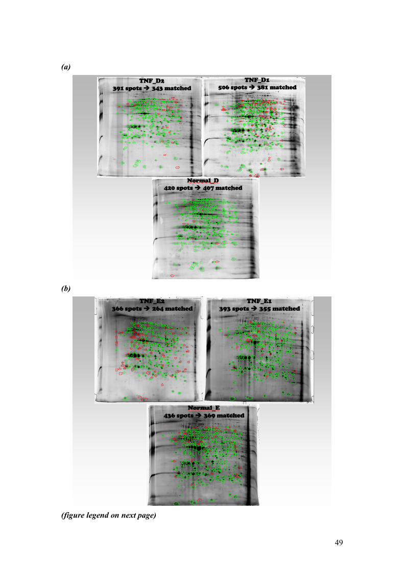
49
(a)
(b)
(figure legend on next page)

50
(c)
Fig. 13: Gels making up batch ‘1’ (a) from donor ‘D’ include the control (nonactivated neutrophils), TNF_D1 and its replicate TNF_D2; whereas batch ‘2’ (b) was made up by control, TNF_E1 and its replicate TNF_E2, all from donor ‘E’. The node (c) was generated by matching the control gels from both batches (Normal_D and Normal_E1 plus a technical replicate from the latter (Normal_E2).
Log2 transformation produces a continuous spectrum of values, whereas ratio measurements compresses the data below 1 and expands it above 1.
In the present study, the gels in a given batch were normalized by dividing the volume in each spot by the arithmetic mean volume of all spots of that given gel. The purpose of normalization was to remove, as much as possible, the effects of any systematic sources of variation such as differences in isoelectric focalization, small differences in protein load or stain development time. To measure expression differences between a TNF-treated neutrophils and the control, a logarithmic transformation was applied to the ratio. Although ratios are commonly used to measure expression differences between two proteins (that is, spot volume or intensity), this simple statistical analysis treats up- and down-expressed proteins differently. That is, proteins up-expressed by a factor of 2 have an expression ratio of 2, whereas those down-expressed by the same factor have an expression ratio of ½ (0.5). Consequently, down-expressed proteins are compressed between 1 and 0 while up-expressed proteins ranges from 0 to ∝. Transformation of the ratios to a logarithmic scale (logarithm base 2) minimizes this problem by providing a similar

51
representation of down- or up-expressed proteins and producing a continuous spectrum of values (Figure 14 and 15).
(a)
(b)
Fig. 14: Histogram of protein expression ratios for 309 spots matched between TNF-treated (TNF_D1) and control gels before (a) and after (b) Log2 transformation showing a more normalized distribution pattern after logarithmic transformation.
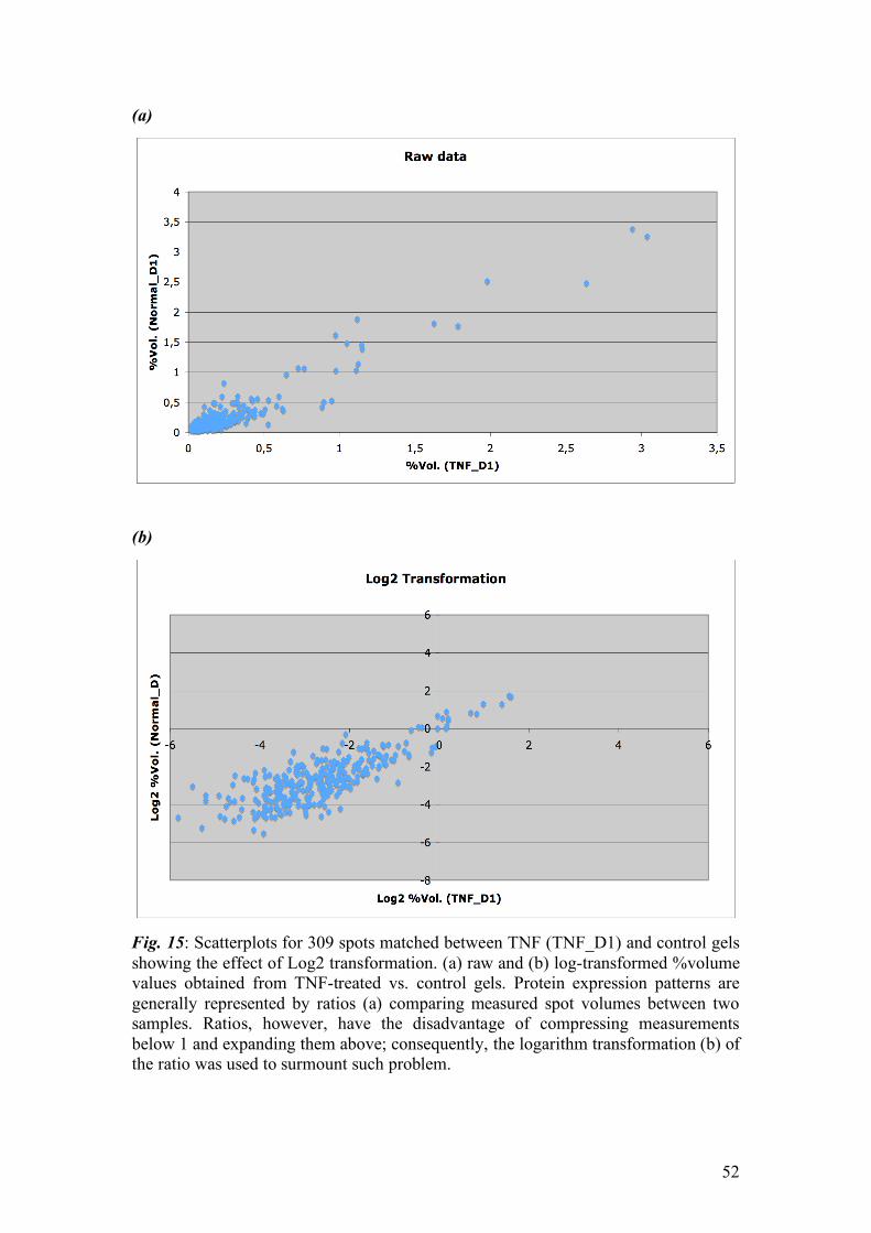
52
(a)
(b)
Fig. 15: Scatterplots for 309 spots matched between TNF (TNF_D1) and control gels showing the effect of Log2 transformation. (a) raw and (b) log-transformed %volume values obtained from TNF-treated vs. control gels. Protein expression patterns are generally represented by ratios (a) comparing measured spot volumes between two samples. Ratios, however, have the disadvantage of compressing measurements below 1 and expanding them above; consequently, the logarithm transformation (b) of the ratio was used to surmount such problem.

53
Differential Expression Analysis of 2D Gels. Differential analysis of proteins by spot volume, intensity, or area is generally handled by using commercial software packages that propose statistical tools to help to conclude on the significance of variation. Unfortunately, most of image analysis packages available still require manual editing to improve detection and matching. Thus, there is some room for user interpretation variability and errors. The ‘two-fold’ change ‘rule’ to determine statistically significant differential protein expression does not conclusively implicate in a biological significance.
In the present study, a simple differential expression gel analysis was performed to show the potentiality of differential protein expression analysis using the ‘Divide-and-Conquer’ workflow. Although ImageMaster software offers a package for such statistical analyses which includes two-sample t test, Mann-Whitney U test and the Kolmogorov-Smirnov test, these statistic tests are limited to univariate analyses of a very strict software workflow. To carry out further statistical analysis, the raw data containing information on the every spot volume was exported into Excel tables. As shown on the workflow on figure 11 and 12, after exporting data into Excel tables, each spot was normalized to mean value of all spots in the gel. Then, the normalized mean for each spot was obtained by taking the average between a given spot and a its replicate in a technical ‘replicate’ gel. The differential expression between TNF and control (normal) gels were determined by calculating the Log2 of the ratio TNF[mean]:Normal[mean]. An eminent issue in 2D gel analysis is related to the choice of arbitrary cut-offs (e.g., two-fold up- or down-regulation as significant, or equivalently, log2 (ratio) values greater than 1 or less than -1). Such arbitrary, fixed cut-offs may or may not be supported by the underlying biology. By taking into consideration the commonly used ‘two-fold change’ (equivalent to one-fold change in log2 ratio) in expression of proteins as significant, 18 and 9 proteins (outlined in figure 17) were found to be upregulated and downregulated, respectively, between TNF_D spots (mean of a technical duplicate) and their matched spots in the control gel (figure 16a). Importantly, the analysis of a biological replicate showed great variability such that most down- and up-regulated proteins from one biological replicate did not overlap with the other replicate (figure 16b).
In conclusion, despite the development of techniques for accurate identification of differentially expressed proteins and their statistical significance, extracting clear and coherent hypotheses from 2DE data remains a daunting task. Statistics cannot decide what is biologically relevant in a 2D gel experiment, but can propose objective methods based on the data, to suggest both what could be interesting, and what should be moved aside or corrected. In fact, the difficulty in 2DE analysis lies mainly in the interpretation of the biological relevance of the data. In order to assess differential expression in a way that controls both false positives and false negatives, the standard approach should be based on statistical significance and hypothesis testing, with careful attention paid to multiple comparisons issues.
The use of fold change between spot volume in different conditions has been commonly used in 2D gel analysis in an attempt to determine what is ‘biologically’ relevant (Marengo, E. et al., 2005). Nevertheless, a two-fold change in expression of a given protein between two conditions does not conclusively implicate in a biological significance of such protein in terms of differentiation between the two conditions. Transcriptomics studies have shown that stress-induced genes have a more variable expression, albeit other less induced genes may present a greater biological relevancy

54
(Dudoit, S. and Speed, T. et al., 2000). A statistically more powerful approach is to calculate the mean µ and standard deviation σ for the distribution of Log2 (ratio) values. Differential expression at the 95% confidence level can then be identified as Log2 (ratio) values more than 1.96 standard deviations from the mean. In fact, a handful statistic tests have emerged mainly from the microarray community for analysis of long and lean datasets such as 2D gel data.
(a)
(b)
Fig. 16: Histogram of protein expression (a) log2 ratio (TNF_D[mean]:Normal) and (b) log2 ratio (TNF_E[mean]:Normal) showing the number of up- and down-regulated (bars in dark blue) proteins considering a cut-off of one-fold in log2 ratio. Values for spot %Vol in this analysis were obtained after averaging two technical replicate for each, TNF_D and TNF_E gels. Most proteins found up- or down-regulated in one of the biological replicates (e.g., TNF_D) do not follow up in the other replicate (e.g., TNF_E) in terms of number and match.

55
The transformation of the ratio to Log2 and application of more statistic-oriented tests for differential expression are promising for gel analysis. In summary, there is still a need for more powerful tools that can help to rationalize the decisions on significance and can draw attention on the imperfections of gels, spot mismatches and other artifacts of 2-DE gels. This study was not meant to find appropriate approaches for statistical treatment of 2D gel data, yet to demonstrate the potentiality of the ‘Divide-and-Conquer’ strategy in terms of simplicity.
Fig.17: Differentially expressed spots obtained from figure 16a. The proteins with log2 ratio (TNF_D[mean]:Normal_D) greater or lower than one-fold are outlined on Normal_D gel.

56
[4.4] Subcellular Fractionation of Cytosolic and Plasma Membrane Compartments Rationalizing Subcellular Fractionation in Neutrophils – an In Silico Analysis - Neutrophils contain effector proteins of the innate immune system, which are stored in granules and mobilized in response to various immunoregulatory mediators (Faurschou, M. and Borregaard, N. 2003; Borregaard N. and Cowland J. 1997; Lominadze, G. et al., 2005). Synthesis of granule and secretory vesicle proteins is initiated in early promyelocytes, and ends in segmented neutrophils. During neutrophil circulation as a naïve cell, synthesis of granule and vesicle proteins comed to a halt, and if no activation stimulus is given to the neutrophil during circulation, programmed cell death is ensued. Upon activation, neutrophils discharge their microbicidal arsenal stored in granules and vesicles to phagosomes or the extracellular environment in a stepwise and highly orchestrated fashion. Granule membrane proteins can be added to the phagosome or plasma membrane membranes, whereas granule luminal contents are released into the extracellular space or phagocytic vacuole. Therefore, granule content accrued during cell development is thought to remain unchanged in terms of new synthesis of proteins. In this regard, granule and vesicle proteins subcellular localization can be seen as the functional proteomics of neutrophils; that is, the mobilization of these proteins from one compartment to another will determine the phenotype of the neutrophil. Secretory vesicles for example fuse with the plasma membrane of neutrophils in response to a wide variety of inflammatory stimuli (Faurschou, M. and Borregaard, N. 2003). This fusion results in the incorporation of a spectrum of membrane-associated receptors including the β2-integrin CD11b/CD18 (Mac-1, CR3) (Segelov, H. et al., 1993), and the metalloprotease leukolysin (Kang, T. et al., 2001), both of which are essential for neutrophil exocytosis. At the cell signaling transduction level, neutrophil phenotype (e.g., granule discharge, NAPH oxidase assembly and chemotaxis) is finely regulated by signals received by membrane receptors that are then relayed into the cell interior by a combination of small intracellular signaling molecules such as diacylglycerol, cyclic AMP and Ca2+, and intracellular signaling proteins, most of which are located primarily in the cytosol. The resulting chain of intracellular signaling events ultimately alters target proteins, resulting in a specific cellular behavior. For future work in neutrophil signaling transduction, we have hypothesized that most of proteins involved in signaling transduction leading to neutrophil migration, extravasation, phagocytosis, degranulation and apoptosis reside in cytosol or plasma membrane. To corroborate with this hypothesis, DNA microarray data from neutrophils of septic patients (Coldren, C et al., 2006) were re-analyzed in silico in order to (1) determine the subcellular localization of the most upregulated genes and compare this list with the proteins found by Lominadze et al., in neutrophil granules; and (2) find potential protein phosphorylation in each subcompartment. The findings of this analysis are summarized on table 3. It is noteworthy that the 357 genes (almost 40%) out of 912 of most upregulated genes pos-endotoxin stimulation have their protein cognate localized on the cytoplasm (268 primarily localized on cytoplasm and 89 primarily localize on plasma membrane or nucleus and with alternative cytoplasmic localization). Similar analysis for plasma membrane proteins reveals that a total number of 267 genes (approximately 30%) out of the same 912 most upregulated genes have their protein cognate localized on the plasma membrane (183 primary to the plasma membrane and 84 alternatively localized on the membrane).

57
Table 3: In silico re-analysis summary of the 912 most upregulated genes reported by Coldren et al., The table contains the primary and alternative localization for the 912 most upregulated genes from the microarray analysis of lung-infiltrating neutrophils (compared to circulating neutrophils) pos-endotoxin administration to healthy human subjects.
Cellular Compartment Primary Localization
Alternative Localization
Proteins Potentially
Phosphorylated
Cytoplasm & Cytoskeleton 268 89 149
Plasma membrane 183 84 6
Source of Protein Localization and potential phosphorylation: Human Protein Reference Database (www.hrpd.org) and Phospho Site (www.phosphosite.org). In another comparative analysis, the list of proteins found in the three granule subsets of neutrophils (Lominadze, G. et al., 2005) was cross-examined against the list of proteins found during phagosome maturation in neutrophils (Burlak, C. et al., 2006). Notably, nineteen proteins from a list of 29 host defense proteins found in neutrophil phagosomes (Burlak, C. et al., 2006) have also been identified by proteomics in at least one of the three granule subsets (Table 4). This indicates that the key role that granule proteins play in delivering its microbicidal arsenal to the phagosomes by membrane fusion. Other proteins typically found in the ER (e.g., calnexin, ERp29, GRP58/ERp57, GRP78/BiP, and PDI) and mitochondria (e.g., F1 ATPase subunits, peroxiredoxin 3 [PRDX3)] and prohibitin) were also found associated with neutrophil phagosomes. The finding that neutrophil phagosomes contain components of the three subsets of granules, endoplasmic reticulum and mitochondria evokes a complex picture of the phagosome formation and maturation. Although the application of high-scale proteomic technologies has accelerated the discovery of many proteins that play important roles in neutrophil function such as phagocytosis, a central challenge now will be to integrate these proteins into pathways and networks that account for the diversity of neutrophil function. High-scale gene expression profiling has become an important tool for investigating transcriptional activity in a variety of biological samples. DNA microarray-based gene expression profiling relies on nucleic acid hybridization and the use of nucleic acid polymers, immobilized on a solid surface, as probes for complementary gene sequences. This prominent experimental tool offers a snapshot of the gene expression in a given sample, permitting the measurement of the level of expression of thousands of genes simultaneously. Despite the technical (robustness, cost and throughput) advantages of using DNA microarrays over proteomics technologies, not all transcripts generate protein, and alternate translation efficiency and posttranslational turnover may result in differential protein accumulation. These differences may underlie at least in part the modest correspondence reported between quantitative measurements of gene expression and cognate protein levels (Gygi, S. et

58
al., 1999). Therefore, caution should be applied when comparing genes and their protein cognate.
Table 4: Comparison between two subcellular proteomic studies. The first column corresponds to the 29 host defense proteins from Burlak et al. and the second column whether the protein was found in one of the three granule subsets in Lominadze et al. Abbreviations: GE – gelatinase granules, SP – specific granules and AZ – azurophil granules. “Rejected protein” means that the protein was identified but did not pass the trustworthy criteria.
Phagosome host defense proteins Granule proteins Anti-inflammatory factor 1 (AIF-1) Not found Arginase 1 (ARG1) Not found BPI GE, SP, AZ Calgizzarin Gelatinase granule rejected protein Calgranulin A Not found Calgranulin B Not found Calgranulin C SP Calprotectin L1L subunit Not found Cathepsin G GE, SP, AZ Cathepsin G-like Not found Cathepsin S Not found Azurocidin 1; CAP37 GE, SP, AZ Chitinase 1; chitotriosidase GE, SP, AZ Defesin α1 preproprotein GE, SP Defesin α3 precursor Not found Elastase 2 SP, AZ Eosinophil preperoxidase GE, SP, AZ Cathelicidin antimicrobial peptide; CAP18 Not found Ficolin GE, SP Lactoferrin GE, SP, AZ Lipocalin; NGAL GE, SP, AZ Lysozyme C GE, SP, AZ MMP8; neutrophil collagenase SP Myeloperoxidase (MPO) GE, SP, AZ Myeloperoxidase (pre-pro-MPO) Not found Myeloperoxidase, chain B (light chain) GE, SP, AZ Myeloperoxidase, chain D (heavy chain) GE, SP, AZ Proteinase-3 GE, SP, AZ Ribonuclease, mase A family 3; eosinophil cationic protein
Specific granule rejected protein
Abbreviations: GE – gelatinase granules, SP – specific granules and AZ – azurophil granules. “Rejected protein” means that the protein was identified but did not pass the trustworthy criteria determined by the authors of the article.
From the abovementioned comparisons, it is obvious that proper biological

59
activity and cellular homeostasis depend on spatially and temporally restricted partitioning of functionally related sets of proteins. In fact, many specialized functions are compartmentalized within organelles. Nevertheless, there is an important caveat in analyzing a given protein at one time point, namely the dynamic nature of proteins. Cell compartments are composed of proteins that are present almost all of the time ('resident' proteins), proteins that are in transit and proteins that transiently interact with the organelle to carry out defined functions. In fact, many proteins seem to have an alternative subcellular localization to its primary one. Hence, whether a given protein will be associated with a specific organelle may depend on very tightly regulated temporal window during the cell signaling process.
[4.5] Optimizing Cytosolic Protein Enrichment in Neutrophils Sample preparation is one of the most crucial stages in proteomics analysis. Proper experimental model and careful sample preparation is vital to obtain significant and trustworthy results. The power of most of proteomic platforms is such that minimal contamination with proteins that are not bona fide components of the enriched compartment will be equivocally identified as being a part of that proteome.
The success of proteomic analysis depends very much on the complexity of samples, sample preparation methods and analytical methods. Currently, the proteomic approaches using many strategies and technologies show certain limitations of detectability. In the past years, efforts in reducing these limitations and enhancing detectability have been a major focus of several proteomics groups worldwide. The technical development in the fields of sample preparation, protein separation and identification, and bioinformatics can be integrated to solve the limitations in proteome studies and to improve the effective proteomic approaches for determining the differentially expressed proteins or novel biomarkers. Digitonin extraction is highly effective for cytosolic proteins Cytosolic proteins from neutrophil were fractionated by repeated washes in digitonin buffer containing sucrose, PIPES, MgCl2, protease and phosphatase inhibitors. To confirm the effectiveness of the method, four goals were established here. The first method based on the quantitative measurement of proteins from the digitonin fractions. On average, the first digitonin wash generated approximately 210 µg of protein per 3x10⁶ neutrophils. Since the subsequent digitonin washes did not generate a satisfactory amount of proteins to perform 2DE analysis, only the first wash was processed further downstream this point. In the second validation approach, the neutrophils were imaged by confocal microscopy to confirm that, after digitonin treatment, the cells retain their morphology, but become impermeable. It was hypothesized that if the digitonin-treated cells were incubated with the DNA-intercalating dye propidium iodide, which normally does not permeate neutrophils, the nuclei would appear stained only if the digitonin form ‘holes’ on the cell membrane. To create a contrast between the nucleus and the cell cytoplasm, the neutrophils were also incubated with the membrane permeable rhodamine 123 (DHR 123 pre-oxidized, see material and methods for oxidative burst flow cytometry analysis). Figure 18 shows that after digitonin treatment the neutrophil nucleus stained with the propidium iodide dye forming a multibolular shape as commonly described in the literature. In addition, the rhodamine was nicely scattered throughout the cytoplasm. The third method to evaluate effectiveness of the extraction method

60
based on the comparison of the 1D gel electrophoresis patterns between cytosolic fraction and the remaining of the neutrophil proteins. Clearly, the 1D gel pattern of the cytosolic proteins differs from that of the remaining proteins (figure 19).
Fig. 18: Confocal optical sections (s.1-s.6) of digitonin-treated neutrophils incubated with rhodamine 123 and propidium iodide. White bar = 10µm.
Fig. 19: Coomassie-stained 1D gel of cytosolic fraction (lanes 1 and 2) and membrane/organelle fraction (lanes 3 and 4). Protein load: lane 1 (50µg), lane 2 (10 µg), lane 3 (10 µg) and lane 4 (10 µg).

61
Although highly useful, the abovementioned experiments lack a conclusive
assertiveness for the enrichment strategy. The ultimate test is, in fact, a work in progress, and consists in the identification of the enriched proteins followed by literature and/or experimental confirmation of the subcellular localization of that protein. To this end, enriched proteins were fractionated by 2D gel electrophoresis, the spots were excised digested and identified by mass spectrometry (MALDI-TOF/TOF). To date, 15 proteins have been identified (figure 20) with a high degree of confidence (table 5). Most notably, all identified proteins have been reported to be primarily located in the cytoplasm.
Fig. 20: Spots outlined were excised for further charaterization by mass spectrometry (MALDI-TOF/TOF).

62
Table 5: Proteins identified by mass spectrometry in this study. Out of 20 spots excised from the 2D gel, 15 were identified with a high degree of confidence. Spot number corresponds to the spots highlighted in figure 20. For spots number 4, 6 and 15, the information gathered from MS and MSMS were not sufficient to distinguished between different isoforms. Detailed information on each identification is given on Appendix 1.
Protein
Name
Mascot
Score
PMF
Coverage (%)
Number of Peptides
Sequenced /
Coverage (%)
Cellular
Compartment
1. β-Actin 311 64% 3/16% Cytosol
2. γ-Actin 280 66% 3/16% Cytosol
3. Annexin A3 153 56% 2/5% Cytosol
4. Annexin A1 isoform B 4. Annexin A1 isoform C
297 289
44 % 40 %
2/10% 2/10%
Cytosol
5. S100-A9 209 67 % 3/37% Cytosol
6. Annexin A5 isoform C
6. Annexin A5
164
164
84 %
82 %
2/6%
2/5%
Cytosol
7. L-plastin isoform 2 342 57 % 2/4% Cytosol
8. Enolase 1 147 49 % 3/10% Cytosol
9. Phosphoglycerato mutase
188 47 % 2/11% Cytosol
10. Proteinase 3 151 48 % 2/7% Cytosol
11. Peptidyl isomerase A 224 69 % 3/28% Cytosol
12. Cofilin 1 128 67 % 1/10% Cytosol
14. Annexin A6 126 19 % 2/3% Cytosol
15. Gelsolin isoform A
15. Gelsolin isoform B
108
111
22 %
23 %
1/2%
1/3%
Cytosol
20. Gluthatione-S transferase P
195 54 % 3/21% Cytosol
Digitonin extraction of cytosolic proteins confers a high degree of reproducibility A high degree of gel-to-gel variation in spot patterns can make comparative 2-DE analysis difficult to achieve, and any true biological variation could be masked or amplified by experimental variation. Slight technical variability from gel to gel inherent to the sample preparation often yields deviations in the number and volume

63
of spots. Gel heterogeneities caused by sample preparation must be minimized and methods that lack reproducibility are in general not suitable for proteomics analysis. To assess the reproducibility of the proposed method for cytosolic proteins enrichment, sample preparation procedure (including blood withdrawn, neutrophil purification and cytosolic protein extraction) were carried out separately (in different days). To minimize the influence of the biological variability as much as possible, the same sample donor for the three replicates was used. Then, every step in the preparation procedure for 2DE and staining were carried our in parallel to reduce 2D gel experimental variability.
A high degree of reproducibility was reached among the three sample preparations. As shown in figure 21, gels ‘1’ and ‘3’ have respectively 70% and 75% of their spots matched with gel ‘2’. Importantly, this analysis was executed uniquely with the ‘automatic matching’ features of the ImageMaster Platinum software, with no manual editing (operator editing) carried out. Manual matching can significantly increase the degree of matching among the gels. Moreover, spot volumes among the three gels showed excellent Pearson correlation coefficient (table 6).
Fig. 21: Digitonin extraction provides a high degree of reproducibility. Automatic matching generated by ImageMaster Platinum software (v.6) assigned matches in 70 and 75% of the spots present in gels ‘1’ and ‘3’, respectively. Spots outlined in green represent the matches in each gel, while spots in red are unmatched. Blue vectors represent the direction of the match between the reference and the matched gel.
In the present Thesis, a simple, fast subcellular fractionation method for cytosolic protein extraction of neutrophils was developed taking as the optimal

64
parameters the number of cytosolic proteins identified, the reproducibility of separation, and the sensitivity of staining detection. The results presented here suggest that this method offers a great efficiency for enriching cytosolic protein from neutrophils, and excellent reproducibility. The percentage of cytosolic proteins identified using the digitonin extraction method was 100%; that is, thus far, all 15 proteins identified by mass spectrometry are cytoplasmic resident proteins. The other parameter taken into consideration during the optimization process, namely reproducibility in spot volumes among the 2D gels, was also quite satisfactory. However, the small variability shown for some of the spots among the replicates could not be discerned whether it came from sample preparation or the staining procedure.
Table 6 Pearson correlation coefficient between any two digitonin gels. Pairwise comparisons of 389 spots (%vol) present in all three gels.
Gel_1 Gel_2 Gel_3
Gel_1 1
Gel_2 0,856424824 1
Gel_3 0,904034608 0,749812798 1 The optimal concentration of 100 µM digitonin affects selectively the
cytoplasmic membranes of neutrophils, releasing the cytosolic proteins with little contamination from the organelles or plasma membrane. Extraction of the remaining cellular extracts with sequential washes with the same concentration of digitonin was ineffective for releasing more proteins as shown by protein quantification of the different washing fractions. Treating the left over pellet with 0.5% of Triton X-100 allowed the recovery of a different set of proteins, which has not been identified yet. As the extraction using digitonin neither denature nor alter the conformation of proteins, cytosolic protein complexes would be expected to be recovered, which would enable future studies of protein-protein interactions.
Other clear advantage this method is that it provides some clues on protein subcellular localization. That is, proteins identified from the digitonin-extracted fraction are likely to reside or transitorily reside in the cell cytoplasm. Overall, the results presented here clearly suggest that subcellular fractionation by digitonin extraction offers great efficiency and reproducibility, least time consuption, and least cost. This protocol is robust and may be well-suited for analyses of other complex specimens.

65
5. Conclusions The study of signaling transduction in neutrophils and current proteomics approaches for biomarker discovery pose a significant technical challenge. Although a variety of proteomic approaches have been attempted so far, the number of protein identifications has been quite modest in many studies. The selection of an appropriate sample preparation method is important for confident results. Moreover, clinical proteomics studies have special needs, namely practicability and feasibility of the sample preparation methods in the clinical context. To this end, we have implemented a sample preparation platform comprising of a simple neutrophil purification method (Percoll gradient purification) and a highly robust and practical technology for cellular characterization at the cellular and molecular levels (flow cytometry). Overall, both methods meet the sample preparation requirements for clinical and proteomics studies, namely simplicity, reproducibility, yield and purity of the preparation.
In the recent years, a general trend towards subcellular proteomics analysis in many different cell types, particularly in neutrophils, has emerged as a powerful and promising alternative to the whole cell proteomics studies. In the present study, we have demonstrated the feasibility of cytosolic protein enrichment from human neutrophils. By reducing sample complexity, we were able to achieve highly reproducible 2D gel patterns. Overall, digitonin extraction is simple, reproducible and highly specific for cytosolic proteins. In addition, a novel workflow for 2D gel analysis has been proposed in this thesis, namely the ‘Divide-and-conquer’ workflow. Perhaps, the major advantages of this novel approach are the improved matching capacity, the reduced gel variability, and the significant reduction in time dedicated for gel analysis. The small degree of variability still observed after the application of the ‘Divide-and-conquer’ workflow may be caused by either technical 2D electrophoresis limitations or gel staining issues. In the present study, ammoniacal silver staining protocol was used to increase sensitivity of detection. Generally speaking, ammonical silver protocols use fast staining development steps (2 min or less), which are difficult to make very reproducible. This may be a problem when staining consistency is a critical parameter since the gel-to-gel reproducibility may be reduced.

66
6. Perspectives The clinical utility of proteomics is a promising new field that has the potential to have many applications, including the identification of biomarker for early diagnosis and prognosis, disease monitoring, and prediction of response to therapy. Further advancement of proteomics technologies could usher in an era of personalized molecular medicine, where diseases are diagnosed at earlier stages using robust molecular markers. Then, the clinicians would use the patient’s specific proteomic profile to rationally tailor and monitor molecular therapy. In clinical proteomics studies, samples from normal and disease subjects must be compared to identify the protein (or a panel of them) differentially expressed between the two conditions. Moreover, expression proteomics may shed some light into our understanding of the disease progression. Although the challenges are great for global analysis of proteomes, the proteomic approaches for reduction of limitations and enhancement of proteomic detectability exposed in the present Thesis have the potential to be translated into the clinical settings to study neutrophil-related diseases in the proteomic context.
The importance of a better understanding of the mechanisms of neutrophil signaling transduction during sepsis development has been highlighted by several experts in the field over the past decades (see Introduction). In this regard, flow cytometry and proteomics have emerged as suitable tools designed to address this need from the level of signal transduction, biomarker discovery and validation, and phenotype analysis. Currently, a multicenter study proposed to profile cytosolic and membrane proteins differentially expressed among healthy subjects, patients with severe sepsis, and patients with shock septic is under development. This study was projected in a subcellular strategy that divides the samples into subproteomes to be further analyzed by proteomics and immulogical approaches.

67
Resumo da dissertação em português.
Pesquisas biomédica comumente começa com a elaboração de uma hipotese para resolver um problema. Nesse contexto, cientistas selecionam o(s) metodo(s) mais apropriado(s) para responder a questão e solucionar um dilema. Nos últimas anos, nós temos testemunhado uma revolução de novas tecnologias em biologia molecular – a ciência -Omica. Tecnologias de alta-escala tais como metabolômica, citômica, genômica e proteômica estão mudando o modo que estudamos sistemas biológicos complexos. Métodos contemporâneos para o entendimento da diversidade funcional em um dado organismo preferencialmente abordam uma visão de biologia de sistemas onde primeiro a classificação fenótipa de um citoma é alcançado antes de uma tentativa de caracterizar o proteoma de tal célula. Dentro desse contexto, e para proporcionar um melhor entendimento dos componentes envolvidos na ativação e morte celular programada dos neutrófilos nos estados patológicos e sano, esse estudo propõe a integração de métodos em biologia celular tal como citometria de fluxo com uma robusta plataforma proteômica em uma tentativa de integrar dados a nível molecular com dados fenótipicos de neutrófilos. Fracionamento subcelular usando um método de extração e enriquecimento de proteínas citosólicas com o detergente digitonina foi otimizado nesse trabalho, e encontrado ser altamente reprodutível, fácil de realizar, e de baixo custo. Objetivo O objetivo geral dessa dissertação de Máster é a construção de uma plataforma sólida e robusta para integração de pesquisa básica em proteômica e pesquisa médica. Para tal objetivo, uma série de otimizações para processamento de amostras foram propostas para avaliar nossa capacidade de gerar dados biológicos de alta qualidade das amostras trabalhadas. As metas principais são as seguintes:
1. Implementar um método efetivo e de baixo custo para purificação de neutrofilos humano no Laboratório de Bioquímica e Química de Proteínas na Universidade de Brasília.
2. Desenhar e provar uma abordagem prática para avaliar características funcionais e morfológicas dos neutrofilos sob análize.
3. Determinar as condições apropriadas e estratégias para estudos proteomicos de neutrofilos. Esta meta foi subdividida em outras duas “metas específicas”:
a. Padronizar as condições optimas para eletroforese bidimensional do proteoma total do neutrofilo
b. Examinar a viabilidade de realizar fracionamento subcelular do proteoma dos neutrofilos.
Resultados Citômica Purificação com Percoll em gradientes de duas camadas proporciona uma população altamente pura de neutrofilos sem afetar o estado original de repouso dos neutrofilos. No geral, nosso protocolo de separação de neutrofilos humano direto do sangue é altamente reproduzível, e produz uma alta quantidade de células. Utilizando apenas duas camadas de Percoll (a 57% and 67%), neutrofilos foram separados com um grau de pureza superior a 96%, e uma viabilidade de aproximadamente 98%.

68
Agentes pro-inflamatorios, em particular fMLP, evocam a geração de espécies reativas do oxigênio (EROs) em neutrofilos. Em comparação com outros autores (Botha, A et al. 1995) que hipotetizam um efeito estimulante (‘priming effect’) de certos agentes pro-inflamatórios, nossos resultados e testes in vitro com citometria de fluxo desafia esta hipótese já que não encontramos um aumento significativo quando os neutrofilos foram pré-ativados (‘primed’) com TNFα ou PAF seguido de ativação com LPS ou fMLP, respectivamente. Esses resultados estão representados na figura 10 da dissertação. Proteômica Devido a variabilidade técnica comumente observada em experimentos de géis bidimensionais, nesse estudo, uma nova metodologia/fluxograma para analise de gel bidimensional foi empregada, sendo nomidada ‘Divide-e-Conquiste’. Ao contrario de métodos mais tradicionais utilizados em programas de análise de imagem de géis bidimensionais, nosso método consiste no pareamento de géis dentro de um “grupo”, que tem em comum o fato que correram juntos; isto é, focalização isoelétrica, separação electroforética em gel de poliacrilamida e coloração foram realizadas concomitantemente. Essa estratégia pode ser utilizada com vários grupos de géis. Dois fluxogramas dessa estratégia estão representado na figura 11 e 12 da dissertação. No geral, os géis que fazem parte de um grupo, ou seja, que correram juntos, apresentaram melhor reproducibilidade em termos de pareamento (fig. 13a e 13b) em comparação a géis pareados de dois grupos distintos (fig. 13c). Tratamento estatístico dos dados para análise de géis bidimensionais é uma das etapas mais importantes para extração de resultados significantes. Dois passos foram encontrados serem altamente relevantes para análise estatística nesse trabalho. O primeiro, a normalização do volumes das manchas (spots) selecionadas para análise é capaz de remover os efeitos de erros sistemáticos devidos a problemas técnicos tais como diferenças em focalização isoelétrica, em coloração, entre outros problemas. Outro passo extremamente vantajoso é a transformação das taxas de expressão diferencial (relação volume da mancha gel A:volume da mancha gel B) de cada pareamento em uma função logarítmica na base 2. Embora taxas de expressão sejam largamente utilizadas em análise de géis bidimensionais, esse tipo de análise trata dados de sub- e sobre-expressão diferencial diferentemente. Proteínas duas vezes mais sobre-expressas apresentam uma taxa de 2, enquanto proteínas duas vezes sub-expressas apresentam uma taxa de ½ (0.5). Consequentemente, proteínas sub-expressas estarão comprimidas entre um valor 1e 0, enquanto proteínas subre-expressas apresentarão um valor entre 0 e ∝. Transformação das taxas de expressão para uma escala logarítmica, tal como Log2, minimiza esse problema gerando uma representação similar do que esta sub- e sobre-expresso, e com um espectro de valores em forma contínua (ver figuras 14 e 15). A manutenção homoestática celular depende em grande parte de uma funcionalidade protéica regulada em tempo e espaço. Nesse contexto, uma grande maioria das funções celulares especializadas são compartimentalizadas; isto é, acontecem dentro de um especifico compartimento celular. Uma importante limitação dos estudos proteômicos portanto refere-se a natureza dinâmica das proteínas. Em outras palavras, caracterização de uma dada proteína a um especifico ponto em tempo é uma análise incompleta que não revela com exatidão a função dessa proteína dentro de um processo. No geral, proteínas apresentam uma alto dinamismo em termos de localização subcelular que em muitos casos esta diretamente relacionado com a

69
função de uma proteína. Do ponto de vista técnico, o sucesso de separação de proteínas em géis bidimensionais esta diretamente relacionado, entre outros fatores, com a complexidade da amostra. Uma abordagem que tem ganhado especial atenção na comunidade proteômica nos últimos anos para redução da complexidade da amostra é a Análise Proteômica Subcelular. Nesse tipo de análise, compartimentos subcelulares são separados com um alto grau de pureza (mínimo de contaminação), e então o proteoma de cada fração é analisado separadamente. Uma vantagem adicional desse tipo de análise, além da redução da complexidade, é a obtenção de informação sobre a dinâmica das proteínas em termos de sub-localização.
Em termos de sinalização celular, neutrófilos, assim como a maioria das células vivas, são regulados através de sinais recebidos por proteínas de membranas, esses sinais são traduzidos em funções por uma rede de proteínas presentes principalmente na fração citosólica das células. Nesse trabalho de dissertação e para trabalhos futuro nesse campo, nós hipotetizamos que a maioria das proteínas com um aumento ou diminuição significativa nos níveis de expressão estariam co-localizadas nas frações citosólicas e membranicas. Para corroborar tal hipótese, dados provenientes de microarranjos de ADN de neutrófilos de indivíduos após administração de endotoxina (Coldren, C. et al., 2006) foram re-analisados in silico com o intuito de caracterizar a localização teórica das proteínas referentes aos genes mais expressos (tabela 3). Interessantemente, cerca de 40% dessas proteínas tem localização primária ou secundária no citoplasma. Com esse fato em mente, um método para extração e caracterização de proteínas citosólicas foi desenhado e otimizado. Dentre os diversos métodos existentes para fracionamento subcelular, nós elegemos o método com digitonina para purificar proteínas citosólicas devido a simplicidade, baixo custo desse método. Digitonina é um detergente não-ionico que se liga especificamente e rapidamente a esteróis, precipitando-os, e permitindo portanto a formação de poros na membrana e a liberação do proteínas citosólicas.
Para confirmar a efetividade do nosso método de fracionamento para proteínas citosólicas, quatro metas foram estabelecidas. Primeiramente, através de uma medida da quantidade de proteínas gerada após o fracionamento com digitonina, a concentração ideal e o número de lavagens necessários foram determinados. Em resumo, uma concentração de digitonina de 100 µM e apenas uma única incubação de 10 minutos foram suficientes para extração de proteínas de citosol. Com o segundo método de verificação, microscopia confocal, verificou-se que após tratamento com digitonina, a integridade do núcleo e o ‘contorno’ celular do neutrófilo são mantidos (fig. 18). O padrão do proteoma da fração citosólica foi também perfilado por separação em gel de poliacrilamida (1D gel) e comparado com a fração restante (membranas e organelas) (fig. 19). E finalmente, o teste mais conclusivo foi realizado com identificação de um número de proteínas presentes na fração citosólica. Esse experimento foi realizado inicialmente com a separação das proteínas da suposta fração citosólicas por electroforese bidimensional, seguido por excisão de 20 manchas aleatoriamente escolhidas (fig. 20), e identificação das proteínas por espectrometria de massa (MALDI-TOF/TOF). De 20 manchas digeridas e analisadas, 15 proteinas foram identificadas com um alto grau de confiabilidade (MASCOT > 100, e com 1 a 3 peptideos sequenciados) (tabela 5). De suma importância, todas as proteínas identificadas são primariamente, e algumas secundariamente, presentes no citosol segundo análise literária.
Como já mencionado acima, uma das principais e mais conhecidas limitações em análise de géis bidimensionais esta vinculada a problemas de reproducibilidade.

70
Heterogenidade causada durante preparação da amostra deve ser minimizadas, e métodos que violam essa regra devem ser considerados inapropriados para estudos proteômicos. Para avaliar reproducibilidade do nosso método de enriquecimento de proteínas citosólicas, o procedimento de purificação de neutrófilos e extração de proteínas citosólicas foi repetido em três vezes cada um em dias diferentes. Para minimizar a influência causada pela variabilidade biológica, as amostras para cada preparação foram obtidas do mesmo individuo. E também, para minimizar a variabilidade oriunda da electroforese bidimensional, todas as etapas de electroforese e coloração foram realizadas em um único experimento. Em resumo, um alto grau de reproducibilidade foi alcançado em termos de padrão apresentados pela triplicata de géis. Mais de 70% de manchas entre os três géis foram pareadas utilizando unicamente recursos automáticos do programa de análise de imagem conhecido como ImageMaster Platinum (Amersham Bioscience, parte da GE Healthcare) (fig. 21). Obviamente, edição manual do pareamento produziria um aumento significativo no número de pares entre os três géis. Outro resultado significativo em termos de reproducibilidade é a correlação positiva (calculada como o coeficiente de correlação de Pearson) entre o volume normalizado de cada mancha em um gel e seu par correspondente em outros géis (tabela 6).

71
7. References Abdolzade-Bavil, A., S. Hayes, et al. (2004). "Convenient and versatile subcellular extraction procedure, that facilitates classical protein expression profiling and functional protein analysis." Proteomics 4(5): 1397-405.
Abraham, E. (2003). "Neutrophils and acute lung injury." Crit Care Med 31(4 Suppl): S195-9.
Adams, J. M., C. J. Hauser, et al., (2001). "Early trauma polymorphonuclear neutrophil responses to chemokines are associated with development of sepsis, pneumonia, and organ failure." J Trauma 51(3): 452-6; discussion 456-7.
Aebersold, R. and M. Mann (2003). "Mass spectrometry-based proteomics." Nature 422(6928): 198-207.
Alban, A., S. O. David, et al., (2003). "A novel experimental design for comparative two-dimensional gel analysis: two-dimensional difference gel electrophoresis incorporating a pooled internal standard." Proteomics 3(1): 36-44.
Anderson, N. L. and N. G. Anderson (2002). "The human plasma proteome: history, character, and diagnostic prospects." Mol Cell Proteomics 1(11): 845-67.
Antal, P., S. Sipka, et al. (1995). "Flow cytometric assay of phagocytic activity of human neutrophils and monocytes in whole blood by neutral red uptake." Ann Hematol 70(5): 259-65.
Arnljots, K., O. Sorensen, et al., (1998). "Timing, targeting and sorting of azurophil granule proteins in human myeloid cells." Leukemia 12(11): 1789-95.
Avram, D., E. P. Romijn, et al., (2004). "Identification of proteins in activated human neutrophils susceptible to tyrosyl radical attack. A proteomic study using a tyrosylating fluorophore." Proteomics 4(8): 2397-407.
Babior, B. M. (1999). "NADPH oxidase: an update." Blood 93(5): 1464-76.
Babior, B. M., J. D. Lambeth, et al., (2002). "The neutrophil NADPH oxidase." Arch Biochem Biophys 397(2): 342-4.
Bainton, D. F. (1973). "Sequential degranulation of the two types of polymorphonuclear leukocyte granules during phagocytosis of microorganisms." J Cell Biol 58(2): 249-64.
Bainton, D. F., J. L. Ullyot, et al., (1971). "The development of neutrophilic polymorphonuclear leukocytes in human bone marrow." J Exp Med 134(4): 907-34.
Baldwin, M. A. (2005). "Mass spectrometers for the analysis of biomolecules." Methods Enzymol 402: 3-48.
Baldwin, S. R., R. H. Simon, et al., (1986). "Oxidant activity in expired breath of patients with adult respiratory distress syndrome." Lancet 1(8471): 11-4.

72
Bassoe, C. F., I. Smith, et al. (2000). "Concurrent measurement of antigen- and antibody-dependent oxidative burst and phagocytosis in monocytes and neutrophils." Methods 21(3): 203-20.
Bijleveld, C. and M. J. Geelen (1987). "Measurement of acetyl-CoA carboxylase activity in isolated hepatocytes." Biochim Biophys Acta 918(3): 274-83.
Blonder, J., M. B. Goshe, et al. (2002). "Enrichment of integral membrane proteins for proteomic analysis using liquid chromatography-tandem mass spectrometry." J Proteome Res 1(4): 351-60.
Blonder, J., M. B. Goshe, et al., (2002). "Enrichment of integral membrane proteins for proteomic analysis using liquid chromatography-tandem mass spectrometry." J Proteome Res 1(4): 351-60.
Borregaard, N. and J. B. Cowland (1997). "Granules of the human neutrophilic polymorphonuclear leukocyte." Blood 89(10): 3503-21.
Borregaard, N., J. M. Heiple, et al., (1983). "Subcellular localization of the b-cytochrome component of the human neutrophil microbicidal oxidase: translocation during activation." J Cell Biol 97(1): 52-61.
Borregaard, N., L. Kjeldsen, et al., (1994). "Changes in subcellular localization and surface expression of L-selectin, alkaline phosphatase, and Mac-1 in human neutrophils during stimulation with inflammatory mediators." J Leukoc Biol 56(1): 80-7.
Botha, A. J., F. A. Moore, et al., (1995). "Postinjury neutrophil priming and activation states: therapeutic challenges." Shock 3(3): 157-66.
Boussac, M. and J. Garin (2000). "Calcium-dependent secretion in human neutrophils: a proteomic approach." Electrophoresis 21(3): 665-72.
Bradford, M. M. (1976). "A rapid and sensitive method for the quantitation of microgram quantities of protein utilizing the principle of protein-dye binding." Anal Biochem 72: 248-54.
Brazil, T. J., A. G. Rossi, et al., (1998). "Priming induces functional coupling of N-formyl-methionyl-leucyl-phenylalanine receptors in equine neutrophils." J Leukoc Biol 63(3): 380-8.
Brealey, D. and M. Singer (2000). "Multi-organ dysfunction in the critically ill: effects on different organs." J R Coll Physicians Lond 34(5): 428-31.
Brown, K. A., S. D. Brain, et al., (2006). "Neutrophils in development of multiple organ failure in sepsis." Lancet 368(9530): 157-69.
Brown, K. A., S. M. Lewis, et al., (2001). "Leucodepletion and the interaction of polymorphonuclear cells with endothelium in systemic inflammatory response syndrome." Perfusion 16 Suppl: 75-83.

73
Brunet, S., P. Thibault, et al., (2003). "Organelle proteomics: looking at less to see more." Trends Cell Biol 13(12): 629-38.
Burlak, C., A. R. Whitney, et al., (2006). "Maturation of human neutrophil phagosomes includes incorporation of molecular chaperones and endoplasmic reticulum quality control machinery." Mol Cell Proteomics 5(4): 620-34.
Cadwallader, K. A., A. M. Condliffe, et al. (2002). "Regulation of phosphatidylinositol 3-kinase activity and phosphatidylinositol 3,4,5-trisphosphate accumulation by neutrophil priming agents." J Immunol 169(6): 3336-44.
Campostrini, N., L. B. Areces, et al., (2005). "Spot overlapping in two-dimensional maps: a serious problem ignored for much too long." Proteomics 5(9): 2385-95.
Carboni, L., C. Piubelli, et al. (2002). "Proteomic analysis of rat brain tissue: comparison of protocols for two-dimensional gel electrophoresis analysis based on different solubilizing agents." Electrophoresis 23(24): 4132-41.
Chevallet, M., S. Luche, et al., (2006). "Silver staining of proteins in polyacrylamide gels." Nat Protoc 1(4): 1852-8.
Chollet-Martin, S., B. Jourdain, et al., (1996). "Interactions between neutrophils and cytokines in blood and alveolar spaces during ARDS." Am J Respir Crit Care Med 154(3 Pt 1): 594-601.
Cieutat, A. M., P. Lobel, et al., (1998). "Azurophilic granules of human neutrophilic leukocytes are deficient in lysosome-associated membrane proteins but retain the mannose 6-phosphate recognition marker." Blood 91(3): 1044-58.
Cinamon, G., V. Shinder, et al., (2001). "Shear forces promote lymphocyte migration across vascular endothelium bearing apical chemokines." Nat Immunol 2(6): 515-22.
Clauser, K. R., P. Baker, et al., (1999). "Role of accurate mass measurement (+/- 10 ppm) in protein identification strategies employing MS or MS/MS and database searching." Anal Chem 71(14): 2871-82.
Clauser, K. R., P. Baker, et al., (1999). "Role of accurate mass measurement (+/- 10 ppm) in protein identification strategies employing MS or MS/MS and database searching." Anal Chem 71(14): 2871-82.
Colbeau, A., J. Nachbaur, et al. (1971). "Enzymic characterization and lipid composition of rat liver subcellular membranes." Biochim Biophys Acta 249(2): 462-92.
Corthals, G. L., V. C. Wasinger, et al., (2000). "The dynamic range of protein expression: a challenge for proteomic research." Electrophoresis 21(6): 1104-15.
Cowland, J. B. and N. Borregaard (1999). "The individual regulation of granule protein mRNA levels during neutrophil maturation explains the heterogeneity of neutrophil granules." J Leukoc Biol 66(6): 989-95.

74
Creasy, D. M. and J. S. Cottrell (2002). "Error tolerant searching of uninterpreted tandem mass spectrometry data." Proteomics 2(10): 1426-34.
Dahms, N. M., P. Lobel, et al., (1989). "Mannose 6-phosphate receptors and lysosomal enzyme targeting." J Biol Chem 264(21): 12115-8.
de Hoog, C. L. and M. Mann (2004). "Proteomics." Annu Rev Genomics Hum Genet 5: 267-93.
Desjardins, M. (2003). "ER-mediated phagocytosis: a new membrane for new functions." Nat Rev Immunol 3(4): 280-91.
Diggs, L. (1985). The Morphology of Human Blood Cells. Abbott Park, IL, Abbott Laboratories.
Dowsey, A. W., M. J. Dunn, et al., (2003). "The role of bioinformatics in two-dimensional gel electrophoresis." Proteomics 3(8): 1567-96.
Dudoit, S. and T. P. Speed (2000). "A score test for the linkage analysis of qualitative and quantitative traits based on identity by descent data from sib-pairs." Biostatistics 1(1): 1-26.
Emmendorffer, A., M. Nakamura, et al. (1994). "Evaluation of flow cytometric methods for diagnosis of chronic granulomatous disease variants under routine laboratory conditions." Cytometry 18(3): 147-55.
Faurschou, M. and N. Borregaard (2003). "Neutrophil granules and secretory vesicles in inflammation." Microbes Infect 5(14): 1317-27.
Fenn, J. B. (2003). "Electrospray wings for molecular elephants (Nobel lecture)." Angew Chem Int Ed Engl 42(33): 3871-94.
Fessler, M. B., K. C. Malcolm, et al., (2002). "A genomic and proteomic analysis of activation of the human neutrophil by lipopolysaccharide and its mediation by p38 mitogen-activated protein kinase." J Biol Chem 277(35): 31291-302.
Fessler, M. B., K. C. Malcolm, et al., (2002). "Lipopolysaccharide stimulation of the human neutrophil: an analysis of changes in gene transcription and protein expression by oligonucleotide microarrays and proteomics." Chest 121(3 Suppl): 75S-76S.
Feterowski, C., H. Weighardt, et al., (2001). "Immune protection against septic peritonitis in endotoxin-primed mice is related to reduced neutrophil apoptosis." Eur J Immunol 31(4): 1268-77.
Fey, E. G., K. M. Wan, et al. (1984). "Epithelial cytoskeletal framework and nuclear matrix-intermediate filament scaffold: three-dimensional organization and protein composition." J Cell Biol 98(6): 1973-84.
Fisher, A. G. (2002). "Cellular identity and lineage choice." Nat Rev Immunol 2(12): 977-82.

75
Fiskum, G., S. W. Craig, et al. (1980). "The cytoskeleton of digitonin-treated rat hepatocytes." Proc Natl Acad Sci U S A 77(6): 3430-4.
Forsberg, M., R. Lofgren, et al. (2001). "Tumour necrosis factor-alpha potentiates CR3-induced respiratory burst by activating p38 MAP kinase in human neutrophils." Immunology 103(4): 465-72.
Foucher, A. L., B. Papadopoulou, et al. (2006). "Prefractionation by digitonin extraction increases representation of the cytosolic and intracellular proteome of Leishmania infantum." J Proteome Res 5(7): 1741-50.
Freeland, H. S., R. P. Schleimer, et al., (1988). "Generation of leukotriene B4 by human lung fragments and purified human lung mast cells." Am Rev Respir Dis 138(2): 389-94.
Friedman, D. B., S. Hill, et al., (2004). "Proteome analysis of human colon cancer by two-dimensional difference gel electrophoresis and mass spectrometry." Proteomics 4(3): 793-811.
Garin, J., R. Diez, et al. (2001). "The phagosome proteome: insight into phagosome functions." J Cell Biol 152(1): 165-80.
Garwicz, D., A. Lindmark, et al., (1998). "On the role of the proform-conformation for processing and intracellular sorting of human cathepsin G." Blood 92(4): 1415-22.
Gharbi, S., P. Gaffney, et al., (2002). "Evaluation of two-dimensional differential gel electrophoresis for proteomic expression analysis of a model breast cancer cell system." Mol Cell Proteomics 1(2): 91-8.
Goding, J. W. (2000). "Ecto-enzymes: physiology meets pathology." J Leukoc Biol 67(3): 285-311.
Gordon, J. R., P. R. Burd, et al., (1990). "Mast cells as a source of multifunctional cytokines." Immunol Today 11(12): 458-64.
Gorg, A., W. Weiss, et al., (2004). "Current two-dimensional electrophoresis technology for proteomics." Proteomics 4(12): 3665-85.
Gross, J. W. (2004). Mass Spectrometry: a textbook. Heidelberg, Germany, Springer-Verlag.
Gross, J. W., A. D. Hegeman, et al., (2000). "Characterization of enzymatic processes by rapid mix-quench mass spectrometry: the case of dTDP-glucose 4,6-dehydratase." Biochemistry 39(45): 13633-40.
Hampton, M. B., A. J. Kettle, et al., (1998). "Inside the neutrophil phagosome: oxidants, myeloperoxidase, and bacterial killing." Blood 92(9): 3007-17.
Han, D., E. Williams, et al. (2001). "Mitochondrial respiratory chain-dependent generation of superoxide anion and its release into the intermembrane space." Biochem J 353(Pt 2): 411-6.

76
Hanash, S. and J. E. Celis (2002). "The Human Proteome Organization: a mission to advance proteome knowledge." Mol Cell Proteomics 1(6): 413-4.
Hazen, S. L., J. R. Crowley, et al., (1997). "Mass spectrometric quantification of 3-chlorotyrosine in human tissues with attomole sensitivity: a sensitive and specific marker for myeloperoxidase-catalyzed chlorination at sites of inflammation." Free Radic Biol Med 23(6): 909-16.
Heinecke, J. W. (2002). "Oxidized amino acids: culprits in human atherosclerosis and indicators of oxidative stress." Free Radic Biol Med 32(11): 1090-101.
Heinecke, J. W., W. Li, et al., (1993). "Dityrosine, a specific marker of oxidation, is synthesized by the myeloperoxidase-hydrogen peroxide system of human neutrophils and macrophages." J Biol Chem 268(6): 4069-77.
Helling, S., E. Schmitt, et al., (2006). "2-D differential membrane proteome analysis of scarce protein samples." Proteomics 6(16): 4506-13.
Henningsen, R., B. L. Gale, et al. (2002). "Application of zwitterionic detergents to the solubilization of integral membrane proteins for two-dimensional gel electrophoresis and mass spectrometry." Proteomics 2(11): 1479-88.
Henson, P. M. and R. B. Johnston, Jr. (1987). "Tissue injury in inflammation. Oxidants, proteinases, and cationic proteins." J Clin Invest 79(3): 669-74.
Henzel, W. J., J. T. Stults, et al., (1989). "The primary structure of a protein carboxyl methyltransferase from bovine brain that selectively methylates L-isoaspartyl sites." J Biol Chem 264(27): 15905-11.
Holman, J. M., Jr. and T. M. Saba (1988). "Hepatocyte injury during post-operative sepsis: activated neutrophils as potential mediators." J Leukoc Biol 43(3): 193-203.
Horgan, G. W. (2007). "Sample size and replication in 2D gel electrophoresis studies." J Proteome Res 6(7): 2884-7.
Horn, D. M., Y. Ge, et al. (2000). "Activated ion electron capture dissociation for mass spectral sequencing of larger (42 kDa) proteins." Anal Chem 72(20): 4778-84.
Hu, Y., J. P. Malone, et al., (2005). "Comparative proteomic analysis of intra- and interindividual variation in human cerebrospinal fluid." Mol Cell Proteomics 4(12): 2000-9.
J., G. (2001). Biological Centrifugation. Oxford, England, BIOS scientific publishers,.
Jacob, J. S., D. P. Cistola, et al., (1996). "Human phagocytes employ the myeloperoxidase-hydrogen peroxide system to synthesize dityrosine, trityrosine, pulcherosine, and isodityrosine by a tyrosyl radical-dependent pathway." J Biol Chem 271(33): 19950-6.
Jensen, O. N., A. Podtelejnikov, et al., (1996). "Delayed extraction improves specificity in database searches by matrix-assisted laser desorption/ionization peptide maps." Rapid Commun Mass Spectrom 10(11): 1371-8.

77
Kang, T., J. Yi, et al., (2001). "Subcellular distribution and cytokine- and chemokine-regulated secretion of leukolysin/MT6-MMP/MMP-25 in neutrophils." J Biol Chem 276(24): 21960-8.
Karas, M. and F. Hillenkamp (1988). "Laser desorption ionization of proteins with molecular masses exceeding 10,000 daltons." Anal Chem 60(20): 2299-301.
Kasper, B., H. H. Thole, et al., (1997). "Cytosolic proteins from neutrophilic granulocytes: a comparison between patients with severe chronic neutropenia and healthy donors." Electrophoresis 18(1): 142-9.
Kettle, A. J. (1996). "Neutrophils convert tyrosyl residues in albumin to chlorotyrosine." FEBS Lett 379(1): 103-6.
Kikuchi, M., N. Hatano, et al. (2004). "Proteomic analysis of rat liver peroxisome: presence of peroxisome-specific isozyme of Lon protease." J Biol Chem 279(1): 421-8.
Kindt T, G. R., Osborne B. (2002). Cells and Organs of the Immune System. In Kuby Immunology, WH Freeman.
Kishimoto, T. K., M. A. Jutila, et al. (1989). "Neutrophil Mac-1 and MEL-14 adhesion proteins inversely regulated by chemotactic factors." Science 245(4923): 1238-41.
Kjeldsen, L., H. Sengelov, et al., (1994). "Isolation and characterization of gelatinase granules from human neutrophils." Blood 83(6): 1640-9.
Kjeldsen, L., H. Sengelov, et al., (1999). "Subcellular fractionation of human neutrophils on Percoll density gradients." J Immunol Methods 232(1-2): 131-43.
Kjeldsen, L., O. W. Bjerrum, et al., (1992). "Subcellular localization and release of human neutrophil gelatinase, confirming the existence of separate gelatinase-containing granules." Biochem J 287 ( Pt 2): 603-10.
Klebanoff, S. J. (2005). "Myeloperoxidase: friend and foe." J Leukoc Biol 77(5): 598-625.
Knowles, M. R., S. Cervino, et al., (2003). "Multiplex proteomic analysis by two-dimensional differential in-gel electrophoresis." Proteomics 3(7): 1162-71.
Krapfenbauer, K., M. Fountoulakis, et al. (2003). "A rat brain protein expression map including cytosolic and enriched mitochondrial and microsomal fractions." Electrophoresis 24(11): 1847-70.
Krutzik, P. O., J. M. Irish, et al. (2004). "Analysis of protein phosphorylation and cellular signaling events by flow cytometry: techniques and clinical applications." Clin Immunol 110(3): 206-21.
Kultima, K., B. Scholz, et al., (2006). "Normalization and expression changes in predefined sets of proteins using 2D gel electrophoresis: a proteomic study of L-

78
DOPA induced dyskinesia in an animal model of Parkinson's disease using DIGE." BMC Bioinformatics 7: 475.
Lander, E. S., L. M. Linton, et al., (2001). "Initial sequencing and analysis of the human genome." Nature 409(6822): 860-921.
Le Cabec, V., J. B. Cowland, et al., (1996). "Targeting of proteins to granule subsets is determined by timing and not by sorting: The specific granule protein NGAL is localized to azurophil granules when expressed in HL-60 cells." Proc Natl Acad Sci U S A 93(13): 6454-7.
Le, Y., P. M. Murphy, et al., (2002). "Formyl-peptide receptors revisited." Trends Immunol 23(11): 541-8.
Lehner, I., M. Niehof, et al. (2003). "An optimized method for the isolation and identification of membrane proteins." Electrophoresis 24(11): 1795-808.
Lenk, R., L. Ransom, et al. (1977). "A cytoskeletal structure with associated polyribosomes obtained from HeLa cells." Cell 10(1): 67-78.
Lenstra, J. A. and H. Bloemendal (1983). "The major proteins from HeLa cells. Identification and intracellular localization." Eur J Biochem 130(2): 419-26.
Lindmark, A., D. Garwicz, et al., (1999). "Characterization of the biosynthesis, processing, and sorting of human HBP/CAP37/azurocidin." J Leukoc Biol 66(4): 634-43.
Lippolis, J. D. and T. A. Reinhardt (2005). "Proteomic survey of bovine neutrophils." Vet Immunol Immunopathol 103(1-2): 53-65.
Liu, L. and T. Ganz (1995). "The pro region of human neutrophil defensin contains a motif that is essential for normal subcellular sorting." Blood 85(4): 1095-103.
Liu, T., W. J. Qian, et al. (2006). "High dynamic range characterization of the trauma patient plasma proteome." Mol Cell Proteomics 5(10): 1899-913.
Lollike, K., M. Lindau, et al., (2002). "Compound exocytosis of granules in human neutrophils." J Leukoc Biol 71(6): 973-80.
Lominadze, G., D. W. Powell, et al., (2005). "Proteomic analysis of human neutrophil granules." Mol Cell Proteomics 4(10): 1503-21.
Mackall, J., M. Meredith, et al. (1979). "A mild procedure for the rapid release of cytoplasmic enzymes from cultured animal cells." Anal Biochem 95(1): 270-4.
Mann, M. and M. Wilm (1994). "Error-tolerant identification of peptides in sequence databases by peptide sequence tags." Anal Chem 66(24): 4390-9.
Mann, M., R. C. Hendrickson, et al., (2001). "Analysis of proteins and proteomes by mass spectrometry." Annu Rev Biochem 70: 437-73.

79
Marengo, E., E. Robotti, et al. (2005). "Numerical approaches for quantitative analysis of two-dimensional maps: a review of commercial software and home-made systems." Proteomics 5(3): 654-66.
Marengo, E., E. Robotti, et al., (2005). "Numerical approaches for quantitative analysis of two-dimensional maps: a review of commercial software and home-made systems." Proteomics 5(3): 654-66.
Marquez, L. A. and H. B. Dunford (1995). "Kinetics of oxidation of tyrosine and dityrosine by myeloperoxidase compounds I and II. Implications for lipoprotein peroxidation studies." J Biol Chem 270(51): 30434-40.
Matute-Bello, G., W. C. Liles, et al., (1997). "Neutrophil apoptosis in the acute respiratory distress syndrome." Am J Respir Crit Care Med 156(6): 1969-77.
McCarthy, F. M., S. C. Burgess, et al. (2005). "Differential detergent fractionation for non-electrophoretic eukaryote cell proteomics." J Proteome Res 4(2): 316-24.
McLeish, K. R., C. Knall, et al. (1998). "Activation of mitogen-activated protein kinase cascades during priming of human neutrophils by TNF-alpha and GM-CSF." J Leukoc Biol 64(4): 537-45.
Medzihradszky, K. F., J. M. Campbell, et al., (2000). "The characteristics of peptide collision-induced dissociation using a high-performance MALDI-TOF/TOF tandem mass spectrometer." Anal Chem 72(3): 552-8.
Mencia-Huerta, J. M., R. A. Lewis, et al., (1983). "Antigen-initiated release of platelet-activating factor (PAF-acether) from mouse bone marrow-derived mast cells sensitized with monoclonal IgE." J Immunol 131(6): 2958-64.
Merril, C. R., D. Goldman, et al., (1984). "Gel protein stains: silver stain." Methods Enzymol 104: 441-7.
Meunier, B., J. Bouley, et al., (2005). "Data analysis methods for detection of differential protein expression in two-dimensional gel electrophoresis." Anal Biochem 340(2): 226-30.
Mootha, V. K., J. Bunkenborg, et al. (2003). "Integrated analysis of protein composition, tissue diversity, and gene regulation in mouse mitochondria." Cell 115(5): 629-40.
Nagasawa, T., K. Ebisu, et al., (2003). "A new role of Pro-73 of p47phox in the activation of neutrophil NADPH oxidase." Arch Biochem Biophys 416(1): 92-100.
Nathan, C. (2003). "Specificity of a third kind: reactive oxygen and nitrogen intermediates in cell signaling." J Clin Invest 111(6): 769-78.
Nathan, C. (2006). "Neutrophils and immunity: challenges and opportunities." Nat Rev Immunol 6(3): 173-82.

80
Nathan, C. F. (1987). "Neutrophil activation on biological surfaces. Massive secretion of hydrogen peroxide in response to products of macrophages and lymphocytes." J Clin Invest 80(6): 1550-60.
Nathan, C., Q. W. Xie, et al., (1993). "Albumin inhibits neutrophil spreading and hydrogen peroxide release by blocking the shedding of CD43 (sialophorin, leukosialin)." J Cell Biol 122(1): 243-56.
Nathens, A. B., R. Bitar, et al., (1997). "Pyrrolidine dithiocarbamate attenuates endotoxin-induced acute lung injury." Am J Respir Cell Mol Biol 17(5): 608-16.
Nauseef, W. M., S. McCormick, et al., (1992). "Roles of heme insertion and the mannose-6-phosphate receptor in processing of the human myeloid lysosomal enzyme, myeloperoxidase." Blood 80(10): 2622-33.
Nick, J. A., N. J. Avdi, et al. (1997). "Common and distinct intracellular signaling pathways in human neutrophils utilized by platelet activating factor and FMLP." J Clin Invest 99(5): 975-86.
Nielsen, P. A., J. V. Olsen, et al. (2005). "Proteomic mapping of brain plasma membrane proteins." Mol Cell Proteomics 4(4): 402-8.
Nusse, O. and M. Lindau (1988). "The dynamics of exocytosis in human neutrophils." J Cell Biol 107(6 Pt 1): 2117-23.
Nuytinck, H. K., X. J. Offermans, et al., (1988). "Whole-body inflammation in trauma patients. An autopsy study." Arch Surg 123(12): 1519-24.
O'Gorman, M. R. and V. Corrochano (1995). "Rapid whole-blood flow cytometry assay for diagnosis of chronic granulomatous disease." Clin Diagn Lab Immunol 2(2): 227-32.
Omenn, G. S., D. J. States, et al., (2005). "Overview of the HUPO Plasma Proteome Project: results from the pilot phase with 35 collaborating laboratories and multiple analytical groups, generating a core dataset of 3020 proteins and a publicly-available database." Proteomics 5(13): 3226-45.
Owen, C. A. and E. J. Campbell (1999). "The cell biology of leukocyte-mediated proteolysis." J Leukoc Biol 65(2): 137-50.
Pappin, D. J., P. Hojrup, et al., (1993). "Rapid identification of proteins by peptide-mass fingerprinting." Curr Biol 3(6): 327-32.
Parsey, M. V., R. M. Tuder, et al., (1998). "Neutrophils are major contributors to intraparenchymal lung IL-1 beta expression after hemorrhage and endotoxemia." J Immunol 160(2): 1007-13.
Patterson, S. D. and R. H. Aebersold (2003). "Proteomics: the first decade and beyond." Nat Genet 33 Suppl: 311-23.

81
Pedersen, S. K., J. L. Harry, et al., (2003). "Unseen proteome: mining below the tip of the iceberg to find low abundance and membrane proteins." J Proteome Res 2(3): 303-11.
Pfeiffer, S., K. Schmidt, et al., (2000). "Dityrosine formation outcompetes tyrosine nitration at low steady-state concentrations of peroxynitrite. Implications for tyrosine modification by nitric oxide/superoxide in vivo." J Biol Chem 275(9): 6346-52.
Piubelli, C., D. Cecconi, et al., (2005). "Proteomic changes in rat serum, polymorphonuclear and mononuclear leukocytes after chronic nicotine administration." Proteomics 5(5): 1382-94.
Piubelli, C., M. Galvani, et al., (2002). "Proteome analysis of rat polymorphonuclear leukocytes: a two-dimensional electrophoresis/mass spectrometry approach." Electrophoresis 23(2): 298-310.
Rabilloud, T. (1990). "Mechanisms of protein silver staining in polyacrylamide gels: a 10-year synthesis." Electrophoresis 11(10): 785-94.
Rabilloud, T. (1999). "Silver staining of 2-D electrophoresis gels." Methods Mol Biol 112: 297-305.
Rabilloud, T. (2003). "Membrane proteins ride shotgun." Nat Biotechnol 21(5): 508-10.
Rabilloud, T., L. Vuillard, et al., (1994). "Silver-staining of proteins in polyacrylamide gels: a general overview." Cell Mol Biol (Noisy-le-grand) 40(1): 57-75.
Ramsby, M. L., G. S. Makowski, et al. (1994). "Differential detergent fractionation of isolated hepatocytes: biochemical, immunochemical and two-dimensional gel electrophoresis characterization of cytoskeletal and noncytoskeletal compartments." Electrophoresis 15(2): 265-77.
Raptis, S. Z., S. D. Shapiro, et al., (2005). "Serine protease cathepsin G regulates adhesion-dependent neutrophil effector functions by modulating integrin clustering." Immunity 22(6): 679-91.
Reeves, E. P., H. Lu, et al., (2002). "Killing activity of neutrophils is mediated through activation of proteases by K+ flux." Nature 416(6878): 291-7.
Rodgers, W. (2002). "Making membranes green: construction and characterization of GFP-fusion proteins targeted to discrete plasma membrane domains." Biotechniques 32(5): 1044-6, 1048, 1050-1.
Rothe, G., A. Emmendorffer, et al. (1991). "Flow cytometric measurement of the respiratory burst activity of phagocytes using dihydrorhodamine 123." J Immunol Methods 138(1): 133-5.
Roy A, S.-M. F., Lee KR, Hang Y, Marten M and Raman B (2003). "Analyzing Two-Dimensional Gel Images." Chance 16: 13-18.

82
Santos, J. L., M. J. Montes, et al. (1995). "Evaluation of phagocytic capacity with a modified flow cytometry technique." Immunol Lett 45(1-2): 1-4.
Schirmer, E. C., L. Florens, et al. (2003). "Nuclear membrane proteins with potential disease links found by subtractive proteomics." Science 301(5638): 1380-2.
Schulz, I. (1990). "Permeabilizing cells: some methods and applications for the study of intracellular processes." Methods Enzymol 192: 280-300.
Segal, A. W., J. Dorling, et al., (1980). "Kinetics of fusion of the cytoplasmic granules with phagocytic vacuoles in human polymorphonuclear leukocytes. Biochemical and morphological studies." J Cell Biol 85(1): 42-59.
Segal, A. W., J. Dorling, et al., (1980). "Kinetics of fusion of the cytoplasmic granules with phagocytic vacuoles in human polymorphonuclear leukocytes. Biochemical and morphological studies." J Cell Biol 85(1): 42-59.
Sengelov, H., L. Kjeldsen, et al., (1993). "Control of exocytosis in early neutrophil activation." J Immunol 150(4): 1535-43.
Sengelov, H., M. H. Nielsen, et al., (1992). "Separation of human neutrophil plasma membrane from intracellular vesicles containing alkaline phosphatase and NADPH oxidase activity by free flow electrophoresis." J Biol Chem 267(21): 14912-7.
Sengelov, H., P. Follin, et al., (1995). "Mobilization of granules and secretory vesicles during in vivo exudation of human neutrophils." J Immunol 154(8): 4157-65.
Shevchenko, A., A. Loboda, et al., (2001). "Archived polyacrylamide gels as a resource for proteome characterization by mass spectrometry." Electrophoresis 22(6): 1194-203.
Shevchenko, A., M. Wilm, et al., (1996). "A strategy for identifying gel-separated proteins in sequence databases by MS alone." Biochem Soc Trans 24(3): 893-6.
Shiose, A. and H. Sumimoto (2000). "Arachidonic acid and phosphorylation synergistically induce a conformational change of p47phox to activate the phagocyte NADPH oxidase." J Biol Chem 275(18): 13793-801.
Simpson, R. (2003). Protein Visualization Procedures. In Proteins and Proteomics - a laboratory manual. Cold Spring Harbor, New York, Cold Spring Harbor Laboratory Press.
Spicer, S. S., W. B. Greene, et al., (1969). "Ultrastructural localization of acid mucosubstance and antimonate-precipitable cation in human and rabbit platelets and megakaryocytes." J Histochem Cytochem 17(12): 781-92.
Steinberg, K. P., J. A. Milberg, et al., (1994). "Evolution of bronchoalveolar cell populations in the adult respiratory distress syndrome." Am J Respir Crit Care Med 150(1): 113-22.
Strupat, K. (2005). "Molecular weight determination of peptides and proteins by ESI and MALDI." Methods Enzymol 405: 1-36.

83
Subramanian, A., P. Tamayo, et al., (2005). "Gene set enrichment analysis: a knowledge-based approach for interpreting genome-wide expression profiles." Proc Natl Acad Sci U S A 102(43): 15545-50.
Switzer, R. C., 3rd, C. R. Merril, et al., (1979). "A highly sensitive silver stain for detecting proteins and peptides in polyacrylamide gels." Anal Biochem 98(1): 231-7.
Syka, J. E., J. A. Marto, et al., (2004). "Novel linear quadrupole ion trap/FT mass spectrometer: performance characterization and use in the comparative analysis of histone H3 post-translational modifications." J Proteome Res 3(3): 621-6.
Sze, S. K., Y. Ge, et al., (2002). "Top-down mass spectrometry of a 29-kDa protein for characterization of any posttranslational modification to within one residue." Proc Natl Acad Sci U S A 99(4): 1774-9.
Theilgaard-Monch, K., L. C. Jacobsen, et al., (2005). "The transcriptional program of terminal granulocytic differentiation." Blood 105(4): 1785-96.
Thijs, A. and L. G. Thijs (1998). "Pathogenesis of renal failure in sepsis." Kidney Int Suppl 66: S34-7.
Tian, L., S. A. Greenberg, et al., (2005). "Discovering statistically significant pathways in expression profiling studies." Proc Natl Acad Sci U S A 102(38): 13544-9.
Todd, J. (1991). "Recommendations for nomenclature and symbolism for mass spectroscopy." Pure Appl. Chem. 63: 1541-1566.
Tonks, N. K. (2005). "Redox redux: revisiting PTPs and the control of cell signaling." Cell 121(5): 667-70.
Trautmann, M., C. Meincke, et al. (1992). "Intracellular bactericidal activity of fosfomycin against staphylococci: a comparison with other antibiotics." Infection 20(6): 350-4.
Treacher, D. F., M. Sabbato, et al., (2001). "The effects of leucodepletion in patients who develop the systemic inflammatory response syndrome following cardiopulmonary bypass." Perfusion 16 Suppl: 67-73.
Unlu, M., M. E. Morgan, et al., (1997). "Difference gel electrophoresis: a single gel method for detecting changes in protein extracts." Electrophoresis 18(11): 2071-7.
Veitch, N. J., D. A. Maugeri, et al. (2004). "Transketolase from Leishmania mexicana has a dual subcellular localization." Biochem J 382(Pt 2): 759-67.
Vestal, M. L., Juhasz, P., Hines, W., and Martin, S. A. (2000). A new delayed extraction MALDI-TOF MS-MS for characterization of protein digests. In ‘‘Mass Spectrometry in Biology and Medicine’’. Totowa, NJ, Humana Press.
Viswanathan, S., M. Unlu, et al., (2006). "Two-dimensional difference gel electrophoresis." Nat Protoc 1(3): 1351-8.

84
Voss, T. and P. Haberl (2000). "Observations on the reproducibility and matching efficiency of two-dimensional electrophoresis gels: consequences for comprehensive data analysis." Electrophoresis 21(16): 3345-50.
Vowells, S. J., S. Sekhsaria, et al. (1995). "Flow cytometric analysis of the granulocyte respiratory burst: a comparison study of fluorescent probes." J Immunol Methods 178(1): 89-97.
Vuorte, J. (2001). "Evaluation of Red Blood Cell Lysing Solutions in the Study of Neutrophil Oxidative Burst by the DCFH Assay." Cytometry 43: 290 –296.
Vuorte, J., S. E. Jansson, et al. (2001). "Evaluation of red blood cell lysing solutions in the study of neutrophil oxidative burst by the DCFH assay." Cytometry 43(4): 290-6.
Ware, L. B. and M. A. Matthay (2000). "The acute respiratory distress syndrome." N Engl J Med 342(18): 1334-49.
Washburn, M. P., D. Wolters, et al., (2001). "Large-scale analysis of the yeast proteome by multidimensional protein identification technology." Nat Biotechnol 19(3): 242-7.
Wattiez, R. and P. Falmagne (2005). "Proteomics of bronchoalveolar lavage fluid." J Chromatogr B Analyt Technol Biomed Life Sci 815(1-2): 169-78.
Weigel, P. H., D. A. Ray, et al. (1983). "Quantitation of intracellular membrane-bound enzymes and receptors in digitonin-permeabilized cells." Anal Biochem 133(2): 437-49.
Weiss, S. J. (1989). "Tissue destruction by neutrophils." N Engl J Med 320(6): 365-76.
Wilkins, M. R., E. Gasteiger, et al., (1998). "Two-dimensional gel electrophoresis for proteome projects: the effects of protein hydrophobicity and copy number." Electrophoresis 19(8-9): 1501-5.
Willard, K. E., A. K. Thorsrud, et al., (1982). "Two-dimensional electrophoretic analysis of human leukocyte proteins from patients with rheumatoid arthritis." Clin Chem 28(4 Pt 2): 1067-73.
Williams, C. M. and J. W. Coleman (1995). "Induced expression of mRNA for IL-5, IL-6, TNF-alpha, MIP-2 and IFN-gamma in immunologically activated rat peritoneal mast cells: inhibition by dexamethasone and cyclosporin A." Immunology 86(2): 244-9.
Wilm, M. and M. Mann (1996). "Analytical properties of the nanoelectrospray ion source." Anal Chem 68(1): 1-8.
Wilm, M., G. Neubauer, et al., (1996). "Parent ion scans of unseparated peptide mixtures." Anal Chem 68(3): 527-33.

85
Windsor, A. C., P. G. Mullen, et al., (1993). "Role of the neutrophil in adult respiratory distress syndrome." Br J Surg 80(1): 10-7.
Wu, C. C., M. J. MacCoss, et al. (2004). "Organellar proteomics reveals Golgi arginine dimethylation." Mol Biol Cell 15(6): 2907-19.
Wu, C. C., M. J. MacCoss, et al., (2004). "Metabolic labeling of mammalian organisms with stable isotopes for quantitative proteomic analysis." Anal Chem 76(17): 4951-9.
Yaffe, M. B., J. Xu, et al. (1999). "Priming of the neutrophil respiratory burst is species-dependent and involves MAP kinase activation." Surgery 126(2): 248-54.
Yates, J. R., 3rd (1998). "Mass spectrometry and the age of the proteome." J Mass Spectrom 33(1): 1-19.
Yates, J. R., 3rd, A. Gilchrist, et al., (2005). "Proteomics of organelles and large cellular structures." Nat Rev Mol Cell Biol 6(9): 702-14.
Yates, J. R., 3rd, S. Speicher, et al., (1993). "Peptide mass maps: a highly informative approach to protein identification." Anal Biochem 214(2): 397-408.
Zenobi, R. a. K., R (1998). "Ion formation in MALDI mass spectrometry." Mass Spectrom. Rev(17): 337-366.
Zhang, W., G. Zhou, et al. (2003). "Affinity enrichment of plasma membrane for proteomics analysis." Electrophoresis 24(16): 2855-63.

Mascot Search Results Protein View
Match to: gi|119582950 Score: 297 Expect: 3.9e-25annexin A1, isoform CRA_b [Homo sapiens]
Nominal mass (Mr): 40475; Calculated pI value: 6.57
NCBI BLAST search of gi|119582950 against nr Unformatted sequence string for pasting into other applications
Taxonomy: Homo sapiens
Fixed modifications: Carbamidomethyl (C) Variable modifications: Gln->pyro-Glu (N-term Q),Oxidation (M) Cleavage by Trypsin: cuts C-term side of KR unless next residue is P Sequence Coverage: 44%
Matched peptides shown in Bold Red
1 MSPRMECSDT FSKMAMVSEF LKQAWFIENE EQEYVQTVKS SKGGPGSAVS 51 PYPTFNPSSD VAALHKAIMV KGVDEATIID ILTKRNNAQR QQIKAAYLQE 101 TGKPLDETLK KALTGHLEEV VLALLKTPAQ FDADELRAAM KGLGTDEDTL 151 IEILASRTNK EIRDINRVYR EELKRDLAKD ITSDTSGDFR NALLSLAKGD 201 RSEDFGVNED LADSDARALY EAGERRKGTD VNVFNTILTT RSYPQLRRVF 251 QKYTKYSKHD MNKVLDLELK GDIEKCLTAI VKCATSKPAF FAEKLHQAMK 301 GVGTRHKALI RIMVSRSEID MNDIKAFYQK MYGISLCQAI LDETKGDYEK 351 ILVALCGGN
Residue Number Increasing Mass Decreasing Mass
Start - End Observed Mr(expt) Mr(calc) ppm Miss Sequence 5 - 22 2140.9690 2139.9617 2139.9349 13 1 R.MECSDTFSKMAMVSEFLK.Q (No match 5 - 22 2188.9890 2187.9817 2187.9196 28 1 R.MECSDTFSKMAMVSEFLK.Q 3 Oxidation (M) ( 23 - 39 2123.9485 2122.9412 2122.9847 -21 0 K.QAWFIENEEQEYVQTVK.S Gln->pyro 43 - 66 2356.1000 2355.0927 2355.1495 -24 0 K.GGPGSAVSPYPTFNPSSDVAALHK.A ( 43 - 66 2356.1057 2355.0984 2355.1495 -22 0 K.GGPGSAVSPYPTFNPSSDVAALHK.A ( 72 - 84 1387.7245 1386.7172 1386.7606 -31 0 K.GVDEATIIDILTK.R (No match) 95 - 110 1776.8908 1775.8835 1775.9305 -26 0 K.AAYLQETGKPLDETLK.K (No match 95 - 111 1904.9998 1903.9925 1904.0255 -17 1 K.AAYLQETGKPLDETLKK.A (No match 127 - 137 1262.5789 1261.5716 1261.5939 -18 0 K.TPAQFDADELR.A (No match) 199 - 217 2067.8284 2066.8211 2066.8777 -27 1 K.GDRSEDFGVNEDLADSDAR.A (No match 202 - 217 1739.6904 1738.6832 1738.7282 -26 0 R.SEDFGVNEDLADSDAR.A (No match 227 - 241 1678.8667 1677.8594 1677.9050 -27 1 R.KGTDVNVFNTILTTR.S (No match) 228 - 241 1550.7000 1549.6927 1549.8100 -76 0 K.GTDVNVFNTILTTR.S (Ions score 113 228 - 241 1550.7755 1549.7682 1549.8100 -27 0 K.GTDVNVFNTILTTR.S (No match) 264 - 270 829.4769 828.4696 828.4957 -31 0 K.VLDLELK.G (No match) 331 - 345 1741.7902 1740.7829 1740.8426 -34 0 K.MYGISLCQAILDETK.G (No match) 331 - 345 1757.8043 1756.7970 1756.8375 -23 0 K.MYGISLCQAILDETK.G Oxidation (M) ( 331 - 350 2334.0515 2333.0442 2333.0919 -20 1 K.MYGISLCQAILDETKGDYEK.I (No match 331 - 350 2350.0606 2349.0533 2349.0868 -14 1 K.MYGISLCQAILDETKGDYEK.I Oxidation (M) (
Show predicted peptides also
Sort Peptides By nmlkji nmlkj nmlkj
Page 1 of 4Mascot Search Results: Protein View
9/3/2007http://www.matrixscience.com/cgi/protein_view.pl?file=../data/20070827/FtnAfaTTE.dat&hit=1

LOCUS EAW62546 359 aa linear PRI 18-DEC-2006 DEFINITION annexin A1, isoform CRA_b [Homo sapiens]. ACCESSION EAW62546 VERSION EAW62546.1 GI:119582950 DBSOURCE accession CH471089.1KEYWORDS . SOURCE Homo sapiens (human) ORGANISM Homo sapiens Eukaryota; Metazoa; Chordata; Craniata; Vertebrata; Euteleostomi; Mammalia; Eutheria; Euarchontoglires; Primates; Haplorrhini; Catarrhini; Hominidae; Homo. REFERENCE 1 (residues 1 to 359) AUTHORS Venter,J.C., Adams,M.D., Myers,E.W., Li,P.W., Mural,R.J., Sutton,G.G., Smith,H.O., Yandell,M., Evans,C.A., Holt,R.A., Gocayne,J.D., Amanatides,P., Ballew,R.M., Huson,D.H., Wortman,J.R., Zhang,Q., Kodira,C.D., Zheng,X.H., Chen,L., Skupski,M., Subramanian,G., Thomas,P.D., Zhang,J., Gabor Miklos,G.L., Nelson,C., Broder,S., Clark,A.G., Nadeau,J., McKusick,V.A., Zinder,N., Levine,A.J., Roberts,R.J., Simon,M., Slayman,C., Hunkapiller,M., Bolanos,R., Delcher,A., Dew,I., Fasulo,D., Flanigan,M., Florea,L., Halpern,A., Hannenhalli,S., Kravitz,S., Levy,S., Mobarry,C., Reinert,K., Remington,K., Abu-Threideh,J., Beasley,E., Biddick,K., Bonazzi,V., Brandon,R., Cargill,M., Chandramouliswaran,I., Charlab,R., Chaturvedi,K., Deng,Z., Di Francesco,V., Dunn,P., Eilbeck,K., Evangelista,C., Gabrielian,A.E., Gan,W., Ge,W., Gong,F., Gu,Z., Guan,P., Heiman,T.J., Higgins,M.E., Ji,R.R., Ke,Z., Ketchum,K.A., Lai,Z., Lei,Y., Li,Z., Li,J., Liang,Y., Lin,X., Lu,F., Merkulov,G.V., Milshina,N., Moore,H.M., Naik,A.K., Narayan,V.A., Neelam,B., Nusskern,D., Rusch,D.B., Salzberg,S., Shao,W., Shue,B., Sun,J., Wang,Z., Wang,A., Wang,X., Wang,J., Wei,M., Wides,R., Xiao,C., Yan,C., Yao,A., Ye,J., Zhan,M., Zhang,W., Zhang,H., Zhao,Q., Zheng,L., Zhong,F., Zhong,W., Zhu,S., Zhao,S., Gilbert,D., Baumhueter,S., Spier,G., Carter,C., Cravchik,A., Woodage,T., Ali,F., An,H., Awe,A., Baldwin,D., Baden,H., Barnstead,M., Barrow,I., Beeson,K., Busam,D., Carver,A., Center,A., Cheng,M.L., Curry,L., Danaher,S., Davenport,L., Desilets,R., Dietz,S., Dodson,K., Doup,L., Ferriera,S., Garg,N., Gluecksmann,A., Hart,B., Haynes,J., Haynes,C., Heiner,C., Hladun,S., Hostin,D., Houck,J., Howland,T., Ibegwam,C., Johnson,J., Kalush,F., Kline,L., Koduru,S., Love,A., Mann,F., May,D., McCawley,S., McIntosh,T., McMullen,I., Moy,M., Moy,L., Murphy,B., Nelson,K., Pfannkoch,C., Pratts,E., Puri,V., Qureshi,H., Reardon,M., Rodriguez,R., Rogers,Y.H., Romblad,D., Ruhfel,B., Scott,R., Sitter,C., Smallwood,M., Stewart,E., Strong,R., Suh,E., Thomas,R., Tint,N.N., Tse,S., Vech,C., Wang,G., Wetter,J., Williams,S., Williams,M., Windsor,S., Winn-Deen,E., Wolfe,K., Zaveri,J., Zaveri,K., Abril,J.F., Guigo,R., Campbell,M.J., Sjolander,K.V., Karlak,B., Kejariwal,A., Mi,H., Lazareva,B., Hatton,T., Narechania,A., Diemer,K., Muruganujan,A., Guo,N., Sato,S., Bafna,V., Istrail,S., Lippert,R., Schwartz,R., Walenz,B., Yooseph,S., Allen,D., Basu,A., Baxendale,J., Blick,L., Caminha,M., Carnes-Stine,J., Caulk,P., Chiang,Y.H., Coyne,M., Dahlke,C., Mays,A., Dombroski,M., Donnelly,M., Ely,D., Esparham,S., Fosler,C., Gire,H., Glanowski,S., Glasser,K., Glodek,A., Gorokhov,M.,
Page 2 of 4Mascot Search Results: Protein View
9/3/2007http://www.matrixscience.com/cgi/protein_view.pl?file=../data/20070827/FtnAfaTTE.dat&hit=1

Mascot Search Results Protein View
Match to: gi|119582952 Score: 289 Expect: 2.4e-24annexin A1, isoform CRA_c [Homo sapiens]
Nominal mass (Mr): 40285; Calculated pI value: 7.60
NCBI BLAST search of gi|119582952 against nr Unformatted sequence string for pasting into other applications
Taxonomy: Homo sapiens
Fixed modifications: Carbamidomethyl (C) Variable modifications: Gln->pyro-Glu (N-term Q),Oxidation (M) Cleavage by Trypsin: cuts C-term side of KR unless next residue is P Sequence Coverage: 40%
Matched peptides shown in Bold Red
1 MNLILRYTFS KMAMVSEFLK QAWFIENEEQ EYVQTVKSSK GGPGSAVSPY 51 PTFNPSSDVA ALHKAIMVKG VDEATIIDIL TKRNNAQRQQ IKAAYLQETG 101 KPLDETLKKA LTGHLEEVVL ALLKTPAQFD ADELRAAMKG LGTDEDTLIE 151 ILASRTNKEI RDINRVYREE LKRDLAKDIT SDTSGDFRNA LLSLAKGDRS 201 EDFGVNEDLA DSDARALYEA GERRKGTDVN VFNTILTTRS YPQLRRVFQK 251 YTKYSKHDMN KVLDLELKGD IEKCLTAIVK CATSKPAFFA EKLHQAMKGV 301 GTRHKALIRI MVSRSEIDMN DIKAFYQKMY GISLCQAILD ETKGDYEKIL 351 VALCGGN
Residue Number Increasing Mass Decreasing Mass
Start - End Observed Mr(expt) Mr(calc) ppm Miss Sequence 21 - 37 2123.9485 2122.9412 2122.9847 -21 0 K.QAWFIENEEQEYVQTVK.S Gln->pyro 21 - 37 2140.9690 2139.9617 2140.0113 -23 0 K.QAWFIENEEQEYVQTVK.S (No match 41 - 64 2356.1000 2355.0927 2355.1495 -24 0 K.GGPGSAVSPYPTFNPSSDVAALHK.A ( 41 - 64 2356.1057 2355.0984 2355.1495 -22 0 K.GGPGSAVSPYPTFNPSSDVAALHK.A ( 70 - 82 1387.7245 1386.7172 1386.7606 -31 0 K.GVDEATIIDILTK.R (No match) 93 - 108 1776.8908 1775.8835 1775.9305 -26 0 K.AAYLQETGKPLDETLK.K (No match 93 - 109 1904.9998 1903.9925 1904.0255 -17 1 K.AAYLQETGKPLDETLKK.A (No match 125 - 135 1262.5789 1261.5716 1261.5939 -18 0 K.TPAQFDADELR.A (No match) 197 - 215 2067.8284 2066.8211 2066.8777 -27 1 K.GDRSEDFGVNEDLADSDAR.A (No match 200 - 215 1739.6904 1738.6832 1738.7282 -26 0 R.SEDFGVNEDLADSDAR.A (No match 225 - 239 1678.8667 1677.8594 1677.9050 -27 1 R.KGTDVNVFNTILTTR.S (No match) 226 - 239 1550.7000 1549.6927 1549.8100 -76 0 K.GTDVNVFNTILTTR.S (Ions score 113 226 - 239 1550.7755 1549.7682 1549.8100 -27 0 K.GTDVNVFNTILTTR.S (No match) 262 - 268 829.4769 828.4696 828.4957 -31 0 K.VLDLELK.G (No match) 329 - 343 1741.7902 1740.7829 1740.8426 -34 0 K.MYGISLCQAILDETK.G (No match) 329 - 343 1757.8043 1756.7970 1756.8375 -23 0 K.MYGISLCQAILDETK.G Oxidation (M) ( 329 - 348 2334.0515 2333.0442 2333.0919 -20 1 K.MYGISLCQAILDETKGDYEK.I (No match 329 - 348 2350.0606 2349.0533 2349.0868 -14 1 K.MYGISLCQAILDETKGDYEK.I Oxidation (M) (
Show predicted peptides also
Sort Peptides By nmlkji nmlkj nmlkj
Page 1 of 3Mascot Search Results: Protein View
9/3/2007http://www.matrixscience.com/cgi/protein_view.pl?file=../data/20070827/FtnAfaTTE.dat&hit=3

Mascot Search Results Protein View
Match to: gi|4826643 Score: 153 Expect: 9.7e-11annexin A3 [Homo sapiens]
Nominal mass (Mr): 36524; Calculated pI value: 5.63
NCBI BLAST search of gi|4826643 against nr Unformatted sequence string for pasting into other applications
Taxonomy: Homo sapiensLinks to retrieve other entries containing this sequence from NCBI Entrez: gi|113954 from Homo sapiensgi|1942126 from Homo sapiensgi|178697 from Homo sapiensgi|307115 from Homo sapiensgi|410202 from Homo sapiensgi|12654115 from Homo sapiensgi|119626227 from Homo sapiens
Fixed modifications: Carbamidomethyl (C) Variable modifications: Gln->pyro-Glu (N-term Q),Methyl (C-term),Methyl (DE),Oxidation (M) Cleavage by Trypsin: cuts C-term side of KR unless next residue is P Sequence Coverage: 56%
Matched peptides shown in Bold Red
1 MASIWVGHRG TVRDYPDFSP SVDAEAIQKA IRGIGTDEKM LISILTERSN 51 AQRQLIVKEY QAAYGKELKD DLKGDLSGHF EHLMVALVTP PAVFDAKQLK 101 KSMKGAGTNE DALIEILTTR TSRQMKDISQ AYYTVYKKSL GDDISSETSG 151 DFRKALLTLA DGRRDESLKV DEHLAKQDAQ ILYKAGENRW GTDEDKFTEI 201 LCLRSFPQLK LTFDEYRNIS QKDIVDSIKG ELSGHFEDLL LAIVNCVRNT 251 PAFLAERLHR ALKGIGTDEF TLNRIMVSRS EIDLLDIRTE FKKHYGYSLY 301 SAIKSDTSGD YEITLLKICG GDD
Residue Number Increasing Mass Decreasing Mass
Start - End Observed Mr(expt) Mr(calc) ppm Miss Sequence 1 - 9 1086.5714 1085.5641 1085.5440 19 0 -.MASIWVGHR.G Methyl (C-term); Oxidation (M) ( 10 - 29 2237.1201 2236.1128 2236.1012 5 1 R.GTVRDYPDFSPSVDAEAIQK.A 3 Methyl (DE) ( 14 - 29 1781.8187 1780.8115 1780.8156 -2 0 R.DYPDFSPSVDAEAIQK.A (No match 14 - 29 1795.8325 1794.8252 1794.8312 -3 0 R.DYPDFSPSVDAEAIQK.A Methyl (C 14 - 29 1837.8462 1836.8389 1836.8782 -21 0 R.DYPDFSPSVDAEAIQK.A Methyl (C 40 - 48 1075.5896 1074.5823 1074.6107 -26 0 K.MLISILTER.S (No match) 40 - 48 1089.6038 1088.5965 1088.6263 -27 0 K.MLISILTER.S Methyl (C-term) ( 40 - 48 1091.6007 1090.5934 1090.6056 -11 0 K.MLISILTER.S Oxidation (M) (No match 40 - 48 1105.6051 1104.5978 1104.6212 -21 0 K.MLISILTER.S Methyl (C-term); Oxidation (M) ( 59 - 66 943.4460 942.4388 942.4447 -6 0 K.EYQAAYGK.E Methyl (C-term) ( 59 - 66 957.4581 956.4508 956.4603 -10 0 K.EYQAAYGK.E Methyl (C-term); Methyl (DE) ( 59 - 69 1313.6543 1312.6470 1312.6663 -15 1 K.EYQAAYGKELK.D Methyl (C-term) ( 105 - 120 1673.8596 1672.8523 1672.8632 -6 0 K.GAGTNEDALIEILTTR.T (No match 124 - 137 1751.8220 1750.8147 1750.8600 -26 1 R.QMKDISQAYYTVYK.K Methyl (C-term) ( 127 - 137 1350.6501 1349.6429 1349.6503 -6 0 K.DISQAYYTVYK.K (No match) 127 - 137 1378.6162 1377.6089 1377.6816 -53 0 K.DISQAYYTVYK.K Methyl (C-term); Methyl (DE) ( 155 - 163 929.5243 928.5170 928.5342 -18 0 K.ALLTLADGR.R (No match) 177 - 184 989.4698 988.4625 988.5229 -61 0 K.QDAQILYK.A Gln->pyro-Glu (N- 190 - 204 1882.8972 1881.8899 1881.8931 -2 1 R.WGTDEDKFTEILCLR.S (No match) 190 - 204 1896.9103 1895.9030 1895.9087 -3 1 R.WGTDEDKFTEILCLR.S Methyl (C- 197 - 204 1051.5464 1050.5391 1050.5532 -13 0 K.FTEILCLR.S (No match)
Show predicted peptides also
Sort Peptides By nmlkji nmlkj nmlkj
Page 1 of 4Mascot Search Results: Protein View
9/3/2007http://www.matrixscience.com/cgi/protein_view.pl?file=../data/20070827/FtnAfaTEL.dat&hit=1

197 - 210 1779.8778 1778.8705 1778.9753 -59 1 K.FTEILCLRSFPQLK.L Methyl (C-term); Methyl (DE) ( 211 - 217 943.4000 942.3927 942.4447 -55 0 K.LTFDEYR.N (Ions score 38) 211 - 217 957.4000 956.3927 956.4603 -71 0 K.LTFDEYR.N Methyl (DE) (Ions score 36 249 - 257 1018.5225 1017.5152 1017.5243 -9 0 R.NTPAFLAER.L (No match) 249 - 257 1032.5386 1031.5313 1031.5400 -8 0 R.NTPAFLAER.L Methyl (C-term) ( 264 - 274 1222.6043 1221.5970 1221.5990 -2 0 K.GIGTDEFTLNR.I (No match) 264 - 274 1236.6189 1235.6116 1235.6146 -2 0 K.GIGTDEFTLNR.I Methyl (C-term) ( 264 - 279 1808.8000 1807.7927 1807.9251 -73 1 K.GIGTDEFTLNRIMVSR.S (No match 264 - 279 1808.8314 1807.8241 1807.9251 -56 1 K.GIGTDEFTLNRIMVSR.S (No match 264 - 279 1822.8467 1821.8394 1821.9407 -56 1 K.GIGTDEFTLNRIMVSR.S Methyl (C 264 - 279 1836.8611 1835.8538 1835.9564 -56 1 K.GIGTDEFTLNRIMVSR.S Methyl (C 264 - 279 1838.8628 1837.8555 1837.9356 -44 1 K.GIGTDEFTLNRIMVSR.S Methyl (C 280 - 288 1073.5658 1072.5585 1072.5764 -17 0 R.SEIDLLDIR.T (No match) 280 - 288 1087.5850 1086.5777 1086.5921 -13 0 R.SEIDLLDIR.T Methyl (C-term) ( 293 - 304 1429.7387 1428.7314 1428.7401 -6 1 K.KHYGYSLYSAIK.S (No match) 294 - 304 1301.6000 1300.5927 1300.6452 -40 0 K.HYGYSLYSAIK.S (Ions score 80 294 - 304 1301.6470 1300.6397 1300.6452 -4 0 K.HYGYSLYSAIK.S (No match) 305 - 317 1441.7202 1440.7129 1440.6984 10 0 K.SDTSGDYEITLLK.I (No match) 305 - 317 1455.7221 1454.7148 1454.7141 0 0 K.SDTSGDYEITLLK.I Methyl (DE) (
LOCUS NP_005130 323 aa linear PRI 03-SEP-2007 DEFINITION annexin A3 [Homo sapiens]. ACCESSION NP_005130 VERSION NP_005130.1 GI:4826643 DBSOURCE REFSEQ: accession NM_005139.2KEYWORDS . SOURCE Homo sapiens (human) ORGANISM Homo sapiens Eukaryota; Metazoa; Chordata; Craniata; Vertebrata; Euteleostomi; Mammalia; Eutheria; Euarchontoglires; Primates; Haplorrhini; Catarrhini; Hominidae; Homo. REFERENCE 1 (residues 1 to 323) AUTHORS Park,J.E., Lee,D.H., Lee,J.A., Park,S.G., Kim,N.S., Park,B.C. and Cho,S. TITLE Annexin A3 is a potential angiogenic mediator JOURNAL Biochem. Biophys. Res. Commun. 337 (4), 1283-1287 (2005) PUBMED 16236264 REMARK GeneRIF: ANXA3 is a novel angiogenic factor that induces vascular endothelial growth factor production through the hypoxia-inducible factor-1 pathway REFERENCE 2 (residues 1 to 323) AUTHORS Bruneel,A., Labas,V., Mailloux,A., Sharma,S., Royer,N., Vinh,J., Pernet,P., Vaubourdolle,M. and Baudin,B. TITLE Proteomics of human umbilical vein endothelial cells applied to etoposide-induced apoptosis JOURNAL Proteomics 5 (15), 3876-3884 (2005) PUBMED 16130169REFERENCE 3 (residues 1 to 323) AUTHORS Stelzl,U., Worm,U., Lalowski,M., Haenig,C., Brembeck,F.H., Goehler,H., Stroedicke,M., Zenkner,M., Schoenherr,A., Koeppen,S., Timm,J., Mintzlaff,S., Abraham,C., Bock,N., Kietzmann,S., Goedde,A., Toksoz,E., Droege,A., Krobitsch,S., Korn,B., Birchmeier,W., Lehrach,H. and Wanker,E.E. TITLE A human protein-protein interaction network: a resource for annotating the proteome
Page 2 of 4Mascot Search Results: Protein View
9/3/2007http://www.matrixscience.com/cgi/protein_view.pl?file=../data/20070827/FtnAfaTEL.dat&hit=1

Mascot Search Results Protein View
Match to: gi|119625664 Score: 164 Expect: 7.7e-12annexin A5, isoform CRA_c [Homo sapiens]
Nominal mass (Mr): 32996; Calculated pI value: 4.95
NCBI BLAST search of gi|119625664 against nr Unformatted sequence string for pasting into other applications
Taxonomy: Homo sapiens
Fixed modifications: Carbamidomethyl (C) Variable modifications: Gln->pyro-Glu (N-term Q),Glu->pyro-Glu (N-term E),Oxidation (M) Cleavage by Trypsin: cuts C-term side of KR unless next residue is P Sequence Coverage: 84%
Matched peptides shown in Bold Red
1 MKGLGTDEES ILTLLTSRSN AQRQEISAAF KTLFGRDLLD DLKSELTGKF 51 EKLIVALMKP SRLYDAYELK HALKGAGTNE KVLTEIIASR TPEELRAIKQ 101 VYEEEYGSSL EDDVVGDTSG YYQRMLVVLL QANRDPDAGI DEAQVEQDAQ 151 ALFQAGELKW GTDEEKFITI FGTRSVSHLR KVFDKYMTIS GFQIEETIDR 201 ETSGNLEQLL LAVVKSIRSI PAYLAETLYY AMKGAGTDDH TLIRVMVSRS 251 EIDLFNIRKE FRKNFATSLY SMIKGDTSGD YKKALLLLCG EDD
Residue Number Increasing Mass Decreasing Mass
Start - End Observed Mr(expt) Mr(calc) ppm Miss Sequence 2 - 18 1833.2894 1832.2821 1831.9891 160 1 M.KGLGTDEESILTLLTSR.S (No match 3 - 18 1705.2751 1704.2678 1703.8941 219 0 K.GLGTDEESILTLLTSR.S (No match 3 - 23 2260.6448 2259.6375 2260.1659 -234 1 K.GLGTDEESILTLLTSRSNAQR.Q (No match 24 - 31 876.4473 875.4400 875.4389 1 0 R.QEISAAFK.T Gln->pyro-Glu (N- 24 - 31 893.2166 892.2093 892.4654 -287 0 R.QEISAAFK.T (No match) 32 - 43 1406.0509 1405.0436 1404.7613 201 1 K.TLFGRDLLDDLK.S (No match) 37 - 49 1447.0825 1446.0752 1445.7613 217 1 R.DLLDDLKSELTGK.F (No match) 53 - 62 1127.9645 1126.9572 1126.6896 238 0 K.LIVALMKPSR.L (No match) 63 - 70 1014.7349 1013.7276 1013.5069 218 0 R.LYDAYELK.H (No match) 75 - 90 1659.2500 1658.2427 1657.8999 207 1 K.GAGTNEKVLTEIIASR.T (No match 82 - 90 1001.8231 1000.8158 1000.5917 224 0 K.VLTEIIASR.T (No match) 82 - 96 1727.2902 1726.2829 1725.9625 186 1 K.VLTEIIASRTPEELR.A (No match) 100 - 124 2888.8721 2887.8648 2887.2308 220 0 K.QVYEEEYGSSLEDDVVGDTSGYYQR.M ( 125 - 134 1156.7000 1155.6927 1155.6798 11 0 R.MLVVLLQANR.D (Ions score 25) 125 - 134 1156.9492 1155.9419 1155.6798 227 0 R.MLVVLLQANR.D (No match) 125 - 134 1172.9490 1171.9417 1171.6747 228 0 R.MLVVLLQANR.D Oxidation (M) ( 135 - 159 2658.8389 2657.8316 2657.2457 221 0 R.DPDAGIDEAQVEQDAQALFQAGELK.W ( 160 - 174 1800.2906 1799.2833 1798.8890 219 1 K.WGTDEEKFITIFGTR.S (No match) 167 - 174 954.6000 953.5927 953.5335 62 0 K.FITIFGTR.S (Ions score 31) 167 - 174 954.7597 953.7524 953.5335 230 0 K.FITIFGTR.S (No match) 182 - 200 2292.5974 2291.5901 2291.1144 208 1 K.VFDKYMTISGFQIEETIDR.E (No match 182 - 200 2308.6533 2307.6460 2307.1093 233 1 K.VFDKYMTISGFQIEETIDR.E Oxidation (M) ( 186 - 200 1803.2620 1802.2547 1801.8556 221 0 K.YMTISGFQIEETIDR.E (No match) 186 - 215 3398.4246 3397.4173 3396.7486 197 1 K.YMTISGFQIEETIDRETSGNLEQLLLAVVK.S 201 - 215 1596.2495 1595.2422 1594.8930 219 0 R.ETSGNLEQLLLAVVK.S Glu->pyro- 201 - 215 1614.2596 1613.2523 1612.9036 216 0 R.ETSGNLEQLLLAVVK.S (No match) 216 - 233 2106.5098 2105.5025 2105.0867 198 1 K.SIRSIPAYLAETLYYAMK.G Oxidation (M) ( 219 - 233 1734.2820 1733.2747 1732.8745 231 0 R.SIPAYLAETLYYAMK.G (No match) 219 - 233 1750.2719 1749.2646 1748.8695 226 0 R.SIPAYLAETLYYAMK.G Oxidation (M) ( 219 - 244 2885.8726 2884.8653 2885.4269 -195 1 R.SIPAYLAETLYYAMKGAGTDDHTLIR.V
Show predicted peptides also
Sort Peptides By nmlkji nmlkj nmlkj
Page 1 of 4Mascot Search Results: Protein View
9/3/2007http://www.matrixscience.com/cgi/protein_view.pl?file=../data/20070822/FtntCzTaT.dat&hit=1

250 - 258 1106.8314 1105.8241 1105.5768 224 0 R.SEIDLFNIR.K (No match) 263 - 274 1403.0558 1402.0485 1401.7326 225 1 R.KNFATSLYSMIK.G (No match) 264 - 274 1274.9224 1273.9151 1273.6376 218 0 K.NFATSLYSMIK.G (No match) 264 - 274 1290.9353 1289.9280 1289.6326 229 0 K.NFATSLYSMIK.G Oxidation (M) (
LOCUS EAX05259 293 aa linear PRI 18-DEC-2006 DEFINITION annexin A5, isoform CRA_c [Homo sapiens]. ACCESSION EAX05259 VERSION EAX05259.1 GI:119625664 DBSOURCE accession CH471056.2KEYWORDS . SOURCE Homo sapiens (human) ORGANISM Homo sapiens Eukaryota; Metazoa; Chordata; Craniata; Vertebrata; Euteleostomi; Mammalia; Eutheria; Euarchontoglires; Primates; Haplorrhini; Catarrhini; Hominidae; Homo. REFERENCE 1 (residues 1 to 293) AUTHORS Venter,J.C., Adams,M.D., Myers,E.W., Li,P.W., Mural,R.J., Sutton,G.G., Smith,H.O., Yandell,M., Evans,C.A., Holt,R.A., Gocayne,J.D., Amanatides,P., Ballew,R.M., Huson,D.H., Wortman,J.R., Zhang,Q., Kodira,C.D., Zheng,X.H., Chen,L., Skupski,M., Subramanian,G., Thomas,P.D., Zhang,J., Gabor Miklos,G.L., Nelson,C., Broder,S., Clark,A.G., Nadeau,J., McKusick,V.A., Zinder,N., Levine,A.J., Roberts,R.J., Simon,M., Slayman,C., Hunkapiller,M., Bolanos,R., Delcher,A., Dew,I., Fasulo,D., Flanigan,M., Florea,L., Halpern,A., Hannenhalli,S., Kravitz,S., Levy,S., Mobarry,C., Reinert,K., Remington,K., Abu-Threideh,J., Beasley,E., Biddick,K., Bonazzi,V., Brandon,R., Cargill,M., Chandramouliswaran,I., Charlab,R., Chaturvedi,K., Deng,Z., Di Francesco,V., Dunn,P., Eilbeck,K., Evangelista,C., Gabrielian,A.E., Gan,W., Ge,W., Gong,F., Gu,Z., Guan,P., Heiman,T.J., Higgins,M.E., Ji,R.R., Ke,Z., Ketchum,K.A., Lai,Z., Lei,Y., Li,Z., Li,J., Liang,Y., Lin,X., Lu,F., Merkulov,G.V., Milshina,N., Moore,H.M., Naik,A.K., Narayan,V.A., Neelam,B., Nusskern,D., Rusch,D.B., Salzberg,S., Shao,W., Shue,B., Sun,J., Wang,Z., Wang,A., Wang,X., Wang,J., Wei,M., Wides,R., Xiao,C., Yan,C., Yao,A., Ye,J., Zhan,M., Zhang,W., Zhang,H., Zhao,Q., Zheng,L., Zhong,F., Zhong,W., Zhu,S., Zhao,S., Gilbert,D., Baumhueter,S., Spier,G., Carter,C., Cravchik,A., Woodage,T., Ali,F., An,H., Awe,A., Baldwin,D., Baden,H., Barnstead,M., Barrow,I., Beeson,K., Busam,D., Carver,A., Center,A., Cheng,M.L., Curry,L., Danaher,S., Davenport,L., Desilets,R., Dietz,S., Dodson,K., Doup,L., Ferriera,S., Garg,N., Gluecksmann,A., Hart,B., Haynes,J., Haynes,C., Heiner,C., Hladun,S., Hostin,D., Houck,J., Howland,T., Ibegwam,C., Johnson,J., Kalush,F., Kline,L., Koduru,S., Love,A., Mann,F., May,D., McCawley,S., McIntosh,T., McMullen,I., Moy,M., Moy,L., Murphy,B., Nelson,K., Pfannkoch,C., Pratts,E., Puri,V., Qureshi,H., Reardon,M., Rodriguez,R., Rogers,Y.H., Romblad,D., Ruhfel,B., Scott,R., Sitter,C., Smallwood,M., Stewart,E., Strong,R., Suh,E., Thomas,R., Tint,N.N., Tse,S., Vech,C., Wang,G., Wetter,J., Williams,S., Williams,M., Windsor,S., Winn-Deen,E., Wolfe,K., Zaveri,J., Zaveri,K., Abril,J.F., Guigo,R., Campbell,M.J., Sjolander,K.V., Karlak,B., Kejariwal,A., Mi,H., Lazareva,B., Hatton,T., Narechania,A., Diemer,K., Muruganujan,A., Guo,N., Sato,S., Bafna,V., Istrail,S., Lippert,R., Schwartz,R., Walenz,B.,
Page 2 of 4Mascot Search Results: Protein View
9/3/2007http://www.matrixscience.com/cgi/protein_view.pl?file=../data/20070822/FtntCzTaT.dat&hit=1

Mascot Search Results Protein View
Match to: gi|71773329 Score: 126 Expect: 4.9e-08annexin VI isoform 1 [Homo sapiens]
Nominal mass (Mr): 76168; Calculated pI value: 5.42
NCBI BLAST search of gi|71773329 against nr Unformatted sequence string for pasting into other applications
Taxonomy: Homo sapiensLinks to retrieve other entries containing this sequence from NCBI Entrez: gi|113962 from Homo sapiensgi|219551 from Homo sapiensgi|16877589 from Homo sapiensgi|119582088 from Homo sapiensgi|119582090 from Homo sapiensgi|123981414 from synthetic constructgi|364966 from Homo sapiens
Fixed modifications: Carbamidomethyl (C) Variable modifications: Gln->pyro-Glu (N-term Q),Oxidation (M) Cleavage by Trypsin: cuts C-term side of KR unless next residue is P Sequence Coverage: 19%
Matched peptides shown in Bold Red
1 MAKPAQGAKY RGSIHDFPGF DPNQDAEALY TAMKGFGSDK EAILDIITSR 51 SNRQRQEVCQ SYKSLYGKDL IADLKYELTG KFERLIVGLM RPPAYCDAKE 101 IKDAISGIGT DEKCLIEILA SRTNEQMHQL VAAYKDAYER DLEADIIGDT 151 SGHFQKMLVV LLQGTREEDD VVSEDLVQQD VQDLYEAGEL KWGTDEAQFI 201 YILGNRSKQH LRLVFDEYLK TTGKPIEASI RGELSGDFEK LMLAVVKCIR 251 STPEYFAERL FKAMKGLGTR DNTLIRIMVS RSELDMLDIR EIFRTKYEKS 301 LYSMIKNDTS GEYKKTLLKL SGGDDDAAGQ FFPEAAQVAY QMWELSAVAR 351 VELKGTVRPA NDFNPDADAK ALRKAMKGLG TDEDTIIDII THRSNVQRQQ 401 IRQTFKSHFG RDLMTDLKSE ISGDLARLIL GLMMPPAHYD AKQLKKAMEG 451 AGTDEKALIE ILATRTNAEI RAINEAYKED YHKSLEDALS SDTSGHFRRI 501 LISLATGHRE EGGENLDQAR EDAQVAAEIL EIADTPSGDK TSLETRFMTI 551 LCTRSYPHLR RVFQEFIKMT NYDVEHTIKK EMSGDVRDAF VAIVQSVKNK 601 PLFFADKLYK SMKGAGTDEK TLTRIMVSRS EIDLLNIRRE FIEKYDKSLH 651 QAIEGDTSGD FLKALLALCG GED
Residue Number Increasing Mass Decreasing Mass
Start - End Observed Mr(expt) Mr(calc) ppm Miss Sequence 12 - 34 2524.0896 2523.0823 2523.1376 -22 0 R.GSIHDFPGFDPNQDAEALYTAMK.G (No match 85 - 99 1703.8146 1702.8073 1702.8898 -48 0 R.LIVGLMRPPAYCDAK.E (No match) 192 - 206 1782.7905 1781.7833 1781.8737 -51 0 K.WGTDEAQFIYILGNR.S (No match) 192 - 206 1782.8000 1781.7927 1781.8737 -45 0 K.WGTDEAQFIYILGNR.S (Ions score 37 213 - 220 1026.4928 1025.4855 1025.5433 -56 0 R.LVFDEYLK.T (No match) 213 - 220 1026.5000 1025.4927 1025.5433 -49 0 R.LVFDEYLK.T (Ions score 43) 457 - 465 999.5699 998.5626 998.6124 -50 0 K.ALIEILATR.T (No match) 484 - 498 1621.6815 1620.6742 1620.7380 -39 0 K.SLEDALSSDTSGHFR.R (No match) 547 - 554 1057.5113 1056.5041 1056.5096 -5 0 R.FMTILCTR.S Oxidation (M) (No match 588 - 598 1176.6069 1175.5997 1175.6550 -47 0 R.DAFVAIVQSVK.N (No match) 630 - 638 1072.5276 1071.5203 1071.5924 -67 0 R.SEIDLLNIR.R (No match) 648 - 663 1717.7621 1716.7548 1716.8319 -45 0 K.SLHQAIEGDTSGDFLK.A (No match
Show predicted peptides also
Sort Peptides By nmlkji nmlkj nmlkj
Page 1 of 5Mascot Search Results: Protein View
9/3/2007http://www.matrixscience.com/cgi/protein_view.pl?file=../data/20070903/FtnAobYwR.dat&hit=2

LOCUS NP_001146 673 aa linear PRI 27-JUN-2007 DEFINITION annexin VI isoform 1 [Homo sapiens]. ACCESSION NP_001146 VERSION NP_001146.2 GI:71773329 DBSOURCE REFSEQ: accession NM_001155.3KEYWORDS . SOURCE Homo sapiens (human) ORGANISM Homo sapiens Eukaryota; Metazoa; Chordata; Craniata; Vertebrata; Euteleostomi; Mammalia; Eutheria; Euarchontoglires; Primates; Haplorrhini; Catarrhini; Hominidae; Homo. REFERENCE 1 (residues 1 to 673) AUTHORS Chi,A., Valencia,J.C., Hu,Z.Z., Watabe,H., Yamaguchi,H., Mangini,N.J., Huang,H., Canfield,V.A., Cheng,K.C., Yang,F., Abe,R., Yamagishi,S., Shabanowitz,J., Hearing,V.J., Wu,C., Appella,E. and Hunt,D.F. TITLE Proteomic and bioinformatic characterization of the biogenesis and function of melanosomes JOURNAL J. Proteome Res. 5 (11), 3135-3144 (2006) PUBMED 17081065REFERENCE 2 (residues 1 to 673) AUTHORS Balcerzak,M., Bandorowicz-Pikula,J., Buchet,R. and Pikula,S. TITLE A novel retinoid binding property of human annexin A6 JOURNAL FEBS Lett. 580 (13), 3065-3069 (2006) PUBMED 16674946 REMARK GeneRIF: It is a first report supporting the hypothesis of a direct implication of AnxA6 in vitamin A-dependent tissue mineralization. REFERENCE 3 (residues 1 to 673) AUTHORS Cuschieri,J., Bulger,E., Garcia,I. and Maier,R.V. TITLE Oxidative-induced calcium mobilization is dependent on annexin VI release from lipid rafts JOURNAL Surgery 138 (2), 158-164 (2005) PUBMED 16153422 REMARK GeneRIF: Macrophage reprogramming during oxidative stress occurs through the cytosolic mobilization of annexin VI from lipid rafts. REFERENCE 4 (residues 1 to 673) AUTHORS Tiacci,E., Orvietani,P.L., Bigerna,B., Pucciarini,A., Corthals,G.L., Pettirossi,V., Martelli,M.P., Liso,A., Benedetti,R., Pacini,R., Bolli,N., Pileri,S., Pulford,K., Gambacorta,M., Carbone,A., Pasquarello,C., Scherl,A., Robertson,H., Sciurpi,M.T., Alunni-Bistocchi,G., Binaglia,L., Byrne,J.A. and Falini,B. TITLE Tumor protein D52 (TPD52): a novel B-cell/plasma-cell molecule with unique expression pattern and Ca(2+)-dependent association with annexin VI JOURNAL Blood 105 (7), 2812-2820 (2005) PUBMED 15576473 REMARK GeneRIF: TPD52 bound to annexin VI in a Ca(2+)-dependent manner suggesting that these molecules may act in concert to regulate secretory processes in plasma cells REFERENCE 5 (residues 1 to 673) AUTHORS Riquelme,G., Llanos,P., Tischner,E., Neil,J. and Campos,B. TITLE Annexin 6 modulates the maxi-chloride channel of the apical membrane of syncytiotrophoblast isolated from human placenta JOURNAL J. Biol. Chem. 279 (48), 50601-50608 (2004)
Page 2 of 5Mascot Search Results: Protein View
9/3/2007http://www.matrixscience.com/cgi/protein_view.pl?file=../data/20070903/FtnAobYwR.dat&hit=2
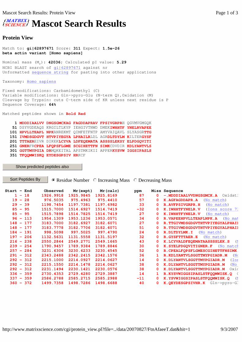
Mascot Search Results Protein View
Match to: gi|62897671 Score: 311 Expect: 1.5e-26beta actin variant [Homo sapiens]
Nominal mass (M r ): 42036; Calculated pI value: 5.29
NCBI BLAST search of gi|62897671 against nr Unformatted sequence string for pasting into other applications
Taxonomy: Homo sapiens
Fixed modifications: Carbamidomethyl (C) Variable modifications: Gln->pyro-Glu (N-term Q),Ox idation (M) Cleavage by Trypsin: cuts C-term side of KR unless next residue is P Sequence Coverage: 64%
Matched peptides shown in Bold Red
1 MDDDIAALVV DNGSGMCKAG FAGDDAPRAV FPSIVGRPRH QGVMVGMGQK 51 DSYVGDEAQS KRGILTLKYP IEHGIVTNWD DMEKIWHHTF YNELRVAPEE 101 HPVLLTEAPL NPKANREKMT QIMFETFNTP AMYVAIQAVL SLYASGRTTG 151 IVMDSGDGVT HTVPIYEGYA LPHAILRLDL AGRDLTDYLM KILTERGYSF 201 TTTAEREIVR DIKEK LCYVA LDFEQEMATA ASSSSLEKSY ELPDGQVITI 251 GNERFRCPEA LFQPSFLGME SCGIHETTFN SIMKCDVDIR KDLYANTVLS 301 GGTTMYPGIA DRMQKEITAL APSTMKIKII APPER KYSVW IGGSIPASLS 351 TFQQMWISKQ EYDESGPSIV HRKCF
Residue Number Increasing Mass Decreasing Mass
Start - End Observed Mr(expt) Mr(calc) ppm Miss Sequence 1 - 18 1926.9918 1925.9845 1925.8169 87 0 -.MDDDIAALVVDNGSGMCK.A Oxidation (M) ( 19 - 28 976.5035 975.4963 975.4410 57 0 K.AGFAGDDAPR.A ( No match ) 29 - 39 1198.7454 1197.7381 1197.6982 33 0 R.AVFPSIVGRPR.H ( No match ) 85 - 95 1515.7000 1514.6927 1514.7419 -32 0 K.IWHHTFYNELR.V ( Ions score 73 85 - 95 1515.7898 1514.7825 1514.7419 27 0 K.IWHHTFYNELR.V ( No match ) 96 - 113 1954.1309 1953.1236 1953.0571 34 0 R.VAPEEHPVLLTEAPLNPK.A ( No match 148 - 177 3183.7000 3182.6927 3182.6071 27 0 R.TTGIVMDSGDGVTHTVPIYEGYALPHAILR.L 148 - 177 3183.7778 3182.7706 3182.6071 51 0 R.TTGIVMDSGDGVTHTVPIYEGYALPHAILR.L 184 - 191 998.5098 997.5025 997.4790 24 0 R.DLTDYLMK.I ( No match ) 197 - 206 1132.5631 1131.5558 1131.5197 32 0 R.GYSFTTTAER.E ( No match ) 216 - 238 2550.2844 2549.2771 2549.1665 43 0 K.LCYVALDFEQEMATAASSSSLEK.S ( No match 239 - 254 1790.9457 1789.9384 1789.8846 30 0 K.SYELPDGQVITIGNER.F ( No match 257 - 284 3231.6306 3230.6233 3230.4545 52 0 R.CPEALFQPSFLGMESCGIHETTFNSIMK.C 291 - 312 2343.2488 2342.2415 2342.1576 36 1 R.KDLYANTVLSGGTTMYPGIADR.M ( No match 292 - 312 2215.1000 2214.0927 2214.0627 14 0 K.DLYANTVLSGGTTMYPGIADR.M ( Ions score 123 292 - 312 2215.1550 2214.1478 2214.0627 38 0 K.DLYANTVLSGGTTMYPGIADR.M ( No match 292 - 312 2231.1494 2230.1421 2230.0576 38 0 K.DLYANTVLSGGTTMYPGIADR.M Oxidation (M) ( 336 - 359 2730.4353 2729.4280 2729.3887 14 1 R.KYSVWIGGSIPASLSTFQQMWISK.Q Oxidation (M) ( 337 - 359 2586.2788 2585.2715 2585.2988 -11 0 K.YSVWIGGSIPASLSTFQQMWISK.Q ( No match 360 - 372 1499.7358 1498.7286 1498.6688 40 0 K.QEYDESGPSIVHR.K Gln->pyro- Glu (N
Show predicted peptides also
Sort Peptides By nmlkji nmlkj nmlkj
Page 1 of 3Mascot Search Results: Protein View
9/3/2007http://www.matrixscience.com/cgi/protein_view.pl?file=../data/20070827/FtnAfaeeT.dat&hit=1

LOCUS BAD96775 375 aa linear PRI 26-APR-2005 DEFINITION beta actin variant [Homo sapiens]. ACCESSION BAD96775 VERSION BAD96775.1 GI:62897671 DBSOURCE accession AK223055.1KEYWORDS . SOURCE Homo sapiens (human) ORGANISM Homo sapiens Eukaryota; Metazoa; Chordata; Craniata; Vertebrata; Euteleostomi; Mammalia; Eutheria; Euarchontoglires; P rimates; Haplorrhini; Catarrhini; Hominidae; Homo. REFERENCE 1 AUTHORS Maruyama,K. and Sugano,S. TITLE Oligo-capping: a simple method to repla ce the cap structure of eukaryotic mRNAs with oligoribonucleoti des JOURNAL Gene 138 (1-2), 171-174 (1994) PUBMED 8125298REFERENCE 2 AUTHORS Suzuki,Y., Yoshitomo-Nakagawa,K., Maruy ama,K., Suyama,A. and Sugano,S. TITLE Construction and characterization of a full length-enriched and a 5'-end-enriched cDNA library JOURNAL Gene 200 (1-2), 149-156 (1997) PUBMED 9373149REFERENCE 3 (residues 1 to 375) AUTHORS Suzuki,Y., Sugano,S., Totoki,Y., Toyoda ,A., Takeda,T., Sakaki,Y., Tanaka,A. and Yokoyama,S. TITLE Direct Submission JOURNAL Submitted (22-APR-2005) Akiko Tanaka, R IKEN Yokohama Institute, Protein Research Group; 1-7-22 Suehiro, Tsurumi, Yokohama, Kanagawa 230-0045, Japan (E-mail:aktanaka@riken. jp, URL:http://protein.gsc.riken.jp/, Tel:8 1-45-503-9452, Fax:81-45-503-9450) COMMENT This work was supported in part by the National Project on Protein Structural and Functional Analysis, Min istry of Education, Culture, Sports, Science and Technology of Japan . Sumio Sugano, Yutaka Suzuki Laboratory of Functional Genomics Depar tment of Medical Genome Sciences Graduate School of Frontier Sc iences The University of Tokyo, 4-6-1 Shirokanedai, Minato-ku, T okyo 108-8639 Japan email: [email protected] URL: http://www.k.u - tokyo.ac.jp/index.html.en . FEATURES Location/Qualifiers source 1..375 /organism="Homo sapiens" /db_xref="taxon: 9606 " /clone="KAT00430" /cell_line="KATO III" /clone_lib="KAT" /note="cloning vector: pME18SF L3 this clone is also named as hs s001002538" Protein 1..375 /product="beta actin variant" Region 3..375
Page 2 of 3Mascot Search Results: Protein View
9/3/2007http://www.matrixscience.com/cgi/protein_view.pl?file=../data/20070827/FtnAfaeeT.dat&hit=1

Mascot Search Results Protein View
Match to: gi|5031635 Score: 128 Expect: 3.1e-08cofilin 1 (non-muscle) [Homo sapiens]
Nominal mass (M r ): 18719; Calculated pI value: 8.22
NCBI BLAST search of gi|5031635 against nr Unformatted sequence string for pasting into other applications
Taxonomy: Homo sapiensLinks to retrieve other entries containing this seq uence from NCBI Entrez: gi|57099669 from Canis lupus familiarisgi|114638608 from Pan troglodytesgi|149725588 from Equus caballusgi|116848 from Homo sapiensgi|50513339 from Homo sapiensgi|50513340 from Homo sapiensgi|219545 from Homo sapiensgi|736400 from Homo sapiensgi|1177471 from Homo sapiensgi|15012201 from Homo sapiensgi|15126676 from Homo sapiensgi|15147369 from Homo sapiensgi|17390594 from Homo sapiensgi|30582531 from Homo sapiensgi|61360378 from synthetic constructgi|119594855 from Homo sapiensgi|119594858 from Homo sapiensgi|123981732 from synthetic construct
Fixed modifications: Carbamidomethyl (C) Variable modifications: Gln->pyro-Glu (N-term Q),Me thyl (C-term),Methyl (DE),Oxidation (M) Cleavage by Trypsin: cuts C-term side of KR unless next residue is P Sequence Coverage: 67%
Matched peptides shown in Bold Red
1 MASGVAVSDG VIKVFNDMKV RKSSTPEEVK KRKKAVLFCL SEDKKNIILE 51 EGKEILVGDV GQTVDDPYAT FVKMLPDKDC RYALYDATYE TKESKKEDLV 101 FIFWAPESAP LKSKMIYASS KDAIKKKLTG IK HELQANCY EEVKDRCTLA 151 EKLGGSAVIS LEGKPL
Residue Number Increasing Mass Decreasing Mass
Start - End Observed Mr(expt) Mr(calc) ppm Miss Sequence 1 - 19 2012.0975 2011.0903 2011.0119 39 1 -.MASGVAVSDGVIKVFNDMK.V 2 Methyl (DE); Oxidation (M) ( 1 - 19 2028.0907 2027.0834 2027.0068 38 1 -.MASGVAVSDGVIKVFNDMK.V 2 Methyl (DE); 2 Oxidation (M) ( 35 - 45 1337.6782 1336.6709 1336.7061 -26 1 K.AVLFCLSEDKK.N Methyl (C- term); Methyl (DE) ( 54 - 73 2166.1555 2165.1482 2165.0892 27 0 K.EILVGDVGQTVDDPYATFVK.M ( No match 82 - 92 1351.6759 1350.6686 1350.6343 25 0 R.YALYDATYETK.E Methyl (DE) ( No match 96 - 112 1990.1000 1989.0927 1989.0611 16 1 K.KEDLVFIFWAPESAPLK.S ( Ions score 99 96 - 112 1990.1151 1989.1078 1989.0611 23 1 K.KEDLVFIFWAPESAPLK.S ( No match 96 - 112 2004.1371 2003.1298 2003.0768 26 1 K.KEDLVFIFWAPESAPLK.S Methyl (DE) ( 97 - 112 1862.0243 1861.0170 1860.9662 27 0 K.EDLVFIFWAPESAPLK.S ( No match 133 - 146 1790.8539 1789.8466 1789.8053 23 1 K.HELQANCYEEVKDR.C ( No match ) 133 - 146 1804.8946 1803.8874 1803.8210 37 1 K.HELQANCYEEVKDR.C Methyl (DE) ( 147 - 166 2043.1051 2042.0978 2042.1082 -5 1 R.CTLAEKLGGSAVISLEGKPL.- ( No match 153 - 166 1340.8156 1339.8083 1339.7711 28 0 K.LGGSAVISLEGKPL.- ( No match )
Show predicted peptides also
Sort Peptides By nmlkji nmlkj nmlkj
Page 1 of 6Mascot Search Results: Protein View
9/3/2007http://www.matrixscience.com/cgi/protein_view.pl?file=../data/20070903/FtnAobYSO.dat&hit=1

LOCUS NP_005498 166 aa linear PRI 03-SEP-2007 DEFINITION cofilin 1 (non-muscle) [Homo sapiens]. ACCESSION NP_005498 VERSION NP_005498.1 GI:5031635 DBSOURCE REFSEQ: accession NM_005507.2KEYWORDS . SOURCE Homo sapiens (human) ORGANISM Homo sapiens Eukaryota; Metazoa; Chordata; Craniata; Vertebrata; Euteleostomi; Mammalia; Eutheria; Euarchontoglires; P rimates; Haplorrhini; Catarrhini; Hominidae; Homo. REFERENCE 1 (residues 1 to 166) AUTHORS Mannherz,H.G., Ballweber,E., Galla,M., Villard,S., Granier,C., Steegborn,C., Schmidtmann,A., Jaquet,K. , Pope,B. and Weeds,A.G. TITLE Mapping the ADF/cofilin binding site on monomeric actin by competitive cross-linking and peptide a rray: evidence for a second binding site on monomeric actin JOURNAL J. Mol. Biol. 366 (3), 745-755 (2007) PUBMED 17196218 REMARK GeneRIF: In the absence of any crystal structures of ADF or cofilin in complex with actin, these studies pr ovide further information about the binding sites on F-actin for these important actin regulatory proteins. REFERENCE 2 (residues 1 to 166) AUTHORS Ewing,R.M., Chu,P., Elisma,F., Li,H., T aylor,P., Climie,S., McBroom-Cerajewski,L., Robinson,M.D., O 'Connor,L., Li,M., Taylor,R., Dharsee,M., Ho,Y., Heilbut,A ., Moore,L., Zhang,S., Ornatsky,O., Bukhman,Y.V., Ethier,M., S heng,Y., Vasilescu,J., Abu-Farha,M., Lambert,J.P., Duewel,H.S. , Stewart,I.I., Kuehl,B., Hogue,K., Colwill,K., Gladwish,K., Musk at,B., Kinach,R., Adams,S.L., Moran,M.F., Morin,G.B., Top aloglou,T. and Figeys,D. TITLE Large-scale mapping of human protein-pr otein interactions by mass spectrometry JOURNAL Mol. Syst. Biol. 3, 89 (2007) PUBMED 17353931REFERENCE 3 (residues 1 to 166) AUTHORS Zhu,H., Enaw,J.O., Ma,C., Shaw,G.M., La mmer,E.J. and Finnell,R.H. TITLE Association between CFL1 gene polymorph isms and spina bifida risk in a California population JOURNAL (er) BMC Med. Genet. 8, 12 (2007) PUBMED 17352815 REMARK GeneRIF: The sequence variation of huma n CFL1 gene is a genetic modifier for spina bifida risk in Calif ornia population REFERENCE 4 (residues 1 to 166) AUTHORS Olsen,J.V., Blagoev,B., Gnad,F., Macek, B., Kumar,C., Mortensen,P. and Mann,M. TITLE Global, in vivo, and site-specific phos phorylation dynamics in signaling networks JOURNAL Cell 127 (3), 635-648 (2006) PUBMED 17081983REFERENCE 5 (residues 1 to 166) AUTHORS Vasavda,N., Eichholtz,T., Takahashi,A., Affleck,K., Matthews,J.G., Barnes,P.J. and Adcock,I.M. TITLE Expression of nonmuscle cofilin-1 and s teroid responsiveness in
Page 2 of 6Mascot Search Results: Protein View
9/3/2007http://www.matrixscience.com/cgi/protein_view.pl?file=../data/20070903/FtnAobYSO.dat&hit=1

Mascot Search Results Protein View
Match to: gi|4503571 Score: 147 Expect: 3.9e-10enolase 1 [Homo sapiens]
Nominal mass (M r ): 47481; Calculated pI value: 7.01
NCBI BLAST search of gi|4503571 against nr Unformatted sequence string for pasting into other applications
Taxonomy: Homo sapiensLinks to retrieve other entries containing this seq uence from NCBI Entrez: gi|114552694 from Pan troglodytesgi|119339 from Homo sapiensgi|182114 from Homo sapiensgi|1167843 from Homo sapiensgi|12804749 from Homo sapiensgi|13325287 from Homo sapiensgi|14530765 from Homo sapiensgi|14602814 from Homo sapiensgi|15029814 from Homo sapiensgi|15990505 from Homo sapiensgi|18490320 from Homo sapiensgi|20379496 from Homo sapiensgi|29792061 from Homo sapiensgi|30583165 from Homo sapiensgi|60655645 from synthetic constructgi|60655647 from synthetic constructgi|60812591 from synthetic constructgi|61354812 from synthetic constructgi|66268795 from Homo sapiensgi|119592010 from Homo sapiensgi|119592011 from Homo sapiensgi|123981076 from synthetic constructgi|123995887 from synthetic construct
Fixed modifications: Carbamidomethyl (C) Variable modifications: Gln->pyro-Glu (N-term Q),Ox idation (M) Cleavage by Trypsin: cuts C-term side of KR unless next residue is P Sequence Coverage: 49%
Matched peptides shown in Bold Red
1 MSILKIHARE IFDSR GNPTV EVDLFTSKGL FRAAVPSGAS TGIYEALELR 51 DNDKTRYMGK GVSKAVEHIN KTIAPALVSK KLNVTEQEKI DKLMIEMDGT 101 ENKSKFGANA ILGVSLAVCK AGAVEKGVPL YRHIADLAGN SEVILPVPAF 151 NVINGGSHAG NKLAMQEFMI LPVGAANFRE AMRIGAEVYH NLKNVIKEKY 201 GKDATNVGDE GGFAPNILEN KEGLELLKTA IGKAGYTDKV VIGMDVAASE 251 FFRSGKYDLD FKSPDDPSRY ISPDQLADLY KSFIK DYPVV SIEDPFDQDD 301 WGAWQKFTAS AGIQVVGDDL TVTNPKRIAK AVNEKSCNCL LLKVNQIGSV 351 TESLQACKLA QANGWGVMVS HRSGETEDTF IADLVVGLCT GQIKTGAPCR 401 SERLAKYNQL LRIEEELGSK AKFAGRNFRN PLAK
Residue Number Increasing Mass Decreasing Mass
Start - End Observed Mr(expt) Mr(calc) ppm Miss Sequence 16 - 28 1406.7549 1405.7476 1405.7089 28 0 R.GNPTVEVDLFTSK.G ( No match ) 33 - 50 1804.9000 1803.8927 1803.9366 -24 0 R.AAVPSGASTGIYEALELR.D ( Ions score 9 33 - 50 1804.9761 1803.9688 1803.9366 18 0 R.AAVPSGASTGIYEALELR.D ( No match
Show predicted peptides also
Sort Peptides By nmlkji nmlkj nmlkj
Page 1 of 5Mascot Search Results: Protein View
8/31/2007http://www.matrixscience.com/cgi/protein_view.pl?file=../data/20070831/FtnAoreeT.dat&hit=1

106 - 120 1519.8656 1518.8583 1518.8228 23 0 K.FGANAILGVSLAVCK.A ( No match ) 163 - 179 1908.0308 1907.0235 1906.9797 23 0 K.LAMQEFMILPVGAANFR.E ( No match 203 - 221 1960.9574 1959.9501 1959.9174 17 0 K.DATNVGDEGGFAPNILENK.E ( No match 240 - 253 1540.8000 1539.7927 1539.7756 11 0 K.VVIGMDVAASEFFR.S ( Ions score 11 240 - 253 1540.8185 1539.8112 1539.7756 23 0 K.VVIGMDVAASEFFR.S ( No match ) 240 - 253 1556.8298 1555.8226 1555.7705 33 0 K.VVIGMDVAASEFFR.S Oxidation (M) ( 257 - 269 1554.8475 1553.8403 1553.6998 90 1 K.YDLDFKSPDDPSR.Y ( No match ) 270 - 281 1425.7000 1424.6927 1424.7187 -18 0 R.YISPDQLADLYK.S ( Ions score 29 270 - 281 1425.7615 1424.7542 1424.7187 25 0 R.YISPDQLADLYK.S ( No match ) 286 - 306 2510.1809 2509.1736 2509.1074 26 0 K.DYPVVSIEDPFDQDDWGAWQK.F ( No match 307 - 326 2033.0896 2032.0823 2032.0477 17 0 K.FTASAGIQVVGDDLTVTNPK.R ( No match 344 - 358 1633.8552 1632.8479 1632.8141 21 0 K.VNQIGSVTESLQACK.L ( No match ) 359 - 372 1525.8003 1524.7930 1524.7620 20 0 K.LAQANGWGVMVSHR.S ( No match ) 373 - 394 2353.2168 2352.2095 2352.1519 24 0 R.SGETEDTFIADLVVGLCTGQIK.T ( No match
LOCUS NP_001419 434 aa linear PRI 29-JUL-2007 DEFINITION enolase 1 [Homo sapiens]. ACCESSION NP_001419 NP_005936 VERSION NP_001419.1 GI:4503571 DBSOURCE REFSEQ: accession NM_001428.2KEYWORDS . SOURCE Homo sapiens (human) ORGANISM Homo sapiens Eukaryota; Metazoa; Chordata; Craniata; Vertebrata; Euteleostomi; Mammalia; Eutheria; Euarchontoglires; P rimates; Haplorrhini; Catarrhini; Hominidae; Homo. REFERENCE 1 (residues 1 to 434) AUTHORS Newman,S.F., Sultana,R., Perluigi,M., C occia,R., Cai,J., Pierce,W.M., Klein,J.B., Turner,D.M. an d Butterfield,D.A. TITLE An increase in S-glutathionylated prote ins in the Alzheimer's disease inferior parietal lobule, a pro teomics approach JOURNAL J. Neurosci. Res. 85 (7), 1506-1514 (20 07) PUBMED 17387692 REMARK GeneRIF: alpha-crystallin B, glyceralde hyde phosphate dehydrogenase (GAPDH), and alpha-enolase identified a s significantly S-glutathionylated in Alzheimer's disea se infeior parietal lobule REFERENCE 2 (residues 1 to 434) AUTHORS Ito,S., Honma,T., Ishida,K., Wada,N., S asaoka,S., Hosoda,M. and Nohno,T. TITLE Differential expression of the human al pha-enolase gene in oral epithelium and squamous cell carcinoma JOURNAL Cancer Sci. 98 (4), 499-505 (2007) PUBMED 17284257 REMARK GeneRIF: Our results indicate that diff erential subcellular localization of ENO1 products may be cl osely related to carcinogenesis of the oral epithelium REFERENCE 3 (residues 1 to 434) AUTHORS Ghosh,A.K., Steele,R. and Ray,R.B. TITLE Knockdown of MBP-1 in human prostate ca ncer cells delays cell cycle progression JOURNAL J. Biol. Chem. 281 (33), 23652-23657 (2 006) PUBMED 16762917 REMARK GeneRIF: Depletion of MBP-1 in prostate cancer cells perturbs cell proliferation by inhibiting cyclin A an d cyclin B1 expression. REFERENCE 4 (residues 1 to 434)
Page 2 of 5Mascot Search Results: Protein View
8/31/2007http://www.matrixscience.com/cgi/protein_view.pl?file=../data/20070831/FtnAoreeT.dat&hit=1

Mascot Search Results Protein View
Match to: gi|809561 Score: 66 Expect: 0.18gamma-actin [Mus musculus]
Nominal mass (Mr): 41335; Calculated pI value: 5.56
NCBI BLAST search of gi|809561 against nr Unformatted sequence string for pasting into other applications
Taxonomy: Mus musculus
Fixed modifications: Carbamidomethyl (C) Variable modifications: Gln->pyro-Glu (N-term Q),Oxidation (M) Cleavage by Trypsin: cuts C-term side of KR unless next residue is P Sequence Coverage: 66%
Matched peptides shown in Bold Red
1 LVIDNGSGMC KAGFAGDDAP RAVFPSIVGR PRHQGVMVGM GQKDSYVGDE 51 AQSKRGILTL KYPIEHGIVT NWDDMEKIWH HTFYNELRVA PEEHPVLLTE 101 APLNPKANRE KMTQIMFETF NTPAMYVAIQ AVLSLYASGR TTGIVMDSGD 151 GVTHTVPIYE GYALPHAILR LDLAGRDLTD YLMKILTERG YSFTTTAERE 201 IVRDIKEKLC YVALDFEQEM ATAASSSSLE KSYELPDGQV ITIGNERFRC 251 PEALFQPSFL GMESCGIHET TFNSIMKCDV DIRKDLYANT VLSGGTTMYP 301 GIADRMQKEI TALAPSTMKI KIIAPPERKY SVWIGGSILA SLSTFQQMWI 351 SKQEYDESGP SIVHRKCF
Residue Number Increasing Mass Decreasing Mass
Start - End Observed Mr(expt) Mr(calc) ppm Miss Sequence 1 - 21 2167.1250 2166.1177 2165.9834 62 1 -.LVIDNGSGMCKAGFAGDDAPR.A Oxidation (M) ( 1 - 21 2167.1500 2166.1427 2165.9834 74 1 -.LVIDNGSGMCKAGFAGDDAPR.A Oxidation (M) ( 78 - 88 1515.7874 1514.7801 1514.7419 25 0 K.IWHHTFYNELR.V (No match) 89 - 106 1954.1155 1953.1082 1953.0571 26 0 R.VAPEEHPVLLTEAPLNPK.A (No match 112 - 140 3285.6526 3284.6453 3284.5920 16 0 K.MTQIMFETFNTPAMYVAIQAVLSLYASGR.T 141 - 170 3183.7036 3182.6963 3182.6071 28 0 R.TTGIVMDSGDGVTHTVPIYEGYALPHAILR.L 190 - 199 1132.5737 1131.5665 1131.5197 41 0 R.GYSFTTTAER.E (No match) 209 - 231 2550.2407 2549.2334 2549.1665 26 0 K.LCYVALDFEQEMATAASSSSLEK.S (No match 232 - 247 1790.9336 1789.9263 1789.8846 23 0 K.SYELPDGQVITIGNER.F (No match 250 - 277 3231.5635 3230.5562 3230.4545 31 0 R.CPEALFQPSFLGMESCGIHETTFNSIMK.C 284 - 305 2343.2270 2342.2198 2342.1576 27 1 R.KDLYANTVLSGGTTMYPGIADR.M (No match 285 - 305 2215.1201 2214.1128 2214.0627 23 0 K.DLYANTVLSGGTTMYPGIADR.M (No match 285 - 305 2231.1235 2230.1163 2230.0576 26 0 K.DLYANTVLSGGTTMYPGIADR.M Oxidation (M) ( 329 - 352 2730.3877 2729.3804 2729.4251 -16 1 R.KYSVWIGGSILASLSTFQQMWISK.Q ( 329 - 352 2746.3499 2745.3426 2745.4200 -28 1 R.KYSVWIGGSILASLSTFQQMWISK.Q Oxidation (M) ( 353 - 365 1499.7263 1498.7190 1498.6688 33 0 K.QEYDESGPSIVHR.K Gln->pyro-Glu (N
Show predicted peptides also
Sort Peptides By nmlkji nmlkj nmlkj
Page 1 of 2Mascot Search Results: Protein View
9/3/2007http://www.matrixscience.com/cgi/protein_view.pl?file=../data/20070827/FtnAfaeat.dat&hit=4

Mascot Search Results Protein View
Match to: gi|4504165 Score: 108 Expect: 3.1e-06gelsolin isoform a precursor [Homo sapiens]
Nominal mass (M r ): 86043; Calculated pI value: 5.90
NCBI BLAST search of gi|4504165 against nr Unformatted sequence string for pasting into other applications
Taxonomy: Homo sapiensLinks to retrieve other entries containing this seq uence from NCBI Entrez: gi|121116 from Homo sapiensgi|736249 from Homo sapiensgi|19684181 from Homo sapiensgi|55960299 from Homo sapiensgi|61364370 from synthetic constructgi|119607897 from Homo sapiensgi|123982744 from synthetic constructgi|225304 from Homo sapiens
Fixed modifications: Carbamidomethyl (C) Variable modifications: Gln->pyro-Glu (N-term Q),Ox idation (M) Cleavage by Trypsin: cuts C-term side of KR unless next residue is P Sequence Coverage: 22%
Matched peptides shown in Bold Red
1 MAPHRPAPAL LCALSLALCA LSLPVRAATA SRGASQAGAP QGRVPEARPN 51 SMVVEHPEFL KAGKEPGLQI WRVEKFDLVP VPTNLYGDFF TGDAYVILKT 101 VQLRNGNLQY DLHYWLGNEC SQDESGAAAI FTVQLDDYLN GRAVQHREVQ 151 GFESATFLGY FKSGLKYKKG GVASGFKHVV PNEVVVQRLF QVKGRRVVRA 201 TEVPVSWESF NNGDCFILDL GNNIHQWCGS NSNRYERLKA TQVSKGIRDN 251 ERSGRARVHV SEEGTEPEAM LQVLGPKPAL PAGTEDTAKE DAANRKLAKL 301 YK VSNGAGTM SVSLVADENP FAQGALKSED CFILDHGKDG KIFVWKGKQA 351 NTEERKAALK TASDFITKMD YPKQTQVSVL PEGGETPLFK QFFKNWRDPD 401 QTDGLGLSYL SSHIANVERV PFDAATLHTS TAMAAQHGMD DDGTGQKQIW 451 RIEGSNKVPV DPATYGQFYG GDSYIILYNY RHGGRQGQII YNWQGAQSTQ 501 DEVAASAILT AQLDEELGGT PVQSRVVQGK EPAHLMSLFG GKPMIIYKGG 551 TSREGGQTAP ASTRLFQVRA NSAGATRAVE VLPKAGALNS NDAFVLKTPS 601 AAYLWVGTGA SEAEKTGAQE LLRVLRAQPV QVAEGSEPDG FWEALGGKAA 651 YRTSPRLKDK KMDAHPPRLF ACSNKIGRFV IEEVPGELMQ EDLATDDVML 701 LDTWDQVFVW VGKDSQEEEK TEALTSAKRY IETDPANRDR RTPITVVKQG 751 FEPPSFVGWF LGWDDDYWSV DPLDRAMAEL AA
Residue Number Increasing Mass Decreasing Mass
Start - End Observed Mr(expt) Mr(calc) ppm Miss Sequence 62 - 72 1254.7000 1253.6927 1253.6880 4 1 K.AGKEPGLQIWR.V ( No match ) 62 - 72 1254.7311 1253.7238 1253.6880 29 1 K.AGKEPGLQIWR.V ( No match ) 65 - 72 998.5847 997.5774 997.5345 43 0 K.EPGLQIWR.V ( No match ) 148 - 162 1722.9037 1721.8964 1721.8301 39 0 R.EVQGFESATFLGYFK.S ( No match ) 178 - 188 1275.7577 1274.7504 1274.7095 32 0 K.HVVPNEVVVQR.L ( No match ) 303 - 327 2463.3230 2462.3157 2462.2111 42 0 K.VSNGAGTMSVSLVADENPFAQGALK.S ( 369 - 390 2464.3137 2463.3064 2463.2356 29 1 K.MDYPKQTQVSVLPEGGETPLFK.Q ( No match 369 - 390 2480.2944 2479.2872 2479.2305 23 1 K.MDYPKQTQVSVLPEGGETPLFK.Q Oxidation (M) ( 374 - 390 1813.0083 1812.0010 1811.9306 39 0 K.QTQVSVLPEGGETPLFK.Q Gln- >pyro 374 - 390 1830.0370 1829.0297 1828.9571 40 0 K.QTQVSVLPEGGETPLFK.Q ( No match 398 - 419 2387.2151 2386.2078 2386.1401 28 0 R.DPDQTDGLGLSYLSSHIANVER.V ( No match
Show predicted peptides also
Sort Peptides By nmlkji nmlkj nmlkj
Page 1 of 5Mascot Search Results: Protein View
9/3/2007http://www.matrixscience.com/cgi/protein_view.pl?file=../data/20070903/FtnAobSwO.dat&hit=3

420 - 447 2873.4060 2872.3987 2872.2756 43 0 R.VPFDAATLHTSTAMAAQHGMDDDGTGQK.Q 420 - 447 2889.4016 2888.3943 2888.2706 43 0 R.VPFDAATLHTSTAMAAQHGMDDDGTGQK.Q 598 - 615 1837.9666 1836.9593 1836.8894 38 0 K.TPSAAYLWVGTGASEAEK.T ( No match 627 - 648 2272.1000 2271.0927 2271.0808 5 0 R.AQPVQVAEGSEPDGFWEALGGK.A ( Ions score 64 627 - 648 2272.1790 2271.1717 2271.0808 40 0 R.AQPVQVAEGSEPDGFWEALGGK.A ( No match
LOCUS NP_000168 782 aa linear PRI 30-JUL-2007 DEFINITION gelsolin isoform a precursor [Homo sapi ens]. ACCESSION NP_000168 VERSION NP_000168.1 GI:4504165 DBSOURCE REFSEQ: accession NM_000177.4KEYWORDS . SOURCE Homo sapiens (human) ORGANISM Homo sapiens Eukaryota; Metazoa; Chordata; Craniata; Vertebrata; Euteleostomi; Mammalia; Eutheria; Euarchontoglires; P rimates; Haplorrhini; Catarrhini; Hominidae; Homo. REFERENCE 1 (residues 1 to 782) AUTHORS Huerva,V., Velasco,A., Sanchez,M.C., Ma teo,A.J. and Matias-Guiu,X. TITLE Lattice corneal dystrophy type II: clin ical, pathologic, and molecular study in a Spanish family JOURNAL Eur J Ophthalmol 17 (3), 424-429 (2007) PUBMED 17534828 REMARK GeneRIF: A new family with Meretoja syn drome is reported. This is the first documented family with Mereto ja syndrome in Spain and in the Mediterranean countries. The molecu lar study shows the same mutation of reported families. REFERENCE 2 (residues 1 to 782) AUTHORS Frum,R., Busby,S.A., Ramamoorthy,M., De b,S., Shabanowitz,J., Hunt,D.F. and Deb,S.P. TITLE HDM2-binding partners: interaction with translation elongation factor EF1alpha JOURNAL J. Proteome Res. 6 (4), 1410-1417 (2007 ) PUBMED 17373842REFERENCE 3 (residues 1 to 782) AUTHORS Roustan,C., Ferjani,I., Maciver,S.K., F attoum,A., Rebiere,B. and Benyamin,Y. TITLE Calcium-induced conformational changes in the amino-terminal half of gelsolin JOURNAL FEBS Lett. 581 (4), 681-686 (2007) PUBMED 17258204 REMARK GeneRIF: Calcium induced conformational changes in the G1-2 and G1-3 sub-domains of gelsolin, and the b inding affinities for the three type II sites, are reported. REFERENCE 4 (residues 1 to 782) AUTHORS Qiao,H. and McMillan,J.R. TITLE Gelsolin segment 5 inhibits HIV-induced T-cell apoptosis via Vpr-binding to VDAC JOURNAL FEBS Lett. 581 (3), 535-540 (2007) PUBMED 17254575 REMARK GeneRIF: gelsolin G5 domain inhibits HI V-Vpr-induced T-cell apoptosis by blocking the interaction b etween Vpr and VDAC REFERENCE 5 (residues 1 to 782) AUTHORS Ewing,R.M., Chu,P., Elisma,F., Li,H., T aylor,P., Climie,S., McBroom-Cerajewski,L., Robinson,M.D., O 'Connor,L., Li,M.,
Page 2 of 5Mascot Search Results: Protein View
9/3/2007http://www.matrixscience.com/cgi/protein_view.pl?file=../data/20070903/FtnAobSwO.dat&hit=3

Mascot Search Results Protein View
Match to: gi|38044288 Score: 111 Expect: 1.5e-06gelsolin isoform b [Homo sapiens]
Nominal mass (M r ): 80876; Calculated pI value: 5.58
NCBI BLAST search of gi|38044288 against nr Unformatted sequence string for pasting into other applications
Taxonomy: Homo sapiensLinks to retrieve other entries containing this seq uence from NCBI Entrez: gi|119607895 from Homo sapiensgi|123232805 from Homo sapiens
Fixed modifications: Carbamidomethyl (C) Variable modifications: Gln->pyro-Glu (N-term Q),Ox idation (M) Cleavage by Trypsin: cuts C-term side of KR unless next residue is P Sequence Coverage: 23%
Matched peptides shown in Bold Red
1 MVVEHPEFLK AGKEPGLQIW RVEKFDLVPV PTNLYGDFFT GDAYVILKTV 51 QLRNGNLQYD LHYWLGNECS QDESGAAAIF TVQLDDYLNG RAVQHREVQG 101 FESATFLGYF KSGLKYKKGG VASGFKHVVP NEVVVQRLFQ VKGRRVVRAT 151 EVPVSWESFN NGDCFILDLG NNIHQWCGSN SNRYERLKAT QVSKGIRDNE 201 RSGRARVHVS EEGTEPEAML QVLGPKPALP AGTEDTAKED AANRKLAKLY 251 K VSNGAGTMS VSLVADENPF AQGALKSEDC FILDHGKDGK IFVWKGKQAN 301 TEERKAALKT ASDFITKMDY PKQTQVSVLP EGGETPLFKQ FFKNWRDPDQ 351 TDGLGLSYLS SHIANVERVP FDAATLHTST AMAAQHGMDD DGTGQKQIWR 401 IEGSNKVPVD PATYGQFYGG DSYIILYNYR HGGRQGQIIY NWQGAQSTQD 451 EVAASAILTA QLDEELGGTP VQSRVVQGKE PAHLMSLFGG KPMIIYKGGT 501 SREGGQTAPA STRLFQVRAN SAGATRAVEV LPKAGALNSN DAFVLKTPSA 551 AYLWVGTGAS EAEKTGAQEL LRVLRAQPVQ VAEGSEPDGF WEALGGKAAY 601 RTSPRLKDKK MDAHPPRLFA CSNKIGRFVI EEVPGELMQE DLATDDVMLL 651 DTWDQVFVWV GKDSQEEEKT EALTSAKRYI ETDPANRDRR TPITVVKQGF 701 EPPSFVGWFL GWDDDYWSVD PLDRAMAELA A
Residue Number Increasing Mass Decreasing Mass
Start - End Observed Mr(expt) Mr(calc) ppm Miss Sequence 11 - 21 1254.7000 1253.6927 1253.6880 4 1 K.AGKEPGLQIWR.V ( No match ) 11 - 21 1254.7311 1253.7238 1253.6880 29 1 K.AGKEPGLQIWR.V ( No match ) 14 - 21 998.5847 997.5774 997.5345 43 0 K.EPGLQIWR.V ( No match ) 97 - 111 1722.9037 1721.8964 1721.8301 39 0 R.EVQGFESATFLGYFK.S ( No match ) 127 - 137 1275.7577 1274.7504 1274.7095 32 0 K.HVVPNEVVVQR.L ( No match ) 252 - 276 2463.3230 2462.3157 2462.2111 42 0 K.VSNGAGTMSVSLVADENPFAQGALK.S ( 318 - 339 2464.3137 2463.3064 2463.2356 29 1 K.MDYPKQTQVSVLPEGGETPLFK.Q ( No match 318 - 339 2480.2944 2479.2872 2479.2305 23 1 K.MDYPKQTQVSVLPEGGETPLFK.Q Oxidation (M) ( 323 - 339 1813.0083 1812.0010 1811.9306 39 0 K.QTQVSVLPEGGETPLFK.Q Gln- >pyro 323 - 339 1830.0370 1829.0297 1828.9571 40 0 K.QTQVSVLPEGGETPLFK.Q ( No match 347 - 368 2387.2151 2386.2078 2386.1401 28 0 R.DPDQTDGLGLSYLSSHIANVER.V ( No match 369 - 396 2873.4060 2872.3987 2872.2756 43 0 R.VPFDAATLHTSTAMAAQHGMDDDGTGQK.Q 369 - 396 2889.4016 2888.3943 2888.2706 43 0 R.VPFDAATLHTSTAMAAQHGMDDDGTGQK.Q 547 - 564 1837.9666 1836.9593 1836.8894 38 0 K.TPSAAYLWVGTGASEAEK.T ( No match 576 - 597 2272.1000 2271.0927 2271.0808 5 0 R.AQPVQVAEGSEPDGFWEALGGK.A ( Ions score 64 576 - 597 2272.1790 2271.1717 2271.0808 40 0 R.AQPVQVAEGSEPDGFWEALGGK.A ( No match
Show predicted peptides also
Sort Peptides By nmlkji nmlkj nmlkj
Page 1 of 6Mascot Search Results: Protein View
9/3/2007http://www.matrixscience.com/cgi/protein_view.pl?file=../data/20070903/FtnAobSwO.dat&hit=2

LOCUS NP_937895 731 aa linear PRI 03-SEP-2007 DEFINITION gelsolin isoform b [Homo sapiens]. ACCESSION NP_937895 VERSION NP_937895.1 GI:38044288 DBSOURCE REFSEQ: accession NM_198252.2KEYWORDS . SOURCE Homo sapiens (human) ORGANISM Homo sapiens Eukaryota; Metazoa; Chordata; Craniata; Vertebrata; Euteleostomi; Mammalia; Eutheria; Euarchontoglires; P rimates; Haplorrhini; Catarrhini; Hominidae; Homo. REFERENCE 1 (residues 1 to 731) AUTHORS Huerva,V., Velasco,A., Sanchez,M.C., Ma teo,A.J. and Matias-Guiu,X. TITLE Lattice corneal dystrophy type II: clin ical, pathologic, and molecular study in a Spanish family JOURNAL Eur J Ophthalmol 17 (3), 424-429 (2007) PUBMED 17534828 REMARK GeneRIF: A new family with Meretoja syn drome is reported. This is the first documented family with Mereto ja syndrome in Spain and in the Mediterranean countries. The molecu lar study shows the same mutation of reported families. REFERENCE 2 (residues 1 to 731) AUTHORS Frum,R., Busby,S.A., Ramamoorthy,M., De b,S., Shabanowitz,J., Hunt,D.F. and Deb,S.P. TITLE HDM2-binding partners: interaction with translation elongation factor EF1alpha JOURNAL J. Proteome Res. 6 (4), 1410-1417 (2007 ) PUBMED 17373842REFERENCE 3 (residues 1 to 731) AUTHORS Roustan,C., Ferjani,I., Maciver,S.K., F attoum,A., Rebiere,B. and Benyamin,Y. TITLE Calcium-induced conformational changes in the amino-terminal half of gelsolin JOURNAL FEBS Lett. 581 (4), 681-686 (2007) PUBMED 17258204 REMARK GeneRIF: Calcium induced conformational changes in the G1-2 and G1-3 sub-domains of gelsolin, and the b inding affinities for the three type II sites, are reported. REFERENCE 4 (residues 1 to 731) AUTHORS Qiao,H. and McMillan,J.R. TITLE Gelsolin segment 5 inhibits HIV-induced T-cell apoptosis via Vpr-binding to VDAC JOURNAL FEBS Lett. 581 (3), 535-540 (2007) PUBMED 17254575 REMARK GeneRIF: gelsolin G5 domain inhibits HI V-Vpr-induced T-cell apoptosis by blocking the interaction b etween Vpr and VDAC REFERENCE 5 (residues 1 to 731) AUTHORS Ewing,R.M., Chu,P., Elisma,F., Li,H., T aylor,P., Climie,S., McBroom-Cerajewski,L., Robinson,M.D., O 'Connor,L., Li,M., Taylor,R., Dharsee,M., Ho,Y., Heilbut,A ., Moore,L., Zhang,S., Ornatsky,O., Bukhman,Y.V., Ethier,M., S heng,Y., Vasilescu,J., Abu-Farha,M., Lambert,J.P., Duewel,H.S. , Stewart,I.I., Kuehl,B., Hogue,K., Colwill,K., Gladwish,K., Musk at,B., Kinach,R., Adams,S.L., Moran,M.F., Morin,G.B., Top aloglou,T. and Figeys,D.
Page 2 of 6Mascot Search Results: Protein View
9/3/2007http://www.matrixscience.com/cgi/protein_view.pl?file=../data/20070903/FtnAobSwO.dat&hit=2

Mascot Search Results Protein View
Match to: gi|4504183 Score: 195 Expect: 6.1e-15glutathione transferase [Homo sapiens]
Nominal mass (Mr): 23569; Calculated pI value: 5.43
NCBI BLAST search of gi|4504183 against nr Unformatted sequence string for pasting into other applications
Taxonomy: Homo sapiensLinks to retrieve other entries containing this sequence from NCBI Entrez: gi|121746 from Homo sapiensgi|4389047 from Homo sapiensgi|4389048 from Homo sapiensgi|4389051 from Homo sapiensgi|4389052 from Homo sapiensgi|4389404 from Homo sapiensgi|4389405 from Homo sapiensgi|4389406 from Homo sapiensgi|4389407 from Homo sapiensgi|4389412 from Homo sapiensgi|4389413 from Homo sapiensgi|4389414 from Homo sapiensgi|4389415 from Homo sapiensgi|4699781 from Homo sapiensgi|4699782 from Homo sapiensgi|5822521 from Homo sapiensgi|5822522 from Homo sapiensgi|109157106 from Homo sapiensgi|109157107 from Homo sapiensgi|109157108 from Homo sapiensgi|109157109 from Homo sapiensgi|31946 from Homo sapiensgi|31948 from Homo sapiensgi|579940 from Homo sapiensgi|598158 from Homo sapiensgi|763405 from Homo sapiensgi|32187525 from Homo sapiensgi|47496669 from Homo sapiensgi|54696760 from Homo sapiensgi|54696762 from Homo sapiensgi|61356575 from synthetic constructgi|61356585 from synthetic constructgi|119595055 from Homo sapiens
Fixed modifications: Carbamidomethyl (C) Variable modifications: Gln->pyro-Glu (N-term Q),Oxidation (M) Cleavage by Trypsin: cuts C-term side of KR unless next residue is P Sequence Coverage: 54%
Matched peptides shown in Bold Red
1 MPPYTVVYFP VRGRCAALRM LLADQGQSWK EEVVTVETWQ EGSLKASCLY 51 GQLPKFQDGD LTLYQSNTIL RHLGRTLGLY GKDQQEAALV DMVNDGVEDL 101 RCKYISLIYT NYEAGKDDYV KALPGQLKPF ETLLSQNQGG KTFIVGDQIS 151 FADYNLLDLL LIHEVLAPGC LDAFPLLSAY VGRLSARPKL KAFLASPEYV 201 NLPINGNGKQ
Show predicted peptides also
Page 1 of 5Mascot Search Results: Protein View
9/3/2007http://www.matrixscience.com/cgi/protein_view.pl?file=../data/20070903/FtnAoecnT.dat&hit=1

Residue Number Increasing Mass Decreasing Mass
Start - End Observed Mr(expt) Mr(calc) ppm Miss Sequence 1 - 12 1484.7664 1483.7591 1483.7534 4 0 -.MPPYTVVYFPVR.G Oxidation (M) ( 2 - 12 1337.7000 1336.6927 1336.7180 -19 0 M.PPYTVVYFPVR.G (Ions score 45 2 - 12 1337.7168 1336.7095 1336.7180 -6 0 M.PPYTVVYFPVR.G (No match) 20 - 30 1292.7105 1291.7032 1291.6231 62 0 R.MLLADQGQSWK.E Oxidation (M) ( 56 - 71 1883.9000 1882.8927 1882.9425 -26 0 K.FQDGDLTLYQSNTILR.H (Ions score 44 56 - 71 1883.9650 1882.9577 1882.9425 8 0 K.FQDGDLTLYQSNTILR.H (No match 83 - 101 2116.9810 2115.9737 2115.9743 -0 0 K.DQQEAALVDMVNDGVEDLR.C (No match 104 - 121 2155.0774 2154.0701 2154.0521 8 1 K.YISLIYTNYEAGKDDYVK.A (No match 104 - 121 2155.2000 2154.1927 2154.0521 65 1 K.YISLIYTNYEAGKDDYVK.A (Ions score 40 122 - 141 2126.1765 2125.1692 2125.1531 8 0 K.ALPGQLKPFETLLSQNQGGK.T (No match 192 - 209 1904.0067 1902.9994 1902.9839 8 0 K.AFLASPEYVNLPINGNGK.Q (No match
LOCUS NP_000843 210 aa linear PRI 20-AUG-2007 DEFINITION glutathione transferase [Homo sapiens].ACCESSION NP_000843 VERSION NP_000843.1 GI:4504183 DBSOURCE REFSEQ: accession NM_000852.2KEYWORDS . SOURCE Homo sapiens (human) ORGANISM Homo sapiens Eukaryota; Metazoa; Chordata; Craniata; Vertebrata; Euteleostomi; Mammalia; Eutheria; Euarchontoglires; Primates; Haplorrhini; Catarrhini; Hominidae; Homo. REFERENCE 1 (residues 1 to 210) AUTHORS Vibhuti,A., Arif,E., Deepak,D., Singh,B. and Qadar Pasha,M.A. TITLE Genetic polymorphisms of GSTP1 and mEPHX correlate with oxidative stress markers and lung function in COPD JOURNAL Biochem. Biophys. Res. Commun. 359 (1), 136-142 (2007) PUBMED 17532303 REMARK GeneRIF: In conclusion, 105V/114V alleles of GSTP1 and 113H/139H alleles of mEPHX and the combination of genotypes with same alleles associated with imbalanced oxidative stress and lung function in patients. REFERENCE 2 (residues 1 to 210) AUTHORS Huang,G., Mills,L. and Worth,L.L. TITLE Expression of human glutathione S-transferase P1 mediates the chemosensitivity of osteosarcoma cells JOURNAL Mol. Cancer Ther. 6 (5), 1610-1619 (2007) PUBMED 17513610 REMARK GeneRIF: GSTP1 contributes to doxorubicin and cisplatin resistance in osteosarcoma, which may be mediated in part by the activation of extracellular signal-regulated kinase 1/2 REFERENCE 3 (residues 1 to 210) AUTHORS Povey,A.C., Jury,F., Dippnall,W.M., Smith,A.E., Thomson,S., Mackness,B., Mackness,M., Durrington,P. and Cherry,N.M. TITLE GST CYP and PON1 polymorphisms in farmers attributing ill health to organophosphate-containing sheep dip JOURNAL Biomarkers 12 (2), 188-202 (2007) PUBMED 17536768 REMARK GeneRIF: Risk of ill health attributed to organophosphate-containing sheep dip is associated with PON1
Sort Peptides By nmlkji nmlkj nmlkj
Page 2 of 5Mascot Search Results: Protein View
9/3/2007http://www.matrixscience.com/cgi/protein_view.pl?file=../data/20070903/FtnAoecnT.dat&hit=1

Mascot Search Results Protein View
Match to: gi|4504965 Score: 137 Expect: 1.1e-07L-plastin [Homo sapiens]
Nominal mass (Mr): 70815; Calculated pI value: 5.20
NCBI BLAST search of gi|4504965 against nr Unformatted sequence string for pasting into other applications
Taxonomy: Homo sapiensLinks to retrieve other entries containing this sequence from NCBI Entrez: gi|114651523 from Pan troglodytesgi|114651525 from Pan troglodytesgi|114651527 from Pan troglodytesgi|114651529 from Pan troglodytesgi|114651531 from Pan troglodytesgi|114651533 from Pan troglodytesgi|114651535 from Pan troglodytesgi|114651537 from Pan troglodytesgi|114651539 from Pan troglodytesgi|114651541 from Pan troglodytesgi|114651543 from Pan troglodytesgi|114651545 from Pan troglodytesgi|114651547 from Pan troglodytesgi|114651549 from Pan troglodytesgi|114651551 from Pan troglodytesgi|1346733 from Homo sapiensgi|189502 from Homo sapiensgi|14043359 from Homo sapiensgi|16307448 from Homo sapiensgi|123988880 from synthetic constructgi|123999207 from synthetic construct
Fixed modifications: Carbamidomethyl (C) Variable modifications: Gln->pyro-Glu (N-term Q),Oxidation (M) Cleavage by Trypsin: cuts C-term side of KR unless next residue is P Number of mass values searched: 123Number of mass values matched: 35Sequence Coverage: 57%
Matched peptides shown in Bold Red
1 MARGSVSDEE MMELREAFAK VDTDGNGYIS FNELNDLFKA ACLPLPGYRV 51 REITENLMAT GDLDQDGRIS FDEFIKIFHG LKSTDVAKTF RKAINKKEGI 101 CAIGGTSEQS SVGTQHSYSE EEKYAFVNWI NKALENDPDC RHVIPMNPNT 151 NDLFNAVGDG IVLCKMINLS VPDTIDERTI NKKKLTPFTI QENLNLALNS 201 ASAIGCHVVN IGAEDLKEGK PYLVLGLLWQ VIKIGLFADI ELSRNEALIA 251 LLREGESLED LMKLSPEELL LRWANYHLEN AGCNKIGNFS TDIKDSKAYY 301 HLLEQVAPKG DEEGVPAVVI DMSGLREKDD IQRAECMLQQ AERLGCRQFV 351 TATDVVRGNP KLNLAFIANL FNRYPALHKP ENQDIDWGAL EGETREERTF 401 RNWMNSLGVN PRVNHLYSDL SDALVIFQLY EKIKVPVDWN RVNKPPYPKL 451 GGNMKKLENC NYAVELGKNQ AKFSLVGIGG QDLNEGNRTL TLALIWQLMR 501 RYTLNILEEI GGGQKVNDDI IVNWVNETLR EAEKSSSISS FKDPKISTSL 551 PVLDLIDAIQ PGSINYDLLK TENLNDDEKL NNAKYAISMA RKIGARVYAL 601 PEDLVEVNPK MVMTVFACLM GKGMKRV
Residue Number Increasing Mass Decreasing Mass
Show predicted peptides also
Sort Peptides By nmlkji nmlkj nmlkj
Page 1 of 6Mascot Search Results: Protein View
9/3/2007http://www.matrixscience.com/cgi/protein_view.pl?file=../data/20070822/FtntCzunL.dat&hit=3

Start - End Observed Mr(expt) Mr(calc) ppm Miss Sequence 124 - 132 1154.8450 1153.8377 1153.5920 213 0 K.YAFVNWINK.A 133 - 141 1089.6979 1088.6906 1088.4557 216 0 K.ALENDPDCR.H 142 - 165 2638.9114 2637.9041 2637.3043 227 0 R.HVIPMNPNTNDLFNAVGDGIVLCK.M 166 - 178 1503.0802 1502.0729 1501.7446 219 0 K.MINLSVPDTIDER.T 218 - 233 1856.5044 1855.4971 1855.0971 216 0 K.EGKPYLVLGLLWQVIK.I 234 - 244 1233.9460 1232.9387 1232.6765 213 0 K.IGLFADIELSR.N 245 - 253 1012.8333 1011.8260 1011.6076 216 0 R.NEALIALLR.E 273 - 285 1577.0359 1576.0286 1575.6888 216 0 R.WANYHLENAGCNK.I 273 - 294 2552.7939 2551.7866 2551.1914 233 1 R.WANYHLENAGCNKIGNFSTDIK.D 286 - 294 994.7431 993.7358 993.5131 224 0 K.IGNFSTDIK.D 298 - 309 1432.0619 1431.0546 1430.7558 209 0 K.AYYHLLEQVAPK.G 310 - 326 1744.2310 1743.2237 1742.8509 214 0 K.GDEEGVPAVVIDMSGLR.E 327 - 333 903.6476 902.6403 902.4457 216 1 R.EKDDIQR.A 334 - 343 1235.8157 1234.8084 1234.5434 215 0 R.AECMLQQAER.L 348 - 357 1118.8245 1117.8172 1117.5768 215 0 R.QFVTATDVVR.G Gln->pyro-Glu (N 348 - 357 1135.8553 1134.8480 1134.6033 216 0 R.QFVTATDVVR.G 362 - 373 1406.0924 1405.0851 1404.7877 212 0 K.LNLAFIANLFNR.Y 374 - 395 2539.7754 2538.7681 2538.2139 218 0 R.YPALHKPENQDIDWGALEGETR.E 402 - 412 1287.8983 1286.8910 1286.6190 211 0 R.NWMNSLGVNPR.V 402 - 412 1303.8969 1302.8896 1302.6139 212 0 R.NWMNSLGVNPR.V Oxidation (M) 413 - 432 2367.7341 2366.7268 2366.2158 216 0 R.VNHLYSDLSDALVIFQLYEK.I 433 - 441 1126.8794 1125.8721 1125.6295 216 1 K.IKVPVDWNR.V 435 - 441 885.6453 884.6380 884.4505 212 0 K.VPVDWNR.V 442 - 449 942.7171 941.7098 941.5334 187 0 R.VNKPPYPK.L 473 - 488 1676.1942 1675.1869 1674.8326 212 0 K.FSLVGIGGQDLNEGNR.T 489 - 500 1459.1587 1458.1514 1457.8428 212 0 R.TLTLALIWQLMR.R 489 - 500 1475.1609 1474.1536 1473.8377 214 0 R.TLTLALIWQLMR.R Oxidation (M) 489 - 501 1615.2804 1614.2731 1613.9439 204 1 R.TLTLALIWQLMRR.Y 501 - 515 1691.2394 1690.2321 1689.9050 194 1 R.RYTLNILEEIGGGQK.V 502 - 515 1535.1405 1534.1332 1533.8038 215 0 R.YTLNILEEIGGGQK.V 516 - 530 1800.3142 1799.3069 1798.9214 214 0 K.VNDDIIVNWVNETLR.E 546 - 570 2699.0918 2698.0845 2697.4840 223 0 K.ISTSLPVLDLIDAIQPGSINYDLLK.T 597 - 610 1586.1804 1585.1731 1584.8399 210 0 R.VYALPEDLVEVNPK.M 611 - 622 1387.9591 1386.9518 1386.6532 215 0 K.MVMTVFACLMGK.G 611 - 625 1719.3149 1718.3076 1718.8050 -289 1 K.MVMTVFACLMGKGMK.R Oxidation (M)
No match to: 868.6578, 870.7202, 901.6448, 911.6748, 912.6750, 917.6542, 1002.6913, 1018.7136, 1026.8501, 1032.7258
LOCUS NP_002289 627 aa linear PRI 27-JUN-2007 DEFINITION L-plastin [Homo sapiens]. ACCESSION NP_002289 VERSION NP_002289.1 GI:4504965 DBSOURCE REFSEQ: accession NM_002298.2KEYWORDS . SOURCE Homo sapiens (human) ORGANISM Homo sapiens Eukaryota; Metazoa; Chordata; Craniata; Vertebrata; Euteleostomi; Mammalia; Eutheria; Euarchontoglires; Primates; Haplorrhini; Catarrhini; Hominidae; Homo. REFERENCE 1 (residues 1 to 627) AUTHORS Klemke,M., Rafael,M.T., Wabnitz,G.H., Weschenfelder,T., Konstandin,M.H., Garbi,N., Autschbach,F., Hartschuh,W. and Samstag,Y. TITLE Phosphorylation of ectopically expressed L-plastin enhances
Page 2 of 6Mascot Search Results: Protein View
9/3/2007http://www.matrixscience.com/cgi/protein_view.pl?file=../data/20070822/FtntCzunL.dat&hit=3

Mascot Search Results Protein View
Match to: gi|10863927 Score: 224 Expect: 7.7e-18peptidylprolyl isomerase A [Homo sapiens]
Nominal mass (Mr): 18229; Calculated pI value: 7.68
NCBI BLAST search of gi|10863927 against nr Unformatted sequence string for pasting into other applications
Taxonomy: Homo sapiensLinks to retrieve other entries containing this sequence from NCBI Entrez: gi|74136179 from Macaca mulattagi|113429091 from Homo sapiensgi|113429184 from Homo sapiensgi|114613182 from Pan troglodytesgi|114613184 from Pan troglodytesgi|114613186 from Pan troglodytesgi|114613188 from Pan troglodytesgi|114613190 from Pan troglodytesgi|114613192 from Pan troglodytesgi|114613194 from Pan troglodytesgi|51702775 from Homo sapiensgi|51702776 from Chlorocebus aethiopsgi|51702777 from Macaca mulattagi|51702778 from Papio anubisgi|118572694 from Pongo pygmaeusgi|118573115 from Gorilla gorilla gorillagi|118573116 from Hylobates largi|118573117 from Nomascus leucogenysgi|118573118 from Symphalangus syndactylusgi|118573119 from Pan paniscusgi|118573120 from Pan troglodytesgi|443368 from Homo sapiensgi|1000006 from Homo sapiensgi|1000007 from Homo sapiensgi|1000008 from Homo sapiensgi|1000009 from Homo sapiensgi|1000010 from Homo sapiensgi|1000011 from Homo sapiensgi|1000012 from Homo sapiensgi|1000013 from Homo sapiensgi|1000014 from Homo sapiensgi|1000015 from Homo sapiensgi|1000019 from Homo sapiensgi|1000020 from Homo sapiensgi|1000021 from Homo sapiensgi|1000022 from Homo sapiensgi|1000023 from Homo sapiensgi|1000024 from Homo sapiensgi|1000025 from Homo sapiensgi|1000026 from Homo sapiensgi|1000027 from Homo sapiensgi|1000028 from Homo sapiensgi|1310885 from Homo sapiensgi|1310886 from Homo sapiensgi|1310887 from Homo sapiensgi|1431709 from Homo sapiensgi|2098513 from Homo sapiensgi|2624879 from Homo sapiensgi|2624880 from Homo sapiensgi|2781284 from Homo sapiensgi|3402106 from Homo sapiens
Page 1 of 5Mascot Search Results: Protein View
9/3/2007http://www.matrixscience.com/cgi/protein_view.pl?file=../data/20070903/FtnAobEeS.dat&hit=2

gi|3402107 from Homo sapiensgi|3402108 from Homo sapiensgi|3402109 from Homo sapiensgi|3402110 from Homo sapiensgi|3660171 from Homo sapiensgi|3660172 from Homo sapiensgi|4389147 from Homo sapiensgi|4389149 from Homo sapiensgi|4389150 from Homo sapiensgi|24158962 from Homo sapiensgi|24158965 from Homo sapiensgi|24987752 from Homo sapiensgi|29726708 from Homo sapiensgi|29726709 from Homo sapiensgi|31615550 from Homo sapiensgi|31615551 from Homo sapiensgi|31615554 from Homo sapiensgi|31615555 from Homo sapiensgi|31615562 from Homo sapiensgi|31615563 from Homo sapiensgi|31615567 from Homo sapiensgi|31615568 from Homo sapiensgi|31615571 from Human immunodeficiency virus 1gi|31615572 from Human immunodeficiency virus 1gi|31615575 from Homo sapiensgi|31615576 from Homo sapiensgi|31615579 from Human immunodeficiency virus 1gi|31615580 from Human immunodeficiency virus 1gi|55670663 from Homo sapiensgi|55670664 from Homo sapiensgi|55670666 from Homo sapiensgi|62738674 from Homo sapiensgi|62738675 from Homo sapiensgi|99031691 from Homo sapiensgi|99031692 from Homo sapiensgi|30168 from Homo sapiensgi|30309 from Homo sapiensgi|2565299 from Papio hamadryasgi|2565301 from Chlorocebus aethiopsgi|2565303 from Macaca mulattagi|12653801 from Homo sapiensgi|13529080 from Homo sapiensgi|15530260 from Homo sapiensgi|32425484 from Homo sapiensgi|49522214 from Homo sapiensgi|51895760 from Homo sapiensgi|77415502 from Homo sapiensgi|82491985 from Pan paniscusgi|82491987 from Pan troglodytesgi|82491989 from Pongo pygmaeusgi|82491991 from Gorilla gorillagi|82491993 from Hylobates largi|82491995 from Nomascus leucogenysgi|82491997 from Symphalangus syndactylusgi|82491999 from Chlorocebus aethiopsgi|82492001 from Macaca mulattagi|90075960 from Macaca fascicularisgi|123984411 from synthetic constructgi|123998379 from synthetic construct
Fixed modifications: Carbamidomethyl (C) Variable modifications: Gln->pyro-Glu (N-term Q),Oxidation (M) Cleavage by Trypsin: cuts C-term side of KR unless next residue is P Sequence Coverage: 69%
Matched peptides shown in Bold Red
Page 2 of 5Mascot Search Results: Protein View
9/3/2007http://www.matrixscience.com/cgi/protein_view.pl?file=../data/20070903/FtnAobEeS.dat&hit=2

1 MVNPTVFFDI AVDGEPLGRV SFELFADKVP KTAENFRALS TGEKGFGYKG 51 SCFHRIIPGF MCQGGDFTRH NGTGGKSIYG EKFEDENFIL KHTGPGILSM 101 ANAGPNTNGS QFFICTAKTE WLDGKHVVFG KVKEGMNIVE AMERFGSRNG 151 KTSKKITIAD CGQLE
Residue Number Increasing Mass Decreasing Mass
Start - End Observed Mr(expt) Mr(calc) ppm Miss Sequence 2 - 19 1946.0494 1945.0422 1944.9946 24 0 M.VNPTVFFDIAVDGEPLGR.V (No match 2 - 19 1946.1000 1945.0927 1944.9946 50 0 M.VNPTVFFDIAVDGEPLGR.V (Ions score 78 20 - 28 1055.5909 1054.5837 1054.5335 48 0 R.VSFELFADK.V (No match) 20 - 31 1379.7987 1378.7914 1378.7496 30 1 R.VSFELFADKVPK.T (No match) 56 - 69 1598.7000 1597.6927 1597.7381 -28 0 R.IIPGFMCQGGDFTR.H (Ions score 23 56 - 69 1598.7913 1597.7840 1597.7381 29 0 R.IIPGFMCQGGDFTR.H (No match) 56 - 69 1614.7820 1613.7747 1613.7331 26 0 R.IIPGFMCQGGDFTR.H Oxidation (M) ( 77 - 91 1831.9000 1830.8927 1830.9039 -6 1 K.SIYGEKFEDENFILK.H (Ions score 58 77 - 91 1831.9557 1830.9484 1830.9039 24 1 K.SIYGEKFEDENFILK.H (No match) 83 - 91 1154.6211 1153.6138 1153.5655 42 0 K.FEDENFILK.H (No match) 92 - 118 2791.3879 2790.3807 2790.3218 21 0 K.HTGPGILSMANAGPNTNGSQFFICTAK.T 92 - 118 2807.3603 2806.3531 2806.3167 13 0 K.HTGPGILSMANAGPNTNGSQFFICTAK.T 132 - 144 1505.8036 1504.7963 1504.7377 39 1 K.VKEGMNIVEAMER.F (No match) 134 - 148 1741.8474 1740.8401 1740.7923 27 1 K.EGMNIVEAMERFGSR.N Oxidation (M) ( 155 - 165 1247.6686 1246.6613 1246.6227 31 1 K.KITIADCGQLE.- (No match)
LOCUS NP_066953 165 aa linear PRI 03-SEP-2007 DEFINITION peptidylprolyl isomerase A [Homo sapiens]. ACCESSION NP_066953 NP_001008741 VERSION NP_066953.1 GI:10863927 DBSOURCE REFSEQ: accession NM_021130.3KEYWORDS . SOURCE Homo sapiens (human) ORGANISM Homo sapiens Eukaryota; Metazoa; Chordata; Craniata; Vertebrata; Euteleostomi; Mammalia; Eutheria; Euarchontoglires; Primates; Haplorrhini; Catarrhini; Hominidae; Homo. REFERENCE 1 (residues 1 to 165) AUTHORS Zhang,S., Joseph,G., Pollok,K., Berthoux,L., Sastry,L., Luban,J. and Cornetta,K. TITLE G2 cell cycle arrest and cyclophilin A in lentiviral gene transfer JOURNAL Mol. Ther. 14 (4), 546-554 (2006) PUBMED 16901758 REMARK GeneRIF: cyclophilin A and G2 cell cycle arrest appear to be two independent factors important for efficient lentiviral gene transfer REFERENCE 2 (residues 1 to 165) AUTHORS Kim,S.C., Sprung,R., Chen,Y., Xu,Y., Ball,H., Pei,J., Cheng,T., Kho,Y., Xiao,H., Xiao,L., Grishin,N.V., White,M., Yang,X.J. and Zhao,Y. TITLE Substrate and functional diversity of lysine acetylation revealed by a proteomics survey
Show predicted peptides also
Sort Peptides By nmlkji nmlkj nmlkj
Page 3 of 5Mascot Search Results: Protein View
9/3/2007http://www.matrixscience.com/cgi/protein_view.pl?file=../data/20070903/FtnAobEeS.dat&hit=2

Mascot Search Results Protein View
Match to: gi|4505753 Score: 183 Expect: 9.7e-14phosphoglycerate mutase 1 (brain) [Homo sapiens]
Nominal mass (Mr): 28900; Calculated pI value: 6.67
NCBI BLAST search of gi|4505753 against nr Unformatted sequence string for pasting into other applications
Taxonomy: Homo sapiensLinks to retrieve other entries containing this sequence from NCBI Entrez: gi|89035644 from Homo sapiensgi|109090155 from Macaca mulattagi|109090157 from Macaca mulattagi|109149555 from Macaca mulattagi|114632193 from Pan troglodytesgi|130348 from Homo sapiensgi|551174 from Homo sapiensgi|9956014 from Homo sapiensgi|14603144 from Homo sapiensgi|15079726 from Homo sapiensgi|44890768 from Homo sapiensgi|49456279 from Homo sapiensgi|49522678 from Homo sapiensgi|56081766 from Homo sapiensgi|57209137 from Homo sapiensgi|67970637 from Macaca fascicularisgi|119570323 from Homo sapiensgi|119570324 from Homo sapiensgi|119570329 from Homo sapiensgi|119618138 from Homo sapiensgi|119618141 from Homo sapiensgi|123992778 from synthetic constructgi|123999524 from synthetic construct
Fixed modifications: Carbamidomethyl (C) Variable modifications: Gln->pyro-Glu (N-term Q),Oxidation (M) Cleavage by Trypsin: cuts C-term side of KR unless next residue is P Sequence Coverage: 47%
Matched peptides shown in Bold Red
1 MAAYKLVLIR HGESAWNLEN RFSGWYDADL SPAGHEEAKR GGQALRDAGY 51 EFDICFTSVQ KRAIRTLWTV LDAIDQMWLP VVRTWRLNER HYGGLTGLNK 101 AETAAKHGEA QVKIWRRSYD VPPPPMEPDH PFYSNISKDR RYADLTEDQL 151 PSCESLKDTI ARALPFWNEE IVPQIKEGKR VLIAAHGNSL RGIVKHLEGL 201 SEEAIMELNL PTGIPIVYEL DKNLKPIKPM QFLGDEETVR KAMEAVAAQG 251 KAKK
Residue Number Increasing Mass Decreasing Mass
Start - End Observed Mr(expt) Mr(calc) ppm Miss Sequence 11 - 21 1312.6022 1311.5949 1311.5956 -1 0 R.HGESAWNLENR.F (No match) 22 - 39 1979.8951 1978.8879 1978.8697 9 0 R.FSGWYDADLSPAGHEEAK.R (No match 22 - 40 2136.0051 2134.9979 2134.9708 13 1 R.FSGWYDADLSPAGHEEAKR.G (No match 47 - 61 1779.7000 1778.6927 1778.7822 -50 0 R.DAGYEFDICFTSVQK.R (Ions score 62 47 - 61 1779.7975 1778.7902 1778.7822 5 0 R.DAGYEFDICFTSVQK.R (No match) 118 - 138 2417.1582 2416.1509 2416.1045 19 0 R.SYDVPPPPMEPDHPFYSNISK.D (No match
Show predicted peptides also
Sort Peptides By nmlkji nmlkj nmlkj
Page 1 of 4Mascot Search Results: Protein View
8/31/2007http://www.matrixscience.com/cgi/protein_view.pl?file=../data/20070831/FtnAorenR.dat&hit=2
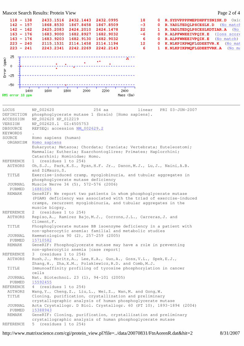
118 - 138 2433.1516 2432.1443 2432.0995 18 0 R.SYDVPPPPMEPDHPFYSNISK.D Oxidation (M) ( 142 - 157 1868.8530 1867.8458 1867.8509 -3 0 R.YADLTEDQLPSCESLK.D (No match 142 - 162 2425.2083 2424.2010 2424.1478 22 1 R.YADLTEDQLPSCESLKDTIAR.A (No match 163 - 176 1683.9000 1682.8927 1682.9032 -6 0 R.ALPFWNEEIVPQIK.E (Ions score 46 163 - 176 1683.9203 1682.9130 1682.9032 6 0 R.ALPFWNEEIVPQIK.E (No match) 223 - 240 2115.1531 2114.1458 2114.1194 12 0 K.NLKPIKPMQFLGDEETVR.K (No match 223 - 241 2243.2341 2242.2269 2242.2143 6 1 K.NLKPIKPMQFLGDEETVRK.A (No match
LOCUS NP_002620 254 aa linear PRI 03-JUN-2007 DEFINITION phosphoglycerate mutase 1 (brain) [Homo sapiens]. ACCESSION NP_002620 XP_012219 VERSION NP_002620.1 GI:4505753 DBSOURCE REFSEQ: accession NM_002629.2KEYWORDS . SOURCE Homo sapiens (human) ORGANISM Homo sapiens Eukaryota; Metazoa; Chordata; Craniata; Vertebrata; Euteleostomi; Mammalia; Eutheria; Euarchontoglires; Primates; Haplorrhini; Catarrhini; Hominidae; Homo. REFERENCE 1 (residues 1 to 254) AUTHORS Oh,S.J., Park,K.S., Ryan,H.F. Jr., Danon,M.J., Lu,J., Naini,A.B. and DiMauro,S. TITLE Exercise-induced cramp, myoglobinuria, and tubular aggregates in phosphoglycerate mutase deficiency JOURNAL Muscle Nerve 34 (5), 572-576 (2006) PUBMED 16881065 REMARK GeneRIF: We report two patients in whom phosphoglycerate mutase (PGAM) deficiency was associated with the triad of exercise-induced cramps, recurrent myoglobinuria, and tubular aggregates in the muscle biopsy. REFERENCE 2 (residues 1 to 254) AUTHORS Repiso,A., Ramirez Bajo,M.J., Corrons,J.L., Carreras,J. and Climent,F. TITLE Phosphoglycerate mutase BB isoenzyme deficiency in a patient with non-spherocytic anemia: familial and metabolic studies JOURNAL Haematologica 90 (2), 257-259 (2005) PUBMED 15710582 REMARK GeneRIF: Phosphoglycerate mutase may have a role in preventing non-spherocytic anemia [case report] REFERENCE 3 (residues 1 to 254) AUTHORS Rush,J., Moritz,A., Lee,K.A., Guo,A., Goss,V.L., Spek,E.J., Zhang,H., Zha,X.M., Polakiewicz,R.D. and Comb,M.J. TITLE Immunoaffinity profiling of tyrosine phosphorylation in cancer cells JOURNAL Nat. Biotechnol. 23 (1), 94-101 (2005) PUBMED 15592455REFERENCE 4 (residues 1 to 254) AUTHORS Wang,Y., Cheng,Z., Liu,L., Wei,Z., Wan,M. and Gong,W. TITLE Cloning, purification, crystallization and preliminary crystallographic analysis of human phosphoglycerate mutase JOURNAL Acta Crystallogr. D Biol. Crystallogr. 60 (PT 10), 1893-1894 (2004) PUBMED 15388943 REMARK GeneRIF: Cloning, purification, crystallization and preliminary crystallographic analysis of human phosphoglycerate mutase REFERENCE 5 (residues 1 to 254)
Page 2 of 4Mascot Search Results: Protein View
8/31/2007http://www.matrixscience.com/cgi/protein_view.pl?file=../data/20070831/FtnAorenR.dat&hit=2

Mascot Search Results Protein View
Match to: gi|190533 Score: 151 Expect: 1.5e-10proteinase 3 [Homo sapiens]
Nominal mass (Mr): 26150; Calculated pI value: 8.51
NCBI BLAST search of gi|190533 against nr Unformatted sequence string for pasting into other applications
Taxonomy: Homo sapiens
Fixed modifications: Carbamidomethyl (C) Variable modifications: Gln->pyro-Glu (N-term Q),Glu->pyro-Glu (N-term E),Oxidation (M) Cleavage by Trypsin: cuts C-term side of KR unless next residue is P Sequence Coverage: 48%
Matched peptides shown in Bold Red
1 AARAAEIVGG HEAQPHSRPY MASLQMRGNP GSHFCGGTLI HPSFVLTAAH 51 CLRDIPQRLV NVVLGAHNVR TQEPTQQHFS VAQVFLNNYD AENKLNDVLL 101 IQLSSPANLS ASVATVQLPQ QDQPVPHGTQ CLAMGWGRVG AHDPPAQVLQ 151 ELNVTVVTFF CRPHNICTFV PRRKAGICFG DSGGPLICDG IIQGIDSFVI 201 WGCATRLFPD FFTRVALYVD WIRSTLRRVE AKGRP
Residue Number Increasing Mass Decreasing Mass
Start - End Observed Mr(expt) Mr(calc) ppm Miss Sequence 1 - 27 2949.9934 2948.9861 2949.4450 -156 1 -.AARAAEIVGGHEAQPHSRPYMASLQMR.G 4 - 27 2635.8489 2634.8416 2635.2747 -164 0 R.AAEIVGGHEAQPHSRPYMASLQMR.G ( 28 - 53 2805.9570 2804.9497 2805.3592 -146 0 R.GNPGSHFCGGTLIHPSFVLTAAHCLR.D 59 - 70 1291.0577 1290.0504 1289.7568 228 0 R.LVNVVLGAHNVR.T (No match) 175 - 206 3412.3865 3411.3792 3411.6051 -66 0 K.AGICFGDSGGPLICDGIIQGIDSFVIWGCATR.L 175 - 206 3413.3889 3412.3816 3411.6051 228 0 K.AGICFGDSGGPLICDGIIQGIDSFVIWGCATR.L 207 - 214 1042.7000 1041.6927 1041.5284 158 0 R.LFPDFFTR.V (Ions score 59) 207 - 214 1042.7714 1041.7641 1041.5284 226 0 R.LFPDFFTR.V (No match) 215 - 223 1134.8000 1133.7927 1133.6233 149 0 R.VALYVDWIR.S (Ions score 69) 215 - 223 1134.8892 1133.8819 1133.6233 228 0 R.VALYVDWIR.S (No match)
LOCUS AAB59493 235 aa linear PRI 19-NOV-2002 DEFINITION proteinase 3 [Homo sapiens]. ACCESSION AAB59493 VERSION AAB59493.1 GI:190533 DBSOURCE locus HUMPRT3A01 accession M96838.1 locus HUMPRT3A02 accession M96837.1 locus HUMPRT3A03 accession M96839.1KEYWORDS . SOURCE Homo sapiens (human)
Show predicted peptides also
Sort Peptides By nmlkji nmlkj nmlkj
Page 1 of 2Mascot Search Results: Protein View
9/3/2007http://www.matrixscience.com/cgi/protein_view.pl?file=../data/20070822/FtntCzYmh.dat&hit=1

Mascot Search Results Protein View
Match to: gi|4506773 Score: 208 Expect: 3.1e-16S100 calcium-binding protein A9 [Homo sapiens]
Nominal mass (Mr): 13291; Calculated pI value: 5.71
NCBI BLAST search of gi|4506773 against nr Unformatted sequence string for pasting into other applications
Taxonomy: Homo sapiensLinks to retrieve other entries containing this sequence from NCBI Entrez: gi|115444 from Homo sapiensgi|7417327 from Homo sapiensgi|34771 from Homo sapiensgi|386958 from Homo sapiensgi|516621 from Homo sapiensgi|29126810 from Homo sapiensgi|49457408 from Homo sapiensgi|49457442 from Homo sapiensgi|56205191 from Homo sapiensgi|60822417 from synthetic constructgi|60822435 from synthetic constructgi|119573718 from Homo sapiensgi|119573719 from Homo sapiensgi|225793 from Homo sapiens
Fixed modifications: Carbamidomethyl (C) Variable modifications: Gln->pyro-Glu (N-term Q),Methyl (C-term),Methyl (DE),Oxidation (M) Cleavage by Trypsin: cuts C-term side of KR unless next residue is P Sequence Coverage: 67%
Matched peptides shown in Bold Red
1 MTCKMSQLER NIETIINTFH QYSVKLGHPD TLNQGEFKEL VRKDLQNFLK 51 KENKNEKVIE HIMEDLDTNA DKQLSFEEFI MLMARLTWAS HEKMHEGDEG 101 PGHHHKPGLG EGTP
Residue Number Increasing Mass Decreasing Mass
Start - End Observed Mr(expt) Mr(calc) ppm Miss Sequence 11 - 25 1806.8000 1805.7927 1805.9312 -77 0 R.NIETIINTFHQYSVK.L (Ions score 85 11 - 25 1806.8972 1805.8899 1805.9312 -23 0 R.NIETIINTFHQYSVK.L (No match) 11 - 25 1820.9000 1819.8927 1819.9468 -30 0 R.NIETIINTFHQYSVK.L Methyl (DE) ( 11 - 25 1820.9108 1819.9035 1819.9468 -24 0 R.NIETIINTFHQYSVK.L Methyl (C- 26 - 38 1455.6000 1454.5927 1454.7154 -84 0 K.LGHPDTLNQGEFK.E (Ions score 79 26 - 38 1455.6851 1454.6778 1454.7154 -26 0 K.LGHPDTLNQGEFK.E (No match) 26 - 38 1469.7000 1468.6927 1468.7310 -26 0 K.LGHPDTLNQGEFK.E Methyl (DE) ( 26 - 38 1469.7076 1468.7004 1468.7310 -21 0 K.LGHPDTLNQGEFK.E Methyl (C-term) ( 58 - 72 1742.7000 1741.6927 1741.8192 -73 0 K.VIEHIMEDLDTNADK.Q (Ions score 34 58 - 72 1742.7863 1741.7790 1741.8192 -23 0 K.VIEHIMEDLDTNADK.Q (No match) 58 - 72 1756.8027 1755.7955 1755.8349 -22 0 K.VIEHIMEDLDTNADK.Q Methyl (C- 58 - 72 1828.8854 1827.8781 1827.8924 -8 0 K.VIEHIMEDLDTNADK.Q Methyl (C- 58 - 72 1842.8905 1841.8832 1841.9081 -13 0 K.VIEHIMEDLDTNADK.Q Methyl (C- 73 - 85 1630.7621 1629.7548 1629.7895 -21 0 K.QLSFEEFIMLMAR.L Oxidation (M) ( 94 - 114 2175.9084 2174.9012 2174.9552 -25 0 K.MHEGDEGPGHHHKPGLGEGTP.- (No match 94 - 114 2189.9260 2188.9188 2188.9709 -24 0 K.MHEGDEGPGHHHKPGLGEGTP.- Methyl (C 94 - 114 2191.9277 2190.9205 2190.9501 -14 0 K.MHEGDEGPGHHHKPGLGEGTP.- Oxidation (M) (
Show predicted peptides also
Sort Peptides By nmlkji nmlkj nmlkj
Page 1 of 4Mascot Search Results: Protein View
9/3/2007http://www.matrixscience.com/cgi/protein_view.pl?file=../data/20070827/FtnAfaTam.dat&hit=3

LOCUS NP_002956 114 aa linear PRI 20-AUG-2007 DEFINITION S100 calcium-binding protein A9 [Homo sapiens]. ACCESSION NP_002956 VERSION NP_002956.1 GI:4506773 DBSOURCE REFSEQ: accession NM_002965.3KEYWORDS . SOURCE Homo sapiens (human) ORGANISM Homo sapiens Eukaryota; Metazoa; Chordata; Craniata; Vertebrata; Euteleostomi; Mammalia; Eutheria; Euarchontoglires; Primates; Haplorrhini; Catarrhini; Hominidae; Homo. REFERENCE 1 (residues 1 to 114) AUTHORS Benedyk,M., Sopalla,C., Nacken,W., Bode,G., Melkonyan,H., Banfi,B. and Kerkhoff,C. TITLE HaCaT keratinocytes overexpressing the S100 proteins S100A8 and S100A9 show increased NADPH oxidase and NF-kappaB activities JOURNAL J. Invest. Dermatol. 127 (8), 2001-2011 (2007) PUBMED 17429438 REMARK GeneRIF: HaCaT keratinocytes overexpressing the S100 proteins S100A8 and S100A9 show increased NADPH oxidase and NF-kappaB activities. REFERENCE 2 (residues 1 to 114) AUTHORS Anceriz,N., Vandal,K. and Tessier,P.A. TITLE S100A9 mediates neutrophil adhesion to fibronectin through activation of beta2 integrins JOURNAL Biochem. Biophys. Res. Commun. 354 (1), 84-89 (2007) PUBMED 17222807 REMARK GeneRIF: In contrast, neutrophil adhesion to fibronectin was completely inhibited by anti-beta2 integrins, suggesting that S100A9-induced specific activation of beta2 integrin is essential to neutrophil adhesion. REFERENCE 3 (residues 1 to 114) AUTHORS Klingelhofer,J., Senolt,L., Baslund,B., Nielsen,G.H., Skibshoj,I., Pavelka,K., Neidhart,M., Gay,S., Ambartsumian,N., Hansen,B.S., Petersen,J., Lukanidin,E. and Grigorian,M. TITLE Up-regulation of metastasis-promoting S100A4 (Mts-1) in rheumatoid arthritis: putative involvement in the pathogenesis of rheumatoid arthritis JOURNAL Arthritis Rheum. 56 (3), 779-789 (2007) PUBMED 17328050 REMARK GeneRIF: The pattern of S100A4 expression differed significantly from that of the proinflammatory proteins S100A9 and S100A12, which were restricted to phagocytes and granulocytes. REFERENCE 4 (residues 1 to 114) AUTHORS Shimada,S., Nakamura,M., Tanaka,Y., Tsutsumi,K., Katano,M., Masuko,K., Yudoh,K., Koizuka,I. and Kato,T. TITLE Crosslinking of the CD69 molecule enhances S100A9 production in activated neutrophils JOURNAL Microbiol. Immunol. 51 (1), 87-98 (2007) PUBMED 17237603 REMARK GeneRIF: data suggest unidentified natural ligands for CD69 and/or CD69 autoantibodies possibly affect joint-composing cell types through increased production of S100A9 in neutrophils, providing insight into functions of CD69 on neutrophils in rheumatoid
Page 2 of 4Mascot Search Results: Protein View
9/3/2007http://www.matrixscience.com/cgi/protein_view.pl?file=../data/20070827/FtnAfaTam.dat&hit=3