aohs foundations of anatomy and physiology ii lesson 14...
TRANSCRIPT

Copyright © 2014‒2016 NAF. All rights reserved.
AOHS Foundations of Anatomy and Physiology II
Lesson 14
The Cardiovascular System
Student Resources
Resource Description
Student Resource 14.1 Think, Pair, Find Out: The Heart
Student Resource 14.2 Diagram: Anatomy of the Heart
Student Resource 14.3 Reading: Anatomy of the Heart
Student Resource 14.4 Reading: Blood Vessels
Student Resource 14.5 Lab 1: Heart Sounds and Blood Pressure
Student Resource 14.6 Drawing: Path of Blood through the Heart
Student Resource 14.7 Lab 2: Sheep Heart Dissection
Student Resource 14.8 Vocabulary: Heart Puzzle
Student Resource 14.9 Notes: Components of Blood
Student Resource 14.10 Reading: Components of Blood
Student Resource 14.11 Lab 3: Blood Typing
Student Resource 14.12 Mystery: Crime Solving
Student Resource 14.13 Cases: Cardiovascular Conditions
Student Resource 14.14 Glossary: The Cardiovascular System (separate Word file)

AOHS Foundations of Anatomy and Physiology II Lesson 14 The Cardiovascular System
Copyright © 2014‒2016 NAF. All rights reserved.
Student Resource 14.1
Think, Pair, Find Out: The Heart
Student Name:_______________________________________________________ Date:___________
Instructions: For each question below, write down what you think the answer is in the “My Guess” row. Then find out what your partner thinks and write that down in the “My partner’s guess” row. As you watch the presentation, look for the answers to the questions and write them down in the “I found out” row.
Why does your heart stay in one place instead of bobbing around inside your chest?
My guess:
My partner’s guess:
I found out:
How many times a minute does your heart usually beat?
My guess:
My partner’s guess:
I found out:
Why don’t you have to make your heart beat?
My guess:
My partner’s guess:
I found out:

AOHS Foundations of Anatomy and Physiology II Lesson 14 The Cardiovascular System
Copyright © 2014‒2016 NAF. All rights reserved.
Student Resource 14.2
Diagram: Anatomy of the Heart
Student Name:_______________________________________________________ Date:___________
Directions: Label the diagram of the heart as you watch the presentation Anatomy of the Heart. Include the blood vessels, chambers of the heart, and valves.

AOHS Foundations of Anatomy and Physiology II Lesson 14 The Cardiovascular System
Copyright © 2014‒2016 NAF. All rights reserved.
Student Resource 14.3
Reading: Anatomy of the Heart

AOHS Foundations of Anatomy and Physiology II Lesson 14 The Cardiovascular System
Copyright © 2014‒2016 NAF. All rights reserved.

AOHS Foundations of Anatomy and Physiology II Lesson 14 The Cardiovascular System
Copyright © 2014‒2016 NAF. All rights reserved.
When you’re scared, your brain and endocrine system tell your heart to speed up, and sometimes you
can even feel it pounding in your chest. Most of the time, though, your heart is beating about 75 times a
minute and you’re totally unaware of it. Your heart beats like this, about 100,000 times a day, every
minute of your life. Imagine if you had to contract some other muscle in your body, like your biceps, 75
times a minute for your whole life. Your arm would get pretty tired. The muscle of your heart and the way
it contracts are specially designed to last a lifetime while doing the job of constantly pushing your 5 quarts
of blood around your body.

AOHS Foundations of Anatomy and Physiology II Lesson 14 The Cardiovascular System
Copyright © 2014‒2016 NAF. All rights reserved.
The heart is the only organ in the body that contains cardiac muscle. Cardiac muscle contains structures
that help spread the electrical impulse evenly through the tissue so that all the cells of the atria or the
ventricles contract at exactly the same time. The design of cardiac muscle is why the heart can contract
and relax continuously.

AOHS Foundations of Anatomy and Physiology II Lesson 14 The Cardiovascular System
Copyright © 2014‒2016 NAF. All rights reserved.
As hard as your heart can pound, it’s only about the size of your fist, much smaller than many other
muscles in your body. It is shaped like a cone, which, with a bit of artistic license, could be imagined as
the heart shape we’re familiar with. The heart sits between the lungs and is protected by the sternum. It’s
tilted a bit. The base of the cone, called the apex, points down and to the left.
At the top of the heart are the largest blood vessels in your body. Blood enters and exits your heart
through these vessels.

AOHS Foundations of Anatomy and Physiology II Lesson 14 The Cardiovascular System
Copyright © 2014‒2016 NAF. All rights reserved.
The pericardium is an important sac that encloses the heart and separates it from the other organs in the
thoracic cavity. The pericardium has three layers and is made of fibrous connective tissue. One layer of
the pericardium produces a slippery fluid that keeps the other layers of the pericardium from sticking to
each other when they move past each other as the heart beats.

AOHS Foundations of Anatomy and Physiology II Lesson 14 The Cardiovascular System
Copyright © 2014‒2016 NAF. All rights reserved.
In this image, we’re looking at the heart as you’d see it from the front, if you could see through someone’s
sternum. Imagine that you wanted to put this heart inside your own chest. In order to position it properly,
you would have to turn it around so that it’s facing the other direction. So, what you see on the left side of
this image would be on the right side of your body after you turn the heart around and put it in your chest.
And what you see on the right side of the image would be on the left side of your body. Being able to
know which side of the heart is which can be confusing, but it is also very important, because the two
sides of the heart have different roles.

AOHS Foundations of Anatomy and Physiology II Lesson 14 The Cardiovascular System
Copyright © 2014‒2016 NAF. All rights reserved.
The two sides of the heart have similar structures, but they look a bit different. For the most part, the atria
serve as a space for gathering the next dose of blood waiting to be pushed out. When the atria contract,
they push blood into the ventricles. The ventricles are the chambers that do the mighty work of propelling
your blood through your body. The heart is divided into its right and left sides by a wall of muscle tissue
called the septum.

AOHS Foundations of Anatomy and Physiology II Lesson 14 The Cardiovascular System
Copyright © 2014‒2016 NAF. All rights reserved.
A valve separates the atrium and ventricle on each side of the heart. The valve keeps the blood from
flowing backward into the atria when the ventricles contract. Without these valves, the heart would have
to work a lot harder to get blood where it needs to go. The valve on the right side of the heart has three
flaps and the valve on the left side has two. When the heart is between beats and the atria are filling with
blood, the flaps are hanging loosely downward. When the ventricles are full of blood and contract, the
pressure of the blood on the flaps pushes them up, closing the valves. The valves keep the blood flowing
in one direction, from atrium to ventricle.

AOHS Foundations of Anatomy and Physiology II Lesson 14 The Cardiovascular System
Copyright © 2014‒2016 NAF. All rights reserved.
The blood that enters the right side of the heart has given up its oxygen to be used for various things,
particularly for generating energy to fuel other functions. This blood comes from throughout the body. It
enters the right side of the heart through two large veins called the inferior and superior venae cavae. It
goes through the atrium into the ventricle and is pushed out to the lungs through the pulmonary artery.
This large artery splits into two branches, with one branch going to each lung. At the base of both the
venae cavae and the pulmonary artery there are valves which, like the valves inside the heart, keep blood
from flowing backward.
AOHS Foundations of Anatomy and Physiology II Lesson 14 The Cardiovascular System
Copyright © 2014‒2016 NAF. All rights reserved.
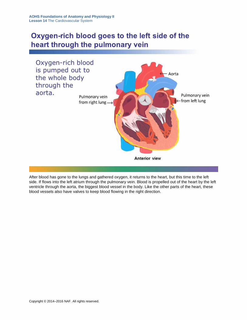
After blood has gone to the lungs and gathered oxygen, it returns to the heart, but this time to the left
side. If flows into the left atrium through the pulmonary vein. Blood is propelled out of the heart by the left
ventricle through the aorta, the biggest blood vessel in the body. Like the other parts of the heart, these
blood vessels also have valves to keep blood flowing in the right direction.

AOHS Foundations of Anatomy and Physiology II Lesson 14 The Cardiovascular System
Copyright © 2014‒2016 NAF. All rights reserved.
If you look at the heart from the front, as we did before, the two ventricles look fairly alike. But if you made
a transverse cut through the heart and viewed it from above, you’d see that they are quite different. The
left ventricle makes up much more of the heart. It has a much rounder shape, while the right ventricle is
shaped something like a crescent. The left ventricle has much thicker walls of muscle. That’s because it
has a bigger job to do, pumping blood through the entire body, sometimes against gravity, while the right
side only pumps blood to the nearby lungs.

AOHS Foundations of Anatomy and Physiology II Lesson 14 The Cardiovascular System
Copyright © 2014‒2016 NAF. All rights reserved.
The heart does two jobs at the same time: the right side of the heart pushes blood to your lungs to get
oxygen, and the left side of the heart pushes blood through the circuit of blood vessels that brings
nutrients and oxygen to the rest of your body.
Before 1616, physicians thought that there were two different systems that moved blood through the
body, and that the lungs played a role. Then, in 1616, an English physician named William Harvey
discovered that the heart pumps all of the body’s blood through a single system (the circulatory system).
Now we know that the heart does this at a remarkable rate: every minute, your heart pumps about 5 liters
(that’s about 14 cans of soda) of blood. When you’re exercising as hard as you can, your heart can pump
over 20 liters a minute.
Image retrieved from
http://commons.wikimedia.org/wiki/File%3APSM_V69_D574_William_Harvey.png on September 23,
2014. From Popular Science Monthly Volume 69.

AOHS Foundations of Anatomy and Physiology II Lesson 14 The Cardiovascular System
Copyright © 2014‒2016 NAF. All rights reserved.
Unlike all the other muscle tissue in your body, cardiac muscle doesn’t need to be stimulated by a neuron.
Each heartbeat is initiated by a bundle of cardiac cells called the sinoatrial node, or SA node. Within the
muscle that makes up the heart are other special bundles of tissue that transmit electrical impulses.
These impulses spread through other special tissue bundles and result in a heartbeat. When your heart is
functioning normally, the muscle cells contract in a precise rhythm, which is what gives you your
heartbeat. If they didn’t contract in a coordinated way, your heart wouldn’t be as effective at moving your
blood through and wouldn’t be as strong a pump. The bundle of tissue that starts this contraction process
rolling also determines how often your heart beats and is considered your heart’s pacemaker. The atria
contract first, pushing blood into the ventricles, and then the ventricles contract, pushing blood to the
lungs and to the rest of the body.
Although you don’t need neurons to initiate a heartbeat, your nervous and endocrine systems can affect
how often your heart beats. When you’re watching that scary movie, your endocrine and nervous systems
work together to make your SA node send signals more often, making your heart beat faster.

AOHS Foundations of Anatomy and Physiology II Lesson 14 The Cardiovascular System
Copyright © 2014‒2016 NAF. All rights reserved.
A doctor can see whether the chambers of your heart are contracting normally by doing a test called an
electrocardiogram, or ECG. To do an ECG, the doctor puts electrodes on your chest. The electrodes pick
up the signals being sent to your heart that make it contract. The signals are transferred to paper, where
you can see a distinctive pattern in the heartbeat. The short peaks represent the signal to the atria to
contract. The tall peaks represent the signal to the ventricles to contract. The small peak afterward is the
heart muscle relaxing.

AOHS Foundations of Anatomy and Physiology II Lesson 14 The Cardiovascular System
Copyright © 2014‒2016 NAF. All rights reserved.
You can live without some of your organs but not without your heart. Your heart has an essential job and
it never gets a break, so it’s important to do what you can to keep your heart healthy. In the same way
that using your arms and legs in exercise will make them stronger, using your heart will keep it in shape
and make it stronger, too. Aerobic exercise keeps your pulse rate up for an extended period of time,
which makes your heart work consistently and helps make your whole cardiovascular system stronger.
Because your heart is so vital to everything your body does, strengthening your heart helps your whole
body in a variety of ways. There are few things you can do that are better for your body than regular
aerobic exercise!

AOHS Foundations of Anatomy and Physiology II Lesson 14 The Cardiovascular System
Copyright © 2014‒2016 NAF. All rights reserved.
Student Resource 14.4
Reading: Blood Vessels
Student Name: _________________________________________ Date: _______________________
Directions: As you complete the reading, answer the questions in each section.
Look at the inside of your wrist or the back of your hand. You can probably see some bluish, branching
lines running just under the skin. These are just a few inches of the 60,000 miles of blood vessels in your
body, the vascular part of the cardiovascular system. They may look as if they’re just sitting there, but
your blood vessels, like your heart, are active organs in your cardiovascular system, providing every cell
in your body with all the necessary substances carried in the blood, and keeping things moving along in
the right direction.
Types of blood vessels
There are three types of blood vessels, each with its own job to do:
Arteries carry blood away from the heart. As they get farther from the heart, arteries branch off into vessels with smaller and smaller diameters. For example, blood leaves the heart through the body’s largest artery, the aorta, which is about 34 mm (1.3 inches) wide. The aorta quickly branches into smaller arteries. By the time they reach your arms, the largest arteries are only about 2 or 3 mm (around 1/10 of an inch) wide. As your right ventricle contracts, it pushes blood through your arteries with great force. To adapt to that force, your arteries have elastic walls that contain a layer of involuntary muscle. Neurons in the autonomic nervous system can stimulate these arterial muscles to contract or expand in response to blood flow, hormones, or other factors. This layer of muscle can also help to keep pushing blood through your arteries.
Question #1: What else happens in your cardiovascular system in response to your sympathetic nervous
system? Why might your blood vessels need to expand to accommodate this?
When arteries become so tiny their walls are only a cell or two thick, they become capillaries. Capillaries are where the real
action happens. Across those thin capillary walls, oxygen, nutrients, hormones, and other substances diffuse from the blood into the fluid that surrounds cells, and waste products diffuse from that fluid into the blood. This process is called capillary exchange, and it is the source of supply for
every one of the trillions of cells in your body. Capillaries connect the arteries to veins in structures called capillary beds. At the end

AOHS Foundations of Anatomy and Physiology II Lesson 14 The Cardiovascular System
Copyright © 2014‒2016 NAF. All rights reserved.
of an artery, the blood vessel branches into a netting of tiny capillaries that rejoin again on the other side. Capillary beds are all over your body, and they account for over half of your 60,000 miles of blood vessels. Along with being the site of gas and nutrient exchange, capillaries also play a role in regulating your body temperature and blood pressure.
At the end of the capillary bed where the vessels rejoin, the veins begin. Veins carry blood from capillary beds back to the heart, where it can be sent to the lungs for oxygen. The walls of veins are thinner than the walls of arteries, and veins tend to be flatter and don’t contain as much muscle. Blood in your veins is under much less pressure than blood in your arteries. In your arms and legs, blood often has to flow upward against gravity to return to your heart. In your arteries, the pressure and muscular walls keep blood flowing in the right direction. Your larger veins, instead, have one-way valves that keep blood from flowing backward. Your movements (well, really, the contractions of your skeletal muscles) also help move blood through your veins and back to your heart.
As a general rule, the blood in your veins is dark purple because the oxygen that gave it a bright red color
has been given up to your cells. (If you cut a vein, you will still see bright red blood, but this is because it
binds oxygen in the air.) But there are two important exceptions to this rule. The pulmonary artery brings
deoxygenated blood away from the heart and to the lungs. The pulmonary veins bring oxygenated blood
from the lungs to the heart.
The inside of all your vessels, including the chambers of your heart, is lined with one continuous layer of
epithelial cells. Blood vessels also contain mechanical sensory receptors called baroreceptors that can
sense the amount of pressure on the blood vessel walls and send signals to your autonomic nerve
system that affect heart rate and level of constriction of the blood vessels. For example, if the receptors
sense that your blood pressure is low, baroreceptors may send a signal to your brain to tell your heart to
beat faster. This increase in heart rate will increase the blood flow out of your heart and increase blood
pressure.
Question #2: You might have heard the phrase “his blood was pulsing through his veins.” What’s
incorrect about that phrase?
Two paths of circulation
In order to do its job, every red blood cell in your
body makes its way through two different circuits of
blood vessels. The pulmonary circuit brings
blood to your lungs to get oxygenated, and returns
it to the left side the heart. The systemic circuit
brings that oxygenated blood to all of your cells
and then returns it to the right side of the heart to
get sent back to the lungs. In about a minute’s
time, each of your trillion red blood cells makes its
way through these two circuits.
In the pulmonary circuit, blood leaves the right
ventricle through the pulmonary artery to the
arteries of the lungs. Blood goes through millions
of capillaries in the lungs that lie next to structures
in the lungs that collect air when you breathe in.

AOHS Foundations of Anatomy and Physiology II Lesson 14 The Cardiovascular System
Copyright © 2014‒2016 NAF. All rights reserved.
Oxygen diffuses into the blood and latches on to the iron ions in the red blood cells. Then the blood
returns to the heart and gets dumped into the left atrium via the pulmonary vein.
Question #3: Why do you need two different circuits of blood vessels in your body?
The systemic circuit is more extensive, supplying your entire body with blood. Often in this circuit, arteries
extend to their destination, branch out again and again until they become capillaries, and continue,
becoming veins that return to the heart alongside or near where the arteries are located. In many cases,
these pairs of blood vessels share the same name. Arteries are usually deeper than veins, protected by
bone and soft tissues. In some cases, there are two veins where there is only one artery. One vein is
superficial and the other is deep. This pairing allows your blood vessels to play a role in regulating your
body temperature. When it’s hot and you want to dissipate heat, more blood flows through the superficial
capillaries and into superficial veins near your skin. When it’s cold and you want to conserve heat, more
blood runs through a deeper set of vessels and into the deeper veins.
This long circuit, which contains thousands of miles of blood vessels, begins as the left ventricle pumps
blood into the aorta, the largest artery in the body. From there, the blood either goes toward the head and
arms through the branches leaving the aortic arch, or continues downward, through the descending
aorta.
The aortic arch, which crosses over the top of the heart, has three branches that give rise to all the
vessels that supply your head, neck, and arms. The right and left subclavian arteries (where would you
guess they are located?) provide blood to the arms. Carotid arteries go up either side of the head to the
face and neck. You can feel your carotid artery―a strong pulse on each side of your neck below your jaw
bone. Branches of the carotid and subclavian arteries supply the brain. All these arteries lead to capillary
beds throughout the head, neck and arms. Once through the capillary beds, the blood from your head,
neck, and brain returns to the heart through the jugular veins, and blood from your arms returns via the
right and left subclavian veins.
The descending aorta supplies blood to everything below the heart. The main pipe that descends behind
your heart is called the thoracic aorta as it passes through your thoracic cavity, and the abdominal
aorta as it passes through your abdomen. This large vessel has branches that supply all your internal
organs, including your digestive, urinary, respiratory, and reproductive systems. Your heart muscle needs
blood, too. It’s supplied by branches of the ascending aorta (where it leaves the heart) called the
coronary arteries.
At about the same height as the top of your pelvis, the abdominal aorta branches off into two vessels, the
right and left common iliac arteries, that supply blood to your legs. Blood returns to your heart via the
right and left common iliac veins.

AOHS Foundations of Anatomy and Physiology II Lesson 14 The Cardiovascular System
Copyright © 2014‒2016 NAF. All rights reserved.

AOHS Foundations of Anatomy and Physiology II Lesson 14 The Cardiovascular System
Copyright © 2014‒2016 NAF. All rights reserved.
Pulse and Blood Pressure
Your pulse and your blood pressure can both be used to give some insight into the state of your
cardiovascular system. When you’re taking a pulse, you’re sensing the blood pulsing through an artery as
the heart is pumping blood through the systemic circuit, and each pulse reflects a heartbeat. Blood
pressure, on the other hand, is a way to see an aspect of the whole circuit at once—the pressure exerted
on your blood vessels when your ventricles are contracting and when they’re at rest.
The two easiest places to feel your pulse are the carotid artery in your neck, just under the square part of
your jaw, and the radial artery on the inside of your wrist, toward your thumb. See if you can feel your
heartbeat in an artery in either of these places. The average heart rate—the number of times your heart
beats in a minute—is about 70 beats per minute. Your heart rate can be affected by several factors. Your
nervous system can speed up your heart rate when you’ve been exercising or when you’re frightened. A
person carrying extra weight may also have an increased heart rate, as can a person whose heart is
beating inefficiently.
If you’ve ever had your blood pressure taken, you know that it’s recorded as two numbers and said as “X
number over Y number,” for example, 115 over 70. The top number is the pressure of your blood against
your blood vessels when your ventricles are contracting, that is, when blood is being pushed into the
circuits. This is called your systolic blood pressure. That same pressure is the heartbeat you feel in
someone’s artery when you take their pulse. The bottom number is a measure of the pressure when your
ventricles are relaxed and is called diastolic blood pressure. A normal blood pressure has a systolic
reading of 120 or less and a diastolic reading of 80 or less.
The chart below shows the American Heart Association’s categories for different blood pressure
measurements. Hypertension is another term for high blood pressure. If a person consistently has blood
pressure readings just a bit above normal, doctors say he has prehypertension, meaning if he doesn’t
change his habits, he will have high blood pressure (similar to a person being prediabetic).
Everyone’s blood pressure varies throughout the day, depending on what they’re doing. When you’re
sleeping, your blood pressure will be low. When you’re exercising, it will be high. A lot of people’s blood
pressure goes up a bit when they’re at the doctor because they’re nervous—just the opposite of what you
want your blood pressure to do when you visit the doctor! Your “real” blood pressure is what the
measurement would be if you’re sitting quietly.

AOHS Foundations of Anatomy and Physiology II Lesson 14 The Cardiovascular System
Copyright © 2014‒2016 NAF. All rights reserved.
Many factors besides your activity can affect blood pressure. How much blood you have altogether can
make a difference. A hormone released by your kidneys controls how much plasma is in your blood. The
more plasma, the higher the blood volume and blood pressure.
Arteries can also expand and contract in response to your blood pressure. If your blood pressure goes
up, the arteries can increase their diameter ((by expanding, dilating, or relaxing). Some vascular
conditions can make a person’s arteries stiffen and become harder to expand, which can raise blood
pressure. If you have consistently high blood pressure, you may not know it, because high blood pressure
doesn’t have any symptoms, but it does have medical dangers. High blood pressure can damage your
arteries and make them less able to expand. If your blood pressure is consistently high, your left ventricle
has to work a lot harder to pump blood all around your body. Eventually, tiny but important blood vessels
get clogged up, particularly in your brain, heart, and kidneys. When cells in these organs can’t get oxygen
and nutrients, it can have life-threatening effects.
Question #4: How do you imagine the heart of a person with prolonged high blood pressure might look
different from the heart of someone with normal blood pressure?
For many people with high blood pressure, the problem arises because of genetic factors or other
reasons that are hard to pinpoint. One way to help keep your blood pressure within a healthy range is to
exercise. That’s because, in response to regular exercise, your body creates more blood vessels to
supply more oxygen and nutrients to the muscles you are using. Those additional blood vessels help
spread out the pressure across more vessels. The result is less pressure on each individual vessel, not to
mention a stronger heart that doesn’t have to work as hard. In fact, exercise can lower a person’s
numbers as much as some blood pressure medications can.

AOHS Foundations of Anatomy and Physiology II Lesson 14 The Cardiovascular System
Copyright © 2014‒2016 NAF. All rights reserved.
Student Resource 14.5
Lab 1: Heart Sounds and Blood Pressure
Student Name:_______________________________________________________ Date:___________
Directions: Listen to your partner’s heart, take his or her blood pressure, and record the results.
Heart Sounds
When you listen to a heartbeat through a stethoscope, you hear a pair of sounds that repeat over and
over. People often describe these sounds by saying it sounds like “lub-dub.” The pattern of the sounds is
lub-dub, pause, lub-dub, pause. What you hear is the sounds of the valves in the person’s heart closing
as the heart is going through its pumping cycle. The ”lub” sound happens after the atria have contracted.
It’s the sound of the valves closing between each atrium and ventricle (which keeps blood in the ventricle
from flowing back into the atrium). The “dub,” which is often more distinct, is the sound of the valves at the
bases of the large blood vessels—the pulmonary artery and the aorta—closing after the ventricles have
contracted. These valves keep blood from flowing back into the ventricles.
Listening to the pulse
Compare your partner’s pulse when sitting and just after exercising. One set of lub-dub is one heartbeat.
The average heart rate for a man is around 68 beats a minute and for a woman around 75 beats per
minute.
1. One partner (partner A) should have the stopwatch and the other (partner B) should have the
stethoscope.
2. Partner A places the stethoscope against partner B’s chest, near the sternum, and lets partner B
know he or she hears the sound.
3. Partner B tells partner A to start counting the heartbeats and at the same time, starts the
stopwatch.
4. After 15 seconds, partner B tells partner A to stop counting.
5. Record the pulse on the chart below.
6. Partner B gives the stopwatch to partner A, and jogs in place or does jumping jacks for one
minute.
7. Partner A gives the stopwatch back to partner B.
8. Partner A takes partner B’s pulse with the stethoscope again for 15 seconds.
9. Record the pulse in the chart below.
10. Switch roles so that partner B listens to partner A’s heart.
Partner Heart rate sitting Heart rate after exercise
A
B

AOHS Foundations of Anatomy and Physiology II Lesson 14 The Cardiovascular System
Copyright © 2014‒2016 NAF. All rights reserved.
What factors influence your heart rate?
Why does your heart need to beat faster when you exercise?
Blood Pressure
Blood pressure measures the pressure of blood within the arteries. It is measured in millimeters of
mercury (mm Hg), which is the distance that the pressure in the arteries makes the level of mercury rise
to in a device called a sphygmomanometer, or a blood pressure cuff.
Your sphygmomanometer has a cuff, a bulb to use to inflate the cuff, and some kind of attachment that
will show you a measurement in mm Hg (a digital sphygmomanometer may just give you numbers). The
bulb will have a valve on it that will open and close, letting air out or keeping it in. You’ll use the
stethoscope, too, to take someone’s blood pressure.
Be sure to read through these instructions before you take a partner’s blood pressure!
1. Have the student whose blood pressure is being measured roll up his or her sleeve and extend an
arm onto the table, with the palm facing up.
2. Wrap the cuff of the sphygmomanometer around the student’s upper arm so it is snug but leaving
room to insert two fingers. If your cuff has markings showing how it should be placed, be sure that it
is placed correctly on the student’s arm.
3. Place the stethoscope directly below the cuff in the bend of the elbow joint. Here, it will be on top of
the brachial artery. Place the ends of the stethoscope in your ears.
4. Close the valve of the bulb by turning it clockwise.
5. Pump air into the cuff until the pressure gauge goes just past 160 mm Hg. You should not be able to
hear any sounds in the stethoscope coming from the artery in the arm. At this point, the pressure of
the cuff on the artery is higher than the pressure of the blood inside. This means the cuff is keeping
blood from flowing through the artery.
6. WARNING: The cuff should not be kept inflated for more than one minute. If you have any trouble obtaining a reading within this time, deflate the cuff, wait one or two minutes, and try again. (A prolonged interruption of blood flow can cause fainting.)
7. Turn the valve of the bulb counterclockwise and gradually release the air from the cuff. Listen for a sound to start coming from the artery.
8. When you first hear a sound coming from the artery, note the pressure on the gauge. This is the
systolic pressure, or the pressure in the arteries when the ventricles are contracting. At this point,
the pressure of the blood in the artery is as great as the pressure from the cuff, and the blood begins
to flow through the artery again.
9. Continue to slowly release air and listen until the clear thumping sound of the pulse becomes strong
and then fades. When you last hear the full heartbeat, note the pressure. This is the diastolic
pressure, or the pressure in the artery when the heart is at rest.
10. Repeat the measurement two more times and determine the average systolic and diastolic pressure.
11. Record the results in the chart below.

AOHS Foundations of Anatomy and Physiology II Lesson 14 The Cardiovascular System
Copyright © 2014‒2016 NAF. All rights reserved.
Trial Systolic pressure Diastolic pressure
1
2
3
Average of the three systolic pressures (add all three and divide by 3): _________________
Average of the three diastolic pressures (add all three and divide by 3): _________________
Is this person’s blood pressure in the normal range?
What factors do you know of that affect blood pressure?

AOHS Foundations of Anatomy and Physiology II Lesson 14 The Cardiovascular System
Copyright © 2014‒2016 NAF. All rights reserved.
Student Resource 14.6
Drawing: Path of Blood through the Heart
Student Name:_______________________________________________________ Date:___________
Directions: Fill in the blanks in the sentences below to describe what is happening at each of the arrows.

AOHS Foundations of Anatomy and Physiology II Lesson 14 The Cardiovascular System
Copyright © 2014‒2016 NAF. All rights reserved.
1. Blood from the _____________ enters the _______________ side of the heart through the
_________________________ and goes into the ________________________________.
2. Blood from the __________________ goes through the _______________________and into the
_________________________.
3. Blood from the ____________________ goes to through the ____________________to the
_______________________________.
4. Blood from the ______________________ enters the ______________ side of the heart through
the ____________________ and goes into the _____________________________.
5. Blood from the __________________ goes through the _____________________and into
the ____________________________.
6. Blood from the ______________________ goes to through the _____________________to the
______________________________.

AOHS Foundations of Anatomy and Physiology II Lesson 14 The Cardiovascular System
Copyright © 2014‒2016 NAF. All rights reserved.
Student Resource 14.7
Lab 2: Sheep Heart Dissection
Student Name:_______________________________________________________ Date:___________
Directions: Follow the observation and dissection instructions below and answer the questions.
1. Look for three types of tissue on the heart: fatty tissue, heart muscle, and blood vessels. Describe
how they look and feel different from each other.
2. Locate the anterior side of the heart. To help you orient the heart, the anterior side has a diagonal
line of blood vessels running along its surface. The right and left chambers of the heart are
separated by this line. Position the heart in front of your chest as it would be situated if it were
inside you. The blood vessels coming out of the heart (or their openings if they are cut short)
should be oriented superiorly as you hold it.
3. Hold the heart with the anterior side facing you again. Gently squeeze the right ventricle, and then
the left. What difference do you notice between them?
4. Your teacher will project an image of the heart. Try to determine which vessel is which. If you are
doing the actual dissection, use a probe or your fingers to locate the superior and inferior venae
cavae and see how they lead into the right atrium. Do the same with the pulmonary vein. The
blood vessels may be cut so short that what remains is mostly a hole leading to the heart
chambers.
Label the following parts of the heart, either with dissection pins on the actual heart or arrows on
the heart image. If you are using dissection pins, attach a piece of tape to each pin with a label to
identify the part.
- Vena cava
- Pulmonary artery
- Pulmonary vein
- Aorta
- Right side of the heart
- Left side of the heart
- Front (anterior) of heart
5. Your teacher will project the same image, but this one will have labels. Check your work, and ask
your teacher if you have any questions.
6. Your teacher will then project an image of the heart cut in half. If you are using a real heart, cut
the heart in half along the frontal plane, as was demonstrated in the video, so that you have
anterior and posterior halves of the heart.

AOHS Foundations of Anatomy and Physiology II Lesson 14 The Cardiovascular System
Copyright © 2014‒2016 NAF. All rights reserved.
7. Again, label the following parts of the heart, either with dissection pins and tape on the actual
heart or arrows on the heart image.
- Left atrium
- Left ventricle
- Right atrium
- Right ventricle
- Bicuspid valve
8. As before, your teacher will project the same image, but with labels. Check your work, and ask
your teacher to clarify any questions you have.

AOHS Foundations of Anatomy and Physiology II Lesson 14 The Cardiovascular System
Copyright © 2014‒2016 NAF. All rights reserved.
Student Resource 14.8
Vocabulary: Heart Puzzle
Student Name:_______________________________________________________ Date:___________
Directions: Write the answers to the 10 clues on the corresponding line in the Words section, one letter per space. Then unscramble the circled letters to find the answer to the riddle.
Riddle
What sound is music to a cardiologist’s ear?
Clues
1. Number of chambers in the heart
2. Top chambers of the heart
3. Valve with two flaps: ______ valve
4. Bottom chambers of the heart
5. Heart muscle that separates the ventricles
6. Organ that pumps blood throughout the body
7. Bottom tip of the heart; points to the left
8. Largest artery
9. Veins that bring blood to right atrium
10. Takes blood away from the heart to the lungs: ______artery

AOHS Foundations of Anatomy and Physiology II Lesson 14 The Cardiovascular System
Copyright © 2014‒2016 NAF. All rights reserved.
Words
1.___ ___ ___ ___
2.___ ___ ___ ___ ___
3.___ ___ ___ ___ ___ ___ ___ ___
4.___ ___ ___ ___ ___ ___ ___ ___ ___ ___
5.___ ___ ___ ___ ___ ___
6.___ ___ ___ ___ ___
7.___ ___ ___ ___
8.___ ___ ___ ___ ___
9.___ ___ ___ ___ / ___ ___ ___ ___
10. ___ ___ ___ ___ ___ ___ ___ ___ ___
Answer (unscramble!):
__________________________________________________

AOHS Foundations of Anatomy and Physiology II Lesson 14 The Cardiovascular System
Copyright © 2014‒2016 NAF. All rights reserved.
Student Resource 14.9
Notes: Components of Blood
Student Name:_______________________________________________________ Date:___________
Directions: Complete these notes as you follow the presentation.
1. What are the four main components of blood?
2. In addition to these main components, what are six other substances carried in your blood?
3. How do red blood cells carry oxygen?
4. a) What is the role of white blood cells?
b) Why is it important that white blood cells can leave the bloodstream?
5. What is the process of phagocytosis?
6. How does a tattoo demonstrate that macrophages can live for decades?
7. What role do platelets play?

AOHS Foundations of Anatomy and Physiology II Lesson 14 The Cardiovascular System
Copyright © 2014‒2016 NAF. All rights reserved.
8. Where are the cells of blood made?
9. How does your blood plasma reflect what’s happening in your body?
10. What is an antigen?
11. Why is it important to know a patient’s blood group when he or she is getting a transfusion?
12. Fill in the chart below. (Note: This chart is similar to, but not the same as, the one in the presentation.)
Person with blood group: Can donate blood to people with
blood group(s):
A
B
AB
O
13. What can happen if a woman who is Rh negative has a second baby that is Rh positive?
AOHS Foundations of Anatomy and Physiology II Lesson 14 The Cardiovascular System
Copyright © 2014‒2016 NAF. All rights reserved.
Student Resource 14.10
Reading: Components of Blood

AOHS Foundations of Anatomy and Physiology II Lesson 14 The Cardiovascular System
Copyright © 2014‒2016 NAF. All rights reserved.

AOHS Foundations of Anatomy and Physiology II Lesson 14 The Cardiovascular System
Copyright © 2014‒2016 NAF. All rights reserved.
You may not faint at the sight of blood, but chances are that when you see it—yours or someone else’s—
the sight has some kind of effect on you. The idea of blood is such a strong one that we have lots of
phrases and sayings that reflect how blood captures our imagination. If there’s something you really love
to do, you might say, “It’s in my blood.” If someone does something that makes you angry, you might say,
“That made my blood boil.” And if you get in a fight over it, there might be “bad blood” between you.
Writers often equate blood with life. Their equation is correct: blood is absolutely essential to the
functioning of your body.

AOHS Foundations of Anatomy and Physiology II Lesson 14 The Cardiovascular System
Copyright © 2014‒2016 NAF. All rights reserved.
Blood is the only tissue in your body that is a fluid. The average adult has about 5 quarts (which is
approximately 5 liters) of blood. Every cell is dependent on the materials blood delivers, and if your blood
stops circulating (in other words, if your heart isn’t beating) for just a couple of minutes, cells begin to die.
Nutrients and oxygen carried by blood keep your cells powered. Ions delivered in the blood balance the
concentrations of substances inside and outside of your cells while removal of acids helps maintain a
normal pH. Hormones convey important messages to cells about what they need to be doing at a given
time.

AOHS Foundations of Anatomy and Physiology II Lesson 14 The Cardiovascular System
Copyright © 2014‒2016 NAF. All rights reserved.
Blood has other important functions that don’t involve ferrying materials from one site to another. When
you are injured—including when the injury is inside of you—blood responds with substances that kick off
the process of forming a blood clot. When the clot forms, it keeps you from losing more blood and
provides protection to the injured area while it is healing. Blood also contains many special cells and
proteins that can detect toxic substances and carry them away, or identify infectious invaders like viruses
and bacteria and destroy them. In addition, when you move your muscles and generate heat, that warmth
is absorbed by your blood and spread throughout your body, helping to regulate your body temperature.

AOHS Foundations of Anatomy and Physiology II Lesson 14 The Cardiovascular System
Copyright © 2014‒2016 NAF. All rights reserved.
Though your blood looks like a red liquid, it is made of four different components, only one of which is red.
Red blood cells contain iron, which gives them—and your blood—its color. Each component of blood has
a different role to play, making blood a very complex tissue.

AOHS Foundations of Anatomy and Physiology II Lesson 14 The Cardiovascular System
Copyright © 2014‒2016 NAF. All rights reserved.
Red blood cells are shaped sort of like a donut that hasn’t had its hole completely cut out. Because you
have—and need—so many RBCs, your body is making new ones all the time. In fact, in the time it takes
you to read this sentence, your body will produce about 10 million RBCs. Red blood cells, which are also
called erythrocytes, are made in the red bone marrow of your bones. It takes about two days for an RBC
to mature and be ready to circulate. Once it’s in your bloodstream, an RBC will live for about 120 days
(around four months), and travel about 700 miles through your blood vessels.

AOHS Foundations of Anatomy and Physiology II Lesson 14 The Cardiovascular System
Copyright © 2014‒2016 NAF. All rights reserved.
Hemoglobin is a protein that contains iron. The iron is in the form of a positively charged ion that can bind
oxygen molecules and transport them around the body. Hemoglobin can also bind carbon dioxide
molecules, which are usually waste products that need to be carried away.
Red blood cells are basically tiny bags of hemoglobin and little else. Before they’re released into your
bloodstream, RBCs lose their nuclei and all their organelles, and most of the space inside the cell is filled
with the oxygen-carrying hemoglobin. Hemoglobin in your blood is the main way your body has of getting
oxygen to your cells, transporting 99% of the oxygen. (The other 1% is dissolved in the plasma—similar
to the oxygen dissolved in water that fish can breathe). You take the oxygen in when you breathe, but
without your RBCs there would be no way to get the oxygen to the rest of your body.

AOHS Foundations of Anatomy and Physiology II Lesson 14 The Cardiovascular System
Copyright © 2014‒2016 NAF. All rights reserved.
White blood cells provide an important part of the body’s defense against infections and foreign
substances. There are several different types of WBCs, and red bone marrow produces each type. Since
bacteria, viruses, and toxins usually do their damage in cells outside of your bloodstream, WBCs have to
be able to follow those invaders wherever they go. It would be disastrous if your RBCs left your blood
vessels, but it would be equally disastrous if your WBCs didn’t. White blood cells slip through tiny spaces
between the cells that make up your blood vessels and then migrate to the places they’re needed.

AOHS Foundations of Anatomy and Physiology II Lesson 14 The Cardiovascular System
Copyright © 2014‒2016 NAF. All rights reserved.
One important trick your WBCs can perform is to hunt down, recognize, and consume potentially harmful
microorganisms and foreign substances, wherever they are in your body. When a WBC gets a signal that
it is needed somewhere, it races to the scene. Proteins on the cell’s surface help it tell the difference
between familiar cells that make up body tissues and foreign cells like bacteria. When they find something
unknown, many WBCs wrap themselves around the cell or substance in question. Then they take it inside
of themselves, destroy it, and send any usable molecules out for recycling. This process of consuming
and digesting invaders is called phagocytosis.

AOHS Foundations of Anatomy and Physiology II Lesson 14 The Cardiovascular System
Copyright © 2014‒2016 NAF. All rights reserved.
For the vast majority of WBCs, phagocytosis is a suicidal act. Though they are protecting the body from
infection, they will die from doing it. White blood cells, called macrophages, that live in the tissue instead
of in the bloodstream can live very long lives, continually consuming and digesting things that are
potentially harmful to the rest of the body. Macrophages are the cells we can thank for the persistence of
tattoos. When a tattoo artist injects ink just below a person’s skin, it activates macrophages. The
macrophages come to the site and eat the droplets of tattoo ink. The macrophages stay put, filled with
tiny drops of ink, until they die off. If they died quickly, the tattoo would fade away. But because they last
so long, so does the tattoo.

AOHS Foundations of Anatomy and Physiology II Lesson 14 The Cardiovascular System
Copyright © 2014‒2016 NAF. All rights reserved.
Platelets aren’t cells, but they do have cell membranes. Platelets are bits and pieces of much larger cells
produced in your bone marrow. When it’s time for one of these cells to become platelets, small sections
of its cell membrane pinch off, trapping contents (including proteins that set off the chain reaction that
leads to blood clotting) inside. One of these special cells can give rise to thousands of platelets. Platelets
float freely in the blood and are much smaller than RBCs or WBCs.

AOHS Foundations of Anatomy and Physiology II Lesson 14 The Cardiovascular System
Copyright © 2014‒2016 NAF. All rights reserved.
The reason your blood is a liquid is because of your blood plasma. Plasma is 90% water. The rest of it is
made up of all the things your body takes in, produces, and needs to get rid of. When your pituitary
secretes TSH that goes to your thyroid, it gets there by traveling in your blood plasma. When you eat, the
fat, proteins, and other nutrients reach your cells via your blood plasma. Virtually everything that needs to
be sent from one part of your body to another travels in your plasma. Because your body is up to different
things at different times—for example, using insulin to bring sugar into cells after a meal versus circulating
glucagon when your blood sugar is low—the exact measurements of things in your blood plasma are
constantly changing. Overall, though, homeostasis keeps the amounts of most substances in plasma
within a certain range. Plasma also contains many proteins with special functions, such as producing
material to make a blood clot.

AOHS Foundations of Anatomy and Physiology II Lesson 14 The Cardiovascular System
Copyright © 2014‒2016 NAF. All rights reserved.
On the surface of your RBC membranes are special proteins called antigens. Your immune system uses
antigens to help your body tell the difference between cells and substances that belong to you and foreign
cells and substances. Antigens that recognize things that belong to you cause immune cells to move
along and look at other cells, continuing their job of scouting out invaders. Antigens that recognize foreign
substances trigger alerts that “this is an invader” and produce a response that destroys the cell. There are
several different types of antigens on the surface of your blood cells. Your blood type is determined by
which antigens you carry.

AOHS Foundations of Anatomy and Physiology II Lesson 14 The Cardiovascular System
Copyright © 2014‒2016 NAF. All rights reserved.
One very important set of blood antigens determines an aspect of your blood type called your ABO blood
group. There are two different antigens, called antigen A and antigen B, that occur in four different
combinations. People whose blood cells contain both antigens belong to the AB blood group. People with
only one antigen belong to either the A or B blood group. Most people have neither antigen, and belong to
the O blood group.

AOHS Foundations of Anatomy and Physiology II Lesson 14 The Cardiovascular System
Copyright © 2014‒2016 NAF. All rights reserved.
When someone is injured and losing blood or has a disease that kills a lot of blood cells, that person will
need a blood transfusion. A transfusion is the process of giving a patient blood that has been donated by
someone else. It’s very important to know a person’s blood group (type) when he or she receives a
transfusion. If someone with blood group O is given blood that contains either the A or B antigens, that
person’s immune system will think the new blood cells are invaders and will initiate a response to destroy
those cells. This is dangerous not only because the patient won’t get the new blood he or she needs but
also because the destroyed blood cells will release all their hemoglobin, which can block vessels in the
kidneys. The kidneys may shut down, and the patient may even die as a result.
People with blood type O are called the universal donor because anyone can accept blood type O, no
matter what their ABO group is. People with AB blood are known as the universal recipient because they
can accept blood from any donor regardless of ABO blood group.

AOHS Foundations of Anatomy and Physiology II Lesson 14 The Cardiovascular System
Copyright © 2014‒2016 NAF. All rights reserved.
Another antigen that may be present on RBC membranes is called an Rh antigen. If you have it, you are
called Rh positive (Rh+). If you don’t, you’re Rh negative (Rh
-). People who are Rh
- will not have an
immune reaction to Rh+ blood the first time they receive it, but they will have a reaction every time after
that. Because blood groups are inherited, a woman who is Rh- can become pregnant with a baby that is
Rh+ if the father carries the antigen. If some of the baby’s blood gets into the mother’s circulation (which
most commonly occurs during delivery), the mother’s immune system will become activated against the
Rh factor. If the mother gets pregnant again and her baby is Rh+, her immune system may cross the
placenta and attack the baby’s red blood cells, and the baby may require blood transfusions even before
it is born. To prevent this condition, Rh- women pregnant with Rh
+ babies can take medication that will
prevent the mother’s immune system from acting against the Rh antigen. This will protect any babies in
future pregnancies.

AOHS Foundations of Anatomy and Physiology II Lesson 14 The Cardiovascular System
Copyright © 2014‒2016 NAF. All rights reserved.
In popular mythology, blood is nourishment for vampires. In nature, it really is the food of creatures like
mosquitoes and vampire bats. It is a symbol of the life force in many cultures and religions. These
associations may be one reason the sight of blood elicits such a strong reaction in most of us. But the
truth about blood is more remarkable than any science fiction novelist could ever dream up. Scientists are
still discovering what the function of blood is and how it can be the saving factor for patients with life-
threatening diseases.

AOHS Foundations of Anatomy and Physiology II Lesson 14 The Cardiovascular System
Copyright © 2014‒2016 NAF. All rights reserved.
Student Resource 14.11
Lab 3: Blood Typing
Student Name:_______________________________________________________ Date:___________
Directions: Read through the information below. Then follow the instructions to determine the types of blood in each vial.
Health care professionals can determine a person’s blood type using simple tests. They use liquids called
antiserums to test the blood for A, B, and Rh antigens. If a blood sample forms clumps when an
antiserum is added to it, it means that blood sample contains that antigen. For example, when you add
anti-A serum to a type O blood sample, it won’t clump. But if you add the same serum to a type A blood
sample, it will clump.
In this activity, you’ll do a version of these tests and determine the ABO and Rh blood groups of several
vials of “blood.” Make sure that you have a blood typing slide and at least 12 wooden mixing sticks. You’ll
follow these instructions four times, once for each blood sample.
1. Bring your slide to the station with the blood sample that you haven’t
yet tested. Make sure to note the number of the sample you’ll be using.
Place one drop of the sample in each well of the slide.
2. Bring your blood typing slide to the other station and place one drop of
each antiserum into the appropriately labeled well.
3. Bring your blood typing slide back to the place where your mixing sticks
are. Using a different mixing stick for each well, gently stir each mixture
for about 30 seconds.
In the chart below, record what you see in each well: use Y if the blood clumped and N if it didn’t.
Serum Sample 1 Sample 2 Sample 3 Sample 4
Anti-A
Anti-B
Anti-Rh
Blood type?

AOHS Foundations of Anatomy and Physiology II Lesson 14 The Cardiovascular System
Copyright © 2014‒2016 NAF. All rights reserved.
Student Resource 14.12
Mystery: Crime Solving
Student Name:_______________________________________________________ Date:___________
Directions: Read through the crime scenario and decide which suspect(s) you think might have done it and why.
Investigators at a crime scene use many kinds of evidence to put together a picture of how a crime
occurred and who committed it. One important tool is learning about any blood at the crime scene.
Finding that the blood type at a scene matches that of a suspect isn’t nearly enough evidence to show
that that suspect committed the crime, but it can be enough evidence to rule out other suspects.
The Crime Scene
Allyssa was doing a summer internship at City Hall, where she was working for the mayor. One morning,
she got to the office very early, before anyone else arrived. As she walked past the mayor’s office on the
way to her desk, she noticed some droplet-sized dark stains on the carpet. The door to the mayor’s office
was open. Allyssa gasped as she looked in and saw a body on the floor beside the mayor’s desk. She
immediately called the building security, and soon investigators were on the scene.
Allyssa watched as investigators collected blood samples that she knew would later be tested to help
police determine who might have committed the crime. They took bits of the stained carpet and samples
from the victim. One officer came in saying that he had found a bloody knife in the stairwell, which he had
put in a plastic bag and then handed over to investigators. Investigators later identified the body as one of
the mayor’s aides; they rounded up four suspects and got blood samples from each of them as well.
The Blood Tests
Investigators brought all the blood samples back to the lab and tested them using the technique you just
used. Below are the results they saw on their blood typing slides. Fill in the blood types and answer the
questions that follow.
Victim Suspect 1 Suspect 2 Suspect 3 Suspect 4 Weapon Carpet
Anti-A N Y N N Y N Y
Anti-B N N Y N N N N
Anti-Rh Y N Y N N Y N
Type?
Whose blood matches the blood on the weapon?
Explain what you think might have happened that led to these results.

AOHS Foundations of Anatomy and Physiology II Lesson 14 The Cardiovascular System
Copyright © 2014‒2016 NAF. All rights reserved.
Are there any suspects that you think did not commit the crime?
Are there any suspects that you think may have committed the crime? Why?
What would you do next as an investigator to find more proof of who committed the crime?

AOHS Foundations of Anatomy and Physiology II Lesson 14 The Cardiovascular System
Copyright © 2014‒2016 NAF. All rights reserved.
Student Resource 14.13
Cases: Cardiovascular Conditions
Student Name:_______________________________________________________ Date:___________
Directions: Read the descriptions of five common cardiovascular conditions described below. Then read through the three cases and, based on the symptoms, decide which of the five conditions each patient is experiencing.
Conditions of the cardiovascular system cause almost one in every three deaths in the United States
each year. Other less life-threatening conditions are incredibly common. Below are some of the most
prevalent cardiovascular-related conditions that health care providers frequently encounter.
Atherosclerosis
You’ve probably heard the phrase “hardening of the arteries.” Normally, our arteries are soft and supple
and able to expand and contract with changes in blood pressure. As people age, their blood vessels
thicken and stiffen, and they respond less well to changing conditions. Atherosclerosis is a specific type
of hardening of the arteries, when fatty deposits, called plaque, build up on the inside of the arteries. The
plaque contains fats, cholesterol, waste products, and other substances. When it lines the arterial walls, it
decreases the space for blood to flow through and sometimes blocks the vessel entirely.
Doctors don’t know exactly how atherosclerosis starts, but most
believe that it begins when the inner lining of an artery is damaged.
High blood pressure, cigarette smoking, and high blood cholesterol
all damage the insides of arteries. An abundance of certain types of
cholesterol in the blood increases the chance that it will stick to the
vessel wall. In a person with high blood pressure, the blood is
constantly being forced against the vessel walls, which can damage
them. Smoking constricts blood vessels and so brings about this
same problem, which is magnified if the person already has high
blood pressure. Ironically, high blood pressure can be both a cause
and a result of atherosclerosis.
Atherosclerosis can happen in any artery of the body, and often
people with this condition have it in many arteries. The disease often
starts early in life, even as early as the teenage years, and
progresses slowly over time. Often people with atherosclerosis don’t
know they have the problem until a blood vessel is so blocked off that
it causes pain or numbness. The symptoms of atherosclerosis vary,
depending on which blood vessels are most affected. Symptoms can
include chest pain, vision problems, numbness in the limbs or face,
and high blood pressure.
The most dangerous places for this plaque buildup are in the aorta
and in the arteries that supply blood to the heart muscle and to the
brain. When blood flow to the heart is reduced, it can cause a heart
attack. When blood flow to part of the brain is blocked, it can bring
about a stroke.
A healthy artery, an artery
developing atherosclerosis, and
a diseased artery.

AOHS Foundations of Anatomy and Physiology II Lesson 14 The Cardiovascular System
Copyright © 2014‒2016 NAF. All rights reserved.
Heart Attack
Almost a million Americans have heart attacks each year, about the same number of women as men. A
heart attack, medically known as a myocardial infarction, occurs when blood flow to part of the heart
muscle is blocked. If blood flow to the muscle is cut off for too long, muscle cells will begin to die and the
heart will become damaged.
Most often, people who have heart attacks have atherosclerosis of the coronary arteries, which provide
blood supply to the heart. The plaque lining that has built up inside the arteries can rupture. The body
perceives this as a ruptured blood vessel, and platelets at the scene kick off the forming of a clot that can
block the blood flow. The clot may break up and dissipate quickly or may remain until the patient receives
some medical care.
The most familiar symptom of a heart attack is
crushing chest pain, but there are many other
indicators. In fact, some heart attack victims—women
in particular—can feel more pain in their arms than in
their chest. They may also feel short of breath, dizzy,
nauseous, or very anxious. These symptoms can
come on strongly or be barely noticeable or even
nonexistent. When symptoms aren’t painful or
debilitating, heart attack victims may think there’s
nothing particularly wrong and resist getting help.
Damage from a heart attack can leave a person with abnormal heart rhythms, called arrhythmias, which
arise from the parts of the heart not beating properly. A doctor treating a heart attack patient will listen to
the patient’s heart using a stethoscope. The doctor can also do a test called an electrocardiogram, or
ECG, that records the electrical activity of the heart. The familiar peaks and valleys that you’ve seen that
indicate a heartbeat are a readout from an ECG. By seeing how the electrical impulse passes through the
heart, doctors can get clues as to whether a heart attack has happened and which part of the heart was
affected.

AOHS Foundations of Anatomy and Physiology II Lesson 14 The Cardiovascular System
Copyright © 2014‒2016 NAF. All rights reserved.
Stroke
A stroke is, in some ways, similar to a heart attack, except that the organ that’s affected is the brain rather
than the heart. Like a heart attack, stroke can be caused by a blockage of blood vessels that feed
important tissue, in this case, brain tissue. A stroke can also arise when a blood vessel in the brain that’s
weakened or under pressure ruptures.
When a stroke occurs, it deprives brain tissue of oxygen. A person
experiencing a stroke may feel dizzy or off balance, may struggle
with speaking or understanding language, or feel numb in his or her
limbs or face. A common sign of a person having a stroke is if one
side of the face is drooping (provided the person hasn’t just had
Novocain at the dentist!).
A stroke can be anything from very mild, with few symptoms, to very
severe, with extensive loss of function. While there are many new
treatments, a stroke can still be fatal. Many stroke victims suffer
from the effects of damaged brain tissue. The damage may affect
speech, muscle control, vision, and even the ability to think clearly.
Sometimes a stroke patient can recover some lost abilities, and
other times the patient doesn’t recover.
The leading causes of strokes are high blood pressure and
atherosclerosis of arteries in the brain. So when assessing a
possible stroke victim, the doctor will measure a patient’s blood
pressure and listen to his or her heart and carotid artery using a
stethoscope. With the stethoscope, a doctor may be able to hear
arrhythmias in the heart and unusual sounds in the blood flow of the
neck that could indicate the presence of plaque built up on the walls
of the body’s blood vessels.
A brain scan showing a stroke in
the cerebellum.
Arrhythmia
In a healthy heart, the atria contract at the same time, pushing blood into the ventricles, and then the
ventricles contract in unison, pushing blood out of the heart and into the lungs and the rest of the body.
The heart’s effectiveness in doing this job depends a lot on the precise and coordinated rhythm of these
contractions. An arrhythmia is a disruption of this coordinated rhythm of a heartbeat, caused by some
kind of faulty transmission of the electrical impulses that make the heart contract.

AOHS Foundations of Anatomy and Physiology II Lesson 14 The Cardiovascular System
Copyright © 2014‒2016 NAF. All rights reserved.
There are many kinds of arrhythmias. They
may occur in either the atria or a ventricle,
or they may affect the whole contraction of
the heart, making it beat too fast or too slow.
An arrhythmia called an atrial fibrillation,
when the atria flutter rather than contract in
a coordinated way, is not uncommon. Often
a-fibs, as they are sometimes called, have
no symptoms, or mild ones, such as feeling
an occasional skipped heartbeat. Some
people experience more notable symptoms,
like dizziness, shortness of breath, and
fatigue that arise from tissues not getting
enough oxygen. A-fibs greatly increase the
chance of developing blood clots that travel
to the brain, causing a stroke, so it’s
important that they are treated.
Ventricular fibrillations, which can happen during heart attacks, are a much more serious matter. When
the walls of the ventricles don’t contract in a coordinated way, they’re unable to move much blood into the
body. This will make a person’s blood pressure drop sharply, and suddenly blood flow stops to vital
organs. Ventricular fibrillations are a medical emergency. Some people develop less serious arrhythmias
of the ventricles. While these aren’t fatal, they need to be addressed because they could lead to
ventricular fibrillation.
Many different conditions can lead to arrhythmias, some of them related to a person’s cardiovascular
health. High blood pressure or atherosclerosis in the coronary arteries can trigger an irregular heartbeat.
Sometimes the condition arises when scar tissue from a previous heart attack impedes the way electrical
signals pass though the tissue. Other factors that can contribute to risk for arrhythmia are smoking,
chronic stress, diet or herbal supplements that can affect the heart rate, and an excess of thyroid
hormone.
Doctors can look for arrhythmias in a number of ways. The first test they usually do is an
electrocardiogram. The ECG can help a doctor see not only whether the heart rhythm is thrown off but
also at what point in the contraction cycle it occurs and whether the problem is with the atria or the
ventricles. If the ECG doesn’t provide a clear enough picture, the doctor may give the patient a portable
ECG that will monitor the heart for a day or two, through all the daily activities.
Anemia
Anemia is the cardiovascular condition most common among teenagers. It is the most common blood
condition in the United States. Anemia is a condition in which you don’t have enough red blood cells, or
your red blood cells don’t contain enough iron or hemoglobin to carry as much oxygen as you need.
Teenagers are susceptible to it because they are going through growth spurts, and their bodies have big
demands to keep up with. Teenage girls are 10 times more likely to have anemia than teen boys,
because this is the time when girls begin menstruating, and their bodies are adjusting to the loss of blood
each month. In addition, teenagers often don’t eat regularly or have unhealthy eating habits. Dieting or
skipping meals can also leave a teen more prone to anemia. The Centers for Disease Control estimates
that 75% of teenage girls don’t get enough iron in their diets, compared with only 17% of teenage boys.
Iron or other nutrient deficiency is the most common reason for anemia. But there are many other
conditions that can also lead to anemia, some of them very serious. Anemia can also arise when a

AOHS Foundations of Anatomy and Physiology II Lesson 14 The Cardiovascular System
Copyright © 2014‒2016 NAF. All rights reserved.
person has a cancer or a cancer-like condition in his or her blood or red bone marrow. Some diseases
like HIV/AIDS, certain autoimmune disorders, and Crohn’s disease can affect the ability of a patient’s
body to produce red blood cells. Patients may also be anemic if they’re losing blood cells in some way,
either bleeding internally or if the have a condition that makes their body destroy more blood cells than it’s
producing. Pregnant women can develop anemia temporarily because they need to provide extra blood to
support the developing fetus. And many older people experience a natural decline in how many blood
cells they produce.
Although there are many types and causes
of anemia, some of the symptoms are
similar. Many anemic people feel easily tired
or fatigued, and may get out of breath or
dizzy from simple exertion. Some people
with anemia have a pale complexion and
might even experience chest pain. Because
having too little hemoglobin or too few blood
cells ultimately means a person’s body gets
too little oxygen, anemia can make it hard to
concentrate and can have a negative impact
on the ability to work or study.
Other cardiovascular conditions can cause similar symptoms. To be sure the patient has anemia and not
another condition, a doctor will check for pale membranes in a patient’s eyes and nose. The doctor will
also order blood tests that measure how many red blood cells and hemoglobin the patient has, as well as
the levels of other nutrients that his or her body needs to produce blood cells. From there, the doctor can
order other tests if he or she thinks the anemia is caused by a more serious underlying disease.
Cases
Patient 1
A man visits the clinic and says that he’s had a hard time focusing and had a recent bout of feeling quite
dizzy and nauseous. The doctor listens to arteries in his neck and hears sounds that indicate there may
be plaque built up. The doctor then does an ECG and finds that the heart rhythm is normal. The patient’s
blood pressure is 140/90.
Doctor: Have you been having any vision problems recently?
Patient: Yes. A couple of days ago I have this weird tunnel vision for a little while. That happened a
couple of times.
Doctor: Did you feel the nausea and dizziness on that same day?
Patient: Yes. That was kind of a bad day. I didn’t have any coffee, either, and I think that gave me a bit of
a headache.
Doctor: Was the headache all over your head or just on one side?
Patient: More on the right side. I get headaches there sometimes.
The doctor then asks the patient to move the fingers of each hand in a certain pattern. The man does fine
with his left hand but has a bit of trouble with his coordination of his right hand.
What condition do you think this patient might be experiencing, and why?

AOHS Foundations of Anatomy and Physiology II Lesson 14 The Cardiovascular System
Copyright © 2014‒2016 NAF. All rights reserved.
Patient 2
A young woman visits her doctor because she’s been feeling short of breath and having slight chest pains
when she’s exercising. The doctor listens to her heart, and her heartbeat sounds normal. The doctor then
does an ECG test and finds that her heart rhythm is also normal. The patient’s blood pressure is 110/70.
Doctor: Have you been having any headaches or vision problems?
Patient: No, not really. I just get tired a lot, but not headachy.
Doctor: Have you been feeling anxious or dizzy?
Patient: Well, I just started a new job, plus I’m finishing college at night school. So yeah, it’s been kind of
stressful, so I’ve been anxious. And yeah, I get dizzy sometimes when I exercise, too.
Doctor: Ah, with all that going on, have you been able to concentrate and study?
Patient: Not very well. I used to be able to really focus, but now I just get kind of sleepy.
Doctor: Have you been eating well?
Patient: Ah…. I barely have time to eat, really. So I get a burger or pizza when I can, usually late at night.
The doctor looks at the patient’s eyes and nose, and at her complexion generally, and notices she is
somewhat pale and drawn.
What condition do you think this patient might be experiencing, and why?
Patient 3
A woman comes to the clinic because she has been feeling dizzy and nauseous. The doctor listens to her
heart and hears some irregular sounds. The doctor then does an ECG and finds that the patient has an
atrial fibrillation. The patient’s blood pressure is 150/85.
Doctor: How long have you been feeling like this?
Patient: Oh, a while now. Maybe a week.
Doctor: Have you had any chest pain?
Patient: No, not really.
Doctor: Any other pain in your upper body?
Patient: One of my arms hurt last week, but I think that might have been from this funky dance move I
tried the day before.
Doctor: How about headaches?
Patient: No, I haven’t really had those either.
The patient tells the doctor she recently quit smoking.
What condition do you think this patient might be experiencing, and why?