antimetastatic activity of the photodynamic agent hypericin in the dark
TRANSCRIPT
ANTIMETASTATIC ACTIVITY OF THE PHOTODYNAMIC AGENT HYPERICININ THE DARKMichael BLANK
1, Gad LAVIE2, Mathilda MANDEL
2, Sadick HAZAN2, Arie ORENSTEIN
3, Daniel MERUELO4 and Yona KEISARI
1*1Department of Human Microbiology, Sackler Faculty of Medicine, Tel-Aviv University, Tel-Aviv, Israel2Institute of Hematology and Blood Transfusion Center, Tel-Hashomer, Israel3Center of Advanced Technologies, Sheba Medical Center, Tel-Hashomer, Israel4Department of Pathology, NYU Medical Center, New York, NY, USA
A unique property of the photodynamic signal transduc-tion inhibitor hypericin (HY) is high functionality in the dark,which has been shown to result in portfolio of anticanceractivities both in vitro and in vivo. Here we show that treat-ment with HY significantly reduces growth rate of metasta-ses in 2 murine models: breast adenocarcinoma (DA3) andsquamous cell carcinoma (SQ2). Focus on metastases wasachieved by resection of primary tumors at stages in whichmicrometastases exist in lungs. Long-term animal survival inDA3 tumor-excised groups increased from 15.6% in controlsto 34.5% following supplementary treatment with HY. Inmice bearing SQ2 tumor metastases, therapy with HY in-creased animal survival from 17.7% in controls to 46.1%.Using Laser-induced fluorescence and multipixel spectral im-age analyses, we demonstrate that HY has a high tendency toaccumulate in primary and metastatic tumors; HY content inlungs bearing metastases was approximately 2-fold higherthan in the lungs of healthy animals. The tendency of HY topreferentially concentrate in lung metastases, combined withits potent antiproliferative activities, may render HY as auseful supplementary modality in the treatment of meta-static cancer irrespective of photoactivation.© 2004 Wiley-Liss, Inc.
Key words: hypericin; Hsp90 inhibitor; antitumoral agent; metasta-ses
Cancer is currently viewed as a cell cycle disease. Strategies totreat malignancies are thus shifting from toxic chemotherapy toagents that switch-off positive cell replication transducers specif-ically or to activators of endogenous negative cell cycle regulators(p53, p21waf1, Kip and INK families of proteins).1 The feasibilityof the novel approach is exemplified by recent success in achievingclinical remissions by Imatinib Mesylate (Gleevec), a rationallydesigned catalytic domain inhibitor of Abl, c-Kit and PDGFRtyrosine kinases.2–4 Other agents such as the cyclin-dependentkinase inhibitors, flavopiridol, 7-hydroxystaurosporine (UCN-01)and staurosporine are at different phases of clinical develop-ment.5,6
Additional attractive targets for intracellular signaling inhibitionare modulators of the PKC family of isoenzymes. Perihydroxy-lated perylene quinones constitute a unique class of photoactivatedinhibitors of ser/thr kinases. In this group, hypericin (HY) has beenreported to inhibit PKC,7,8 Erk1/2 kinases and also epidermalgrowth factor receptor tyrosine kinase.9–11 Most of these signal-transduction inhibitory activities of hypericin have been attributedto the photodynamic properties of the compound that wereachieved when HY was excited by light at wavelengths absorbedby the molecule.11 However, recent observations suggest that HY,in addition to its light-dependent anticancer activities,12 possessespotent anticancer activities and ability to inhibit different signaltransduction pathways also in the absence of light activation (darkeffects).13–15
We recently demonstrated that the newly identified anti-canceractivities of HY in the dark differ from the well-characterizedlight-induced effects of the compound in kinetics, modes of tumorcell death and their underling mechanisms.13 Cell death inductionby HY with light occurs rapidly. Within hours after light irradia-tion, cells undergo apoptosis, or if the light dose is high, also
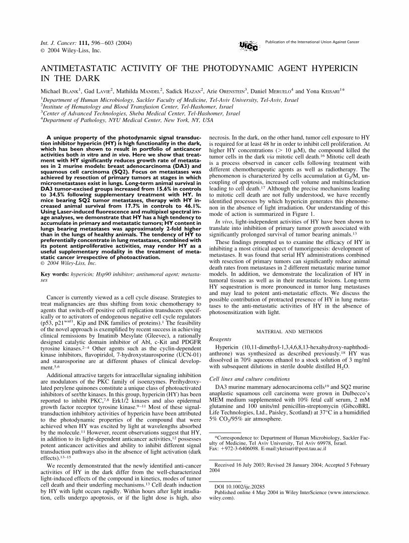
necrosis. In the dark, on the other hand, tumor cell exposure to HYis required for at least 48 hr in order to inhibit cell proliferation. Athigher HY concentrations (� 10 �M), the compound killed thetumor cells in the dark via mitotic cell death.16 Mitotic cell deathis a process observed in cancer cells following treatment withdifferent chemotherapeutic agents as well as radiotherapy. Thephenomenon is characterized by cells accumulation at G2/M, un-coupling of apoptosis, increased cell volume and multinucleationleading to cell death.17 Although the precise mechanisms leadingto mitotic cell death are not fully understood, we have recentlyidentified processes by which hypericin generates this phenome-non in the absence of light irradiation. Our understanding of thismode of action is summarized in Figure 1.
In vivo, light-independent activities of HY have been shown totranslate into inhibition of primary tumor growth associated withsignificantly prolonged survival of tumor bearing animals.13
These findings prompted us to examine the efficacy of HY ininhibiting a most critical aspect of tumorigenesis: development ofmetastases. It was found that serial HY administrations combinedwith resection of primary tumors can significantly reduce animaldeath rates from metastases in 2 different metastatic murine tumormodels. In addition, we demonstrate the localization of HY intumoral tissues as well as in their metastatic lesions. Long-termHY sequestration is more pronounced in tumor lung metastasesand may lead to potent anti-metastatic effects. We discuss thepossible contribution of protracted presence of HY in lung metas-tases to the anti-metastatic activities of HY in the absence ofphotosensitization with light.
MATERIAL AND METHODS
ReagentsHypericin (10,11-dimethyl-1,3,4,6,8,13-hexahydroxy-naphthodi-
anthrone) was synthesized as described previously.18 HY wasdissolved in 70% aqueous ethanol to a stock solution of 3 mg/mlwith subsequent dilutions in sterile double distilled H2O.
Cell lines and culture conditionsDA3 murine mammary adenocarcinoma cells19 and SQ2 murine
anaplastic squamous cell carcinoma were grown in Dulbecco’sMEM medium supplemented with 10% fetal calf serum, 2 mMglutamine and 100 units/ml penicillin-streptomycin (GibcoBRLLife Technologies, Ltd., Paisley, Scotland) at 37°C in a humidified5% CO2/95% air atmosphere.
*Correspondence to: Department of Human Microbiology, Sackler Fac-ulty of Medicine, Tel Aviv University, Tel Aviv 69978, Israel.Fax: �972-3-6406098. E-mail:[email protected]
Received 16 July 2003; Revised 28 January 2004; Accepted 5 February2004
DOI 10.1002/ijc.20285Published online 4 May 2004 in Wiley InterScience (www.interscience.
wiley.com).
Int. J. Cancer: 111, 596–603 (2004)© 2004 Wiley-Liss, Inc.
Publication of the International Union Against Cancer
AnimalsBALB/c mice, 9–10 weeks old, were purchased from Harlan,
Ltd. (Jerusalem, Israel) grown and maintained on Purina. Maleswere used in the SQ2 experiments and females in the DA3 exper-iments. Experiments were conducted in compliance with animalwelfare regulations of the ministry of health as approved by theinstitutional animal care committee.
Generation of tumors and design of experimental therapyThe primary DA3- or SQ2-derived murine tumors were gener-
ated by inoculating 5�105 tumor cells in 0.2 ml serum-free Dul-becco’s medium intradermally into depilated dorsa of the mice.When DA3 tumors reached sizes of 5–6 mm in diameter and SQ2tumors reached sizes of 7–8 mm, the animals were randomizedinto 4 experimental groups and injected intraperitoneally (i.p.)either with HY (5 mg/kg) or with vehicle alone. Five days later, asthe primary tumors were 7–8 mm in diameter in mice bearing DA3tumors or 10–12 mm in mice bearing SQ2 carcinoma, the animalswere anaesthetized with Ketamine (Parke-Davis, Eastleigh, Hamp-shire, UK)-Xylazine (Vitamed, Bat-Yam, Israel) mix, 100 mg/kgand 6.25 mg/kg, respectively, and the primary tumors removed byexcision. The HY-treated mice received 3–5 doses of HY at 5-dayintervals. From the onset of treatments with HY animals weremaintained in metal cages with covers that contain 1 cm diameterpores designed to minimize penetration of light and allow suffi-cient exchange of air. Laboratory lights were shut off upon animalremoval from cages and ambient light kept below 0.03 mW/cm2
(light intensities were quantified using the IL 1350 Radiometer/Photometer, from International Light, Inc., Massachusetts).
Histological studiesMice bearing DA3 tumor in the dorsum were injected with a
single dose of HY (10 mg/kg) i.p., approximately 50 days aftertumor cell inoculation. The animals were then maintained in thedark for time intervals ranging between 24–72 hr. At the end ofeach incubation period the animals were killed, their lungs re-moved and fixed in 4% buffered formaldehyde (Sigma ChemicalCo., St. Louis, MO). The specimens were embedded in paraffin,sections 5 �m in depth prepared and stained with hematoxylin-eosin (H&E), and subjected to morphological examinations.
HY accumulation in murine organsThe concentration of HY in internal organs was analyzed using
fiber-optic laser induced fluorescence (LIF) and multipixel spectralimaging (MSI) systems. LIF, which is based on a point measure-ment of signals from a fluorescence probe (fluorophore) followinglaser-induced activation of the tissue-sequestered compound,20
was used for quantitative assessment of HY distributed in organsof tumor-free and tumor-bearing (TB) animals. BALB/c miceinoculated with primary DA3 or SQ2 tumors and bearing lungmetastases (approximately 50 days post tumor inoculation), wereinjected with HY (10 mg/kg; i.p.). The animals were maintained in
the dark for time intervals ranging from 3 to 72 hr. At the end ofeach incubation period the animals were killed, their organs ex-tracted and placed on a plate with attached bifurcated optical fiber(Oriel, Statford, CT, model 77533). One leg of the fiber wasattached to an argon ion laser source (Melles Griot, 43 series), withan output of 10 mW/cm2 emitting at 477 nm for excitation. Theother leg was connected to a spectrofluorophotometer (RF-5301PC. Shimadzu, Japan) calibrated to detect emission spectrum offluorophores at 550–750 nm range. Fluorescent intensity emittedby HY from tissue samples was measured in 5–7 different sectionsof the sample. Background signals, originating from the tissues ofanimals not administered with HY, were subtracted from thespectra, and fluorescence intensities at 602 nm were used asmaxima for pharmacokinetic curves.
Fluorescence images of normal lungs and lungs with metastases,extracted from TB mice injected with HY (10 mg/kg; i.p.), wereobtained using miltipixel spectral image-analysis system (Spec-traCube™ SD300, Applied Spectral Imaging, Migdal Ha’Emek,Israel) as previously described.21 Fluorescence was acquired in thespectral range of 550–750 nm at excitation 500�20 nm.
Statistical analysisAnimal survival data were analyzed using the Logrank/Mantel-
Cox test. For ex-vivo studies the data was analyzed using a 2-tailStudent’s t-test. p-values of less than 0.05 were considered to bestatistically significant.
RESULTS
Effects of HY on the growth of DA3 breast carcinoma-derivedlung metastases
HY can interfere with the growth of primary tumors;13 however,since growth of metastases constitutes the major cause for treat-ment failure in cancer, the effects of HY on the advanced stages ofthe disease, when metastases already exist, were examined.
DA3 cell-derived tumors develop metastases predominantly in thelung.12 The anti-metastatic efficacy of HY was evaluated as animalsurvival measured up to 300 days. This parameter was chosen becauseweight differences between lungs bearing metastases and normallungs in this tumor model were minimal. On occasions the weight oflungs burdened with metastases exceeded lung weight in tumor-freeanimals, by only 38–45% (285�18 mg vs. 210�20 mg in healthymice) even at advanced stages of the disease.
To focus selectively on the effects of HY on metastases, theprimary tumors were surgically resected at a stage, when micro-metastases have already existed. Calibrations determined that ex-cision of primary tumors at a diameter of 8 mm (approximately 15days post DA3 cell inoculation) result in death rates of approxi-mately 85% of the mice.
The tumor bearing mice were randomized into 4 groups. Thefirst received 6 i.p. HY administrations at declining dosages at
FIGURE 1 – Molecular effects of hypericin in cancer cells that culminate in mitotic cell death.
597ANTI-METASTATIC EFFECTS OF HYPERICIN IN THE DARK
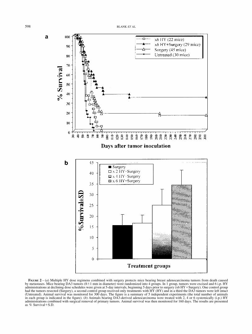
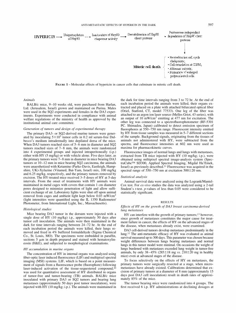
FIGURE 2 – (a) Multiple HY dose regimens combined with surgery protects mice bearing breast adenocarcinoma tumors from death causedby metastases. Mice bearing DA3 tumors (8�1 mm in diameter) were randomized into 4 groups. In 1 group, tumors were excised and 6 i.p. HYadministrations at declining dose schedules were given at 5-day intervals, beginning 5 days prior to surgery (x6 HY�Surgery). One control grouphad the tumors resected (Surgery), a second control group received only treatments with HY (HY) and in a third the DA3 tumors were left intact(Untreated). Animal survival was monitored for 300 days. The figure is a summary of 5 independent experiments (the total number of animalsin each group is indicated in the figure). (b) Animals bearing DA3-derived adenocarcinoma were treated with 2, 4 or 6 systemically (i.p.) HYadministrations combined with surgical removal of primary tumors. Animal survival was then monitored for 160 days. The results are presentedas % Survival�S.D.
598 BLANK ET AL.
5-day intervals between schedules. The first HY dose of 5mg/kg was initiated 5 days prior to tumor resection. The seconddose was 2.5 mg/kg and was followed by 4 maintenance dosesof 1.25 mg/kg each. The second treatment group consisted ofsurgical resection with no HY dosing and the third groupreceived only treatments with HY applied in parallel to the firstgroup. A fourth group consisted of untreated DA3 tumor-bearing mice. The declining HY dose regimens and relativelylong intervals between HY administrations were selected due tothe long half-life of this compound in rodents (discussed here-under). Animal survival was monitored for 300 days after tumorcell inoculation.
The results (Fig. 2a) show that combining HY therapy withsurgical resection of the primary tumors led to a cure rate of 34.5%of the mice compared to 15.6% cured by surgery alone or 0%survival in untreated group (p � 0.001). Although, treatment withHY without tumor resection resulted in death of all mice in thegroup, a modest prolongation of animal survival from 52�7.2 daysin the untreated control group to 58�12.4 days was observed (p �0.03).
Examination of dose-dependent effects of HY on animal curefrom metastases in DA3 tumor-excised mice revealed that the curerates of the treatment increased from 16% following tumor exci-sion to 25% and 35% when primary tumor resection was combinedwith 4 or 6 regimens of HY, respectively (Fig. 2b). Two doseregimens of HY were insufficient and death rates remained similarto the control groups in which tumor resection was the soletreatment (Fig. 2b).
The effect of HY on lung metastases has been examined histo-logically. Animals bearing DA3 tumor metastases and adminis-tered with a single dose of HY 10 mg/kg i.p were sacrificed after24, 48 and 72 hr. The lungs were removed, sections prepared andstained with H&E. The histological examination (Fig. 3) revealedcomplete destruction of several, but not all, of the metastatic focifollowing treatment with HY. Such destruction became evidentonly �48 hr after HY administration as shown in Figure 3a takenat the 72 hr time-point. No destruction of metastatic foci could bedetected in the untreated control mice (Fig. 3b). These findingssuggest that treatment of TB animals with HY can reduce themetastatic load, although a single dose of the compound wasinsufficient to cure the animals or prolong their survival (data notshown).
Immunological considerations in animal cure from metastaticdisease
An attempt was made to test whether mice cured by primaryDA3 tumor resection followed by treatment with HY developimmune-mediated resistance to this cell line. Animals that sur-vived DA3 metastases were reinjected with 2.5�105 live DA3cells/mouse. The mean survival time (MST) of mice that havepreviously been cured with 6 dose schedules of HY and primarytumor excision (62.4�13.4 days) did not differ significantly fromthe MST of naı̈ve mice inoculated with DA3 cells (59.5�5.9days), or from mice subjected to tumor excision alone(63.4�12.3). The data does not support development of any sig-nificant anti-tumoral protective activity in animals previouslycured with HY.
Anti-metastatic effect of HY on SQ2 murine squamous cellcarcinoma-derived metastases
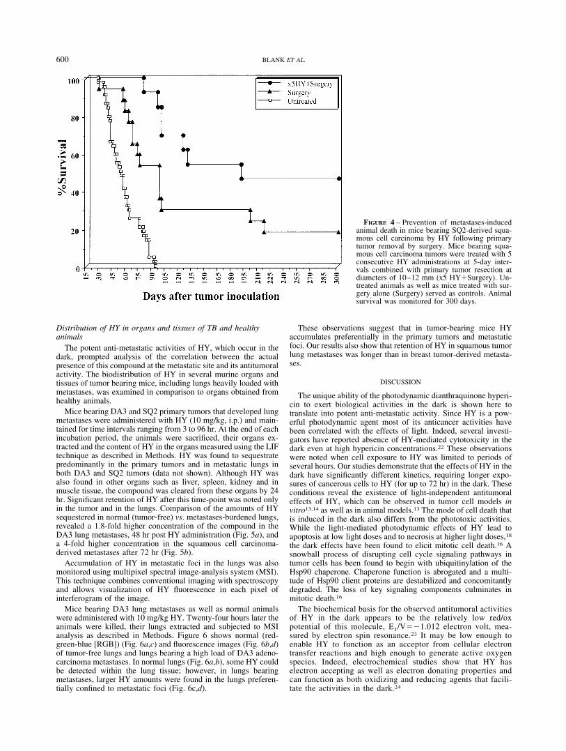
In order to corroborate observations in the breast carcinomatumor model, which show improved cure rates from metastaticdisease by treatment with HY, our studies were extended toanalysis of effects on metastases in a squamous cell carcinomatumor model. Calibration studies indicated that excision ofprimary SQ2 cell-derived tumors at a diameter of 10 –12 mm,resulted in metastases-related death of approximately 80 – 85%of the animals. Effects of HY on SQ2 metastatic developmentwas examined following primary tumor excision as they at-
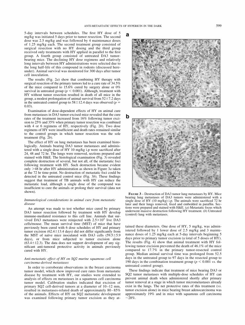
tained these diameters. One dose of HY, 5 mg/kg, was admin-istered followed by 1 lower dose of 2.5 mg/kg and 3 mainte-nance doses of 1.25 mg/kg each at 5-day intervals beginning 5days prior to primary tumor excision (a total of 5 doses of HY).The results (Fig. 4) show that animal treatment with HY fol-lowing tumor excision prevented the death of 46.1% of the micecompared to 17.7% in the primary tumor-resected controlgroup. Median animal survival time was prolonged from 52.5days in the untreated group to 97 days in the resected group to190 days in the combination treatment group (p � 0.001 vs. theuntreated control group).
These findings indicate that treatment of mice bearing DA3 orSQ2 tumor metastases with multiple-dose schedules of HY canprevent animal death when administered shortly after primarytumor removal at a stage in which tumor micrometastases alreadyexist in the lungs. The net protective rates of this treatment (vs.tumor excision alone) in mice bearing breast adenocarcinoma wasapproximately 19% and in mice with squamous cell carcinoma�28%.
FIGURE 3 – Destruction of DA3 tumor lung metastases by HY. Micebearing lung metastases of DA3 tumors were administered with asingle dose of HY (10 mg/kg) i.p. The animals were sacrificed 72 hrlater and their lungs removed, fixed and embedded in paraffin. Sec-tions were prepared and stained with H&E. (a) Metastatic focus whichunderwent massive destruction following HY treatment. (b) Untreated(control) lung with metastases.
599ANTI-METASTATIC EFFECTS OF HYPERICIN IN THE DARK
Distribution of HY in organs and tissues of TB and healthyanimals
The potent anti-metastatic activities of HY, which occur in thedark, prompted analysis of the correlation between the actualpresence of this compound at the metastatic site and its antitumoralactivity. The biodistribution of HY in several murine organs andtissues of tumor bearing mice, including lungs heavily loaded withmetastases, was examined in comparison to organs obtained fromhealthy animals.
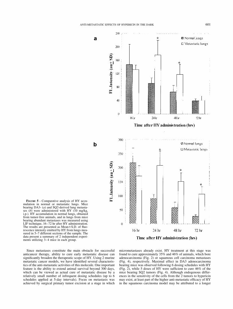
Mice bearing DA3 and SQ2 primary tumors that developed lungmetastases were administered with HY (10 mg/kg, i.p.) and main-tained for time intervals ranging from 3 to 96 hr. At the end of eachincubation period, the animals were sacrificed, their organs ex-tracted and the content of HY in the organs measured using the LIFtechnique as described in Methods. HY was found to sequestratepredominantly in the primary tumors and in metastatic lungs inboth DA3 and SQ2 tumors (data not shown). Although HY wasalso found in other organs such as liver, spleen, kidney and inmuscle tissue, the compound was cleared from these organs by 24hr. Significant retention of HY after this time-point was noted onlyin the tumor and in the lungs. Comparison of the amounts of HYsequestered in normal (tumor-free) vs. metastases-burdened lungs,revealed a 1.8-fold higher concentration of the compound in theDA3 lung metastases, 48 hr post HY administration (Fig. 5a), anda 4-fold higher concentration in the squamous cell carcinoma-derived metastases after 72 hr (Fig. 5b).
Accumulation of HY in metastatic foci in the lungs was alsomonitored using multipixel spectral image-analysis system (MSI).This technique combines conventional imaging with spectroscopyand allows visualization of HY fluorescence in each pixel ofinterferogram of the image.
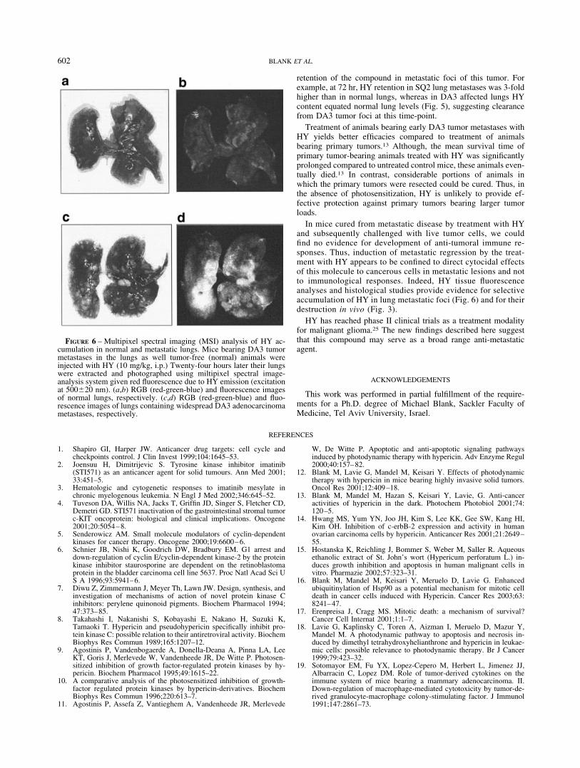
Mice bearing DA3 lung metastases as well as normal animalswere administered with 10 mg/kg HY. Twenty-four hours later theanimals were killed, their lungs extracted and subjected to MSIanalysis as described in Methods. Figure 6 shows normal (red-green-blue [RGB]) (Fig. 6a,c) and fluorescence images (Fig. 6b,d)of tumor-free lungs and lungs bearing a high load of DA3 adeno-carcinoma metastases. In normal lungs (Fig. 6a,b), some HY couldbe detected within the lung tissue; however, in lungs bearingmetastases, larger HY amounts were found in the lungs preferen-tially confined to metastatic foci (Fig. 6c,d).
These observations suggest that in tumor-bearing mice HYaccumulates preferentially in the primary tumors and metastaticfoci. Our results also show that retention of HY in squamous tumorlung metastases was longer than in breast tumor-derived metasta-ses.
DISCUSSION
The unique ability of the photodynamic dianthraquinone hyperi-cin to exert biological activities in the dark is shown here totranslate into potent anti-metastatic activity. Since HY is a pow-erful photodynamic agent most of its anticancer activities havebeen correlated with the effects of light. Indeed, several investi-gators have reported absence of HY-mediated cytotoxicity in thedark even at high hypericin concentrations.22 These observationswere noted when cell exposure to HY was limited to periods ofseveral hours. Our studies demonstrate that the effects of HY in thedark have significantly different kinetics, requiring longer expo-sures of cancerous cells to HY (for up to 72 hr) in the dark. Theseconditions reveal the existence of light-independent antitumoraleffects of HY, which can be observed in tumor cell models invitro13,14 as well as in animal models.13 The mode of cell death thatis induced in the dark also differs from the phototoxic activities.While the light-mediated photodynamic effects of HY lead toapoptosis at low light doses and to necrosis at higher light doses,18
the dark effects have been found to elicit mitotic cell death.16 Asnowball process of disrupting cell cycle signaling pathways intumor cells has been found to begin with ubiquitinylation of theHsp90 chaperone. Chaperone function is abrogated and a multi-tude of Hsp90 client proteins are destabilized and concomitantlydegraded. The loss of key signaling components culminates inmitotic death.16
The biochemical basis for the observed antitumoral activitiesof HY in the dark appears to be the relatively low red/oxpotential of this molecule, E1/V�1.012 electron volt, mea-sured by electron spin resonance.23 It may be low enough toenable HY to function as an acceptor from cellular electrontransfer reactions and high enough to generate active oxygenspecies. Indeed, electrochemical studies show that HY haselectron accepting as well as electron donating properties andcan function as both oxidizing and reducing agents that facili-tate the activities in the dark.24
FIGURE 4 – Prevention of metastases-inducedanimal death in mice bearing SQ2-derived squa-mous cell carcinoma by HY following primarytumor removal by surgery. Mice bearing squa-mous cell carcinoma tumors were treated with 5consecutive HY administrations at 5-day inter-vals combined with primary tumor resection atdiameters of 10–12 mm (x5 HY�Surgery). Un-treated animals as well as mice treated with sur-gery alone (Surgery) served as controls. Animalsurvival was monitored for 300 days.
600 BLANK ET AL.
Since metastases constitute the main obstacle for successfulanticancer therapy, ability to cure early metastatic disease cansignificantly broaden the therapeutic scope of HY. Using 2 murinemetastatic cancer models, we have identified several characteris-tics of the anti-metastatic activities of this molecule. One importantfeature is the ability to extend animal survival beyond 300 days,which can be viewed as actual cure of metastatic disease by arelatively small number of infrequent dosing schedules (up to 6schedules applied at 5-day intervals). Focus on metastasis wasachieved by surgical primary tumor excision at a stage in which
micrometastases already exist. HY treatment at this stage wasfound to cure approximately 35% and 46% of animals, which boreadenocarcinoma (Fig. 2) or squamous cell carcinoma metastases(Fig. 4), respectively. Maximal effect in DA3 adenocarcinomabearing mice was observed following 6 dosing schedules with HY(Fig. 2), while 5 doses of HY were sufficient to cure 46% of themice bearing SQ2 tumors (Fig. 4). Although endogenous differ-ences in the sensitivity of the cells from the 2 tumors to hypericinmay exist, at least part of the higher anti-metastatic efficacy of HYin the squamous carcinoma model may be attributed to a longer
FIGURE 5 – Comparative analysis of HY accu-mulation in normal or metastatic lungs. Micebearing DA3- (a) and SQ2-derived lung metasta-ses (b) were administered with HY (10 mg/kg,i.p.). HY accumulation in normal lungs, obtainedfrom tumor-free animals, and in lungs from micebearing abundant metastases was measured usingLIF technique, 16–72 hr after HY administration.The results are presented as Mean�S.D. of fluo-rescence intensity emitted by HY from lungs mea-sured in 5–7 different sections of the sample. Thedata present a summary of 2 independent experi-ments utilizing 3–4 mice in each group.
601ANTI-METASTATIC EFFECTS OF HYPERICIN IN THE DARK
retention of the compound in metastatic foci of this tumor. Forexample, at 72 hr, HY retention in SQ2 lung metastases was 3-foldhigher than in normal lungs, whereas in DA3 affected lungs HYcontent equated normal lung levels (Fig. 5), suggesting clearancefrom DA3 tumor foci at this time-point.
Treatment of animals bearing early DA3 tumor metastases withHY yields better efficacies compared to treatment of animalsbearing primary tumors.13 Although, the mean survival time ofprimary tumor-bearing animals treated with HY was significantlyprolonged compared to untreated control mice, these animals even-tually died.13 In contrast, considerable portions of animals inwhich the primary tumors were resected could be cured. Thus, inthe absence of photosensitization, HY is unlikely to provide ef-fective protection against primary tumors bearing larger tumorloads.
In mice cured from metastatic disease by treatment with HYand subsequently challenged with live tumor cells, we couldfind no evidence for development of anti-tumoral immune re-sponses. Thus, induction of metastatic regression by the treat-ment with HY appears to be confined to direct cytocidal effectsof this molecule to cancerous cells in metastatic lesions and notto immunological responses. Indeed, HY tissue fluorescenceanalyses and histological studies provide evidence for selectiveaccumulation of HY in lung metastatic foci (Fig. 6) and for theirdestruction in vivo (Fig. 3).
HY has reached phase II clinical trials as a treatment modalityfor malignant glioma.25 The new findings described here suggestthat this compound may serve as a broad range anti-metastaticagent.
ACKNOWLEDGEMENTS
This work was performed in partial fulfillment of the require-ments for a Ph.D. degree of Michael Blank, Sackler Faculty ofMedicine, Tel Aviv University, Israel.
REFERENCES
1. Shapiro GI, Harper JW. Anticancer drug targets: cell cycle andcheckpoints control. J Clin Invest 1999;104:1645–53.
2. Joensuu H, Dimitrijevic S. Tyrosine kinase inhibitor imatinib(STI571) as an anticancer agent for solid tumours. Ann Med 2001;33:451–5.
3. Hematologic and cytogenetic responses to imatinib mesylate inchronic myelogenous leukemia. N Engl J Med 2002;346:645–52.
4. Tuveson DA, Willis NA, Jacks T, Griffin JD, Singer S, Fletcher CD,Demetri GD. STI571 inactivation of the gastrointestinal stromal tumorc-KIT oncoprotein: biological and clinical implications. Oncogene2001;20:5054–8.
5. Senderowicz AM. Small molecule modulators of cyclin-dependentkinases for cancer therapy. Oncogene 2000;19:6600–6.
6. Schnier JB, Nishi K, Goodrich DW, Bradbury EM. G1 arrest anddown-regulation of cyclin E/cyclin-dependent kinase-2 by the proteinkinase inhibitor staurosporine are dependent on the retinoblastomaprotein in the bladder carcinoma cell line 5637. Proc Natl Acad Sci US A 1996;93:5941–6.
7. Diwu Z, Zimmermann J, Meyer Th, Lawn JW. Design, synthesis, andinvestigation of mechanisms of action of novel protein kinase Cinhibitors: perylene quinonoid pigments. Biochem Pharmacol 1994;47:373–85.
8. Takahashi I, Nakanishi S, Kobayashi E, Nakano H, Suzuki K,Tamaoki T. Hypericin and pseudohypericin specifically inhibit pro-tein kinase C: possible relation to their antiretroviral activity. BiochemBiophys Res Commun 1989;165:1207–12.
9. Agostinis P, Vandenbogaerde A, Donella-Deana A, Pinna LA, LeeKT, Goris J, Merlevede W, Vandenheede JR, De Witte P. Photosen-sitized inhibition of growth factor-regulated protein kinases by hy-pericin. Biochem Pharmacol 1995;49:1615–22.
10. A comparative analysis of the photosensitized inhibition of growth-factor regulated protein kinases by hypericin-derivatives. BiochemBiophys Res Commun 1996;220:613–7.
11. Agostinis P, Assefa Z, Vantieghem A, Vandenheede JR, Merlevede
W, De Witte P. Apoptotic and anti-apoptotic signaling pathwaysinduced by photodynamic therapy with hypericin. Adv Enzyme Regul2000;40:157–82.
12. Blank M, Lavie G, Mandel M, Keisari Y. Effects of photodynamictherapy with hypericin in mice bearing highly invasive solid tumors.Oncol Res 2001;12:409–18.
13. Blank M, Mandel M, Hazan S, Keisari Y, Lavie, G. Anti-canceractivities of hypericin in the dark. Photochem Photobiol 2001;74:120–5.
14. Hwang MS, Yum YN, Joo JH, Kim S, Lee KK, Gee SW, Kang HI,Kim OH. Inhibition of c-erbB-2 expression and activity in humanovarian carcinoma cells by hypericin. Anticancer Res 2001;21:2649–55.
15. Hostanska K, Reichling J, Bommer S, Weber M, Saller R. Aqueousethanolic extract of St. John’s wort (Hypericum perforatum L.) in-duces growth inhibition and apoptosis in human malignant cells invitro. Pharmazie 2002;57:323–31.
16. Blank M, Mandel M, Keisari Y, Meruelo D, Lavie G. Enhancedubiquitinylation of Hsp90 as a potential mechanism for mitotic celldeath in cancer cells induced with Hypericin. Cancer Res 2003;63:8241–47.
17. Erenpreisa J, Cragg MS. Mitotic death: a mechanism of survival?Cancer Cell Internat 2001;1:1–7.
18. Lavie G, Kaplinsky C, Toren A, Aizman I, Meruelo D, Mazur Y,Mandel M. A photodynamic pathway to apoptosis and necrosis in-duced by dimethyl tetrahydroxyhelianthrone and hypericin in leukae-mic cells: possible relevance to photodynamic therapy. Br J Cancer1999;79:423–32.
19. Sotomayor EM, Fu YX, Lopez-Cepero M, Herbert L, Jimenez JJ,Albarracin C, Lopez DM. Role of tumor-derived cytokines on theimmune system of mice bearing a mammary adenocarcinoma. II.Down-regulation of macrophage-mediated cytotoxicity by tumor-de-rived granulocyte-macrophage colony-stimulating factor. J Immunol1991;147:2861–73.
FIGURE 6 – Multipixel spectral imaging (MSI) analysis of HY ac-cumulation in normal and metastatic lungs. Mice bearing DA3 tumormetastases in the lungs as well tumor-free (normal) animals wereinjected with HY (10 mg/kg, i.p.) Twenty-four hours later their lungswere extracted and photographed using miltipixel spectral image-analysis system given red fluorescence due to HY emission (excitationat 500�20 nm). (a,b) RGB (red-green-blue) and fluorescence imagesof normal lungs, respectively. (c,d) RGB (red-green-blue) and fluo-rescence images of lungs containing widespread DA3 adenocarcinomametastases, respectively.
602 BLANK ET AL.

20. Malik Z, Kostenich G, Roitman L, Orenstein, A. Topical applicationof 5-aminolevulinic acid, DMSO and EDTA: protoporphyrin IX ac-cumulation in skin and tumours of mice. J. Photochem PhotobiolB:Biology 1995;25:213–8.
21. Orenstein A, Kostenich G, Rothmann C, Barshack I, Malik, Z. Im-aging of human skin lesions using Multipixel Fourier TransformSpectroscopy. Lasers Med Sci 1998;13:112–8.
22. Vandenbogaerde AL, Cuveele JF, Proot P, Himpens BE, Merlevede WJ,de Witte PA. Differential cytotoxic effects induced after photosensitiza-tion by hypericin. J Photochem Photobiol B 1997;38:136–42.
23. Gerson F, Gescheidt G, Haering P, Mazur Y, Freeman D, Spreitzer H,Daub G. Electron acceptor properties of hypericin and its salts: anESR/ENDOR and electrochemical study. J Am Chem Soc 1995;117:11861–6.
24. Redepenning J, Tao N. Measurement of formal potentials forhypericin in dimethyl sulfoxide. Photochem Photobiol 1993;58:532–5.
25. Couldwell WT, Gopalakrishna R, Hinton DR, He S, Weiss MH, LawRE, Apuzzo ML, Law RE. Hypericin: a potential antiglioma therapy.Neurosurgery 1994;35:705–10.
603ANTI-METASTATIC EFFECTS OF HYPERICIN IN THE DARK