animal cytokinesis: from parts list to mechanisms - bio 5068
TRANSCRIPT

ANRV277-BI75-21 ARI 3 May 2006 10:16
Animal Cytokinesis: FromParts List to MechanismsUlrike S. Eggert,1 Timothy J. Mitchison,2
and Christine M. Field2
1Dana-Farber Cancer Institute and Department of Biological Chemistry andMolecular Pharmacology, 2Department of Systems Biology, Harvard Medical School,Boston, Massachusetts 02115; email: ulrike [email protected],timothy [email protected], christine [email protected]
Annu. Rev. Biochem.2006. 75:543–66
The Annual Review ofBiochemistry is online atbiochem.annualreviews.org
doi: 10.1146/annurev.biochem.74.082803.133425
Copyright c© 2006 byAnnual Reviews. All rightsreserved
0066-4154/06/0707-0543$20.00
Key Words
cleavage furrow, contractile ring, midzone microtubules, RNAi
AbstractThe mechanism underlying cytokinesis, the final step in cell divi-sion, remains one of the major unsolved questions in basic cell bi-ology. Thanks to advances in functional genomics and proteomics,we are now able to assemble a “parts list” of proteins involved incytokinesis. In this review, we discuss how to relate this parts list tobiological mechanism. For easier analysis, we split cytokinesis intodiscrete steps: cleavage plane specification, rearrangement of mi-crotubule structures, contractile ring assembly, ring ingression, andcompletion. We report on the advances that have been made to un-derstand these steps and how they can be integrated into a globalunderstanding of cytokinesis. We also discuss the extent to whichclassic questions have been answered and identify major outstand-ing questions.
543
Ann
u. R
ev. B
ioch
em. 2
006.
75:5
43-5
66. D
ownl
oade
d fr
om a
rjou
rnal
s.an
nual
revi
ews.
org
by W
ashi
ngto
n U
nive
rsity
Lib
rary
on
06/1
3/06
. For
per
sona
l use
onl
y.
ANRV277-BI75-21 ARI 3 May 2006 10:16
Contents
INTRODUCTION ANDRESEARCH APPROACHES . . . . 544
A PARTS LIST FORCYTOKINESIS . . . . . . . . . . . . . . . . . 546
SUBPROCESSES INCYTOKINESIS . . . . . . . . . . . . . . . . . 549Timing Cytokinesis . . . . . . . . . . . . . . . 549Cleavage Plane Specification:
Evaluation of Classic Models . . . 550Cleavage Plane Specification:
Molecular Mechanisms . . . . . . . . 552Organization of
Cytokinesis-AssociatedMicrotubules . . . . . . . . . . . . . . . . . . 553
Contractile Ring Assembly . . . . . . . 555Furrow Ingression . . . . . . . . . . . . . . . . 557Completion . . . . . . . . . . . . . . . . . . . . . 558
CYTOKINESIS AND CANCER. . . . 560
INTRODUCTION ANDRESEARCH APPROACHES
Cytokinesis, the physical separation of onecell into two, is the last step in the cell cy-cle. It requires coordinated actions of the cy-toskeleton, membrane systems, and the cellcycle engine, which are precisely controlledin space and time. Cytokinesis was describedmore than 100 years ago, and the roles ofactin and myosin in cleavage, as well as that ofthe mitotic spindle in specifying the cleavageplane, were discovered more than 20 years ago(reviewed in Reference 1). The last few yearshave seen intensive focus on the identificationof proteins involved in cytokinesis, using clas-sical genetics and biochemistry. This effort,coupled with recent systematic RNA inter-ference (RNAi) screens, has culminated in amore or less complete “parts lists.” The nextchallenge, and the focus of this review, is toconvert such lists into an interlinked systemof molecular mechanisms.
RNAi: RNAinterference
In this review, we focus on cytokinesis inanimal cells, especially the two model organ-
isms whose parts lists are now available, ne-matode (Caenorhabditis elegans) embryos andcultured insect (Drosophila) cells. Other im-portant systems include cultured mammaliancells, echinoderm and amphibian (Xenopus)eggs, and Drosophila embryos and sperma-tocytes. For recent reviews of cytokinesis infungi and plants, see References 2 and 3. Eachmodel system has unique features, includingdistinct regulatory and mechanical challengesfaced by particular cell types to effect cytoki-nesis, as well as the more obvious technicalpros and cons for different research methods.In cytokinesis research, as in other areas ofbasic cell biology, our molecular understand-ing has progressed to the point where thedifferences between cell types are becomingas interesting as their conserved features (4).For example, large eggs require longer micro-tubules to position spindles and furrows, andthey may sacrifice accuracy-enhancing check-points in exchange for faster division. Also, theamount of new plasma membrane required tocreate two cells from one varies with the sizeand surface area of a given cell type, leadingto differing levels of importance for vesicu-lar trafficking pathways. These biological dif-ferences require that conserved mechanismsbe implemented in distinct ways in differ-ent cells and may, perhaps, also require ad-ditional or different mechanisms in particu-lar cell types. When controversies arise in thefield, it is important to determine which dif-ferences arise from genuine differences be-tween systems and which arise from personalinterpretation of particular experiments in afast-developing field.
Some differences between systems hingeprimarily on the technical ease of im-plementing the three most important ap-proaches in modern cell biology: genetics/genomics, microscopy, and biochemistry. Forgenetics/genomics, C. elegans embryos andDrosophila tissue culture cells are currently themost advanced, thanks to genome-wide RNAiscreens. RNAi is far from a perfect genetictechnology, typically providing an incompleteknockdown of protein levels that may take
544 Eggert · Mitchison · Field
Ann
u. R
ev. B
ioch
em. 2
006.
75:5
43-5
66. D
ownl
oade
d fr
om a
rjou
rnal
s.an
nual
revi
ews.
org
by W
ashi
ngto
n U
nive
rsity
Lib
rary
on
06/1
3/06
. For
per
sona
l use
onl
y.

ANRV277-BI75-21 ARI 3 May 2006 10:16
several days in cultured cells. It is also diffi-cult to score a role in cytokinesis using RNAifor a protein that is important for earlier stepsin the cell cycle, such as mitosis. Thus, partslists determined solely by RNAi screens areincomplete. However, the power of this tech-nology and its applicability to systems thatlack traditional genetics have led to an explo-sion of interest and information. Given thecurrent availability of technology and RNAilibraries, we expect publication of a list of pro-teins involved in cytokinesis in human cancercells soon. These will be accomplished first forthe commonly used HeLa cell line, originallyderived from a cervical carcinoma, which isextensively adapted for life in culture and thusnot truly representative of any normal humancell type. Generating parts lists for cytokinesisin untransformed human cells, or cancer cellsin situ, will be more difficult.
Protein lists are not by themselves mecha-nistically informative, but the phenotypes ob-served using RNAi and traditional geneticsprovide functional information, especially inconjunction with high resolution microscopy.Microscopy has always been an importanttool in cytokinesis research, and the past fewyears have seen dramatic improvement in op-tical methods. Genetically encoded fluores-cent proteins of different colors are now inroutine use in many systems, and the dynamicinformation they provide is being enhancedby methods for localized photobleaching andphotoactivation. Instrumentation has also ad-vanced, with highly sensitive digital camerasand new confocal technologies widely avail-able. Together, these imaging methods are al-lowing the field to observe molecular events atplay inside living cells. The confluence of mi-croscopy with perturbations using genetics/genomics methods, or in some cases physi-cal perturbation using microneedles or UVmicrobeams, is currently driving the fieldforward.
The biochemistry of cytokinesis, in con-trast, has seen slower progress in the past fewyears. An extract system for all or part of cy-
tokinesis has yet to be reported, and recon-stitution with purified components has beenachieved for only a few processes and proteins(5, 6). We believe this lack of biochemistrymay become limiting in the quest for molec-ular mechanism, particularly as quantitativeunderstanding is sought through modeling.For mathematical models to approach reality,it will be necessary to use real values for pro-tein concentrations and association constants.Recently, a first attempt at protein quantita-tion has been made in fission yeast (6a).
Two less commonly used research ap-proaches deserve comment. Modeling has adistinguished history in cytokinesis research(e.g., 7), and its importance is likely to grow inthe future, driven in part by an influx into ba-sic cell biology of young scientists with physi-cal and mathematical training. Small moleculeapproaches have also been historically impor-tant, notably in the discovery of the role ofactin in cytokinesis via the cytochalasins (re-viewed in Reference 8). Because cytokinesisis a rapid and dynamic process that occursduring a small portion of the cell cycle, smallmolecules that can rapidly enter living cellsand perturb specific processes are especiallyvaluable tools. We also believe that interfer-ing with specific aspects of cytokinesis mayprovide novel and useful anticancer therapeu-tics, with the recently introduced Aurora ki-nase inhibitors as examples (9–11). Despitetheir dual importance for research and po-tential therapeutics and the large number ofpotential protein targets, the number of well-characterized small molecules available forcytokinesis research is small (Table 1). Wecarried out a screen for small molecule in-hibitors of cytokinesis and identified 50 smallmolecules that caused the formation of bin-ucleate cells in Drosophila tissue culture cells(20). We are currently investigating the cel-lular targets and biochemical mechanisms ofmany of these compounds and hope to providethe field with more diverse and useful smallmolecule tools to study cytokinesis in thefuture.
www.annualreviews.org • Animal Cytokinesis 545
Ann
u. R
ev. B
ioch
em. 2
006.
75:5
43-5
66. D
ownl
oade
d fr
om a
rjou
rnal
s.an
nual
revi
ews.
org
by W
ashi
ngto
n U
nive
rsity
Lib
rary
on
06/1
3/06
. For
per
sona
l use
onl
y.

ANRV277-BI75-21 ARI 3 May 2006 10:16
Table 1 Small molecules that affect cytokinesis
Small molecule Mechanism of action ReferenceActin depolymerization:
CytochalasinsLatrunculinSwinholide
Bind barbed ends of actinSequesters actin monomersSevers actin filaments
(12)(13)(14)
Jasplakinolide Stabilization of actin filaments (15)(−) Blebbistatina Inhibition of myosin II ATPase (16)Hesperadin,b ZM447439,b VX-680b Inhibition of Aurora B kinase (9–11)Y27632b Inhibition of Rho kinase (17)W7,b ML7b Inhibition of myosin light chain kinase (18, 19)
aBlebbistatin is sensitive to UV and blue light.bPlease note that these drugs target the active ATPase site of the kinase and can also inhibit other kinases,especially at higher concentrations.
A PARTS LIST FORCYTOKINESIS
A prerequisite for a complete understandingof cytokinesis is a parts list, or inventory, of allthe molecules involved. Effective RNAi tech-nology and complete genome sequence infor-mation have allowed a preliminary listing ofthe full complement of genes involved in cy-tokinesis for two cell types and more fragmen-tary lists for several others (Table 2). Becauseof the limitations of RNAi mentioned above,we believe that the most significant omissionsfrom current lists may be proteins involvedboth in mitosis and cytokinesis, for which thecytokinesis defect can be difficult to score. Forexample, Polo kinase has a central role in cy-tokinesis but does not score in the screens be-cause it is required for mitosis.
Proteomics provides a complementarystrategy for genome-wide understanding. Ithas the potential to supply more direct infor-mation on biochemical mechanisms by pro-viding data on relative protein concentrations,posttranslational modifications, and the com-position of protein complexes. The method-ology for making such measurements on aglobal scale using mass spectrometry is onlynow emerging and has yet to be applied tocytokinesis. A major limitation in applyingproteomic methods to cytokinesis has beenthe challenge of isolating relevant structuresand complexes from defined cell cycle states.
Skop et al. (25) approached this by isolat-ing midbodies, representing a very late stageof cytokinesis, from synchronized Chinesehamster ovary cells. Five hundred seventy-seven proteins enriched in midbodies wereidentified, and 160 candidates were testedfor relevance by RNAi of orthologs in C.elegans (25). Some of the cytokinesis pro-teins identified in this study are included inTable 2.
Detailed examination of recently pub-lished screens (20–25, 29, 31, 34, 41, 49, 74)reveals that certain proteins appear in ev-ery screen, whereas many proteins were onlyfound in one or a few screens. Table 2 liststhose that scored in two or more screens,as well as well-described cytokinesis proteinsfrom the literature. Proteins that scored inonly one screen are presumably a mixture ofconserved proteins that escaped detection inother screens for technical reasons, and pro-teins required in only a subset of systems.[See Supplemental Tables 1–3 for a summaryof hits (follow the Supplemental Materiallink from the Annual Reviews home page athttp://www.annualreviews.org)]. Only twoscreens, one in worms (23) and one in flies(20), covered most of a genome so it is cur-rently difficult to distinguish these possibil-ities. Given the unprecedented rate of newdata generation, Table 2 will soon be out ofdate. The European MitoCheck consortium
546 Eggert · Mitchison · Field
Ann
u. R
ev. B
ioch
em. 2
006.
75:5
43-5
66. D
ownl
oade
d fr
om a
rjou
rnal
s.an
nual
revi
ews.
org
by W
ashi
ngto
n U
nive
rsity
Lib
rary
on
06/1
3/06
. For
per
sona
l use
onl
y.

ANRV277-BI75-21 ARI 3 May 2006 10:16
Table 2 Parts list of proteins involved in cytokinesisa
Mammalian geneDrosophila
geneC. elegans
gene Predicted protein functionScreen
reference ReferenceActin cytoskeleton
Actin Act5C act-1 Actin (20–23) (12)Myosin heavy chain zip nmy-2 Nonmuscle myosin II heavy
chain(20, 21, 24, 25) (26–28)
MRLC sqh mlc-4 Myosin II regulatory lightchain
(20–23, 25, 29) (30)
Anillin ani ani-1 Anillin, actin binding (20, 21, 24, 29,31)
(32, 33)
Arp3 arp66B arx-1 Actin nucleation (20, 25)CAPZ cpb cap-2 Capping protein (20, 25)Profilin chic pfn-1 Binding of actin monomers (21, 23–25, 34) (35)Cofilin tsr unc-60 Actin severing (20, 21, 24, 25,
29, 31)(36)
mDia dia cyk-1 Formin-Rho effector, actinnucleator
(20–25, 31) (37, 38)
RhoA Rho1 rho-1 Rho GTPase (20, 21, 24, 29,31)
(39)
Ect2 pbl let-21 RhoGEF (20, 21, 23, 25,29, 31)
(40)
MgcRacGAP RacGAP50C cyk-4 RhoGAP (20–24, 29, 41) (42, 43)Citron kinase CG10522 F59A6.5/
W02B8.2
Kinase-Rho effector (20, 21, 24, 25,31)
(44)
ROCK rok let-502 Rho kinase-Rho effector (20, 21) (45)Microtubule associated
Tubulin tub84D tba-2 Tubulin (21, 25)γTubulin γTub tbg-1 Microtubule nucleation (25, 41)PRC1 feo spd-1 Microtubule bundling (20, 21) (46, 47)CLASP1/2 orbit cls-2 Microtubule-tip binding (25) (48)MKLP1 pav zen-4 Kinesin-6, microtubule motor (20, 21, 23, 25,
29, 31, 49)(50–52)
MKLP2/rab kinesin6 Kinesin-6, microtubule motor (49) (53)Kif4A/B klp3A klp-12,
klp-19
Kinesin-4, microtubule motor (25, 49) (54)
KIFC1 ncd klp-16 Kinesin-14,microtubule motor (20, 41)KIF18 klp67A Kinesin-8, microtubule motor (55)
Vesicle transportClathrin heavy chain chc chc-1 Endocytosis (20, 25) (56)Dynamin shi dyn-1 Endocytosis (20, 25, 31) (57)Syntaxin 1A syx1A unc-64 Vesicle fusion (21, 29)Syntaxin 5 syx5 syn-3 Vesicle fusion (20, 21) (58)betaCOP betaCOP Y25C1A.5 COP1 coatomer (20, 25)gammaCOP gammaCOP COP1 coatomer (20, 21, 24, 31)NSF attachment protein SNAP phi-29 SNARE-mediated membrane
fusion(20, 21)
Arfophilin/Fib3-
Rab11
Recycling endosome (59)
(Continued )
www.annualreviews.org • Animal Cytokinesis 547
Ann
u. R
ev. B
ioch
em. 2
006.
75:5
43-5
66. D
ownl
oade
d fr
om a
rjou
rnal
s.an
nual
revi
ews.
org
by W
ashi
ngto
n U
nive
rsity
Lib
rary
on
06/1
3/06
. For
per
sona
l use
onl
y.

ANRV277-BI75-21 ARI 3 May 2006 10:16
Table 2 (Continued)
Mammalian geneDrosophila
geneC. elegans
gene Predicted protein functionScreen
reference ReferenceRegulation
Aurora B kinase ial air-2 Aurora B kinase complex (20, 21, 31) (60)INCENP INCENP icp-1 Aurora B kinase complex (61)Survivin bir-1 Aurora B kinase complex (62)Borealin Borr csc-1 Aurora B kinase complex (20) (63–65)Cyclin B3 cycB3 cyb-3 Cyclin (41) (66)CyclinB cycB cyb-1,
cyb2.1Cyclin (41) (66)
Polo polo plk-1 Polo kinase (67)Other
Annexin11 Annexin (68)BRCA2 Oncogene (69)centriolin Centrosome binding (70)Nir2 rdgB M01F1.7 PI transferase (71)Orc6 orc6 Initiation of DNA replication (72)PI4 kinase fwd PI4 kinase (20) (73)SEPT2 b SEPT9 b Sep2b,
pnutbunc-59b,unc-61b
Septin (21) (73a–c)
SNW1 Bx42 Splicing factor (20, 74)CG7236 Kinase (20, 21, 31)Tra1 Transcription factor (20, 21)
lin-5 Unknown (41) (75)spk-1 Kinase (22, 23)
aGene products that scored in a screen and at least one other study as well as proteins identified in detailed studies are included. Putatvive human,Drosophila, and C. elegans orthologs are shown for each protein. If several copies of a gene are present in the genome (for example actin), only one isshown for clarity. For a full list of screening hits, see Supplemental Tables 1–3. Follow the Supplemental Material link from the Annual Reviewshome page at http://www.annualreviews.org. Genes implicated in cytokinesis are shown in bold font; orthologs that have not yet been implicatedare shown in regular font.bSeptins have been implicated in cytokinesis in a number of systems, but the ortholog relationships are unclear at this point.
Cleavage furrow orcytokinetic furrowor contractile ring:forms at cell equatorduring anaphase andingresses duringcytokinesis. Containsactin, myosin andother proteins
(http://www.mitocheck.org) is making asystematic effort to identify and annotate allproteins involved in mitosis and cytokinesisin human cells and has created a growingdatabase. This resource will incorporate theresults of genome-wide screening in humancells starting some time in 2006. Table 2, andrelated lists, are the first effort toward a com-plete parts list for cytokinesis.
Not all the molecules involved in cytoki-nesis will be proteins that can be identifiedby techniques such as RNAi; small moleculemetabolites and nonprotein macromoleculeswill also have important roles. Because cy-tokinesis intimately involves the plasma mem-
brane and organelles, we expect specificroles for lipids or their metabolites. Recentstudies show that phosphatidylinositol-4,5-bisphosphate [PtdIns(4,5)P2] accumulates at thecleavage furrow and is required for cytoki-nesis in HeLa cells and in Drosophila sper-matocytes (76, 77), and Table 2 includestwo enzymes of lipid metabolism, the PtdInstransfer protein Nir2 and a PtdIns 4 kinase.Also, a glycosphingolipid, psychosine (1-β-D-galactosylsphingosine) is known to nega-tively regulate cytokinesis in certain cell types(78, 79). Further analysis of the role of specificlipids and other metabolites might provide in-teresting insights.
548 Eggert · Mitchison · Field
Ann
u. R
ev. B
ioch
em. 2
006.
75:5
43-5
66. D
ownl
oade
d fr
om a
rjou
rnal
s.an
nual
revi
ews.
org
by W
ashi
ngto
n U
nive
rsity
Lib
rary
on
06/1
3/06
. For
per
sona
l use
onl
y.

ANRV277-BI75-21 ARI 3 May 2006 10:16
SUBPROCESSES INCYTOKINESIS
In the rest of this review, we address newmechanistic information on various subpro-cesses in cytokinesis and relate these to theparts list in Table 2. Figure 1 shows a break-down of cytokinesis into a series of subpro-cesses based on timing and morphology. Thisis the traditional method of making cytokine-sis more manageable from a reductionist per-spective, but caution is required because bio-chemical subsystems may not fit neatly intocategories defined by morphology and tim-ing. For example, the biochemically definedRho pathway is probably involved in severalsubprocesses, and some subprocesses that ap-pear unitary, such as completion, may in factbe highly complex, requiring multiple bio-chemical pathways. The timing and physicalrequirements for cytokinesis were extensivelyexplored in classic experiments, notably inechinoderm embryos by Rappaport (1). Theseconstitute a platform for current research thathas been extensively reviewed elsewhere (80).Determining the molecular mechanisms un-derlying the Rappaport phenomenology canbe viewed as the central goal of modern cy-tokinesis research.
Timing Cytokinesis
An important Rappaport conclusion was thatalthough the whole cell cortex can supportassembly of a cleavage furrow, this ability istightly restricted in time. In echinoderm em-bryos, the ability to form a cleavage furrowis only expressed in a short window start-ing after anaphase onset. Later, Margolis andcoworkers (81) found that inhibition of cy-tokinesis in tissue culture cells with a drug thatblocks actin depolymerization was reversiblebut only if the drug was washed out withina window of ∼45 min following initiation ofcytokinesis. Canman et al. (82) extended thiswork and coined the term “C phase” for theperiod during the cell cycle in which cytoki-nesis can occur. C phase is not part of the
a
b
c
Midzone
d
Midbody
Figure 1Schematic and immunofluorescence illustrations showing different stages ofcytokinesis: DNA (light blue), microtubules (green), and the cleavage furrowprotein Anillin (red) are shown. (a) Cell is in metaphase. Chromosomes arealigned at the metaphase plate by the microtubules of the mitotic spindle.(b) Cell is in late anaphase. Microtubules have elongated and contact thecortex. They have also rearranged to create a region of bundledmicrotubules between chromosomes termed the midzone. Cleavage furrowcomponents (red) have assembled at the equator. (c) Cell is in earlytelophase. The cleavage furrow ingresses. (d) Cell is in late telophase. Thecleavage furrow has fully ingressed compressing the midzone and creatingan intercellular bridge containing a microtubule midbody. Completionoccurs when the intercellular bridge is resolved creating two daughter cells.
www.annualreviews.org • Animal Cytokinesis 549
Ann
u. R
ev. B
ioch
em. 2
006.
75:5
43-5
66. D
ownl
oade
d fr
om a
rjou
rnal
s.an
nual
revi
ews.
org
by W
ashi
ngto
n U
nive
rsity
Lib
rary
on
06/1
3/06
. For
per
sona
l use
onl
y.

ANRV277-BI75-21 ARI 3 May 2006 10:16
Cell cortex: ameshwork attachedto the plasmamembrane thatcontains actin andother proteins
C phase: the time(∼1 h) during whichthe cortex remainscapable ofcontraction afteranaphase onset
GEF: guaninenucleotide exchangefactor
fundamental cell cycle oscillator like S or Mphase but responds to that oscillator and ap-pears to be a conserved aspect of animal cy-tokinesis. None of the proteins in Table 2 hasan obvious role in timing cytokinesis, suggest-ing C phase regulation may involve proteinsrequired elsewhere in the cell cycle.
One might expect that onset of C phaseis triggered by the reduction of Cdc2/Cdk1kinase activity that accompanies anaphase on-set because lowered Cdc2 activity is thoughtto trigger other cytoplasmic rearrangementsat anaphase, such as nuclear envelope ref-ormation (83). Consistent with this expecta-tion, inhibiting Cdc2 in mammalian cells withdrugs rapidly triggers cytokinesis-associatedchanges in cortical activity (83a). Studies inechinoderm embryos, however, suggest a dif-ferent role for Cdc2. When anaphase spin-dles were pushed close to the cortex in echin-oderm embryos in which Cdc2 kinase activitywas kept artificially high, furrowing was in-duced (84). Thus, lowering Cdc2 kinase maynot regulate the cortex directly in that sys-tem, rather it may function to increase thelength of astral microtubules required for sig-naling (discussed below). The same manip-ulation did not induce furrowing when theanaphase-promoting complex (APC) was in-hibited (85). APC is the E3 ligase that reg-ulates exit from mitosis by ubiquitination ofcyclin B and securin (86). Thus C phaseentry may be induced by APC-dependentproteolysis. C phase exit may also involveubiquitin-mediated proteolysis. A pharmaco-logical study in cultured human cells foundthat, out of many drugs tested, the only onethat altered the duration of C phase was a pro-teosome inhibitor, which approximately dou-bled its length when added after C phase wasinitiated (16).
Rape & Kirschner (87) recently proposedthat the duration of G1 is regulated by or-dered proteolysis of different APC substrates,with the ordering generated by different ubiq-uitination kinetics for varied substrates. Per-haps ordered proteolysis, mediated by APCand proteosomes, regulates both onset and
exit from C phase in a similar manner. Consis-tent with this idea, Lindon & Pines (88) foundthat ordered proteolysis of mitotic regulatorssuch as Polo-like kinase can contribute to thetiming of mitotic exit and can influence theduration of C phase, and Echard & O’Farrell(66) proposed that sequential degradation ofcyclin B and cyclin B3 controls the timingof C phase in Drosophila, possibly by regu-lating the Rho guanine nucleotide exchangefactor (GEF) Ect2/pebble. In a screen forAPC substrates in mammalian cells, two im-portant cytokinesis proteins, the actin-myosinII-binding protein Anillin (89) and Aurora Bkinase (89a), were found to be ubiquitinatedand degraded late in the M to G1 transition.It seems likely that regulation of C phase in-volves both kinase-phosphatase and regulatedprotolysis systems. Dissecting their relativecontributions might resolve apparent discrep-ancies in the literature.
Cleavage Plane Specification:Evaluation of Classic Models
Cleavage always occurs perpendicular to theaxis of chromosome segregation, thus ensur-ing equal partition of the genome. Typically,it occurs in the middle of the cell, partitioningthe cytoplasm equally. Unequal cleavage, fol-lowing off-center positioning of the spindle,is common during embryonic developmentand in stem cell divisions during adult home-ostasis. The special mechanisms involved inunequal division have recently been reviewed(90, 91), so we do not discuss them. It haslong been known that cleavage plane specifi-cation involves communication between mi-crotubules and the actin cortex (1), and themolecular mechanisms involved are now thesubject of intense research efforts in many lab-oratories (92–95).
In both classical models and recent re-search, a large effort has been made to dis-tinguish possible contributions of differenttypes of signals from microtubules to the cor-tex (see Figure 2). One distinction focuseson the nature of the signal delivered. Polar
550 Eggert · Mitchison · Field
Ann
u. R
ev. B
ioch
em. 2
006.
75:5
43-5
66. D
ownl
oade
d fr
om a
rjou
rnal
s.an
nual
revi
ews.
org
by W
ashi
ngto
n U
nive
rsity
Lib
rary
on
06/1
3/06
. For
per
sona
l use
onl
y.

ANRV277-BI75-21 ARI 3 May 2006 10:16
relaxation refers to a negative signal from mi-crotubules to the cortex at the poles (redarrows in Figure 2), preventing furrow as-sembly there, and specifying assembly atthe equator by default. Equatorial stimulationrefers to a positive signal from microtubulesto the cortex at the equator (blue arrows inFigure 2), directly stimulating furrow assem-bly there. A second distinction focuses onthe identity of the microtubules deliveringthe signal, and the primacy of asters vs mid-zones (Figure 2). Asters (pale green lines inFigure 2) are approximately radial micro-tubule arrays nucleated by centrosomes onthe outsides of the spindle. Midzones (darkgreen lines in Figure 2) are antiparallel ar-rays of microtubules that assemble in be-tween the separated chromosome massesduring cytokinesis. Midzone organizationmechanisms are discussed below. The two de-bates (relaxation vs stimulation, asters vs mid-zone) are related in the sense that any re-laxing signal at the poles would presumablybe delivered only by astral microtubules be-cause only these come near the polar cor-tex. A stimulating signal at the equator, incontrast, could come from either type of mi-crotubule, or both, because plus ends of as-tral and midzone microtubules intermix nearthe equatorial cortex in many cell types (seeFigure 2 [this review] and figure 5 in Ref-erence 96). Although these hypotheses wereclassically seen as alternatives, its now seemslikely that all of them may in fact operate in asingle cell.
The asters vs midzone debate was recentlyresolved by an experiment proving that botharrays send signals to the cortex. These sig-nals were distinguished by physically separat-ing the two arrays, causing the induction oftwo distinct furrows (see Figure 3) (97). A UVmicrobeam was used to sever the connectionbetween one aster and the midzone early incytokinesis in C. elegans embryos (Figure 3a).The strong pulling forces that act on corti-cal microtubules in this system then causedthe single aster and the midzone with the at-tached aster to move further apart than during
a b
–
–
–
–
–
–
+++
+ + +
Figure 2Organization of anaphase microtubule arrays and models for their roles insignaling to the cortex. Adapted from immunofluorescence images of PtK2cells (99). (a) In anaphase, astral microtubules (light green) emanating fromthe centrosomes (circles) have elongated and many of their plus ends extendto the equatorial cortex. The midzone microtubule array (dark green) ismade up of bundled, overlapping microtubules that extend between thechromatin masses (light blue). Note that some of the midzone microtubulesextend to the equatorial cortex. The midzone microtubule array is typicallymore dense but has been reduced for clarity. (b) Models of how anaphasemicrotubules signal to the cortex. Polar relaxation signals (red arrows) andequatorial stimulation signals (blue arrows) are shown.
a b
c d
Figure 3Cartoon of the experiments performed by Bringmann & Hyman (97).(a) Asymmetric spindle severing. (b) Severed aster is pulled away from themidzone and other aster. (c) First furrow ingresses at a point equidistant toboth asters. (d) A second furrow ingresses at the middle of the midzone.
www.annualreviews.org • Animal Cytokinesis 551
Ann
u. R
ev. B
ioch
em. 2
006.
75:5
43-5
66. D
ownl
oade
d fr
om a
rjou
rnal
s.an
nual
revi
ews.
org
by W
ashi
ngto
n U
nive
rsity
Lib
rary
on
06/1
3/06
. For
per
sona
l use
onl
y.
ANRV277-BI75-21 ARI 3 May 2006 10:16
Midzone: bundledmicrotubule arraybetween separatingchromosomes, firstformed duringanaphase, sometimescalled central spindle
Actomyosin:structures containingactin and nonmusclemyosin II
GAP:GTPase-activatingprotein
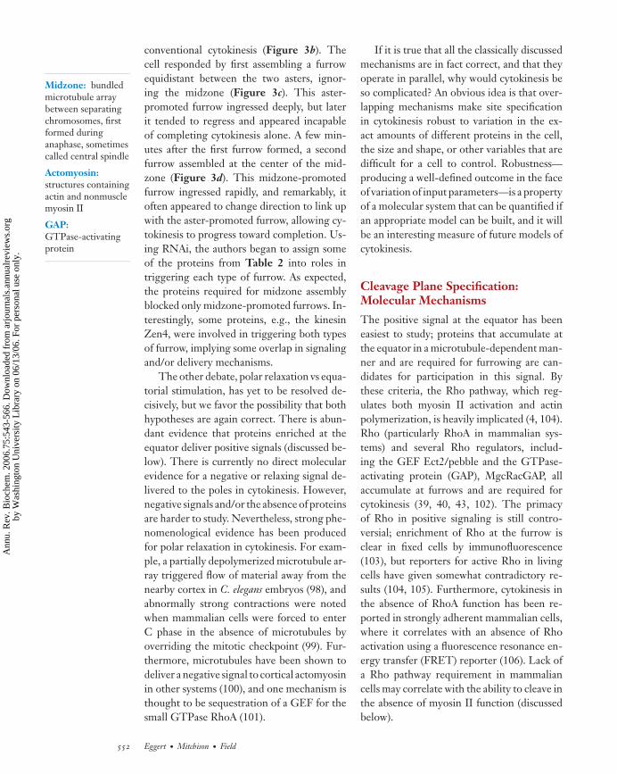
conventional cytokinesis (Figure 3b). Thecell responded by first assembling a furrowequidistant between the two asters, ignor-ing the midzone (Figure 3c). This aster-promoted furrow ingressed deeply, but laterit tended to regress and appeared incapableof completing cytokinesis alone. A few min-utes after the first furrow formed, a secondfurrow assembled at the center of the mid-zone (Figure 3d). This midzone-promotedfurrow ingressed rapidly, and remarkably, itoften appeared to change direction to link upwith the aster-promoted furrow, allowing cy-tokinesis to progress toward completion. Us-ing RNAi, the authors began to assign someof the proteins from Table 2 into roles intriggering each type of furrow. As expected,the proteins required for midzone assemblyblocked only midzone-promoted furrows. In-terestingly, some proteins, e.g., the kinesinZen4, were involved in triggering both typesof furrow, implying some overlap in signalingand/or delivery mechanisms.
The other debate, polar relaxation vs equa-torial stimulation, has yet to be resolved de-cisively, but we favor the possibility that bothhypotheses are again correct. There is abun-dant evidence that proteins enriched at theequator deliver positive signals (discussed be-low). There is currently no direct molecularevidence for a negative or relaxing signal de-livered to the poles in cytokinesis. However,negative signals and/or the absence of proteinsare harder to study. Nevertheless, strong phe-nomenological evidence has been producedfor polar relaxation in cytokinesis. For exam-ple, a partially depolymerized microtubule ar-ray triggered flow of material away from thenearby cortex in C. elegans embryos (98), andabnormally strong contractions were notedwhen mammalian cells were forced to enterC phase in the absence of microtubules byoverriding the mitotic checkpoint (99). Fur-thermore, microtubules have been shown todeliver a negative signal to cortical actomyosinin other systems (100), and one mechanism isthought to be sequestration of a GEF for thesmall GTPase RhoA (101).
If it is true that all the classically discussedmechanisms are in fact correct, and that theyoperate in parallel, why would cytokinesis beso complicated? An obvious idea is that over-lapping mechanisms make site specificationin cytokinesis robust to variation in the ex-act amounts of different proteins in the cell,the size and shape, or other variables that aredifficult for a cell to control. Robustness—producing a well-defined outcome in the faceof variation of input parameters—is a propertyof a molecular system that can be quantified ifan appropriate model can be built, and it willbe an interesting measure of future models ofcytokinesis.
Cleavage Plane Specification:Molecular Mechanisms
The positive signal at the equator has beeneasiest to study; proteins that accumulate atthe equator in a microtubule-dependent man-ner and are required for furrowing are can-didates for participation in this signal. Bythese criteria, the Rho pathway, which reg-ulates both myosin II activation and actinpolymerization, is heavily implicated (4, 104).Rho (particularly RhoA in mammalian sys-tems) and several Rho regulators, includ-ing the GEF Ect2/pebble and the GTPase-activating protein (GAP), MgcRacGAP, allaccumulate at furrows and are required forcytokinesis (39, 40, 43, 102). The primacyof Rho in positive signaling is still contro-versial; enrichment of Rho at the furrow isclear in fixed cells by immunofluorescence(103), but reporters for active Rho in livingcells have given somewhat contradictory re-sults (104, 105). Furthermore, cytokinesis inthe absence of RhoA function has been re-ported in strongly adherent mammalian cells,where it correlates with an absence of Rhoactivation using a fluorescence resonance en-ergy transfer (FRET) reporter (106). Lack ofa Rho pathway requirement in mammaliancells may correlate with the ability to cleave inthe absence of myosin II function (discussedbelow).
552 Eggert · Mitchison · Field
Ann
u. R
ev. B
ioch
em. 2
006.
75:5
43-5
66. D
ownl
oade
d fr
om a
rjou
rnal
s.an
nual
revi
ews.
org
by W
ashi
ngto
n U
nive
rsity
Lib
rary
on
06/1
3/06
. For
per
sona
l use
onl
y.

ANRV277-BI75-21 ARI 3 May 2006 10:16
Two kinases, Aurora B and Polo, also meetthe criteria for involvement in the positivesignal (reviewed in Reference 83). Their rolein cytokinesis has been difficult to dissectbecause both are also involved in mitosis.The recent availability of small molecule Au-rora B inhibitors (Table 1) may help eluci-date its specific role in cytokinesis, and sim-ilar reagents for Polo are needed. To makeprogress in understanding the function of Au-rora B and Polo in cytokinesis, we also needto identify substrates whose phosphorylationplays a specific role in regulating cytokinesis.There will probably be many such substrates.Two likely Aurora B substrates involved in cy-tokinesis are MgcRacGAP (107) and the ki-nesin Zen4/MKLP (108).
The mechanism by which microtubulesspatially regulate the activity of the Rho path-way and the kinases is currently quite mys-terious. Models under consideration includetransport of signaling complexes along mi-crotubules by motors, signaling by plus-endtracking complexes, and control of signalingsimply by the local concentration of tubulinpolymer. In support of the motor hypoth-esis, the plus-end-directed motor, kinesin-6 (Pavarotti/MKLP1/Zen4) has been impli-cated in cytokinesis in all systems examined(Table 2). This motor physically interactswith the Rho GAP, MgcRacGAP, (109) andparticipates in a defined molecular complexwith the Rho GEF, Ect2/pebble, possiblythrough a Ect2/pebble-MgcRacGAP interac-tion (42, 110). Thus the motor protein couldtransport key Rho regulators to the cortex.In support of signaling by plus ends, a plus-end-tracking protein, CLASP/orbit, has beenshown to be important for cytokinesis (48).However, taxol-stabilized asters can success-fully signal (111, 112), implying dynamic plusends are not always required. Local micro-tubule polymer concentration alone may alsobe important. An actin nucleator and essen-tial cytokinesis protein, Diaphanous, binds tomicrotubules independent of Rho signalingand microtubule dynamics (113). Perhaps thepresence of microtubules locally sequesters
Intercellularbridge: connectstwo daughter cellsprior to abscissionand is microtubulerich with themidbody at its center
Midbody: thecenter of theintercellular bridge.It containsmicrotubules and ahigh protein densityarea, the stembody
this protein (or other signaling factors) awayfrom the plasma membrane. We still havemuch to learn about how microtubules di-rect the signaling pathways required forcytokinesis.
Organization ofCytokinesis-AssociatedMicrotubules
Given their role in delivering spatiallyrestricted signals to the cortex, it is impor-tant that microtubules are properly orga-nized in space and time during cytokine-sis. Cytokinesis-associated microtubules aredominated by asters and midzones (Figure 2).Astral microtubules elongate greatly atanaphase in many systems, so they often touchthe cortex and elongate toward the equa-tor. This elongation plays a central role inmorphogenesis of the microtubule cytoskele-ton during cytokinesis, and it is thoughtto be important for microtubule signalingto the cortex (84, 112). Elongation may bedriven by a decrease in catastrophe rate af-ter anaphase (114). This is correlated withdecreased Cdc2 levels, although exactly howCdc2 regulates microtubule dynamics is notunderstood. Rappaport (1) measured that themicrotubule-derived signal propagates to thecortex in echinoderm eggs at a rate of 6 to 7μm/min, and perhaps anaphase microtubuleelongation sets this rate.
The midzone is initially formed betweenthe separating chromosomes by bundling ofelongating overlapping microtubules associ-ated with the spindle (see the movies inReference 115). Later, the midzone becomesself-organizing, and it can persist for manyminutes, if furrow contraction is blocked (16).Although initially formed by the reorganiza-tion of existing microtubules, it is likely thatnew microtubules are nucleated within themidzone (115a). Some authors refer to mid-zones as “central spindles,” which is confus-ing, as the same name has been applied tooverlapping microtubules of the mitotic spin-dle during metaphase. Although midzones
www.annualreviews.org • Animal Cytokinesis 553
Ann
u. R
ev. B
ioch
em. 2
006.
75:5
43-5
66. D
ownl
oade
d fr
om a
rjou
rnal
s.an
nual
revi
ews.
org
by W
ashi
ngto
n U
nive
rsity
Lib
rary
on
06/1
3/06
. For
per
sona
l use
onl
y.

ANRV277-BI75-21 ARI 3 May 2006 10:16
Figure 4Thin section electron micrographs of a dividing HeLa cell. The upperpanel shows an intercellular bridge between two daughter cells. Thenucleus of the right-hand cell is visible. The lower panel is a higherresolution image of the same bridge. Note the bundled microtubules inthe midbody and the electron-dense material (stembody) concentrated ina discrete zone at the center of the bridge (lower panel). Stembodiestypically bulge outward at their center. The bar shown in the lower panelcorresponds to 2 μm (upper panel) and 250 nm (lower panel). Imagescourtesy of Margaret Coughlin.
EM: electronmicroscopy
Stembody: thesmall electron-densedisk at the center ofthe midbody
originate from spindle microtubules, distinctmicrotubule-associated proteins (MAPs) andmotors organize the two arrays.
The midzone has several functions duringcytokinesis. One is to help deliver the equa-torial stimulation signal (discussed above), butthis probably involves microtubules that elon-gate from the midzone to the cortex, ratherthan the midzone itself (48, 116). A second isto keep the separated genomes apart prior tocompletion; when microtubules were depoly-merized before completion in mammaliancells, the nuclei collapsed back together (16).A third is to participate in completion and cellcycle regulation. These are functions of themidbody, a microtubule array within the in-tercellular bridge that connects two daugthercells. The midbody is a derivative of the mid-zone that forms by compression of the furrowduring ingression, and its role is discussed be-low in the section on completion.
At the electron microscopy (EM) level,the midzone is dominated by electron-densematerial that accumulates at the equator on
overlapping microtubule bundles starting atanaphase (117). The electron-dense materialcoalesces as the midzone microtubule bun-dles are compressed by the furrow, eventuallyforming a small disc that was called a stem-body, from the German Stemmkorper “push-ing body” (see Figure 4). Despite its domi-nance in EM views, the molecular nature ofthe electron-dense material has not been de-termined, and its precise function is unknown.
The principal microtubule-interactingproteins implicated in morphogenesis ofthe midzone are bundling factors and ki-nesins. A conserved bundling factor, PRC1/Fascetto/spd-1 accumulates at the center ofthe midzone and ablating it blocks midzoneassembly in all systems (Table 2). PRC1 bindsand bundles microtubules in vitro and in cells(46). It is regulated by the cell cycle, possiblythrough phosphorylation by Cdc2-cyclinB. As mitosis progresses and Cdc2 activitydecreases, PRC1 is dephosphorylated andbecomes active (118). RNAi-mediated knock-down of PRC1 in mammalian cells preventedlocalization of other midzone markers andcompletely blocked midzone assembly (116).Signaling proteins such as Aurora B kinasestill accumulated at the furrow, presumablybecause astral microtubules were unaffected.Ingression was normal, but completion failed.Given its biochemistry, localization, and ge-netics, PRC1 family members are probablythe main bundling factor in midzones; aninteresting unanswered question is whetherthey enforce antiparallel organization.
Two classes of plus-end-directed kinesinshave been implicated in midzone assembly.In all systems, a kinesin-6 family member(MKLP1/Pavarotti/Zen4) accumulates at thecenter of the midzone and ablating it blocksmidzone assembly (Table 2). MKLP1 is aplus-end-directed motor that can cross-linkmicrotubules and slide one microtubule overanother (52), making it an ideal candidatefor organizing overlap interactions within themidzone. CHO1, a splice variant of MKLP1,may be the most relevant isoform for midzonemorphogenesis in mammalian cells (119).
554 Eggert · Mitchison · Field
Ann
u. R
ev. B
ioch
em. 2
006.
75:5
43-5
66. D
ownl
oade
d fr
om a
rjou
rnal
s.an
nual
revi
ews.
org
by W
ashi
ngto
n U
nive
rsity
Lib
rary
on
06/1
3/06
. For
per
sona
l use
onl
y.

ANRV277-BI75-21 ARI 3 May 2006 10:16
A second plus-end-directed kinesin from thekinesin-4 family plays a less defined rolein midzone assembly. Mammalian Kif4 is achromokinesin during mitosis, with a poorlydefined role in spindle assembly/function(120). At anaphase, Kif4 relocalizes to micro-tubule bundles and accumulates at the centerof the midzone (120). Kif4 binds PRC1, witha preference for the dephosphorylated form,and may help to localize this microtubule-bundling protein correctly in the midzone(118).
Other proteins might also contribute tomidzone formation. The plus-end-trackingprotein CLASP/orbit may be involved (48).Annexin 11 is a new player in midzone orga-nization in mammalian cells. Its biochemicalfunction is not known, but it localizes stronglyto midzones and removing it blocks theirassembly (68).
Microtubule organization during cell di-vision has typically been considered indepen-dent of the actin cytoskeleton, and signalingfrom microtubules to the cortex was classi-cally considered unidirectional, but recent ev-idence has questioned this view. During mito-sis, spindle organization depends on corticalactomyosin in some systems (121), and duringcytokinesis, there may be feedback from thecortex to midzone organization. In Drosophilacells, damage to the actin cytoskeleton pre-vents assembly of a normal midzone (35); and,in mammalian cells, evidence from speckleimaging suggested that microtubule plus endscontacting the equatorial cortex are specif-ically stabilized against catastrophes (99). Afeedback loop from the cortex to microtubuleorganization is appealing as a way to ensurerobust self-organization of both cytoskeletalsystems during cytokinesis.
Contractile Ring Assembly
The question of how the furrow assemblesis closely connected to that of how micro-tubules signal to the cortex. The furrow con-sists of a contractile ring together with theplasma membrane to which it is connected.
We currently understand the biochemistry ofthe ring, which is dominated by actin andmyosin II, much better than that of the mem-brane. Contractile rings are stable biochemi-cal entities in the sense that they can be iso-lated and can be induced to contract in vitro(122), but these rings are highly dynamic incells, with fast turnover of both actin andmyosin (123, 124).
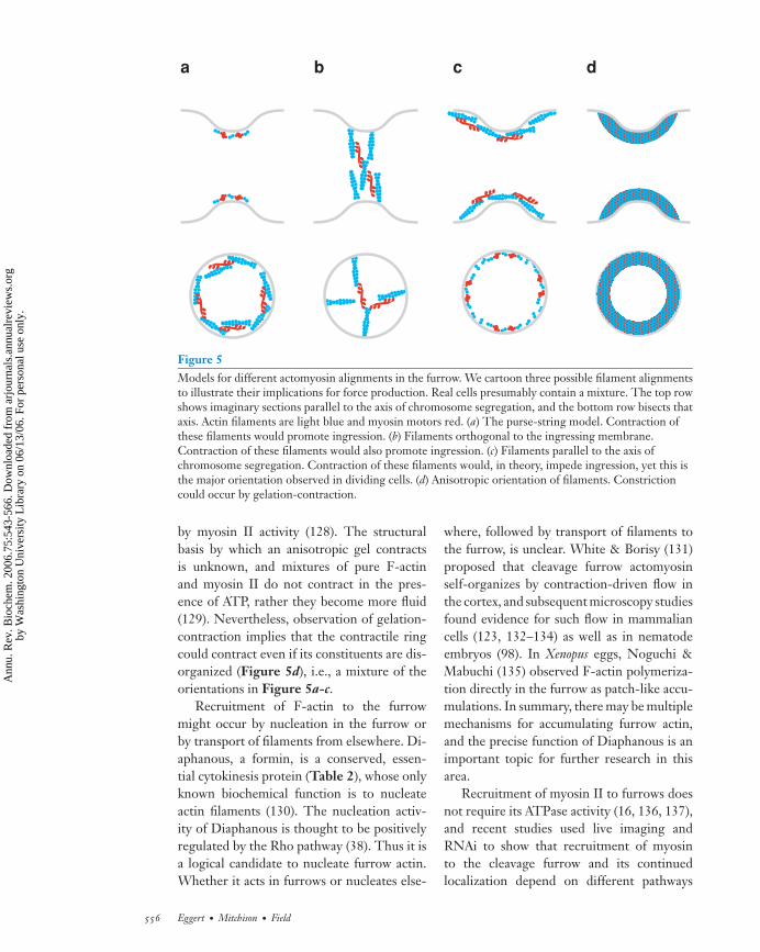
The organization of actin and myosin II incontractile rings is an unsolved problem. InFigure 5a-c, we depict three possible orthog-onal alignments of actomyosin filaments toclarify their implications for force generation,noting that real cells presumably contain somemixture of these alignments. Schroeder pro-posed the “purse-string” model (Figure 5a)(125) on the basis of EM observations inechinoderm embryos. In this model, filamentsliding shortens the ring, and ingression forcecomes from the component of the slidingforce that is directed inward, which is given bycontractile force in the ring multiplied by thereciprocal of the radius. In other words, thismechanism becomes more efficient as the ringgets smaller. Schroeder’s purse string domi-nates textbooks, yet there is surprisingly lit-tle structural evidence for alignment of con-tractile fibers in this orientation. Fishkind &Wang (126) observed actin bundles connect-ing the dorsal part of the furrow to the ventralsurface in adherent mammalian cells, lead-ing them to propose the model in Figure 5b.In this model, all contractile force is di-rected inward. Paradoxically, the majority offluorescently-labeled actin filaments observedin mammalian cells tend to show the organiza-tion in Figure 5c (33, 126, 127). Contractionof filaments with this orientation has no in-wardly directed force component, if anythingit would tend to oppose ingression. Perhapsthese filaments do not participate in force gen-eration and align passively in response to in-gression. Finally, we note that anisotropic or-ganization could potentially generate inwardforce. Cytoplasmic extracts containing actinand myosin II undergo “gelation-contraction”in which an anisotropic F-actin gel contracts
www.annualreviews.org • Animal Cytokinesis 555
Ann
u. R
ev. B
ioch
em. 2
006.
75:5
43-5
66. D
ownl
oade
d fr
om a
rjou
rnal
s.an
nual
revi
ews.
org
by W
ashi
ngto
n U
nive
rsity
Lib
rary
on
06/1
3/06
. For
per
sona
l use
onl
y.
ANRV277-BI75-21 ARI 3 May 2006 10:16
a b c d
Figure 5Models for different actomyosin alignments in the furrow. We cartoon three possible filament alignmentsto illustrate their implications for force production. Real cells presumably contain a mixture. The top rowshows imaginary sections parallel to the axis of chromosome segregation, and the bottom row bisects thataxis. Actin filaments are light blue and myosin motors red. (a) The purse-string model. Contraction ofthese filaments would promote ingression. (b) Filaments orthogonal to the ingressing membrane.Contraction of these filaments would also promote ingression. (c) Filaments parallel to the axis ofchromosome segregation. Contraction of these filaments would, in theory, impede ingression, yet this isthe major orientation observed in dividing cells. (d) Anisotropic orientation of filaments. Constrictioncould occur by gelation-contraction.
by myosin II activity (128). The structuralbasis by which an anisotropic gel contractsis unknown, and mixtures of pure F-actinand myosin II do not contract in the pres-ence of ATP, rather they become more fluid(129). Nevertheless, observation of gelation-contraction implies that the contractile ringcould contract even if its constituents are dis-organized (Figure 5d), i.e., a mixture of theorientations in Figure 5a-c.
Recruitment of F-actin to the furrowmight occur by nucleation in the furrow orby transport of filaments from elsewhere. Di-aphanous, a formin, is a conserved, essen-tial cytokinesis protein (Table 2), whose onlyknown biochemical function is to nucleateactin filaments (130). The nucleation activ-ity of Diaphanous is thought to be positivelyregulated by the Rho pathway (38). Thus it isa logical candidate to nucleate furrow actin.Whether it acts in furrows or nucleates else-
where, followed by transport of filaments tothe furrow, is unclear. White & Borisy (131)proposed that cleavage furrow actomyosinself-organizes by contraction-driven flow inthe cortex, and subsequent microscopy studiesfound evidence for such flow in mammaliancells (123, 132–134) as well as in nematodeembryos (98). In Xenopus eggs, Noguchi &Mabuchi (135) observed F-actin polymeriza-tion directly in the furrow as patch-like accu-mulations. In summary, there may be multiplemechanisms for accumulating furrow actin,and the precise function of Diaphanous is animportant topic for further research in thisarea.
Recruitment of myosin II to furrows doesnot require its ATPase activity (16, 136, 137),and recent studies used live imaging andRNAi to show that recruitment of myosinto the cleavage furrow and its continuedlocalization depend on different pathways
556 Eggert · Mitchison · Field
Ann
u. R
ev. B
ioch
em. 2
006.
75:5
43-5
66. D
ownl
oade
d fr
om a
rjou
rnal
s.an
nual
revi
ews.
org
by W
ashi
ngto
n U
nive
rsity
Lib
rary
on
06/1
3/06
. For
per
sona
l use
onl
y.

ANRV277-BI75-21 ARI 3 May 2006 10:16
(96, 126a). Phosphorylated myosin regulatorylight chain is observed at the furrow dur-ing early anaphase (138) and is needed to as-semble myosin into the ring. DeBiasio et al.(127) carried out a comprehensive analysisof myosin II during cytokinesis in live 3T3cells. They found that myosin fibers flow to-ward the equator during anaphase and form ameshwork, which contains fibers that are bothparallel and perpendicular to the plane ofcleavage. Cortical flow of myosin has alsobeen observed in Xenopus eggs (135). Myosinis very dynamic at the furrow; phosphoryla-tion of the heavy chain is required to maintainits dynamic behavior (124). Myosin transportalong microtubules (139) is an appealing ideabut has not been substantiated by biochem-istry. In summary, cortical flow appears to con-tribute to myosin localization but is unlikelyto be the only mechanism given the observa-tions of recruitment in the absence of its ownmotor activity.
Furrow Ingression
Furrow ingression proceeds by some combi-nation of force generation from the cytoskele-ton (Figure 5), coupled to an increase ofplasma membrane surface area. The textbookview of cytoskeletal force generation focuseson the purse-string contraction model pro-posed by Schroeder. Questions concerningthis model were discussed above, highlightingthe need for more information on the struc-tural organization of contractile rings. Inhi-bition of myosin II immediately blocks fur-row ingression in some mammalian cells (16),and myosin II subunits score as conserved,essential cytokinesis proteins (Table 2).It is thus very puzzling how Dictyosteliumcells and highly adherent mammalian cells,under some conditions, are capable of exe-cuting a form of cytokinesis with myosin IIabsent or greatly reduced in activity (28, 124,140–142). Wang (143) has proposed an al-ternative model for furrow ingression, named“equatorial relaxation,” which posits that thewhole cortex is under tension, and furrow in-
gression occurs at the equator because thecortex is softer there. Much of the evidencethat supports this model comes from lo-cal drug perfusion experiments. Cytochalasinapplied to the furrow tended to promote fur-rowing, whereas application to the poles in-hibited it (144). Local application of the F-actin-stabilizing drug jasplakinolide, or themyosin II inhibitor blebbistatin, had essen-tially the opposite effects (136). These datasupport a model in which selective actin de-polymerization at the equator is important forcytokinesis. However, direct measurementsof cortical stiffness by atomic force mea-surements found increased stiffness at theequator (145). These observations might bereconciled if the equator was stiffer, yet de-polymerized more rapidly, than the bulk cor-tex. In our view, the mechanics of cytoskeletalforce production in cytokinesis is still an openquestion, and it is possible that more than onemechanism operates.
Independent of its effect on cortical stiff-ness, it is clear that actin depolymerizationis an important aspect of furrow ingression.Actin and myosin both turn over rapidly infurrows (123, 124), implying that their localconcentration depends on a dynamic balancebetween polymerization/recruitment and de-polymerization/dissociation. Local concen-tration remains approximately constant dur-ing ingression, implying continuous decreasein contractile ring volume (125). The precisemechanism of actin depolymerization in cellsis controversial, but proteins from the actindepolymerization factor (ADF)-cofilin familyare key factors. Cofilin (twinstar in flies) isneeded for cytokinesis (Table 2). In sperma-tocytes of twinstar Drosophila mutants, actinaccumulates into abnormally large furrows,resulting in inhibition of cleavage (36). Onemodel for keeping F-actin concentration con-stant while volume decreases would make therate of recruiting new actin dependent on thesurface area of the furrow, and the depolymer-ization rate dependent on the total amount ofactin present. This could occur if recruitmentdepended on formin-driven nucleation within
www.annualreviews.org • Animal Cytokinesis 557
Ann
u. R
ev. B
ioch
em. 2
006.
75:5
43-5
66. D
ownl
oade
d fr
om a
rjou
rnal
s.an
nual
revi
ews.
org
by W
ashi
ngto
n U
nive
rsity
Lib
rary
on
06/1
3/06
. For
per
sona
l use
onl
y.

ANRV277-BI75-21 ARI 3 May 2006 10:16
the furrow or, perhaps, if it also depended oncortical flow from neighboring regions.
It has been known for years that furrow in-gression can be coupled to deposition of newplasma membrane (146). The source of thismembrane is not well characterized. Embry-onic systems contain stored membranes in theform of vesicles, but in general this problemis unsolved, and recent investigations have fo-cused on vesicle trafficking pathways. Actin-independent addition of new membrane hasbeen observed in Xenopus eggs, where themembrane is delivered by exocytic vesiclesthat seem to travel along microtubules (147).New membrane addition was also observed insea urchin eggs, using the extracellular matrixprotein hyalin as a membrane marker (148).Shuster & Burgess (148) show that new mem-brane is added specifically to the ingressingfurrow after mitotic exit and that membraneaddition is dependent on astral microtubulesand calcium. Both of these studies were car-ried out in eggs, which are larger than mostcells and have a greater surface area, thus re-quiring more membrane to be inserted dur-ing ingression. Drosophila embryo cellulariza-tion is a special form of cytokinesis that ex-hibits a particularly large requirement for newsurface area, and the process by which newmembrane is inserted just behind the ingress-ing furrows in this system has been analyzedby genetics and microscopy (149). It is possi-ble that specialized mechanisms have evolvedin embryonic systems, but a general require-ment for membrane addition during ingres-sion seems likely. RNAi screens have revealeda conserved requirement for the endocytosisproteins clathrin and dynamin (Table 2), sug-gesting that plasma membrane recycling maybe important during ingression, in additionto the addition of membrane from vesicularstores.
Completion
Completion, also termed abscission or scis-sion, is the final step of cytokinesis. It is insome sense an optional step in cytokinesis and
the cell cycle. In embryos, blastomeres oftenremain connected by intercellular bridges formany cell cycles, perhaps explaining why someproteins involved in completion fail to scorein cyokinesis screens performed in embryos(Table 2). Beyond its importance in cytokine-sis, an emerging concept is that completion isa regulated process with a role in preventingaccumulation of aneuploid cells (150). Thisexciting development may account for someof the biochemical complexity of completion.As the very last step in the old cell cycle or thefirst in the new one, it is logical to use com-pletion as a sensor and regulator of cell cycleprogression.
Completion occurs after the actomyosinring has contracted and the cleavage furrowhas ingressed to its fullest extent, creating anintercellular bridge. The bridge is packed withtightly bundled, antiparallel microtubulesembedded in phase- and electron-densematerial called the stembody (Figure 4).The morphology of completion in mam-malian cells was described by live analysis andEM almost 30 years ago (151). When fur-row ingression is complete, the intercellularbridge is approximately 1–1.5 μm in diame-ter. Separation of daughter cells is precededby a reduction in the diameter of the bridgeto approximately 0.2 μm. This occurs overthe entire length of the bridge, except at thestembody, which retains its original diame-ter and projects out as a bulge in the centerof the bridge (Figure 4). Microtubule bun-dles become further compacted and also be-gin to disappear across the entire length of thebridge, an observation duplicated recently bylive imaging (21, 152).
The microtubule bundles and stembody inthe intercellular bridge are required for com-pletion. They form by compaction of the mid-zone during ingression of the furrow (16).Blocking midzone assembly prevents assem-bly of normal microtubule/stembody struc-tures and causes completion to fail (68, 109,116, 153). There have been recent sugges-tions that midbody microtubules are dynamic(153a), but the function of microtubules and
558 Eggert · Mitchison · Field
Ann
u. R
ev. B
ioch
em. 2
006.
75:5
43-5
66. D
ownl
oade
d fr
om a
rjou
rnal
s.an
nual
revi
ews.
org
by W
ashi
ngto
n U
nive
rsity
Lib
rary
on
06/1
3/06
. For
per
sona
l use
onl
y.

ANRV277-BI75-21 ARI 3 May 2006 10:16
the stembody in completion is unknown.Current ideas focus on a possible role ofmicrotubules in directing vesicles to the stem-body and a possible role of septins, polymer-izing GTPases that accumulate in the bridge(Table 2), in directing vesicle fusion (153b).
To study completion, it is important to dis-tinguish it from late stages of ingression. Oneway to do this is to test for dependence onF-actin. During ingression and before bridgematuration, low doses of the actin depoly-merizing compound Latrunculin B cause thefurrow to reopen. Once the bridge maturesand completion begins, it becomes Latrun-culin insensitive (21), implying that the plasmamembrane is linked to the midbody by aconnection that does not involve dynamicF-actin.
A conserved cleavage furrow componentthat may be involved in bridge stability isAnillin. This multidomain protein binds to F-actin, myosin II, septin complexes, and per-haps also membranes via its pleckstrin ho-mology (PH) domain (5, 33, 96, 154, 155).Anillin plays a nonessential role during ingres-sion (96, 154) but is required for completionin several systems (21, 29, 96). Anillin and itsinteraction partners, the septins, remain in thematuring intercellular bridge after myosin IIand most F-actin have dissociated (21, 32, 33).They also remain permanently in the narrowintercellular bridges that persist after incom-plete cytokinesis in Drosophila spermatocytes(156). Microtubules are absent in these long-lived spermatocyte bridges, implying the exis-tence of a stable, membrane-bound collar thatdoes not depend on a microtubule scaffold.Biochemically, septins polymerize into rela-tively stable filaments that associate with, andmay regulate, membrane trafficking proteins(reviewed in References 157–159). We hy-pothesize that Anillin and septins together as-semble into a stable, filamentous array, whichshapes the plasma membrane of the stable in-tercellular bridge, and that may also regulatethe vesicle fusion required for completion.
Completion requires remodeling ofplasma membranes to create sister cells, and
several recent reports focused on the role ofvesicle trafficking components. Inhibition ofthe midbody-localized t-SNARE/v-SNAREpair syntaxin 2 and endobrevin/VAMP-8by overexpression of dominant-negativeconstructs blocked completion but not in-gression (160). Both the cellular localizationand completion defects were specific for thisSNARE pair. α-SNAP, part of a complexrequired for SNARE-mediated fusion, wasfound in a screen for completion defectsin Drosophila cells (21). In SNAP-depletedcells, intercellular bridges formed normallybut later disassembled without completion.Together, these studies imply that comple-tion involves membrane fusion mediatedby specific SNAREs, using mechanisms incommon with other types of intracellularvesicle fusion. Whether these SNAREspromote direct fusion of plasma membranes,or work less directly by fusing transportvesicles to plasma membranes, remains tobe determined, as does the possible roleof midbody microtubules and septins intargeting fusion.
Centrosomes have long been implicatedin cytokinesis biology as the nucleating sitesof asters (Figure 2), but recently a new, andsomewhat mysterious, role of centrosomes incompletion has been discovered. Live imagingof GFP-tagged centrosomes revealed move-ment of one centrosome into the intercel-lular bridge late in cytokinesis. Completioncorrelated with this movement, suggesting acausal connection (152). The connection be-tween centrosomes and completion was re-inforced by the observation that RNAi de-pletion of the centrosome protein centriolinblocks completion (70). Centriolin localizesto centrosomes throughout the cell cycle,but during cytokinesis, it localizes in one ortwo spots adjacent to the stembody. Deple-tion of centriolin caused a defect in comple-tion with persistent, elongated bridges andformation of multinucleated syncytia. De-fects were also observed in cell cycle tim-ing (70). Recently, centriolin has been shownto be required for the localization of exocyst
www.annualreviews.org • Animal Cytokinesis 559
Ann
u. R
ev. B
ioch
em. 2
006.
75:5
43-5
66. D
ownl
oade
d fr
om a
rjou
rnal
s.an
nual
revi
ews.
org
by W
ashi
ngto
n U
nive
rsity
Lib
rary
on
06/1
3/06
. For
per
sona
l use
onl
y.

ANRV277-BI75-21 ARI 3 May 2006 10:16
components and SNARE proteins to a ring onthe stembody, indicating a role in both vesicletargeting and fusion (160a). A role for inter-cellular bridge components in cell cycle tim-ing was also found in a microsurgery study,where cells that failed to inherit a bridge expe-rienced cell cycle delays (161). The biologicallogic behind the centrosome/completion/cellcycle connection is not yet clear, but cluesmay come from what is known about mitoticexit in yeast cells (162, 163). The mammaliancentriolin implicated in completion shares ho-mology with the MEN/SIN proteins Nud1p(budding yeast) and Cdc11p (fission yeast)(70). The relevance of the yeast MEN/SINmitotic exit pathways for mammalian biologyhas been unclear; these data suggest that a re-lated pathway may regulate completion.
A new direction in completion researchwas opened by a paper describing a connec-tion between mistakes in chromosome seg-regation and completion failure in culturedhuman cells (150). Spontaneously arising bin-ucleate cells, which result from failed com-pletion, were found to exhibit a high fre-quency of chromosome missegregation, anddrug treatments that promoted missegrega-tion increased the frequency of cytokinesisfailure. This study implies that failure in chro-mosome segregation that escapes detection bythe mitotic checkpoint can nevertheless be de-tected by the cell, which responds by block-ing completion. Shi & King (150) proposethat this mechanism evolved to reduce car-cinogenesis because, in their view, single chro-mosome aneuploidy is more dangerous thantetraploidy. The molecular basis of the con-nection between missegregation and failedcompletion is unknown. BRCA2, a proteininvolved in genome stability and protectionfrom cancer, was recently implicated in cy-tokinesis, providing a possible molecular clue(69).
CYTOKINESIS AND CANCER
Failure in cytokinesis very likely contributesto cancer progression. Many cancers are an-
euploid, facilitating genomic plasticity thatallows rapid evolution of aggressive geno-types. Common solid tumors tend to exhibitpolyploidy as well as single chromosome ab-normalities, presumably resulting from fail-ures in both mitosis and cytokinesis. Thetwo may be closely connected both by chro-mosome missegregation triggering failure ofcompletion (150) and by centrosome abnor-malities, which cause defects in both spindleassembly and cytokinesis (reviewed in Refer-ence 164). Pellman and coworkers (165) re-cently found that blocking cytokinesis causesprimary cells lacking p53 to become muchmore carcinogenic in mice, directly demon-strating a causal connection between failedcytokinesis and carcinogenesis for the firsttime. Analysis of cytokinesis defects in humancancers is an important direction for futureresearch.
Cytokinesis is also a point of possible ther-apeutic intervention in cancer. The first testof this idea will come from Aurora kinase in-hibitors, currently in clinical trials (11). Cellstreated with these drugs become polyploidbefore eventually dying, a mechanism of cellkilling distinct from mitotic spindle poisonssuch as taxol. Given that blocking cytokinesisin p53− cells can cause cancer in mice (165),there is a risk that this treatment will causecancer as well as treat it, a concept familiarfor DNA-damaging agents. Cancer drugs thattarget generic cell division mechanisms killnormal stem cells and thus cause bone mar-row and gut toxicity, which limits their ther-apeutic dose. It would be better to find drugsthat selectively blocked cytokinesis (or mito-sis) in cancer cells while sparing normal stemcells. Cytokinesis is highly conserved, and wecannot expect major mechanistic differences.However, different cell types probably vary inthe extent to which overlapping cytokinesispathways are used, so selective inhibition isnot out of the question. In that light, it willbe useful to extend research on cytokinesismechanisms to comparative studies of stemand cancer cells, using both genome-wide anddetailed mechanism approaches.
560 Eggert · Mitchison · Field
Ann
u. R
ev. B
ioch
em. 2
006.
75:5
43-5
66. D
ownl
oade
d fr
om a
rjou
rnal
s.an
nual
revi
ews.
org
by W
ashi
ngto
n U
nive
rsity
Lib
rary
on
06/1
3/06
. For
per
sona
l use
onl
y.

ANRV277-BI75-21 ARI 3 May 2006 10:16
SUMMARY POINTS
1. A parts list of genes involved in cytokinesis has been assembled through a combinationof RNAi and previous efforts.
2. We dissect cytokinesis into six subprocesses and discuss mechanistic progress in eachsubprocess:� Timing: Proteolysis is involved in regulating C phase, the time during which the
cortex can contract.
� Cleavage plane specification: Multiple mechanisms operate in parallel; the Rhopathway and kinases are involved.
� Rearrangement of microtubule structures: Microtubules rearrange into differentarrays that have varied functions.
� Ring assembly: Cortical flow and local nucleation contribute to assembly of acto-myosin filaments.
� Ring ingression: Force generation mechanisms for different orientations relativeto the furrow are discussed. The traditional purse-string model is most likely anoversimplification.
� Completion: Midzone microtubules, vesicle transport, and centrosomes are impor-tant for completion.
3. The relevance of cytokinesis to cancer is discussed.
FUTURE ISSUES TO BE RESOLVED
1. We are beginning to generate a parts list of all proteins involved in cytokinesis butstill know little about how they interact with each other to accomplish this process.
2. We have partial information for most of the fundamental mechanisms underlyingcytokinesis, but our understanding of biochemical mechanisms is only just emerging.
ACKNOWLEDGMENTS
We thank Margaret Coughlin for the images in Figure 4. U.S.E was supported by a Merck-sponsored fellowship from the Helen Hay Whitney foundation. Cytokinesis research in theMitchison lab is supported by National Institutes of Health grant R01 GM023928–25.
LITERATURE CITED
1. Rappaport R. 1996. Cytokinesis in Animal Cells. Cambridge, UK: Cambridge Univ. Press2. Jurgens G. 2005. Trends Cell Biol. 15:277–833. Balasubramanian MK, Bi EF, Glotzer M. 2004. Curr. Biol. 14:R806–184. Uyeda TQ, Nagasaki A, Yumura S. 2004. Int. Rev. Cytol. 240:377–4325. Kinoshita M, Field CM, Coughlin ML, Straight AF, Mitchison TJ. 2002. Dev. Cell
3:791–8026. Mishima M, Pavicic V, Gruneberg U, Nigg EA, Glotzer M. 2004. Nature 430:908–13
www.annualreviews.org • Animal Cytokinesis 561
Ann
u. R
ev. B
ioch
em. 2
006.
75:5
43-5
66. D
ownl
oade
d fr
om a
rjou
rnal
s.an
nual
revi
ews.
org
by W
ashi
ngto
n U
nive
rsity
Lib
rary
on
06/1
3/06
. For
per
sona
l use
onl
y.
ANRV277-BI75-21 ARI 3 May 2006 10:16
6a. Wu JQ, Pollard TD. 2005. Science 310:310–147. Harris AK, Gewalt SL. 1989. J. Cell Biol. 109:2215–238. Peterson JR, Mitchison TJ. 2002. Chem. Biol. 9:1275–859. Hauf S, Cole RW, LaTerra S, Zimmer C, Schnapp G, et al. 2003. J. Cell Biol. 161:281–94
10. Ditchfield C, Johnson VL, Tighe A, Ellston R, Haworth C, et al. 2003. J. Cell Biol.161:267–80
11. Harrington EA, Bebbington D, Moore J, Rasmussen RK, Ajose-Adeogun AO, et al.2004. Nat. Med. 10:262–67
12. Schroeder TE. 1973. Proc. Natl. Acad Sci. USA 70:1688–9213. Coue M, Brenner SL, Spector I, Korn ED. 1987. FEBS Lett. 213:316–1814. Bubb MR, Spector I, Bershadsky AD, Korn ED. 1995. J. Biol. Chem. 270:3463–6615. Bubb MR, Senderowicz AM, Sausville EA, Duncan KL, Korn ED. 1994. J. Biol. Chem.
269:14869–7116. Straight AF, Cheung A, Limouze J, Chen I, Westwood NJ, et al. 2003. Science 299:1743–
4717. Ishizaki T, Uehata M, Tamechika I, Keel J, Nonomura K, et al. 2000. Mol. Pharmacol.
57:976–8318. Nishikawa M, Tanaka T, Hidaka H. 1980. Nature 287:863–6519. Silverman-Gavrila RV, Forer A. 2001. Cell Motil. Cytoskelet. 50:180–9720. Eggert US, Kiger AA, Richter C, Perlman ZE, Perrimon N, et al. 2004. PLoS Biol.
2:e37921. Echard A, Hickson GR, Foley E, O’Farrell PH. 2004. Curr. Biol. 14:1685–9322. Gonczy P, Echeverri C, Oegema K, Coulson A, Jones SJ, et al. 2000. Nature 408:331–3623. Sonnichsen B, Koski LB, Walsh A, Marschall P, Neumann B, et al. 2005. Nature 434:462–
6924. Rogers SL, Wiedemann U, Stuurman N, Vale RD. 2003. J. Cell Biol. 162:1079–8825. Skop AR, Liu H, Yates J 3rd, Meyer BJ, Heald R. 2004. Science 305:61–6626. Mabuchi I, Okuno M. 1977. J. Cell Biol. 74:251–6327. Knecht DA, Loomis WF. 1987. Science 236:1081–8628. De Lozanne A, Spudich JA. 1987. Science 236:1086–9129. Somma MP, Fasulo B, Cenci G, Cundari E, Gatti M. 2002. Mol. Biol. Cell 13:2448–6030. Karess RE, Chang XJ, Edwards KA, Kulkarni S, Aguilera I, Kiehart DP. 1991. Cell
65:1177–8931. Kiger AA, Baum B, Jones S, Jones M, Coulson A, et al. 2003. J. Biol. 2:2732. Field CM, Alberts BM. 1995. J. Cell Biol. 131:165–7833. Oegema K, Savoian MS, Mitchison TJ, Field CM. 2000. J. Cell Biol. 150:539–5234. Zipperlen P, Fraser AG, Kamath RS, Martinez-Campos M, Ahringer J. 2001. EMBO J.
20:3984–9235. Giansanti MG, Bonaccorsi S, Williams B, Williams EV, Santolamazza C, et al. 1998.
Genes Dev. 12:396–41036. Gunsalus KC, Bonaccorsi S, Williams E, Verni F, Gatti M, Goldberg ML. 1995. J. Cell
Biol. 131:1243–5937. Castrillon DH, Wasserman SA. 1994. Development 120:3367–7738. Watanabe N, Madaule P, Reid T, Ishizaki T, Watanabe G, et al. 1997. EMBO J. 16:3044–
5639. Mabuchi I, Hamaguchi Y, Fujimoto H, Morii N, Mishima M, Narumiya S. 1993. Zygote
1:325–3140. Prokopenko SN, Brumby A, O’Keefe L, Prior L, He Y, et al. 1999. Genes Dev. 13:2301–
14
562 Eggert · Mitchison · Field
Ann
u. R
ev. B
ioch
em. 2
006.
75:5
43-5
66. D
ownl
oade
d fr
om a
rjou
rnal
s.an
nual
revi
ews.
org
by W
ashi
ngto
n U
nive
rsity
Lib
rary
on
06/1
3/06
. For
per
sona
l use
onl
y.

ANRV277-BI75-21 ARI 3 May 2006 10:16
41. Piano F, Schetter AJ, Morton DG, Gunsalus KC, Reinke V, et al. 2002. Curr. Biol.12:1959–64
42. Somers WG, Saint R. 2003. Dev. Cell 4:29–3943. Hirose K, Kawashima T, Iwamoto I, Nosaka T, Kitamura T. 2001. J. Biol. Chem.
276:5821–2844. Madaule P, Eda M, Watanabe N, Fujisawa K, Matsuoka T, et al. 1998. Nature 394:491–
9445. Kosako H, Yoshida T, Matsumura F, Ishizaki T, Narumiya S, Inagaki M. 2000. Oncogene
19:6059–6446. Mollinari C, Kleman JP, Jiang W, Schoehn G, Hunter T, Margolis RL. 2002. J. Cell
Biol. 157:1175–8647. Verni F, Somma MP, Gunsalus KC, Bonaccorsi S, Belloni G, et al. 2004. Curr. Biol.
14:1569–7548. Inoue YH, Savoian MS, Suzuki T, Mathe E, Yamamoto MT, Glover DM. 2004. J. Cell
Biol. 166:49–6049. Zhu CJ, Zhao J, Bibikova M, Leverson JD, Bossy-Wetzel E, et al. 2005. Mol. Biol. Cell
16:3187–9950. Adams RR, Tavares AA, Salzberg A, Bellen HJ, Glover DM. 1998. Genes Dev. 12:1483–9451. Goshima G, Vale RD. 2003. J. Cell Biol. 162:1003–1652. Nislow C, Lombillo VA, Kuriyama R, McIntosh JR. 1992. Nature 359:543–4753. Fontijn RD, Goud B, Echard A, Jollivet F, van Marle J, et al. 2001. Mol. Cell. Biol.
21:2944–5554. Williams BC, Riedy MF, Williams EV, Gatti M, Goldberg ML. 1995. J. Cell Biol.
129:709–2355. Gatt MK, Savoian MS, Riparbelli MG, Massarelli C, Callaini G, Glover DM. 2005. J.
Cell Sci. 118:2671–8256. Niswonger ML, O’Halloran TJ. 1997. Proc. Natl. Acad Sci. USA 94:8575–7857. Thompson HM, Skop AR, Euteneuer U, Meyer BJ, McNiven MA. 2002. Curr. Biol.
12:2111–1758. Xu H, Brill JA, Hsien J, McBride R, Boulianne GL, Trimble WS. 2002. Dev. Biol.
251:294–30659. Hickson GR, Matheson J, Riggs B, Maier VH, Fielding AB, et al. 2003. Mol. Biol. Cell
14:2908–2060. Terada Y, Tatsuka M, Suzuki F, Yasuda Y, Fujita S, Otsu M. 1998. EMBO J. 17:667–7661. Adams RR, Maiato H, Earnshaw WC, Carmena M. 2001. J. Cell Biol. 153:865–8062. Fraser AG, James C, Evan GI, Hengartner MO. 1999. Curr. Biol. 9:292–30163. Romano A, Guse A, Krascenicova I, Schnabel H, Schnabel R, Glotzer M. 2003. J. Cell
Biol. 161:229–3664. Gassmann R, Carvalho A, Henzing AJ, Ruchaud S, Hudson DF, et al. 2004. J. Cell Biol.
166:179–9165. Sampath SC, Ohi R, Leismann O, Salic A, Pozniakovski A, Funabiki H. 2004. Cell
118:187–20266. Echard A, O’Farrell PH. 2003. Curr. Biol. 13:373–8367. Carmena M, Riparbelli MG, Minestrini G, Tavares AM, Adams R, et al. 1998. J. Cell
Biol. 143:659–7168. Tomas A, Futter C, Moss SE. 2004. J. Cell Biol. 165:813–2269. Daniels MJ, Wang Y, Lee M, Venkitaraman AR. 2004. Science 306:876–7970. Gromley A, Jurczyk A, Sillibourne J, Halilovic E, Mogensen M, et al. 2003. J. Cell Biol.
161:535–45
www.annualreviews.org • Animal Cytokinesis 563
Ann
u. R
ev. B
ioch
em. 2
006.
75:5
43-5
66. D
ownl
oade
d fr
om a
rjou
rnal
s.an
nual
revi
ews.
org
by W
ashi
ngto
n U
nive
rsity
Lib
rary
on
06/1
3/06
. For
per
sona
l use
onl
y.

ANRV277-BI75-21 ARI 3 May 2006 10:16
71. Litvak V, Tian D, Carmon S, Lev S. 2002. Mol. Cell. Biol. 22:5064–7572. Prasanth SG, Prasanth KV, Stillman B. 2002. Science 297:1026–3173. Brill JA, Hime GR, Scharer-Schuksz M, Fuller MT. 2000. Development 127:3855–64
73a. Neufeld TP, Rubin GM. 1994. Cell 77:371–7973b. Surka MC, Tsang CW, Trimble WS. 2002. Mol. Biol. Cell 13:3532–4573c. Kinoshita M, Kumar S, Mizoguchi A, Ide C, Kinoshita A, et al. 1997. Genes Dev. 11:1535–
4774. Kittler R, Putz G, Pelletier L, Poser I, Heninger AK, et al. 2004. Nature 432:1036–4075. Lorson MA, Horvitz HR, van den Heuvel S. 2000. J. Cell Biol. 148:73–8676. Field SJ, Madson N, Kerr ML, Galbraith KA, Kennedy CE, et al. 2005. Curr. Biol.
15:1407–1277. Wong R, Hadjiyanni I, Wei HC, Polevoy G, McBride R, et al. 2005. Curr. Biol. 15:1401–678. Im DS, Heise CE, Nguyen T, O’Dowd BF, Lynch KR. 2001. J. Cell Biol. 153:429–3479. Kanazawa T, Nakamura S, Momoi M, Yamaji T, Takematsu H, et al. 2000. J. Cell Biol.
149:943–5080. Pollard TD. 2004. J. Exp. Zool. A 301:9–1481. Martineau SN, Andreassen PR, Margolis RL. 1995. J. Cell Biol. 131:191–20582. Canman JC, Hoffman DB, Salmon ED. 2000. Curr. Biol. 10:611–1483. Nigg EA. 2001. Nat. Rev. Mol. Cell Biol. 2:21–32
83a. Niiya F, Xie X, Lee KS, Inoue H, Miki T. 2005. J. Biol. Chem. 280:36502–984. Shuster CB, Burgess DR. 1999. J. Cell Biol. 146:981–9285. Shuster CB, Burgess DR. 2002. Curr. Biol. 12:854–5886. King RW, Deshaies RJ, Peters JM, Kirschner MW. 1996. Science 274:1652–5987. Rape M, Kirschner MW. 2004. Nature 432:588–9588. Lindon C, Pines J. 2004. J. Cell. Biol. 164:233–4189. Zhao WM, Fang G. 2005. J. Biol. Chem. 280:33516–24
89a. Stewart S, Fang G. 2005. Cancer Res. 65:8730–3590. Betschinger J, Knoblich JA. 2004. Curr. Biol. 14:R674–8591. Roegiers F, Jan YN. 2004. Curr. Opin. Cell Biol. 16:195–20592. Maddox AS, Oegema K. 2003. Nat. Cell Biol. 5:773–7693. Mandato CA, Benink HA, Bement WM. 2000. Cell Motil. Cytoskelet. 45:87–9294. Glotzer M. 2004. J. Cell Biol. 164:347–5195. D’Avino PP, Savoian MS, Glover DM. 2005. J. Cell Sci. 118:1549–5896. Straight AF, Field CM, Mitchison TJ. 2005. Mol. Biol. Cell 16:193–20197. Bringmann H, Hyman AA. 2005. Nature 436:731–3498. Hird SN, White JG. 1993. J. Cell Biol. 121:1343–5599. Canman JC, Cameron LA, Maddox PS, Straight A, Tirnauer JS, et al. 2003. Nature
424:1074–78100. Canman JC, Bement WM. 1997. J. Cell Sci. 110(Pt.16):1907–17101. Krendel M, Zenke FT, Bokoch GM. 2002. Nat. Cell Biol. 4:294–301102. Tatsumoto T, Xie X, Blumenthal R, Okamoto I, Miki T. 1999. J. Cell Biol. 147:921–28103. Yonemura S, Hirao-Minakuchi K, Nishimura Y. 2004. Exp. Cell Res. 295:300–14104. Bement WM, Benink HA, von Dassow G. 2005. J. Cell Biol. 170:91–101105. Yoshizaki H, Ohba Y, Kurokawa K, Itoh RE, Nakamura T, et al. 2003. J. Cell Biol.
162:223–32106. Yoshizaki H, Ohba Y, Parrini MC, Dulyaninova NG, Bresnick AR, et al. 2004. J. Biol.
Chem. 279:44756–62107. Minoshima Y, Kawashima T, Hirose K, Tonozuka Y, Kawajiri A, et al. 2003. Dev. Cell
4:549–60
564 Eggert · Mitchison · Field
Ann
u. R
ev. B
ioch
em. 2
006.
75:5
43-5
66. D
ownl
oade
d fr
om a
rjou
rnal
s.an
nual
revi
ews.
org
by W
ashi
ngto
n U
nive
rsity
Lib
rary
on
06/1
3/06
. For
per
sona
l use
onl
y.

ANRV277-BI75-21 ARI 3 May 2006 10:16
108. Guse A, Mishima M, Glotzer M. 2005. Curr. Biol. 15:778–86109. Jantsch-Plunger V, Gonczy P, Romano A, Schnabel H, Hamill D, et al. 2000. J. Cell
Biol. 149:1391–404110. Yuce O, Piekny A, Glotzer M. 2005. J. Cell Biol. 170:571–82111. Shannon KB, Canman JC, Moree CB, Tirnauer JS, Salmon ED. 2005. Mol. Biol. Cell
16:4423–36112. Strickland LI, Donnelly EJ, Burgess DR. 2005. Mol. Biol. Cell 16:4485–94113. Kato T, Watanabe N, Morishima Y, Fujita A, Ishizaki T, Narumiya S. 2001. J. Cell Sci.
114:775–84114. Desai A, Mitchison TJ. 1997. Annu. Rev. Cell Dev. Biol. 13:83–117115. Rusan NM, Wadsworth P. 2005. J. Cell Biol. 168:21–28
115a. Shu HB, Li Z, Palacios MJ, Li Q, Joshi HC. 1995. J. Cell Sci. 108:2955–62116. Mollinari C, Kleman JP, Saoudi Y, Jablonski SA, Perard J, et al. 2005. Mol. Biol. Cell
16:1043–55117. Buck RC, Tisdale JM. 1962. J. Cell Biol. 13:109–15118. Zhu CJ, Jiang W. 2005. Proc. Natl. Acad Sci. USA 102:343–48119. Kuriyama R, Gustus C, Terada Y, Uetake Y, Matuliene J. 2002. J. Cell Biol. 156:783–90120. Vernos I, Raats J, Hirano T, Heasman J, Karsenti E, Wylie C. 1995. Cell 81:117–27121. Rosenblatt J, Cramer LP, Baum B, McGee KM. 2004. Cell 117:361–72122. Mabuchi I, Tsukita S, Tsukita S, Sawai T. 1988. Proc. Natl. Acad Sci. USA 85:5966–70123. Murthy K, Wadsworth P. 2005. Curr. Biol. 15:724–31124. Yumura S. 2001. J. Cell Biol. 154:137–46125. Schroeder TE. 1972. J. Cell Biol. 53:419–34126. Fishkind DJ, Wang YL. 1993. J. Cell Biol. 123:837–48
126a. Dean SO, Rogers SL, Stuurman N, Vale RD, Spudich JA. 2005. Proc. Natl. Acad. Sci.USA 102:13473–78
127. DeBiasio RL, LaRocca GM, Post PL, Taylor DL. 1996. Mol. Biol. Cell 7:1259–82128. Pollard TD. 1976. J. Cell Biol. 68:579–601129. Humphrey D, Duggan C, Saha D, Smith D, Kas J. 2002. Nature 416:413–16130. Zigmond SH. 2004. Curr. Opin. Cell Biol. 16:99–105131. White JG, Borisy GG. 1983. J. Theor. Biol. 101:289–316132. Cao LG, Wang YL. 1990. J. Cell Biol. 111:1905–11133. Cao LG, Wang YL. 1990. J. Cell Biol. 110:1089–95134. Wang YL, Silverman JD, Cao LG. 1994. J. Cell Biol. 127:963–71135. Noguchi T, Mabuchi I. 2001. J. Cell Sci. 114:401–12136. Guha M, Zhou M, Wang YL. 2005. Curr. Biol. 15:732–36137. Zang JH, Spudich JA. 1998. Proc. Natl. Acad Sci. USA 95:13652–57138. Matsumura F, Ono S, Yamakita Y, Totsukawa G, Yamashiro S. 1998. J. Cell Biol. 140:119–
29139. Foe VE, Field CM, Odell GM. 2000. Development 127:1767–87140. Neujahr R, Heizer C, Gerisch G. 1997. J. Cell Sci. 110(Pt. 2):123–37141. Kanada M, Nagasaki A, Uyeda TQ. 2005. Mol. Biol. Cell 16:3865–72142. Gerisch G, Weber I. 2000. Curr. Opin. Cell Biol. 12:126–32143. Wang YL. 2001. Cell. Struct. Funct. 26:633–38144. O’Connell CB, Warner AK, Wang Y. 2001. Curr. Biol. 11:702–7145. Matzke R, Jacobson K, Radmacher M. 2001. Nat. Cell Biol. 3:607–10146. Bluemink JG, de Laat SW. 1973. J. Cell Biol. 59:89–108147. Danilchik MV, Bedrick SD, Brown EE, Ray K. 2003. J. Cell Sci. 116:273–83
www.annualreviews.org • Animal Cytokinesis 565
Ann
u. R
ev. B
ioch
em. 2
006.
75:5
43-5
66. D
ownl
oade
d fr
om a
rjou
rnal
s.an
nual
revi
ews.
org
by W
ashi
ngto
n U
nive
rsity
Lib
rary
on
06/1
3/06
. For
per
sona
l use
onl
y.

ANRV277-BI75-21 ARI 3 May 2006 10:16
148. Shuster CB, Burgess DR. 2002. Proc. Natl. Acad Sci. USA 99:3633–38149. Lecuit T, Wieschaus E. 2000. J. Cell Biol. 150:849–60150. Shi Q, King R. 2005. Nature 437:1038–42151. Mullins JM, Biesele JJ. 1977. J. Cell Biol. 73:672–84152. Piel M, Nordberg J, Euteneuer U, Bornens M. 2001. Science 291:1550–53153. Matuliene J, Kuriyama R. 2002. Mol. Biol. Cell 13:1832–45
153a. Rosa J, Canovas P, Islam A, Altieri DC, Doxsey SJ. 2006. Mol. Biol. Cell 17:1483–93153b. Finger FP, White JG. 2002. Cell 108:127–30154. Field CM, Coughlin M, Doberstein S, Marty T, Sullivan W. 2005. Development
132:2849–60155. Maddox AS, Habermann B, Desai A, Oegema K. 2005. Development 132:2837–48156. Robinson DN, Cooley L. 1996. Trends Cell Biol. 6:474–79157. Versele M, Thorner J. 2005. Trends Cell Biol. 15:414–24158. Joo E, Tsang CW, Trimble WS. 2005. Traffic 6:626–34159. Beites CL, Campbell KA, Trimble WS. 2005. Biochem. J. 385:347–53160. Low SH, Li X, Miura M, Kudo N, Quinones B, Weimbs T. 2003. Dev. Cell 4:753–59
160a. Gromley A, Yeaman C, Rosa J, Redick S, Chen CT, et al. 2005. Cell 123:75–87161. Murata-Hori M, Sluder G, Wang YL. 2004. BMC Cell Biol. 5:49162. McCollum D, Gould KL. 2001. Trends Cell Biol. 11:89–95163. Pereira G, Schiebel E. 2001. Curr. Opin. Cell Biol. 13:762–69164. Saunders W. 2005. Semin. Cancer Biol. 15:25–32165. Fujiwara T, Bandi M, Nitta M, Ivanova EV, Bronson RT, Pellman D. 2005. Nature
437:1043–47
566 Eggert · Mitchison · Field
Ann
u. R
ev. B
ioch
em. 2
006.
75:5
43-5
66. D
ownl
oade
d fr
om a
rjou
rnal
s.an
nual
revi
ews.
org
by W
ashi
ngto
n U
nive
rsity
Lib
rary
on
06/1
3/06
. For
per
sona
l use
onl
y.

Contents ARI 22 April 2006 9:16
Annual Reviewof Biochemistry
Volume 75, 2006Contents
Wanderings of a DNA Enzymologist: From DNA Polymerase to ViralLatencyI. Robert Lehman � � � � � � � � � � � � � � � � � � � � � � � � � � � � � � � � � � � � � � � � � � � � � � � � � � � � � � � � � � � � � � � � � � � � � � � � � � � � � � � 1
Signaling Pathways in Skeletal Muscle RemodelingRhonda Bassel-Duby and Eric N. Olson � � � � � � � � � � � � � � � � � � � � � � � � � � � � � � � � � � � � � � � � � � � � � � � � � � � � � �19
Biosynthesis and Assembly of Capsular Polysaccharides inEscherichia coliChris Whitfield � � � � � � � � � � � � � � � � � � � � � � � � � � � � � � � � � � � � � � � � � � � � � � � � � � � � � � � � � � � � � � � � � � � � � � � � � � � � � � � �39
Energy Converting NADH:Quinone Oxidoreductase (Complex I)Ulrich Brandt � � � � � � � � � � � � � � � � � � � � � � � � � � � � � � � � � � � � � � � � � � � � � � � � � � � � � � � � � � � � � � � � � � � � � � � � � � � � � � � � � �69
Tyrphostins and Other Tyrosine Kinase InhibitorsAlexander Levitzki and Eyal Mishani � � � � � � � � � � � � � � � � � � � � � � � � � � � � � � � � � � � � � � � � � � � � � � � � � � � � � � �93
Break-Induced Replication and Recombinational Telomere Elongationin YeastMichael J. McEachern and James E. Haber � � � � � � � � � � � � � � � � � � � � � � � � � � � � � � � � � � � � � � � � � � � � � � � 111
LKB1-Dependent Signaling PathwaysDario R. Alessi, Kei Sakamoto, and Jose R. Bayascas � � � � � � � � � � � � � � � � � � � � � � � � � � � � � � � � � � � � � � 137
Energy Transduction: Proton Transfer Through the RespiratoryComplexesJonathan P. Hosler, Shelagh Ferguson-Miller, and Denise A. Mills � � � � � � � � � � � � � � � � � � � � � � 165
The Death-Associated Protein Kinases: Structure, Function, andBeyondShani Bialik and Adi Kimchi � � � � � � � � � � � � � � � � � � � � � � � � � � � � � � � � � � � � � � � � � � � � � � � � � � � � � � � � � � � � � � � 189
Mechanisms for Chromosome and Plasmid SegregationSantanu Kumar Ghosh, Sujata Hajra, Andrew Paek,
and Makkuni Jayaram � � � � � � � � � � � � � � � � � � � � � � � � � � � � � � � � � � � � � � � � � � � � � � � � � � � � � � � � � � � � � � � � � � � 211
Chromatin Modifications by Methylation and Ubiquitination:Implications in the Regulation of Gene ExpressionAli Shilatifard � � � � � � � � � � � � � � � � � � � � � � � � � � � � � � � � � � � � � � � � � � � � � � � � � � � � � � � � � � � � � � � � � � � � � � � � � � � � � � � 243
v
Ann
u. R
ev. B
ioch
em. 2
006.
75:5
43-5
66. D
ownl
oade
d fr
om a
rjou
rnal
s.an
nual
revi
ews.
org
by W
ashi
ngto
n U
nive
rsity
Lib
rary
on
06/1
3/06
. For
per
sona
l use
onl
y.

Contents ARI 22 April 2006 9:16
Structure and Mechanism of the Hsp90 Molecular ChaperoneMachineryLaurence H. Pearl and Chrisostomos Prodromou � � � � � � � � � � � � � � � � � � � � � � � � � � � � � � � � � � � � � � � � � � 271
Biochemistry of Mammalian Peroxisomes RevisitedRonald J.A. Wanders and Hans R. Waterham � � � � � � � � � � � � � � � � � � � � � � � � � � � � � � � � � � � � � � � � � � � � 295
Protein Misfolding, Functional Amyloid, and Human DiseaseFabrizio Chiti and Christopher M. Dobson � � � � � � � � � � � � � � � � � � � � � � � � � � � � � � � � � � � � � � � � � � � � � � � � 333
Obesity-Related Derangements in Metabolic RegulationDeborah M. Muoio and Christopher B. Newgard � � � � � � � � � � � � � � � � � � � � � � � � � � � � � � � � � � � � � � � � � 367
Cold-Adapted EnzymesKhawar Sohail Siddiqui and Ricardo Cavicchioli � � � � � � � � � � � � � � � � � � � � � � � � � � � � � � � � � � � � � � � � � � 403
The Biochemistry of SirtuinsAnthony A. Sauve, Cynthia Wolberger, Vern L. Schramm, and Jef D. Boeke � � � � � � � � � � � 435
Dynamic Filaments of the Bacterial CytoskeletonKatharine A. Michie and Jan Lowe � � � � � � � � � � � � � � � � � � � � � � � � � � � � � � � � � � � � � � � � � � � � � � � � � � � � � � � � 467
The Structure and Function of Telomerase Reverse TranscriptaseChantal Autexier and Neal F. Lue � � � � � � � � � � � � � � � � � � � � � � � � � � � � � � � � � � � � � � � � � � � � � � � � � � � � � � � � � 493
Relating Protein Motion to CatalysisSharon Hammes-Schiffer and Stephen J. Benkovic � � � � � � � � � � � � � � � � � � � � � � � � � � � � � � � � � � � � � � � 519
Animal Cytokinesis: From Parts List to MechanismsUlrike S. Eggert, Timothy J. Mitchison, and Christine M. Field � � � � � � � � � � � � � � � � � � � � � � � � 543
Mechanisms of Site-Specific RecombinationNigel D.F. Grindley, Katrine L. Whiteson, and Phoebe A. Rice � � � � � � � � � � � � � � � � � � � � � � � � � � 567
Axonal Transport and Alzheimer’s DiseaseGorazd B. Stokin and Lawrence S.B. Goldstein � � � � � � � � � � � � � � � � � � � � � � � � � � � � � � � � � � � � � � � � � � � 607
Asparagine Synthetase ChemotherapyNigel G.J. Richards and Michael S. Kilberg � � � � � � � � � � � � � � � � � � � � � � � � � � � � � � � � � � � � � � � � � � � � � � � 629
Domains, Motifs, and Scaffolds: The Role of Modular Interactions inthe Evolution and Wiring of Cell Signaling CircuitsRoby P. Bhattacharyya, Attila Remenyi, Brian J. Yeh, and Wendell A. Lim � � � � � � � � � � � � � 655
Ribonucleotide ReductasesPar Nordlund and Peter Reichard � � � � � � � � � � � � � � � � � � � � � � � � � � � � � � � � � � � � � � � � � � � � � � � � � � � � � � � � � � 681
Introduction to the Membrane Protein Reviews: The Interplay ofStructure, Dynamics, and Environment in Membrane ProteinFunctionJonathan N. Sachs and Donald M. Engelman � � � � � � � � � � � � � � � � � � � � � � � � � � � � � � � � � � � � � � � � � � � � � 707
vi Contents
Ann
u. R
ev. B
ioch
em. 2
006.
75:5
43-5
66. D
ownl
oade
d fr
om a
rjou
rnal
s.an
nual
revi
ews.
org
by W
ashi
ngto
n U
nive
rsity
Lib
rary
on
06/1
3/06
. For
per
sona
l use
onl
y.

Contents ARI 22 April 2006 9:16
Relations Between Structure and Function of the MitochondrialADP/ATP CarrierH. Nury, C. Dahout-Gonzalez, V. Trezeguet, G.J.M. Lauquin,G. Brandolin, and E. Pebay-Peyroula � � � � � � � � � � � � � � � � � � � � � � � � � � � � � � � � � � � � � � � � � � � � � � � � � � � � � � 713
G Protein–Coupled Receptor RhodopsinKrzysztof Palczewski � � � � � � � � � � � � � � � � � � � � � � � � � � � � � � � � � � � � � � � � � � � � � � � � � � � � � � � � � � � � � � � � � � � � � � � � 743
Transmembrane Traffic in the Cytochrome b6 f ComplexWilliam A. Cramer, Huamin Zhang, Jiusheng Yan, Genji Kurisu,
and Janet L. Smith � � � � � � � � � � � � � � � � � � � � � � � � � � � � � � � � � � � � � � � � � � � � � � � � � � � � � � � � � � � � � � � � � � � � � � � 769
INDEXES
Subject Index � � � � � � � � � � � � � � � � � � � � � � � � � � � � � � � � � � � � � � � � � � � � � � � � � � � � � � � � � � � � � � � � � � � � � � � � � � � � � � � � � � � 791
Author Index � � � � � � � � � � � � � � � � � � � � � � � � � � � � � � � � � � � � � � � � � � � � � � � � � � � � � � � � � � � � � � � � � � � � � � � � � � � � � � � � � � � 825
ERRATA
An online log of corrections to Annual Review of Biochemistry chapters (if any, 1977 tothe present) may be found at http://biochem.annualreviews.org/errata.shtml
Contents vii
Ann
u. R
ev. B
ioch
em. 2
006.
75:5
43-5
66. D
ownl
oade
d fr
om a
rjou
rnal
s.an
nual
revi
ews.
org
by W
ashi
ngto
n U
nive
rsity
Lib
rary
on
06/1
3/06
. For
per
sona
l use
onl
y.