angiotensin ii potentiates inflammatory edema in rats: role of mast cell degranulation
TRANSCRIPT

gy 540 (2006) 175–182www.elsevier.com/locate/ejphar
European Journal of Pharmacolo
Angiotensin II potentiates inflammatory edema in rats:Role of mast cell degranulation
Raquel F. Carvalho a, Ronaldo A. Ribeiro a, Ryan A. Falcão a, Rodrigo C. Lima a,Renata F.C. Leitão a, Cirle Alcantara d,e, Marcellus H.L.P. Souza a,
Fernando Q. Cunha c, Gerly A.C. Brito b,⁎
a Department of Physiology and Pharmacology of the Federal University of Ceará, Fortaleza, CE, Brazilb Department of Morphology of the Federal University of Ceará, Fortaleza, CE, Brazil
c Department of Pharmacology of the University of Sao Paulo, Ribeirão Preto, SP, Brazild National Institutes of Health Philippines, Philippines
e Center for Global Health, University of Virginia, Charlottesville, VA, USA
Received 13 October 2005; received in revised form 27 March 2006; accepted 10 April 2006Available online 26 April 2006
Abstract
The aim of this study was to evaluate the effect of angiotensin II onmodels of acute inflammation. This study shows that angiotensin II potentiatesthe carrageenan- and dextran-induced paw edema. The administration of angiotensin II does not change the myeloperoxidase activity, neither thetissue content of interleukin-1 beta and tumor necrosis alpha nor the neutrophil migration to the peritoneal cavity, but induces significant enhancementof mast cell degranulation. The anti-histamine, mepyramine, and the anti-serotonin, metisergyde, reduce the angiotensin II-facilitated dextran-induced edema. Our results suggest that angiotensin II increases the vascular permeability through induction of mast cell degranulation and that thiseffect is mediated by the angiotensin AT2 receptor, since the angiotensin AT1 receptor antagonist and the angiotensin AT2 receptor agonist potentiatedthe paw edema.© 2006 Elsevier B.V. All rights reserved.
Keywords: Angiotensin II; Edema; Mast cell; Carrageenan; Dextran
1. Introduction
Angiotensin II is an effector hormone of the renin–angiotensinsystem, which plays a major role in the control of peripheralvascular resistance, blood pressure, and fluid and electrolytehomeostasis through its multiple effects on the vasculature,adrenal glands, kidneys, and brain (Reid, 1992). These actions ofangiotensin II are mediated by specific receptors located on targettissues. Two distinct angiotensin II receptors subtypes (AT1 andAT2) have been identified by their sensitivity to dithiothreitol
⁎ Corresponding author. Departamento de Fisiologia e Farmacologia,Faculdade de Medicina da Universidade Federal do Ceará, Rua Cel Nunes deMelo, 1127, CEP 60.430-270, Fortaleza, CE, Brazil. Tel.: +55 85 40098349;fax: +55 85 40098333.
E-mail address: [email protected] (G.A.C. Brito).
0014-2999/$ - see front matter © 2006 Elsevier B.V. All rights reserved.doi:10.1016/j.ejphar.2006.04.014
(Chiu et al., 1989; Whitebread et al., 1989). Most of the hyper-tensive actions of the angiotensin II, such as vasoconstriction,stimulation of aldosterone secretion, and increased renal tubularsodium resorption, have been shown to be mediated by theangiotensin AT1 receptor (Dzau et al., 1993). The angiotensin AT2
receptor appears to be important in fetal development, celldifferentiation, apoptosis, and regeneration of various tissues(Cao et al., 2000; Chung et al., 1998; Csikos et al., 1998). Up-regulation of angiotensin AT2 receptors can be observed in myo-cardial infarction, cardiac hypertrophy and skins wounds (Ichikiet al., 1996; Viswanathan and Saavedra, 1992; Viswanathan et al.,1996; Walsh et al., 1997). Recent evidence has revealed that thefunction of angiotensin AT1 and AT2 receptors are mutuallyantagonistic (De Gasparo et al., 2000; Horiuchi et al., 1999).
In additional to the effects on the cardiovascular system, recentstudies have investigated a role for the rennin–angiotensin systemin the modulation of inflammatory responses. Brasier et al.
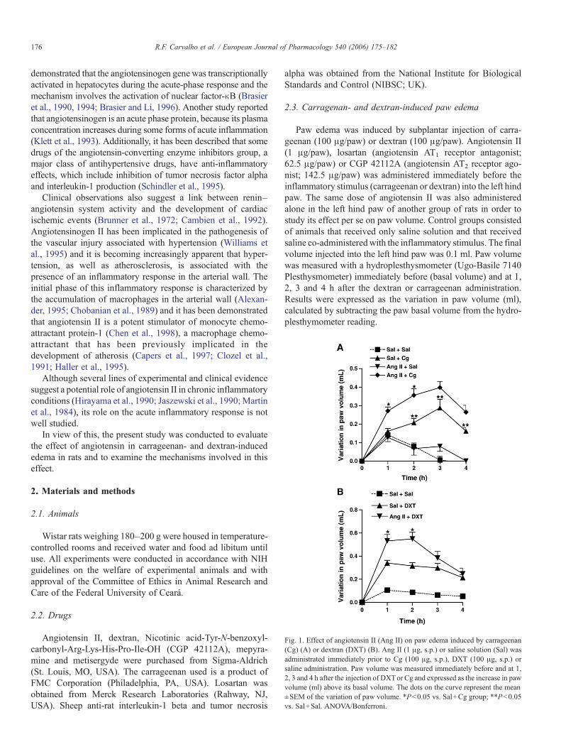
Fig. 1. Effect of angiotensin II (Ang II) on paw edema induced by carrageenan(Cg) (A) or dextran (DXT) (B). Ang II (1 μg, s.p.) or saline solution (Sal) wasadministrated immediately prior to Cg (100 μg, s.p.), DXT (100 μg, s.p.) orsaline administration. Paw volume was measured immediately before and at 1,2, 3 and 4 h after the injection of DXTor Cg and expressed as the increase in pawvolume (ml) above its basal volume. The dots on the curve represent the mean±SEM of the variation of paw volume. *Pb0.05 vs. Sal+Cg group; **Pb0.05vs. Sal+Sal. ANOVA/Bonferroni.
176 R.F. Carvalho et al. / European Journal of Pharmacology 540 (2006) 175–182
demonstrated that the angiotensinogen gene was transcriptionallyactivated in hepatocytes during the acute-phase response and themechanism involves the activation of nuclear factor-κB (Brasieret al., 1990, 1994; Brasier and Li, 1996). Another study reportedthat angiotensinogen is an acute phase protein, because its plasmaconcentration increases during some forms of acute inflammation(Klett et al., 1993). Additionally, it has been described that somedrugs of the angiotensin-converting enzyme inhibitors group, amajor class of antihypertensive drugs, have anti-inflammatoryeffects, which include inhibition of tumor necrosis factor alphaand interleukin-1 production (Schindler et al., 1995).
Clinical observations also suggest a link between renin–angiotensin system activity and the development of cardiacischemic events (Brunner et al., 1972; Cambien et al., 1992).Angiotensinogen II has been implicated in the pathogenesis ofthe vascular injury associated with hypertension (Williams etal., 1995) and it is becoming increasingly apparent that hyper-tension, as well as atherosclerosis, is associated with thepresence of an inflammatory response in the arterial wall. Theinitial phase of this inflammatory response is characterized bythe accumulation of macrophages in the arterial wall (Alexan-der, 1995; Chobanian et al., 1989) and it has been demonstratedthat angiotensin II is a potent stimulator of monocyte chemo-attractant protein-1 (Chen et al., 1998), a macrophage chemo-attractant that has been previously implicated in thedevelopment of atherosis (Capers et al., 1997; Clozel et al.,1991; Haller et al., 1995).
Although several lines of experimental and clinical evidencesuggest a potential role of angiotensin II in chronic inflammatoryconditions (Hirayama et al., 1990; Jaszewski et al., 1990; Martínet al., 1984), its role on the acute inflammatory response is notwell studied.
In view of this, the present study was conducted to evaluatethe effect of angiotensin in carrageenan- and dextran-inducededema in rats and to examine the mechanisms involved in thiseffect.
2. Materials and methods
2.1. Animals
Wistar rats weighing 180–200 g were housed in temperature-controlled rooms and received water and food ad libitum untiluse. All experiments were conducted in accordance with NIHguidelines on the welfare of experimental animals and withapproval of the Committee of Ethics in Animal Research andCare of the Federal University of Ceará.
2.2. Drugs
Angiotensin II, dextran, Nicotinic acid-Tyr-N-benzoxyl-carbonyl-Arg-Lys-His-Pro-Ile-OH (CGP 42112A), mepyra-mine and metisergyde were purchased from Sigma-Aldrich(St. Louis, MO, USA). The carrageenan used is a product ofFMC Corporation (Philadelphia, PA, USA). Losartan wasobtained from Merck Research Laboratories (Rahway, NJ,USA). Sheep anti-rat interleukin-1 beta and tumor necrosis
alpha was obtained from the National Institute for BiologicalStandards and Control (NIBSC; UK).
2.3. Carragenan- and dextran-induced paw edema
Paw edema was induced by subplantar injection of carra-geenan (100 μg/paw) or dextran (100 μg/paw). Angiotensin II(1 μg/paw), losartan (angiotensin AT1 receptor antagonist;62.5 μg/paw) or CGP 42112A (angiotensin AT2 receptor ago-nist; 142.5 μg/paw) was administered immediately before theinflammatory stimulus (carrageenan or dextran) into the left hindpaw. The same dose of angiotensin II was also administeredalone in the left hind paw of another group of rats in order tostudy its effect per se on paw volume. Control groups consistedof animals that received only saline solution and that receivedsaline co-administeredwith the inflammatory stimulus. The finalvolume injected into the left hind paw was 0.1 ml. Paw volumewas measured with a hydroplesthysmometer (Ugo-Basile 7140Plesthysmometer) immediately before (basal volume) and at 1,2, 3 and 4 h after the dextran or carrageenan administration.Results were expressed as the variation in paw volume (ml),calculated by subtracting the paw basal volume from the hydro-plesthymometer reading.

177R.F. Carvalho et al. / European Journal of Pharmacology 540 (2006) 175–182
2.4. Effect of mepyramine and metisergyde in the dextran-induced paw edema
In order to investigate whether histamine and serotonin wereinvolved in the mediation of dextran-induced paw edema, ratswere injected intraperitoneally with the anti-histamine mepir-amine (10 mg/kg) or the anti-serotonin metisergyde (1 mg/kg)1 h prior to the dextran injection (100 μg/paw). Angiotensin IIor saline was locally injected immediately prior to dextraninjection.
2.5. Effect of angiotesin II on mast cell degranulation in pawtissue
Angiotensin II (1 μg/ml) or saline solution was administeredin the left hind paws of the animals immediately prior to thelocal injection of carrageenan (100 μg) or dextran (100 μg). Thesame concentration of angiotensin II was also co-administeredwith saline to study its effect per se on mast cell degranulation.A control group received two injections of saline into thesubplantar region of the left hind paw. One hour later, theanimals were sacrificed and the paw tissues were fixed with10% neutral buffered formalin. Fixed tissues samples wererinsed in PBS and embedded in paraffin according to standardtechniques. Sections (5 μm) were collected on microscope
Fig. 2. Effect of the angiotensin AT1 receptor antagonist losartan (LOS) and the ancarrageenan (Cg) (A and C) or dextran (DXT) (B and D). LOS (62.5 μg/paw), CGP (1(100 μg, s.p.) or DXT (100 μg, s.p.) administration. Paw volume was measured immedas the increase in paw volume (ml) above its basal volume. The dots on the curve repand C) or Sal+DXT (B and D). Student t test.
slides. The hydrated tissue sections were immersed in a solutionof 0.1% toluidine blue (in 0.9% sodium chloride) for 60 sfollowed by extensive rinsing in deionized water as describedpreviously (Zhong et al., 2001). The percentage of degranulatedmast cells was determined by counting one hundred stainedcells per tissue section.
2.6. Myeloperoxidase activity
Angiotensin II (1 μg/paw) or saline was administeredimmediately before the inflammatory stimulus (Cg;100 μg/paw)or saline into the left hind paw. Three hours after the admin-istration of carrageenan, animals were sacrificed and the wholeskin from the plantar region of the left paws was harvested. Afterhomogenization and centrifugation (4500 rpm, 20 min), myelo-peroxidase activity, an enzyme found in azurophil neutrophilgranules, was determined by a colorimetric method describedpreviously (Souza et al., 2003) and expressed as units ofmyeloperoxidase activity per 5 mg of tissue.
2.7. Stimulation of neutrophil migration into peritoneal cavities
Carrageenan (100 μg/0.5 ml) or saline (0.5 ml) was injectedintraperitoneally (i.p.) in rats pretreated 1 hour earlier withangiotensin II (0.5 μg/0.5 ml, i.p.) or saline (0.5 ml; i.p.). Three
giotensin AT2 receptor agonist CGP 42112A (CGP) on paw edema induced by42.5 μg/paw) or saline solution (Sal) was administrated immediately prior to Cgiately before and at 1, 2, 3 and 4 h after the injection of DXTor Cg and expressedresent the mean±SEM of the variation of paw volume. *Pb0.05 vs. Sal+Cg (A

Fig. 3. Effect of the anti-histamine mepyramine, (MEP; A) and the anti-serotoninmetisergyde (MET) on the Ang II modulation on the dextran (DXT; B) inducedpaw edema. Rats were injected intraperitoneally with MEP (10 mg/kg), MET(1 mg/kg) or saline solution (Sal) 1 h prior to the DXT injection (100 μg). Ang II(1 μg, s.p.) or saline was locally injected immediately prior to DXT injection. Pawvolume was measured immediately before and at 1, 2, 3 and 4 h after the DXTinjection and expressed as the increase in paw volume (ml) above its basal volume.The dots on the curve represent the mean±SEM of the variation of paw volume.*Pb0.05 vs. Sal/Sal+DXT; **Pb0.05 vs. Sal/Ang II+DXT. ANOVA/Bonferroni.
Fig. 4. Effect of angiotensin II (Ang II) on mast cell degranulation in pawsinjected with carrageenan (Cg) or dextran (DXT). Ang II (1 μg/ml) or salinesolution (Sal) was administered immediately prior to the local injection of Cg(100 μg), DXT (100 μg) or saline. One hour later, the animals were sacrificed andthe skin and subcutaneous paw tissue samples were stained with toluidine blue.The percentages of degranulated mast cells was determined by counting onehundred stained cells in different fields (×400). The dots on the curve representthe mean±SEMof the percentage of mast cells degranulation. *Pb0.05 vs. Sal+Sal; **Pb0.05 vs. Sal+Cg; #Pb0.05 vs. Sal+DXT. ANOVA/Bonferroni.
178 R.F. Carvalho et al. / European Journal of Pharmacology 540 (2006) 175–182
hours after the carrageenan injection, the animals weresacrificed and peritoneal fluid was collected. Total anddifferential cell counts were performed as described elsewhere(Souza and Ferreira, 1985).
2.8. Quantification of tumor necrosis alpha and interleukin1 beta
Angiotensin II (1 μg, subplantar injection) or saline wasinjected immediately prior to carrageenan (100 μg, s.p.)administration. One and two hours later, the paw tissue washarvested for tumor necrosis factor alpha and interleukin-1 betadetermination , respectively, by ELISA, according to a previousdescription of time course production of these cytokines aftercarrageenan injection (Cunha et al., 2003). Briefly, paw skinwas homogenized in 500 μl of the appropriate buffer containingprotease inhibitors. Microtiter plates (Nunc-Maxisorb) wereincubated overnight at 4 °C with a sheep anti-rat interleukin-1 beta or tumor necrosis alpha polyclonal antibody. Afterblocking the plates, samples and standards at various dilutionswere added in triplicate and maintained at room temperature for
2 h. The plates were washed three times with buffer and asecond biotiny-lated polyclonal antibody against interleukin-1(1:500 dilution) or tumor necrosis alpha (1:1000 dilution) wasadded followed by incubation at room temperature for 1 h.Finally, 100 μl of avidin-HRP (1:5000 dilution) was added toeach well and, after 30 min, the plates were washed and thecolor reagent o-phenylenediamine (40 μl well−1) was added.After 15 min, the reaction was terminated with H2SO4 (1M,50 μl well−1) and the optical density measured at 490 nm. Theresults were adjusted to 500 μl, the volume used to extract thecytokine from the paw skin, and were expressed as nanogramsof respective cytokine per paw.
2.9. Statistical analysis
Results were presented as means and standard errors of themean for groups of six animals each. The differences betweenthe experimental groups were compared by Analysis of variance(ANOVA) followed by Bonferroni's t-test. The level ofsignificance was set at Pb0.05.
3. Results
3.1. Paw edema
Subplantar injection of carrageenan or dextran inducedsignificant paw edema with peak at 3 h for carrageenan and 1 hfor dextran. Local injection of angiotensin II resulted in a significantincrease of paw edema in the first 2 h after the injection ofcarrageenan (Pb0.05; Fig. 1A) and dextran (Pb0.05; Fig. 1B).However, the local administration of angiotensin II alone did notinduce any significant change in paw volume (Fig. 1A) comparedto the saline group. Local administration of losartan enhanced thecarrageenan- and dextran-induced increase in paw volume (Fig. 2Aand B). CGP 42112A also potentiated carrageenan- and dextran-induced edema within the first 2 h of treatment (Fig. 2C and D).
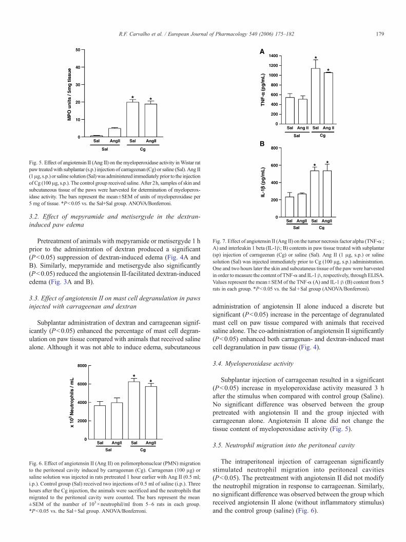
Fig. 5. Effect of angiotensin II (Ang II) on the myeloperoxidase activity inWistar ratpaw treatedwith subplantar (s.p.) injection of carrageenan (Cg) or saline (Sal). Ang II(1μg, s.p.) or saline solution (Sal)was administered immediately prior to the injectionof Cg (100 μg, s.p.). The control group received saline. After 2h, samples of skin andsubcutaneous tissue of the paws were harvested for determination of myeloperox-idase activity. The bars represent the mean±SEM of units of myeloperoxidase per5 mg of tissue. *Pb0.05 vs. the Sal+Sal group. ANOVA/Bonferroni.
Fig. 7. Effect of angiotensin II (Ang II) on the tumor necrosis factor alpha (TNF-α ;A) and interleukin 1 beta (IL-1β; B) contents in paw tissue treated with subplantar(sp) injection of carrageenan (Cg) or saline (Sal). Ang II (1 μg, s.p.) or salinesolution (Sal) was injected immediately prior to Cg (100 μg, s.p.) administration.One and two hours later the skin and subcutaneus tissue of the paw were harvestedin order tomeasure the content of TNF-α and IL-1β, respectively, through ELISA.Values represent the mean±SEM of the TNF-α (A) and IL-1 β (B) content from 5rats in each group. *Pb0.05 vs. the Sal+Sal group (ANOVA/Bonferroni).
179R.F. Carvalho et al. / European Journal of Pharmacology 540 (2006) 175–182
3.2. Effect of mepyramide and metisergyde in the dextran-induced paw edema
Pretreatment of animals with mepyramide or metisergyde 1 hprior to the administration of dextran produced a significant(Pb0.05) suppression of dextran-induced edema (Fig. 4A andB). Similarly, mepyramide and metisergyde also significantly(Pb0.05) reduced the angiotensin II-facilitated dextran-inducededema (Fig. 3A and B).
3.3. Effect of angiotensin II on mast cell degranulation in pawsinjected with carrageenan and dextran
Subplantar administration of dextran and carrageenan signif-icantly (Pb0.05) enhanced the percentage of mast cell degran-ulation on paw tissue compared with animals that received salinealone. Although it was not able to induce edema, subcutaneous
Fig. 6. Effect of angiotensin II (Ang II) on polimorphonuclear (PMN) migrationto the peritoneal cavity induced by carrageenan (Cg). Carragenan (100 μg) orsaline solution was injected in rats pretreated 1 hour earlier with Ang II (0.5 ml;i.p.). Control group (Sal) received two injections of 0.5 ml of saline (i.p.). Threehours after the Cg injection, the animals were sacrificed and the neutrophils thatmigrated to the peritoneal cavity were counted. The bars represent the mean±SEM of the number of 103×neutrophil/ml from 5–6 rats in each group.*Pb0.05 vs. the Sal+Sal group. ANOVA/Bonferroni.
administration of angiotensin II alone induced a discrete butsignificant (Pb0.05) increase in the percentage of degranulatedmast cell on paw tissue compared with animals that receivedsaline alone. The co-administration of angiotensin II significantly(Pb0.05) enhanced both carragenan- and dextran-induced mastcell degranulation in paw tissue (Fig. 4).
3.4. Myeloperoxidase activity
Subplantar injection of carrageenan resulted in a significant(Pb0.05) increase in myeloperoxidase activity measured 3 hafter the stimulus when compared with control group (Saline).No significant difference was observed between the grouppretreated with angiotensin II and the group injected withcarrageenan alone. Angiotensin II alone did not change thetissue content of myeloperoxidase activity (Fig. 5).
3.5. Neutrophil migration into the peritoneal cavity
The intraperitoneal injection of carrageenan significantlystimulated neutrophil migration into peritoneal cavities(Pb0.05). The pretreatment with angiotensin II did not modifythe neutrophil migration in response to carrageenan. Similarly,no significant difference was observed between the group whichreceived angiotensin II alone (without inflammatory stimulus)and the control group (saline) (Fig. 6).
180 R.F. Carvalho et al. / European Journal of Pharmacology 540 (2006) 175–182
3.6. Quantification of tumor necrosis alpha and interleukin-1
Paw tissue injected with carrageenan and harvested 1 h laterexhibited a 100% increase in tumor necrosis alpha levels com-pared with animals that received saline injection alone (Fig. 7A).Similar results were obtained with interleukin 1 beta, harvested2 h later (Fig. 7B), which also revealed a 100% increase. An-giotensin II alone did not have a significant effect on the tumornecrosis alpha or interleukin-1 beta levels, with or without car-rageenan (Fig. 7A and B).
4. Discussion
In this study we demonstrated that angiotensin II potentiatesthe carrageenan and dextran induced paw edema, suggesting anactivity of angiotensin II in inflammatory events. It has beenpreviously reported that the renin–angiotensin system plays arole in the modulation of inflammatory response. Angiotensino-gen is considered an acute phase protein, because its plasmaconcentration increases during acute inflammation (Klett et al.,1993). Moreover, it has been previously demonstrated that gra-nuloma macrophages release angiotensin-converting enzyme,which produces angiotensin II, which in turn modulatesmonocyte/macrophage activity (Simon et al., 1991). Angioten-sin II receptors (AT1 and AT2) and angiotensin-convertingenzyme develop sequentially during angiogenesis in the ratsubcutaneous sponge granuloma (Walsh et al., 1997). Thesefindings suggest the possibility of expression of renin–angio-tensin system within the tissue at the site of inflammation. Localproduction of angiotensin II occurs and modulates the ongoinginflammatory processes. However, in the current report theadministration of angiotensin II alone did not induce edema,suggesting that triggering mechanisms are essential for angio-tensin II to initiate inflammatory action.
The present study also showed that the locally administeredangiotensin AT1 receptor antagonist, losartan, potentiated car-rageenan-induced paw edema. This data is in accordance with aprevious report which shows that local administration oflosartan, as well as angiotensin II, enhanced the carraggenan-induced increase in paw volume in a dose-dependent manner(Raghavendra and Kulkarni, 2000). It has been reported also thatangiotensin AT1 receptor antagonists cause elevated levels ofangiotensin II, which selectively binds to unblocked angiotensinAT2 receptors (Bernstein and Alexander, 1992). Thus, the po-tentiation of carrageenan-induced edema by losartan could be aconsequence of the activation of angiotensin AT2 receptors byendogenous angiotensin II produced during inflammation, sincethe angiotensin AT1 receptors are blocked. Consistent with thishypothesis, we found that the angiotensin AT2 receptor agonistCGP 42112A also potentiated the carrageenan-induced inflam-mation. According to our data, it has been demonstrated that sti-mulation of angiotensin AT2 receptors are thought to activateprostaglandins, kinins and the nitric oxide system (Schiefer etal., 1994) which are known mediators of carrageenan-inducedinflammation.
The intraplantar injection of carrageenan elicits an inflam-matory response characterized by a time-dependent increase in
paw edema. Edema formation in rat hind paw following in-jection of carrageenan has been described as a biphasic eventconsisting of a relatively rapid early phase followed by a moresustained late phase (Vinegar et al., 1969). It is suggested thatthe early inflammation response of carrageenan-induced edemain rats results from the release of histamine and serotonin frommast cells (Kulkarni et al., 1986). On the other hand, the latephase of carrageenan-induced edema is known to be dependenton cytokine production by resident cells and neutrophil mi-gration (DiRosa et al., 1971; Vinegar et al., 1969, 1982;Wedmore and Williams, 1981).
In the present study we demonstrated that angiotensin II po-tentiates the first 2 h of carrageenan-induced paw edema, knownto be mediated by the release of mast cell derived mediators(Capasso et al., 1975; Kulkarni et al., 1986). Accordingly, wedemonstrated that angiotensin II, as well as losartan and CGP42112A, has potentiated the dextran-induced paw edema, whichis also dependent of mast cell degranulation and release ofseveral inflammatory mediators such as histamine and serotonin(Cahill et al., 1996; Chu et al., 1997; Kulkarni et al., 1986;Ribeiro et al., 1997). Consistent with these data, we showed thatthe pretreatment with a specific inhibitor of histamine H1 recep-tor, mepyramine, reduced both the dextran and angiotensin II-facilitated dextran induced paw edema. Similar results wereobserved with the pretreatment with the serotoninergic blocker,metisergyde. These results suggest that the release of histamineand serotonin appears to be involved in the angiotensin II poten-tiation of carrageenan- and dextran-induced paw edema.
These findings point toward the hypothesis that angiotensin IIinterferes with mast cell degranulation, which is mainly respon-sible for the dextran edema and for the early phase of carrageenanpaw edema (DiRosa, 1971). In fact, we also demon-strated thatthe administration of angiotensin II induced significant enhance-ment of mast cell degranulation in the dextran and carrageenan-induced paw tissue. Similar result was obtained in the mesenterictissue, where angiotensin II potentiated the mast cell degranula-tion induced by 48/80 compound (data not shown).
To further investigate the participation of angiotensin II inthe late phase of carrageenan-induced inflammation, which isassociated with cytokine production and neutrophil migration(DiRosa et al., 1971; Vinegar et al., 1969, 1982; Wedmore andWilliams, 1981), we examined the effects of angiotensin II inthe cytokine levels and myeloperoxidase activity in carrageen-an-injected paw tissue, and in the neutrophil migration to peri-toneal cavity in response to carrageenan. The local pretreatmentwith angiotensin II did not change the tissue content of the pro-inflammatory cytokines tumor necrosis alpha and interleukin1 beta, suggesting that these cytokines are not involved in an-giotensin II-potentiation of carrageenan-induced inflammation.Furthermore, pretreatment with angiotensin II did not modifythe neutrophil migration to the peritoneal cavity in response tocarrageenan and did not enhanced the increase in myeloperox-idase activity induced by carrageenan in paw tissue. Takentogether, these data suggest that angiotensin II does not interferewith the late phase carragenan-induced paw edema.
In conclusion, the present study suggests an important role ofangiotensin II in inflammatory response, increasing vascular
181R.F. Carvalho et al. / European Journal of Pharmacology 540 (2006) 175–182
permeability through induction of mast cells degranulation.Furthermore, our results suggest that angiotensin II enhancesedema response through the angiotensin AT2 receptors, sincethe angiotensin AT1 antagonist and angiotensin AT2 agonist alsopotentiated the paw edema. Although, further investigation isrequired, these data provide basic information on the role ofangiotensin II on acute inflammation and support future studiesof ligands of angiotensin II receptors on the modulation ofinflammatory conditions.
Acknowledgements
We gratefully acknowledge the technical assistance of MariaSilvandira França Pinheiro. This work was supported by grantsfrom Conselho Nacional de Pesquisa (CNPq) and FundaçãoCearense de Pesquisa e Cultura (FUNCAP).
References
Alexander, R.W., 1995. Hypertension and the pathogenesis of atherosclerosis:oxidative stress and the mediation of arterial inflammatory response. Hyper-tension 25, 155–161.
Bernstein, K.E., Alexander, R.W., 1992. Counterpoint: molecular analysis of theangiotensin II receptor. Endocr. Rev. 13, 381–386.
Brasier, A.R., Li, J., 1996. Mechanisms for inducible control of angiotensinogengene transcription. Hypertension 27, 465–475.
Brasier, A.R., Ron, D., Tate, J.E., Habener, J.F., 1990. A family of constitutiveC/EBP-like DNA binding proteins attenuate the IL-1 alpha induced, NFkappa B mediated trans-activation of the angiotensinogen gene acute-phaseresponse element. EMBO J. 9, 3933–3944.
Brasier, A.R., Li, J., Copland, A., 1994. Transcription factors modulating an-giotensinogen gene expression in hepatocytes. Kidney Inter., Suppl. 46,1564–1566.
Brunner, H.R., Laragh, J.H., Baer, L., Newton,M.A.,Goodwin, F.T., Krakoff, L.R.,Bard, R.H., Buhler, F.R., 1972. Essential hypertension: renin and aldosterone,heart attack and stroke. N. Engl. J. Med. 286, 441–449.
Cahill, C.M., Waterman, W.R., Xie, Y., Auron, P.E., Calderwood, S.K., 1996.Transcriptional repression of the prointerleukin 1β gene by heat shockfactor-1. J. Biol. Chem. 271, 17724–17732.
Cambien, F., Poirier, O., Lecerf, L., Evans, A., Cambou, J.P., Arveiler, D., Luc,G., Bard, J.M., Bara, L., Ricard, S., et al., 1992. Deletion polymorphism inthe gene for angiotensin-converting enzyme is a potent risk factor formyocardial infarction. Nature 359, 641–644.
Cao, Z., Kelly, D.G., Cox, A., Casley, D., Forbes, J.M., Martinello, P., Dean, R.,Gilbert, R.E., Cooper, M.E., 2000. Angiotensin type 2 receptor is expressedin the adult rat kidney and promotes cellular proliferation and apoptosis.Kidney Int. 58, 2437–2451.
Capasso, F., Dunn, C.J., Yamamoto, S., Willoughby, D.A., Giround, J.P., 1975.Further studies on carrageenan-induced pleurisy in rats. J. Pathol. 116,117–124.
Capers, I.V.Q., Alexander, R.W., Lou, P., DeLeon, H., Wilcox, J.N., Ishizaka, N.,Howard, A.B., Taylor, W.R., 1997. Monocyte chemoattractant protein-1expression in aortic tissues of hypertensive rats. Hypertension 30, 1397–1402.
Chen, X.L., Tummala, P.E., Olbrych, M.T., Alexander, R.W., Medford, R.M.,1998. Angiotensin II induces monocyte chemoattractant protein-1 geneexpression in rat vascular smooth muscle cells. Circ. Res. 83, 952–959.
Chiu, A.T., Herblin,W.F.,McCall, D.E., Ardecky, R.J., Carini, D.J., Duncia, J.V.,Pease, L.J., Wong, P.C., Wixler, R.R., Jonhson, A.L., et al., 1989.Identification of angiotensin II receptor subtypes. Biochem. Biophys. Res.Commun. 165, 196–203.
Chobanian, A.V., Lichtenstein, A.H., Nilakhe, V., Haudenschild, C.C., Drago,R., Nickerson, C., 1989. Influence of hypertension on aortic atherosclerosisin the watanable rabbit. Hypertension 14, 203–209.
Chu, E.K., Ribeiro, S.P., Slutsky, A.S., 1997. Heat stress increases survival ratesin lipopolysaccharide-stimulated rats. Crit. Care Med. 25, 1727–1732.
Chung,O.,Kuhl,H., Stoll,M.,Unger, T., 1998. Physiological and pharmacologicalimplications of AT1 versus AT2 receptors. Kidney Inter., Suppl. 67, 95–99(Review).
Clozel, M., Kuhn, H., Hefti, F., Baumgartner, H.R., 1991. Endothelial dysfunctionand subendothelial monocyte macrophages in hypertension. Effect of an-giotensin converting enzyme inhibition. Hypertension 18, 132–141.
Csikos, T., Chung, O., Unger, T., 1998. Receptors and their classification: focuson angiotensin II and the AT2 receptor. J. Hum. Hypertens. 12, 311–318.
Cunha, J.M., Sachs, D., Canetti, C.A., Poole, S., Ferreira, S.H., Cunha, F.Q.,2003. The critical role of leukotriene B4 in antigen-induced mechanicalhyperalgesia in immunised rats. Br. J. Pharmacol. 139, 1135–1145.
De Gasparo, M., Catt, K.J., Inagami, T., Wright, J.W., Unger, T., 2000. Inter-national union of pharmacology: XXIII. The angiotensin II receptors. Phar-macol. Rev. 52, 415–472.
DiRosa, M., Giroud, J.P., Willoughby, D.A., 1971. Studies of the mediators ofthe acute inflammatory response induced in rats in different sites by car-rageenan and turpentine. J. Pathol. 104, 15–29.
Dzau, V.J., Sasamura, H., Hein, L., 1993. Heterogeneity of angiotensin syntheticpathways and receptor subtypes: physiological and pharmacological im-plications. J. Hypertens., Suppl. 11, 13–18.
Haller, H., Behrend, M., Park, J.K., Luft, F.C., Distler, A., 1995. Monocyte infil-tration and c-fms expression in hearts of spontaneously hypertensive rats.Hypertension 25, 132–138.
Hirayama, K., Fukoyama, K., Epstein, W.L., 1990. Angiotensin II productingproteases from granulomatous tissue reaction in mice infected with Schis-tosoma mansoni. Comp. Biochem. Physiol. 96, 553–557.
Horiuchi,M., Akishita,M., Dzau, V.J., 1999.Recent progress in angiotensin II type2 receptor research in the cardiovascular system. Hypertension 33, 613–622.
Ichiki, T., Kambayashi, Y., Inagami, T., 1996. Differential inducibility ofangiotensin II AT2 receptor between SHR and WKY vascular smooth mus-cle cells. Kidney (Suppl. 55), 14–17.
Jaszewski, R., Tolia, V., Ehrinpreis, M.N., Bodzin, J.H., Peleman, R.R.,Korlipara, R., Weinstock, J.V., 1990. Increased colonic mucosal angiotensinI and II concentrations in Crohn's colitis. Gastroenterology 98, 1543–1548.
Klett, C., Hellmann, W., Ganten, D., Hakenthal, E., 1993. Tissue distribution ofangiotensinogen mRNA during experimental inflammation. Inflammation17, 183–197.
Kulkarni, S.K., Mehta, A.K., Kunchandy, J., 1986. Anti-inflammatory actions ofclonidine, guanfacine and B-HT 920 against various inflammagen-inducedacute paw oedema in rats. Arch. Int. Pharmacodyn. Ther. 279, 324–334.
Martín,M.F., Surrall, K.E.,McKenna, F., Dixon, J.S., Bird, H.A.,Wright, V., 1984.Captopril: a new treatment for rheumatoid arthritis? Lancet 1, 1325–1328.
Raghavendra, V., Kulkarni, S.K., 2000. AT1 receptor antagonism enhancesangiotensin-II-facilitated carrageenan-induced paw edema. Methods Find.Exp. Clin. Pharmacol. 22, 633–636.
Reid, I.A., 1992. Interactions between ANG II, sympathetic nervous system andbaroreceptor reflexes in regulation of blood pressure. Am. J. Physiol. 262,763–778.
Ribeiro, R.A., Souza Filho, M.V.P., Souza, M.H.L.P., Oliveira, S.H.P., Costa,C.H.S., Cunha, F.Q., Ferreira, S.H.P., 1997. Role of resident mast cells andmacrophages in the neutrophil migration induced by LTB4, fMLP and C5ades arg. Int. Arch. Allergy Immunol. 112, 27–35.
Schiefer, B., Wirger, A., Meybrunn,M., Seitz, S., Holtz, J., Riede, U.N., Drexler,H., 1994. Comparative effects of chronic angiotensin converting enzymeinhibition and angiotensin II type 1 receptor bloked on cardiac remodelingafter myocardial infarction in the rat. Circulation 89, 2273–2282.
Schindler, R., Dinarello, C.A., Koch, K.M., 1995. Angiotensin-converting-enzyme inhibitors suppress synthesis of tumor necrosis factor andinterleukin 1 by humam peripheral bllod mononuclear cells. Cytokine 7,526–533.
Simon, M.R., Kamlay, M.T., Desai, S.G., Majumdar, A.P., 1991. Angiotensin IIaugmentation of tyrosine kinase activity in human adherent mononuclearcells. Biochem. Med. Metabol. Biol. 45, 48–55.
Souza, G.E.P., Ferreira, S.H., 1985. Blockade by antimacrophage serum of themigration of PMN neutrophils into the inflamed peritoneal cavity. AgentsActions 17, 97–103.
Vinegar, R., Schreiber, W., Hugo, R., 1969. Biphasic development of carrageenanoedema in rat. J. Pharmacol. Exp. Ther. 166, 96–103.

182 R.F. Carvalho et al. / European Journal of Pharmacology 540 (2006) 175–182
Vinegar, R., Truax, J.F., Selph, J.L., Voelker, F.A., 1982. Pathway of onset, de-velopment, and decay of carrageenan pleurisy in the rat. Fed. Proc. 41,2588–2595.
Viswanathan, M., Saavedra, J.M., 1992. Expression of angiotensin II AT2
receptors in the rat skin during experimental wound healing. Peptides 13,783–786.
Viswanathan, M., Johren, O., de Oliveira, M., Saavedra, J.M., 1996. Increasednon-angiotensin II [125I] CGP 42112 binding in rat carotid artery afterballon injury. Peptides 17, 695–699.
Walsh, D.A., Hu, D.E., Wharton, J., Catravas, J.D., Blake, D.R., Fan, T.P., 1997.Sequential development of angiotensin receptors and angiotensin-I convert-ing enzyme during angiogenesis in the rat subcutaneous sponge granuloma.Br. J. Pharmacol. 120, 1302–1311.
Wedmore, C.V., Williams, T.J., 1981. Control of vascular permeability bypolimorphonuclear leukocytes in inflammation. Nature 289, 646–650.
Whitebread, S., Mele, M., Kamber, B., de Gasparo, M., 1989. Preliminarybiochemical characterization of two angiotensin II receptor subtypes.Biochem. Biophys. Res. Commun. 163, 284–291.
Williams, B., Baker, A.Q., Gallacher, B., Lodwick, D., 1995. Angiotensin IIincreases vascular permeability factor gene expression by human vascularsmooth muscle cells. Hypertension 25, 913–917.
Zhong, H., Chunn, J.L., Volmer, J.B., Fozard, J.R., Blackburn, M.R., 2001.Adenosine-mediated mast cell degranulation in adenosine deaminase-deficient mice. J. Pharmacol. Exp. Ther. 298, 433–440.