anatomy of appendix
TRANSCRIPT

VERMIFORM APPENDIX
DR.SUPRITIDEMONSTRATOR(DEPT OF ANATOMY)TMMC&RC

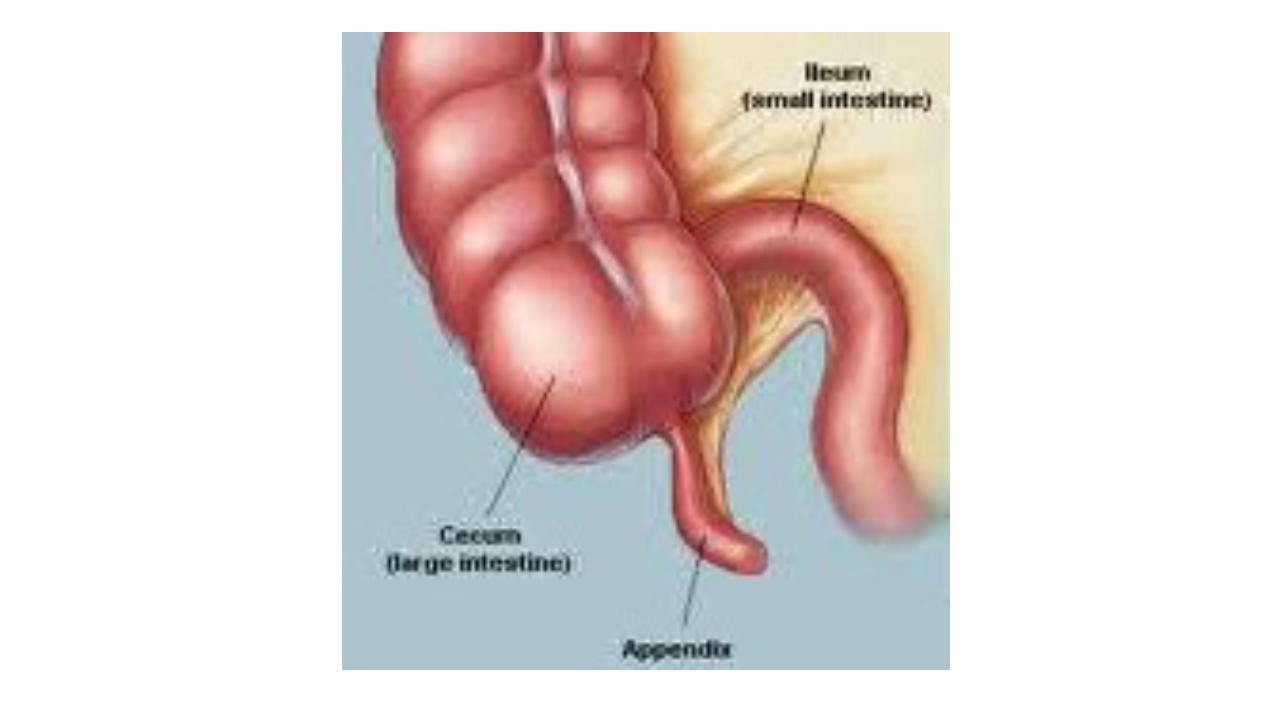
• It is a blind-ended muscular tube attached to the
posteromedial wall of caecum, about 2cm below ileocaecal jxn.
• Suspended by peritoneal fold, MESOAPPENDIX.
• Devoid of taenia coli, sacculations, appendices epiploicae.


MESOAPPENDIX
• The mesoappendix (mesentery of appendix) is short, triangular and variable.
• It extends the whole length of appendix.The breadth of mesoappendix usually falls short of length of appendix.
• The body of appendix is kinked on itself where the free border of mesoappendix ends,hence it is coiled like worm and is named the vermiform.
• Appendicular vessels pass through free margin of mesoappendix.

MEASUREMENTS
• The appendix is 9 cm (7 to 11cm) in length but can range from 2 to 20 cm. The diameter of the appendix is 1 to 7 mm.
• It is relatively longer in children and decreases after 40 years of age.
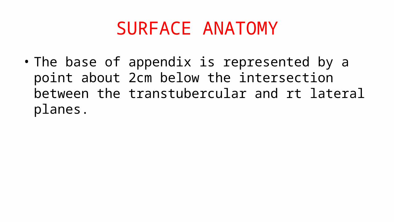
SURFACE ANATOMY
• The base of appendix is represented by a point about 2cm below the intersection between the transtubercular and rt lateral planes.

McBurney’s point
• Represented by a point at the junction of medial two third and lateral one third of a line which extends from the umblicus to rt ant. Sup.illiac spine.
• This point indicate maximum tenderness in a patient suffering from inflammation of the appendix.


PARTS OF APPENDIX
A) Base B) Body c) Tip

BASE
Base – it is attached to posteromedial wall of caecum about 2 m below the ileocaecal junction.
All taenia of caecum converge to the base and serve as a guide for the identification of the appendix.


BODY
Body is narrow,tubular and contains a canal which opens into the caecum.
The caecal opening is guarded by an incomplete mucous fold called as,”THE VALVE OF GERLACH”

TIP
It is least vascular and is directed in various direction.

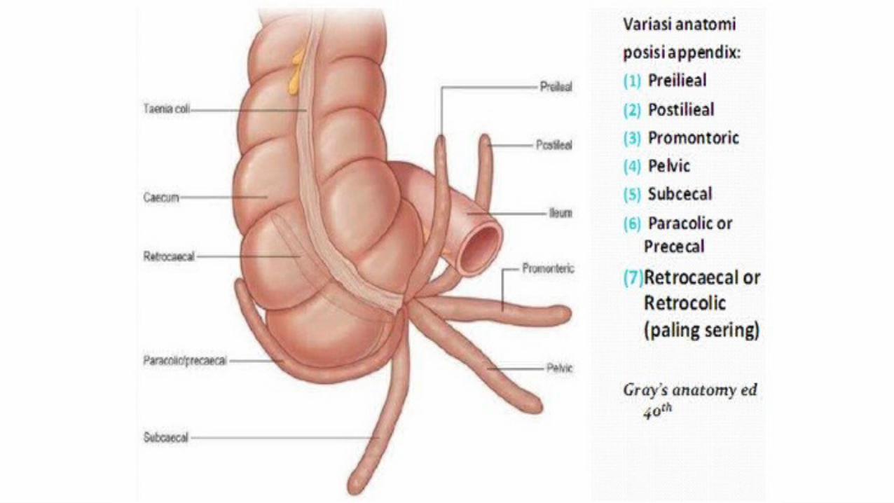
TYPES
A) Retro-caecal: 12’o clock (65%) B) Splenic: 2’o clock (1-2%) Preileal (Dangerous) & Postileal C) Promontoric: 3’o clock , towards sacrum D) Pelvic: 4’o clock (30%), downwards &
medially (rt uterine tube & ovary) E) MID-INGUINAL: 6’o clock Vertically
downwards. F)PARACOLIC:11o’clock,appendix pass upwards
or towards right.

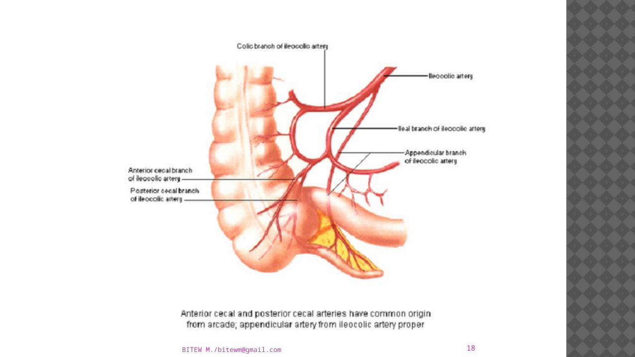
BLOOD SUPPLY
Appendicular A br. Of inferior div of ileocolic art
Recurrent br supplies base of appendix and anastomose with posterior caecal artery.
End artery
18BITEW M./[email protected]

NERVE SUPPLY
Parasympathetic: VAGUS
Sympathetic: T10 segment of spinal cord.

LYMPHATIC DRAINAGE
Lymphatics drain into superior mesenteric lymph nodes via ilea-colic nodes

APPLIED ANATOMY
Inflammation of appendix is known as appendicitis,seen in adoloscent age.In this condition it is usually necessary to remove the appendix.
The operation for removal of appendix is called as appendectomy.

The anatomial factors producing inflammation may be as follows:-
Appendix is a blind tube,a faecolith may obstruct the lumen and precipitate the attack of appendicitis.
Supplied by an end artery. Presence of hiatus muscularis. Presence of lymphatic follicles in submuosa.