anatomy and histology of the human and murine prostate
TRANSCRIPT

Anatomy and Histology of the Humanand Murine Prostate
Michael Ittmann
Department of Pathology and Immunology, Baylor College of Medicine, Houston, Texas 77030
Correspondence: [email protected]
The human and murine prostate glands have similar functional roles in the generation ofseminal fluid to assist in reproduction. There are significant differences in the anatomy andhistology of murine and human prostate and knowledge of the normal anatomy and histologyof the murine prostate is essential to interpreting changes in genetically engineered mousemodels. In this review, the normal anatomy and histology of both human andmouse prostatewill be described.
Analysis of molecular alterations in humanprostate cancer tissues has been critical in
understanding the molecular basis of prostatecancer. Initially, changes were analyzed for sin-gle genes, proteins, or genomic regions. Howev-er, over the last decade, high-throughput meth-odologies have allowed examination of geneticand epigenetic changes and associated changesin gene expression in prostate cancer on an un-precedented scale for both clinically localizedand metastatic disease. Although there aresome disease states that not well characterizedto date (i.e., metastatic treatment naïve cancer),overall, these efforts have allowed for the begin-ning of a clinically and biologically meaningfulclassification of prostate cancer.
Ultimately, analysis of molecular alterationsin human cancer tissues is descriptive, and func-tional studies are critical in defining the under-lying biology induced by the genetic and epige-netic changes seen in the cancer tissues. Mousemodels have played a central role in defining the
biological importance of the annotated lesionsin human prostate cancer by engineering thedescribed lesions into mouse models (Ittmannet al. 2013). Genetically engineered models havesignificant advantages in that they reflect tumorprogression over time from the initiation of pre-invasive lesions to invasive and, in some cases,metastatic lesions within the prostatic microen-vironment, including a fully intact immune sys-tem. Particularly with the increasing impor-tance of immunotherapy, the latter is a keyadvantage over xenograft models.
ANATOMY AND HISTOLOGY OF THEHUMAN PROSTATE
The human prostate gland is a pyramid-shapedorgan located beneath the bladder with the apex(corresponding to apex of the pyramid) contact-ing the penile urethra and the base contacting tothe bladder (Paner 2010). The prostate lies belowthe urinary bladder and is located in front of the
Editors: Michael M. Shen and Mark A. RubinAdditional Perspectives on Prostate Cancer available at www.perspectivesinmedicine.org
Copyright © 2018 Cold Spring Harbor Laboratory Press; all rights reserved; doi: 10.1101/cshperspect.a030346Cite this article as Cold Spring Harb Perspect Med 2018;8:a030346
1
ww
w.p
ersp
ecti
vesi
nm
edic
ine.
org
Press on December 1, 2021 - Published by Cold Spring Harbor Laboratoryhttp://perspectivesinmedicine.cshlp.org/Downloaded from

rectum. The prostate surrounds the prostaticurethra that is the conduit for urine flow fromthe bladder. The normal prostate weighs about15–20 g. The seminal vesicles are located bilat-erally at the base of the prostate. The humanprostate is a single gland with different histolog-ical zones (peripheral, transition, and centralzones).As shown inFigure 1, theperipheral zoneis wrapped around the outer portion of the pros-tate distally and is the site of origin of the ma-jority of prostate cancers. It constitutes about70% of tissue in the normal prostate (Paner2010). The transition zone is located near theprostatic urethra and is inconspicuous in mostyoung men, constituting about 5% of the pros-tate. In the majority of older men, the transitionzone is enlarged considerably by benign prostat-ic hyperplasia, an extremely common benignproliferation in transition zone tissue. Most rad-ical prostatectomy specimens show evidence ofvariable degrees of benign prostatic hyperplasia(see Fig. 2, for example). Cancers also arise inthe transition zone and there is considerable ev-idence that cancers arising in the transition zoneare clinically and biologically different than pe-ripheral zone cancers, although there is consid-erable overlap. The central zone is a cone-shapedregion, with the wider portion at the base of the
prostate and the apex at the verumontanum sur-rounding the ejaculatory ducts. It is not the siteof origin of any disease process but of course canbe secondarily involved by cancer.
The human prostate glandular epithelium iscomposed of acini and ducts lined by three typesof cells: luminal, basal, and neuroendocrine (seeFig. 1C). The acini have an undulating to papil-lary appearance in most cases (Fig. 2A–D). Thispapillary configuration is noticeably more pro-nounced in the central zone (Paner 2010). Theluminal cells are columnar, with pale eosino-philic cytoplasm and round nuclei near thebase of the cell (Fig. 2D). Luminal cells are spe-cialized cells that secrete a variety of productsinto the lumen, which contribute to the forma-tion of the seminal fluid. These products includeprostate-specific antigen (PSA), and luminalcells are strongly positive for PSA immunohis-tochemistry. Basal cells are adjacent to the base-ment membrane and have ovoid nuclei and in-conspicuous cytoplasm. The number of basalcells can be variable between glands in an indi-vidual prostate. They can generally be identifiedby careful examination of routine H&E sectionsbut are easier to identify using immunohisto-chemistry for p63 (nuclear) and high-molecu-lar cytokeratins (cytoplasmic). Neuroendocrine
Central zone
Peripheralzone
Transitionzone
Prostaticsphincter
Adult human(sagittal section)
Ductus deferensSeminalvesicle
Ventral prostate
Lateral prostate
Urethra
Adult mouse(lateral view)
Dorsal prostate
Anteriorprostate
Bladder
A B
Figure 1. Schematic illustration of human and mouse prostate and adjacent structures. (A) Human prostateshowing the location of the three zones of the prostate. (B) Mouse prostate showing different lobes of the prostateand their relation to adjacent structures.
M. Ittmann
2 Cite this article as Cold Spring Harb Perspect Med 2018;8:a030346
ww
w.p
ersp
ecti
vesi
nm
edic
ine.
org
Press on December 1, 2021 - Published by Cold Spring Harbor Laboratoryhttp://perspectivesinmedicine.cshlp.org/Downloaded from

A B
C D
E F
G H
Figure 2. Human prostate and seminal vesicles. (A) Low, (B) medium, and (C,D) high-power views of theperipheral zone of the human prostate. Note the loosely lobulated arrangement of acini (A,B). Basal cells arecharacterized by ovoid nuclei and lie near the basement membrane (C, arrows). The acini can have small papillae(D). (E) Laminated corpora amylacea are commonly seen with the prostatic acini (arrows). (F) Benign prostatichyperplasia is characterized by nodular proliferation of epithelium and stroma. This nodule shown at low powerhas both epithelial and stromal hyperplasia. Cystic atrophy is common in hyperplastic nodules (arrow). Chronicinflammation is present in the lower left. (G) Low power of seminal vesicle with central lumen indicated by anarrowwith numerous smaller glandular structures. (H ) High-power view of seminal vesicle; the abundant yellowliposfuschin pigment is epithelium. The amount of pigment is variable and in this example is particularlyabundant.
Anatomy and Histology of the Human and Murine Prostate
Cite this article as Cold Spring Harb Perspect Med 2018;8:a030346 3
ww
w.p
ersp
ecti
vesi
nm
edic
ine.
org
Press on December 1, 2021 - Published by Cold Spring Harbor Laboratoryhttp://perspectivesinmedicine.cshlp.org/Downloaded from

cells cannot be reliably identified on H&E sec-tions but can be highlighted by immunohisto-chemistry for neuroendocrine markers such aschromogranin and synaptophysin. Roundedlaminated eosinophilic corpora amylacea arecommonly seen within the lumens of the acini(Fig. 2E).
The prostatic stroma is fibromuscular, withabundant smooth muscle cells admixed with fi-broblasts, blood vessels, and nerves. No adiposetissue is present in the prostate. This fibromus-cular stroma is much more prominent than therelatively thin fibromuscular stroma in themouse prostate. Skeletal muscle fibers are pre-dominantlyoutside the prostate but often extendinto the outer portion of the prostate as well.
As noted above, most prostates removed forthe treatment of prostate cancer show variabledegrees of benign prostatic hyperplasia such thatthe majority of tissue in most radical prostatec-tomies shows benign prostatic hyperplasia. Thisis manifested by nodular proliferation of tissuewith variable components of epithelium andstroma, ranging from predominantly epithelialto pure stromal nodules (Fig. 2F).
The human prostate commonly shows awide variety of alterations in the epitheliumand stroma. Atrophy of the epithelium is ex-tremely common as is basal cell hyperplasia.Chronic inflammation is also extremely com-mon and variable degrees of acute inflammationfrom focal to extensive with abscess formationcan be seen. Infarcts, in which tissues die owingto compromise of the blood supply, can be seenin areas of benign prostatic hyperplasia, andsquamousmetaplasia can be present at the edgesof these infarcts.
The seminal vesicles in humans are bilateraltubular structures with a central lumen intowhich empties a complex of smaller glands, alllined by a columnar epithelium with eosino-philic cytoplasm (Fig. 2G). Characteristic yellowlipofuschin pigment is present in the epithelialcells (Fig. 2H). The nuclei can be hyperchro-matic and display degenerative atypia. The epi-thelium is surrounded by prominent smoothmuscle layers with an inner circular and outerlongitudinal layer. Primary neoplasia of theseminal vesicles is exceedingly rare (Paner
2010) but invasion of one or both seminal ves-icles by prostatic adenocarcinoma is not uncom-mon and is associated with a significantly worseprognosis.
ANATOMY AND HISTOLOGY OF MOUSEPROSTATE
The mouse prostate consists of four lobes: theanterior, ventral, dorsal, and lateral lobes (Fig.1B). The latter two are sometimes grouped to-gether as the dorsolateral lobe. The anterior lobeis also known as the coagulating gland. It is bi-lateral and located cranial to the other lobes. Theanterior lobes are elongated and tubular and at-tached to the lesser curvature of the seminal ves-icles. Grossly, they are thin and glistening. Theventral lobe is a single structure located ventrallyin the midline above the urethra. It is also thinand delicate. The dorsal lobe is located dorsallynear the urethra and is flanked on each side bythe lateral lobes (Knoblaugh and True 2012).
The lining epithelium of all lobes consists ofsecretoryepithelial cells, basal cellswithminimalcytoplasm located beneath the secretory epithe-lial cells, and a small number of neuroendocrinecells as in the human prostate. The latter cells arenot easily identifiable by routine histology.
The lobes are covered by a thinmesotheliumthat also contains adipose tissue, nerves, andblood vessels. The glands of each lobe have adistinct histology. They are also surrounded bystroma of different thicknesses in different lobes.Based on immunohistochemistry with CD-34and smooth muscle actin (SMA) and location,four stromal subtypes have been identified (Penget al. 2013). In addition to smooth muscle cells(CD34+/SMA+), there are three distinct popula-tions of CD34/SMA−
fibroblastic or fibroblast-like cells. These three types are: subepithelialcells, wrapping cells (tightly associated with theouter surface of smooth muscle cells), and inter-stitial fibroblasts (between ducts). The first twocell types along with the smooth muscle cellsconstitute the duct wall. The histology of eachlobe is shown in Figure 3. The ventral prostatehas moderate-to-large acini lined by cubodalcells with sparse infolding and tufting (Fig.3A). The stroma is very thin. The secretions are
M. Ittmann
4 Cite this article as Cold Spring Harb Perspect Med 2018;8:a030346
ww
w.p
ersp
ecti
vesi
nm
edic
ine.
org
Press on December 1, 2021 - Published by Cold Spring Harbor Laboratoryhttp://perspectivesinmedicine.cshlp.org/Downloaded from
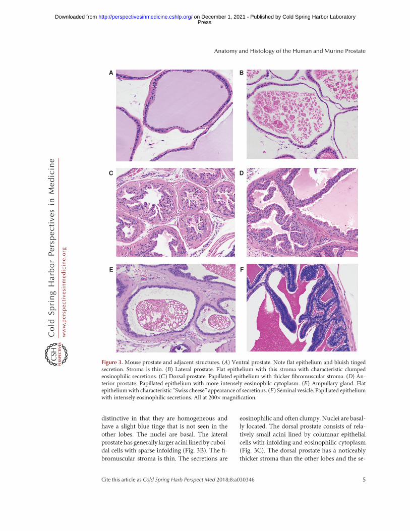
distinctive in that they are homogeneous andhave a slight blue tinge that is not seen in theother lobes. The nuclei are basal. The lateralprostate has generally larger acini lined by cuboi-dal cells with sparse infolding (Fig. 3B). The fi-bromuscular stroma is thin. The secretions are
eosinophilic and often clumpy. Nuclei are basal-ly located. The dorsal prostate consists of rela-tively small acini lined by columnar epithelialcells with infolding and eosinophilic cytoplasm(Fig. 3C). The dorsal prostate has a noticeablythicker stroma than the other lobes and the se-
A
C
E
B
D
F
Figure 3. Mouse prostate and adjacent structures. (A) Ventral prostate. Note flat epithelium and bluish tingedsecretion. Stroma is thin. (B) Lateral prostate. Flat epithelium with this stroma with characteristic clumpedeosinophilic secretions. (C) Dorsal prostate. Papillated epithelium with thicker fibromuscular stroma. (D) An-terior prostate. Papillated epithelium with more intensely eosinophilc cytoplasm. (E) Ampullary gland. Flatepitheliumwith characteristic “Swiss cheese” appearance of secretions. (F) Seminal vesicle. Papillated epitheliumwith intensely eosinophilic secretions. All at 200× magnification.
Anatomy and Histology of the Human and Murine Prostate
Cite this article as Cold Spring Harb Perspect Med 2018;8:a030346 5
ww
w.p
ersp
ecti
vesi
nm
edic
ine.
org
Press on December 1, 2021 - Published by Cold Spring Harbor Laboratoryhttp://perspectivesinmedicine.cshlp.org/Downloaded from

cretions are homogeneous and lightly eosino-philic. The nuclei are centrally located. Theanterior prostate is lined by a cuboidal to colum-nar epithelium with a more eosinophilic cyto-plasm and a papillary growth pattern (Fig. 3D).The cytoplasmhas a slight granularityandnucleiare centrally located. Based on the above,each lobe can be recognized based on its distinc-tive histology, although when the lobes are in-volved with pathological processes this can beproblematic.
Other structures adjacent to the prostate areof some importance because they can be secon-darily involved in pathological processes arisingin the prostate and/or be the site of primarypathology in genetically engineered models.The ampullary glands are bilateral outpouch-ings from the proximal ductus deferens andare of mesodermal origin. It does not have ahuman counterpart (Knoblaugh and True2012). The acini are small and are mainly flatwith slight infolding (Fig. 3E). The lining cellsare cuboidal, eosinophilic, and have basal nuclei.The stroma is similar in thickness to the dorsalprostate. The most characteristic feature of theampullary gland is the dense secretions with a“Swiss cheese” or bubbly appearance.
The two seminal vesicles are dorsolateral tothe bladder. The seminal vesicles are lined tallcolumnar cells with large intricate branchingfolds and are surrounded by a modest fibromus-cular stroma (Fig. 3F). The epithelial cells havebasal nuclei that are surrounded by hematoxy-philic cytoplasm with apical pale vacuolated cy-toplasm that is distinctive. The secretions arehomogeneous, dense, and brightly eosinophilic.The amount of secretion is variable, and whenthe seminal vesicle is distended the folds areflattened considerably.
Other structures frequently seen near theprostate include the ductus deferens, ganglia,
and their associated nerves, and of course thebladder and the membranous urethra.
CONCLUDING REMARKS
As described above the human and murineprostate have considerable differences in anato-my and histology despite a similar functionalrole in reproduction. To date, there is no firmevidence that any specific murine prostate lobeis more valid as a model for human prostatecancer and models have been described involv-ing every lobe and, frequently, multiple lobes.Although anatomically and histologically differ-ent than human prostate, there is extensive evi-dence that the genetic lesions in human prostatecancer can lead to neoplasia or neoplastic pro-gression in murine prostate, either alone or incombination when engineered in mouse pros-tate. The differences in anatomy and histologybetween human and mouse prostate and theassociated structures must always be taken intoaccount in the pathological analysis of genetical-ly engineered mouse models.
REFERENCES
Ittmann M, Huang J, Radaelli E, Martin P, Signoretti S,Sullivan R, Simons BW, Ward JM, Robinson BD, ChuGC, et al. 2013. Animal models of human prostate cancer:The consensus report of the New York meeting of theMouse Models of Human Cancers Consortium ProstatePathology Committee. Cancer Res 73: 2718–2736.
Knoblaugh S, True L. 2012. Male reproductive system. InComparative anatomy histology. A mouse and human at-las (ed. Treuting P, et al.), pp. 295–303. Elsevier, Amster-dam.
Paner G. 2010. Prostate gland and seminal vesicle. In Diag-nostic pathology: Genitourinary (ed. Amin M, et al.), Sec-tion 3, pp. 4–156. Amirysis, Salt Lake City, UT.
Peng YC, Levine CM, Zahid S, Wilson EL, Joyner AL. 2013.Sonic hedgehog signals to multiple prostate stromal stemcells that replenish distinct stromal subtypes during re-generation. Proc Natl Acad Sci 110: 20611–20116.
M. Ittmann
6 Cite this article as Cold Spring Harb Perspect Med 2018;8:a030346
ww
w.p
ersp
ecti
vesi
nm
edic
ine.
org
Press on December 1, 2021 - Published by Cold Spring Harbor Laboratoryhttp://perspectivesinmedicine.cshlp.org/Downloaded from

October 16, 20172018; doi: 10.1101/cshperspect.a030346 originally published onlineCold Spring Harb Perspect Med
Michael Ittmann Anatomy and Histology of the Human and Murine Prostate
Subject Collection Prostate Cancer
CancerAnatomic and Molecular Imaging in Prostate
Eric T. Miller, Amirali Salmasi and Robert E. ReiterReceptor in Prostate CancerNew Opportunities for Targeting the Androgen
Ebrahimie, et al.Margaret M. Centenera, Luke A. Selth, Esmaeil
The Epidemiology of Prostate Cancer
Wilson, et al.Claire H. Pernar, Ericka M. Ebot, Kathryn M.
Prostate Cancer Research at the CrossroadsMichael M. Shen and Mark A. Rubin
Prostate Stem Cells and Cancer Stem CellsJia J. Li and Michael M. Shen
Immunotherapy for Prostate CancerNicholas J. Venturini and Charles G. Drake
Mechanisms to Clinical ImplicationsProstate Cancer Epigenetics: From Basic
Marzo and William G. NelsonSrinivasan Yegnasubramanian, Angelo M. De Opportunities
Intraepithelial Neoplasia: Challenges and Molecular Pathology of High-Grade Prostatic
al.Levent Trabzonlu, Ibrahim Kulac, Qizhi Zheng, et
PerspectiveThe Genomics of Prostate Cancer: A Historic
Mark A. Rubin and Francesca Demichelis
Metastases in Prostate Cancer
Zoni, et al.Federico La Manna, Sofia Karkampouna, Eugenio
TherapiesCancer: Emerging Biology, Models, and Neuroendocrine Differentiation in Prostate
Himisha BeltranLoredana Puca, Panagiotis J. Vlachostergios and
Cancer in the Postgenomic EraGenetically Engineered Mouse Models of Prostate
Juan M. Arriaga and Cory Abate-Shen
DNA Damage Response in Prostate CancerMatthew J. Schiewer and Karen E. Knudsen of Prostate Cancer
Molecular Biomarkers in the Clinical Management
Aaron M. Udager and Scott A. TomlinsTranscriptional Regulation in Prostate Cancer
David P. Labbé and Myles Brown Diagnostic and Therapeutic OpportunitiesMetabolic Vulnerabilities of Prostate Cancer:
Giorgia Zadra and Massimo Loda
http://perspectivesinmedicine.cshlp.org/cgi/collection/ For additional articles in this collection, see
Copyright © 2018 Cold Spring Harbor Laboratory Press; all rights reserved
Press on December 1, 2021 - Published by Cold Spring Harbor Laboratoryhttp://perspectivesinmedicine.cshlp.org/Downloaded from