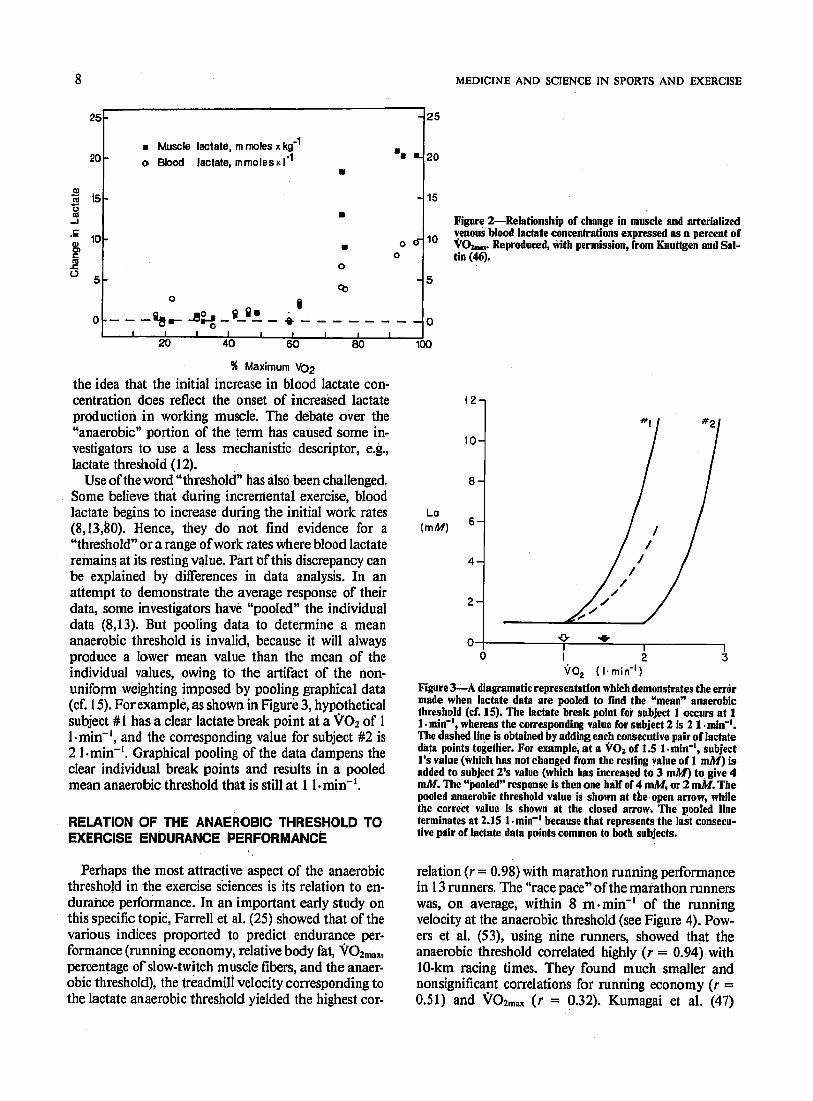
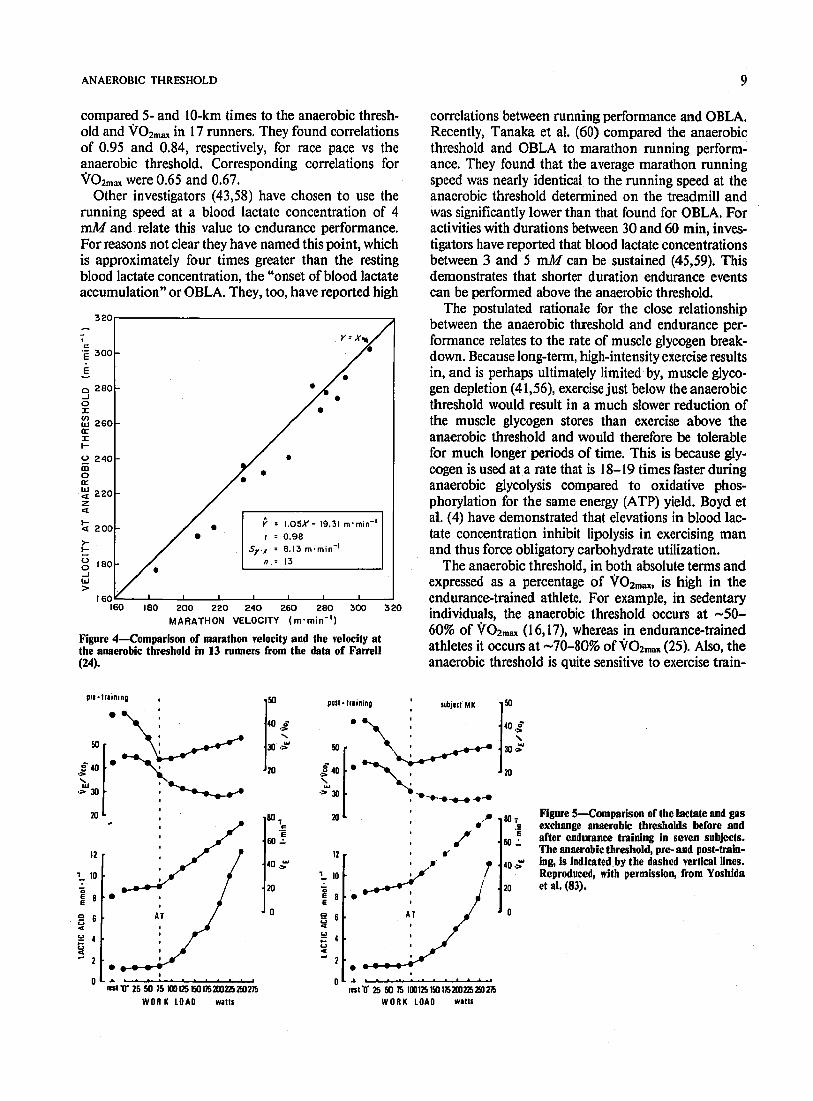
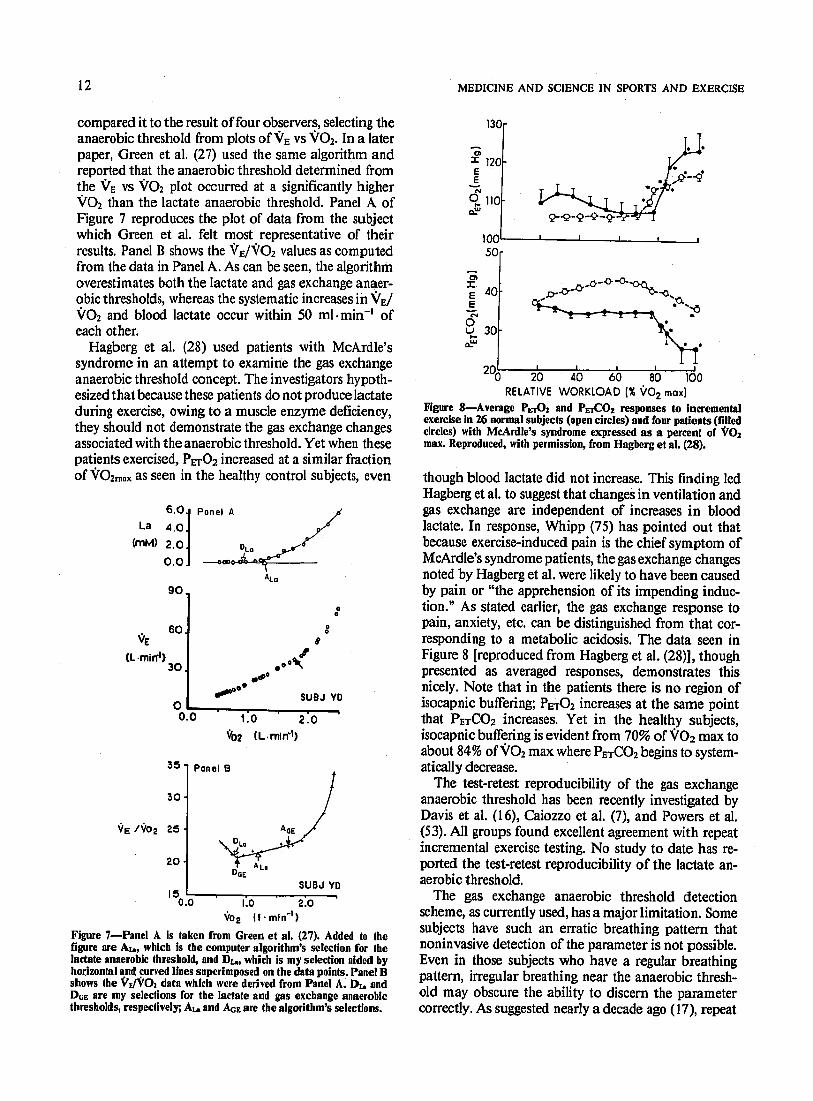
anaerobic threshold and respiratory gas exchange...
TRANSCRIPT

JOURNAL OF APPLIED PHYSIOLOGY Vol. 35, No. 2, August 1973. Printed in U.S.B.
Anaerobic threshold and respiratory gas
exchange during exercise
KARLMAN WASSERMAN, BRIAN Jm WHIPP,
SANKAR N. KOYAL, AND WILLIAM L. BEAVER
Department of Medicine, Harbor General Has-ital, Torrarxe 90509; and
University of California, Los Angeles, School of Medicine, Los Angeles, California 90024 .
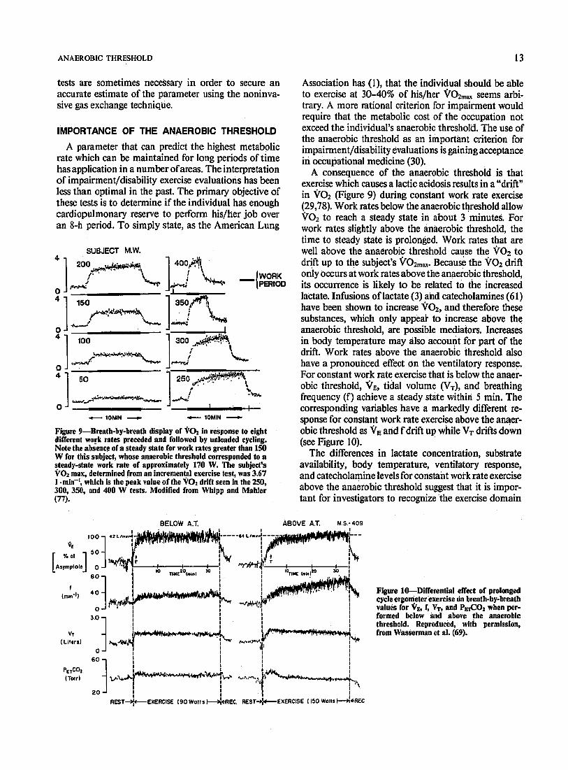
WASSERMAN, KARLMAN, BRIAN J. WHIPP, SANKAR IX KOYAL, AND WILLIAM L. BEAVER. Anaerobic threshold and respiratory gas ex- change during exercise. J. Appl. Physiol. 35(Z): 236-243. 1973.-- Alterations in gas exchange were studied in man during exercise increasing in increments of 15 w each minute, to determine the noninvasive indicators of the onset of metabolic acidosis (anaer- obic metabolism). Expired airflow and CO 2 and 0 2 tensions at the mouth during the breath were continuously monitored with rapid1 y responding gas analyzers. These measurements were recorded di- rectly as well as processed by a minicomputer, on-line, to give minute ventilation (VE), COP production #co& 02 consump- tion (voz), and the gas exchange ratio (R), breath-by-breath. The anaerobic threshold (AT) could be identified by the point of I) nonlinear increase in VE, 2) nonlinear increase in Vco2, 3) an increase in end-tidal 02 without a corresponding decrease in end- tidal COz, and 4) an increase in R, as work rate was increased during an incremental exercise test. Of these measurements, R was found least sensitive. The AT was determined in 85 normal subjects between 17 and 91 years of age, by these techniques. The lower limit of normal was 45 w (KTo 2 = 1 liter/min), while values for very fit normal adults were as high as 180 w. The patients studied with cardiac disease above functional class I have lower anaerobic thresholds than the least fit normal subjects. The I-min incremental work rate test is associated with changes in gas ex- change which can be used as sensitive on-line indicators of the AT, thus bypassing the need for measuring arterial lactate or acid-base parameters to indicate anaerobiosis.
metabolic acidosis; nonlinear changes in VE; end-tidal (220 tension; end-tidal 02 tension; gas exchange ratio; anaerobic metabolism; work performance and fitness; CO2 production; 02 consumption; noninvasive indicators of anaerobic metabolism in man
ALMOST A HALF-CENTURY AGO, Hill, Long, and Lupton (12) recognized that “a study of the respiratory quotient, if undertaken with sufficient caution, may throw light, not so much on the bodies being oxidised as on the acid-base changes occurring as a result of exercise and recovery.” Harrison and Pilcher (11) and Pilcher, Clark, and Harrison (23) later demonstrated that patients with heart failure developed metabolic acidosis and, consequently, a high respiratory gas exchange ratio at low work rates. They were also able to induce this phenomenon by exercising patients with heart disease who had limited work capacity but who were not in overt failure at the time. In more recent years
Issekutz and Rohdahl (17), Issekutz, Birkhead, and Rohdahl (16), and Naimark, Wasserman and McIlroy (22) were able to compute the gas exchange ratio breath-by-breath during exercise, by measuring expired N2 and CO 2 concen- trations with rapidly responding gas analyzers. Nairnark et al. (22) compared the arterial blood lactate and bicarbon- ate concentrations with the breath-by-breath changes in R and found the latter to reflect, reliably, the metabolic acidosis of exercise. Wasserman and McIlroy (26) con- firmed these observations and applied this technique to the determination of the anaerobic threshold1 in a group of patients with heart disease. More recently, Clode and Campbell (6) h ave attempted to apportion the R increase into metabolic, respiratory and blood buffering components.
However, in spite of the potential advantage of detecting the work rate at which a metabolic acidosis occurs during the performance of an incremental exercise test, the anaero- bic threshold has not been utilized widely for patient evaluation due, in large part, to technical difficulties with the N2 analyzer. The introduction of reliable rapidly responding oxygen analyzers and on-line computer processing has en- abled us to compute and visualize the anaerobic threshold, as it occurs during the performance of a test. This has ex- panded our understanding of the disturbances in gas ex- change associated with the exercise metabolic acidosis.
It is now evident that the increase in R caused by the buffering of lactic acid by sodium bicarbonate, is transient and occurs only while lactate is increasing and HCO, is decreasing in concentration. Furthermore, other bloodless approaches to the measurement of the anaerobic threshold have become evident. End-tidal CO2 (FETE& and 02 1 tensions (PET~~), when measured simultaneously, have also been found to be sensitive indicators of the anaerobic thresh- old during incremental work tests. It is also now evident that exercise above the anaerobic threshold results in al-
tered 02 uptake kinetics, with a delay in the 02 uptake
steady-state time and an increase in the 02 deficit and debt (1, 28).
We find the anaerobic threshold to be an invaluable con- cept in understanding changes in gas exchange during ex- ercise and work performance capabilities in normal sub-
1 The anaerobic threshold is defined as the level of work or Cl? consumption just below that at which metabolic acidosis and the associated changes in gas exchange occur.

ANAEROBIC: THRESHOLD DUKING EXERCISE 237
jects and patients. The purpose of this report is to describe the exercise test for detecting the anaerobic threshold which we find most useful, and the physiological basis for the measurement of VE, %O 2, and the combination of PETITE
and PETIT as alternate measurements to R as indicators of the anaerobic threshold.
THEORETICAL CONSIDERATIONS
The relationship between oxygen supply and lactic acid production are related by the Hill-Meyerhof concept of in- adequacy in 0 2 transport (14). Considerations are a) work efficiency is constant, i.e., doubling the work rate requires doubling the high-energy phosphate utilized for muscle contraction (5), b) the work rate determines the number of muscle units contracting (3), and c) control of the local circulation at the exercising muscle level is predominantly determined by the effects of vasodilator metabolites on the vascular resistance ( 19).
If the local circulation is adequate for the work rate being performed, all of the energy requirements may be supplied by ATPs generated by aerobic mechanisms. However, if the number of muscle units which must contract to generate the required power exceeds the oxygen delivery and ex- hausts the 02 stores, the oxygen level will drop to critical levels in each muscle unit and prevent the ATP, which is needed for the muscle contraction, from being generated at an adequate rate by the respiratory enzymes in the mito- chondria. This will result in increased anaerobic glycolysis to sustain the availability of ATP. The consequence is an increased rate of lactic acid production.
The physiological changes in respiratory gas exchange resulting from the inadequate 02 supply for the energy transformations are, as we have measured them, described in Fig. 1. The first consequence of the inadequate 02 sup- plv is the formation of lactic acid. Because of its low pR, lactic acid will be more than 99 % dissociated and buffered predominantly by the bicarbonate system (27). This is a d
lnadequate O2 delivery
1 !I
highly effective buffer system because of the volatile nature of the acid component. CO2 can be readily exhaled into the atmosphere, thereby preventing accumulation of this acid in the body tissues. The additional CO2 formed by this buffering is exhaled via the lungs, resulting in an increase in J&o2 and R. A stimulus resulting from the increase in J&02; provides an additional ventilatory stimulus. The de- crease in local tissue and blood bicarbonate results in a component of respiratory compensation for the metabolic acidosis e
Failure to supply the quantity of 0 2 required for the work rate being performed alters 0 2 uptake kinetics (28). If the subject could do the work completely aerobically, the steady-state VOW would be predicted by the work efficiency and the work rate. However, if all the energy required can- not be provided by reactions involving molecular oxygen, the oxygen uptake would be lower than expected for the work being performed, but it would gradually increase as the circulation readjusts to meet the energy demands. Re- distribution of blood flow, which contributes to the increase in Vo2 during work with an anaerobic component, is probably secondary to the regional acidosis and hypoxia of the heavily working muscles (19). Thus, the steady-state time for vo2 is delayed during a constant work rate above the anaerobic threshold. This contrasts with the Q92 pat- tern for the same work rate performed by a subject who is more fit and is able to meet all the energy requirements with reactions involving molecular oxygen (28).
METHODS
Eighty-five normal subjects” between 17 and 91 years of age were given incremental exercise tests. Studies on patients with cardiac disease of functional significance were con- trasted with those of the normal subjects.
Expired airflow and CO2 and 02 tensions at the mouth were continuously measured and recorded. The expired airflow was measured by use of a Fleisch model 3 pneu- motachograph (linear through peak flows of 600 liters/min at normal exercise respiratory frequencies) and Statham model PM97 strain gauge. Expired CO 2 and 0, were sampled at the mouthpiece and measured with a Beckman model LB-l or LB-2 CO 12 analyzer and a Westinghouse M-21 1 oxygen analyzer, respectively. There was an 0.08- to 0.12-set delay in each measurement. The 90 % response time of the instrument in the case of CO 2 was 0.160 and 0.200 set in the case of 0 2. More recently, we have used a
Anaerobic ‘Metabolism (t lactic acid)
1 Buffering
(JHCOJ, t&o,, tR)
“l k
a) Non-linear increase (Incremental work test)
I Delayed steady
state in \;io2
(t O2 deficit )
I b) Delayed steady state (Constant work test) *
Respiratory compensation for metabolic acidosis
(4 P%o*) . .
mass spectrometer (Perkin-Elmer, Pomona, Calif.) with an instrument 90 % response time of less than 0.06 sec.
The electrocardiogram was also continuously monitored on an oscilloscope and the heart rate continuously recorded. Some subjects had arterial blood gas and pH measure- ments using Radiometer equipment (London Company, Cleveland, Ohio) and arterial lactate and pyruvate meas- urements by enzymatic techniques (4, 15). Blood was sampled as previously described (27).
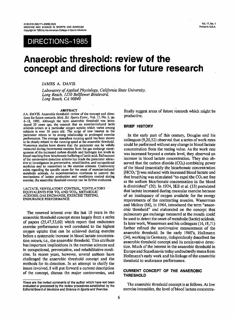
Sixty-one subjects were studied during an incremental work test in which the initial work rate consisted of 4 min of pedaling on an unloaded (“0” w) cycle ergometer c
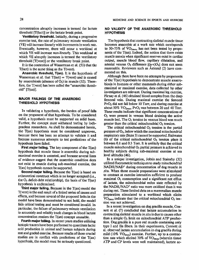
FIG. 1. ;2lterations in gas exchange which result from exerclsmg _I_____ at work rates above the anaerobic threshold. See text for a complete 2 These subjects were predominantly sedentary, but included fit
description of the flow of physiological responses depicted in this figure. subjects as they became available for the study.

238
(Lanooy, Instrumentation Associates, N-Y.) following which the work rates were incremented 15 w every minute. In the 24 other subjects, 25-w work rate increases were used.
The expired airflow, CO% and 02 tensions, and heart rate measurements were recorded on a Beckman type RM Dynograph and the data simultaneously transmitted to a Varian 620i minicomputer. The computed breath-by- breath VE, h02, vo2, and R (2) were displayed on-line on the recorder in addition to the directly recorded expired flow, and CO2 and 02 tensions in the breath and heart rate. The recorder speed of 10 mn/min permits the investigator to easily view the work rate at which CO2 production and minute ventilation deviate from linearity3 as compared with the rate of rise in oxygen consumption as work rate is in- cremented. This nonlinearity, the associated increase in R, and the decrease in the difference in 02 tension between inspired and end-tidal values without a comparable change in end-tidal CO2 (hyperventilation with respect to 0 2) were used to detect the anaerobic threshold.
All data were stored on digital tape during the test so that they might be retrieved and displayed on the recorder through the digital-to-analog converter of the computer for more detailed study using scaling factors which might be more appropriate than those used for the on-line test. The processed data can be played back at any speed, but we find that 1 min of study being displayed on 3-l 2 mm of paper is optimal to recognize those linearity changes of critical significance in detecting the anaerobic threshold. The data processing system is described in a previous report
(2) m All gas analyzers were calibrated before the test with tank
gases analyzed by the micro-Scholander method (24). This procedure was repeated routinely immediately after each test to ensure that the calibration factors had not changed during the course of the study.
RESULTS
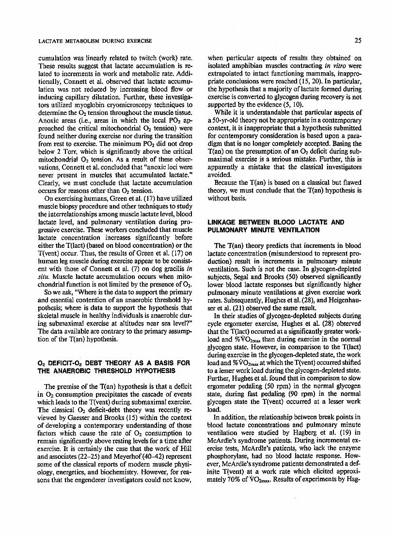
A. Gas exchange ratio (R) during constant, sufvathreshold work. Measurement of R, breath-bv-breath, as related to time for work above the anaerobic’ threshold, after an initial 4- min period of unloaded cycling, is shown in Fig. 2. Note that the gas exchange ratio increases to its peak value at the time that the rate of bicarbonate concentration change is at its maximum. When the bicarbonate concentration no longer changes, or changes minimally, the gas exchange ratio returns to a lower value and stays at this reduced level in spite of the fact that the same work rate is continued. Thus, to see the effects of anaerobic metabolism by study- ing R, one must look at it during the time of maximal bi- carbonate change. R will not remain elevated above the metabolic RQ if the bicarbonate concentration change had already occurred. R should again become equal to the metabolic RQ, when the CO? stores reach a new steady state. This limits the usefulness of the measurement of R
3 Linearity, in this regard, refers to equal increments in response for equal increments in work rate. The “0’‘-w work rate is not used to establish the linear direction of the VE and Vooz curves for the lower work rates because of the unique exercise duration of this work rate and the difficulty in knowing the amount of work being done. Thus the lowest point establishing the relationship of VE and Vco, and work rate was 15 w in this studv.
WASSERMAN, WHIPP, KOYAL, AND BEAVER
when looking for the anaerobic threshold (AT) during incremental work tests of relatively long duration.
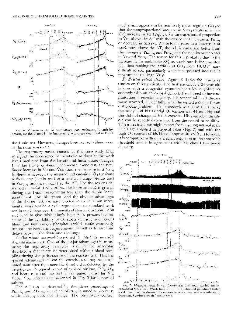
B. Work duration for an incremental work test to detect the anaerobic threshold. In the interest of time and avoiding undue stress to the patient, we concerned ourselves with how short a period we might use for each work rate in an incremental work test, in order to detect the anaerobic threshold. We compared the lactate, lactate/pvruvate ratio, and acid-
’ base parameters for a l- and 4-min incremental work test (Fig. 3). Note that the magnitudes of the lactate increase and the bicarbonate decrease are less for the 1-min test than
--x-----.x --I-. x--x
X
24
22
T \
20 w” E V
18 ‘IO
=: I
I6
0e7?REST+0 WATTS-+ 135 WATTS I I I I I 1
0 2 4 TIME (rnir$ IO I2 14
FIG. 2. Relationship between the increase in the gas exchange ratio (R) during suprathreshold exercise (135 w) and time of bicarbonate decrease. A period of unloaded cycling was done before the supra- threshold work was started, since exercise of any work intensity is usually associated with an increase in total body RQ.
24
16
25
I - I , I , , I , ,
REST “0” 25 50 75 100 125 150 175
WORK RATE (WATTS)
-10
-8
3 \
-6 g Y W
-4 G
: -l
-2
FIG. 3. Changes in lactate, bicarbonate, L/P ratio, pH, and Pacc12 during a I-min (0) and 4-ruin (0) incremental work test.
239 ,lNiZEROBIC THRESHOLD DURING EXERCISE
WATTS WATTS
IO-
FIG. 4. Measurements of ventilatory gas exchange, breath-by- breath, for the l- and 4-min incremental work tests described in Fig. 3.
the 4-min test. However, changes front control values occur at the same work rate.
The respiratory measurements for this same study (Fig. 4) signal the occurrence of metabolic acidosis at the work levels predicted from the lactate and bicarbonate changes. In either the l- or 4-min incremental work test, the non- linear increase in TE and Vc02 and the decrease in APET~, (difference between the inspired and end-tidal 02 tensions) without any (1-min test) or a smaller change (4-min test) in PETIT,, becomes evident at the AT. For the reasons de- scribed in section A of RESULTS, the increase in R is greater during the I-min incremental test than the 4-min incre- mental test. For this reason, and the obvious advantages of the shorter test, we have elected to use a I-min incre- mental work test on a cycle ergometer as a standard work test in our laboratory. Increments of shorter duration (<30 set) tend to give misleadingly high ATs, presumably be- cause of the availability of 02 stores in tissue and venous blood and high energy phosphates which could transiently support the energetic requirements, as well as transit time delays between the tissue and the lungs.
C. One-minute incremental work test to detect the anaerobic threshold during work. One of the major advantages in rneas- uring the respiratory variables to detect the anaerobic threshold is that it can be determined without blood sam- pling during the performance of the exercise test. This has special advantages in that the exercise test may be termi- nated soon after the anaerobic threshold is detected by the investigator. A typical record of expired airflow, (202, 02, and heart rate and Vco 2, 1’02, and R
subject. The AT can be
PETa> 2 and APET~),
while PET~() 2 does
the on-line computed values for VE,
are presented in Fig. 5 for a normal
detected by the direct recordings of in which APET~, is noted to decrease
not change. The respiratory control
mechanism appears to be sensitively set to regulate CO2 so that the nonproportional increase in %k02 results in a par- allel increase in VE (Fig. 5). VE increases out of proportion to vo2 above the AT with the consequent increase in PETIT or decrease in APET~~. While R increases at a faster rate at work rates above the AT, the AT is visualized better from the changes in PETIT, and PET (),, and the nonlinear increases in VE and %02. The reason for this is probably due to the increase in the metabolic RQ as work rate is incremented (1), thus making the additional CO2 from HCO, more dificult to see, particularly when incorporated into the R measurement at high 002s.
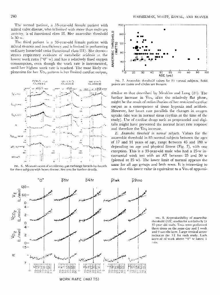
D. Selected patient studies. Figure 6 shows the results of studies on three patients. The ?irst patient is a 24-year-old laborer with a congenital cyanotic heart lesion (Ebstein’s anomaly with an atrioseptal defect). He claimed to have no limitation in exercise capacity. His congenital heart disease was discovered, incidentally, when he visited a doctor for an orthopedic problem. His hematocrit was 80 at the time of the study and his arterial 02 tension was 44 mm Hg and this did not change with this exercise. His anaerobic thresh- old can be readily determined from the record to be 60 w. This is less than one might expect from a young normal male at his age engaged in physical labor (Fig. 7) and with the high 0 2 content of his blood (approx 30 ~01%). However, it is compatible with only a small reduction in the anaerobic threshold and is in agreement with his class I functional capacity.
WATTS
pETco, - (mmHg) -
0
72 I
2.6 I I
1.6 I 1 / , I
I ! I
FIG. 5. Measurements in ventilatory gas exchange during an in- cremental work test. Work load at “0” w (unloaded pedaling) lasted for 4 min. Each additional increment in work rate was one minute in duration. Symbols are defined in text.
240 WASSERMAN, WHIPP, KOYAL, AND BEAVER
The second patient, a 53-vear-old female patient with mitral valve disease, who is limited with more than ordinary activity, is of functional class II. Her anaerobic threshold is 30 w.
The third patient is a 33-year-old female patient with mitral stenosis and insufficiency and is limited in performing ordinary household tasks (functional class III). She demon- strates respiratory evidence of metabolic acidosis at the lowest work rates (“0” w) and has a relatively fixed oxygen consumption, even though the work rate is incremented, until her highest work rate is reached. The most likely ex- planarion for her Voz pattern is her limited cardiac output,
- l
I OIO I I I I 1 20 30 40
GE
60 I 70 1 80 1 90 100 J
(yrs)
CONG. HD. VALV H D. VALV H.D. Cl-ASS I Cl-ASS II: CLASS III
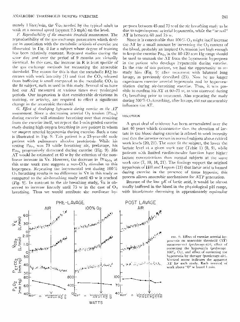
FIG. 7. ,4naerobic threshold values for 85 normal subjects. Solid points are males and circles are females.
similar to that described by Meakins and Long (2 1). The further increase in Vo2, after the relatively flat phase, might be the result of redistribution of her restricted cardiac output as a consequence of tissue hypoxia and acidosis. However, her heart rate parallels the changes in oxygen uptake (she was in normal sinus rhythm at the time of the study). Use of cardiac drugs such as propranolol and digi- talis might have prevented the normal heart rate response
and therefore the Vo2 increase. E. Anaerobic threshold in normal subjects. Values for the
anaerobic threshold in 85 normal subjects between the ages of 17 and 91 years of age, range between 45 and 180 w depending on age and physical fitness (Fig. 7), with one exception. This is a 33-year-old male who had a 25-w in- cremental work test with an AT between 25 and 50 w (plotted at 25 w). The lower limit of normal appears the
FIG. 6. Measurements of ventilatory gas exchange breath-by-breath same for all age groups and both sexes. It is interesting to
for three subjects with heart disease. See text for further details. note that this lower value is equivalent to a voz of approxi-
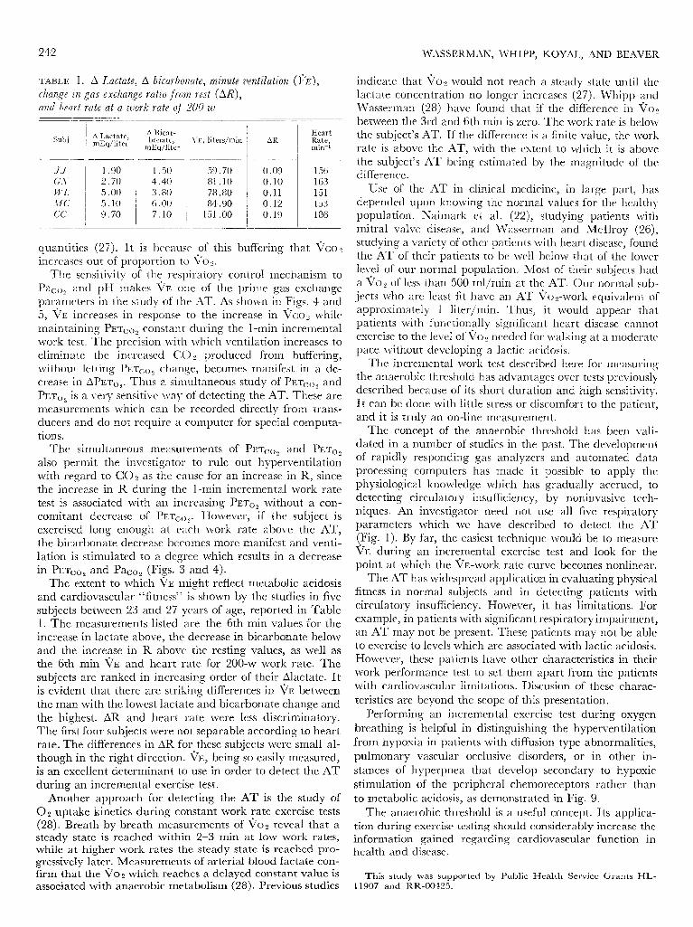
plhr p4hr PI wk p9mo
FIG. 8. Reproducibility of anaerobic threshold (AT) studies for a relatively fit 35-year-old male. Tests were performed three times on the same day and 1 week and 9 months later. Large vertical arrow indicates the ,4T for each study. Each interval of work above “0’” w lasted 1 nrin.
WORK RATE (WATTS)
ANAEROBIC THRESHOLD DURING EXERCISE 241
mately 1 liter,/‘min, the J?oz needed by the typical adult to walk at a normal speed (approx 2.5 mph) on the level.
F. Kejv-oducihility of the anaerobic threshold measurement. The . reproducibility of the gas exchange parameters which devi- ate in association with the metabolic acidosis of exercise are illustrated in Fig. 8 for a subject whose degree of training has been relatively constant. Repeated studies during the same day and over the period of 9 months are virtually identical. In this case, the increase in IX. is least specific of the gas exchange methods for measuring the anaerobic threshold. The reason for this is that the metabolic KQ in- creases with work intensity (1) and that the CO2 released from buffering is small compared to the metabolic CO2 in the fit sub-ject: such as used in this st,udy. Several of us have had our AT measured at various times over prolonged periods. Our impression is that considerable deviations in training, or activity, arc required to effect a significant change in t,hc ana.erobic threshold.
G. Effect of deuelophg /y& 1 ,)oxemia during exercise on the A T measurement. Since a decreasing arterial 0 2 tension (PaOJ during exercise will stimulate breathing over that resulting from the exercise itself, w ve repeat the 1 -min graded exercise study during high oxygen breathing in any patient in whom we suspect arterial hypoxemia during exercise. Such a case is illustrated in Fig. 9. This patient is a 23-year-old male patient with pulmonary alveolar proteinosis. While his resting Pa () 2 was 73 while breathing air, prelavage, his I’;;~~~~ progressively decreased during exercise (Fig. 9). His AT would be estimated at 45 w by the criterion of the non- linear increase in VE. However, the decrease in PETITE at this same work rate suggests a non-CD2 stimulus to this hyperpnea. Repeating the incremental test during 100 % 02 breathing results in no difference in VE in this study as compared to the air-breathing study until 45 w is reached (Fig. 9). In contrast to the air-breathing study, VIZ is ob- served to increase linearly until 75 w in the case of 02 breathing. Thus we would attribute the nonlinear hy-
perpnea between 45 and 75 w of the air breathing study to be due to superimposed arterial hvpoxemia, while the “actual”
’ AT is between 60 and 75 w. Since it is conceivable that 100 % 0 2 might itself increase
the AT by a small amount by increasing the 02 content of the blood, probably an inspired 0 2 tension just high enough to keep the exercise Pa oz in the 80-l 20 mm IIg range should be used to unmask the *4T from the hvpoxemic hyperpnea in the patient who develops hypoxemia during exercise. In the case of this patient, we had the opportunity to re- study him (Fig. 9) after treatment with bilateral lung lavage, as previouslv described (25). Now he no longer experiences exercise ‘arterial hypoxemia and its hyperven- tilation during air-breathing exercise. Thus, it was pos- sible to confirm his *4T at 60--75 w, as was observed during 02 breathing prior to treatment. Also repeating the study during 100 % 02 breathing, after lavage, did not measurably influence the AT.
DISCUSSION
A great deal of evidence has been accumulated over the last 40 years which demonstrate that the elevation of lac- tate in the blood during exercise is related to work intensity and that the increase occurs in normal subjects above critical
work levels (20, 21). The more fit the subject, the lower the lactate level at a given work rate (Table 1 j (8, 91, while patients with limited cardiovascular function have higher lactate concentrations than normal subjects at the same work rate (7, 10, 18, 2 l)* The findings support the original hypothesis of Hill and Lupton (13) that lactic acid is formed during exercise in the presence of tissue hypoxia; this process allows anaerobic mechanisms for ATP generation.
Because of the low pk’ of lactic acid, it would be almost totally buffered in the blood in the physiological pII range, with bicarbonate decreasing in approximately equimolar
POST LAVAGE AIR
FIG. 9. Effect of exercise arterial hy- poxemia on anaerobic threshold (AT j
measurement (prelavage-air > , effect of correcting the hypoxemia (prelavage- 100yc O:), and effect of correcting the hypoxemia by therapy (postlavage-air). Vertical arrow indicates the apparent AT for each study. Each interval of work above ccO” w lasted 1 min.
WATTS
242 WASSERMAN, WHIPP, KO-YAL, AND BEAVER
TABLE I. A Lactate, A bicarbonate, minute ventilation (PI), change in gas exchange ratio from rut (AR), and heart rate at a work rate qj* 200 w --- __l_____l__l_l_--- -_l_~
A Lactate, mEq/liter
1.90 2.70 5.00 5.10 9.70
/ A Bicar- 1 bonate, I I-E, liters/min
mEq/liter I
1.50 , 59.70 4.40 81.10 3.80 78.80 6.00 : 84.90 7.10 / 151 .oo
0.09 0.10 0.11 0.12 0.19
-
Heart Rate, min-1
156 163 151 153 186
quantities (27). It is because of this buffering that VCO~ increases out of proportion to Toz.
The sensitivity of the respiratory control mechanism to
hm, and pH makes VE one of the prime gas exchange parameters in the study of the L%T. As shown in Figs. 4 and 5, VE increases in response to the increase in &o, while maintaining PET c02 constant during the I-min incremental work test. The precision with which ventilation increases to eliminate the increased CO:! produced from buffering, without letting PETITE change, becomes manifest in a de- crease in APET~~. Thus a simultaneous study of PETIT, and PETIT is a very sensitive wav of detecting the AT. These are measurements which can be recorded directly from trans- ducers and do not require a computer for special computa- tions.
The simultaneous measurements of PETIT, and PETIT
also permit the investigator to rule out hyperventilation with regard to CO2 as the cause for an increase in R, since the increase in R during the 1-min incremental work rate test is associated with an increasing PETIT without a con- comitant decrease of PETIT.,. However, if the subject is exercised long enough at each work rate above the AT, the bicarbonate decrease becomes more manifest and venti- lation is stimulated to a degree which results in a decrease in PETIT:, and Pacog (Figs. 3 and 4).
The extent to which VE might reflect metabolic acidosis and cardiovascular “fitness’” is shown by the studies in five subjects between 23 and 27 years of age, reported in Table 1. The measurements listed are the 6th min values for the increase in lactate above, the decrease in bicarbonate below and the increase in R above the resting values, as well as the 6th min VE and heart rate for 200-w work rate. The subjects are ranked in increasing order of their Alactate. It is evident that there are striking differences in VE between the man with the lowest lactate and bicarbonate change and the highest. AR and heart rate were less discriminatory. The first four subjects were not separable according to heart rate. The differences in AR for these subjects were small al- though in the right direction. VE, being so easily measured, is an excellent determinant to use in order to detect the AT during an incremental exercise test.
Another approach for detecting the AT is the study of 02 uptake kinetics during constant work rate exercise tests (28). Breath-by-b reath measurements of Tj02 reveal that a steady state is reached within 2-3 min at low work rates, while at higher work rates the steady state is reached pro- gressively later. Measurements of arterial blood lactate con- firm that the vo2 which reaches a delayed constant value is associated with anaerobic metabolism (28). Previous studies
indicate that VO, would not reach a steady state until the lactate concentration no longer increases (27). Whipp and Wasserman (28) have found that if the difference in Tjo, between the 3rd and 6th min is zero. The work rate is below the subject’s AT. If the difference is a finite value, the work rate is above the AT, with the extent to which it is above the subject’s AT being estimated by the magnitude of the difference.
Use of the AT in clinical medicine, in large part, has depended upon knowing the normal values for the healthy population. Naimark et al. (22), studying patients with mitral valve disease, and Wasserman and McIlroy (26), studying a variety of other patients with heart disease, found the AT of their patients to be well below that of the lower level of our normal population. Most of their subjects had a X70, of less than 500 ml/min at the AT. Our normal sub- jects who are least fit have an AT Voz-work equivalent of approximately 1 liter/min. Thus, it would appear that patients with functionally significant heart disease cannot exercise to the level of Vo2 needed for walking at a moderate pace without developing a lactic acidosis.
The incremental work test described here for measuring the anaerobic threshold has advantages over tests previously described because of its short duration and high sensitivity. It can be done with little stress or discomfort to the patient, and it is truly an on-line measurement.
The concept of the anaerobic threshold has been vali- dated in a number of studies in the p last. The development of rapidly respond ing gas anal vzers and automated data processing computers has made it possible to apply the physiological knowledge which has gradually accrued, to detecting circulatory insufhciency, by noninvasive tech- niques. An investigator need not use all five respiratory parameters which we have described to detect the AT (Fig. 1). By far, the easiest technique would be to measure
VE during an incremental exercise test and look for the point at which the %%-work rate curve becomes nonlinear.
The AT has widespread application in evaluating physical fitness in normal subjects and in detecting patients with circulatory insufficiency. However, it has limitations. For example, in patients with significant respiratory impairment, an AT may not be present. These patients may not be able to exercise to levels which are associated with lactic acidosis. However, these patients have other characteristics in their work performance test to set them apart from the patients with cardiovascular limitations. Discusion of these charac-
teristics are beyond the scope of this presentation. Performing an incremental exercise test during oxygen
breathing is helpful in distinguishing the hyperventilation from hypoxia in patients with diffusion type abnormalities, pulmonary vascular occlusive disorders, or in other in- stances of hyperpnea that develop secondary to hypoxic stimulation of the peripheral chemoreceptors rather than to metabolic acidosis, as demonstrated in Fig. 9.
The anaerobic threshold is a useful concept. Its applica- tion during exercise testing should considerably increase the
information gained regarding cardiovascular function in health and disease.
This study was supper ted by Public Health Service Grants HL- 11907 and RR-00425.

ANA4EROBIC THRESHOLD DURING EXERZISE 243
B. J. Whipp is an Established Investigator of the American Heart Requests for reprints should be sent to: K. Wasserman, Division of Association. Respiratory Medicine, Harbor General Hospital.
W. L. Beaver is a Senior Scientist? Central Research, Varian Asso-
ciates, Palo Alto, Calif. Received for publication 8 January 1973.
REFERENCES
1. ASMWSEN, E. Muscular exercise. In : Handbook of Physiology Respira- tion. Washington, D.C. : Am. Physiol. Sot., 1965, sect. 3, vol. II, chapr. 36, p. 939-978.
2. BEAVER, W. L., K. WASSERMAN, AX;D B. J, WHIPP. On-line com- puter analysis and breath-by-breath graphical display of exercise function tests. J. AppZ. Physiol. 34 : 128-132, 1973.
3. BIGLAND, B., AND 0. C. J. LIPPOLD. Motor unit activity in the voluntary contraction of human muscle. J. Physiol., London 125 : 322-329, 1954.
4. BUCHER, T., R. CZAK, W. LAMPRECHT, AND E. LATZKE. Pyruvate, In: *2lethods of Enzymatic Analysis, edited by H. U. Bergmeyer. New York: Academic, 1965.
5. CARLSON, F. D., AND A. SIGER. The mechanochemistry of muscle contraction. J. Gen. Physiol. 44: 33-60, 1960.
6. CLODE, M., AND E. J. M. CAMPBELL. The relationship between gas exchange and changes in blood lactate concentrations during exercise. CZin. Sci. 37: 263-272, 1969.
7. COTES, J. E. The role of oxygen, carbon dioxide and lactic acid in the ventilatory response to exercise in patients with mitral stenosis. CZin. Sci. 14: 317-328, 1955.
8. EDM’ARDS, H. T., L. BROUHA, AND R. T. JOHNSON. Effect de l’en- trainemente sur le taux de l’acide lactique au tours du travail musculaire. Trav. Humain 8 : l-8, 1940.
9. EKBLOM, B., P. 0. ASTRAND, B. SALTIN, J. STENBERG, AND B. WALLSTROM. Effect of training on circulatory response to exercise. J. AppZ. Physiol. 24 : 5 18-528, 1968.
10. HALLOCK, P. Lactic acid production during rest and after exercise in subjects with various types of heart disease with special reference to congenital heart disease. J. Clin. Invest. 18 : 385-394, 1939.
11. HARRISON, T. R., AND C. PILCHER. Studies in congestive heart failure. II. Respiratory exchange during and after exercise. J. CZin. Invest. 8: 291-315, 1930
12. HILL, A. V., C. N. H. LONG, AND H. LUPTON. Muscular exercise, lactic acid and the supply and utilization of oxygen. 1. Proc. Roy. Sot. London, Ser. B 96 : 438-475, 1924
13. HILL, A. V., AND H. LUPTON. Muscular exercise, lactic acid and supply and utilization of oxygen. Quart. J. Med. 16: 135-171, 1923.
14. HILL, A. V., AND 0. MEYERHOF. Uber die vorgange bei der mus- kelkontrakttion. Ergeb. PhysioZ. 22 : 299-327, 1923.
15. HOHORST, H. J. L( +) lactate. In : Methods of Enzymatic Analysis, edited by H. U. Bergmeyer. New York, :Icademic, 1965.
16. ISSEKUTZ, B., JR., N. C. BIRKHEAD, AND K. ROHDAHL. Use of respiratory quotients in assessment of aerobic work capacity. J. AppZ. Physiol. 17 : 47-50, 1962.
17. ISSEKUTZ, B., JR., AND K. ROHDAHL. Respiratory quotient during exercise J. APPZ. Physiol. 16 : 606-6 10, 196 1.
18. JERVELL, 0. Investigation of the concentration of lactic acid in the blood and urine under physiologic and pathologic conditions. Acta Med. Stand. SuppZ. 24, 1928.
19. KJELLMER, I. On the competition between metabolic vasodilation and neurogenic vasoconstriction in skeletal muscle. Acta Physiol. Stand. 63: 450-459, 1965.
20. MARGARIA, R., H. T. EDWARDS, AND D. B. DILL. The possible mechanisms of contracting and paying the oxygen debt and the role of lactic acid in muscular contraction. Am. J. Physiol. 106 : 689-715, 1933.
21. MEAKINS, J., AND C. N. H. LONG. Oxygen consumption, oxygen
debt and lactic acid in circulatory failure. J. CZin. Invest. 4: 273-293, 1927.
22. NAIMARK, A., K. WASSERMAN, AND M. B. MCILROY. Continuous measurement of ventilatory exchange ratio during exercise. J. AppZ. Physiol. 19 : 644-652, 1964
23. PILCHER, C., G. CLARK, AND T. R. HARRISON. The buffering power of the blood and tissues. J. CZin. Invest. 8 : 3 17-323, 1930.
24. SCHOLANDER, P. F. Analyzer for accurate estimating of respira- tory gases in one-half cubic centimeter samples. J. Biol. Chem. 167 : 235-259, 1947.
25. WASSERMAN, K., N. BLANK, AND G. FLETCHER. Lung lavage (al- veolar washing) in alveolar proteinosis. Am. J. Med. 44: 611-617, 1968.
26. WASSERMAN, K., AND M. B. MCILROY. Detecting the threshold of anaerobic metabolism. Am. J. Cardiol. 14 : 844-852, 1964.
27. WASSERMAN, K., A. L. VAN KESSEL, AND G. G. BURTON. Interac- tion of physiological mechanism during exercise. J. A@Z. Physiol. 22: 71-85, 1967.
28. WHIPP, B. J., AND K. WASSERMAN. Oxygen uptake kinetics for various intensities of constant load work. J. APPZ. Physiol. 33:
351-356, 1972.


HPER 6760 Exercise Science Seminar
Manuscript Review Process
Journal:
Journal of Strength and Conditioning Research
Title of Paper:
The validity of the heart rate deflection threshold test for determination of the maximal lactate steady state
Reviewer Instructions:
1. Choose a Reviewer Recommendation term from the drop-down box at the top of the review page. 2. Complete the rating evaluation form. Please use 1 as the highest score. 3. Enter any Blind Comments to the Author. These comments will be distributed to the author in the decision letter. Do
not indicate acceptance or rejection in these comments. 4. Complete the Confidential Comments to the Editor. Provide answers to the 'yes' or 'no' questions and any frank
comments to the editor. These comments will NOT be shared with the authors or other reviewers. 5. Click "Proceed" to see a proof of your review, then Submit the review to the journal office.
Recommendation:
Accept
Minor Revision
Major Revision
Reject
Rate the practical applications impact of this paper (1 is high impact):
N/A 1 2 3
Reviewer Blind Comments to Author:
Reviewer Confidential Comments to Editor:
(1) Answer yes or no for each of the questions below:
Do you have a conflict of interest in reviewing this paper? Yes______ No______
Do you perceive a conflict of interest for the authors within the paper? Yes______ No______
Do you agree to hold its contents confidential? Yes______ No______
Does this paper meet the IRB standards? Yes______ No______
Is a statistical review needed? Yes______ No______
(2) Provide confidential comments to the Editor:

Journal of Strength and Conditioning Research Manuscript Draft Manuscript Number: JSCR-08-1072 Title: The validity of the heart rate deflection threshold test for determination of the maximal lactate steady state Short Title: The validity of the heart rate deflection threshold Article Type: Original Investigation Keywords: lactate, Conconi test, running intensity. Manuscript Region of Origin: BRAZIL Abstract: The purpose of this study was to investigate the validity of the heart rate deflection threshold (HRDT) test in the determination of the velocity at the maximal lactate steady state (MLSS). Fifteen untrained male took part in a 3-km running performance on 400-m track and completed a comprehensive battery of laboratory tests. Performance velocity at HRDT was strongly correlated with the MLSS running velocity (r = 0.84; R2 = 0.71; P<0.0001). HRDT running velocity (mean ± SD 9.0 ± 1.3-km.h-1) was not significantly different (P > 0.05) from MLSS velocity (9.3 ± 1.3-km.h-1). A high agreement was observed between methods (Bland and Altman analysis). It is concluded that the HRDT was an accurate method to predict MLSS velocity in the present study.
1 2 3 4 5 6 7 8 9 10 11 12 13 14 15 16 17 18 19 20 21 22 23 24 25 26 27 28 29 30 31 32 33 34 35 36 37 38 39 40 41 42 43 44 45 46 47 48 49 50 51 52 53 54 55 56 57 58 59 60 61 62 63 64 65
The validity of the heart rate deflection threshold test for determination of the maximal
lactate steady state
Running head: The validity of the heart rate deflection threshold
Laboratory of Exercise Physiology
Paulo H.S.M. de Azevedo
Vitor K.P. Carrara
Gustavo M. Rissato
João M.P. Duarte
Runer A. Marson
Department of Physical Education
Anhanguera College of Bauru
3, Moussa Nakhl Tobias, Bauru – SP – Brazil
CEP: 17.021.100
Phone: (5514) 3239-9147
e-mail: [email protected]
Financial support: AESA
Manuscript (All Manuscript Text Pages in MS Word format, including References and Figure Legends)

1 2 3 4 5 6 7 8 9 10 11 12 13 14 15 16 17 18 19 20 21 22 23 24 25 26 27 28 29 30 31 32 33 34 35 36 37 38 39 40 41 42 43 44 45 46 47 48 49 50 51 52 53 54 55 56 57 58 59 60 61 62 63 64 65
The purpose of this study was to investigate the validity of the heart rate deflection
threshold (HRDT) test in the determination of the velocity at the maximal lactate steady
state (MLSS). Fifteen untrained male took part in a 3-km running performance on 400-
m track and completed a comprehensive battery of laboratory tests. Performance
velocity at HRDT was strongly correlated with the MLSS running velocity (r = 0.84; R2
= 0.71; P<0.0001). HRDT running velocity (mean ± SD 9.0 ± 1.3-km.h-1
) was not
significantly different (P > 0.05) from MLSS velocity (9.3 ± 1.3-km.h-1
). A high
agreement was observed between methods (Bland and Altman analysis). It is concluded
that the HRDT was an accurate method to predict MLSS velocity in the present study.
Keywords: lactate, Conconi test, running intensity.
1 2 3 4 5 6 7 8 9 10 11 12 13 14 15 16 17 18 19 20 21 22 23 24 25 26 27 28 29 30 31 32 33 34 35 36 37 38 39 40 41 42 43 44 45 46 47 48 49 50 51 52 53 54 55 56 57 58 59 60 61 62 63 64 65
Introduction
The maximal lactate steady state (MLSS) has been defined as the highest blood
lactate concentration that increases by no more than 1 mmol.L-1
between 10 and 30-min
of constant velocity/workload test (3,23). This physiological index is important because
it has been used to prescribe endurance exercise training programs (20). However, two
to five constant workload exercise test of up 20 to 30-min duration is usually required to
determine MLSS (23).
Conconi et al. (8) reported that heart rate (HR) as a function of speed is not
linear up to the maximum and that the speed corresponding to the deflection point also
corresponds to the speed of the second lactate threshold. So, the heart rate deflection
threshold (HRDT) is a non-invasive test of the anaerobic threshold. Thus, determination
of the HRDT is supposed to be too an indirect measure of MLSS utilizing incremental
exercise test, due to its correlation whit ventilatory threshold (VT) (19) and second
lactate threshold (LT2) (14). These investigations suggest that knowledge of the heart
rate kinetics may provide to reveal certain indicators of the intensity of exercise
equivalent to MLSS. But, a number of studies has produced contradictory results
(16,18,24). Thus, the aim of the present research was to establish whether HRDT
velocity corresponding to the MLSS intensity.
Method
Subjects
The Ethics Committee for Human Research at Anhanguera College approved the
methods used in this study (32/2008). All experimental procedures complied with the
current laws for human studies. Potential subjects were introduced to all testing
equipment and procedures. After completing the informed consent procedure, 15

1 2 3 4 5 6 7 8 9 10 11 12 13 14 15 16 17 18 19 20 21 22 23 24 25 26 27 28 29 30 31 32 33 34 35 36 37 38 39 40 41 42 43 44 45 46 47 48 49 50 51 52 53 54 55 56 57 58 59 60 61 62 63 64 65
physically active men (23.4 ± 3.9 years old, 71 ± 10.1 kg body weight, 175 ± 0 cm
height, 19.8 ± 5.1 % body fat), who were students at Anhanguera College, volunteered
to participate in this investigation. The number of subjects was determined for provide
appropriate statistical power (β=80%; α=5%). Body density was estimated by the
skinfold technique, from which body composition was determined using the Siri
formula for estimation of percent body fat and fat-free mass (11). The subjects had been
involved in recreational training programs (e.g. endurance running, resistance training,
and soccer) for the previous 6 months, consisting of at least 20-min of exercise three
times per week on a regular basis. All of the subjects were advised not to have any
extenuating physical practice 48 hours before the tests.
Design
The tests were performed in the Exercise Physiology Laboratory at Anhanguera
College and at 400-m track. Three to six tests were performed on separate days at the
same time of day. The subjects were instructed to have their last meal at least 3-h before
testing, arrive at the laboratory in a rested and fully hydrated state and to avoid
strenuous exercise in the 48-h preceding a test session. The first test was a 3000-m time
trial (V3000) on 400-m track. In the second test the volunteers performed an
incremental test for HRDT determination. The third test was a constant velocity lasted
30-min around HRDT. The incremental and constant velocity test was performed on a
treadmill (Movement LX-150). The heart rate (HR) was monitored continuously during
all tests using a Polar Accurex Plus (Kempele, Finland). Before each test session the
subjects performed a warm-up consisting of low intensity running for 5-min at about
(6–7.5 km h−1
).

1 2 3 4 5 6 7 8 9 10 11 12 13 14 15 16 17 18 19 20 21 22 23 24 25 26 27 28 29 30 31 32 33 34 35 36 37 38 39 40 41 42 43 44 45 46 47 48 49 50 51 52 53 54 55 56 57 58 59 60 61 62 63 64 65
Test 1: The 3000-m velocity test (V3000)
The subjects ran 3000-m as quickly as possible and the mean running velocity
was calculated for each subject (V3000). The V3000 was used to prescribe the
velocities of the treadmill runs during the incremental tests to determine the HRDT
(22).
Test 2: Incremental test for HRDT determination
Initial running velocity was about 65% of the individual’s V3000. The incline
was set at 1% (15), with 0.5-km.h−1
increments at each 3-min stage. The HRDT was
identify when the running velocity at which heart rate began to increase less rapidly
with increments in running velocity during the incremental exercise test, by means of
third order polynomial fit (confidence 95%) (17). The maximal distance (Dmax) between
perpendicular line and polynomial fit was considered HRDT (17). Standardized verbal
encouragement was given to the subjects to continue the test until they were exhausted.
The heart rate (HR) was continuously monitored by a heart rate monitor (Polar Accurex
Plus, Finland) and values recorded every three minutes during incremental exercise test.
The HR corresponding to HRDT (HRDTHR) was the values registered at HRDT velocity
during incremental exercise test and is also reported as percent of the peak HR
(%HRDTHR).
Test 3: Determination of MLSS
On subsequent occasions (3–5 visits), participants completed a series of
constant-speed treadmill runs, each of 30-min duration, for the determination of the
MLSS. An ear lobe capillary blood sample was taken during a 45-s time period for
blood lactate concentration ([Lac-]) analysis at rest and during 5-min intervals

1 2 3 4 5 6 7 8 9 10 11 12 13 14 15 16 17 18 19 20 21 22 23 24 25 26 27 28 29 30 31 32 33 34 35 36 37 38 39 40 41 42 43 44 45 46 47 48 49 50 51 52 53 54 55 56 57 58 59 60 61 62 63 64 65
throughout the run at which point participants were asked to step astride the treadmill.
The velocity of the first constant work rate test corresponded to a 5% below HRDT
measured during the incremental exercise test. The MLSS was defined as the running
speed that produced no more than a 1-mmol.L-1
increase in [Lac-] between 10 and 30-
min of exercise (3). MLSS was determined with a precision of 0.5-km.h-1
. If during the
first constant work-rate test a steady state or a decrease in lactate was observed, further
subsequent 30-min constant work-rate tests from 0.5-km.h-1
higher work-rate were
performed on separate days until no [Lac-] steady state could be maintained. MLSS was
calculated as the mean [Lac-] measured at 10 and 30-min of the MLSS.
The heart rate (HR) was continuously monitored by a heart rate monitor (Polar
Accurex Plus, Finland) and values recorded every five minutes during constant velocity
tests. The HR corresponding to MLSS (MLSSHR) was the mean of the values registered
during MLSS testing and is also reported as percent of the peak HR (%MLLSHR).
Blood collection and laboratory analysis
The 25-μL of capillary blood was collected from the ear lobe using heparinized
and calibrated microcapillaries during MLSS tests. The 25-μL was deposited into
Eppendorf tubes containing 50-μL of 1% sodium fluoride (NaF). The [lac-] was
determined from this sample by using a blood lactate analyzer (Yellow Springs 1500).
The [lac-] results were corrected by the volume of the blood sampled within the
Eppendorf tubes and are presented in mmol.L-1
concentrations.
Statistical Analysis
Firstly, the Shapiro-Wilk test was applied to the sample for normality, and
because data were found to be normal, a parametric statistic test was used. Data are
1 2 3 4 5 6 7 8 9 10 11 12 13 14 15 16 17 18 19 20 21 22 23 24 25 26 27 28 29 30 31 32 33 34 35 36 37 38 39 40 41 42 43 44 45 46 47 48 49 50 51 52 53 54 55 56 57 58 59 60 61 62 63 64 65
presented as means with standard deviations (±SD). Pearson product moment
correlations were used to quantify the relationships between MLSS and HRDT. Paired
t-test was used to compare MLSS and HRDT velocity. The limit of statistical
significance was set at 5% (P ≤ 0.05). The Bland-Altman method was additionally
applied in order to assess the agreement of the results. Data was analyzed using the
Statistical Package for Social Sciences (SPSS), version 13.0 for Windows.
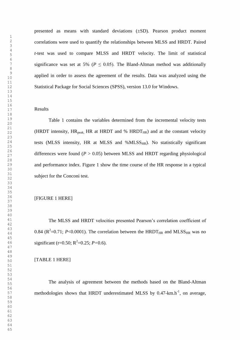
Results
Table 1 contains the variables determined from the incremental velocity tests
(HRDT intensity, HRpeak, HR at HRDT and % HRDTHR) and at the constant velocity
tests (MLSS intensity, HR at MLSS and %MLSSHR). No statistically significant
differences were found (P > 0.05) between MLSS and HRDT regarding physiological
and performance index. Figure 1 show the time course of the HR response in a typical
subject for the Conconi test.
[FIGURE 1 HERE]
The MLSS and HRDT velocities presented Pearson’s correlation coefficient of
0.84 (R2=0.71; P<0.0001). The correlation between the HRDTHR and MLSSHR was no
significant (r=0.50; R2=0.25; P=0.6).
[TABLE 1 HERE]
The analysis of agreement between the methods based on the Bland-Altman
methodologies shows that HRDT underestimated MLSS by 0.47-km.h-1
, on average,
1 2 3 4 5 6 7 8 9 10 11 12 13 14 15 16 17 18 19 20 21 22 23 24 25 26 27 28 29 30 31 32 33 34 35 36 37 38 39 40 41 42 43 44 45 46 47 48 49 50 51 52 53 54 55 56 57 58 59 60 61 62 63 64 65
within a 95% confidence interval (Fig. 2). For MLSSHR and HRDT HR high agreement is
found too, while MLSSHR underestimated HRDT by 1-bpm (Fig. 3).
[FIGURE 2 and 3 HERE]
Discussion
The main finding of this study was that the validity of HRDT to predict MLSS
velocity (Table 1; Figure 2). These suggest that HRDT and MLSS (gold standard) could
be used interchangeably to demarcate the boundary between oxidative and anaerobic
lactic metabolism. Thus, our results indicate that HRDT and MLSS maybe correspond
to the equivalent physiological phenomenon. Others studies have show high correlation
when investigated the relationship between the anaerobic threshold and HRDT, which
occur in response to incremental exercise (8), HRDT and ventilatory threshold (19),
second lactate threshold (LT2) (14).
The MLSS is an important physiological index because it has been used to
prescribe endurance exercise training programs (1,4,20). However, two to five constant
workload exercise test of up 20 to 30-min duration is usually required to determine
MLSS (23), limiting its use for training programs. Therefore, several studies were
performed to propose a simple method for determining the MLSS, time to conduct the
test relatively short and inexpensive marker (8,10,16,24).
Some authors have reported that the HRDT does not occur, but this is a linear
relationship between heart rate and velocity/power during incremental exercise test (16).
Was suggested that the HRDT is protocol dependent (6,12,24). Vachon et al., 1999 (24),
concluded that the HRDT is not an accurate predictor of LT. Jones and Doust (16)
compared HRDT, lactate turnpoint and OBLA (fixed 4-mmol.L-1
) with MLSS. The

1 2 3 4 5 6 7 8 9 10 11 12 13 14 15 16 17 18 19 20 21 22 23 24 25 26 27 28 29 30 31 32 33 34 35 36 37 38 39 40 41 42 43 44 45 46 47 48 49 50 51 52 53 54 55 56 57 58 59 60 61 62 63 64 65
authors suggested that the Conconi test is invalid for the determination of the lactate
turnpoint and MLSS. In the present study high correlation and agreement were observed
between HRDT and MLSS velocity. This difference of results between studies may be
due the specifics protocols used and methodologies for determining deviation of heart
rate from linearity. Our results provided evidence that HRDT can be determined during
an incremental test in nonathlete individuals for this specific protocol and methodology.
The stage length of present study is in accordance of previous researches (7).
According to these authors, 3-min of stage was sufficient for blood and muscle lactate
equilibrium during incremental exercise test. So, blood lactate concentration can not be
considered due to the longer stage as reported by Vachon et al. (24). Additionally, has
been reported that the blood lactate does not promote muscle fatigue (21) and cardiac
muscle using most of the lactate as an oxidative fuel (9).
According to present data, it seems that the methodology here used to determine
HRDT is valid for estimation of MLSS intensity. No statistically significant difference
was observed in the velocity regarding HRDT versus MLSS (Table 1), and a good
correlation was found as well (r = 0.84; R2=0.71; P<0.0001). These data suggest that
MLSS (Fig. 2) can be accurately estimated by using the dynamic response to heart rate
during a 3-min incremental effort increased by 0.5-km.h-1
, pause of 45-s and a total of
6-10 stages.
It was observed good agreement (Fig. 2) between the velocity associated to
HRDT and that associated to MLSS. The MLSS was found to be slightly superior to the
HRDT (0.47-km.h-1
). Six subjects exhibited identical GT and MLSS velocities. These
data suggest that HRDT is a simple method to estimate the MLSS.
The heart rate corresponding to HRDT was not found to be statistically different
from MLSSHR, with good agreement between them (Fig. 3). The percentage HRpeak was
1 2 3 4 5 6 7 8 9 10 11 12 13 14 15 16 17 18 19 20 21 22 23 24 25 26 27 28 29 30 31 32 33 34 35 36 37 38 39 40 41 42 43 44 45 46 47 48 49 50 51 52 53 54 55 56 57 58 59 60 61 62 63 64 65
not different between HRDT and MLSS. These data are clinically important because
heart rate is largely used for controlling the training intensity. In the present study, the
heart rate values regarding MLSS are in agreement with those found elsewhere (2,5,20).
For HRDT heart rate values was found below of the others studies (16,18), but, percent
of maximum heart rate it is similar to previous researches (13,17,25).
We can conclude that the validity of HRDT to predict MLSS velocity. High
agreement was observed between methods. No difference is found for heart rate at
HRDT velocity and MLSS velocity. Our results provided evidence that HRDT can be
determined during an incremental test in nonathlete individuals for this specific protocol
and methodology.
PRACTICAL APPLICATIONS
In the present study, HRDT was in agreement with the running speed
corresponding to the maximal lactate steady state. Utilizing the Dmax methodology
between perpendicular line and polynomial fit by means of third order polynomial fit,
the HRDT could be properly identified in all of the subjects. So, the data from the
current study would be useful when prescribing running exercise programs to allow for
sufficient intensity training sessions.
Acknowledgements
We thank the subjects for participation in this study, AESA for financial support.
The authors wish to thank Giovanna Togashi and Amilton Vieira for blood sampling
analysis.

1 2 3 4 5 6 7 8 9 10 11 12 13 14 15 16 17 18 19 20 21 22 23 24 25 26 27 28 29 30 31 32 33 34 35 36 37 38 39 40 41 42 43 44 45 46 47 48 49 50 51 52 53 54 55 56 57 58 59 60 61 62 63 64 65
References
1. Azevedo, PHSM, Garcia, A, Duarte, JMP, Rissato, GM, Carrara, VKP, Marson, RA.
Anaerobic threshold and bioenergetics: a didactic approach. R. da Educação
Física/UEM. 20:453-464, 2009.
2. Baron, B, Noakes, TD, Dekerle, J, Moullan, F, Robin, S, Matran, R, Pelayo, P. Why
does exercise terminate at the maximal lactate steady state intensity? Br J Sports Med.
42:528-533, 2008.
3. Beneke, R. Maximal lactate steady state concentration (MLSS): experimental and
modelling approaches. Eur J Appl Physiol. 88:361-369, 2003.
4. Beneke, R. Methodological aspects of maximal lactate steady state-implications for
performance testing. Eur J Appl Physiol. 89:95-99, 2003.
5. Beneke, R, Hutler, M, Leithauser, RM. Maximal lactate-steady-state independent of
performance. Med Sci Sports Exerc. 32:1135-1139, 2000.
6. Bosquet, L, Leger, L, Legros, P. Methods to determine aerobic endurance. Sports
Med. 32:675-700, 2002.
7. Chwalbinska-Moneta, J, Robergs, RA, Costill, DL, Fink, WJ. Threshold for muscle
lactate accumulation during progressive exercise. J Appl Physiol. 66:2710-2716, 1989.
8. Conconi, F, Ferrari, M, Ziglio, PG, Droghetti, P, Codeca, L. Determination of the
anaerobic threshold by a noninvasive field test in runners. J Appl Physiol. 52:869-873,
1982.
9. Gladden, LB. A lactatic perspective on metabolism. Med Sci Sports Exerc. 40:477-
485, 2008.
10. Grazzi, G, Alfieri, N, Borsetto, C, Casoni, I, Manfredini, F, Mazzoni, G, Conconi, F.
The power output/heart rate relationship in cycling: test standardization and
repeatability. Med Sci Sports Exerc. 31:1478-1483, 1999.
1 2 3 4 5 6 7 8 9 10 11 12 13 14 15 16 17 18 19 20 21 22 23 24 25 26 27 28 29 30 31 32 33 34 35 36 37 38 39 40 41 42 43 44 45 46 47 48 49 50 51 52 53 54 55 56 57 58 59 60 61 62 63 64 65
11. Guedes, DP, Guedes, JE. Proposed equations for predicting the amount of body fat
in young adults. Semina. 12:61-70, 1991.
12. Hofmann, P, Bunc, V, Leitner, H, Pokan, R, Gaisl, G. Heart rate threshold related to
lactate turn point and steady-state exercise on a cycle ergometer. Eur J Appl Physiol
Occup Physiol. 69:132-139, 1994.
13. Hofmann, P, Pokan, R, Preidler, K, Leitner, H, Szolar, D, Eber, B, Schwaberger, G.
Relationship between heart rate threshold, lactate turn point and myocardial function.
Int J Sports Med. 15:232-237, 1994.
14. Hofmann, P, Pokan, R, von Duvillard, SP, Seibert, FJ, Zweiker, R, Schmid, P. Heart
rate performance curve during incremental cycle ergometer exercise in healthy young
male subjects. Med Sci Sports Exerc. 29:762-768, 1997.
15. Jones, AM, Doust, JH. A 1% treadmill grade most accurately reflects the energetic
cost of outdoor running. J Sports Sci. 14:321-327, 1996.
16. Jones, AM, Doust, JH. The Conconi test in not valid for estimation of the lactate
turnpoint in runners. J Sports Sci. 15:385-394, 1997.
17. Kara, M, Gokbel, H, Bediz, C, Ergene, N, Ucok, K, Uysal, H. Determination of the
heart rate deflection point by the Dmax method. J Sports Med Phys Fitness. 36:31-34,
1996.
18. Passelergue, PA, Cormery, B, Lac, G, Leger, LA. Utility of the Conconi's heart rate
deflection to monitor the intensity of aerobic training. J Strength Cond Res. 20:88-94,
2006.
19. Petit, MA, Nelson, CM, Rhodes, EC. Comparison of a mathematical model to
predict 10-km performance from the Conconi test and ventilatory threshold
measurements. Can J Appl Physiol. 22:562-572, 1997.

1 2 3 4 5 6 7 8 9 10 11 12 13 14 15 16 17 18 19 20 21 22 23 24 25 26 27 28 29 30 31 32 33 34 35 36 37 38 39 40 41 42 43 44 45 46 47 48 49 50 51 52 53 54 55 56 57 58 59 60 61 62 63 64 65
20. Philp, A, Macdonald, AL, Carter, H, Watt, PW, Pringle, JS. Maximal lactate steady
state as a training stimulus. Int J Sports Med. 29:475-479, 2008.
21. Robergs, RA, Ghiasvand, F, Parker, D. Biochemistry of exercise-induced metabolic
acidosis. Am J Physiol Regul Integr Comp Physiol. 287:R502-516, 2004.
22. Simões, HG, Denadai, BS, Baldissera, V, Campbell, CS, Hill, DW. Relationships
and significance of lactate minimum, critical velocity, heart rate deflection and 3 000 m
track-tests for running. J Sports Med Phys Fitness. 45:441-451, 2005.
23. Svedahl, K, MacIntosh, BR. Anaerobic threshold: the concept and methods of
measurement. Can J Appl Physiol. 28:299-323, 2003.
24. Vachon, JA, Bassett, DR, Jr., Clarke, S. Validity of the heart rate deflection point as
a predictor of lactate threshold during running. J Appl Physiol. 87:452-459, 1999.
25. Zacharogiannis, E, Farrally, M. Ventilatory threshold, heart rate deflection point and
middle distance running performance. J Sports Med Phys Fitness. 33:337-347, 1993.
1 2 3 4 5 6 7 8 9 10 11 12 13 14 15 16 17 18 19 20 21 22 23 24 25 26 27 28 29 30 31 32 33 34 35 36 37 38 39 40 41 42 43 44 45 46 47 48 49 50 51 52 53 54 55 56 57 58 59 60 61 62 63 64 65
Table 1: Selected variables obtained from constant and incremental velocity tests (mean±SD).
Figure 1 – Heart rate responses to 1 subject during incremental run tests. Subject
showed an HR plateau on Conconi treadmill protocol, in which 3-min stages were used.
HR deflection threshold (arrow).
Figure 2 – Results of the Bland–Altman analysis of agreement for MSSL and HRDT
measurements. The continued lines indicate de means of the differences and the dashed
lines indicate the limits of agreement between measurements.
Figure 3 – Results of the Bland–Altman analysis of agreement for MLSSHR and
HRDTHR measurements. The continued lines indicate de means of the differences and
the dashed lines indicate the limits of agreement between measurements.

Table 1
HRDT MLSS
Velocity (km.h-1
) 9.0±1.3 9.3±1.3
HRpeak (bpm) 191.7±8.0 -
HR at (bpm) 174.1±7.4 174.2±5.2
%HR 90±2.1 91±4.2
Table

Figure 1
6.0 6.5 7.0 7.5 8.0 8.5 9.0 9.5 10.0
130
140
150
160
170
180
190H
R (
bpm
)
Velocity (km.h-1)
HRDT
Figure

Figure 2
6 7 8 9 10 11 12
AVERAGE of MLSS and HRDT (km/h)
2.0
1.5
1.0
0.5
0.0
-0.5
-1.0
-1.5
MLS
S -
HR
DT
(km
/h)
Mean
0.47
-1.96 SD
-0.84
+1.96 SD
1.77
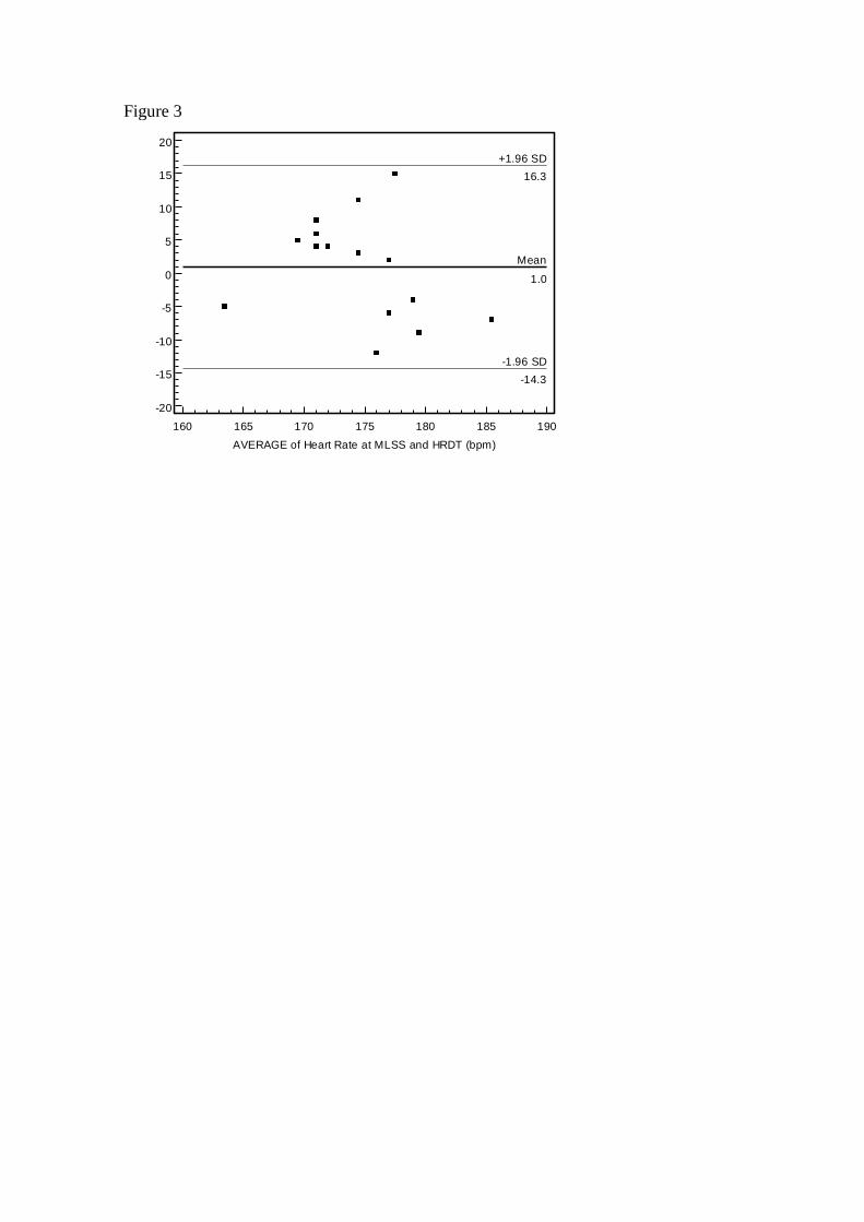
Figure 3
160 165 170 175 180 185 190
AVERAGE of Heart Rate at MLSS and HRDT (bpm)
20
15
10
5
0
-5
-10
-15
-20
Hea
rt R
ate
at
ML
SS
an
d H
RD
T (
bpm
)
Mean
1.0
-1.96 SD
-14.3
+1.96 SD
16.3