an innovative method for teaching anatomy in the ... innovative method for teaching anatomy in the...
TRANSCRIPT

1498 Journal of Dental Education ■ Volume 77, Number 11
An Innovative Method for Teaching Anatomy in the Predoctoral Dental CurriculumEric W. Baker, M.Phil.; Phyllis A. Slott, Ph.D.; Louis Terracio, Ph.D.; Elena P. Cunningham, Ph.D.Abstract: New methods of teaching gross anatomy are being evaluated as medical and dental schools attempt to find time in their curricula for new content without sacrificing essential anatomical knowledge. This article reports on an innovative method of teaching anatomy at New York University College of Dentistry. In 2005, the instructors completely replaced the dissection of wet cadavers with the study of dissected and sliced plastinated specimens. The shift from cadaver dissection to the study of plasti-nated specimens was accompanied by other changes in the anatomy course: students study in small, consistent groups; frequent, low-impact quizzes are administered; and the role of the computer is increased as a tool for self-directed study. To assess the course, this study considered students’ long-term understanding of anatomy as demonstrated by performance on the National Board Dental Examination (NBDE) Part I, hours of instruction, and student evaluation. The results show that, since 2005, stu-dents have had higher NBDE Part I scores, their overall performance has been above the national mean while hours of instruction were 60 percent of the national mean, and student satisfaction increased.
Mr. Baker is Adjunct Associate Professor, Department of Basic Science and Craniofacial Biology, New York University College of Dentistry; Dr. Slott was Adjunct Assistant Professor, Department of Basic Science and Craniofacial Biology, New York Uni-versity College of Dentistry at the time of this study; Dr. Terracio is Vice Dean for Research and Professor, Department of Basic Science and Craniofacial Biology, New York University College of Dentistry, and Professor of Pediatrics, New York University School of Medicine; and Dr. Cunningham is Adjunct Associate Professor, Department of Basic Science and Craniofacial Biology, New York University College of Dentistry. Direct correspondence and requests for reprints to Dr. Elena Cunningham, Depart-ment of Basic Science and Craniofacial Biology, New York University College of Dentistry, VA Hospital, 423 East 23rd Street, #1623, New York, NY 10010; 212-998-9618 phone; 212-995-4087 fax; [email protected].
Keywords: anatomy, gross anatomy, dental education, plastinated specimens, gross anatomy laboratory, cross-sectional anatomy
Submitted for publication 10/2/12; accepted 1/1/13
Dental and medical education programs are struggling to find time in their curricula for new content, while simultaneously look-
ing for ways to manage rising costs.1-5 Anatomy has long been considered one of the foundations of medicine,6,7 but it has become difficult to maintain the many hours typically allocated to a traditional anatomy course.2,5 In dental education, anatomy instruction takes up almost twice as much time as any other area in the biomedical sciences.8 In recent years, the advantages and disadvantages of various methods of teaching anatomy have been debated, not only to manage resources, but also to improve anatomical understanding.2,6 Some programs have minimized the use of cadavers;9 others no longer use cadavers, but use living anatomy and imaging6 or virtual learning packages.10 Some programs use prosections or plastinated specimens.11,12 Plastination, a method invented by Gunther von Hagens, makes it possible for prosections or slices of cadavers to be preserved in a safe, strong, dry polymer medium that is odorless and inert.13,14
Some programs that eliminated dissection re-ported student satisfaction and anatomical knowledge
decreased; 2,15 other programs have reduced or elimi-nated dissection with success.11,15,16 Teaching method may be a key factor in reducing or eliminating dis-section without a loss of anatomical understanding.12 When the University of California, San Francisco (UCSF) replaced dissection with prosections, student exploration was also replaced with faculty mini-lectures. After two years, UCSF returned to full dis-section.2 In contrast, when Yale School of Medicine successfully shortened the amount of time students dissect, the new course was designed to maintain or increase student exploration and engagement.9 There is, however, a paucity of data on the effectiveness of various methods of teaching anatomy, in particular in dental schools.12,15
Prior to 2005, New York University College of Dentistry (NYUCD) had a traditional anatomy course, including dissection of human cadavers. Although the college allocated substantial resources (faculty and staff, hours of instruction, space for dissection) to the course, NYUCD was not satisfied with the students’ knowledge of anatomy as reflected in their National Board Dental Examination (NBDE) Part I scores. In addition, the use of cadavers, which are embalmed

November 2013 ■ Journal of Dental Education 1499
New Anatomy CourseIn 2004, NYUCD invited Gunther von Hagens,
scientific director of the Institute for Plastination, to New York to discuss the possibility of creating a collection of plastinated specimens for our gross anatomy course. Dr. von Hagens joined the faculty of NYUCD and spent hours meeting with all the members of the anatomy faculty to fully grasp our needs. The resulting collection consists of eight dis-sected full bodies, over fifty dissected heads, isolated plastinated brains and spinal cords, several upper and lower limbs, thoracic and abdominal viscera, and hundreds of slices in three planes. All plastinated materials are from cadavers donated to The Body Donation Program of the Institute for Plastination in Heidelberg, Germany. NYUCD paid the institute for the cost of dissecting, processing, and plastinating the teaching collection.
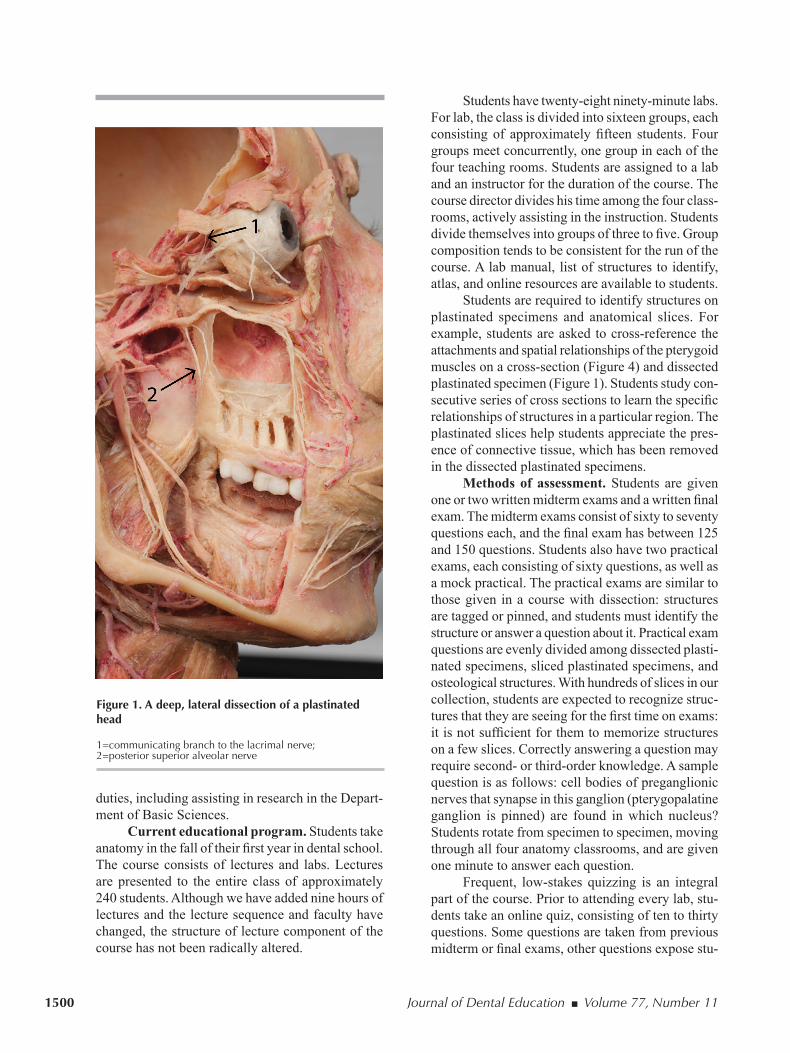
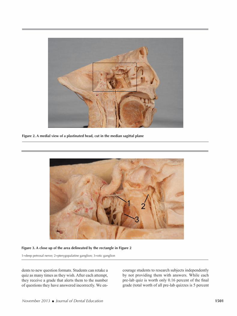
The plastinated specimens exhibit a high de-gree of anatomical detail. Among the structures and relationships visible on a single specimen (Figure 1) are the three branches of the trigeminal nerve as they leave the ganglion, the maxillary nerve entering the pterygopalatine fossa, the nerve of the pterygoid canal entering the pterygopalatine ganglion, the com-municating branch from the zygomatic nerve carry-ing autonomic fibers to the lacrimal nerve and gland, and the posterior superior alveolar nerves innervating the mucosa of the maxillary sinus before innervating the roots of the molars. On another specimen (Figure 2 and Figure 3), students can observe the deep petro-sal nerve leave the carotid plexus to join the greater petrosal nerve and form the nerve of the pterygoid canal before synapsing in the pterygopalatine gan-glion. We supplement the plastinated specimens with over sixty real skulls, including several Beauchene (exploded) skulls, other osteological specimens, and plastic models. Each student is assigned a plastic skull to take home for the duration of the course.
At present, the anatomy course uses four class-rooms (total size: 1,636 square feet), each of which has direct access to a specimen storage closet (total size: 242 square feet). As the specimens are odorless and inert, the storage closets do not have a ventilation system and are not temperature controlled. We have three full-time faculty members and three adjuncts. In addition, we have a curator who sets up the class-rooms, assists with the instruction and administration of the course, and repairs the specimens with silicon and/or acrylic mixtures. When the anatomy course is not in session, the curator is free to pursue other
with formaldehyde, a hazardous chemical,17 entails the risk of accidental overexposure through fumes or direct contact.18,19 Following an incident in which the cadavers were embalmed with an excess amount of formaldehyde, NYUCD administrators and faculty began to explore alternative methods for teaching anatomy. Plastinated specimens seemed like an ideal choice as the specimens allow students to see a high degree of anatomical specificity, yet they are dry, odorless, and non-toxic.14,20
This article reports on the changes in NYUCD’s gross anatomy course as we replaced dissection of cadavers with the study of plastinated specimens. We describe the traditional anatomy course and the design of the new course: students study plas-tinated specimens in small, consistent groups, and we administer frequent, low-stakes quizzes and incorporate use of online learning. We assessed the overall effectiveness of the course by a) comparing student evaluations of the two courses, b) compar-ing student performance on the NBDE Part I before and after 2005, and c) comparing the NBDE Part I scores and hours of instruction of NYUCD students with national averages.
Methods
Traditional Anatomy CourseIn 2004, the last year in which students at
NYUCD dissected, the gross anatomy course consist-ed of forty-five lecture hours, forty-eight laboratory hours, and seven hours of exam. For laboratory, the class of approximately 240 students was divided into two groups. Five or six students were assigned to each cadaver. Students dissected all regions of the upper body (head thorax, upper extremity, and abdominal and pelvic viscera). Students studied radiographs. Attendance was mandatory and was recorded with an in-lab quiz. Five or six faculty members and the diener (individual responsible for procurement, transportation, and preparation of cadavers) usually assisted students in the lab. In 2003 and 2004, the college rented space for dissection; previously, the college had maintained a 4,000-square foot dissec-tion laboratory. Students took two written exams consisting of fifty to seventy-five multiple-choice questions, two practical exams consisting of forty to forty-five questions, and sixteen pre-lab quizzes. The Blackboard management system was used to post assignments and schedules and learning materials.
1500 Journal of Dental Education ■ Volume 77, Number 11
Students have twenty-eight ninety-minute labs. For lab, the class is divided into sixteen groups, each consisting of approximately fifteen students. Four groups meet concurrently, one group in each of the four teaching rooms. Students are assigned to a lab and an instructor for the duration of the course. The course director divides his time among the four class-rooms, actively assisting in the instruction. Students divide themselves into groups of three to five. Group composition tends to be consistent for the run of the course. A lab manual, list of structures to identify, atlas, and online resources are available to students.
Students are required to identify structures on plastinated specimens and anatomical slices. For example, students are asked to cross-reference the attachments and spatial relationships of the pterygoid muscles on a cross-section (Figure 4) and dissected plastinated specimen (Figure 1). Students study con-secutive series of cross sections to learn the specific relationships of structures in a particular region. The plastinated slices help students appreciate the pres-ence of connective tissue, which has been removed in the dissected plastinated specimens.
Methods of assessment. Students are given one or two written midterm exams and a written final exam. The midterm exams consist of sixty to seventy questions each, and the final exam has between 125 and 150 questions. Students also have two practical exams, each consisting of sixty questions, as well as a mock practical. The practical exams are similar to those given in a course with dissection: structures are tagged or pinned, and students must identify the structure or answer a question about it. Practical exam questions are evenly divided among dissected plasti-nated specimens, sliced plastinated specimens, and osteological structures. With hundreds of slices in our collection, students are expected to recognize struc-tures that they are seeing for the first time on exams: it is not sufficient for them to memorize structures on a few slices. Correctly answering a question may require second- or third-order knowledge. A sample question is as follows: cell bodies of preganglionic nerves that synapse in this ganglion (pterygopalatine ganglion is pinned) are found in which nucleus? Students rotate from specimen to specimen, moving through all four anatomy classrooms, and are given one minute to answer each question.
Frequent, low-stakes quizzing is an integral part of the course. Prior to attending every lab, stu-dents take an online quiz, consisting of ten to thirty questions. Some questions are taken from previous midterm or final exams, other questions expose stu-
duties, including assisting in research in the Depart-ment of Basic Sciences.
Current educational program. Students take anatomy in the fall of their first year in dental school. The course consists of lectures and labs. Lectures are presented to the entire class of approximately 240 students. Although we have added nine hours of lectures and the lecture sequence and faculty have changed, the structure of lecture component of the course has not been radically altered.
Figure 1. A deep, lateral dissection of a plastinated head
1=communicating branch to the lacrimal nerve; 2=posterior superior alveolar nerve
November 2013 ■ Journal of Dental Education 1501
courage students to research subjects independently by not providing them with answers. While each pre-lab quiz is worth only 0.16 percent of the final grade (total worth of all pre-lab quizzes is 5 percent
dents to new question formats. Students can retake a quiz as many times as they wish. After each attempt, they receive a grade that alerts them to the number of questions they have answered incorrectly. We en-
Figure 2. A medial view of a plastinated head, cut in the median sagittal plane
Figure 3. A close up of the area delineated by the rectangle in Figure 2
1=deep petrosal nerve; 2=pterygopalatine ganglion; 3=otic ganglion

1502 Journal of Dental Education ■ Volume 77, Number 11
Course evaluation. The Office of Academic Affairs asks students to anonymously evaluate courses on a regular basis. Twenty-five percent of the class is randomly selected to participate in the surveys. The anatomy course and the Department of Basic Sciences also anonymously survey students in order to improve course content and teaching methods. This survey is conducted to get students’ reactions to specific aspects of the anatomy course. The Human Investigations Committee of New York University reviewed this study and granted an exemp-tion for educational studies.
ResultsAlthough the cost of acquiring the collection
of plastinated specimens was significant, the speci-mens have a life span of twenty years or longer,12 and the initial cost was amortized over five years. There are no recurring costs such as those of ca-daver acquisition and embalming associated with a dissection laboratory. In addition to being used by first-year dental students for gross anatomy and a single laboratory in the organ systems (physiology) course, the plastinated specimens are utilized by third-year dental students preparing for the NBDE Part I examination, dental hygiene students, nursing students, and occasionally postgraduate students. As the multipurpose classrooms are used for other courses and meetings when anatomy is not sched-uled, there are no continuing costs of a dedicated space. Nor do we incur the expense of renting a lab: it would cost approximately $40 a square foot per year to rent a dissection laboratory in Manhattan at the present time.
Student Evaluation of Course and Hours of Instruction
For this study, we compared students’ responses to two statements that best summarize their overall evaluation of the course. Although our method of teaching anatomy has changed, the primary course goals as stated in the course syllabus from 2003-04 to 2012-13 remain the same: to “1) present the detailed structures of the head and neck in their structural and functional relationships, and 2) enable students to recognize the application of anatomical information in clinical dental practice.” In 2003 (the last course evaluation before redesign of the course), 80.5 per-cent of the students agreed or strongly agreed that the
of the course grade), students strive to answer the questions correctly: last year the class average on these quizzes was 96 percent. At the end of every laboratory, we give students a five-question quiz, which is based mostly on the material covered that day during laboratory, but also incorporates pre-lab quiz and lecture material. Each “exit quiz” is worth 0.80 points; all together the exit quizzes are worth 20 percent of the course grade. Laboratory attendance is close to 100 percent.
Computer-assisted learning. We use the Blackboard Management System to facilitate self-directed study. Through Blackboard, students have access to the pre-lab quizzes, images of plastinated slices, radiographs, CT scans, a laboratory manual, dissection videos created in our institution, pur-chased copies of commercial dissection videos, and additional content. Prior to each lab, students are assigned structures to identify on the images of the slices. The images are projected and reviewed by the instructors during lab. Students bring their laptops to lab, and we observe students referring to online content while working.
In order for the students to be apprised of their progress through the course, grades are placed in the Blackboard grade book. Grades for online pre-lab quizzes are available upon submission of the quiz, grades for exit quizzes are usually available within twenty-four hours, and grades for major exams are usually available within forty-eight hours.
Figure 4. A transverse cross-section of a head showing attachments of pterygoid muscles

November 2013 ■ Journal of Dental Education 1503
Figure 5. Student response to the statement “Course goals and objectives were met”
Note: Surveys were conducted by Office of Academic Affairs. Graduating Class of 2006 (academic year 2002-03) had response rate of 74.14 percent (n=42); this survey is the last one conducted before redesign of the course. Graduating Class of 2008 (academic year 2004-05) had response rate of 60 percent (n=35); this survey was taken to assess the first year of the redesigned course.
course met its goals and objectives compared to 91.4 percent of the students in 2005 (the first class that took the new anatomy course) (Figure 5). Although this question is no longer asked on the surveys, the grade that students give to the course on the survey also indicates their overall evaluation. From the first introduction of plastinations, students gave the course higher grades than when they dissected cadavers (Figure 6). We have consistently worked on improv-ing the course, and the most recent course evaluation reflects students’ positive impression of the changes. Students have been consistently positive about the Blackboard website and frequent quizzes and have given the value of plastinated specimens especially high grades (Table 1).
First-year NYUCD students have fifty-nine and a half hours of gross anatomy lecture (including five and a half hours of exam) and forty-five hours of lab (including two hours of exam). Third-year students have an additional six hours of gross anatomy labo-ratory and two hours of neuroscience lecture as part of the NBDE Part I review course. In 2008-09, the average U.S. dental student spent more than twice as much time as an NYUCD student in gross anatomy lab (Table 2). NYUCD’s lecture hours are 74 per-
cent of the national mean. NYUCD students spend seventy-six hours less class time than the national mean studying gross anatomy.
Board ScoresStudents’ NBDE Part I scores have improved
since we redesigned the course and introduced plas-tinated specimens. The first students to take the rede-signed anatomy curriculum were the graduating class of 2008. They took anatomy in spring 2005 and the NBDE Part I from August through December 2006. Comparing the NBDE Part I scores for the Class of 2008 with the scores for students who took the exam the previous year, we see an increase of seven points in the average Anatomic Sciences score. The failure rate in Anatomic Sciences decreased from 37 percent to 11 percent. Although there was improvement in all four disciplines (Anatomic Sciences, Biochemistry-Physiology, Microbiology-Pathology, and Dental Anatomy and Occlusion), no other discipline ap-proached the decrease in failure rate achieved in Anatomic Sciences.
In 2007 and in 2012, the Joint Commission on National Dental Examinations (JCNDE) changed

1504 Journal of Dental Education ■ Volume 77, Number 11
has increased dramatically since we implemented changes: from 14 percent in 2005 to 79 percent in 2011.
Factors in addition to changes in the course have probably contributed to higher NBDE Part I scores. There has been improvement in the Dental Admission Test (DAT) scores for incoming students.
the ways in which NBDE Part I results are reported. In 2007, the JCNDE began to report raw scores rather than providing standardized scores in the four disciplines. Therefore, we cannot directly compare discipline scores after 2007 with scores from previ-ous years. However, the percentage of students who scored above the national mean in Anatomic Sciences
Figure 6. Student response to the question “What overall grade, from A to F, would you give this course?”
Note: Surveys were conducted by Office of Academic Affairs. Graduating Class of 2006 (academic year 2002-03) had response rate 74.14 percent (n=42); this survey is the last one conducted before the redesign of the course (D and NA were not choices on this survey). Graduating Class of 2008 (academic year 2004-05) had response rate 60 percent (n=35); this survey was taken to assess the first year of the redesigned course (D and NA were not choices on this survey). Class of 2013 (academic year 2009-10) had response rate 7.67 percent (n=43).
NA=not applicable
Table 1. Student responses to select questions from six surveys conducted between 2005 and 2010
Question Average SD
Value of the course website on Blackboard 1.51a 0.08Value of pre-lab quizzes in helping you prepare for lab 1.92a 0.29Value of in-lab quizzes to motivating you to study before every lab 1.72a 0.19Value of dissected plastinations (hanging figures, limbs, heads, organs) 1.41a 0.12Value of plastinated slices (sectional materials) 1.73a 0.21I acquired detailed knowledge of head and neck anatomy. 1.44b 0.14I acquired basic knowledge of the anatomy of the rest of the body (other than head and neck). 1.72b 0.14
Note: Surveys were conducted by anatomy course and Department of Basic Sciences. Averages are not weighted by number of students responding each year (2005 N=59; 2006 N=48; 2007 N=83; 2008 N=73; 2009 N=37; 2010 N=49).
aResponses to questions were on scale of 1=excellent (extremely helpful), 2=good (generally helpful), 3=adequate (sometimes helpful), 4=unsatisfactory (not helpful). bResponses to questions were on scale of 1=strongly agree, 2=agree, 3=disagree, 4=strongly disagree

November 2013 ■ Journal of Dental Education 1505
226 students in 2011, one failure out of 238 students in 2012, and no failures in 2013.
DiscussionThis study is the first report of a gross anatomy
course in a college of dentistry that has completely replaced dissection of cadavers with the study of plastinated specimens. The current course format has
Figure 7 shows changes in the academic average of DAT scores of entering students and improvements in the percentage of students who scored above the national mean on the NBDE Part I. Although the scales are not comparable, the figure suggests a relationship between improvements in the two sets of scores. However, changes in board scores did not always follow changes in DAT scores: board scores have steadily improved, whereas improvement in DAT scores has not been as consistent. The Office of Academic Affairs instituted changes in academic policy to improve board scores: the most pertinent changes in policy are requirements that students must take the exam within a short time period and must pass the NBDE Part I before they can have a full schedule in the General Dentistry Clinic. In addition, we began to provide students with review materials and enhanced the NBDE Part I review course, in particular the gross anatomy section.
As of this year, the JCNDE reports whether or not students have passed the boards, but not the scores of passing students. Although we do not know individual student’s scores, the mean score of NYUCD students in Anatomic Sciences was 0.9 standard deviation above the national mean in 2012 and 1.5 standard deviations above the national mean in 2013. There were no failures among NYUCD students taking the boards in 2010, one failure out of
Table 2. Comparison of hours of instruction in gross anatomy in U.S. dental schools and at New York University
Mean for U.S. Dental New York Type of Instruction Schools University
Didactic 83.3 61.5Lab 104.8 51Total 188.5 112.5
Sources: For mean for U.S. dental schools (N=57): American Dental Association, 2008-09 Survey of dental education, vol. 4, Figure 14a. For New York University: NYUCD head and neck anatomy course syllabus, 2012-13 (time allocated for exams is included) and NYUCD NBDE Part I review syllabus, 2011-12. There is a discrepancy between the number of hours of instruction for NYUCD in the ADA 2008-09 survey and the hours we report here. The authors were not the source of data for the ADA survey.
Figure 7. Changes in mean DAT scores of entering students and in percentage of NYUCD students scoring above national mean on NBDE Part I
Note: The academic average of DAT scores of the class graduating in 2007 is used as the baseline score. NBDE Part I scores are re-ported for students taking the exam for the first time.

1506 Journal of Dental Education ■ Volume 77, Number 11
assignments and to take pre-lab quizzes. Online quiz-zes highlight important concepts, give students im-mediate feedback, and allow students to assess their own mastery.24 We instituted “exit quizzes” at the end of every laboratory session to discourage students from falling behind in their study and ensure high attendance rate at laboratory sessions. Recent studies have shown that information retrieval, which occurs during assessments, is a critical factor in long-term retention of knowledge.25,26
Perhaps the most valuable resource in a dental school is time. NYUCD students spend 60 percent of the time of an average U.S. dental student in gross anatomy laboratory and lecture, yet in 2011 (the last year in which individual scores were released) almost 80 percent scored above the national mean in the Anatomic Sciences section of the NBDE Part I. These results suggest a highly effective use of time for students studying plastinated specimens. Nnodim et al.11 report similar, but less dramatic, time-saving results with prosections: a course using prosections took 74 percent of the time of a dissection program, but five years later, retention was comparable or slightly better among students who studied prosec-tions. Courses without dissection that require a high level of anatomical detail tend to use wet prosections rather than plastinations.12 Our students, however, have access to odorless specimens with great detail. The specimens are available not only to first-year students, but to more advanced students preparing for national exams and postgraduate students needing to refresh or increase their knowledge. Furthermore, as the anatomy classrooms are used for other purposes, the study of plastinated specimens has made our use of space more economical.
ConclusionWe made substantial changes in our anatomy
course at New York University College of Dentistry: dividing students into small, consistent groups, replacing dissection with the study of plastinated specimens, making cross-sectional anatomy an in-tegral part of the course, increasing online learning, and increasing the number of low-stakes quizzes. In addition, there was improvement in the DAT scores of incoming students, and the college instituted changes in academic policy aimed at improving NBDE Part I scores. Our conclusion, therefore, is not that replac-ing dissection with study of plastinated specimens will lead to positive results, but rather that the study
been operating for over seven years, and its impact can be assessed. Student satisfaction has increased; students’ long-term retention of anatomical knowl-edge, as evidenced by improvement in NBDE Part I scores, has improved; and resources are used more efficiently.
As we are not reporting on the results of an experimental study, but rather evaluating an ongoing course in a large dental school, there are confound-ing factors. In considering improvement in NBDE Part I scores, it is impossible to fully account for the effect of changes in the DAT scores of incoming students or the effect of changes in academic policy that increased the consequences of NBDE Part I failure. There was, however, more improvement in the scores for Anatomic Sciences than in any other discipline, suggesting that the redesign of the course was an important factor.
Neither are we able to isolate and consider the individual components of the course. The effective-ness of the course, however, may be due to the very interaction of the components. The large collection of plastinated prosections and cross-sections makes it possible for students to work in small, self-selected groups, engaging them in active inquiry and devel-oping their small-group, problem-solving abilities. We designed the course to preserve positive aspects of students’ experiences in a traditional course. The type of reasoning and discussions that go on between students in a dissection laboratory takes place among our students: what could this structure be? where is it going? what is its relationship to other structures? Juxtaposing the sliced and prosected specimens pro-motes three-dimensional thinking, focuses attention on anatomical relationships, and deepens students’ knowledge of cross-sectional anatomy and their ability to interpret computed tomography (CT) sec-tions.21 Thanks to the extensive size of the collection, students are exposed to a wide range of variations and some pathology. Without small, student-centered classes, the study of plastinations might have resulted in diminished anatomical knowledge and student sat-isfaction as occurred in other programs that reduced or eliminated dissection.2,15 As their benefits become apparent, an increasing number of dental schools are adopting small-group, student-centered learning models in various parts of their curricula.22
To further encourage self-directed study, we increased the role of computer-assisted learning. Extensive use of computer-aided instruction is an effective learning tool.23 Students use the Blackboard online management system to access resources and

November 2013 ■ Journal of Dental Education 1507
10. Durham JA, Brettell S, Summerside C, McHanwell S. Evaluation of a virtual anatomy course for clinical un-dergraduates. Eur J Dent Educ 2009;13:100-9.
11. Nnodim JO, Ohanaka EC, Osuji CU. A follow-up com-parative study of two modes of learning human anatomy: by dissection and from prosections. Clin Anat 1996;9: 258-62.
12. Cornwall J. The diverse utility of wet prosections and plastinated specimens in teaching gross anatomy in New Zealand. Anat Sci Educ 2011;4:269-74.
13. Von Hagens G. Impregnation of soft biological speci-mens with thermosetting resins and elastomers. Anat Rec 1979;194:247-55.
14. Von Hagens G, Tiedemann K, Kriz W. The current poten-tial of plastination. Anat Embryol 1987;175:411-21.
15. Winkelmann A. Anatomical dissection as a teaching method in medical school: a review of the evidence. Med Educ 2007;41:15-22.
16. Fruhstorfer BH, Palmer J, Brydges S, Abrahams PH. The use of plastinated prosections for teaching anatomy: the view of medical students on the value of this learning resource. Clin Anat 2011;24:246.
17. National Toxicology Program. Final report on car-cinogens background document for formaldehyde. Rep Carcinog Backgr 2010. At: www.ncbi.nlm.nih.gov/pubmed/20737003. Accessed: September 28, 2012.
18. Occupational Safety and Health Administration. Ap-plication of the formaldehyde standard, 1910.1048, to private medical school personnel. 1990. At: www.osha.gov/pls/oshaweb/owadisp.show_document?p_table=INTERPRETATIONS&p_id=19981. Accessed: December 14, 2012.
19. Ohmichi K, Komiyama M, Matsuno Y, Miyamoto H, Kadota T, Maekawa M, et al. Formaldehyde exposure in a gross anatomy laboratory: personal exposure level is higher than indoor concentration. Environ Sci Pollut Res Int 2006;13:120-4.
20. Mehta V. A review on plastination process, uses, and ethi-cal issues. Indmedica Medico-Legal Update 2007;7:2. At: www.indmedica.com/journals.php?journalid=9&issueid=96&articleid=1309&action=article. Accessed: December 14, 2012.
21. De Barros N, Rodrigues CJ, Rodrigues AJ Jr, De Negri Germano MA, Cerri GG. The value of teaching sectional anatomy to improve CT scan interpretation. Clin Anat 2001;14:36-41.
22. Pyle MA. New models of dental education and curricular change: their potential impact on dental education. J Dent Educ 2012;76(1):89-97.
23. McNulty JA, Sonntag B, Sinacore JM. Evaluation of computer-aided instruction in a gross anatomy course: a six-year study. Anat Sci Educ 2009;2:2-8.
24. Lee LMJ, Nagel RW, Gould DJ. The educational value of online mastery quizzes in a human anatomy course for first-year dental students. J Dent Educ 2012;76(9):1195-9.
25. Karpicke JD, Roediger HL. The critical importance of retrieval for learning. Science 2008;319:966-8.
26. Karpicke JD, Blunt JR. Retrieval practice produces more learning than elaborative studying with concept mapping. Science 2011;331:772-5.
of plastinated specimens can be a highly success-ful replacement for dissecting cadavers in dental school. Our experience suggests the importance of pedagogical methodology and of maintaining a large, high-quality collection of plastinations including cross-sections. As none of our students, including those interested in surgery, have experience dissect-ing, further research should examine the performance of our graduates in residency programs.
AcknowledgmentsThe authors thank Gunther von Hagens and
Nadine Diwersi for their inspiration, insights, and hard work in setting up the course; Associate Dean Andrew I. Spielman for continuing support, thought-ful comments, and providing some of the raw data for this study; Eugenia Mejia and Richard Anchundia for providing DAT scores; Curator Joshua Hayes Johnson for assistance and care of specimens; Eliza-beth Appel, Elyse Bloom, Jayne Burke, and Mark Courtney for help with images; Maureen McGovern for helpful notes; and Johanna Warshaw for her contribution to the course and valuable comments on the manuscript.
REFERENCES1. DePaola DP, Slavkin HC. Reforming dental health
professions education: a white paper. J Dent Educ 2004;68(11):1139-50.
2. Rizzolo LJ, Stewart WB. Should we continue teaching anatomy by dissection when . . . ? Anat Rec B New Anat 2006;289:215-8.
3. Pyle MA, Andrieu SC, Chadwick DG, Chmar JE, Cole JE, George MC, et al. The case for change in dental education. J Dent Educ 2006;70(9):921-4.
4. Formicola AJ, Bailit HL, Beazoglou TJ, Tedesco LA. Introduction to the Macy study report. J Dent Educ 2008;72(2 Suppl):5-9.
5. Drake RL, McBride JM, Lachman N, Pawlina W. Medical education in the anatomical sciences: the winds of change continue to blow. Anat Sci Educ 2009;2:253-9.
6. McLachlan JC, Patten D. Anatomy teaching: ghosts of the past, present, and future. Med Educ 2006;40:243-53.
7. Hildebrandt S. Lessons to be learned from the history of anatomical teaching in the United States: the example of the University of Michigan. Anat Sci Educ 2010;3:202-12.
8. American Dental Association. 2008-09 survey of dental education: curriculum, vol. 4. Chicago: American Dental Association, 2010.
9. Rizzolo LJ, Rando WC, O’Brien MK, Haims AH, Abrahams JJ, Stewart WB. Design, implementation, and evaluation of an innovative anatomy course. Anat Sci Educ 2010;3:109-20.