an innovative and integrated model for learning the … · an innovative and integrated model for...
TRANSCRIPT

Critical Care in
Obstetrics:
An Innovative and Integrated Model for
Learning the Essentials

The Morbidly Adherent Placenta
Michael A. Belfort, MD, PhD Professor and Chairman
Department of Obstetrics and Gynecology
Baylor College of Medicine
Obstetrician and Gynecologist-in-Chief
Texas Children’s Hospitals
Houston, TX
Disclosures
• I am the co-inventor of the Ebb
balloon Tamponade system
• I hold stock in Glenveigh Medical
which is the licensee of the device
• I will mention an off-label use of this
device that has been reported
Learning Objectives
Incidence & Causes
Pre-delivery Diagnosis
Management
Summary
Evidence
Outline

Learning objectives
Discuss incidence and causes of morbidly
adherent placenta
Discuss methods used for diagnosis during
pregnancy
Discuss optimal management: preparation,
medical and operative management, and
alternative approaches
Discuss anatomic considerations

Incidence &
Causes
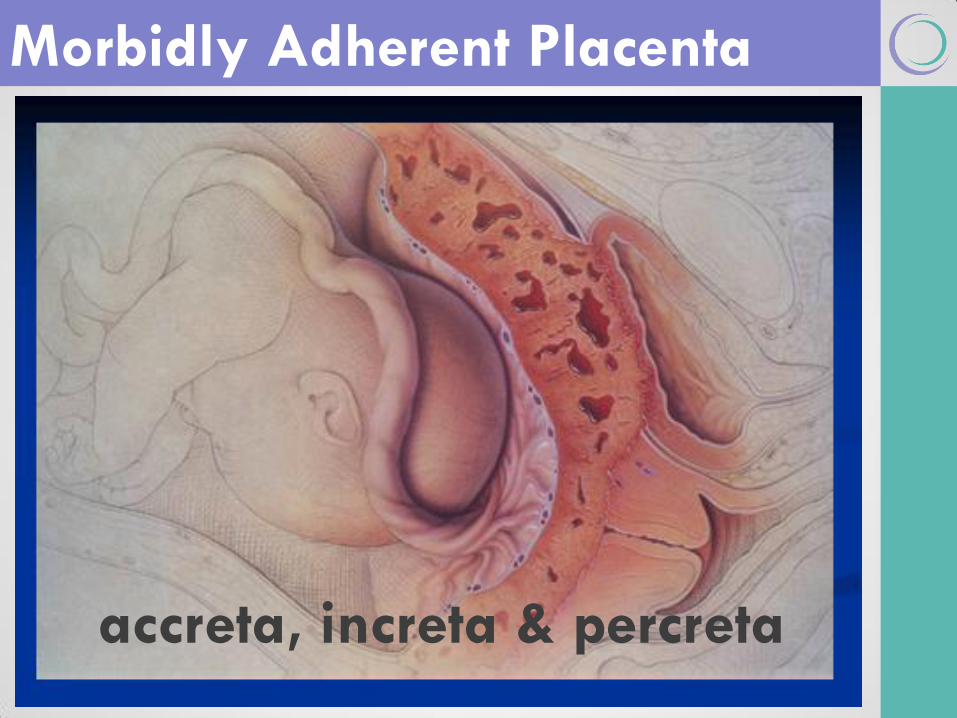
Morbidly Adherent Placenta
accreta, increta & percreta
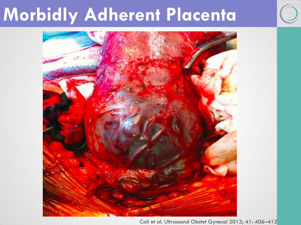
Morbidly Adherent Placenta
Cali et al. Ultrasound Obstet Gynecol 2013; 41: 406–412

Risk factors
Placenta previa – 75%
Prior cesarean section - 66%
Prior accreta
IVF (OR 13.2, p<0.0001)
Prior uterine surgery/inflammation: myomectomy, manual placental removal, D+C,
cornual resection (even endometritis)
Prior hysteroscopic surgery
Prior pelvic irradiation
Rudimentary horn with no connection to uterus

Frequency- Rising!

Prior C/S alone is a risk factor
Risk Factors
Eshkoli, AJOG 2013

Profuse hemorrhage (vaginal and/or
intra-abdominal)
Uterine rupture
Abnormal fetal
growth/oligohydramnios
Maternal death as high as 5%
Fetal/Neonatal death ~10%
Consequences
O’Brien et al. Am J Obstet Gynecol 1996;175:1632-8

Pre-delivery
Diagnosis

Morbidly adherent placenta is not
diagnosed antenatally in 50% of cases
Antenatal diagnosis of increta or
percreta
Reduced levels of hemorrhage
Reduced need for transfusion
Key Points
Fitzpatrick et al, BJOG 2013

Obliteration of retroplacental hypoechoic “clear
space”
Myometrial thickness <1mm
Vessels bridging placenta/ uterine margina
Disruption of hyperchoic placenta/ uterine
wall interface (“bladder line”)
Vessels crossing sites of interface disruption
(hypervascularity)
Main discriminators are last 2
USN Features Percreta (2008)
Wong, J Clin Ultrasound 2008; 36:551-559 (Level II-3)
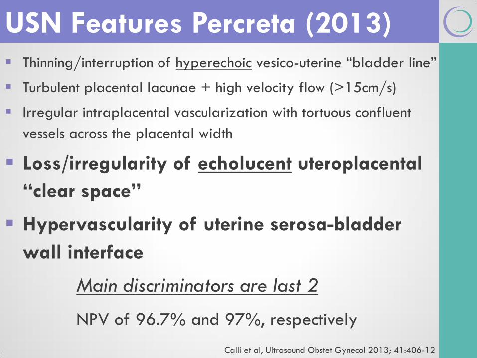
Thinning/interruption of hyperechoic vesico-uterine “bladder line”
Turbulent placental lacunae + high velocity flow (>15cm/s)
Irregular intraplacental vascularization with tortuous confluent
vessels across the placental width
Loss/irregularity of echolucent uteroplacental
“clear space”
Hypervascularity of uterine serosa-bladder
wall interface
Main discriminators are last 2
NPV of 96.7% and 97%, respectively
USN Features Percreta (2013)
Calli et al, Ultrasound Obstet Gynecol 2013; 41:406-12

Loss/irregularity of echolucent uteroplacental “clear space”
Loss of “clear space”
Visible echolucent
uteroplacental “clear space”
Note LACK of echolucent
uteroplacental “clear space”

Thinning/interruption of hyperechoic vesico-uterine
“bladder line”
Loss of “bladder line”
Visible
“bladder line”
Interruption of
bladder line
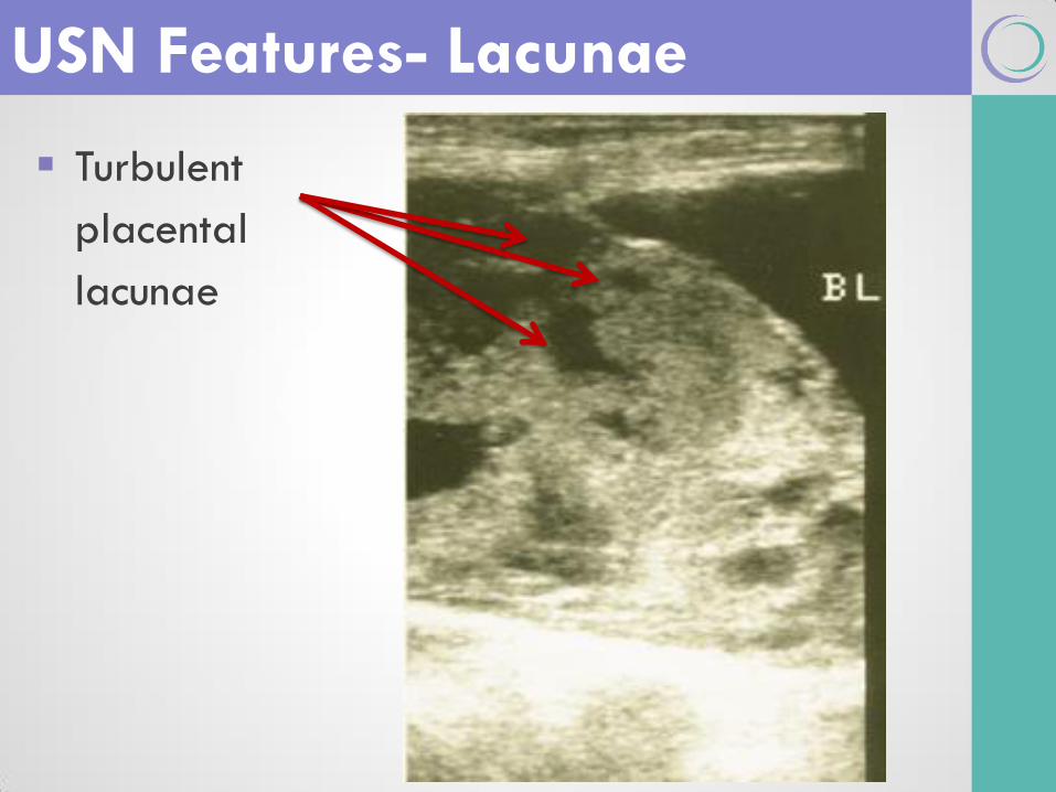
Turbulent
placental
lacunae
USN Features- Lacunae

3 features best seen by 3D power Doppler
Hypervascularity of uterine serosa-bladder wall interface
Turbulent placental lacunae + high velocity flow (>15cm/s)
Irregular intraplacental vascularization with tortuous confluent
vessels across the placental width
Significant associations with percreta
Numerous coherent vessels
Sensitivity 97%; Specificity 92%; PPV 76%
USN Features- Doppler
Shih et al. Ultrasound Obstet Gynecol. 2009 ;33:193-203 (Level III)

Percreta
USN Features- Power Doppler
Shih et al. Ultrasound Obstet Gynecol. 2009;33:193-203
Normal

Management:
Pre-Op

OR Staff and Blood Bank Staff
Anesthesia
Urology
Vascular Surgery
GYN Oncology
Interventional Radiology
Neonatology / NICU
Multidisciplinary Team

Steroids as appropriate
MgSO4 for cerebral protection if delivery
anticipated <32 weeks
In-house management?
Individualize (depending on bleeding)
If stable: admit 33 weeks, deliver 34–35 weeks O’Brien (1996): 93% >35 weeks hemorrhaged
Warshack (2010): 44% >36 weeks hemorrhaged
Robinson (2010): decision analysis, 34 weeks
Belfort (2011): 34 -35 weeks (NIH Workshop)
Antepartum

Vitamins, iron, iron-dextran ± EPO
venofer – 300mg in 250cc NaCl (0.9%) over 2-3hrs IV every other day
darbopoeitin 100 mg after second dose
Can expect 2g% increase in two weeks
Preop transfusion if necessary - < 30%
Factor VIIa availability
direct effect on Factor X leading to “thrombin burst”
Prepare for Blood Loss
Management:
Intra-Op
Preparation

Adequate IV access, A-line, central line Quad sheath
General endotracheal anesthesia +/- epidural
DVT prophylaxis (“plexipulse”) - thigh high
Shock trauma blood infusers and cell saver
Control OR & maternal body temperature
Cystoscopy & ureteral stents (Eller et al, BJOG 2009)
OR Preparation- Maternal Prep

Arterial catheters? (balloons not advised, sheath for SAE may be reasonable)
Auto-transfusion (cell saver)
Pelvic pressure pack equipment
Vascular sets and other specialized equipment
OR Preparation- Blood loss Prep

Prepare for embolization
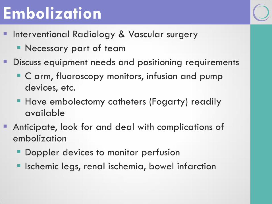
Interventional Radiology & Vascular surgery
Necessary part of team
Discuss equipment needs and positioning requirements
C arm, fluoroscopy monitors, infusion and pump devices, etc.
Have embolectomy catheters (Fogarty) readily available
Anticipate, look for and deal with complications of embolization
Doppler devices to monitor perfusion
Ischemic legs, renal ischemia, bowel infarction
Embolization
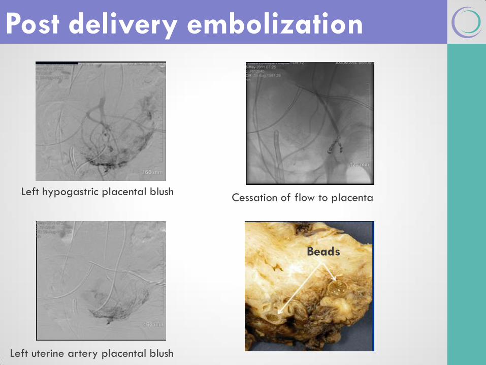
Post delivery embolization
Left hypogastric placental blush
Left uterine artery placental blush
Cessation of flow to placenta
Beads
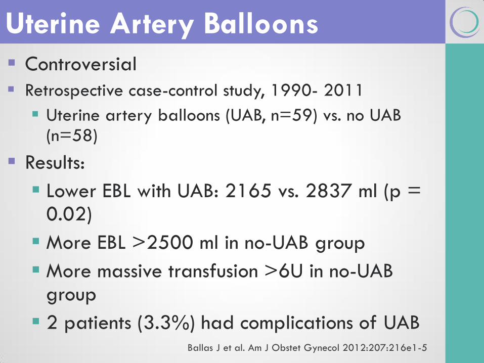
Controversial
Retrospective case-control study, 1990- 2011
Uterine artery balloons (UAB, n=59) vs. no UAB (n=58)
Results:
Lower EBL with UAB: 2165 vs. 2837 ml (p = 0.02)
More EBL >2500 ml in no-UAB group
More massive transfusion >6U in no-UAB group
2 patients (3.3%) had complications of UAB
Uterine Artery Balloons
Ballas J et al. Am J Obstet Gynecol 2012:207:216e1-5
Management:
Operative

Staged hysterectomy with embolization
Cesarean hysterectomy without placental
removal
Conservative treatment
Cesarean section, close up & wait
(preservation of fertility)
Post-surgery
Management: Approaches

Femoral artery catheterization Classic C/S
Uterine/placental embolization Hysterectomy
Cohort study of retrospective & prospective data
n=26 total, n=8 staged procedures
Staged Hysterectomy
Angstmann et al, Am J Obstet Gynecol 2010
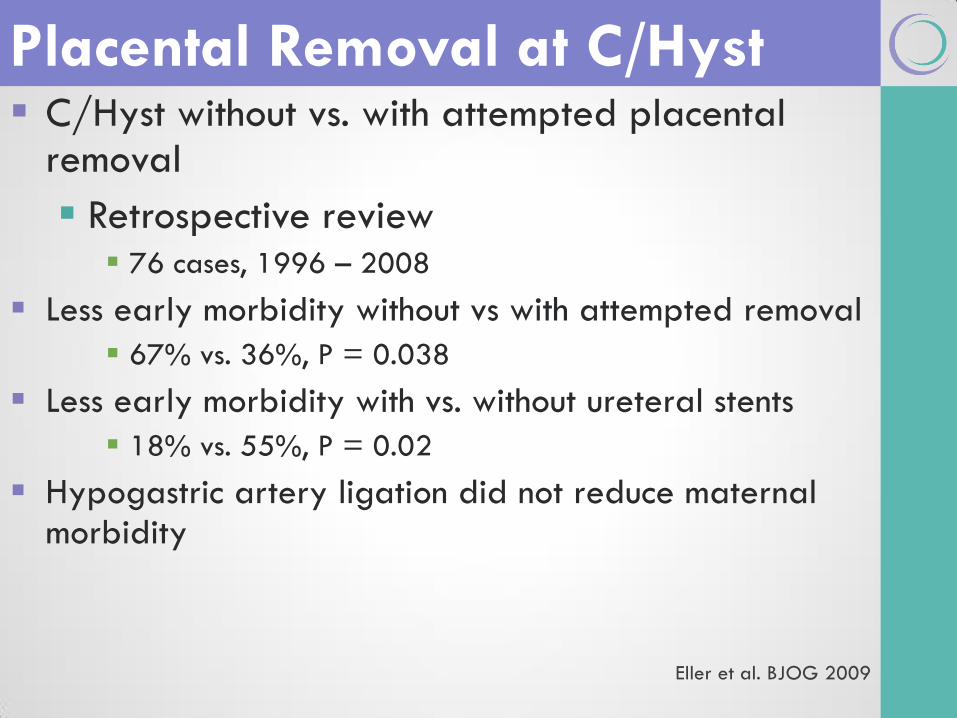
C/Hyst without vs. with attempted placental removal
Retrospective review 76 cases, 1996 – 2008
Less early morbidity without vs with attempted removal
67% vs. 36%, P = 0.038
Less early morbidity with vs. without ureteral stents
18% vs. 55%, P = 0.02
Hypogastric artery ligation did not reduce maternal morbidity
Placental Removal at C/Hyst
Eller et al. BJOG 2009

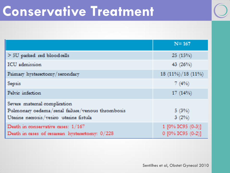
Leave placenta in situ (partially
or totally) in order to preserve
fertility
Retrospective review
1993-2007
n=167
Conservative tx. successful in
78.4%
Likely more risky- individualize
to patient desires/risks
Conservative Treatment
Sentilhes et al, Obstet Gynecol 2010
Conservative Treatment
Sentilhes et al, Obstet Gynecol 2010

Conservative Treatment
Algorithm
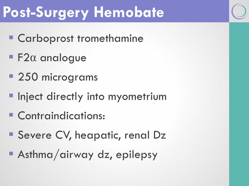
Carboprost tromethamine
F2α analogue
250 micrograms
Inject directly into myometrium
Contraindications:
Severe CV, heapatic, renal Dz
Asthma/airway dz, epilepsy
Post-Surgery Hemobate

Anti-folate metabolite Halts rapid cell growth Three words: Don’t do it
Toxicity without benefit Placental cells
No longer dividing rapidly Huge tissue mass
Post-Surgery- Methotrexate

Suggested
Approach

Be prepared!
Suggested Approach
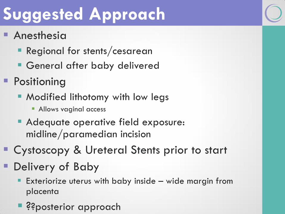
Anesthesia
Regional for stents/cesarean
General after baby delivered
Positioning
Modified lithotomy with low legs Allows vaginal access
Adequate operative field exposure: midline/paramedian incision
Cystoscopy & Ureteral Stents prior to start
Delivery of Baby Exteriorize uterus with baby inside – wide margin from
placenta
??posterior approach
Suggested Approach

Leave placenta in situ and close hysterotomy
Modified radical hysterectomy technique – wide margins
Suggested Approach- Hyst
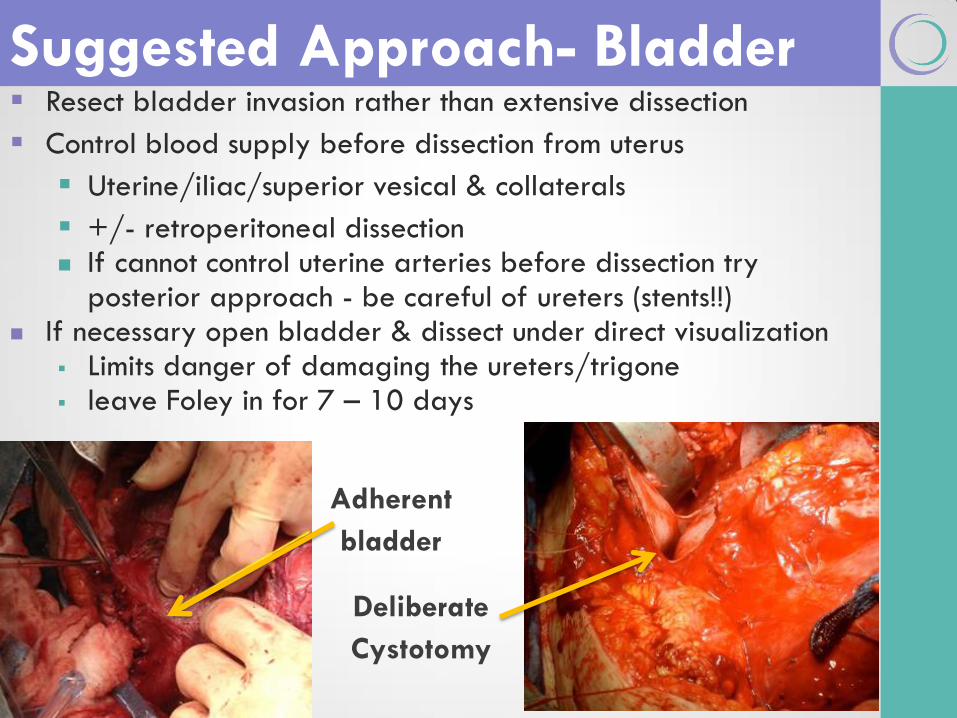
Resect bladder invasion rather than extensive dissection
Control blood supply before dissection from uterus
Uterine/iliac/superior vesical & collaterals
+/- retroperitoneal dissection If cannot control uterine arteries before dissection try
posterior approach - be careful of ureters (stents!!) If necessary open bladder & dissect under direct visualization
Limits danger of damaging the ureters/trigone leave Foley in for 7 – 10 days
Suggested Approach- Bladder
Adherent
bladder
Deliberate
Cystotomy

Pack vagina (Pelosi 1999) or “EEA” device (unpublished)
Suggested Approach- Packing

Summary
Morbidly adherent placenta is an
increasingly common problem
Prior cesarean with previa greatest risk
Pre-delivery diagnosis is important for
planning, optimal management
Optimize patient well-being: anemia rx
Optimize fetal well-being: MgS04, steroids
Keys: mullti-disciplinary team, adjunctive care
Summary

Only 50% will be diagnosed before delivery
Prior c/s & previa are the biggest risk factors
Watch for ultrasound signs
Delivery between 34-35 weeks
No substitute for staff preparation!
Variety of approaches- suggested:
Ureteral stenting, midline incision
Uterine incision away from placenta
Do not attempt to remove placenta
Modified radiacal hysterectomy
Summary- MAP

Evidence

Ultrasound diagnosis (Level II-3)
Wong, J Clin Ultrasound 2008; 36:551-559 (Level II-3)
3D power Doppler diagnosis (Level III)
Shih et al. Ultrasound Obstet Gynecol. 2009 ;33:193-
203 (Level III)
Evidence

U.S. Preventive Services Task Force (USPSTF)[edit]
Systems to stratify evidence by quality have been developed, such as this one by
the United States Preventive Services Task Forcefor ranking evidence about the
effectiveness of treatments or screening:[30]
Level I: Evidence obtained from at least one properly designed randomized controlled
trial.
Level II-1: Evidence obtained from well-designed controlled trials without randomization.
Level II-2: Evidence obtained from well-designed cohort or case-control analytic studies,
preferably from more than one center or research group.
Level II-3: Evidence obtained from multiple time series with or without the intervention.
Dramatic results in uncontrolled trials might also be regarded as this type of evidence.
Level III: Opinions of respected authorities, based on clinical experience, descriptive
studies, or reports of expert committees.
Ex of Evidence

Thank You for Your Attention!
Planning Committee
Mike Foley, Director Shad Deering, co-Director
Helen Feltovich, co-Director Bill Goodnight, co-Director
Loralei Thornburg, Content co-Chair Deirdre Lyell, Content co-Chair
Suneet Chauhan, Testing Chair Mary d’Alton
Daniel O’Keeffe Andrew Satin
Barbara Shaw

Additional materials
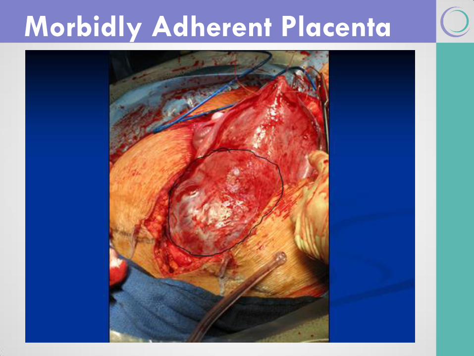
Morbidly Adherent Placenta

Please name finding (loss of clear space? No bladder line?)
USN Features

3D power color Doppler: percreta
USN Features
Shih et al. Ultrasound Obstet Gynecol. 2009;33:193-203

Gross pathology (same patient): percreta
USN Features
Shih et al. Ultrasound Obstet Gynecol. 2009;33:193-203

“Eye of the Typhoon”
USN Features
Yang et al, Arch Gynecol Obstet 2012
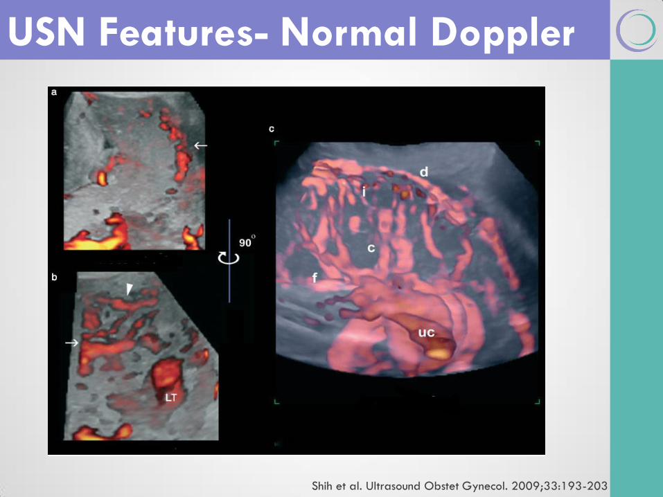
USN Features- Normal Doppler
Shih et al. Ultrasound Obstet Gynecol. 2009;33:193-203

3D power color Doppler: percreta
USN Features- Percreta Doppler
Shih et al. Ultrasound Obstet Gynecol. 2009;33:193-203
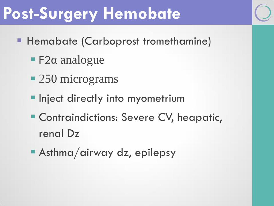
Hemabate (Carboprost tromethamine)
F2α analogue
250 micrograms
Inject directly into myometrium
Contraindictions: Severe CV, heapatic,
renal Dz
Asthma/airway dz, epilepsy
Post-Surgery Hemobate

Post-Surgery Hemobate

Suggested Approach

OR Preparation

USN Features

If not possible to control the uterine arteries before the bladder dissection
Try posterior approach - be careful of ureters (stents!!)
Suggested Approach- Bladder