amino-terminal sequences of the major tryptic - cancer research
TRANSCRIPT

[CANCER RESEARCH 38, 2199-2208, August 1978]0008-5472/78/0038-OOOOS02.00
Amino-terminal Sequences of the Major Tryptic Peptides Obtained from
Carcinoembryonic Antigen by Digestion with Trypsin in the Presenceof Triton X-1001
John E. Shively,2 Michael J. Kessler,3 and Charles W. Todd
Division of Immunology, City of Hope National Medical Center, Duarte, California 91010
ABSTRACT
Sequence studies of Carcinoembryonic antigen (CEA)were initiated to obtain direct chemical evidence regarding the structure of CEA for comparison with that of CEAcross-reacting antigens. To obtain, purify, and performamino acid sequencing on mg amounts of CEA (a glyco-protein with a molecular weight of 180,000, composed of60% carbohydrate by weight), we needed to develop newapproaches and refine existing techniques. This reportdescribes the procedures developed during the course ofthis study and presents initial results. Trypsin digestion inthe presence of 0.25% Triton X-100 produced seven majorglycopeptide fragments that were separated and purifiedby high-pressure liquid chromatography on ion-exchangeresins followed by Sephadex gel chromatography. NH,-terminal sequences were determined on 1- to 2-mgamounts of the glycopeptides by high-pressure liquidchromatography for the analysis of phenylthiohydantoinderivatives of amino acids. Four peptides gave sequencesthrough 20 cycles of Edman degradation, one gave sequences through 13 cycles, and two gave sequencesthrough 10 cycles. Three of these peptides also gavesequences of up to 8 to 13 cycles for a minor component.The abrupt halt in Edman degradation for each peptidewas interpreted as the failure to sequence beyond acarbohydrate substitution on the polypeptide chain. Sinceeach sequence obtained was unique, the results substantiate the claim that the polypeptide chain of CEA is adefinite chemical entity and that the microheterogeneityresides in the carbohydrate portion of the molecule.
INTRODUCTION
CEA4 is a glycoprotein (26) with a molecular weight of
approximately 180,000 (41), composed of about 60% carbohydrate and 40% protein by weight (46). Extensive studies on CEA have revealed details of its physical structure bymolecular sizing techniques and by electron microscopy(41), its carbohydrate structure by periodate oxidation (8,21) and by methylation-linkage analysis (7, 21), and its NH2-
1Supported in part by National Cancer Institute Grants CA16434 andCA19163 from the National Large Bowel Cancer Program.
2To whom requests for reprints should be addressed, at Division ofImmunology, City of Hope National Medical Center, 1500 East Duarte RoadDuarte, Calif. 91010.
3 Present address: S. U. N. Y. at Buffalo, Department of Cell and MolecularBiology, Gary Hall, Buffalo, N. Y. 14214.
"The abbreviations used are: CEA, Carcinoembryonic antigen- SOSsodium dodecyl sulfate: PTH, phenylthiohydantoin.
Received January 23, 1978; accepted April 20, 1978.
terminal amino acid sequence (4, 45) by Edman degradation. In addition these studies and others (22, 31, 52, 53)support the current view that the antigenic determinantsrecognized in the radioimmunoassay for CEA reside in theprotein portion of the molecule. Since CEA was reportedlyshown to be heterogeneous in terms of charge and molecular weight polydispersity (3, 5, 17, 23, 42), a structuralfeature attributable to its extensive carbohydrate microheterogeneity, it is reasonable to assume that the antigenicdeterminants should reside in an invariant portion of themolecule, namely, the polypeptide chain.
CEA was first described as a tumor-associated antigen byGold and Freedman (18, 19) because it was detected intumors of the digestive system and in fetal gastrointestinaltissue. Subsequently, several investigators (9, 20, 22, 30, 50)have isolated CEA cross-reacting antigens from normaladult tissues. Recent evidence (33, 34, 48) has shown thatmany of these substances differ in chemical compositionand molecular size from CEA. These findings have demonstrated a real need to characterize chemically the CEA-reactive molecules from different sources.
Significant progress in the structural studies of CEA andCEA cross-reacting antigens has been made in this laboratory. CEA isolated from normal adult colon washings (14)has been shown to be structurally identical to that isolatedfrom liver métastasesof colon adenocarcinoma (40), butsignificant differences were found in the lower-molecular-weight CEA cross-reacting antigens isolated from normal(16) and malignant tissues (25).
Structural studies on the protein portion of CEA and CEA-related antigens are necessary to establish the basis fortheir observed antigenic differences. The results of suchstudies may lead to the development of a more specific testfor CEA in malignant and other diseased tissues that showelevated CEA levels. Protein sequence studies on CEA arehampered by unusual technical problems since CEA contains over 600 amino acids and contains over 50% carbohydrate. The carbohydrate, all of which is linked to proteinthrough /V-acetylglucosamine to asparagine linkages, isdifficult to remove by chemical cleavage (7, 8, 52). Specificcleavage of peptide bonds by cyanogen bromide treatmenthas been unsuccessful due to the lack of methionine inCEA and the occurrence of hydrolytic cleavage, whichoccurs at low pH (presumably aspartylproline bonds). Theuse of the cyanylation method of cleaving cysteine peptidebonds by the method of Jacobson ef al. (24) formed A/blocked peptides that could not be sequenced (12, 28).
In the present study it was found that glycopeptidescapable of sequence analysis could be obtained from CEA
AUGUST 19782199
Research. on December 30, 2018. © 1978 American Association for Cancercancerres.aacrjournals.org Downloaded from

J. E. Shively et al.
by trypsin digestion in the presence of 0.25% Triton X-100and subsequent separation by ion-exchange high-pressureliquid chromatography. In some cases the resulting glyco-
peptides could be sequenced through 20 degradation cycles before the analysis came to an abrupt halt, presumablydue to the presence of a carbohydrate chain linked to anasparagine residue. Although this report constitutes only astart on the amino acid sequence analysis of CEA, it showsfor the first time that CEA can be broken up into isolataleglycopeptide fragments that are amenable to NH2-terminal
amino acid sequencing. These results support the conclusion, suggested originally by the finding of a single, uniformNH,-terminal amino acid sequence for CEA (45), that indeed
the polypeptide portion of CEA is a definite chemical entity.The authors intend to use the techniques developed here toanswer specific questions about the structural featuresresponsible for the antigenic differences observed amongthe various CEA and CEA cross-reacting entities.
MATERIALS AND METHODS
Preparation of CEA. Isolation and purification of CEAwas carried out as previously described (6) and was measured by double-antibody radioimmune assay (13) with a"Co volume marker (15). In addition, contaminating traces
of mucopolysaccharides were removed by concanavalin ASepharose affinity chromatography as previously described(36); recovery of CEA from this step was 78%. CEA from 1tumor source was used throughout these studies.
Neuraminidase Digestion. Vibrio cholerae neuramini-
dase digestion was performed according to Coligan andTodd (8). A control sample was run in which CEA wasomitted from the reaction. No material capable of sequenceanalysis was found in the control sample.
Reduction and Alkylation. CEA (210 mg) treated withneuraminidase and dissolved in 30 ml of 9.0 M urea bufferedwith 0.1 M Tris-HCI, pH 8.3, and purged with N2was reduced
with 0.3 mmol of dithiothreitol for 1 hr at room temperatureand alkylated with 0.43 mmol of iodoacetamide (0.30 mCiof [14C]iodoacetamide diluted to a final specific activity of
0.70 mCi/mmol) for 1 hr at room temperature in the dark.The sample was dialyzed exhaustively against water, lyoph-
ilized, and dried over P2O5.Trypsin Digestion. CEA (218 mg) treated with neuramini
dase, reduced, alkylated, and dissolved in 15 ml of 0.25%aqueous Triton X-100 (Sigma Chemical Co., St. Louis, Mo.)
was adjusted to pH 8.1 with a dilute ammonia solution anadigested with 2.0 mg of solid u-l-tosylamido-2-phenylethylchloromethyl ketone-treated trypsin (Worthington Biochemical Corp., Freehold, N. J.) for 12 hr at 37°.pH 8.1 was
maintained by further addition of dilute ammonia as necessary. The reaction mixture was lyophilized and dried overP205 (yield, 254 mg). A sample of a,-acid glycoprotein wasobtained from Dr. Yu-Lee Hao, American Red Cross Blood
Research Laboratory, Bethesda, Md., and was used formodel studies of trypsin digestion in the presence of TritonX-100.
Amino Acid Analysis. Amino acid analysis was performedby a modification of the method of Liu and Chang (29).Samples of 100 to 200 ^g were hydrolyzed under vacuum in
heavy-walled ignition tubes at 110°for 48 hr in duplicate
with 0.5 ml of 3 Np-toluenesulfonic acid containing 0.2% 3-(2-aminoethyl)indole. The hydrolyzed samples were ana
lyzed on a Beckman 121H amino acid analyzer (BeckmanInstruments, Inc., Palo Alto, Calif.) The basic amino acidsand amino sugars were eluted from a 20- x 0.9-cm column
of PA35 (Beckman) with sodium citrate buffer, pH 5.26 (0.4N sodium). The acidic and neutral amino acids were elutedfrom a 56- x 0.9-cm column of AA15 (Beckman) with sodiumcitrate buffers of pH's 3.10 (0.16 N sodium), 3.60 (0.02 N
sodium), and 4.20 (0.20 N sodium).Carbohydrate Analysis. Carbohydrate analysis was per
formed on the trimethysilyl derivatives of sugar methylglycosides (37). Sialic acid was determined by the previously described method and by the method of Warren (51).
SDS/Gel Electrophoresis. SDS/gel electrophoresis wasperformed on either 6 or 12% polyacrylamide gels according to the method of Swank and Munkres (44). Gels wereroutinely stained with Coomassie blue or periodic acid-Schiff reagent (38). For location of 14C-labeled peptideswith CEA activity, portions of the extracts of 1-mm-thick gelslices shaken overnight in 0.5 ml of phosphate-buffered
saline (0.075 M sodium phosphate/0.075 M NaCI; pH 7.2)were counted.
Isoelectric Focusing. Isoelectric focusing was performedon an LKB 8100 electrofocusing column of 110-ml capacity
according to the method of Vesterberg and Svensson (49).LKB carrier ampholytes (1%; LKB Instruments, Inc., Rock-
ville, Md.) were used in the pH ranges of 2.5 to 4, 4 to 6,and 5 to 8.
Peptide Separation Procedures. Ion-exchange separa
tions were performed on either DC4A cation or DA8X8 aniónexchangers (Durrum Instrument Corp., Palo Alto, Calif.)with a DuPont Model 830 high-pressure liquid Chromato
graph. Gel filtration was performed on either SephadexG-50 or G-100 (Pharmacia Fine Chemicals, Inc., Piscataway,N. J.). Aliquots were analyzed for 14C-labeled peptides byliquid scintillation counting on a Beckman LS-330 scintilla
tion counter with an Aquasol (New England Nuclear, Boston,Mass.) cocktail. Free amino groups were detected by reaction with fluorescamine (Hoffmann-La Roche Inc., Nutley,
N. J.). Portions of 10 /JL\were evaporated to dryness underN, in 0.6- x 5-cm tubes and mixed with 50 /¿Iof 0.2 M
pyridine and 50 /¿Iof 2 mg fluorescamine per ml acetone(w/v); the relative fluorescence was read in a 0.10-ml cuvet
on an Aminco fluorometer (excitation, 364 nm; emission, 458nm).
Two-Dimensional Mapping Procedures. Chromatography in the vertical dimension with butyl alcohol/acetic acid/water (4/1/5, v/v) for 24 hr and electrophoresis in thehorizontal dimension with pyridine/acetic acid/water (20/2/378, v/v) for 24 hr at 3000 volts was performed according tothe procedure of Bennett (2).
Sequencing Procedures. NH,-termmal peptide sequences were performed with a modified dimethyl allylam-
ine program (Beckman 102974) on a Beckman Model 890Csequencer equipped with a dry ice/acetone trap on its low-
vacuum pump. The program modifications were the following. Benzene/ethyl acetate (1/1, v/v) was used as S1. N2delivery through S2 at Step 14 was 80 sec. N2 dry at Step 17was 80 sec. Steps 38 and 39 were interchanged (new Step
2200 CANCER RESEARCH VOL. 38
Research. on December 30, 2018. © 1978 American Association for Cancercancerres.aacrjournals.org Downloaded from

Tryptic Peptides from CEA
38 was cleavage reaction for 80 sec, and new Step 39 wasN2delivery through S3 for 200 sec). For improvement of thesensitivity of the analysis of PTH-amino acids by thin-layerchromatography, 1 mCi of phenyliso[35S]thiocyanate was
added to 16 mmol of phenylisothiocyanate in R1 of thesequencer (final specific activity, 0.06 mCi/mmol). Thethiazolinone derivatives of the amino acids were convertedto their PTH derivatives by a modification of the procedureof Laursen (27). The PTH derivative of aminoisobutyric acid(4.5 nmol) was added to the chlorobutane solution of eachthiazolinone as an internal standard. Samples were evaporated to dryness on a Buchler Evapomix in 1.5- x 15-cmconical tubes fitted with ground-glass joints, mixed with200 fji\ of 20% aqueous trifluoroacetic acid containing 0.05mg dithiothreitol per ml, allowed to react 15 min at 55°
under N2, and evaporated to dryness on the Buchler Evapomix. The PTH's were extracted 3 times with 25 /¿Iof 1,2-
dichloroethane/methanol (7/3, v/v) according to themethod of Wittman-Liebold (54), transferred to a 0.4- x 4-cm polyethylene microfuge tube (Analytical Aids, Wood-side, Calif.), mixed with 25 ¿¿Iof water, and stored at -20°
until analyzed (the PTH is in the bottom, organic phase).This procedure has the following advantages, (a) The incorporation of an internal standard in the method gives moreaccurate quantitation and monitors transfer losses, (o) Themild conversion conditions prevent amide hydrolysis ofglutamine and asparagine derivatives and give higher yieldsof serine and threonine. (c) The water extraction stepremoves traces of impurities that would otherwise contribute to analysis background levels. The PTH's were sepa
rated and quantitated by gas chromatography according tothe method of Pisano and Bronzert (35) on a Hewlett-Packard 571OA gas Chromatograph and by high-pressureliquid chromatography on a Waters Associates liquid Chromatograph (Waters Associates, Inc., Milford, Mass.), bothequipped with an Autolab System IV peak integrator. Excellent resolution of PTH derivatives of 17 amino acids wasachieved on C1B^Bondapak (Waters Associates) reversephase columns by either Program 1, a linear gradient from0 to 35% Component B over 30 min, or Program 2, a concavegradient from 10 to 40% Component B over 30 min. In bothcases the flow rates were 2 ml/min, Component A was 0.01M^odium acetate/acetonitrile buffer (95/5, v/v; pH 7.6), andComponent B was 100% acetonitrile. These separations aresimilar to those reported by Zimmerman ef al. (55) andDowning and Mann (10). In addition the thin-layer Chromatographie procedure of Summers ef al. (43) was usedwith 2,5-bis-2-(5-ferf-butyl benzoxazolyljthiophene (Packard Instrument Co., Inc., Downers Grove, III.) as the fluor.Thin-layer chromatograms were recorded and compared byUV photography and by autoradiography of the [35S]PTHderivatives on X-ray film. Reagents, solvents, and spermwhale apomyoglobin obtained from Beckman were usedthroughout this work.
RESULTS
Trypsin Cleavage. Initial treatment of CEA (treated withneuraminidase, reduced, and alkylated) with trypsin gaveonly a few peptides in low yields when it was monitored by2-dimensional mapping, gel chromatography, SDS/poly-
acrylamide gel electrophoresis, and ion-exchange chromatography. The failure of trypsin alone to digest denaturedCEA was attributed to the protection of lysyl and arginylpeptide bonds by the presence of extensive amounts ofcarbohydrate on CEA. Model peptide mapping studies ona, acid glycoprotein treated, reduced, and alkylated byneuraminidase established that trypsin digestion was moreeffective in the presence of 0.25% Triton X-100. Similarly,this result was confirmed with CEA. The tryptic map of CEA(not shown here) reveals the formation of 5 to 10 slow-moving, poorly resolved peptides from CEA. In contrast,trypsin digestion of CEA performed in the absence of TritonX-100 gave very few large peptides and no smaller peptides.The tryptic peptides of CEA were eluted and analyzed for[14C]cysteine by scintillation counting, for carbohydrate
content by the phenol/sulfuric acid method of Dubois ef a/.(11), and for NH2-terminal amino acids by the method ofNeuhoff (32). The results of these analyses (not shown here)revealed that the slower-moving peptides were composedof variable amounts of carbohydrate and cysteine, the[14C]carboxyamidomethyl derivative. Each peptide contained several NH2-terminal amino acids. In general the 2-dimensional mapping procedure was found inadequate forthe separation of large glycopeptides.
The attempted separation of CEA tryptic peptides onSDS/gels is shown in Chart 1. Untreated CEA migrates as asingle, diffuse band on 6% gels and as a substantiallysharper band on 12% gels. An increase in the separation ofCEA tryptic peptides is apparent in 12 compared to 6% gels.
400
4.0 6.0 8.0 10.0 12.0
Mobility in cmChart 1. SDS/polyacrylamide gel electrophoresis profiles of CEA tryptic
peptides. Samples were run on either 6 or 12% polyacrylamide gels (0.5 x 18cm). The standards were lysozyme (14,000), 0-lactoglobulin (18,000), carbonic anhydrase (32,000), ovalbumin (45,000), and bovine serum albuminmonomer (68,000) and dimer (136,000). Arrow, migration of CEA treatedwith neuraminidase, reduced, and alkylated: bars, molecular weight rangesfor unfractionated CEA tryptic peptides, as detected by either Coomassieblue staining or location of [14C]carboxyamidomethyl cysteine. The identification of the peptides (T1A1. etc.) corresponds to a separate experimentin which the peptides purified by ion-exchange and gel filtration chromatography were run on separate gels and detected by Coomassie blue staining orlocation of ["C]carboxyamidomethyl cysteine.
AUGUST 1978 2201
Research. on December 30, 2018. © 1978 American Association for Cancercancerres.aacrjournals.org Downloaded from

J. E. Shively et al.
Results similar to these were obtained with a variety ofpeptide detection methods: iodination or fluorescent labeling before separation; Coomassie blue or periodate-Schifi
staining after separation. Peptide bands were eluted fromgels and analyzed for [14C]carboxyamidomethyl cysteine
and NIVtermmal amino acids. A minimum of 10 trypticfragments could be estimated from these experiments.
Isoelectric focusing of CEA tryptic peptides is shown inChart 2. Although CEA treated with neuraminidase, reduced, and alkylated does not give a single sharp peak onisoelectric focusing, it is clear that the profile of its trypsindigest is different and reveals at least 10 separate peaks.The acidic nature of the tryptic peptides was confirmed bythe 2-dimensional tryptic map and by the results of ion-
exchange chromatography. Microanalysis (<5 mg) of thepeptides obtained from either column or polyacrylamide gelisoelectric focusing proved difficult (complete removal ofampholytes and urea from the sample was the major problem).
The most practical preparative method for separation andpurification of the tryptic peptides of CEA was ion exchangefollowed by gel permeation chromatography. Trial separations of the tryptic fragments of sperm whale apomyo-globin on a DC4A cation exchanger by high-pressure liquid
chromatography and fluorescamine detection of elutedpeptides gave 16 peptides in a run time of only 2 hr. However, a similar separation of a, acid glycoprotein treatedwith neuraminidase, reduced, and alkylated gave a largebreakthrough peak of unseparated acidic peptides and6 well-resolved basic peptides. Further analysis revealedthat the acidic a, acid glycoprotein peptides were glyco-
peptides of relatively high molecular weight, which couldbe eventually separated on DA8-X8 anion-exchange resin.Since the CEA glycopeptides were of higher molecularweight (75,000 dallons) and comprised larger amountsof carbohydrate, the separation of the CEA tryptic peptideswas more difficult to achieve than were those obtained from a, acid glycoprotein. The separation outlined in Chart 3 details the purification of 8 tryptic fragments from CEA. The unfractionated digest gave a poorlydefined separation on gel permeation chromatography
alone but, when it was run first on a cation exchanger,it yielded a breakthrough peak (T1) and a retained peak(T2), each of which yielded a breakthrough and a retainedpeak on anión exchanger (T1A, T2A, T1B, and T2B, respectively). Each of the 4 peptide mixtures separated by ionexchange gave 2 well-defined peaks on subsequent gel
permeation chromatography. The gel permeation resultsshown in Chart 3 gave lower estimations of molecularweights than that obtained by SDS/gel electrophoresis(Chart 1). However, the large effect of carbohydrate substitution in glycopeptides on either migration in Sephadexchromatography or SDS/gel electrophoresis precludes accurate molecular weight determinations.
The method of purification shown in Chart 3 was adoptedfor large-scale preparation of CEA tryptic peptides. Table 1
summarizes peptide yields for this procedure. The apparentlow yields for the initial DC4A cation exchange step ispartially due to the conversion of ammonium salts of peptides formed during trypsin digestion in ammonia buffer totheir respective free acid derivatives.
Amino Acid and Carbohydrate Analyses. Table 2 presents the amino acid and carbohydrate analyses for CEAand its tryptic fragments. In general the high-molecular-
weight glycopeptides resemble intact CEA in overall composition, except Peptides T1B2 and T2B2, which are low incarbohydrate, and Peptide T2A2, which was obtained insuch low yield and contaminated with foreign matter thatfurther analysis was impossible.
Sequence Results. It was necessary to refine our sequencing procedures to obtain meaningful quantitativedata on 2- to 3-mg amounts of the CEA tryptic peptides.
Improvements in the sequenator programs, conversion procedure, and PTH identification detailed in "Materials andMethods" made this possible. The high-pressure liquid
Chromatographie separation and quantitation of PTH derivatives was especially useful in distinguishing a major froma minor sequence in several of the peptides that were laterfound to be a mixture (in all cases the minor sequencecomprised 30% or less of the mixture). An example of theresolution and sensitivity achieved with the high-pressure
liquid Chromatographie system is shown in Chart 4, along
Chart 2. Isoelectric focusing of CEA tryptic peptides. Five mg of CEA tryptic digest mixed with ' " l-labeled digest and '"l-labeled CEA treated with
neuraminidase, reduced, and alkylated were applied to a 1- x 40-cm column, and isoelectric
focusing was performed over a pH range of 2.5 to8.0 for 72 hr.
50 60 TO
ml Eluted
90 100 110
M
«O
3.0
IO
100
SO
5«o ú
2202 CANCER RESEARCH VOL. 38
Research. on December 30, 2018. © 1978 American Association for Cancercancerres.aacrjournals.org Downloaded from

Tryptic Peptides from CEA
4.0
2.0
4.0
2.0
2.0
1.0
2.0
1.0
2.0
1.0
A ' \
A»holeD.g«.
on OC44
Fraction T lon DA6X8
â„¢¿�
Froclion T2on DA8X8
4.0
2.0
ÃŒJO
0.5
i Table 1
Summary of isolation and yields of CEA tryptic peptides
40
-Froction TÕA
TIA2 °"s«PhG5°
2.0
1.0
2.0
1.0
,BS
05 IS 20 25
Effluent mlChart 3. Separation of CEA tryptic peptides. A, the peptide mixture (27.4
mg) was applied to a 0.21- x 100-cm stainless steel column packed withDC4A cation exchanger at 60°,with 1100 psi and a flow rate of 1 ml/mm, and
eluted in steps, first with 0.01 M acetic acid and then with pyridinium acetate(PyrAc) buffer (0.2 M pyridine/4.5 M acetic acid; pH 3.1). B and C the peptidemixtures T1 (37.2 mg) and T2 (17.6 mg) were separately applied to a 0.21- x100-cm stainless steel column packed with DA8-X8 aniónexchanger at 40°,with 3000 psi and a flow rate of 0.5 ml/min, and eluted in steps, first with 0.1M A/-ethyl morpholine/0.2 M a-picoline/0.1 M pyridine buffer (pH 8.0) and thenwith pyridinium acetate buffer (2.0 M pyridine/2.4 M acetic acid; pH 5.0). D toG, peptide fractions T1A, T2A, T1B, and T2B were applied to either SephadexG-50 (Seph G50, fine) or G-100 (Seph G100; 0.60- x 48-cm) columns, withflow rates of 0.2 ml/min, and eluted with 0.10 M acetic acid. Arrows, elutionvolumes of the standards BSA (bovine serum albumin), RNase (ribonucle-ase), Ala-Val (L-alanyl-L-valine), and CEA (12*l-labeledCEA). In all cases 0.4-ml fractions were collected, and two 0.01-ml aliquots were withdrawn foranalysis. One fraction was counted for "C. whereas the other was reactedwith fluorescamine and the relative fluorescence was determined. Bars indicate fractions pooled for sequence determination.
with several cycles of an actual sequence. Unidentifiedpeaks in Chart 4 correspond to UV-absorbing impuritiesthat do not change from cycle to cycle and that are generallyencountered when sequencing 2 mg or less of protein. ThePTH-amino acids that could not be separated by thissystem were identified by either gas or thin-layer chroma-tography. The use of [35S]PTH-amino acids on polyamide
plates permitted the unambiguous determination of severalPTH-amino acids that are usually obtained in reducedyields.
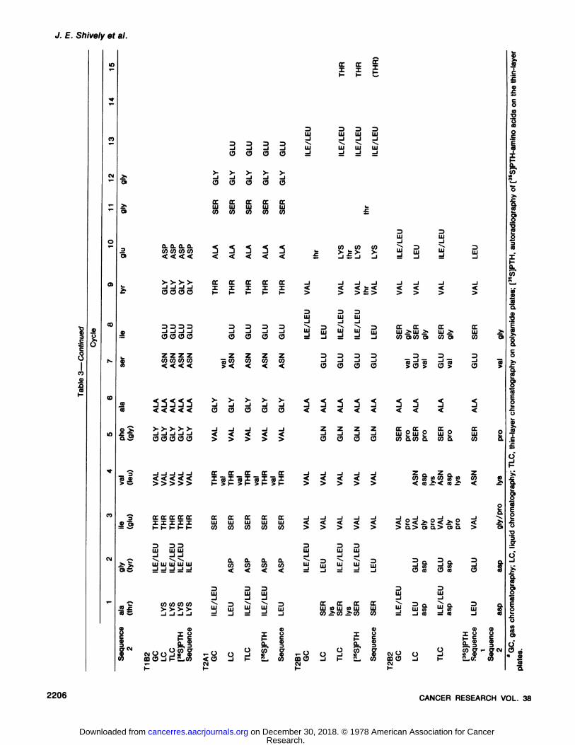
Table 3 summarizes the results obtained for the first 15cycles of Edman degradations performed on 7 tryptic peptides from CEA. Peptides T1A1, T1A2, T1B1, and T2B1 gavesequences through position 15 (yields were extremely lowpast Cycle 15), but T1B2 and T2B2 gave abruptly loweryields at Cycle 10. Absolute yields of PTH-amino acids (e.g.,Cycles 1 and 2) were 30 to 50%, based on the roughmolecular weight estimates for each peptide on SDS/gelelectrophoresis (Chart 1). Since the sequenator programused gave routine yields of 50% for sperm whale apomy-
SeparationmethodDC4Acationexchanger"DA8-X8
aniónexchanger*Sephadex
G-50Sephadex
G-100mg
applied27.4
ofwholedigest37.2
ofT117.6
ofT216.3
ofT1A12.6ofT2A19.2
ofT1B6.9ofT2BPeptideT1T2TotalT1AT1BTotalT2AT2BTotalT1A1T1A2TotalT2A1T2A2TotalT1B1T1B2TotalT2B1T2B1TotalYield
(mg)11.24.115.321.618.640.26.49.215.65.76.412.13.19.212.311.94.716.64.43.17.5%ofyield41155658501083652883539742573986225876345108
" Results were obtained for Batch 1 of whole tryptic digest.
Similar results were obtained for Batches 2 and 3. The percentage of yield is based on weight.
6 Results were obtained for Batch 1 of T1 or T2. Similar results
were obtained for the second batch of each. The percentage ofyield is based on weight.
oglobin, the previous yields are acceptable.The NH2-terminal amino acid determination of each of the
peptides by the dansyl procedure of Neuhoff (32) gave thesame NH2-termini as that shown in Table 3. In all cases thetryptic peptides showed varying amounts of e-dansylaminolysine.
DISCUSSION
Effect of Triton X-100. These experiments demonstratefor the first time that specific cleavage of CEA into peptidefragments capable of sequence analysis can be achieved.In principle the methodology adopted to produce, separate,and sequence the glycopeptides of CEA can be used onother high-molecular-weight, high-carbohydrate-contentglycoproteins. The use of trypsin digestion in the presenceof Triton X-100 gave higher, more uniform yields of trypticpeptides for CEA. The binding of this non-ionic detergent tohydrophobic regions of the protein perhaps unfolded thecollapsed structure of CEA treated, reduced, and alkylatedby neuraminidase [see Slayter and Coligan (41) for a discussion of the electron microscopy of CEA] enough to allowbetter trypsin binding to the polypeptide chain. This explanation of enhanced trypsin digestion is based partly onthe known binding of detergents to hydrophobic portions ofproteins and in part on the resulting NH2-terminal sequences of the tryptic fragments obtained, which are highin hydrophobic amino acids. Control experiments haveshown no change in trypsin activity on a synthetic substratein the presence of 0.25% Triton X-100.
AUGUST 1978 2203
Research. on December 30, 2018. © 1978 American Association for Cancercancerres.aacrjournals.org Downloaded from

J. E. Shively et al.
Table 2Amino acid" and carbohydrate h composition of CEA tryptic peptides
TyrosinePhenylalanineTryptophanLysineHistidineArginine
Carboxymethyl cysteineAspartic acidThreonineSerineGlutamic
acidProlineGlycine
AlanineHalf-cystine
ValineMethionineIsoleucineLeucine%
of amino acidrecoveredFucoseGalactoseMannose
D-GlucosaminerfD-Glucosaminep%
of carbohydrate recoveredCEAr5.04
2.761.172.851.893.50
1.6013.998.119.689.5210.275.25
5.540
6.250.104.238.2534.69.79.07.6
14.114.640.4T1A15.38
3.161.812.841.134.59
1.3717.9711.2311.914.985.545.58
6.390
4.6402.588.8917.56.610.76.9
14.415.238.6T1A22.54
2.6101.570.427.68
1.3921.1011.0512.355.6107.78
5.480
4.9702.1413.3232.65.57.56.5
13.013.132.5T1B14.39
2.702.121.161.252.49
1.7815.208.9711.979.509.304.81
5.820
6.5604.666.8656.96.89.17.2
12.017.135.1T1B2Mol
%3.521.70
01.451.452.64
3.4914.036.1612.1711.168.036.92
6.090
5.5509.056.5917.2%
by weight1.12.22.0
2.83.68.1T2A13.36
1.9702.060.676.88
0.5317.679.5013.045.214.596.27
5.800
5.0504.6312.7736.33.05.48.0
11.812.528.2T2A23.33
2.518.144.271.613.98
2.6210.614.238.4411.35011.01
5.970
6.0709.066.801.70.1<1T2B14.69
2.691.732.281.583.61
0.6014.297.0710.9810.3511.204.47
5.590
6.6205.257.0041.05.45.15.4
8.09.323.9T2B23.48
1.5901.491.293.93
1.1314.146.6710.4711.747.0510.54
6.570
6.8007.336.3218.81.1<1
Apparent molecular wt' 180,000 50.000 40.000 110,000 12,000 50,000 16.000 70.000 28,000" Samples were hydrolyzed in 0.5 ml of 3 N p-toluenesulfonic acid for 48 hr in duplicate at 110°under vacuum and analyzed on a
Beckman 121H amino acid analyzer.6 Samples were treated with 1.5 N methanolic HCI for 24 hr in duplicate at 80°and analyzed as trimethylsilyl derivatives on a Hewlett-
Packard 7620 gas Chromatograph equipped with a flame ionization detector.c CEA treated with neuraminidase, reduced, and alkylated.d Determined by gas chromatography.'' Determined by amino acid analysis.
! Molecular weight values calculated from migration on SDS/gels shown in Chart 1 should be considered approximate.
The potential interference of the carbohydrate chains ofCEA in the trypsin cleavage is great since there are on theaverage 80 carbohydrate chains of about 7 residues/chainpresent and >600 amino acids/mol (39). With the assumption that the carbohydrate substitution on asparagine residues is heterogeneous in terms of degree and extent ofsubstitution, there is the likelihood that different CEA molecules can be cleaved by trypsin at different arginine orlysine residues, depending on the degree of interferencecaused by carbohydrate chains. The beneficial effect ofTriton X-100 may be in part due to a lessening of thecarbohydrate steric effects.
Although CEA contains sufficient arginine and lysineresidues to produce up to 40 tryptic peptides, only 8 wereisolated. Thus it seems likely that many of these residuesare inaccessible to cleavage by trypsin. Also, possibly,several small peptides (<10 amino acids) were lost due tothe small amounts of the CEA fractionated and the fraction-ation procedure adopted. The relatively large-molecular-weight (75,000 daltons) glycopeptides obtained containedvariable amounts of carbohydrate. In each case the sequence was lost before 20 cycles of Edman degradationcould be achieved. Most probably, the sequence was lostdue to the drop in yields, which often occurs when an
asparagine linked to a carbohydrate chain is encountered(47). Amino acid analysis of CEA gives an average of 90asparagine plus aspartic acid residues/mol of CEA and,since there are 80 carbohydrate chains (all linked to proteinthrough asparagine) per molecule, the likelihood of encountering carbohydrate linked to asparagine in sequencestudies on CEA is high. Indeed, 4 of the 7 peptides se-quenced encountered asparagine before 20 degradationcycles. Peptide T2A1 contains the sequence Asn-X-Thr,which appears to be a common recognition sequence forcarbohydrate linked to asparagine in glycoproteins (1). Thefact that these asparagines were identified in the usualchlorobutane washes obtained from the sequenator suggests that these residues had little or no carbohydrate attached, since the presence of hydrophilic carbohydratesubstituents on PTH-asparagine would render these derivatives insoluble in chlorobutane.
The finding that Peptides T1A2, T1B1, and T2B2 gaveboth a major and a minor sequence demonstrates thatthese 3 peptides were not completely pure. However, in nocase did the minor sequence of these 3 peptides correspond to the sequence of the other peptides. Evidently, theminor sequences represent distinct peptides obtained inlower yield. A tentative reason for the lower yields of these
2204 CANCER RESEARCH VOL. 38
Research. on December 30, 2018. © 1978 American Association for Cancercancerres.aacrjournals.org Downloaded from

Tryptic Peptides from CEA
CO CU "D $C£
1 i iì{¡ili"O
:zz Q)co•
lati§1 p gs* u)—¿�•^- to C>,Q)^ fl) fl)u||IN.—
A y) Q}¡¡TÃül*(/3
O flj ¿¿Q)«CO O. CCïiiïl">,
-C .£5 Oyi
-o! *-i-C•¿�-£ CD_£*^.2"ai ««O—¿�£ coc—T3 ** 0> S
O C C U0I:SHî*S
^soà ie Sgco
¿ Ol O t .O« S 2 .5 gcoS
1 •¿�i ïAQ.CD̂ 9üa§l£
SL.u1 u £ S«uÃ
« á1$Õ"?Wc fi »IiÜ«"O ^u£c
Q) O n+*"o ai 8c05à £ £ 3'S"5jQ?c &.c»fgffli™
O ^ ^ £'7ñ0)
Q) C 5 2 *"Oiflfiifg
a co -D SCDil
nüü£ *" a) CT*ggf
|| »V«
£'5.2 ^«CCD CO C *"*cDc
E o cov¡°>^=5.c >•gj£"""^>ci a SS^^ X o araE
û-£52.E185 2 8iiâ„¢¿�o 5o^ÛJ CO Ãico7
a E £cS«2 »fe¡|i||fil
>, _'«o
co â„¢¿�^CO1 "OOa
r: c c £*^£^8CD
.E £T-1
j i *>llcgSa
co o §c
HUÕog •¿�§.0*-m
d) 'w (OSoH^sI
.*--i "O (Q mQ)o
o .SìO) Mc¡SK
E 8BOOintco^cg-0a>Odscom„coCMer
er1-':'.¡ >-^~á1
co t-t-•z.
-z.o
o_l
_J<ñ â„¢¿�^ < STo>> CO O) > CO>LU
_ LU LULUx2 xxiCLaCL CLCL"Z.
"Z.C_J 3 _l3O)C3 oiO01.-
"Z."Z.S
cococo<<er
ce¿"l—h-:
"Z- aza¿
< co <co—
J ^ —¿�J ^< <<«<£/)>>>>•>>•co
coco> >->OOQLU
LU LU LUII IXCL Q. CLCL3
33LULULULU
LU LU C LUCce
er ererLULU 75 LU 7gLUcoco > co >co3
33LULU LU
LU Qj LULUcr
< . ererLU—¿�1m LU LU
CO < to COCOI£<8
s g£Prr
> > >- > LU LU UJ LULU>_i_i _i_i ICOICOICOIMI
C O CD (SOCLTöCLnO-TBu-i-Q.33
c c 5 5 if 5 52 525C3 "ôiBOO <=<<a<a<31
rf tf ^^^i i —¿� i —¿�i^
CO ^ ^ CO ^ TO^coC-<>< <><><UJ
LU LU LU LU LU 3333I XInjX IcBX S. -JL ^ S. -JL 2? •¿�£Ì!-JLa.
CLCL = CL CLmCLcoOoicoOraOolO"Z.
cr ce et a. CL a.a._)I75CII COCOCOXCO^-CO^CO
O 1- > Oll- 1- CO < CO Ol< 01 < Ol<z<<<<<
<<<<<CO-J.J.J _i_j -i-J^-J5¿-I^-I¿uer<<<<<
—¿�zzzz<^<><¿-<¿-< i3
3z
o. CL CL a. er er er ^ er ^ccCO COCO^CO^CO £ IIm"5XoX¿ÕI<<<£<£< £ i-(-=>I-=K=H>
>>>"5)>> >>«>«>«>3
33LULULU—
J —¿�J—¿�J>
<^<^<^ <^< ^ "J â„¢¿�tu J5 "J .2 ^ 3LuO^0)^01^01 > rj) > O) dco _i cc dco _ cc_JLU
LU LU LU LULU O^OnjOnjOcuO0. CL¿'CL¿'CL¿' CL CL £CLaBu.a'ÖlCLa'ÖlCLa'ÖlCL3
33LULU LU -, -,3LU
LU UJ r; LU LU LU 3 —¿�̂ ^_J —¿�3_J —¿�~^J —¿�̂ -i—
- —¿� —¿� >._O> —¿�(3>._O>._O3
33yy y3333a-\\\j3J"öij3!iiQJ
LUOLUOUJO LUOLU O <^ <^j<"cî3<^3<co
dadada dad a >:= >=>=oi>r=oi>3
33333 y wwCDg) CD3 C CL CL— CL^CL > 3 r> ^T 3
LU CDWCD'''IU COmCO n, Lu.^'irLU^-tLH^'^.LH^1ïrLU—¿�1 :=<:=<:= < = < := =¡O1¿'_IO1—=O)¿%^O1¿'_I333
333mLU LU LU LU LU
-1-1-1 _l _l_lCC ^ X —¿� \ •¿�x. ^
LU LU LU =1 LU LU LU 3 LUco LUco>-LUcO>-LUco>-LUw _l _l CD _1 _l _l CD -1-5_|-ñ£_l-5£_|-¡5£_lCD
CU CU0)UTOOUc c: c c~US] ?U CD CL«Z
CMU o S ÕP 0^^^ ^0 ü ÕJ ?cJ-co<a_i(-ÕLCOCO coa _i ¡ 2^coH
H
AUGUST 1978 2205
Research. on December 30, 2018. © 1978 American Association for Cancercancerres.aacrjournals.org Downloaded from
J. E. Shively et al.
IO^T-ooCM^o01C
^*•èOCoT
Kco<Dnta•"
coIO*coCM^'Sfio.a.
o.a.C/3C/3 C/3C/3O)
< < <<^
^ ^^t—¿�J —¿�J —¿�1 —¿�1i- 0000_l
_l -J —¿�I= CD CD OCDZZZZv
co co coco_.
^ ^ ^ ^^__J ~J _J ^ —¿�Jra< < < <<-C
-Õ —¿�1 —¿�1—¿�1 —¿�I —¿�1Q.3COO CDC5O"5
<D < < < < <> = » »>3
er er oc ococ0)"n) I I I II=i i- i- i- i-i-3
33LULULU_J_J^.>
>, LU LU LU LULU-.T
CO CO CO CO.2 £>->>->-CO
S _l _l _J_l8gC
XCCD
n CDD Q--tco
coCD_i KLeoK3(9>
>-CD
0oc
o:LULUcoco<
<<<cc
ccII1—t-oz13LJ
_ìCDCD_1
_l>
>oc
crÕSÌ2cr
crLULUco
coo.
coui\
—¿�*UJUJ—
)_J1-0
o< CD-JCNP_JO>•OocLUco<<irXnezco^oJ>DCilocUJcoo.
coUJ_lLUot-o>0ocUJco<<CIp3
(9zco^oJ^ccfiocUJco0.coLU_lUJXpj^coin
•¿�30>C9ccUJco<<ocIDozco^CD-I>ococUJcoCOLU-¡uence?coccILU
LULU
LUco£
_i_J
_j<
<§
§aia3
3_l_l
OCD^
^^•1W ^
< <<_i
_iCDCDÃ
ÃÕ>
>>3
3LULU—
1Ja
§aoc
ccQJco LU
CO^CO^0
oSenCD _iHi—cc
£ïtLU
LUUJ
LU_J_i£^
coco£-i-i_¡
_j<^ <
> £>3
LU_lÕÕ
§3
3_J_l
CDO^
^^_J_j
-iCDCD_j
_i<
<3LUJä
§oc
trcoUJ UJ
¿•COCO1
icop-inO2-
CO3LUJLU
7~.d
ÕÕJ
J>
>cc
ocLU>-UJ
CO0)CO3il^
^—J —¿�1cc
ocLU2LUCO
CLCOCO>
a>30LU—1üj
§CNJ
0(JmCD_iCM1-3LUJLU_Jj>cr>^
LU >•O) COO)3^__Jo
2j2acoo.&
«CO& coco¿1< co•¿�£•>•
2 < >•2O)O. > D)O.a
3 aco CDcoUJ\
_ci HIB«i«co
=CO|0
COn3LU_l>OCLU
iiC»3d
1^_
j£
8COaZ
CO co<ioä_i
^>nCD
â„¢¿�!3
LU M-JCOQJ
QÕÜO5
§33cr
- CTCMcuaiCi
COCOT'ECo•D'5CO0C1icoL'S1s_coIco"M»—
^s1CO•5fiCoj?2a2coEpE
£O2C£0!•fE0S3.go9ÕIsCOgi»Ucoo
o"?|COa
2206 CANCER RESEARCH VOL. 38
Research. on December 30, 2018. © 1978 American Association for Cancercancerres.aacrjournals.org Downloaded from

Tryptic Peptides from CEA
03
02
01
o>o
03
02
01
CO
I
PTHAmmoAcidStond-ards
LJ
linear (rom 0%B to 35% Bover 30 mm, 2ml/mmA<Ph76, 01MActtoit/CHjCN
95/5 U/»)B- lOOIfcCMjCN
N GA
MPu
PTHAmmoAcidStandards
Program 2concave from K)% 0to 40%B over 30 mmA* B same as above
I
Program 2
i Cycle 2
Alt: CycteS 1
: L.. , » 1
I Cycl«5
! Cyc* 6
Time (seconds)Chart 4. High-pressure liquid Chromatographie separations of PTH deriv
atives. Five to 10 fil of the sample were injected onto a /¿BondapakC„(0.04-x 30-cm) column. Pan A. Program 1 separation of <1 nmol each of 16 PTHstandards, except arginine (R) (6 nmol) and cysteine (C) (3 nmol). Underthese conditions methionine (M) and proline (P) are not separated fromvaline (V). and tryptophan (W) is not separated from isoleucine (/) and leucine(L). The internal standard PTH derivative of aminoisobutyric acid is designated by y. Part B, Program 2 separation of P from M and V. Part C,Program 2 results for the first 6 cycles of Edman degradation performedon CEA Tryptic Peptide T2B1. D, aspartic acid; £, glutamic acid; N,asparagine; S, serine; 7, threonme; G, glycine; A, alanine; H, histidine:C?,glutamine; Y, tyrosine; F, phenylalanine; K, lysine.
peptides may be that a given population of CEA moleculespossesses a spectrum of specific trypsin cleavage sites thatvary in their accessibility to the enzyme. One interestingand perhaps anomalous cleavage has occurred in CEA toproduce peptide T1B2 with a NH2-terminal lysine. Thissomewhat surprising result suggests an unusual structuralfeature such as a cluster of 2 or more basic residues in thesequence of CEA.
Antigen Studies. An important goal of this work was to
determine if the tryptic peptides could be used to makeantisera against specific portions of the CEA molecule. CEAhas been shown to require the presence of intact disulfidebridges to retain activity in the radioimmunoassay (21, 53).We have produced high-liter antiserum against reducedand alkylated CEA, which cross-reacts with intact CEA in aradioimmunoassay although it has no activity against tryp-sinized, reduced, and alkylated CEA. Evidently, the trypticfragments obtained from CEA do not possess sufficientsimilarity in tertiary structures to reduced and alkylatedCEA to yield significant cross-reactivity in this sensitive test.Alternatively, these fragments may not be derived frompeptide regions involved in the antigenic sites recognizedby this antiserum. This result was confirmed for the unfrac-tionated and fractionated CEA tryptic peptides. The unfrac-tionated tryptic fragments were injected into rabbits uncon-jugated and conjugated to either bovine serum albumin ormethylated bovine serum albumin, but in no case wasspecific antibody formed against the injected antigens.Additionally, 500 /.¿g/injection(total, 3 injections) of purifiedCEA tryptic peptides gave no detectable antisera as judgedby direct binding against their respective antigens. Thesenegative results further suggest that tertiary structure is ofparamount importance in determining CEA antigenicity.Possibly, the antigenic determinants in CEA recognized byanti-CEA are present in those portions of the molecule,which are also accessible to cleavage by trypsin. Althoughtrypsin-cleaved CEA is not antigenically active, specificcleavage at cysteine residues in CEA does yield antigenically active peptides (12, 28) that are blocked at their aminotermini. Structural studies on these peptides should yieldmeaningful information on the nature of the antigenicdeterminants of CEA.
The success of these preliminary sequence studies holdsconsiderable hope that CEA and CEA cross-reacting antigens may be directly compared in terms of their proteinchemistry. These studies are currently underway.
ACKNOWLEDGMENTS
The authors are grateful to David Bills, Jaga Nath Singh Glassman, andNancy Buker for their excellent technical help. We thank Jean Warren forperforming all of the radioimmunoassays.
REFERENCES
1. Aubert, J.-P., Biserte, G., and Loucheux-Lefebvre, M.-H. Carbohydrate-Peptide Linkage in Glycoproteins. Arch. Biochem. Biophys., 775: 410-418,1976.
2. Bennett, J. Paper Chromatography and Electrophoresis, Special Procedure for Peptide Maps. Methods Enzymol., 11: 330-339, 1967.
3. Chism, S. E.,-Bell. P. M., and Warner, N. L. Heterogeneity of CEA andCEA-like Preparations Determined by Farr Assays for Lectin Binding. J.Immunol. Methods, 13: 83-89,1976.
4. Chu, T. M., Bhargava, A. K., and Harvey, S. R. Structure Studies of theGlycoproteinsAssociatedwith CarcinoembryonicAntigen (CEA). Federation Proc., 33: 1562,1974.
5. Coligan, J. E., Henkart, P. A., Todd, C. W., and Terry, W. D. Heterogeneity of the Carcinoembryonic Antigen. Immunochemistry, 10: 591-599,1973.
6. Coligan, J. E . Lautenschleger, J. T., Egan, M. L., and Todd, C. W.Isolation and Characterization of Carcinoembryonic Antigen. Immunochemistry, 9: 377-386, 1972.
7. Coligan, J. E., Pritchard, D. G., Schute, W. C., Jr., and Todd, C. W.Methylation Analysis of the Carbohydrate Portion of CarcinoembryonicAntigen. Cancer Res., 36:1915-1917,1976.
8. Coligan, J. E., and Todd, C. W. Structural Studies on Carcinoembryonic
AUGUST 1978 2207
Research. on December 30, 2018. © 1978 American Association for Cancercancerres.aacrjournals.org Downloaded from

J. E. Shively et al.
Antigen: Periodate Oxidation. Biochemistry, 14: 805-810, 1975.9. Darcy, D. A., Turberville, C., and James, R. Immunological Study of
Carcinoembryomc Antigen (CEA) and a Related Glycoprotein. Brit. J.Cancer. 28: 147-160, 1973.
10. Downing, M. R., and Mann, K. G. High-Pressure Liquid Chromatographie Analysis of Amino Acid Phenylthiohydantoins: Comparison withOther Techniques. Anal. Biochem., 74: 298-319, 1976.
11. Dubois, M., Gilles, K. A., Hamilton, J. K., Rebers, P. A., and Smith, F.Colorimetrie Method for Determination of Sugars and Related Substances. Anal. Chem.,28. 350-356, 1956.
12. Egan, M. L., Coligan, J. E., Morris, J. E., Schnute, W. C., Jr.. and Todd,C. W. Antigenic Determinants on Carcinoembryonic Antigen: Chemicaland Immunological Studies. In: P. Bucalossi, U. Veronesi, and N.Cascinelli (eds.), Proceedings of the Eleventh International CancerCongress, Florence. Italy, October 20 to 26, 1974, Vol. 1, pp. 244-248.Amsterdam: Excerpta Medica, 1975.
'3. Egan, M. L., Lautenschleger, J. T., Coligan, J E., and Todd, C. W.
Radioimmune Assay of Carcinoembryonic Antigen. Immunochemistry,9. 289-299, 1972.
14. Egan, M. L., Pritchard, D. G., Todd, C. W., and Go, V. L. W. Isolationand Immunochemical and Chemical Characterization of Carcinoembryonic Antigen-like Substances in Colon Lavages of Healthy Individuals. Cancer Res., 37. 2638-2643, 1977.
15. Egan, M. L., Todd, C. W., and Knight. W. S. "Co: A Volume Marker Forthe Triple Isotope, Double Antibody Radioimmune Assay. Immunochemistry, 74. 611-613, 1977.
16. Engvall, E., Shively, J. E.. and Wrann, M. W. Isolation and Characterization of the Normal Crossreacting Antigen (NCA). Homology of Its N-Terminal Amino Acid Sequence with That of Carcinoembryonic Antigen(CEA). Proc. Nati. Acad. Sei. U. S., 75: 1670-1674, 1978.
17. Eveleigh, J. W. Heterogeneity of Carcinoembryonic Antigen. CancerRes.,34. 2122-2124, 1974.
18. Gold, P., and Freedman, S. 0. Demonstration of Tumor-Specific Antigens in Human Colonie Carcinomata by Immunological Tolerance andAbsorption Techniques. J. Exptl. Med . 121: 439-462, 1965.
19. Gold, P., and Freedman, S. O. Specific Carcinoembryonic Antigens ofthe Human Digestive System. J. Exptl. Med., 722. 467-481, 1965.
20. Häkkinen,I. P. T. Immunological Relationship of the CarcinoembryonicAntigen and the Fetal Sulfoglycoprotein Antigen. Immunochemistry, 9:1115-1119,1972.
21. Hammarström,S., Engvall, E., Johannsson, B. G., Svensson, S., Sund-blad, G., and Goldstein, I. J. Nature of the Tumor-Associated Determi-nant(s) of Carcinoembryonic Antigen. Proc. Nati. Acad. Sei. U. S., 72:1528-1532, 1975.
22. Hammarström, S., Engvall, E., and Sundblad, G. CarcinoembryonicAntigen (CEA): Purification, Structure and Antigenic Properties. In: H.Bostrom, T. Larsson, and N. Ljungstedt (eds).. Health Control in Cancer,pp. 24-39. Stockholm: Almqvist and Wiksells Boktryckeri. 1977.
23. Harvey, S. R., and Chu, T. M. Demonstration of Two Molecular Variantsof Carcinoembryonic Antigen by Concanavalin A Sepharose AffinityChromatography. Cancer Res., 35: 3001-3008,1975.
24. Jacobson, G. P., Schaffer. M. H., Stark, G. R., and Vanaman, T. C.Specific Chemical Cleavage in High Yield at the Amino Peptide Boardsof Cysteine and Cystine Residues. J. Biol. Chem , 248 6583-6591,1973.
25. Kessler, M. J., Shively, J. E., Pritchard, D. G., and Todd, C. W. Isolation,Immunological Characterization, and Structural Studies of a TumorAntigen Related to Carcinoembryonic Antigen. Cancer Res., 38: 1041-1048, 1978.
26. Krupey, J., Gold, P., and Freedman, S. 0. Physicochemical Studies ofthe Carcinoembryonic Antigens of the Human Digestive System. J.Exptl. Med., 728. 387-398, 1968.
27. Laursen, R. A. Solid Phase Edman Degradation. An Automatic PeptideSequencer. European J. Biochem.,20: 89-102, 1971.
28. Leung, J. P., Eshdat, Y., and Marchesi, V. T. Colonie Tumor Membrane-Associated Glycoprotein: Isolation of Antigen ¡cally-Active Peptides afterChemical Cleavage. J. Immunol., 779: 664-670, 1977.
29. Liu, T.-Y., and Chang, Y. H. Hydrolysis of Proteins with p-Toluenesul-fonic Acid. Determination of Tryptophan. J. Biol. Chem., 246: 2842-2848, 1971.
30. Mach, J. P., and Pusztaszeri, G. Carcinoembryonic Antigen (CEA):Demonstration of a Partial Identity between CEA and a Normal Glycoprotein. Immunochemistry, 70: 197-204, 1973.
31. Morris, J. E., Egan, M. L., and Todd, C. W. The Binding of Carcinoembryonic Antigen by Antibody and Its Fragments. Cancer Res., 35: 1804-
1808, 1975.32. Neuhoff, V. Micro-Determination of Amino Acids and Related Com
pounds with Dansyl Chloride. In: Neuhoff, V., (ed.), Micromethods inMolecular Biology, pp. 85-133. Berlin: Springer-Verlag, 1973.
33. Newman, E. S., Petras, S. E., Georgiadis, A., and Hansen, H. J.Interrelationship of Carcinoembryonic Antigen and Colon CarcinomaAntigen-Ill. Cancer Res.,34: 2125-2130, 1974.
34. Orjasaeter, H. Study of Substances Related to Carcinoembryonic Antigens, CEA-NCA and Association with a-Anti-chymotrypsin. Acta Pathol.Microbiol. Scand., 84: 235-244, 1976.
35. Pisano, J. J., and Bronzert, T. J. Analysis of Amino Acid Phenylthiohydantoins by Gas Chromatography. J. Biol. Chem.,244. 5597-5607,1969.
36. Pritchard, D. G., and Todd, C. W. Purification of CarcinoembryonicAntigen by Removal of Contaminating Mucopolysaccharides. CancerRes..36: 4699-4701, 1976.
37. Pritchard, D. G., and Todd, C. W. Gas Chromatography of MethylGlycosides as Their Trimethylsilylethers. The Methanolysis and Re-rV-acetylation Steps. J. Chromatog., 733: 133-139, 1977.
38. Segrest, J. P., and Jackson, R. L. Molecular Weight Determination ofGlycoproteins by Polyacrylamide Gel Electrophoresis in Sodium Dode-cyl Sulfate. Methods Enzymol., 28: 54-63, 1974.
39. Shively, J. E., and Todd, C. W. Carcinoembryonic Antigen. Scand. J.Immunol., 7: (Suppl. 6): 19-32, 1978.
40. Shively, J. E., Todd, C. W., Go, V. L. W., and Egan, M. L. Amino-terminalSequence of a Carcinoembryonic Antigen-like Glycoprotein Isolatedfrom the Colonie Lavages of Healthy Individuals. Cancer Res., 38: SOS-SOS,1978.
41. Slayter, H. S., and Coligan, J. E. Electron Microscopy and PhysicalCharacterization of the Carcinoembryonic Antigen. Biochemistry, 14:2323-2330, 1975.
42. Slayter, H. S., and Coligan, J. E. Characterization of CarcinoembryonicAntigen Fractionated by Concanavalin A Chromatography. Cancer Res.,36:1696-1704,1976.
43. Summers, M. R., Smythers, G. W. and Oroszlan, S. Thin-Layer Chromatography of Sub-Nanomole Amounts of Phenylthiohydantoin (PTH)Amino Acids on Polyamide Sheets. Anal. Biochem., 53: 624-628, 1973.
44. Swank, R. T., and Munkres, K. D. Molecular Weight Analysis of Oligo-peptides by Electrophoresis in Polyacrylamide Gel with Sodium DodecylSulfate. Anal. Biochem., 39. 462-477, 1971.
45. Terry, W. D., Henkart, P. A., Coligan, J. E., and Todd, C. W. StructuralStudies of the Major Glycoprotein in Preparations with Carcinoembryonic Antigen Activity. J. Exptl. Med., 736: 200-204,1972.
46. Terry, W. D., Henkart, P. A., Coligan, J. E., and Todd, C. W. Carcinoembryonic Antigen: Characterization and Clinical Applications. Transplantation Rev., 20: 100-129, 1974.
47. Tornita, M., and Marchesi, V. Amino-acid Sequence and OligosaccharideAttachment Sites of Human Erythrocyte Glycophorin. Proc. Nati. Acad.Sei. U. S., 72: 2964-2968, 1975.
48. Tuberville. C , Darcy. D. A., Laurence, D. J. R., Jones, E. W., and Neville,A. M. Studies on Carcinoembryonic Antigen (CEA) and a RelatedGlycoprotein, CCEA-2. Preparation and Chemical Characterization. Immunochemistry, 70: 841-843, 1973.
49. Vesterberg, O., and Svensson, H. isoelectric Fractionation, Analysis,and Characterization of Ampholytes in Natural pH Gradients. Acta Chem.Scand., 20: 820-834, 1966.
50. von Kleist, S., Chavanel, G., and Burtin, P. Identification of an Antigenfrom Normal Human Tissue That Crossreacts with the CarcinoembryonicAntigen. Proc. Nati. Acad. Sei. U. S., 69: 2492-2494, 1972.
51. Warren, L. The Thiobarbituric Acid Assay of Sialic Acids. J. Biol. Chem.,234. 1971-1975, 1959.
52. Westwood, J. H., Bessel, E. M., Bukhari, M. A., Thomas, P., and Walker,J. M. Studies on the Structure of the Carcinoembryonic Antigen-l.Some Deductions on the Basis of Chemical Degradations. Immunochemistry, 77: 811-818, 1974.
53. Westwood, J. H., and Thomas, P. Studies on the Structure and Immunological Activity of Carcinoembryonic Antigen —¿�TheRole of DisulphideBonds. Brit. J. Cancer, 32. 708-719, 1975.
54. Wittman-Liebold, B. Amino Acid Sequence Studies on Ten RibosomalProteins of Escherichia coli with an Improved Sequenator Equipped withan Automatic Conversion Device. Hoppe-Seyler's Z. Physiol. Chem.,354: 1415-1431,1973.
55. Zimmerman, C. L., Appella, E., and Pisano, J. J. Advances in theAnalysis of Amino Acid Phenylthiohydantoins by High PerformanceLiquid Chromatography. Aanl. Biochem., 75: 77-85, 1976.
2208 CANCER RESEARCH VOL. 38
Research. on December 30, 2018. © 1978 American Association for Cancercancerres.aacrjournals.org Downloaded from

1978;38:2199-2208. Cancer Res John E. Shively, Michael J. Kessler and Charles W. Todd Trypsin in the Presence of Triton X-100Obtained from Carcinoembryonic Antigen by Digestion with Amino-terminal Sequences of the Major Tryptic Peptides
Updated version
http://cancerres.aacrjournals.org/content/38/8/2199
Access the most recent version of this article at:
E-mail alerts related to this article or journal.Sign up to receive free email-alerts
Subscriptions
Reprints and
To order reprints of this article or to subscribe to the journal, contact the AACR Publications
Permissions
Rightslink site. Click on "Request Permissions" which will take you to the Copyright Clearance Center's (CCC)
.http://cancerres.aacrjournals.org/content/38/8/2199To request permission to re-use all or part of this article, use this link
Research. on December 30, 2018. © 1978 American Association for Cancercancerres.aacrjournals.org Downloaded from