altered axonal targeting and short-term plasticity in the
TRANSCRIPT
Altered axonal targeting and short-term plasticity inthe hippocampus of Disc1 mutant miceMirna Kvajoa,b,1, Heather McKellarc,1, Liam J. Drewb, Aude-Marie Lepagnol-Bestelb,2, Lan Xiaod,e, Rebecca J. Levyf,Richard Blazeskig, P. Alexander Arguellof, Clay O. Lacefieldf, Carol A. Masong, Michel Simonneauh, James M. O’Donnelld,e,Amy B. MacDermottb,f, Maria Karayiorgoua,i,3, and Joseph A. Gogosb,f,3
aDepartment of Psychiatry, Columbia University Medical Center, New York, NY 10032; bDepartment of Physiology and Cellular Biophysics, Columbia UniversityMedical Center, New York, NY 10032; cIntegrated Program in Cellular, Molecular, and Biophysical Studies, Columbia University, New York, NY 10032;dDepartment of Behavioral Medicine and Psychiatry, West Virginia University Health Sciences Center, Morgantown, WV 26505-2854; eDepartment ofNeurobiology and Anatomy, West Virginia University Health Sciences Center, Morgantown, WV 26506; fDepartment of Neuroscience, Columbia UniversityMedical Center, New York, NY 10032; gDepartment of Pathology, College of Physicians and Surgeons, Columbia University, New York, NY 10032; hInstitutNational de la Santé et de la Recherche Médicale U675, Institut Fédératif de Recherche 02, Faculté de Médecine Xavier Bichat, Université Paris Diderot-Paris 7,75018 Paris, France; and iDivision of Psychiatric Genetics, New York State Psychiatric Institute, New York, NY 10032
Edited* by Gerald D. Fischbach, The Simons Foundation, New York, NY, and approved October 7, 2011 (received for review September 1, 2011)
Carefully designed animalmodels of genetic risk factors are likely toaid our understanding of the pathogenesis of schizophrenia. Here,we study a mouse strain with a truncating lesion in the endogenousDisc1 ortholog designed to model the effects of a schizophrenia-predisposing mutation and offer a detailed account of the conse-quences that this mutation has on the development and functionof a hippocampal circuit. We uncover widespread and cumulativecytoarchitectural alterations in the dentate gyrus during neonataland adult neurogenesis, which include errors in axonal targetingand are accompanied by changes in short-term plasticity at themossy fiber/CA3 circuit. We also provide evidence that cAMP levelsare elevated as a result of the Disc1 mutation, leading to alteredaxonal targeting and dendritic growth. The identified structuralalterations are, for the most part, not consistent with the growth-promoting and premature maturation effects inferred from previ-ous RNAi-based Disc1 knockdown. Our results provide support tothe notion that modest disturbances of neuronal connectivity andaccompanying deficits in short-term synaptic dynamics is a generalfeature of schizophrenia-predisposing mutations.
psychiatric disease | rare mutation | mouse model
The schizophrenia (SCHZ) susceptibility gene DISC1 wasidentified through a balanced chromosomal translocation
(1;11)(q42.1;q14.3) segregating with SCHZ and mood disordersin a large Scottish pedigree. This is the only confirmed diseaserisk allele within the DISC1 locus. The biology of DISC1 is beinginterrogated with a variety of approaches, such as acute RNAi-mediated gene knockdown (1–4), overexpression of truncatedforms of the human DISC1 (5, 6), and chemical mutagenesis (7).Although they provide information about potential pathwaysrelated to DISC1, these approaches are not meant to recapi-tulate the integrated effects of the pathogenic translocation, andthe relevance of these findings to SCHZ pathogenesis remainsuncertain.We used a disease-focused knockin approach to introduce a
truncating lesion in the endogenous murine Disc1 orthologdesigned to model the effects of the (1;11) translocation (MouseGenome Informatics nomenclatureDisc1Tm1Kara) (8, 9). Althoughthe divergence in the human and mouseDisc1 gene does not allowan exact replication of the human translocation, this mouse modelapproximates very closely the Scottish mutation. One advantage ofour approach is that it retains endogenous levels as well as spatialand temporal patterns of Disc1 expression, and it preserves shortN-terminal isoforms that are presumably unaffected by the Scot-tish translocation and seem to be expressed at relatively higherlevels in the hippocampus (HPC) of patients with SCHZ (10). Acomprehensive behavioral analysis has shown that Disc1Tm1Kara
mice display a unique profile of cognitive impairments, includingspecific and robust deficiencies in working memory (WM) tests
(8, 9), which may relate to similar cognitive deficits prominent inpsychotic disorders.DISC1 is widely expressed during embryonic and early postnatal
development, with high expression persisting in the adult HPC andespecially, the dentate gyrus (DG) (11). Abnormal neuronal de-velopment and function of the DG has been proposed as a riskfactor for a number of psychiatric disorders, including depressionand SCHZ (12), and thus, it could contribute to the increased ratesof SCHZ and mood disorders found in the Scottish pedigree.Neuropathological studies of patients with SCHZ have found anumber of variable and modest cytoarchitectural disturbances inthe DG-CA3 regions, including impaired dendritic arborization(13, 14), as well as alterations in synaptic density and mossy fibers(MFs) terminal structure (12). DG receives, as a primary input,sensory information from the entorhinal cortex and projects ex-clusively to the CA3 region of the HPC, acting as an importantprocessor of sensory input for integration in hippocampal net-works. The output of the DG, the MFs, is characterized by precisetopography and remarkable activity-dependent short-term synap-tic plasticity (STP) and plays a critical role in regulating activity inits target CA3 area. There is considerable evidence that variationin axonal cytoarchitecture and alterations in neural activity andsynaptic plasticity in DG may affect cognition (15, 16), includingperformance in some spatial WM tasks (17, 18) similar to the onesimpaired by Disc1Tm1Kara. Disrupting synaptic plasticity within theDG also impairs performance on spatial WM paradigms (19).Using our mouse model, here, we uncover widespread cyto-
architectural alterations in the DG granule cells (GCs) both earlyin neonatal development and in the adult brain, including pre-viously unsuspected errors in axon pathfinding. We also show thatdeficits in neural architecture are accompanied by altered excit-ability as well as impaired STP at MF synapses. Finally, we provideevidence that cAMP levels are elevated because of the Disc1mutation andmodulate axonal pathfinding and dendritic growth ofhippocampal neurons by altering expression of axon guidancemolecules and local cAMP signaling at the growth cone. Our work
Author contributions: M. Kvajo, H.M., L.J.D., A.-M.L.-B., C.A.M., M.S., J.M.O., A.B.M.,M. Karayiorgou, and J.A.G. designed research; M. Kvajo, H.M., L.J.D., A.-M.L.-B., L.X.,R.J.L., R.B., P.A.A., and C.O.L. performed research; C.O.L. contributed new reagents;M. Kvajo, H.M., L.J.D., A.-M.L.-B., L.X., and R.J.L. analyzed data; and M. Kvajo, H.M.,L.J.D., M. Karayiorgou, and J.A.G. wrote the paper.
The authors declare no conflict of interest.
*This Direct Submission article had a prearranged editor.1M.Kvajo and H.M. contributed equally to this work.2Present address: Laboratoire de Génomique Fonctionnelle, Commissariat à l’EnergieAtomique, 91057 Evry, France.
3To whom correspondence may be addressed. E-mail: [email protected] or [email protected].
See Author Summary on page 19471.
This article contains supporting information online at www.pnas.org/lookup/suppl/doi:10.1073/pnas.1114113108/-/DCSupplemental.
www.pnas.org/cgi/doi/10.1073/pnas.1114113108 PNAS | December 6, 2011 | vol. 108 | no. 49 | E1349–E1358
NEU
ROSC
IENCE
PNASPL
US
offers a clear and detailed account of the effect of an SCHZ riskmutation on the development, structure, and synaptic propertiesof HPC circuits.
ResultsDisc1Tm1Kara Impairs Development of Neonatally Born GCs. The DG isformed by two distinct populations of GCs: neonatally born neu-rons that are generated during the first 2 postnatal wk (neonatalneurogenesis) representing the majority of GCs (20), and adult-born neurons that populate the mature DG through continuousneurogenesis (21). Previous analysis inmice carrying theDisc1Tm1Kara
mutation showed decreased numbers of proliferating cells andimmature adult-born neurons (9). To assess whetherDisc1Tm1Kara
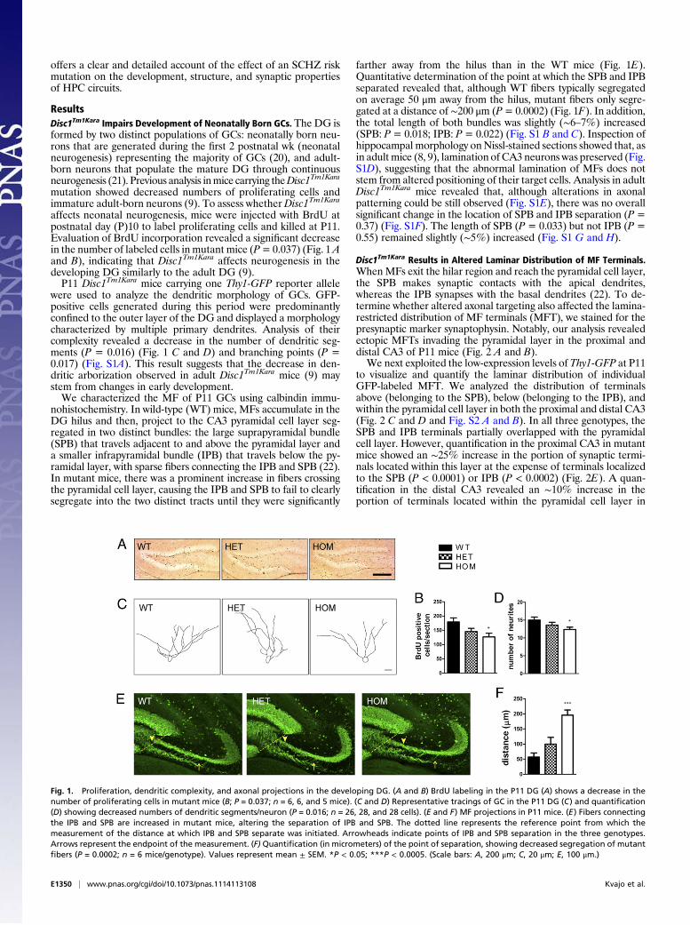
affects neonatal neurogenesis, mice were injected with BrdU atpostnatal day (P)10 to label proliferating cells and killed at P11.Evaluation of BrdU incorporation revealed a significant decreasein the number of labeled cells in mutant mice (P=0.037) (Fig. 1Aand B), indicating that Disc1Tm1Kara affects neurogenesis in thedeveloping DG similarly to the adult DG (9).P11 Disc1Tm1Kara mice carrying one Thy1-GFP reporter allele
were used to analyze the dendritic morphology of GCs. GFP-positive cells generated during this period were predominantlyconfined to the outer layer of the DG and displayed a morphologycharacterized by multiple primary dendrites. Analysis of theircomplexity revealed a decrease in the number of dendritic seg-ments (P = 0.016) (Fig. 1 C and D) and branching points (P =0.017) (Fig. S1A). This result suggests that the decrease in den-dritic arborization observed in adult Disc1Tm1Kara mice (9) maystem from changes in early development.We characterized the MF of P11 GCs using calbindin immu-
nohistochemistry. In wild-type (WT) mice, MFs accumulate in theDG hilus and then, project to the CA3 pyramidal cell layer seg-regated in two distinct bundles: the large suprapyramidal bundle(SPB) that travels adjacent to and above the pyramidal layer anda smaller infrapyramidal bundle (IPB) that travels below the py-ramidal layer, with sparse fibers connecting the IPB and SPB (22).In mutant mice, there was a prominent increase in fibers crossingthe pyramidal cell layer, causing the IPB and SPB to fail to clearlysegregate into the two distinct tracts until they were significantly
farther away from the hilus than in the WT mice (Fig. 1E).Quantitative determination of the point at which the SPB and IPBseparated revealed that, although WT fibers typically segregatedon average 50 μm away from the hilus, mutant fibers only segre-gated at a distance of ∼200 μm (P= 0.0002) (Fig. 1F). In addition,the total length of both bundles was slightly (∼6–7%) increased(SPB: P= 0.018; IPB: P= 0.022) (Fig. S1 B and C). Inspection ofhippocampal morphology onNissl-stained sections showed that, asin adult mice (8, 9), lamination of CA3 neurons was preserved (Fig.S1D), suggesting that the abnormal lamination of MFs does notstem from altered positioning of their target cells. Analysis in adultDisc1Tm1Kara mice revealed that, although alterations in axonalpatterning could be still observed (Fig. S1E), there was no overallsignificant change in the location of SPB and IPB separation (P =0.37) (Fig. S1F). The length of SPB (P = 0.033) but not IPB (P =0.55) remained slightly (∼5%) increased (Fig. S1 G and H).
Disc1Tm1Kara Results in Altered Laminar Distribution of MF Terminals.WhenMFs exit the hilar region and reach the pyramidal cell layer,the SPB makes synaptic contacts with the apical dendrites,whereas the IPB synapses with the basal dendrites (22). To de-termine whether altered axonal targeting also affected the lamina-restricted distribution of MF terminals (MFT), we stained for thepresynaptic marker synaptophysin. Notably, our analysis revealedectopic MFTs invading the pyramidal layer in the proximal anddistal CA3 of P11 mice (Fig. 2 A and B).We next exploited the low-expression levels of Thy1-GFP at P11
to visualize and quantify the laminar distribution of individualGFP-labeled MFT. We analyzed the distribution of terminalsabove (belonging to the SPB), below (belonging to the IPB), andwithin the pyramidal cell layer in both the proximal and distal CA3(Fig. 2 C and D and Fig. S2 A and B). In all three genotypes, theSPB and IPB terminals partially overlapped with the pyramidalcell layer. However, quantification in the proximal CA3 in mutantmice showed an ∼25% increase in the portion of synaptic termi-nals located within this layer at the expense of terminals localizedto the SPB (P < 0.0001) or IPB (P < 0.0002) (Fig. 2E). A quan-tification in the distal CA3 revealed an ∼10% increase in theportion of terminals located within the pyramidal cell layer in
Fig. 1. Proliferation, dendritic complexity, and axonal projections in the developing DG. (A and B) BrdU labeling in the P11 DG (A) shows a decrease in thenumber of proliferating cells in mutant mice (B; P = 0.037; n = 6, 6, and 5 mice). (C and D) Representative tracings of GC in the P11 DG (C) and quantification(D) showing decreased numbers of dendritic segments/neuron (P = 0.016; n = 26, 28, and 28 cells). (E and F) MF projections in P11 mice. (E) Fibers connectingthe IPB and SPB are increased in mutant mice, altering the separation of IPB and SPB. The dotted line represents the reference point from which themeasurement of the distance at which IPB and SPB separate was initiated. Arrowheads indicate points of IPB and SPB separation in the three genotypes.Arrows represent the endpoint of the measurement. (F) Quantification (in micrometers) of the point of separation, showing decreased segregation of mutantfibers (P = 0.0002; n = 6 mice/genotype). Values represent mean ± SEM. *P < 0.05; ***P < 0.0005. (Scale bars: A, 200 μm; C, 20 μm; E, 100 μm.)
E1350 | www.pnas.org/cgi/doi/10.1073/pnas.1114113108 Kvajo et al.
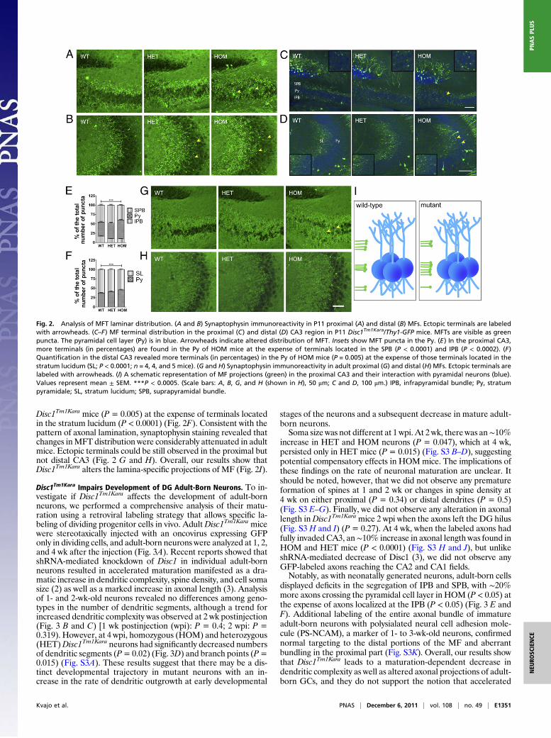
Disc1Tm1Kara mice (P = 0.005) at the expense of terminals locatedin the stratum lucidum (P < 0.0001) (Fig. 2F). Consistent with thepattern of axonal lamination, synaptophysin staining revealed thatchanges inMFT distribution were considerably attenuated in adultmice. Ectopic terminals could be still observed in the proximal butnot distal CA3 (Fig. 2 G and H). Overall, our results show thatDisc1Tm1Kara alters the lamina-specific projections of MF (Fig. 2I).
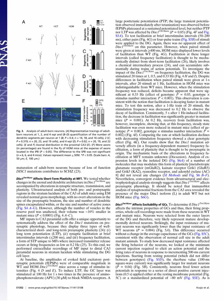
Disc1Tm1Kara Impairs Development of DG Adult-Born Neurons. To in-vestigate if Disc1Tm1Kara affects the development of adult-bornneurons, we performed a comprehensive analysis of their matu-ration using a retroviral labeling strategy that allows specific la-beling of dividing progenitor cells in vivo. AdultDisc1Tm1Kara micewere stereotaxically injected with an oncovirus expressing GFPonly in dividing cells, and adult-born neurons were analyzed at 1, 2,and 4 wk after the injection (Fig. 3A). Recent reports showed thatshRNA-mediated knockdown of Disc1 in individual adult-bornneurons resulted in accelerated maturation manifested as a dra-matic increase in dendritic complexity, spine density, and cell somasize (2) as well as a marked increase in axonal length (3). Analysisof 1- and 2-wk-old neurons revealed no differences among geno-types in the number of dendritic segments, although a trend forincreased dendritic complexity was observed at 2 wk postinjection(Fig. 3 B and C) [1 wk postinjection (wpi): P = 0.4; 2 wpi: P =0.319). However, at 4 wpi, homozygous (HOM) and heterozygous(HET)Disc1Tm1Kara neurons had significantly decreased numbersof dendritic segments (P=0.02) (Fig. 3D) and branch points (P=0.015) (Fig. S3A). These results suggest that there may be a dis-tinct developmental trajectory in mutant neurons with an in-crease in the rate of dendritic outgrowth at early developmental
stages of the neurons and a subsequent decrease in mature adult-born neurons.Soma size was not different at 1 wpi. At 2 wk, there was an∼10%
increase in HET and HOM neurons (P = 0.047), which at 4 wk,persisted only in HET mice (P = 0.015) (Fig. S3 B–D), suggestingpotential compensatory effects in HOMmice. The implications ofthese findings on the rate of neuronal maturation are unclear. Itshould be noted, however, that we did not observe any prematureformation of spines at 1 and 2 wk or changes in spine density at4 wk on either proximal (P = 0.34) or distal dendrites (P = 0.5)(Fig. S3 E–G). Finally, we did not observe any alteration in axonallength inDisc1Tm1Karamice 2 wpi when the axons left the DG hilus(Fig. S3 H and I) (P = 0.27). At 4 wk, when the labeled axons hadfully invaded CA3, an∼10% increase in axonal length was found inHOM and HET mice (P < 0.0001) (Fig. S3 H and J), but unlikeshRNA-mediated decrease of Disc1 (3), we did not observe anyGFP-labeled axons reaching the CA2 and CA1 fields.Notably, as with neonatally generated neurons, adult-born cells
displayed deficits in the segregation of IPB and SPB, with ∼20%more axons crossing the pyramidal cell layer in HOM (P < 0.05) atthe expense of axons localized at the IPB (P < 0.05) (Fig. 3 E andF). Additional labeling of the entire axonal bundle of immatureadult-born neurons with polysialated neural cell adhesion mole-cule (PS-NCAM), a marker of 1- to 3-wk-old neurons, confirmednormal targeting to the distal portions of the MF and aberrantbundling in the proximal part (Fig. S3K). Overall, our results showthat Disc1Tm1Kara leads to a maturation-dependent decrease indendritic complexity as well as altered axonal projections of adult-born GCs, and they do not support the notion that accelerated
Fig. 2. Analysis of MFT laminar distribution. (A and B) Synaptophysin immunoreactivity in P11 proximal (A) and distal (B) MFs. Ectopic terminals are labeledwith arrowheads. (C–F) MF terminal distribution in the proximal (C) and distal (D) CA3 region in P11 Disc1Tm1Kara/Thy1-GFP mice. MFTs are visible as greenpuncta. The pyramidal cell layer (Py) is in blue. Arrowheads indicate altered distribution of MFT. Insets show MFT puncta in the Py. (E) In the proximal CA3,more terminals (in percentages) are found in the Py of HOM mice at the expense of terminals located in the SPB (P < 0.0001) and IPB (P < 0.0002). (F)Quantification in the distal CA3 revealed more terminals (in percentages) in the Py of HOM mice (P = 0.005) at the expense of those terminals located in thestratum lucidum (SL; P < 0.0001; n = 4, 4, and 5 mice). (G and H) Synaptophysin immunoreactivity in adult proximal (G) and distal (H) MFs. Ectopic terminals arelabeled with arrowheads. (I) A schematic representation of MF projections (green) in the proximal CA3 and their interaction with pyramidal neurons (blue).Values represent mean ± SEM. ***P < 0.0005. (Scale bars: A, B, G, and H (shown in H), 50 μm; C and D, 100 μm.) IPB, infrapyramidal bundle; Py, stratumpyramidale; SL, stratum lucidum; SPB, suprapyramidal bundle.
Kvajo et al. PNAS | December 6, 2011 | vol. 108 | no. 49 | E1351
NEU
ROSC
IENCE
PNASPL
US
maturation of adult-born neurons because of loss of functionDISC1 mutations contributes to SCHZ (23).
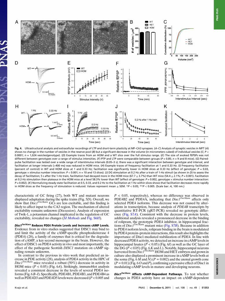
Disc1Tm1Kara Affects Short-Term Plasticity at MFT.We tested whetherchanges in the axonal and dendritic architecture inDisc1Tm1Kara areaccompanied by alterations in synaptic structure, transmission, andplasticity. Ultrastructural analysis of both pre- and postsynapticregions in the stratum lucidum of the CA3 of adult mice using EMrevealed normal gross morphology, with no overt alterations in thesize of the presynaptic boutons, the size and number of dendriticspines encapsulated within, or the size and number of active zones(Fig. S4 A–G). However, although the number of vesicles in thereserve pool was unaltered, their volume was ∼10% smaller inmutant mice (P < 0.0001) (Fig. 4 A–C).MF inputs to CA3 pyramidal cells offer a unique opportunity to
systematically address the effect of the Disc1 mutations on pre-synaptic physiology, because they display three types of well-characterized short- and long-term presynaptic plasticity (24): (i)long term potentiation (LTP), (ii) synaptic facilitation at briefinterstimulus intervals (≤200 ms), and (iii) frequency facilitation,a form of STP unique to MFs where increased transmitter releaseoccurs at firing frequencies as low as 0.2 Hz (25). To this end, weperformed extracellular recordings of MF input to distal CA3pyramidal neurons in stratum lucidum while stimulating the DGcell layer.At baseline, the amplitudes of evoked field excitatory post-
synaptic potentials (fEPSPs) were of comparable magnitude inWT and HOM Disc1Tm1Kara mice across a range of stimulus in-tensities (Fig. 4 D and E). To induce LTP, the GC layer wasstimulated at 100 Hz for 1 s two times in the presence of amino-phosphonovalerate (APV) (30 μM) to block NMDA receptors. A
large posttetanic potentiation (PTP; the large transient potentia-tion observed immediately after tetanization) was observed beforefEPSPs plateaued at a sustained potentiated level, but neither PTPnor LTP was affected by Disc1Tm1Kara (P = 0.85) (Fig. 4F and Fig.S5A). To test facilitation at brief interstimulus intervals (50–200ms), either pairs (Fig. 4G) or four-pulse trains (Fig. S5B) of stimuliwere applied to the DG. Again, there was no apparent effect ofDisc1Tm1Kara on this parameter. However, when paired stimuliwere given at intervals ≥400 ms, HOMmice displayed lower levelsof facilitation than WT (Fig. 4G). Facilitation at these longerintervals (called frequency facilitation) is thought to be mecha-nistically distinct from short-term facilitation (26), likely involvesa chemical intermediary process (24), and can accumulate sub-stantially during trains of action potentials. To investigate theimpact of the Disc1Tm1Kara on frequency facilitation, the DG wasstimulated 20 times at 1, 0.5, and 0.33 Hz (Fig. 4H and I). Despitedifferences in facilitation when paired stimuli were given at 1-sintervals, after 20 stimuli at 1 Hz, facilitation in HOM mice wasindistinguishable from WT mice. However, when the stimulationfrequency was reduced, deficits became apparent that were sig-nificant at 0.33 Hz (effect of genotype: P = 0.03, genotype ×stimulus number interaction: P < 0.001). This observation is con-sistent with the notion that facilitation is decaying faster in mutantmice. To test this notion, after a 1-Hz train of 20 stimuli, thestimulation frequency was decreased to 0.2 Hz to observe thedecay of facilitation. Consistently, 5 s after 1 Hz-induced facilita-tion, the decrease in facilitation was significantly greater in mutantmice (P = 0.001). At 0.2 Hz, recovery from facilitation was,however, incomplete, showing that, at this frequency, steady statefacilitation was even more reduced in mutant mice (effect of ge-notype: P = 0.002, genotype × stimulus number interaction: P =0.002) (Fig. 4J). Comparing the rate at which facilitation declineswith decreasing stimulation frequency showed a clear genotypiceffect (Fig. 4K). Our results show that the Disc1 mutation ad-versely affects (in a frequency-dependent manner) frequency fa-cilitation, a form of plasticity that is thought to be presynaptic inorigin. The mechanistic basis of the alterations in frequency fa-cilitation at MFT remains unknown (Discussion). Analysis of ex-pression levels in the isolated DG (Fig. S6A) of a number ofmolecules that may modulate this form of plasticity [metabotropicglutamate receptor (mGluR2), kainate receptor Grik1 (GluR5)and Grik5 (KA2), ryanodine receptor, and adenylyl cyclase (AC)8] did not reveal any changes (SI Methods and Fig. S6 B–F).Nevertheless, convergent evidence from ultrastructural and elec-trophysiological analysis strongly suggests thatDisc1Tm1Kara affectspresynaptic physiology. It should be noted that immunoblotanalysis of synaptosomal fractions from the CA3 area revealed thepresence of the major Disc1 isoform (L) in adult WT but notHOM mice (Fig. S6G).
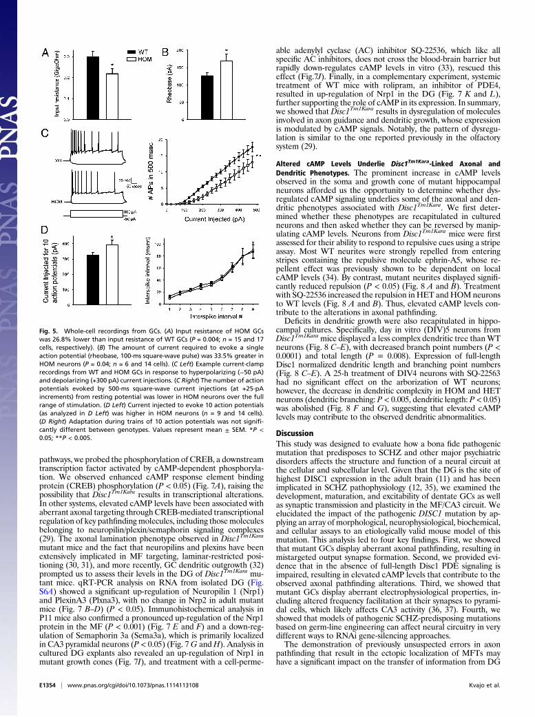
Disc1Tm1Kara Affects Excitability of GCs. To determine ifDisc1Tm1Kara
affects the intrinsic properties of GCs and thus, their firing prop-erties, whole-cell recordings were made from these neurons inWTand mutant mice. Neurons were selected from the outer layersof the DG and therefore, very likely represent mature develop-mentally derived neurons. Strikingly, the input resistance of mu-tant neurons was significantly lower than the input resistance ofWT neurons (P = 0.004) (Fig. 5A). This difference occurredwithout a change in the average capacitance of the GCs (Fig. S5C),consistent with the observation of normal cell size in the adultmutant animals. To study how decreased input resistance affectedthe firing behavior of the neurons, we looked at the minimumcurrent injection required to elicit an action potential and thefiring rates of neurons in response to incrementing 500-ms currentinjections. Starting from resting potential (which did not differbetween genotypes) (Fig. S5D), the rheobase value (100-mssquare-wave current) was significantly larger for mutant neurons(P = 0.04) (Fig. 5B). Consistently, neurons fired fewer actionpotentials in response to a series of direct positive current injec-tions (0.5 s) applied either at the resting membrane potential (Fig.5C) or a standardized potential of −80 mV (Fig. S5E). As is
Fig. 3. Analysis of adult-born neurons. (A) Representative tracings of adult-born neurons at 1, 2, and 4 wpi and (B–D) quantification of the number ofdendritic segments per neuron at 1 (B; P = 0.4; n = 14, 18, and 16 cells), 2 (C;P = 0.319; n = 20, 23, and 19 cells), and 4 wpi (D; P = 0.02; n = 20, 19, and 22cells). (E and F) Axonal distribution in the proximal CA3 (E). (F) More axons(in percentages) are found in the Py of HOM mice at the expense of axonslocated in the IPB (P < 0.05). The difference to the SPB was not significant(n = 6, 6, and 4 mice). Values represent mean ± SEM. *P < 0.05. (Scale bars: A,50 μm; E, 100 μm.)
E1352 | www.pnas.org/cgi/doi/10.1073/pnas.1114113108 Kvajo et al.
characteristic of GC firing (27), both WT and mutant neuronsdisplayed adaptation during the spike trains (Fig. 5D). Overall, weshow that Disc1Tm1Kara GCs are less excitable, and this finding islikely to affect input to the CA3 region. The mechanism of alteredexcitability remains unknown (Discussion). Analysis of expressionof Twik-1, a potassium channel implicated in the regulation of GCexcitability, revealed no changes (SI Methods and Fig. S6H).
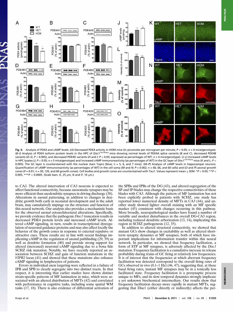
Disc1Tm1Kara Reduces PDE4 Protein Levels and Increases cAMP Levels.Evidence from in vitro studies suggested that DISC1 may bind toand limit the activity of the cAMP-specific phosphodiesterase 4(PDE4) (28), a family of enzymes that is critical for the degrada-tion of cAMP, a key second messenger in the brain. However, theeffect of DISC1 on PDE4 activity in vivo andmost importantly, theeffect of the pathogenic Scottish DISC1 mutation on cAMP sig-naling remain unknown.In contrast to the previous in vitro work that predicted an in-
crease in PDE activity (28), analysis of PDE4 activity in theHPC ofDisc1Tm1Kara mice revealed a robust (50%) decrease in activity inHOM mice (P < 0.05) (Fig. 6A). Strikingly, immunoblot analysisrevealed a consistent decrease in the levels of several PDE4 iso-forms (Fig. 6B–I). Specifically, PDE4B1,PDE4B3, andPDE4B4aswell asPDE4D3andPDE4D5 levelsweredecreased (P< 0.005 and
P < 0.05, respectively), whereas no difference was observed inPDE4B2 and PDE4A, indicating that Disc1Tm1Kara affects onlyselected PDE4 isoforms. This decrease was not caused by alter-ations in transcription, because analysis of PDE4B transcripts byquantitative RT-PCR (qRT-PCR) revealed no genotypic differ-ence (Fig. S7A). Consistent with the decrease in protein levels,additional analysis revealed a pronounced decrease in the bindingof rolipram, the prototypic PDE4 inhibitor, in hippocampal frac-tions of Disc1Tm1Kara mutant mice (Fig. S7B). Because in additionto PDE4 isoform levels, rolipram binding in the brain is modulatedbyPDE4protein–protein interactions, this result alsohighlights theimportance of Disc1-mediated stabilization of PDE4. In line withdecreasedPDE4 activity, wedetected an increase in cAMP levels inhippocampal lysates (P < 0.05) (Fig. 6J) as well as the GC layer ofthe DG (P< 0.05) (Fig. 6K andL). Notably, hippocampal neuronsharvested fromDisc1Tm1KaraHOMandHETembryos and grown inculture also displayed a prominent increase in cAMP levels both atthe soma (Fig. 6M andN) (P= 0.002) and the axonal growth cone(P=0.01) (Fig. 6O andP), confirming a pervasive effect ofDisc1 inmodulating cAMP levels in mature and developing neurons.
Disc1Tm1Kara Affects cAMP-Dependent Pathways. To test whetherchanges in PDE4 activity have an impact on cAMP-dependent
Fig. 4. Ultrastructural analysis and extracellular recordings of LTP and short-term plasticity at MF–CA3 synapses. (A–C) Analysis of synaptic vesicles in MFT (A)shows no change in the number of vesicles in the reserve pool (B) but a significant decrease in the volume (in micrometers cubed) of individual vesicles (C; P <0.0001; n = 1,024 vesicles/genotype). (D) Example traces from an HOM and a WT slice over the full stimulus range. (E) The size of evoked fEPSPs was notdifferent between genotypes over a range of stimulus intensities. (F) PTP and LTP were comparable between groups (P = 0.85; n = 9 and 8 mice). (G) Paired-pulse facilitation was tested over a wide range of interstimulus intervals (0.05–3 s); there was a significant interaction between genotype and interval, andfacilitation at longer intervals (>400 ms) was reduced in HOM mice. (H) Example traces of frequency facilitation at 1 and 0.33 Hz. (I) Frequency facilitation(percent of control) in WT and HOM slices at 1 and 0.33 Hz; facilitation was significantly lower in HOM slices at 0.33 Hz (effect of genotype: P = 0.03,genotype × stimulus number interaction: P < 0.001; n = 10 and 13 slices). (J) DG stimulation at 0.2 Hz after a train of 1-Hz stimuli (as shown in D) to assess thedecay of facilitation; 5 s after the 1-Hz train, facilitation had decayed more in the HOM mice (67.7 ± 3.7%) than WT mice (54.4 ± 2.1%; P = 0.001). Facilitationat 0.2-Hz stimulation then plateaus in the HOM mice at a level 58.3% lower than WT (effect of genotype: P = 0.002, genotype × stimulus number interaction:P = 0.002). (K) Normalizing steady state facilitation at 0.5, 0.33, and 0.2 Hz to the facilitation at 1 Hz within slices shows that facilitation decreases more rapidlyin HOM slices as the frequency of stimulation is reduced. Values represent mean ± SEM. *P < 0.05; **P < 0.005. (Scale bar: A, 100 nm.)
Kvajo et al. PNAS | December 6, 2011 | vol. 108 | no. 49 | E1353
NEU
ROSC
IENCE
PNASPL
US
pathways, we probed the phosphorylation of CREB, a downstreamtranscription factor activated by cAMP-dependent phosphoryla-tion. We observed enhanced cAMP response element bindingprotein (CREB) phosphorylation (P < 0.05) (Fig. 7A), raising thepossibility that Disc1Tm1Kara results in transcriptional alterations.In other systems, elevated cAMP levels have been associated withaberrant axonal targeting throughCREB-mediated transcriptionalregulation of key pathfinding molecules, including thosemoleculesbelonging to neuropilin/plexin/semaphorin signaling complexes(29). The axonal lamination phenotype observed in Disc1Tm1Kara
mutant mice and the fact that neuropilins and plexins have beenextensively implicated in MF targeting, laminar-restricted posi-tioning (30, 31), and more recently, GC dendritic outgrowth (32)prompted us to assess their levels in the DG of Disc1Tm1Kara mu-tant mice. qRT-PCR analysis on RNA from isolated DG (Fig.S6A) showed a significant up-regulation of Neuropilin 1 (Nrp1)and PlexinA3 (Plxna3), with no change in Nrp2 in adult mutantmice (Fig. 7 B–D) (P < 0.05). Immunohistochemical analysis inP11 mice also confirmed a pronounced up-regulation of the Nrp1protein in the MF (P < 0.001) (Fig. 7 E and F) and a down-reg-ulation of Semaphorin 3a (Sema3a), which is primarily localizedin CA3 pyramidal neurons (P < 0.05) (Fig. 7G andH). Analysis incultured DG explants also revealed an up-regulation of Nrp1 inmutant growth cones (Fig. 7I), and treatment with a cell-perme-
able adenylyl cyclase (AC) inhibitor SQ-22536, which like allspecific AC inhibitors, does not cross the blood-brain barrier butrapidly down-regulates cAMP levels in vitro (33), rescued thiseffect (Fig.7J). Finally, in a complementary experiment, systemictreatment of WT mice with rolipram, an inhibitor of PDE4,resulted in up-regulation of Nrp1 in the DG (Fig. 7 K and L),further supporting the role of cAMP in its expression. In summary,we showed that Disc1Tm1Kara results in dysregulation of moleculesinvolved in axon guidance and dendritic growth, whose expressionis modulated by cAMP signals. Notably, the pattern of dysregu-lation is similar to the one reported previously in the olfactorysystem (29).
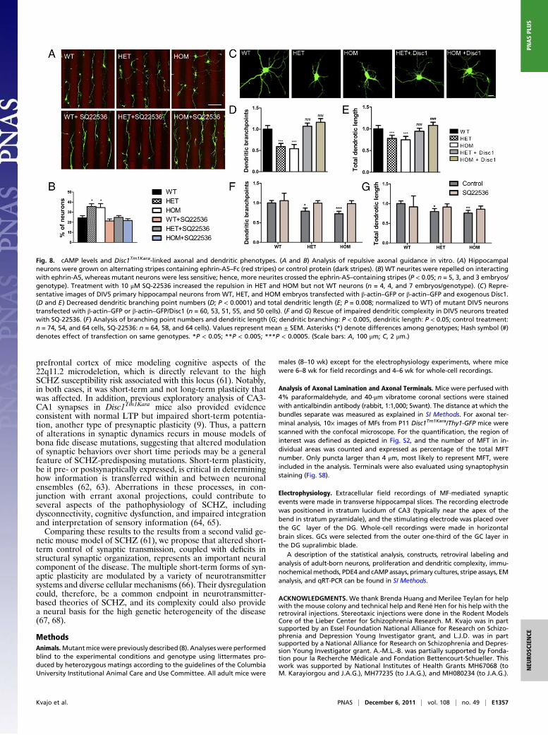
Altered cAMP Levels Underlie Disc1Tm1Kara-Linked Axonal andDendritic Phenotypes. The prominent increase in cAMP levelsobserved in the soma and growth cone of mutant hippocampalneurons afforded us the opportunity to determine whether dys-regulated cAMP signaling underlies some of the axonal and den-dritic phenotypes associated with Disc1Tm1Kara. We first deter-mined whether these phenotypes are recapitulated in culturedneurons and then asked whether they can be reversed by manip-ulating cAMP levels. Neurons from Disc1Tm1Kara mice were firstassessed for their ability to respond to repulsive cues using a stripeassay. Most WT neurites were strongly repelled from enteringstripes containing the repulsive molecule ephrin-A5, whose re-pellent effect was previously shown to be dependent on localcAMP levels (34). By contrast, mutant neurites displayed signifi-cantly reduced repulsion (P < 0.05) (Fig. 8 A and B). Treatmentwith SQ-22536 increased the repulsion in HET and HOM neuronsto WT levels (Fig. 8 A and B). Thus, elevated cAMP levels con-tribute to the alterations in axonal pathfinding.Deficits in dendritic growth were also recapitulated in hippo-
campal cultures. Specifically, day in vitro (DIV)5 neurons fromDisc1Tm1Karamice displayed a less complex dendritic tree thanWTneurons (Fig. 8 C–E), with decreased branch point numbers (P <0.0001) and total length (P = 0.008). Expression of full-lengthDisc1 normalized dendritic length and branching point numbers(Fig. 8 C–E). A 25-h treatment of DIV4 neurons with SQ-22563had no significant effect on the arborization of WT neurons;however, the decrease in dendritic complexity in HOM and HETneurons (dendritic branching: P < 0.005, dendritic length: P < 0.05)was abolished (Fig. 8 F and G), suggesting that elevated cAMPlevels may contribute to the observed dendritic abnormalities.
DiscussionThis study was designed to evaluate how a bona fide pathogenicmutation that predisposes to SCHZ and other major psychiatricdisorders affects the structure and function of a neural circuit atthe cellular and subcellular level. Given that the DG is the site ofhighest DISC1 expression in the adult brain (11) and has beenimplicated in SCHZ pathophysiology (12, 35), we examined thedevelopment, maturation, and excitability of dentate GCs as wellas synaptic transmission and plasticity in the MF/CA3 circuit. Weelucidated the impact of the pathogenic DISC1 mutation by ap-plying an array of morphological, neurophysiological, biochemical,and cellular assays to an etiologically valid mouse model of thismutation. This analysis led to four key findings. First, we showedthat mutant GCs display aberrant axonal pathfinding, resulting inmistargeted output synapse formation. Second, we provided evi-dence that in the absence of full-length Disc1 PDE signaling isimpaired, resulting in elevated cAMP levels that contribute to theobserved axonal pathfinding alterations. Third, we showed thatmutant GCs display aberrant electrophysiological properties, in-cluding altered frequency facilitation at their synapses to pyrami-dal cells, which likely affects CA3 activity (36, 37). Fourth, weshowed that models of pathogenic SCHZ-predisposing mutationsbased on germ-line engineering can affect neural circuitry in verydifferent ways to RNAi gene-silencing approaches.The demonstration of previously unsuspected errors in axon
pathfinding that result in the ectopic localization of MFTs mayhave a significant impact on the transfer of information from DG
Fig. 5. Whole-cell recordings from GCs. (A) Input resistance of HOM GCswas 26.8% lower than input resistance of WT GCs (P = 0.004; n = 15 and 17cells, respectively). (B) The amount of current required to evoke a singleaction potential (rheobase, 100-ms square-wave pulse) was 33.5% greater inHOM neurons (P = 0.04; n = 6 and 14 cells). (C Left) Example current-clamprecordings from WT and HOM GCs in response to hyperpolarizing (−50 pA)and depolarizing (+300 pA) current injections. (C Right) The number of actionpotentials evoked by 500-ms square-wave current injections (at +25-pAincrements) from resting potential was lower in HOM neurons over the fullrange of stimulation. (D Left) Current injected to evoke 10 action potentials(as analyzed in D Left) was higher in HOM neurons (n = 9 and 14 cells).(D Right) Adaptation during trains of 10 action potentials was not signifi-cantly different between genotypes. Values represent mean ± SEM. *P <0.05; **P < 0.005.
E1354 | www.pnas.org/cgi/doi/10.1073/pnas.1114113108 Kvajo et al.
to CA3. The altered innervation of CA3 neurons is expected toaffect functional connectivity, because axosomatic synapses may bemore efficient than axodendritic synapses in driving discharge (38).Alterations in axonal patterning, in addition to changes in den-dritic growth both early in neonatal development and in the adultbrain, may cumulatively impinge on the structure and function ofthis neural network. Our analysis also provides a mechanistic basisfor the observed axonal cytoarchitectural alterations. Specifically,we provide evidence that the pathogenicDisc1 truncation results indecreased PDE4 protein levels and increased cAMP levels. Al-tered cAMP signaling, in turn, results in transcriptional dysregu-lation of neuronal guidance proteins andmay also affect locally thebehavior of the growth cones in response to external repulsive orattractive cues. These results are in line with recent findings im-plicating cAMP in the regulation of axonal pathfinding (29, 39) aswell as dendrite formation (40) and provide strong support foraltered (increased) neuronal cAMP signaling due to a bona fideSCHZ risk mutation. Notably, we have recently reported an as-sociation between SCHZ and gain of function mutations in theVIPR2 locus (41) and showed that these mutations also enhancecAMP signaling in lymphocytes of patients.Errors in individual axon targeting were reflected in a failure of
IPB and SPB to clearly segregate into two distinct tracts. In thatrespect, it is interesting that earlier studies have shown distinctstrain-specific patterns of MF lamination in mice, which were as-sociated with an altered distribution of MFTs (42) and correlatedwith performance in cognitive tasks, including some spatial WMtasks (17, 18). There is also evidence of differential activation of
the SPBs and IPBs of the DG (43), and altered segregation of theSP and IP blades may change the respective connectivities of theseblades with CA3. Although the pattern of MF lamination has notbeen explicitly probed in patients with SCHZ, one study hasreported lower numerical density of MFTs in CA3 (44), and an-other study showed lighter overall staining with an MF specificmarker (45) consistent with changes occurring in this pathway.More broadly, neuropathological studies have found a number ofvariable and modest disturbances in the overall DG-CA3 region,including reduced dendritic arborization (13, 14), implicating thisregion in SCHZ pathogenesis (12, 35).In addition to altered structural connectivity, we showed that
mutant GCs show changes in excitability as well as altered short-term synaptic dynamics at MF synapses, both of which have im-portant implications for information transfer within this neuralnetwork. In particular, we showed that frequency facilitation, aform of STP at MF synapses, is adversely affected by the Disc1mutation. Frequency facilitation is a cumulative increase in releaseprobability during trains of GC firing at relatively low frequencies.It is of interest that the frequencies at which aberrant frequencyfacilitation was detected correspond to the overall firing rates ofGCs observed in vivo (0.1–1 Hz) (46, 47), suggesting that, at thesebasal firing rates, mutant MF synapses may be in a tonically lessfacilitated state. Frequency facilitation is a presynaptic processunique to MFs, and its slow temporal dynamics strongly implicatea yet unknown biochemical intermediary. Our results show thatfrequency facilitation decays more rapidly in mutant MFTs, sug-gesting that Disc1 (either directly or indirectly) affects the per-
Fig. 6. Analysis of PDE4 and cAMP levels. (A) Decreased PDE4 activity in HOM mice (in picomoles per microgram per minute; P < 0.05; n = 4 mice/genotype).(B–I) Analysis of PDE4 isoform protein levels in the HPC of Disc1Tm1Kara mice showing normal levels of PDE4A splice variants (B and C), decreased PDE4Bvariants (D–G; P < 0.005), and decreased PDE4D variants (H and I; P < 0.05; expressed as percentages of WT; n = 4 mice/genotype). (J–L) Increased cAMP levelsin HPC lysates (J; P < 0.05; n = 5 mice/genotype) and increased cAMP immunoreactivity (as percentages of WT) in the GC layer of Disc1Tm1Kara mice (K and L; P <0.005). The GC layer is counterstained with the nuclear stain Topro (blue; n = 5, 6, and 7 mice). (M–P) Analysis of cAMP levels in hippocampal neurons.Quantification of cAMP immunoreactivity (as percentages of WT) in the cell soma (M and N; P = 0.002; n = 60, 66, and 60 cells) and (O and P) axonal growthcones (P = 0.01; n = 50, 120, and 60 growth cones). Cell bodies and growth cones are counterstained with Tau1. Values represent mean ± SEM. *P < 0.05; **P <0.005; ***P < 0.0005. (Scale bars: K, 25 μm; N and P, 10 μm.)
Kvajo et al. PNAS | December 6, 2011 | vol. 108 | no. 49 | E1355
NEU
ROSC
IENCE
PNASPL
US

sistence of this facilitating signal. The signaling molecules thatmediate frequency facilitation, however, remain unclear. Caseshave been made for and against the involvement of presynaptickainate receptors and intraterminal Ca2+ stores, but unequivocaldata are lacking. Elucidating whether cAMP signaling is involvedis confounded by the facts that wholesale manipulations of cAMPlevels through cAMP analogs and AC activation potently and bi-directionally affect release probability and result in diverse neu-rophysiological outputs at MFTs (48, 49). Nevertheless, it is well-established that manipulating cAMP levels at various types ofpresynaptic terminals alters the dynamics of STP (50–53). Ofparticular interest is that chronically elevated cAMP levels canaffect synaptic facilitation through effects on vesicular pools (54),and animals lacking AC8, which acts as a presynaptic calciumsensor, have WM but not long-term memory deficits (55), similartoDisc1Tm1Kara animals. In that respect, it is noteworthy that cyclicnucleotide-dependent vesicle recycling at presynaptic terminals andgrowth cones has been implicated in both synaptic plasticity (24)and axon targeting (56) and therefore, is a potential link betweenthese processes in Disc1Tm1Kara animals. Along these lines, addi-tional work needs to determine whether the volumetric change ofsynaptic vesicles in MFTs reflects abnormal cAMP-dependentvesicle recycling or biogenesis at the presynaptic terminal andwhether it contributes to observed defects in synaptic plasticity.In contrast to robust deficits in STP, an assay of long-term
plasticity showed no effect by the mutation. That LTP is not sig-nificantly changed in mutant animals may suggest differential sen-sitivity of various synaptic processes to the levels of cAMP elevationor emergence of homeostatic mechanisms, which might compen-sate for the effect of chronically elevated cAMP on some but not allphysiological properties. Similarly, the observation that GCs areless excitable in Disc1 mutants, despite the generally pro-excitatory effects of cAMP, may indicate that reduced catabolismof cAMP, may maladaptively induce homeostatic mechanisms toreduce cell activity. Future work will investigate whether physio-logical changes are directly linked to increased cAMP levels orwhether they are part of an adaptive/maladaptive response to theprimary genetic effects.
Although our study shows that intact Disc1 is essential forproper maturation of adult-born GCs, it did not yield datacongruent with results obtained by using oncoretrovirus-medi-ated RNAi approaches (2–4). Previous work on the effect ofDisc1 on the DG structure and function (including on adultneurogenesis) has been based primarily on oncoretrovirus-me-diated RNAi approaches (2–4). A prominent view, largely sha-ped by such approaches, is that Disc1 restricts growth of adult-born neurons and that DISC1 mutations predispose to SCHZ byreleasing this constraint, resulting in robust neuronal overgrowthand misplacement as well as premature maturation (23). Al-though a relatively subtle mispositioning, affecting only a frac-tion of young GCs, has been observed in Disc1Tm1Kara mice (9),our findings overall do not support the notion that DISC1mutations increase disease risk by accelerating the normal mat-uration and growth of adult-born neurons. These discrepanciesraise an important general issue pertaining to the informationobtained by disease models based on RNAi-mediated approachesas opposed to models based on germ-line genetic lesions. Dis-crepant findings from such models are well-established in theliterature (57, 58). Although RNAi-mediated approaches mayprovide information about cell-autonomous functions, they havesignificant limitations when used to model in vivo effects of adisease risk gene disruption. Targeting of a small portion of cellswithin the brain cannot reproduce the timing and magnitude of agerm-line genetic disruption and it may result in unpredictableinteractions with the WT context surrounding them. Anotherlikely explanation for some of these discrepancies is off-target effects,a well-known confounding factor of RNAi-based approaches (59,60). Overall, our findings suggest that results from studies usingRNAi techniques may need to be reevaluated in carefully designedanimal models.Our work has important implications for understanding the
pathophysiology of SCHZ, especially when considered inthe context of recent findings from other genetic mouse modelsof the disease. The pattern of widespread but modest cytoarch-itectural abnormalities and altered short-term synaptic dynamicsdescribed here is similar to the one recently described in the
Fig. 7. Expression of key molecules belonging to neuropilin/plexin/semaphorin signaling complexes. (A) CREB phosphorylation is increased in the HPC ofadult Disc1Tm1Kara mice (P < 0.05; n = 5 mice/genotype). (B–D) qRT-PCR analysis showing increased expression of Nrp1 (P < 0.05; B) and Plxna3 (P < 0.05; C) butnot Nrp2 (D) in the DG of adult mice (n = 8, 7, and 5 mice). (E and F) Increased Nrp1 immunoreactivity in MFs of P11 mice (expressed as percentages of WT; P <0.001; n = 7 mice/genotype). (G and H) Decreased Sema3a immunoreactivity in CA3 pyramidal neurons (expressed as percentages of WT; P < 0.05; n = 7 mice/genotype). (I and J) Nrp1 immunoreactivity in axonal growth cones of DG explants is increased in mutant mice (I). This effect is rescued by treatment with SQ-22536 (J). Quantification (as percentages of WT; P < 0.005; n = 6, 6, and 5 explants/genotype per treatment). Axons were counterstained with Gap43 (green).(K and L) Nrp1 immunoreactivity in MF of P11WT mice treated with rolipram (K). (L) Quantification (as percentages of control; P < 0.05; n = 4 mice/treatment).Values represent mean ± SEM. *P < 0.05; **P < 0.005; ***P < 0.0005. (Scale bars: E, G, and K, 50 μm; I, 2 μm.)
E1356 | www.pnas.org/cgi/doi/10.1073/pnas.1114113108 Kvajo et al.
prefrontal cortex of mice modeling cognitive aspects of the22q11.2 microdeletion, which is directly relevant to the highSCHZ susceptibility risk associated with this locus (61). Notably,in both cases, it was short-term and not long-term plasticity thatwas affected. In addition, previous exploratory analysis of CA3-CA1 synapses in Disc1Tm1Kara mice also provided evidenceconsistent with normal LTP but impaired short-term potentia-tion, another type of presynaptic plasticity (9). Thus, a patternof alterations in synaptic dynamics recurs in mouse models ofbona fide disease mutations, suggesting that altered modulationof synaptic behaviors over short time periods may be a generalfeature of SCHZ-predisposing mutations. Short-term plasticity,be it pre- or postsynaptically expressed, is critical in determininghow information is transferred within and between neuronalensembles (62, 63). Aberrations in these processes, in con-junction with errant axonal projections, could contribute toseveral aspects of the pathophysiology of SCHZ, includingdysconnectivity, cognitive dysfunction, and impaired integrationand interpretation of sensory information (64, 65).Comparing these results to the results from a second valid ge-
netic mouse model of SCHZ (61), we propose that altered short-term control of synaptic transmission, coupled with deficits instructural synaptic organization, represents an important neuralcomponent of the disease. The multiple short-term forms of syn-aptic plasticity are modulated by a variety of neurotransmittersystems and diverse cellular mechanisms (66). Their dysregulationcould, therefore, be a common endpoint in neurotransmitter-based theories of SCHZ, and its complexity could also providea neural basis for the high genetic heterogeneity of the disease(67, 68).
MethodsAnimals.Mutantmicewere previously described (8). Analyseswere performedblind to the experimental conditions and genotype using littermates pro-duced by heterozygous matings according to the guidelines of the ColumbiaUniversity Institutional Animal Care and Use Committee. All adult mice were
males (8–10 wk) except for the electrophysiology experiments, where micewere 6–8 wk for field recordings and 4–6 wk for whole-cell recordings.
Analysis of Axonal Lamination and Axonal Terminals. Mice were perfused with4% paraformaldehyde, and 40-μm vibratome coronal sections were stainedwith anticalbindin antibody (rabbit, 1:1,000; Swant). The distance at which thebundles separate was measured as explained in SI Methods. For axonal ter-minal analysis, 10× images of MFs from P11 Disc1Tm1Kara/Thy1-GFP mice werescanned with the confocal microscope. For the quantification, the region ofinterest was defined as depicted in Fig. S2, and the number of MFT in in-dividual areas was counted and expressed as percentage of the total MFTnumber. Only puncta larger than 4 μm, most likely to represent MFT, wereincluded in the analysis. Terminals were also evaluated using synaptophysinstaining (Fig. S8).
Electrophysiology. Extracellular field recordings of MF-mediated synapticevents were made in transverse hippocampal slices. The recording electrodewas positioned in stratum lucidum of CA3 (typically near the apex of thebend in stratum pyramidale), and the stimulating electrode was placed overthe GC layer of the DG. Whole-cell recordings were made in horizontalbrain slices. GCs were selected from the outer one-third of the GC layer inthe DG supralimbic blade.
A description of the statistical analysis, constructs, retroviral labeling andanalysis of adult-born neurons, proliferation and dendritic complexity, immu-nochemicalmethods, PDE4 and cAMPassays, primary cultures, stripe assays, EManalysis, and qRT-PCR can be found in SI Methods.
ACKNOWLEDGMENTS. We thank Brenda Huang and Merilee Teylan for helpwith the mouse colony and technical help and René Hen for his help with theretroviral injections. Stereotaxic injections were done in the Rodent ModelsCore of the Lieber Center for Schizophrenia Research. M. Kvajo was in partsupported by an Essel Foundation National Alliance for Research on Schizo-phrenia and Depression Young Investigator grant, and L.J.D. was in partsupported by a National Alliance for Research on Schizophrenia and Depres-sion Young Investigator grant. A.-M.L.-B. was partially supported by Fonda-tion pour la Recherche Médicale and Fondation Bettencourt-Schueller. Thiswork was supported by National Institutes of Health Grants MH67068 (toM. Karayiorgou and J.A.G.), MH77235 (to J.A.G.), and MH080234 (to J.A.G.).
Fig. 8. cAMP levels and Disc1Tm1Kara-linked axonal and dendritic phenotypes. (A and B) Analysis of repulsive axonal guidance in vitro. (A) Hippocampalneurons were grown on alternating stripes containing ephrin-A5–Fc (red stripes) or control protein (dark stripes). (B) WT neurites were repelled on interactingwith ephrin-A5, whereas mutant neurons were less sensitive; hence, more neurites crossed the ephrin-A5–containing stripes (P < 0.05; n = 5, 3, and 3 embryos/genotype). Treatment with 10 μM SQ-22536 increased the repulsion in HET and HOM but not WT neurons (n = 4, 4, and 7 embryos/genotype). (C) Repre-sentative images of DIV5 primary hippocampal neurons from WT, HET, and HOM embryos transfected with β-actin–GFP or β-actin–GFP and exogenous Disc1.(D and E) Decreased dendritic branching point numbers (D; P < 0.0001) and total dendritic length (E; P = 0.008; normalized to WT) of mutant DIV5 neuronstransfected with β-actin–GFP or β-actin–GFP/Disc1 (n = 60, 53, 51, 55, and 50 cells). (F and G) Rescue of impaired dendritic complexity in DIV5 neurons treatedwith SQ-22536. (F) Analysis of branching point numbers and dendritic length (G; dendritic branching: P < 0.005, dendritic length: P < 0.05; control treatment:n = 74, 54, and 64 cells, SQ-22536: n = 64, 58, and 64 cells). Values represent mean ± SEM. Asterisks (*) denote differences among genotypes; Hash symbol (#)denotes effect of transfection on same genotypes. *P < 0.05; **P < 0.005; ***P < 0.0005. (Scale bars: A, 100 μm; C, 2 μm.)
Kvajo et al. PNAS | December 6, 2011 | vol. 108 | no. 49 | E1357
NEU
ROSC
IENCE
PNASPL
US
1. Mao Y, et al. (2009) Disrupted in schizophrenia 1 regulates neuronal progenitorproliferation via modulation of GSK3beta/beta-catenin signaling. Cell 136:1017–1031.
2. Duan X, et al. (2007) Disrupted-In-Schizophrenia 1 regulates integration of newlygenerated neurons in the adult brain. Cell 130:1146–1158.
3. Faulkner RL, et al. (2008) Development of hippocampal mossy fiber synaptic outputsby new neurons in the adult brain. Proc Natl Acad Sci USA 105:14157–14162.
4. Kim JY, et al. (2009) DISC1 regulates new neuron development in the adult brain viamodulation of AKT-mTOR signaling through KIAA1212. Neuron 63:761–773.
5. Pletnikov MV, et al. (2008) Inducible expression of mutant human DISC1 in mice isassociated with brain and behavioral abnormalities reminiscent of schizophrenia.MolPsychiatry 13:173–186.
6. Hikida T, et al. (2007) Dominant-negative DISC1 transgenic mice displayschizophrenia-associated phenotypes detected by measures translatable to humans.Proc Natl Acad Sci USA 104:14501–14506.
7. Clapcote SJ, et al. (2007) Behavioral phenotypes of Disc1 missense mutations in mice.Neuron 54:387–402.
8. Koike H, Arguello PA, Kvajo M, Karayiorgou M, Gogos JA (2006) Disc1 is mutated inthe 129S6/SvEv strain and modulates working memory in mice. Proc Natl Acad Sci USA103:3693–3697.
9. Kvajo M, et al. (2008) A mutation in mouse Disc1 that models a schizophrenia riskallele leads to specific alterations in neuronal architecture and cognition. Proc NatlAcad Sci USA 105:7076–7081.
10. Nakata K, et al. (2009) DISC1 splice variants are upregulated in schizophrenia andassociated with risk polymorphisms. Proc Natl Acad Sci USA 106:15873–15878.
11. Austin CP, Ky B, Ma L, Morris JA, Shughrue PJ (2004) Expression of Disrupted-In-Schizophrenia-1, a schizophrenia-associated gene, is prominent in the mousehippocampus throughout brain development. Neuroscience 124:3–10.
12. Kobayashi K (2009) Targeting the hippocampal mossy fiber synapse for the treatmentof psychiatric disorders. Mol Neurobiol 39:24–36.
13. Christison GW, Casanova MF, Weinberger DR, Rawlings R, Kleinman JE (1989) Aquantitative investigation of hippocampal pyramidal cell size, shape, and variabilityof orientation in schizophrenia. Arch Gen Psychiatry 46:1027–1032.
14. Arnold SE, et al. (1995) Smaller neuron size in schizophrenia in hippocampal subfieldsthat mediate cortical-hippocampal interactions. Am J Psychiatry 152:738–748.
15. McHugh TJ, et al. (2007) Dentate gyrus NMDA receptors mediate rapid patternseparation in the hippocampal network. Science 317:94–99.
16. Kobayashi K, Poo MM (2004) Spike train timing-dependent associative modificationof hippocampal CA3 recurrent synapses by mossy fibers. Neuron 41:445–454.
17. Crusio WE, Schwegler H (1987) Hippocampal mossy fiber distribution covaries withopen-field habituation in the mouse. Behav Brain Res 26:153–158.
18. Schwegler H, Crusio WE, Brust I (1990) Hippocampal mossy fibers and radial-mazelearning in the mouse: A correlation with spatial working memory but not with non-spatial reference memory. Neuroscience 34:293–298.
19. Niewoehner B, et al. (2007) Impaired spatial working memory but spared spatialreference memory following functional loss of NMDA receptors in the dentate gyrus.Eur J Neurosci 25:837–846.
20. Altman J, Bayer SA (1990) Migration and distribution of two populations ofhippocampal granule cell precursors during the perinatal and postnatal periods. JComp Neurol 301:365–381.
21. Kempermann G, Kuhn HG, Gage FH (1997) More hippocampal neurons in adult miceliving in an enriched environment. Nature 386:493–495.
22. Amaral DG, Dent JA (1981) Development of the mossy fibers of the dentate gyrus: I. Alight and electron microscopic study of the mossy fibers and their expansions. J CompNeurol 195:51–86.
23. Dranovsky A, Hen R (2007) DISC1 puts the brakes on neurogenesis. Cell 130:981–983.24. Nicoll RA, Schmitz D (2005) Synaptic plasticity at hippocampal mossy fibre synapses.
Nat Rev Neurosci 6:863–876.25. Salin PA, Scanziani M, Malenka RC, Nicoll RA (1996) Distinct short-term plasticity at
two excitatory synapses in the hippocampus. Proc Natl Acad Sci USA 93:13304–13309.26. Gundlfinger A, et al. (2007) Differential modulation of short-term synaptic dynamics
by long-term potentiation at mouse hippocampal mossy fibre synapses. J Physiol 585:853–865.
27. Staley KJ, Otis TS, Mody I (1992) Membrane properties of dentate gyrus granule cells:Comparison of sharp microelectrode and whole-cell recordings. J Neurophysiol 67:1346–1358.
28. Millar JK, et al. (2005) DISC1 and PDE4B are interacting genetic factors inschizophrenia that regulate cAMP signaling. Science 310:1187–1191.
29. Imai T, et al. (2009) Pre-target axon sorting establishes the neural map topography.Science 325:585–590.
30. Cheng HJ, et al. (2001) Plexin-A3 mediates semaphorin signaling and regulates thedevelopment of hippocampal axonal projections. Neuron 32:249–263.
31. Suto F, et al. (2007) Interactions between plexin-A2, plexin-A4, and semaphorin 6Acontrol lamina-restricted projection of hippocampal mossy fibers. Neuron 53:535–547.
32. Tran TS, et al. (2009) Secreted semaphorins control spine distribution andmorphogenesis in the postnatal CNS. Nature 462:1065–1069.
33. Pavan B, Biondi C, Dalpiaz A (2009) Adenylyl cyclases as innovative therapeutic goals.Drug Discov Today 14:982–991.
34. Nicol X, et al. (2007) cAMP oscillations and retinal activity are permissive for ephrinsignaling during the establishment of the retinotopic map. Nat Neurosci 10:340–347.
35. Tamminga CA, Stan AD, Wagner AD (2010) The hippocampal formation inschizophrenia. Am J Psychiatry 167:1178–1193.
36. Lawrence JJ, McBain CJ (2003) Interneuron diversity series: Containing the deto-nation—feedforward inhibition in the CA3 hippocampus. Trends Neurosci 26:631–640.
37. Mori M, Abegg MH, Gähwiler BH, Gerber U (2004) A frequency-dependent switchfrom inhibition to excitation in a hippocampal unitary circuit. Nature 431:453–456.
38. Witter MP (2007) Intrinsic and extrinsic wiring of CA3: Indications for connectionalheterogeneity. Learn Mem 14:705–713.
39. Zou DJ, et al. (2007) Absence of adenylyl cyclase 3 perturbs peripheral olfactoryprojections in mice. J Neurosci 27:6675–6683.
40. Shelly M, et al. (2010) Local and long-range reciprocal regulation of cAMP and cGMPin axon/dendrite formation. Science 327:547–552.
41. Vacic V, et al. (2011) Duplications of the neuropeptide receptor gene VIPR2 confersignificant risk for schizophrenia. Nature 471:499–503.
42. Barber RP, Vaughn JE, Wimer RE, Wimer CC (1974) Genetically-associated variations inthe distribution of dentate granule cell synapses upon the pyramidal cell dendrites inmouse hippocampus. J Comp Neurol 156:417–434.
43. Chawla MK, et al. (2005) Sparse, environmentally selective expression of Arc RNA inthe upper blade of the rodent fascia dentata by brief spatial experience.Hippocampus 15:579–586.
44. Kolomeets NS, Orlovskaya DD, Uranova NA (2007) Decreased numerical density ofCA3 hippocampal mossy fiber synapses in schizophrenia. Synapse 61:615–621.
45. Goldsmith SK, Joyce JN (1995) Alterations in hippocampal mossy fiber pathway inschizophrenia and Alzheimer’s disease. Biol Psychiatry 37:122–126.
46. Jung MW, McNaughton BL (1993) Spatial selectivity of unit activity in thehippocampal granular layer. Hippocampus 3:165–182.
47. Leutgeb JK, Leutgeb S, Moser MB, Moser EI (2007) Pattern separation in the dentategyrus and CA3 of the hippocampus. Science 315:961–966.
48. Kamiya H, Shinozaki H, Yamamoto C (1996) Activation of metabotropic glutamatereceptor type 2/3 suppresses transmission at rat hippocampal mossy fibre synapses. JPhysiol 493:447–455.
49. Weisskopf MG, Castillo PE, Zalutsky RA, Nicoll RA (1994) Mediation of hippocampalmossy fiber long-term potentiation by cyclic AMP. Science 265:1878–1882.
50. Kuromi H, Kidokoro Y (2000) Tetanic stimulation recruits vesicles from reserve poolvia a cAMP-mediated process in Drosophila synapses. Neuron 27:133–143.
51. Davis GW, Schuster CM, Goodman CS (1996) Genetic dissection of structural andfunctional components of synaptic plasticity. III. CREB is necessary for presynapticfunctional plasticity. Neuron 17:669–679.
52. Ghirardi M, et al. (1992) Roles of PKA and PKC in facilitation of evoked andspontaneous transmitter release at depressed and nondepressed synapses in Aplysiasensory neurons. Neuron 9:479–489.
53. Castellucci VF, Nairn A, Greengard P, Schwartz JH, Kandel ER (1982) Inhibitor ofadenosine 3′:5′-monophosphate-dependent protein kinase blocks presynapticfacilitation in Aplysia. J Neurosci 2:1673–1681.
54. Renden RB, Broadie K (2003) Mutation and activation of Galpha s similarly alters pre-and postsynaptic mechanisms modulating neurotransmission. J Neurophysiol 89:2620–2638.
55. Zhang M, et al. (2008) Ca-stimulated type 8 adenylyl cyclase is required for rapidacquisition of novel spatial information and for working/episodic-like memory. JNeurosci 28:4736–4744.
56. Tojima T, Hines JH, Henley JR, Kamiguchi H (2011) Second messengers and membranetrafficking direct and organize growth cone steering. Nat Rev Neurosci 12:191–203.
57. Govek EE, et al. (2004) The X-linked mental retardation protein oligophrenin-1 isrequired for dendritic spine morphogenesis. Nat Neurosci 7:364–372.
58. Khelfaoui M, et al. (2007) Loss of X-linked mental retardation gene oligophrenin1 inmice impairs spatial memory and leads to ventricular enlargement and dendritic spineimmaturity. J Neurosci 27:9439–9450.
59. Ma Y, Creanga A, Lum L, Beachy PA (2006) Prevalence of off-target effects inDrosophila RNA interference screens. Nature 443:359–363.
60. Schultz N, et al. (2011) Off-target effects dominate a large-scale RNAi screen formodulators of the TGF-β pathway and reveal microRNA regulation of TGFBR2. Silence2:3.
61. Fénelon K, et al. (2011) Deficiency of Dgcr8, a gene disrupted by the 22q11.2microdeletion, results in altered short-term plasticity in the prefrontal cortex. ProcNatl Acad Sci USA 108:4447–4452.
62. Abbott LF, Regehr WG (2004) Synaptic computation. Nature 431:796–803.63. Mongillo G, Barak O, Tsodyks M (2008) Synaptic theory of working memory. Science
319:1543–1546.64. Sigurdsson T, Stark KL, Karayiorgou M, Gogos JA, Gordon JA (2010) Impaired
hippocampal-prefrontal synchrony in a genetic mouse model of schizophrenia.Nature 464:763–767.
65. Fletcher PC, Frith CD (2009) Perceiving is believing: A Bayesian approach to explainingthe positive symptoms of schizophrenia. Nat Rev Neurosci 10:48–58.
66. Xu-Friedman MA, Regehr WG (2004) Structural contributions to short-term synapticplasticity. Physiol Rev 84:69–85.
67. Xu B, et al. (2008) Strong association of de novo copy number mutations withsporadic schizophrenia. Nat Genet 40:880–885.
68. Xu B, et al. (2009) Elucidating the genetic architecture of familial schizophrenia usingrare copy number variant and linkage scans. Proc Natl Acad Sci USA 106:16746–16751.
E1358 | www.pnas.org/cgi/doi/10.1073/pnas.1114113108 Kvajo et al.