allan m. campbell 123 · 2017-04-11 · of gpf form an icosa hedral capsid, ... ( ssb), which...
TRANSCRIPT

BacteriophagesALLAN M. CAMPBELL
123
INTRODUCTION
Bacteriophages (phages) are viruses that infect bacterial hosts. Those that grow on Escherichia coli have awide variety of sizes, shapes, and lifestyles. They range from phages with small single-stranded genomes(either DNA or RNA) and as few as four genes up to complex double-stranded DNA phages like T4, withover 200 genes. Some (like T4) are completely virulent, killing and lysing every cell they infect; others(like λ) are temperate, able to join the bacterial genome as quiescent prophages.
Phages have been studied for various reasons. In the decade following their discovery by Twort andd’Herelle 6, 49), research was driven by the hope (largely unfulfilled) that phages might prove useful incombating bacterial diseases. Their most important direct application to medical bacteriology has been asreagents for typing bacterial strains. Starting about 1940, phages became favored objects for basicexperiments in mechanistic (eventually molecular) biology. From the 1970s onward, phages have beenused as cloning vehicles for genetic engineering.
Whereas the role of phages in nature is incompletely understood, they seem to have substantial impacton the growth and evolution of bacteria in their natural habitats. It is likely that bacterial restrictionenzymes developed primarily as a defense against phage infection, and selection for phage resistance hasshaped other bacterial properties as well. Temperate phages affect the antigenicity and pathogenicity ofhosts they lysogenize. Through transduction, they also mediate gene transfer within natural populations.Like all viruses, phages can be viewed as vehicles for packaging specific nucleic acids and delivering themto new cells, where they can direct the cell’s biosynthetic machinery toward synthesis of more phage. Thelife cycle commences when a free phage particle (virion) encounters a susceptible cell and includes several(sometimes overlapping) phases: (i) entry of phage nucleic acid into the cell; (ii) phage gene expression(coordinated in a temporal program and frequently leading to inhibition of host macromolecular synthesis);(iii) replication of viral nucleic acid; (iv) assembly of new virions; and (v) egress of new virions from thecell (generally, but not always, achieved by cell death and lysis). A wide diversity of mechanisms are usedin each of these phases.
In this chapter, the life cycles of representative phages from various groups are outlined. Some of thesephages have been entirely sequenced, and in all of them, extensive genetic studies define the phage-specified determinants used. For all but the most complex phages (P1 and T4), a catalog of genes or majorgenes and their functions is included. These are always listed in their order of occurrence on the genome.
RNA PHAGES
Arguably the most primitive phages are those with RNA genomes. The RNA coliphages are male-specific,using the F-coded pili as attachment organs. Virions bind to the sides of pili, and their RNA is internalizedby an unknown mechanism. A typical RNA coliphage (MS2) has a genome of 3,569 nucleotides (nt) oflinear plus-strand (sense) RNA enclosed in an icosahedral capsid composed of a single protein species withone or a few molecules of a minor protein (Table 1) (50). The RNA encodes four proteins: coat (C), RNA-directed RNA polymerase (P), lysin (L), and adsorption protein (A) (50) (Table 2). The gene order is5′AC(L)P3′, where L overlaps both the 3′ terminus of C and the 5′ terminus of L.

Inside the cell, phage RNA can be translated. Because genome and message are synonymous, there isno transcriptional control, yet an effective temporal program of gene expression is executed. This programdepends on the secondary structure of the RNA, which has extensive self-complementarity. Thus the coatprotein gene, whose product is needed in large amounts (180 copies per virion), is translated throughout theinfectious cycle, whereas polymerase synthesis is positively regulated by coat gene translation (which frees theribosome binding site from pairing with a complementary sequence within the coat gene) and negativelyregulated by coat protein, which binds near the ribosome binding site and sequesters it. The former controlprevents replication from initiating until conditions for translation are favorable, and the latter curtails wastefulsynthesis of polymerase late in infection. The gene A ribosome binding site is buried in secondary structure ofthe folded RNA molecule and thus is accessible only on nascent RNA molecules whose 5′ termini are still open.The A protein remains associated with plus-strand RNA and is packaged with it. Translation of gene L isactivated by termination of coat protein translation. A ribosome bound at the terminator for gene C canapparently loosen the secondary structure near the initiation codon for gene L, allowing either the same oranother ribosome to initiate there (8, 31). This coordinates lysin synthesis with coat protein synthesis, so thatlysis is unlikely before a full yield of intracellular virus has been assembled.
TABLE 1 Features of MS2Virion: Icosahedral, tailless, T=3, single major capsid proteinNucleic acid: Linear plus-strand RNA, 3,569 ntReceptor: F pilus (lateral attachment)Regulation: Translational; sequestration of ribosome binding sitesEffects on host synthesis: Competition for building blocks.Replication: RNA → RNA; phage enzyme plus host translation factors Initiation at 3′ end of template Multiple replication forks on a single templatePackaging substrate: Linear monomerLysis: Phage lysin, mechanism unknownGrowth parameters: Burst size 20,000 Latent period 22 minRelated or similar phages: Qβ, f2, R17, GA, SP
Replication is catalyzed by a complex composed of the phage-coded polymerase and three host translationfactors (ribosomal protein S1 and elongation factors Tu and Ts). The polymerase (most thoroughly characterizedfor phage Qβ) recognizes the terminal sequences of phage RNA and initiates replication from the 3′ end (24).Reinitiation can occur before the first copy has reached the 5′ end, so that multiple copies of minus-strand RNAare made concurrently from a single plus-strand. Full-length minus strands are then copied by the samepolymerase into plus strands that can be packaged into virions.As more RNA copies are made, coat protein accumulates, and plus-strand RNA with its associated A protein ispackaged into virions, which typically number about 20,000. These are released by lysis triggered by the 8-kDaL protein.
TABLE 2 Genes of MS2Gene Product
A Minor virion component, needed for attachment, perhaps for assemblyC Major coat proteinL LysinP RNA-directed RNA polymerase

SINGLE-STRANDED DNA PHAGES
Isometric Phages
The prototypic isometric single-stranded DNA phage is φX174 (Table 3) (26). φX174 was the first DNAvirus discovered to have a single-stranded genome, which proved to be circular. Its replication cycle wasstudied intensively, especially as it became clear that the first round of replication, in which the single-stranded circle is converted to a double-stranded circle, is carried out entirely by host enzymes. Thisprovided the earliest system in which E. coli enzymes synthesized a biologically active DNA in vitro froma defined origin (21).
The φX174 DNA (5,387 nt) encodes 10 proteins, 4 of which are present in the virion. Sixty monomersof gpF form an icosahedral capsid, onto which are added two spike proteins (60 monomers of gpG and 12monomers of gpH). The spike proteins can be physically removed without destroying the integrity of the capsid,but they are essential for infectivity. In normal infection, a molecule of gpH (the pilot protein) accompanies theDNA into the infected cell. However, successful infection can also be achieved through transfection ofprotoplasts by phage DNA. The fourth protein in the virion is a 4-kb internal protein (Table 4) (26).
After entering the cell, the φX174 DNA is used as a template for minus-strand DNA synthesis,producing double-helical DNA. Synthesis is primed by a short RNA transcript made by host primosomes,and host polymerase is used for elongation. Once made, the double-stranded DNA can be transcribed, andphage-specific proteins are made. One of these (gpA) initiates plus-strand synthesis by nicking the plusstrand at a specific site within gene A. The 3′ end of the nicked strand then primes plus-strand synthesis ofthe rolling-circle type, where the 5′ end of the parental plus strand (covalently linked to gpA through atyrosine ester linkage) is displaced by the newly synthesized DNA. When the replication fork has traversedthe circle and reencounters the origin, the displaced 5′ end is ligated to the 3′ end created by a fresh nick,providing one free plus-strand circle and one double-stranded circle, both of which can initiate furtherrounds of replication. Transcription (from several phage promoters) proceeds in the same direction asreplication.
TABLE 3 Features of φX174
Virion: Icosahedral, tailless, T=1; one capsid protein, two spike proteinsNucleic acid: Circular plus-strand DNA, 5,387 ntReceptor: LipopolysaccharideRegulation: None knownEffects on host synthesis: Inhibition of DNA synthesis by gpA*Replication: DNA → DNA. First cycle (ss → ds)a carried out entirely by host enzymes Rolling-circle replication of ds DNA initiated by phage protein gpAPackaging substrate: Single-strand tails of rolling circles, processed into monomer circlesLysis: Phage lysin, mechanism unknownGrowth parameters: Burst size 180 Latent period 13 minRelated or similar phages: G4, S13, α3ass, single stranded; ds, double stranded.
Late in infection, the single-stranded circles formed by replication are encapsidated into new virionsrather than returning to the replicating pool. Single-stranded DNA at the initiation site becomes coatedeither with host single-stranded binding protein (Ssb), which eventually covers the entire single strand thatreturns to the replicating pool, or, competitively, with the phage protein gpC, which signals packaging intovirions. Once gpC has interacted with the initiation complex on a double-stranded template, all further

replication from that template requires interaction with procapsids (2).The cycle terminates by cellular lysis, mediated by viral protein gpE. A cloned copy of gpE provokes lysisof uninfected cells when expressed. The mechanism of lysis is unknown.
TABLE 4 Genes of φX174
Gene Product
A Initiator of rolling-circle replication, nicks and seals DNA ata specific site
A* (within A) Major coat protein
B (within A) Assembly protein
K (overlaps A and C) Nonessential protein
C Single-strand DNA-binding protein directing plus strandstoward packaging
D Scaffolding protein
E (within D) Lysin
J Internal protein
F Capsid protein
G Major spike protein
H Minor spike protein functioning in attachment; enters cellwith DNA
Filamentous Phages
The filamentous DNA phages differ markedly from φX174 in their mode of attachment to and egress fromcells. They are not detectably related to the isometric phages in DNA sequences, yet the replication cyclesof the two groups are strikingly similar. M13 is a typical filamentous phage (Tables 5, 6) (33, 35).
The virion of M13 consists of a flexible helical capsid enclosing a single strand of circular DNA. Thecapsid is composed of a single protein species (gpVIII), except for a few molecules of minor proteins. Atthe leading end (the end that makes contact with the cell surface in infection) are the adsorption proteingpIII and a second protein, gpVI; at the opposite end are gpIII and gpIX. Adsorption is to the tips of pilidetermined by conjugative plasmids. Phages f1, M13, and fd all attach to F pili. The mechanism ofinternalization is unknown; apparently the whole virion, not just the DNA, is internalized.
TABLE 5 Features of M13Virion: Filamentous, one major protein, four minor proteins at endsNucleic acid: Circular plus strand, 6,407 ntReceptor: Tip of F pilusRegulation: None knownEffects on host synthesis: No major effect; infected cells grow and divideReplication: First cycle (ss → ds) carried out by host enzymes Rolling-circle replication of ds DNA initiated by gpIIPackaging substrate: ss tails of rolling circles, processed into circularmonomers, complexed with gpIIEgress: Through intact cell envelopeGrowth parameters: Continuous release Latent period 30 minRelated or similar phages: fd, fl, ZJ/2, Ec9, AE2, HR, δA, Jf1, Jf2, X, Ike

Once within the cell, the DNA replicates in a manner very similar to that of φX174. The role of φXgpCin directing plus-strand DNA toward virions is played by M13 proteins gpV and gpX. The gpV proteincoats the single-stranded DNA to form a precapsid, whereas gpX prevents recycling of single strands intothe replicating pool in a manner competitive with some activity of gpII.
The DNA enclosed in its gpV precapsid makes contact with the inner membrane, where it first interactsin an assembly step requiring gpVII and gpIX and a specific packaging sequence on the DNA, then passesthrough the cell envelope. In traversing the inner membrane, gpV is replaced by gpVIII (an integralmembrane protein). When the end of the virion crosses the membrane, gpIII and gpVI are added, so thatthe leading end (in infection) exits last. Such extrusion, without lysis or inhibition of cell division, cancontinue indefinitely, leading to very high phage yields.
TABLE 6 Genes of M13Gene Product
IV Assembly proteinI Assembly proteinVI Minor protein at leading end of virionIII Minor protein at leading end of virionVIII Major coat proteinIX Minor protein at virion terminusVII Minor protein at virion terminusV ss DNA-binding proteinX Inhibitor of gpII?II Initiator of rolling-circle replication; nicks and seals
DNA at a specific site
Filamentous DNA phages (especially M13) have become popular cloning vectors. Foreign DNA can bespliced either into double-helical replicating form M13 DNA (at a site where it does not disrupt genomefunction) or into plasmids that include signals for M13 replication and packaging and which then can beintroduced by transformation into strains that supply M13 function from defective helper phage that is notitself packaged. The system has the advantages of high yield, unlimited packaging capacity, and single-stranded product suitable for use as a hybridization probe. Gene splicing can also create chimeras betweengpVIII or gpIII and peptide sequences of interest, which are then displayed on the virion surface (4). This isuseful in screening libraries for rare members with specific binding properties, because those virions thatare enriched on affinity columns contain the genetic determinants for specific binding.
Because cells are not killed by infection, cloned genes that alter bacterial phenotype can be scored byobservations of colonies arising from infected cells. For genes of the cloning host, this can be facilitated byusing a phage with conditional mutations in gene II, where the cloned genes are perpetuated by integrationinto their chromosomal homologs (3).
VIRULENT DOUBLE-STRANDED DNA PHAGES
Early students of mechanistic biology agreed to focus their attention on seven natural isolates (numberedT1 through T7), all virulent phages that plate on E. coli B. These T phages fall into four groups: the T-evengroup (T2, T4, T6); T3 and T7; T1; and T5. Members of a group are similar in size, virion morphology,DNA sequence, and life cycle and can coinfect the same cell with production of viable recombinants. Allthe T phages have linear double-stranded DNA genomes.
The T-Even Group
The T-even group (specifically T4) have been studied most intensively. These are large phages, with DNAabout 170 kb in length encoding over 200 gene products, at least 69 of which are essential for plaque

formation (Table 7) (34).In the virion, T4 DNA is linear and circularly permuted (almost randomly) with a terminal redundancy
of about 5 kb. T4 DNA differs from host DNA in that all of its cytosines are hydroxymethylated and manyare glucosylated as well.
The DNA is surrounded by an elongated icosahedral protein capsid. A total of 840 copies of the majorcapsid protein (gp23) form a T=13 lattice with an extra row of capsomeres winding around the lateral faces.Two nonessential proteins (Hoc and Soc) are regularly distributed in the gp23 lattice. A minor protein(gp24) lies at the icosahedral vertices, and several other proteins are located at the portal vertex, which isconnected to the phage tail. The tail (which is joined to the head by a collar structure) consists of a centralhollow core surrounded by a contractile sheath, terminating at a hexagonal baseplate to which six longkinked fibers are attached. The tail fibers tend to stick to the sheath (in which state the virion is unable toattach to cells) and can be released by treatment with tryptophan.
In infection, the tips of the tail fibers first make specific contacts with lipopolysaccharide receptors onthe cell surface, then become anchored to the cell. A remarkable series of mechanical changes ensues. Thebaseplate rearranges to create a central hole through which the core can pass, and contraction of the sheathdrives the core through the cell envelope. DNA is then extruded from the phage head into the cell.
TABLE 7 Features of T4Virion: Elongated icosahedral capsid, T=13, one major and several minorcapsid proteins; tail with core, contractile sheath, hexagonal base plate with sixkinked tail fibersNucleic acid: Linear ds, ∼170 kb, circularly permuted, ∼5-kb terminal repeatReceptor: LipopolysaccharideRegulation: Early and middle transcription by host RNA polymerase Late transcription uses phage-coded factor and replicated DNA Translational repression of early genes at late timesEffects on host synthesis: Massive degradation of host DNA Additional inhibition of host transcriptionReplication: Phage enzymes; first cycle bidirectional, primed by host RNA polymerase Later cycles primed by invading 3′ endsPackaging substrate: Linear concatemerLysis: Holin and lysozymeGrowth parameters: Burst size 200 Latent period 23 minRelated or similar phages: T2, T6
The temporal program of gene expression is then set in motion. Early transcription is initiated atpromoters controlled by the host RNA polymerase holoenzyme associated with the major host sigma factorσ70. The genes expressed from these promoters include all those needed for phage DNA synthesis and alsotwo regulatory genes: motA, whose product stimulates transcription from another set of promoters (middletranscripts), and regA, whose product is a translational repressor of early genes. Middle promoters, whichhave a specific nucleotide sequence (TGCTT) around –35, depend on RNA polymerase holoenzyme plusσ70. One product of middle transcription is a phage-coded sigma factor (gp55), which replaces σ70 andinitiates transcription at late promoters (with a consensus TATAAATA around –10), which controlsynthesis of virion components and lytic enzymes. Transcription from late promoters requires not onlygp55 but also DNA replication. In vitro, the requirement for replication can be replaced by a nick or gapnear the promoter in the nontranscribed strand and requires the participation of three DNA replicationproteins, gp45, gp42, and gp66, which are thought to recognize the interruption and signal a polymerase-associated transcription factor, gp33 (20).
Soon after infection, some T4 early gene products cause a massive degradation of cytosine-containinghost DNA, conversion of cytosine monophosphate to hydroxymethylcytosine monophosphate, andconversion of monophosphates to triphosphates. Bidirectional replication from several origins, primed bytranscripts made by host RNA polymerase and executed by a complex of several T4-encoded enzymes,

proceeds to the molecular ends of the infecting DNA molecule. Because there is no source of a primer forOkazaki fragment synthesis at the 3′ ends of the template strands, the daughter molecules have projecting3′ ends (37).
As infection proceeds, host RNA polymerase becomes associated with gp55, and several ancillaryfactors (not essential for plaque formation by T4) interfere with σ70-requiring host transcription. At thispoint, the early replication origins become inactive, and further replication proceeds from a different source.The 3′ ends of linear molecules invade other molecules by forming D-loops with homologous sequences. Insingle infection, these will initially lie in the repeated DNA near the molecular ends. In subsequent cyclesor in multiple infection, invasions can also be internal. The invading 3′ ends then prime DNA synthesis,producing (after multiple cycles) a complex DNA network that includes multigenomic linear concatemers.Either before or concomitant with packaging, some of the branches are cleaved off by a phage enzyme(gp49), yielding unbranched concatemers suitable as packaging substrates (19).
After early times, most replication is initiated by strand invasion, which mediates genetic recombination aswell. The recombination rate in T4 is correspondingly high. Strand-invasion-associated replication is notthe only source of genetic recombinants, because some treatments that block replication stimulaterecombination still further (42).
Assembly of T4 virions has been studied in depth. Thanks to the availability of conditional mutationsin the structural genes for most virion components and to the facility with which later steps take place invitro, it was possible to deduce a great deal about the assembly pathways simply through mixingappropriate mutant extracts and observing the production of active phage. This provided a frameworkwithin which physical studies of interacting components could be placed. Heads and tails join to formhead-tail complexes, to which tail fibers then attach.
In head formation, the capsid is formed first and then DNA is pumped into it from multigenomicconcatemers. The first step of capsid assembly seems to be nucleation from a unique vertex, whichbecomes the portal vertex through which DNA enters the head and to which the tail later attaches.Nucleation requires some minor capsid proteins, some noncapsid proteins, and a host chaperonin (GroEL).The complex attracts the major capsid protein (gp23), which initiates a hexagonal surface lattice. gp23interacts with a scaffolding protein (gp22), which fills the prohead. Another protein (gp24) locates at theicosahedral vertices. After the shell is constructed, gp22 is degraded, and DNA of the concatemericsubstrate is drawn into the head. Host DNA has been degraded by this time and hence does not compete forpackaging. Packaging requires two phage proteins, gp16 and gp17, where gp17 is the endonucleaseresponsible for cutting a headful of DNA from the concatemer when the head is filled (10). Cuttingprobably takes place at the portal vertex. Gp16 and gp17 are not present in the finished virion. After theDNA is cut, the collar proteins are added at the portal vertex, preparing the head to interact with the tail.
Both the cyclic permutation and the terminal repetition are consequences of headful packaging, whichinitiates at random positions on the concatemer and cuts out a headful length slightly longer than thegenome length. This idea has been tested by independently varying the genome length (through deletions oradditions) and the headful length (through alterations in capsid size.) Virions with abnormal capsid size canhave isometric capsids (smaller than normal T4 and unable to package a complete genome) and elongatedcapsids (with additional equatorial rows of capsomeres that extend the lateral faces until, in very longcapsids, they collectively approach a cylindrical configuration with the diameter of normal capsids.) Aminute fraction of the virions in a wild-type lysate have abnormal capsid size, but the frequency is greatlyenhanced by certain missense mutations in genes 22–24 or by certain treatments such as growth in thepresence of the amino acid analog canavanine (11). All these alterations have the predicted results: ifgenome length is decreased while capsid size remains constant, the extent of terminal repetition increases,so that a tester gene lies within the terminal repeat in a larger than normal fraction of the phage progeny(scored by heterozygosity in mixed infection with genetically marked stocks [44]. If capsid size isdecreased while genome length is held constant, the isometric virions package random segments of about23 genome length; if it is increased, the resulting virions have more DNA than normal, a high frequency of

heterozygosity, and enhanced resistance to UV irradiation (17, 36).Tail assembly commences with the baseplate, a complex structure made of more than 20 different
protein species. Six wedges (composed of several proteins) surround the central hub. Wedge proteinsassemble in a defined order, the innermost protein being added last (which should ensure that wedgescannot associate with the hub until they are complete). Core assembly is then initiated on the baseplate.After core growth is completed, the sheath is added around it. Finished tails can then join to heads.
The tail fibers are linear, with an acute-angle kink. The specificity for host cell receptors lies at the endof the fiber, at the C-terminus of gp37. The distal and proximal segments of the fiber are assembledseparately, then joined at the kink. Completed fibers then attach to the baseplates of head-tail complexes.Determinants near the kink facilitate the addition by interaction with a collar component (Wac). Thisinteraction is not essential in vivo, because wac mutants are viable. Addition of fibers to the baseplate iscatalyzed by a phage protein (gp63), which also has RNA ligase activity. This was one of the firstdocumented cases of catalysis of a noncovalent reaction between protein structures.
Lysis of the infected cell is brought about by two enzymes, a lysozyme (gpe) that hydrolyzes bondsbetween N-acetyglucosamine residues in the rigid murein layer of the cell envelope, and a holin (gpt) thatcreates holes in the inner membrane, allowing the lysozyme to contact its murein substrate and incidentallycollapsing the membrane potential and killing the cell. Once the murein layer is destroyed, osmotic forcesbreak open the cell and release progeny virions.
T4 was the first phage to be scrutinized genetically and was therefore used in many geneticexperiments aimed at elucidating the structure of the T4 genome, the mechanism of T4 replication, the finestructure of the gene, and the mechanism of homologous recombination. T4 genetics was launched by thediscovery that genetic recombinants are produced following mixed infection by parental phages bearingdifferent mutations (29). Once a sufficient variety of mutants had been isolated (many of them conditionallethals, such as amber or ts mutations), a linkage map was constructed that fits with (and provides some ofthe evidence for) the replication cycle already described. Because progeny are packaged from randomstarting sites on a concatemer arising by strand invasions at random positions, the linkage map is circular.As the DNA is terminally repetitious, progeny of mixed infection may carry alleles from one parent at theleft end and from the other parent at the right. Such heterozygous particles are recognizable because theyproduce a mixed yield, leading (for plaque morphology markers) to mottled plaques. The terminal repeat isabout 3% of the genome, so approximately 1.5% of the progeny are heterozygous for a specific gene underexamination. Individual bursts from virions heterozygous for multiple mutations have been analyzed (52).An array of recombinants has been found (indicating that recombination near the molecular ends is veryfrequent), and their pattern is polarized as though the markers closest to the molecular ends arepreferentially lost (indicating that the termini of the infecting molecule, or copies thereof, initiate multiple-strand invasions).
The segment of the T4 genome that has been analyzed most intensively is the rII locus. The locusconsists of two adjacent genes (operationally defined as cistrons, or units of complementation) whose exactfunction in T4 biology is still incompletely understood. On the normal laboratory host, E. coli B, rIImutants make larger plaques than wild type, because they are insensitive to lysis inhibition. When a cellinfected with an r+ phage is superinfected before lysis with a second T4 particle, lysis is delayed, andintracellular phage development continues. Because superinfection within plaques is common, wild-typeplaques are small. When T4 rII mutants infect strains lysogenic for λ, the products of two genes expressedfrom the λ prophage (rexA and rexB) interact with the superinfecting phage to trigger a premature collapseof the membrane potential before any phage is liberated (39). This does not happen with T4 r+. Therefore,T4 rII mutants make large plaques on E. coli B and no plaques on E. coli K-12(λ), whereas T4 r+ plates onboth.
The ease with which spontaneous r plaques can be recognized and picked, plus their failure to plate onK-12(λ), enabled Benzer to map this locus almost to saturation (6). A detailed recombinational map wasconstructed by pairwise infection of E. coli B with independent rII mutants and counting wild-type

recombinant progeny on E. coli K-12(λ). The task was simplified by the identification of some mutationsas deletions rather than point mutations, on the primary criterion that deletions were unable to revert to wildtype. Crosses among the deletions (scoring not for number of wild-type recombinants but only for theirpresence or absence) yielded results consistent with a linear genetic map and also divided the whole locusinto intervals. Point mutations could be assigned to the proper interval by crossing with a set of deletions.Point mutations were found preferentially at certain hot spots, of which the most active proved to berepetitious sequences that allow slippage during replication.
Mixed infection on K-12(λ) tests for physiological complementation. The cistron was defined as a setof mutations that fail to complement one another, as judged by the ability of mixedly infected cells toproduce a yield approaching that found with wild-type T4. The yield was measured by plating on B andconsisted primarily of r mutants, indicating that the basis of the high yield was that coinfecting phages weresharing products rather than recombining to produce many r+ progeny. On that criterion, the rII locuscomprises two cistrons, A and B.
Besides their role in the foundations of molecular genetics, the rII mutants provided material for manysophisticated investigations of T4 genetics. The markers used in the studies with terminal repeatheterozygotes described earlier were rII mutations. More recent experiments indicated that splice-typeheteroduplexes can arise from a single event (most plausibly, a strand invasion from a molecular end [41]).The experimental distinction between heterozygosis due to terminal repetition and that attributable toheteroduplex recombination intermediates, the extent to which more than two parental genomes contributeto a single recombinant, the high correlation between recombinational events in adjacent map intervals, andthe number of coinfecting particles able to contribute genetically to the yield of a single infected cell wereall worked out with the aid of rII mutants.
The T7 Group
Phages T3 and T7 are related in DNA sequence, and several other coliphages belonging to the same grouphave been discovered. T7 DNA is linear and double stranded, 39,936 bp in length. Its sequence isnonpermuted, with a terminal repeat of 160 bp (Table 8) (25).
All genes of T7 are transcribed rightward on the standard map, the leftmost genes being expressed atthe earliest time (Table 9) (45). The class I genes (including that for T7 RNA polymerase), located at theleft end and transcribed by host RNA polymerase, are expressed first. Several promoters are used, with alltranscripts terminating at a common site. The class II genes, toward the middle of the map, are transcribedby T7 RNA polymerase and include the T7 DNA polymerase. The right half of the map, including genesfor virion components and lysis, is also transcribed by T7 RNA polymerase, primarily at late times(although replication is not prerequisite to expression of these class III genes). Again, several promoters areused, the transcripts terminating near the right end of the map.
TABLE 8 Features of T7Virion: Icosahedral headNucleic acid: Linear dsReceptor: LipopolysaccharideRegulation: Early genes transcribed by host RNA polymerase Middle and late genes transcribed by phage RNA polymeraseEffects on host synthesis: DNA degradation Inactivation of host RNA polymeraseReplication: Bidirectional from internal origin Concatemers formed by joining through 160-bp terminal homologyPackaging substrate: Linear concatemersLysis: Phage lysinGrowth parameters: Burst size 300 Latent period 13 minRelated phage: T3

T7 RNA polymerase is a highly efficient competitor for deoxyribonucleoside triphosphates, so much sothat a cloned T7 RNA polymerase gene can convert a cell into a factory for making only those mRNAsprogrammed by the T7 promoter sequence. That sequence does not occur in the E. coli genome, so a genecontrolled under a T7 promoter becomes virtually the only gene expressed in a cell that has beenengineered to contain the T7 polymerase (46).
DNA replication, catalyzed by the T7 DNA polymerase, proceeds bidirectionally from a uniqueinternal site, producing linear double-stranded daughter molecules with the 3′ ends of the template strandsprotruding. Because of the 160-bp repetition, these ends are complementary and can anneal. Trimming orgap filling plus ligation then creates a linear dimer, which can undergo additional cycles of replication. Theeventual product is a linear concatemer in which 160-bp segments alternate with 40 kb of unique sequenceDNA. The 160-bp segments and the 40-kb segments are in equimolar amounts, whereas in the packagedDNA they are in a 2:1 ratio. Before or during packaging, the 160-bp segment must be replicated to allowefficient packaging. This may be achieved by extra replication initiated from a foldback DNA near theterminus (14).
TABLE 9 Major genes of T7Gene Function
Class I (early)0.3 Anti-restriction0.7 Protein kinase1 RNA polymerase1.2 Replication1.3 DNA ligaseClass II (middle)2 Anti-RNA polymerase2.5 DNA binding3 Endonuclease3.5 Lysozyme4 Primase5 DNA polymerase6 ExonucleaseClass III (late)7, 7.3 Host range8 Head-tail joining9 Head assembly10 Capsid11, 12 Tail13, 14, 15, 16 Core17 Tail fibers17.5 Lysis18, 19 DNA maturation
Phages T1 and T5
The remaining two groups of T phages are represented by T1 and T5, both of which have double-strandedDNA genomes with terminal repeats.
T1 DNA is about 46 kb in length, with a terminal repetition of about 2.8 kb. It employs headfulpackaging, but (unlike T4) processive packaging from a concatemer is initiated from a unique pac site, so thecircular permutation is highly nonrandom. T1 infection induces extensive degradation of host DNA.Replication of T1 requires the participation of both phage and host enzymes.
T5 DNA is about 125 kb in length, with a terminal repeat of about 10 kb. The DNA sequence isnonpermuted. Early in infection, T5 induces degradation of host DNA and hydrolysis of thedeoxynucleotides to deoxynucleosides (which are not reutilized) and Pi. A feature unique to T5 and itsrelatives is that DNA injection occurs in two stages. After attachment, about 8 kb from one end of thevirion DNA enters the cell. DNA transfer then stops and awaits transcription and translation of the very

early genes introduced on this first-step transfer DNA. Some of these gene products are needed for thesubsequent entry of the rest of the T5 DNA into the cell.
TEMPERATE DOUBLE-STRANDED DNA PHAGES
All known temperate phages have double-stranded DNA in the virion. They can be classified according totheir mode of association with the bacterial chromosome.
TABLE 10 Features of λ
Virion: Icosahedral head, T=7; two major capsid proteins, several minor onesNucleic acid: Linear ds, 48,502 bp; terminal complementary 12-nt 5′ overhangsReceptor: Maltose transport protein LamB; original isolate also used OmpFRegulation: In lysogens, phage genes turned off by repressor
In lytic cycle, genes turned on by antitermination, early genes turned off through repression by CroEffects on host: Inhibition of host ExoV by λ Gam protein.Replication: Bidirectional from unique origin on circularized DNA, using phage initiator protein and host
primase, helicase, and replicasePackaging substrate: Rolling-circle replication at late times
Linear concatemer (tail of rolling circle)Lysis: Holin and transglycosylaseGrowth parameters: Burst size 100
Latent period 35 minRelated phages: 21, φ80, φ81, φ82, 424, 434, HK022, HK97, Atlas, P22, L, LP-7
Phages That Insert Their DNA into Chromosomes by Site-Specific Recombination
The most thoroughly investigated temperate phage is λ (Tables 10, 11) (28). Both its natural biology andits experimental study are better appreciated in the context of the group of related phages (lambdoids) towhich it belongs. That group is discussed first. Two unrelated phages (P2 and P4) illustrate importantvariations on the λ theme.
Lambdoid Phages as a Natural Gene Pool. Several collections made at widely different times in variousgeographical locations include an assortment of phages all of which can develop together with λ in mixedinfection, have a common genetic map, and can recombine with λ to give viable hybrids. These includecoliphages (such as λ, 21, 434, φ80, and HK022) and Salmonella phages (such as P22). Comparativesequence analysis shows that these phages are all related to one another by homologous recombination innature. Any two of these lambdoid phages share some segments of similar sequence where recombinationcan occur. The natural recombinational pool includes not only active phages but complete and incompleteprophages as well.
As a group, the lambdoid phages are highly polymorphic, both in DNA sequence and in functionalspecificity. In some cases (as in their repressor genes), their elements have equivalent modes of action.Repression in phage 434 follows all the major rules established for λ; the differences between the twophages come to light only when they are tested against each other. A major use of heterologous systems hasbeen in defining the determinants of specificity; for example, helix-swap experiments show that the entiredifference in recognition specificity rests in the helix-turn-helix segment of the repressor protein (51). Ifthat part of the protein is of λ origin, the repressor recognizes operator sites from λ rather than from 434,regardless of the origin of the rest of the protein.
In other cases, various phages use disparate mechanisms to achieve a common end. For example, P22and other Salmonella lambdoids differ from λ in virion shape, mechanism of packaging, and repressorcontrol.

Variation in specificity is seen for repression, integration, early gene regulation, late gene regulation,and attachment to the cell surface. Natural recombination creates a combinatorial array of phage types.
Bacteriophage λλ Life Cycle and Major Variations Thereon.Virion. The λ virion has an isometric T=7 head with two major capsid proteins, gpE and gpD. GpE
can form functional capsids in the absence of gpD, but their diameter and packaging capacity are reduced.In addition, the capsid contains minor protein gpB, processed derivatives of gpB and gpC, and portalproteins gpW and gpFII. Virion DNA is linear and double stranded (48,502 nt) with 12-bp single-strandedcomplementary 5′ ends. The capsid is attached to a long flexible tail composed of several proteins,culminating in a single fiber of gpJ, which recognizes the cellular receptor, maltose transport proteinLamB. Following contact between gpJ and LamB, phage DNA is released from the head through the tailcore. The ends of the entering DNA find one another and are ligated together to form a covalently closedring.
TABLE 11 Major genes of λGenes Function
nul, A DNA packagingW, B, C, nu3, D, E, FI, FII Head formationZ, U, V, G, T, H, M, L, K, I, J Tail formationint, xis Integration, excisionexo, bet, gam RecombinationcIII Stabilization of gpcIIN Early antiterminationcI Repression (primary)cro Repression (secondary)cII Turn on cI, intO, P ReplicationQ Late antiterminatorS, R, Rz Lysis
The virions of most lambdoid coliphages are constructed similarly to λ, but the lambdoid Salmonellaphages follow a different plan. Whereas 11 λ genes are needed for tail formation, the short stubby tail ofP22 consists of a single protein (gp9), probably a trimer, which recognizes lipopolysaccharides of the cellenvelope. Gp9 has an endorhamnosidase activity apparently needed for DNA penetration followingadsorption (7).
Pathway to lysis.
λ being a temperate phage, some of the infected cells follow a pathway leading to phage production andlysis, whereas others survive infection harboring the phage genome in latent form. Although the choicebetween the two pathways is not made immediately, it is convenient to outline first the entire course ofevents in those cells that eventually lyse.
Immediately after infection, short transcripts are initiated from three major promoters, pL, pR, and pR′.The leftward transcript from pL encodes a single protein, gpN; the rightward transcript from pR encodes theCro protein; and the 186-nt pR′ transcript contains no genes. Cro and gpN are both regulatory proteins, Croacting as a repressor and gpN as an antiterminator. Antitermination by gpN is effected by interactionbetween gpN and mRNA containing special sequences (N-utilization, or nut sites) that lie in the pL and pR
transcripts. Several proteins (gpN and host proteins NusA, NusB, NusG, and S10) create an RNA loop thatconnects the nut site to the RNA polymerase downstream from it. This interaction reprograms the RNApolymerase so that it no longer recognizes the ordinary signals for transcription termination but rathercontinues through them. The pL transcript is then extended past a terminator, tL1, downstream from N, toinclude (among others) genes redX and β, which promote genetic recombination and serve an ancillary

function in DNA replication. Beyond the first rightward terminator tR1, the pR transcript includes replicationgenes O and P and the activator of late transcription, Q.
Most lambdoid phages have early antiterminator genes similar to N, although with differentspecificities; λ gpN does not act on φ80 nut sites or vice versa. An exception is phage HK022. In the samelocation as N, HK022 has a gene nun (slightly homologous to N). The nun gene plays no known role inHK022 infection, but it interferes with replication of λ. If λ infects an HK022 lysogen, Nun causespremature termination of λ early messages, competing with λ gpN for binding to nut sites. This exclusionof λ may be important in the natural competition between the two phages. Although Nun does not causeantitermination in HK022, nut site analogs are present and cause downstream antitermination, apparentlyeffected entirely by host genes (38).
As time proceeds, Cro accumulates and binds to the operator sites controlling pL and pR, reducing therate of transcription of the early gene. At the same time, gpQ antiterminates transcription from pR′, whichresults in expression of the late genes controlling lysis and virion formation. By this time, replication iswell under way, increasing the copy number of late genes available for activation. Thus the turn-on of lategenes is controlled both by gpQ concentration and by gene dosage.
Bidirectional replication is initiated from an origin site within the O gene. gpO recognizes the specificorigin sequence and also the second replication protein, gpP; gpP in turn binds to the host preprimingprotein DnaB, recruiting it into the replication complex. (In P22, the gene analogous to P is homologous tohost dnaB, which is unneeded for P22 replication.) Bidirectional replication is succeeded by rolling-circlereplication, where multigenomic tails of double-stranded DNA are spun out from replicating circles.
These concatemers are the substrate for DNA packaging. Empty proheads form and later fill withDNA, which is cut at recognition sequences (cohesive end, or cos sites) at the termini of virion DNA,generating 12-nt protruding 5′ ends. The full cos sequence consists of three functional components (cosQ,cosN, and cosB), extending over about 150 bp. The cutting enzyme, terminase (not present in the maturevirion but probably capsid associated at the time of cutting), specifically recognizes cosB (the binding site).The terminase-cosB interaction is phage specific; λ terminase does not bind to 21 cosB and vice versa. Theactual cutting then takes place at the adjacent nicking site, cosN. To complete packaging, cosQ is needed tosignal cutting of cosN at the other end of the packaged monomer. In cosQ mutants, DNA enters the headbut fails to be cut properly (15). Packaging proceeds from left to right. Several monomers can be packagedprocessively, and cosB recognition is required only to initiate packaging of the leftmost monomer (18).From concatemers with λ cosB at the leftmost cos site and 21 cosB at a subsequent site, λ terminase canpackage the entire chain of monomers, whereas 21 terminase packages only rightward from 21 cosB.
Packaging in P22 differs qualitatively from packaging in λ and resembles more that of T1. Virion DNA doesnot have unique ends as in λ or T7 but rather represents a headful of DNA cut from a concatemer. P22 differsfrom T4 in using a specific interaction site (pac) to initiate packaging, with subsequent rounds of processivepackaging coming progressively out of phase (48). The map location of the P22 pac site is very close to that ofthe λ cos site (between genes for lysis and virion formation), perhaps suggesting an ancient relationship betweenthe two systems.
λ has three lysis genes, S, R, and Rz (54). The R gene encodes a transglycosylase that breaks bondsbetween N-acetylglucosamine residues (like a lysozyme), and the S gene encodes a holin that destroys theinner membrane and allows access of gpR to the murein layer. The action of gpRz is unknown, and it isneeded only under special conditions, such as high concentrations of divalent cations. Unlike λ, all otherlambdoid phages that have been tested make lysozymes rather than transglycosylases. The genes for thesetwo enzymes are unrelated in sequence but allelic in map position. The timing of lysis is apparentlycontrolled by the accumulation of gpS. Gene S is translated from two alternative sites. The larger peptideinhibits the holin activity of the smaller peptide, but this inhibition has no substantial effect on the time oflysis under laboratory conditions (12).

Pathway to lysogeny. Among cells infected by λ, some follow the lytic pathway whereas others surviveto become lysogenic. The critical element determining which pathway is followed is the protein product ofgene cII, which elicits expression of genes needed for two essential aspects of lysogeny, phage generepression and prophage integration. Repression is caused by the product of gene cI (the repressor), whichbinds to the same operator sites as Cro. When these sites are occupied by repressor, transcription ceasesfrom the early promoters pL and pR. Each of these promoters is controlled by a tripartite operator, whoseindividual binding sites have different orders of loading for repressor and Cro. The right operator is moreinteresting, because it controls not only rightward transcription of cro, O, P, and Q but also leftwardtranscription (from promoter pM) of cI.
Repressor binds tightly and cooperatively to the oR1 and oR2 sites. The binding has two consequences:repression of rightward transcription from pR and stimulation of leftward transcription from pM (Table 12). Athigher repressor concentrations, repressor can also bind to oR3 and repress transcription from pM. Cro bindsnoncooperatively to the three sites. Its greatest affinity is for oR3, where it represses leftward transcription.At higher concentrations (such as those realized late in the lytic cycle), Cro binds to oR2 and oR1 andrepresses rightward transcription.
Repressor thus can stimulate its own synthesis from pM, but the low rate of spontaneous transcriptionfrom pM is inadequate to initiate repression, especially in the face of the competing activity of Cro.Establishment of repression is effected by leftward transcription from a promoter, pE, that lies within genecII. Gene cII is part of the rightward transcript immediately downstream from cro and tR1. The pE transcriptproceeds leftward through cro (antisense direction) and into cI (sense direction). So if a sufficient highconcentration of gpcII builds up in the cell, abundant repressor is produced, and its stimulation of cItranscription from pM then makes repressor synthesis self-sustaining. Commitment to lysogeny cantherefore be equated to achievement of a high concentration of gpcII.
TABLE 12 Regulation of λ pR and λ pMa
Leftward transcription of cI from pM Occupancy of operator sitesb Rightwardtranscription ofcro, O, P, Q frompR
oR3 oR2 oR1
Low — — — ONOFF C — — ONOFF C C C OFFON — R R OFFOFF R R R OFFaModified from reference 40. Only the most important states are shown.b—, unoccupied; C, Cro; R, repressor.
Why does not every infected cell achieve such a high gpcII concentration? The gpcII protein ismetabolically unstable (mainly due to proteolysis by a host protease encoded by gene hflA). Another λgene, cIII (to the left of N), makes a protein that stabilizes gpcII to some extent. Because of this instability,high gpcII concentration requires a high rate of synthesis. Because Cro accumulates with time andincreasingly represses cII transcription, a high rate of gpcII synthesis must be attained early or not at all.Once the Cro concentration has reached a high level, the cell is committed to lysis. Thus in each cell thereis a race between Cro synthesis and gpcII synthesis; because gpN is needed to synthesize gpcII but not Cro,gpN promotes lysogeny.
It is noteworthy that, under a wide range of ambient conditions, a substantial fraction of the populationgoes toward lysis. Some conditions, such as high multiplicity of infection or the presence of divalentcations or cyclic AMP, increase the proportion of cells that go toward lysogeny, but seldom to 100%.Uncertainty appears to be built into the system.
gpcII stimulates transcription from promoters with a characteristic –35 sequence (TTGCN6TTGC) ofwhich λ contains three: pE, pI, and pAQ. Transcription from pE makes repressor, whereas pI controls the int

gene, whose product (integrase) inserts λ DNA into the host chromosome by site-specific recombination.Thus in those cells that survive infection by shutting off pL and pR with repressor, phage DNA isefficiently inserted into the chromosome. The integrase gene can also be transcribed from pL, but thatpart of the pL transcript is rapidly degraded by RNase III cleavage of a sequence (sib) downstream from int,followed by exonucleolytic degradation. This degradation does not affect pI transcripts, which terminate atsib, whereas the pL transcript extends through sib because of the antiterminating effect of gpN. The pAQ
promoter lies within the Q gene and initiates an antisense transcript which probably interferes with Qexpression and therefore with the turn-on of late genes (30).
Integration is effected by site-specific recombination between the covalently closed circular phagemolecule and the bacterial chromosome. The sites on both partners are unique and share 15 bp of identicalsequence. The phage site is immediately downstream from the int gene, and the bacterial site is at 17 min on theE. coli map (between chlD and bio). Within the 15-bp identity, two strands of DNA (one from each partner)first exchange to produce a Holliday structure, whose branch then migrates 7 bp to the right, followed by aresolving exchange between the other two DNA strands. All parental atoms are conserved in the product,and no external energy source is required.
Because the phage integration site (attP) is not at the same location as the cutting site for packaging(cos), the linear gene order of the prophage is a cyclic permutation of the gene order on the virion DNA.
Maintenance of and exit from the lysogenic state.
Lysogeny, once established, is relatively stable. Repressor continues to stimulate its own synthesis, and theinserted prophage is passively replicated as part of the bacterial chromosome. Almost all phage genes(including cII and cIII) are turned off. In absence of gpcII, int is transcribed only at a very low rate. Oncethe prophage is inserted, integrase is not needed.
Rarely (about once every 104 cell divisions), repression breaks down, DNA is excised from thechromosome, and the lytic pathway is set in motion. The resulting spontaneous phage production is whatled to the discovery of lysogeny. The rate can be increased by conditions (such as UV irradiation orthymine starvation) that turn on the host SOS repair system. Under such conditions, the host RecA proteinis converted to a form in which it promotes cleavage of λ repressor into inactive fragments. RecA-promoted cleavage of the host LexA repressor initiates the SOS response, and λ repressor mimics LexA inthis respect. A possible reason for the negative regulation of repressor synthesis by repressor binding at oR3
(Table 12) is to keep the repressor concentration low enough so that the phage can readily respond toconditions unfavorable for host growth by entering the lytic cycle and escaping to infect other cells. As inthe establishment of lysogeny, λ seems designed to live at the edge.
Excision of λ prophage from the chromosome requires not only integrase but a second phage-specifiedprotein, excisionase. Exactly how excisionase promotes the reversal of the integration reaction is notunderstood. The excisionase gene is located immediately upstream from int and is cotranscribed with intfrom the pL promoter. This pL transcript is not subject to 3′ → 5′ digestion because the sib site is distal toattP and therefore lies at the other end of the prophage.
Because the sib site (unlike most regulatory sites) lies downstream from the gene it affects, its effecthas been dubbed retroregulation. The term may be misleading, in that sib is not a regulatory site in theusual sense; i.e., its negative effect on int expression is independent of and unresponsive to any externalsignals. Its only differential effect is on the various int transcripts. It reduces int expression from theantiterminated pL transcript of an infecting phage but not from the pI transcript, which terminates at sib, orfrom the pL transcript of a derepressed prophage, which does not contain sib. The status of retroregulationas a general regulatory strategy remains to be established.
Genes that affect host phenotype. At least two genes of λ (lom and bor) confer properties onlysogens that may contribute to the pathogenicity of some E. coli strains (5). Both these genes are

expressed from weak promoters in the prophage. The rexA and rexB genes of λ are cotranscribed with cIand make λ lysogens unable to support growth of rII mutants of phage λ. They may also confer a selectiveadvantage to their bearers under certain growth conditions (13).
Phage P2. Phage P2 and its relatives resemble the lambdoid phages in their mode if integration but differin several other properties. The P2 virion has an icosahedral head (T=9), with one major capsid protein(present mostly in processed form) and at least two minor proteins, and a tail with contractible sheath,baseplate, and tail fibers (Table 13) (9). The linear double-stranded DNA is nonpermuted, with 19-nt 5′-terminal overhangs. Although P2 and λ are not significantly related in DNA sequence, the P2 tail fibergene is related to a λ gene for side tail fibers (present in the original λ isolate but inactivated by mutation inmost laboratory strains [27]) and to genes from several other otherwise unrelated phages (T4, Mu-1, e14)(22). Apparently the advantages of new attachment specificities can sometimes be sufficiently strong thatrare incorporations of determinants from heterologous sources are selected in nature.
TABLE 13 Features of P2Virion: Icosahedral, T=9, sheathed tail with baseplate and fibersNucleic acid: Linear ds DNA, ∼33 kb with terminal 19-nt complementary 5′ overhangsReceptor: LipopolysaccharideRegulation: In lysogens, lytic cycle genes shut off by repression In lytic cycle, late gene expression activated by P2 Ogr proteinEffects on host: P2 Old protein lethal for recB recC hostsReplication: Unidirectional from fixed originPackaging substrate: Monomer circlesLysis: Phage lysinGrowth parameters: Burst size 120 Latent period 30 minRelated phages: 186, 299, 18, PK, W-φ, φ-D, HK239, PSP3
In the lytic pathway, phage genes are transcribed by host RNA polymerase. After replication hasstarted, a phage protein (Ogr) activates late genes for virion structure and lysis (Table 14) (9). Replicationis unidirectional from a unique origin and requires a phage initiation protein, gpA (which acts only in cis[32]), and host proteins DnaB, DnaE, DnaG, and RepA. The major replication products seem to bemonomer circles, which are also the preferred packaging substrate.
In the lysogenic pathway, phage genes are repressed by P2 gpC. Integration takes place by site-specificrecombination: as in λ, efficient excision requires, in addition to integrase, a second phage protein, Cox. Aswith λ, P2 lysogens spontaneously enter the lytic cycle at a low rate and produce some phage, but, unlikeλ, P2 lysogens are not induced to lyse by activation of the SOS system.
Linkage mapping of P2 offers some interesting contrasts to that of λ. The rate of recombination perkilobase between coinfecting phages is much lower in P2 than in λ, even lower than for a λ red mutant,where only the host Rec system is used. Because general recombination is so low, site-specificrecombination at the att site accounts for most of the observed recombination between P2 phages. Thishigh rate of recombination creates an apparently linear map terminating at att. In int mutants of P2, the mapbecomes circular rather than linear. In λ (either int+ or int –), the linkage map is linear, terminating at cos.
These findings may result from the packaging mechanisms of the two phages. In many systems, mostrecombination is provoked by molecular ends; in λ, cos cutting of concatemers apparently is a majorsource of recombinogenic ends (43), hence the higher rate of recombination in λ. Where recombinationgenerates mixed concatemers, cos cutting in λ has the same genetic consequences as a crossover at cos;i.e., markers that flank a cos site are always separated from one another at packaging, hence the linearity ofthe λ map. Packaging of monomer circles in P2 does not have that consequence, because the markersflanking cos end up in the same virion.

TABLE 14 Major genes of P2Genes Function
Q, P, O, N, M, L Head synthesisK LysisR, S, V, J, H, G, F, E, T, U, D Tail synthesisint IntegrationC Repressioncox ExcisionB, A Replication
Phage P4. Phage P4 is a satellite virus which can carry out a complete lytic cycle only in the presence ofP2 or its relatives. P2 can be present either as a coinfecting phage or as a prophage. Most experiments onP4 have used P2 lysogens both as infection hosts and as plating bacteria. In absence of P2, P4 can infect,replicate, and lysogenize, but does not produce virions or lyse the cell.
P4 DNA is linear and double stranded, 11,624 bp in length (Table 15) (33). This is about one-third thesize of P2 DNA, and the head, though built of the major P2 capsid protein, is correspondingly smaller.Following infection, a P4 leftward transcript is made, which includes genes cI, ε, and α (Table 16) (33).This induces some replication (but not excision) of P2 prophage and expression of P2 late genes, activatedby P2 Ogr. P4 replication is initiated by gpα (which performs primase and helicase functions as well),using host DnaE for chain elongation. Ogr also activates the P4 late (rightward) operon, which includes sid,δ, and psu. Although not itself a virion component, Sid reprograms the assembly of P2 capsid proteins toproduce P4-size heads. Gpδ, like P2 Ogr, turns on the P2 late operons.
In those cells that become lysogenic, the cI product accumulates and shuts off early transcription. ThecI product is not a protein repressor but rather a heterogeneous collection of short RNA transcripts of cIDNA. These products are thought to bind to RNA sequences downstream from cI, causing termination oftranscription. Short RNAs from this part of the genome are present in lysogens but also predominate late ininfection, suggesting that they function not only to silence lytic functions in P4 lysogens but also to reduceearly gene expression late in the lytic cycle.
P4 can lysogenize nonlysogens as well as P2 lysogens. The prophage is generally inserted by site-specific recombination catalyzed by P4 integrase. In nonlysogens, P4 can also persist as a plasmid (whetheror not there is also an integrated copy), and the plasmid state can be stabilized by mutations in P4.
TABLE 15 Features of P4Virion: Icosahedral capsid (T=4), made from P2 proteins plus P4 protein Psu P2-determined tail with contractile sheath, baseplate, tail fibersNucleic acid: Linear ds DNA, 11,624 bp, with terminal 19-nt complementary 5′ overhangsReceptor: LipopolysaccharideRegulation: In lysogens, genes turned off by premature termination In lytic cycle, P4 gpE turns on P2 early genes, gpδ turns on P2 genes, and P2 Ogr turns on late genes of both phagesEffects on host: Lethal P4 protein, KilReplication: Bidirectional from unique origin P4 gpα serves as initiator, primase, and helicasePackaging substrate: Monomer circlesLysis: P2 lysinGrowth parameters: Burst size 300 Latent period 60 minRelated phage: φR73
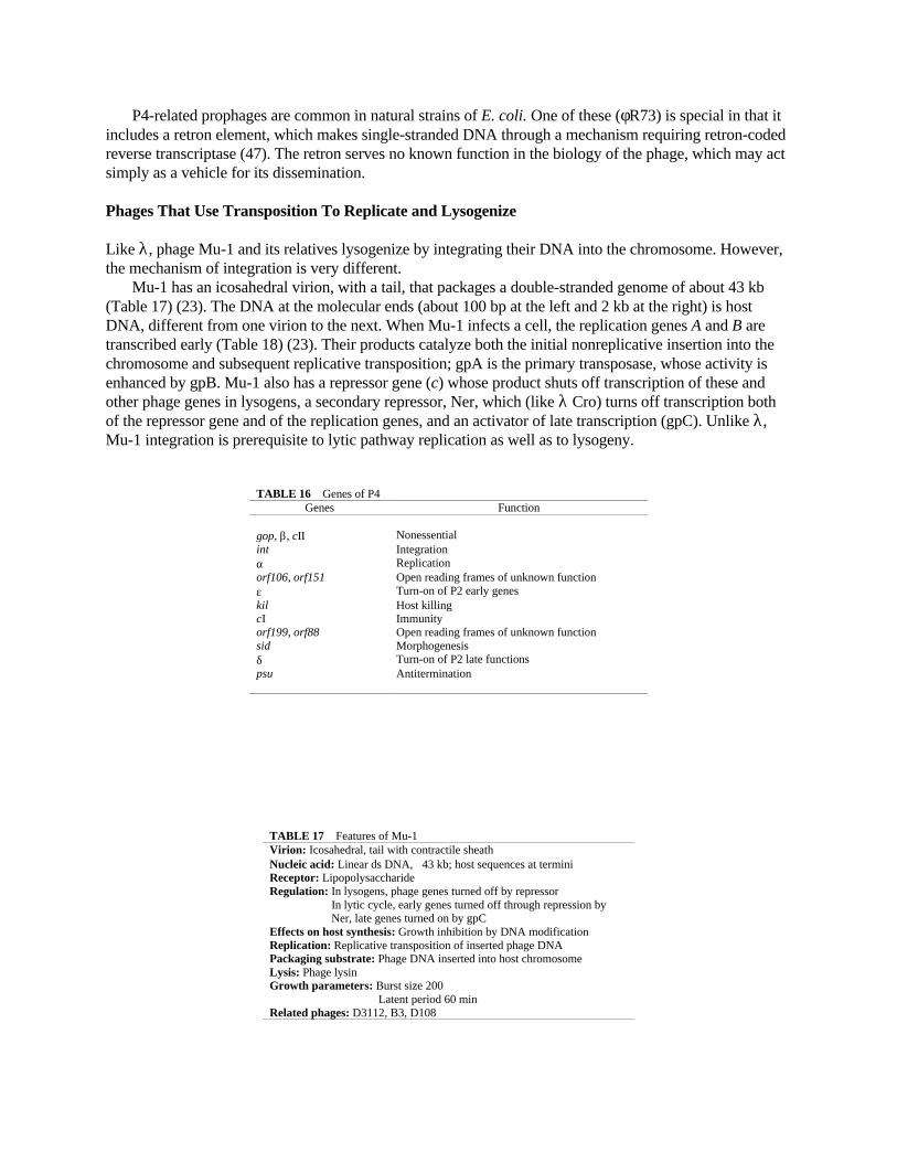
P4-related prophages are common in natural strains of E. coli. One of these (φR73) is special in that itincludes a retron element, which makes single-stranded DNA through a mechanism requiring retron-codedreverse transcriptase (47). The retron serves no known function in the biology of the phage, which may actsimply as a vehicle for its dissemination.
Phages That Use Transposition To Replicate and Lysogenize
Like λ, phage Mu-1 and its relatives lysogenize by integrating their DNA into the chromosome. However,the mechanism of integration is very different.
Mu-1 has an icosahedral virion, with a tail, that packages a double-stranded genome of about 43 kb(Table 17) (23). The DNA at the molecular ends (about 100 bp at the left and 2 kb at the right) is hostDNA, different from one virion to the next. When Mu-1 infects a cell, the replication genes A and B aretranscribed early (Table 18) (23). Their products catalyze both the initial nonreplicative insertion into thechromosome and subsequent replicative transposition; gpA is the primary transposase, whose activity isenhanced by gpB. Mu-1 also has a repressor gene (c) whose product shuts off transcription of these andother phage genes in lysogens, a secondary repressor, Ner, which (like λ Cro) turns off transcription bothof the repressor gene and of the replication genes, and an activator of late transcription (gpC). Unlike λ,Mu-1 integration is prerequisite to lytic pathway replication as well as to lysogeny.
TABLE 16 Genes of P4Genes Function
gop, β, cII Nonessentialint Integrationα Replicationorf106, orf151 Open reading frames of unknown functionε Turn-on of P2 early geneskil Host killingcI Immunityorf199, orf88 Open reading frames of unknown functionsid Morphogenesisδ Turn-on of P2 late functionspsu Antitermination
TABLE 17 Features of Mu-1Virion: Icosahedral, tail with contractile sheathNucleic acid: Linear ds DNA, ∼43 kb; host sequences at terminiReceptor: LipopolysaccharideRegulation: In lysogens, phage genes turned off by repressor In lytic cycle, early genes turned off through repression by Ner, late genes turned on by gpCEffects on host synthesis: Growth inhibition by DNA modificationReplication: Replicative transposition of inserted phage DNAPackaging substrate: Phage DNA inserted into host chromosomeLysis: Phage lysinGrowth parameters: Burst size 200 Latent period 60 minRelated phages: D3112, B3, D108

Like some bacterial transposons, Mu-1 DNA can change location by either replicative or conservativetransposition. In the first cycle following infection, nonreplicative transposition moves the Mu-1 DNAfrom the infecting molecule (where it is flanked by host DNA) into a random location on the chromosome.In the lytic pathway, subsequent cycles of transposition are replicative, so that one semiconserved copyremains at its original location and a second copy is inserted elsewhere. Repeated cycles of replicativetransposition from one chromosomal site to another cause both inversion and fragmentation ofchromosomal sequences. DNA is packaged from such chromosomal sites. The packaging systemrecognizes the left end of Mu-1 and cuts 50 to 150 bp to its left. The right cut site is determined by headfulpackaging, whose imprecision is evident in the length variation of host DNA from about 500 to 3,000 bp.
In the lysogenic pathway, repression is established, and the prophage remains inserted in its originallocation (with rare transpositions to new sites). Insertion can happen at any chromosomal site, sometimesresulting in gene disruption. The name Mu-1 (mutator) was based on the phage’s ability to cause suchdisruptions.
TABLE 18 Genes of Mu-1Genes Function
c Repression (primary)ner Repression (secondary)A, B Transposition, replicationC Stimulation of late transcriptionD, E, H, F, G, I, J Head synthesisK, L, M, Y, N, P, Q, V, W, R, S Tail synthesisgin G segment inversionmom DNA modification
Mu-1 has two additional special features: a DNA modification activity (mom) that helps it to overcomehost restriction systems and a segment (called the G segment) of about 3 kb (found also in P1 phage) thatcan invert by the action of a site-specific recombinase (encoded by the adjacent gin gene) acting on anoligonucleotide inverted repeat at the ends of the segment. The fiber protein of the Mu-1 tail is coded by agene that crosses one boundary of the invertible segment, so inversion switches the C-terminus of thisprotein to one with a different host specificity. This strategy allows Mu-1 to infect a wider range ofpotential hosts than might otherwise be possible.
Phages That Lysogenize by Plasmid Formation
A final group of temperate phages do not insert their DNA into the chromosome, but instead can be stablymaintained as plasmids. The prototype is coliphage P1 (Table 19) (53).
TABLE 19 Features of P1Virion: Icosahedral head; tail with contractile sheath, baseplate, tail fibersNucleic acid: ds DNA, ∼100 kb, circularly permuted, ∼7-kb terminal repeatReceptor: LipopolysaccharideRegulation: In lysogens, some phage genes turned off by repressor binding to numerous dispersed sites In lytic cycle, host polymerase transcribes both early and late genesEffects on host: Introduces restriction-modification systemReplication: Infecting molecules circularize through specific (lox-Cre) or general recombination Prophage replicates as circular plasmid, bidirectionally from oriR In lytic cycle, bidirectional replication from oriL, rolling-circle replication to give ds concatemersPackaging substrate: Linear concatemer (tail of rolling circle)Lysis: Probably lysozyme plus holinGrowth parameters: Burst size 80 Latent period 45 minRelated or similar phages: P7, φW39, j2, p15B, D6

P1 DNA is double stranded and terminally repetitious, about 100 kb in length. Soon after infection, theterminal segments cross over to form a circular molecule, shorter than the linear form by the length of theterminal repeat (about 7 kb). This crossover is frequently effected by a site-specific recombinase (Cre)acting on its target sites (lox) in the terminal repeat. After circle formation, P1 DNA can replicate either astheta forms and later as rolling circles (lytic pathway) or (from a different replication origin) as theta forms,once per division cycle (lysogenic pathway).
In the lytic cycle, the packaging substrates are the linear concatemers formed by rolling-circlereplication. As in P22 and T1, headful packaging is initiated at a specific pac site and continuesprocessively for several rounds. Because the pac site is close to lox, lox is present in the terminal repeats ofvirions formed during the first rounds of packaging and can be used for circularization following infection.
In the lysogenic state, some lytic cycle genes are repressed. Cre continues to be made. Repression iseffected by the product of a phage gene (c1). Like P22, P1 makes an antirepressor, which is itself repressedby a secondary repressor. Replication of the P1 plasmid DNA is determined by a 1.5-kb segment thatincludes a replication origin (oriR), the gene for a replication protein (repA), and a regulatory sequence(incA) that sequesters RepA. Replication requires host proteins, including DnaA (53). Replication controlmay be accomplished through plasmid-plasmid pairing, where the incA sequences of one plasmid bind tothe oriR site of the other through RepA protein, so that the number of plasmid copies is limited to two percell (1). Immediately adjacent to the replication segment is 2.7 kb of DNA that controls partitioning ofthese two plasmids at cell division. Partitioning requires both cis- and trans-acting factors and is facilitatedby the lox-Cre system, which resolves into monomers any dimeric molecules generated during replicationor general recombination. The resulting high fidelity of transmission is measured by a rate of plasmid lossof about 10–5 per cell per generation.
SUMMARY
Bacteriophages are important components of the natural environments of E. coli and Salmonellatyphimurium and (through lysogeny) of their genomes as well. Phages are a highly diverse group, whoseonly common feature is the packaging of their genomes into protein coats to form infectious virions. Ineach phage’s life cycle, there is a continual interplay between host products and phage products.
LITERATURE CITED
1. Abeles, A. L., and S. J. Austin. 1991. Antiparallel plasmid-plasmid pairing may control P1 plasmidreplication. Proc. Natl. Acad. Sci. USA 88:9011–9015.
2. Aoyama, A., and M. Hayashi. 1986. Synthesis of bacteriophage φX174 in vitro: mechanism of switchfrom DNA replication to DNA packaging. Cell 47:99–106.
3. Artz, S., D. Holzschu, P. Blum, and R. Shand. 1983. Use of M13 mp phages to study generegulation, structure and function: cloning and recombinational analysis of genes of the Salmonellatyphimurium histidine operon. Gene 26:147–158.
4. Barbas, C. F., A. S. Kang, R. A. Lerner, and S. Benkovic. 1991. Assembly of combinatorialantibody libraries on phage surfaces. The gene IV site. Proc. Natl. Acad. Sci. USA 88:7978–7982.
5. Barondess, J. J., and J. Beckwith. 1990. A bacterial virulence determinant encoded by lysogeniccoliphage lambda. Nature (London) 346:871–874.
6. Benzer, S. 1961. On the topography of the genetic fine structure. Proc. Natl. Acad. Sci. USA 47:403–415.
7. Berget, P. T., and A. R. Poteete. 1980. Structure and function of the bacteriophage P22 tail protein. J.Virol. 34:234–243.
8. Berkhout, B., B. F. Schmidt, A. Van Strien, J. H. Van Boom, J. Van Westrenen, and J. Van

Duin. 1987. Lysis gene of bacteriophage MS2 is activated by translation termination at theoverlapping coat gene. J. Mol. Biol. 195:517–524.
9. Bertani, L. E., and E. W. Six. 1988. The P2-like phages and their parasite, P4, p. 73–144. In R.Calendar (ed.), The Bacteriophages, vol. 2. Plenum Publishing Corp., New York.
10. Bhattacharyya, S. P., and V. B. Rao. 1993. A novel terminase activity associated with the DNApackaging protein gp17 of bacteriophage T4. Virology 196:34–44
11. Black, L., and M. Showe. 1983. Morphogenesis of the T4 head, p. 219–245. In C. Mathews, E.Kutter, G. Mosig, and P. Berget (ed.), Bacteriophage T4. American Society for Microbiology,Washington, D.C.
12. Bonovitch, M. T., and R. Young. 1991. Dual start motif in two lambdoid S genes unrelated to λ S. J.Bacteriol. 173:2897–2905.
13. Campbell, J. H., D. Dykhuizen, and B. G. Rolfe. 1978. Effects of the rex gene on phage λ lysogeny.Genet. Res. 32:257–263.
14. Chung, Y.-B., C. Nardone, and D. Hinkle. 1990. Bacteriophage T7 packaging. III. A “hairpin” endformed on T7 concatemers may be an intermediate in the primary reaction. J. Mol. Biol. 216:939–948.
15. Cue, D., and M. Feiss. 1993. A site required for termination of packaging of the phage λchromosome. Proc. Natl. Acad. Sci. USA 90:9290–9294.
16. d’Herelle, F. 1917. Sur un microbe invisible antagoniste des bacilles dysentériques. C. R. Acad. Sci.165:373–375.
17. Doermann, A. H., F. A. Eiserling, and L. Boehner. 1973. Genetic control of capsid length inbacteriophage T4. I. Isolation and preliminary description of four new mutants. J. Virol. 12:374–385.
18. Feiss, M., J. Sippy, and G. Miller. 1985. Processive action of terminase during sequential packagingof bacteriophage λ chromosomes. J. Mol. Biol. 186:759–771.
19. Flemming, M., B. Deumling, and B. Kemper. 1993. Function of gene 49 of bacteriophage T4. III.Isolation of Holliday structures from very fast-sedimenting DNA. Virology 196:910–913.
20. Geiduschek, E. P. 1991. Regulation of expression of the late genes of bacteriophage T3. Annu. Rev.Genet. 25:437–460.
21. Goulian, M., A. Kornberg, and R. L. Sinsheimer. 1967. Enzymatic synthesis of DNA. XXIV.Synthesis of infectious phage φX174 DNA. Proc. Natl. Acad. Sci. USA 58:2321–2328.
22. Haggard-Ljungquist, E., C. Halling, and R. Calendar. 1992. DNA sequences of the tail fiber genesof bacteriophage P2: evidence for horizontal transfer of tail fiber genes among unrelated bacteriophages.J. Bacteriol. 174:1462–1467.
23. Harshey, R. M. 1988. Phage Mu, p. 193–234. In R. Calendar (ed.), The Bacteriophages, vol. 2.Plenum Publishing Corp., New York.
24. Haruna, I., and S. Spiegelman. 1965. Specific template requirements of RNA replicases. Proc. Natl.Acad. Sci. USA 54:579–587.
25. Hausmann, R. 1988. The T7 group, p. 259–290. In R. Calendar (ed.), The Bacteriophages, vol. 1.Plenum Publishing Corp., New York.
26. Hayashi, M., A. Aoyama, D. L. Richardson, Jr., and M. N. Hayashi. 1988. Biology of thebacteriophage φX174, p. 1–72. In R. Calendar (ed.), The Bacteriophages, vol. 2. Plenum PublishingCorp., New York.
27. Hendrix, R. W., and R. L. Duda. 1992. λ Papa is not the mother of all λ phages. Science 258:1154–1158.
28. Hendrix, R. W., J. W. Roberts, F. W. Stahl, and R. A. Weisberg. 1983. Lambda II. Cold SpringHarbor Laboratory, Cold Spring Harbor, N.Y.
29. Hershey, A. D., and R. Rotman. 1949. Genetic recombination between host-range and plaque-typemutants of bacteriophage in single bacterial cells. Genetics 34:44–71.
30. Ho, Y. S., and M. Rosenberg. 1988. Structure and function of the activator protein cII and itsregulatory signals, p. 725–756. In R. Calendar (ed.), The Bacteriophages, vol. 2. Plenum Publishing

Corp., New York.31. Kastelein, R. A., E. Remaut, W. Feiss, and J. van Duin. 1982. Lysis gene expression of RNA phage
MS2 depends on a frameshift during translation of the overlapping coat protein gene. Nature (London)285:35–41.
32. Lindahl, G. 1970. Bacteriophage P2: replication of the chromosome requires a protein which actsonly on the genome that coded for it. Virology 42:522–533.
33. Lindquist, B. H., G. Dehó, and R. Calendar. 1993. Mechanisms of genome propagation and helperexploitation by satellite phage P4. Microbiol. Rev. 57:683–702.
34. Mathews, C. K., E. M. Kutter, G. Mosig, and P. B. Berget (ed.). 1983. Bacteriophage T4. AmericanSociety for Microbiology, Washington, D.C.
35. Model, P., and M. Russell. 1988. Filamentous bacteriophage, p. 375–456. In R. Calendar (ed.), TheBacteriophages, vol. 2. Plenum Publishing Corp., New York.
36. Mosig, G. 1988. A map of distances along the DNA molecule of phage T4. Genetics 59:137–151.37. Mosig, G., and F. Eiserling. 1988. Phage T4 structure and metabolism, p. 521–606. In R. Calendar
(ed.), The Bacteriophages, vol. 2. Plenum Publishing Corp., New York.38. Oberto, J., R. A. Weisberg, and M. E. Gottesman. 1989. Structure and function of the nun gene and
the immunity region of the lambdoid phage HK022. J. Mol. Biol. 207:675–693.39. Parma, D. H., M. Snyder, S. Sobolevski, M. Mowray, E. Brady, and L. Gold. 1992. The Rex
system of bacteriophage λ: tolerance and altruistic cell death. Genes Dev. 6:497–510.40. Ptashne, M. 1988. A Genetic Switch. Cell Press and Blackwell, Cambridge, Mass.41. Shcherbakov, V. P., L. A. Plugina, and M. L. Nesheva. 1992. Genetic recombination in bacteriophage
T4: single-burst analysis of cosegregants and evidence in favor of a splice/patch coupling model. Genetics131:769–781.
42. Simon, E. 1965. Recombination in bacteriophage T4: a mechanism. Science 150:759–763.43. Stahl, F. W., I. Kobayashi, and M. M. Stahl. 1985. In phage λ, cos is a recombinator in the Red pathway.
J. Mol. Biol. 181:199–209.44. Streisinger, G., J. Emrich, and M. M. Stahl. 1967. Chromosome structure in phage T4. III. Terminal
redundancy and length determination. Proc. Natl. Acad. Sci. USA 57:292–295.45. Studier, F. W., and J. J. Dunn. 1983. Organization and expression of bacteriophage T7 DNA. Cold
Spring Harbor Symp. Quant. Biol. 47:999–1007.46. Studier, F. W., A. H. Rosenberg, J. J. Dunn, and J. W. Dubendoff. 1990. Use of T7 DNA polymerase to
direct expression of cloned genes. Methods Enzymol. 185:60–89.47. Sun, J., M. Inouye, and S. Inouye. 1991. Association of a retroelement with a P4 like cryptic prophage
integrated into the selenocysteinyl tRNA gene of Escherichia coli. J. Bacteriol. 173:4171–4181.48. Susskind, M. M., and D. Botstein. 1978. Molecular genetics of bacteriophage P22. Microbiol. Rev.
42:385–413.49. Twort, F. W. 1915. An investigation on the nature of ultramicroscopic viruses. Lancet 189:1241–1243.50. van Duin, J. 1988. The single-stranded RNA bacteriophages, p. 117–168. In R. Calendar (ed.), The
Bacteriophages, vol. 1. Plenum Publishing Corp., New York.51. Wharton, R. P., E. L. Brown, and M. Ptashne. 1984. Substituting an α-helix switches the sequence
specific DNA interactions of a repressor. Cell 38:361–369.52. Womack, F. C. 1963. An analysis of single-burst progeny of bacteria singly infected with a bacteriophage
heterozygote. Virology 21:232–241.53. Yarmolinsky, M. R., and N. A. Sternberg. 1988. Bacteriophage P1, p. 291–438. In R. Calendar (ed.),
The Bacteriophages, vol. 1. Plenum Publishing Corp., New York.54. Young, R. 1992. Bacteriophage lysis: mechanism and regulation. Microbiol. Rev. 56:430–481.