adhesive hydrocellular dressing vs hydrocolloid dressing … · adhesive hydrocellular dressing vs...
TRANSCRIPT
ADHESIVE HYDROCELLULAR DRESSING VS HYDROCOLLOID DRESSING IN THE
MANAGEMENT OF 2nd and 3rd DEGREE PUS
A Prospective, controlled randomized comparative multi-center clinical evaluation
A. Avanzi,# M. Martinelli,##, S. Accardi###, C. Giraudi#, P. Peroli####,
A. Andriessen##### .
#Geriatric ward, Stuart Hospital, PARMA, ## Misericordia, Hospital, GROSSETO, ###Nursing
home, Sopra BG, BREMBATE, ####Geriatric hospital, Borgo Trento, VERONA, ITALY#####
Smith & Nephew WM GBU, MALDEN, NETHERLANDS.
ABSTRACT
Studies have shown the effectiveness of hydrocolloids^ (HDC) in the treatment of various wound
types. The evaluated adhesive hydrocellular* (AA) dressing has been shown to be more absorbent
and therefore able to remain in place longer than HDC. The purpose of this study was to evaluate
and to compare the performance of these dressings in the management of patients with stage II and
III Pressure Ulcers (PU). Eighty patients in a hospital and nursing home setting were recruited to
the study. The % of sloughy tissue present at the start and at the end of the study went from 17.3%
to 9.3% in the AA group vs 11.8% to 16% in the HDC group.(p<0.04). All AA dressings were
removed in less than 3 minutes vs a range of 3 to 5 minutes in the HDC group.(p<0.006) At day 21
in the AA group 90% of the dressings stayed in place for 4 days vs 35% in the HDC group.
(p<0.0007). 95% In the AA group had normal skin condition after dressing removal, 1% had slight
redness and no damage, vs 54% normal, 7% redness and 17% damage upon dressing removal in the
HDC group (p<0.008).
INTRODUCTION
One of the problems frequently associated with the management of bed-ridden patients, is the
occurrence of PU. These ulcers often appear in specific areas, difficult to manage with conventional
dressings. The AA dressing has been reported to enable optimal management of several wound types.
The dressing is easy to use, alleviates pain and does not cause injury to the wound bed when it is
removed (1)(2)(3)(6).
Studies have shown the effectiveness of HDC in the treatment of various wound types. The
evaluated AA dressing has been shown to be more absorbent and therefore able to remain in place
longer than the HDC dressing.(5) This suggests that AA dressings may be of particular use in the
management of PU. The purpose of this study was to evaluate the performance of a hydrocellular
dressing (Allevyn Adhesive) and to compare it with that of a hydrocolloid dressing (Duoderm
CGF) in the management of stage II and III PU.
MATERIAL AND METHODS
The AA dressing used in this study consists of 3 layers. The external layer, a vapour-permeable
polyurethane film, creates a bacterial barrier (4) and inhibits fluid from leaking. Ameen et al.(4)
showed that AA inhibits bacterial transmission over an11-day challenge period. The investigators
suggested that AA may facilitate infection control by acting as a physical barrier to the transmission of
pathogenic and antibiotic-resistant wound bacteria (4). The hydrophilic core accounts for the high
absorbency of the dressing (10 times its weight) and for its structural integrity; the dressing can be left
in the wound for more than four days.(5)(6) The third layer, a polyurethane hypoallergenic adhesive
film which adheres to the surrounding skin, offers protection due to friction. This layer in contact with
the wound prevents the dressing from sticking to the wound bed. It makes changing the dressing easier
and less painful (3)(5)(6).
The hydrocolloid dressing under evaluation consists of a thin layer of a semi-open-cell polyurethane
foam bonded onto a polyurethane film which acts as a carrier for the hydrocolloid base.
This was a prospective multi-center comparative randomized study. Eighty patients from a hospital
– and home care setting were recruited, who fulfilled the following inclusion criteria: Patients of
either sex, aged at least 18 years, with 2nd and 3rd degree PU,(7) were compliant with medical
treatment and able to give consent.
Exclusion criteria were: Pregnant women; patients with plasma proteins <6mg/100ml,
Hb<10mg/100ml, or those with clinically infected wounds. A clinical infection was defined as
follows: Inflammation or cellulitis around the ulcers, purulent exudate, fever, these patients could
only be recruited once the infection had cleared up. At the initial evaluation the following was
assessed: The time since occurrence of the PU, the stage of the ulcer, ulcer site, ulcer size, description
of the wound bed and peri-wound skin condition. For assessment of the condition of the wound bed a
red/yellow/black color classification model was used. The three colors were documented for each
wound and the percentages present were indicated on ulcer tracings/photographs upon each dressing
change and at the end of the study. Tracings were taken using a measuring grid (Opsite Flexigrid); the
number of whole and half squares were counted. Photographs were taken using a standardized
technique and equipment. Test and control ulcers were photographed on the same standard blue
background (disposable surgical drape) and aperture, exposure time and distance were kept constant.
The color and odor of the exudate in the dressing were used as parameters to assess possible clinical
infection. The peri-wound skin condition was assessed for information on any increase in pain reported
by the patient. To analyze pain, a 10cm scale was used to record. At each dressing removal, subjects
were asked to put a single stroke on the line to indicate their level of pain (0=no pain, 10=unbarable
pain).
The trial and control dressings were changed at the discretion of the clinician, depending on the
amount of exudate produced, at a maximum every four days. The treatment lasted until 10 dressing
changes were performed or until one of the following endpoints had been reached: re-
epithelialisation; an adverse incident; patient withdrawn for other reasons. At each dressing removal
the reason for changing the dressing was described, as well as the size and depth of the wound, and rate
of exudate production. At the same time an evaluation of the ease of application and removal of the
dressing took place. The final evaluation included the clinicians' subjective assessment of the features
of the trial dressing compared to the control dressing.
METHOD OF ANALYSIS
Data from the completed and validated questionnaires were entered into a program written in
Microsoft Access. A randomization code was used to define and allocate the patients to the trial -
and control treatment. Summary baseline statistics were compiled to help evaluate the validity of
statistical assumptions. Discrete variables (e.g. gender) were analyzed using the Chi-square statistic
or Fisher’s exact test where appropriate. Continuous variables (e.g. age) were analyzed using
Student’s t-test. To evaluate the effect on wound healing (i.e. percent change in area) a repeated
measure analysis of variance was used, in which the effect of ulcers in the trial group vs. ulcers in
the control group, time (days 0 through the end of study) and their interaction were assessed.
Statistical analysis included the chi-square test and variance analysis (one-way Anova).
RESULTS
The study population consisted of 80 patients with stage II and III PU(7).
The Clinical Investigator sought the permission of the relevant consultant for their patients to be
included in the study. Before being admitted to the clinical evaluation, the patient consented to
participate, after the nature, scope and possible consequences of the study had been explained in an
understandable form. Patients were questioned on their previous medical and social history.
Assessments were made to determine the main cause of ulceration and risk assessment was performed
using an adapted Braden Risk Score.
Patients were recruited in several centers in Italy between December 1996 and December 1999
(Table 1 and 2). Of the 80 patients that were recruited, 63 were evaluated. 9 Patients were lost to
follow up, 3 patients died and 5 were withdrawn for reasons not related to the PU treatment. There was
no difference in age, stage of the ulcers and mobility between the two cohorts. As shown in table 3,
upon recruitment to the study the ulcers in the AA group were significantly deeper.
Evolution in ulcer size and appearance of the wound bed
As shown in table 4, at day14 there was a difference in ulcer size reduction in favor of the AA group.
Comparing the evolution in wound bed appearance, the % of sloughy tissue present at the start and end
of the study went from 22% to 6% in the AA group vs. 13% to 14% in the HDC group. This difference
was statistically significant p< 0.05. Complete
epithelialisation at 10 dressing changes, end of the study period is 85% (estimate); treatment difference:
25%; significance: 5%; power: 80%.
Dressing application, removal, time in situ
The % of dressings that stayed in place for 4 days was significantly higher in the AA group at day
7,(p<0.001) day 14 (p<0.0007) and day 21 (p<0.0007).
At days 7 and 14 the level of pain reported upon dressing removal, was lower for AA, compared to
HDC (p< 0.07). Further into the trial, days 21 and 28 there was no difference reported between the two
groups.
Time taken for dressing removal: All AA dressings were removed in less than 3 minutes vs. a range of
3 to 5 minutes in the HDC group. At day 21 assessment, there was a significant difference shown in
time required for dressing removal in the AA group vs. the
HDC group ( p< 0.006).
Ease of dressing removal is shown in table 5. Regarding adherence of the dressing to the wound
bed, there was a highly significant difference at day 28, when most of the wounds assessed are
granulating wounds. At day 7, there was a significant difference in peri-wound skin condition in
favor of the AA group, 89% had normal skin condition after dressing removal, 11% slight redness
and no damage, vs. 48% normal skin condition, 34% redness and 18% skin damage in the HDC
group (p< 0.007).
At day 14 in the AA group 95% had normal skin condition after dressing removal, 1% redness and
no damage, vs. 54% normal, 7% redness and 17% damage upon dressing removal in the HDC
group ( p< 0.008).
DISCUSSION
In conducting Randomized Controlled Trials comparing Moist Wound Healing dressings using
chronic wounds as a model, one encounters difficulties in defining standards and categories on
chronic wounds. Methodological problems regarding patient numbers and difficulties regarding
relevance on time, i.e. path, may influence the quality of such studies. Factors influencing quality
of life, equally important for the clinician to make an appropriate choice are often left out of the
equation. There may be methodological approaches that support avoiding bias, which are complex,
time consuming and therefore costly and not necessarily producing better results.
The defined study period was limited for reasons of practicality and ethics. This comported that we
developed the above study design focussing on practical aspects of PU management rather than
healing time.
Occlusive dressings possess the characteristic of promoting a local moist environment in chronic
wounds (8). With the growing number of dressing types available a better understanding of properties
is essential for the caregiver to provide appropriate management for each wound type (6). Apart from
the biophysical properties, practical aspects of the dressing chosen may influence the outcome. PU
often appear in specific areas, such as the sacral area, complex to manage, especially in incontinent
patients (3). Depending on exudate production and the absorbent capacity of the dressing, maceration
of the wound edges may occur. Typical disadvantages observed with adhesive dressings are damage to
the peri-wound skin, which may be enhanced when maceration occurs. The results shown in the
present study regarding skin condition, suggest that an enhanced absorbent capacity of the dressing
may aid to inhibit skin damage.
CONCLUSIONS
The overall performance of the AA dressing was shown to have clinical advantages over the HDC
dressing in all the parameters under evaluation.
The use of AA proved to be safe and did not cause mechanical injuries upon dressing changes in
comparison to the HDC group, where skin damage was reported in up to 18% of the cases. The patients
reported little or no pain upon dressing removal in the AA group vs. significantly more pain in the
HDC group. The AA dressing was shown to be easier to apply and to remove, as well as having a
greater absorbent capacity than the HDC.
*Trademark of Smith & Nephew Medical Ltd
^Trademarks of E.R. Squibb & Sons, Inc. ConvaTec Limited, authorised user.
We greatly acknowledge Prof. Boddi and Dr. Vannucchi, Angiology Department of the Santa
Maria Nuova Hospital, Florence, Italy and H. van Druten, PhD of the Bio-statistical Department,
University of Nijmegen, Netherlands, for statistical advice and analysis.
The authors from the Italian Clinics have no commercial, proprietary, or financial interest
in the products or companies described in this article. A. Andriessen is employed by the Wound
Management Global Business Unit of Smith & Nephew as an International Clinical Specialist. (e-
mail: [email protected])
REFERENCES
1) Foster A.V.M., Greenhill M.T., Edmonds M.E. Comparing two dressings in
the treatment of diabetic foot ulcers. J. Wound Care 1994: 3, 224-228.
2) Manniche, L., Data on file S&N. 5363 AUS/CLP/UK/992
3) Shutler, S., Stock, J., Bales, S., Harding KG., A multi-centre comparison of a Hydrocellular
adhesive dressing and a Hydrocolloid dressing in the management of stage 2 & 3 pressure sores,
Poster, 5th EWMA Conference 1995. CT89/16.
4) Ameen, H. et al. Investigating the bacterial barrier properties of four contemporary wound
dressings, J Wound Care 2000; 9, 455-459
5) Kurring, P.A., Roberts C.D., Quinlan D. Evaluation of a hydrocellular
dressing in the management of exuding wounds in the community. Br. J. Nurs.
1994: 3, 1049-50.
6) Reali, UM., Martini, L., Andriessen, A., An adhesive hydrocellular dressing in the treatment of
partial thickness skin graft donor sites, J. Wound Care 1999: 9, 457- 460
7) Nano, M., Ricci E., Le piaghe da decubito nel paziente anziano, Edizioni Minerva Medica,
Torino 1994, P26-27
8) Tsukada, K., Tokunaga, K., Iwama, T., et al; The ph changes of Pressure Ulcers related to the
healing process of wounds. Wounds 1992; 4(1): 16-20
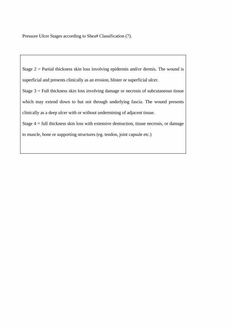
Pressure Ulcer Stages according to Shea# Classification (7).
Stage 2 = Partial thickness skin loss involving epidermis and/or dermis. The wound is
superficial and presents clinically as an erosion, blister or superficial ulcer.
Stage 3 = Full thickness skin loss involving damage or necrosis of subcutaneous tissue
which may extend down to but not through underlying fascia. The wound presents
clinically as a deep ulcer with or without undermining of adjacent tissue.
Stage 4 = full thickness skin loss with extensive destruction, tissue necrosis, or damage
to muscle, bone or supporting structures (eg. tendon, joint capsule etc.)
Patient population