additions to the cercosporoid fungi from the brazilian ... · cerrado: 1. new species on hosts...
TRANSCRIPT
http://dx.doi.org/10.12664/mycobiota.2015.05.06
Copyright ©2015 ■ MYCOBIOTA
MYCOBIOTA 5: 33–64 (2015) RESEARCH ARTICLE ISSN 1314-7129 (print)doi: 10.12664/mycobiota.2015.05.06 ISSN 1314-7781 (online)www.mycobiota.com
Additions to the cercosporoid fungi from the Brazilian Cerrado: 1. New species on hosts belonging in family Fabaceae, and reallocations of four Stenella species into Zasmidium
Antonio Hernández-Gutiérrez ¹, Zuleide Martins Chaves ², Denise Dornelo-Silva ² & José Carmine Dianese ²*
¹ Departamento de Fitopatologia, Universidade Federal do Pará, Rua Augusto Corrêa 01, Campus do Guamá, 66071–110 Belém, Pará, Brazil
² Departamento de Fitopatologia, Universidade de Brasília, 70910-900, Brasília, Distrito Federal, Brazil
Received 25 November 2015 / Accepted 8 December 2015 / Published 18 December 2015
Hernández-Gutiérrez, A., Chaves, Z.M., Dornelo-Silva, D. & Dianese, J.C. 2015. Additions to the cercosporoid fungi from the Brazilian Cerrado: 1. New species on hosts belonging in family Fabaceae, and reallocations of four Stenella species into Zasmidium. – Mycobiota 5: 33–64. doi: 10.12664/mycobiota.2015.05.06
Abstract. Nine new species of cercosporoid fungi were found on leaves of plants in family Fabaceae from the Brazilian Cerrado: Asperisporium galactiae on Galactia peduncularis, Sirosporium sclerolobii and Zasmidium sclerolobii on Sclerolobium paniculatum, Pseudocercospora acosmii-subelegantis on Acosmium subelegans, and Pseudocercospora stryphnodendri on Stryphnodendron adstringens. In addition, four other Pseudocercospora species were found on Cassia s. lat., including Chamaecrista and Senna: Pseudocercospora aquae-emendadasensis on Chamaecrista orbiculata; Pseudocercospora sennae, Pseudocercospora sennae-rugosae, and Pseudocercospora subcuticularis on Senna rugosa. Descriptions, illustrations, and discussions of the new taxa are supplemented by keys to Pseudocercospora species on hosts belonging to Cassia s. lat., and to Zasmidium species on Fabaceae.
Key words: Asperisporium, foliicolous fungi, fungal taxonomy, leaf spots, Neotropical mycodiversity, Pseudocercospora, Sirosporium
* Corresponding author: e-mail: [email protected]

34 Hernández-Gutiérrez, A. et al. — Additions to the cercosporoid fungi of the Cerrado
IntroductionA survey accomplished in 2008 indicated that the Fabaceae (including subfamilies Caesalpinoideae, Faboideae, and Mimosoideae), known as the largest family of fl owering plants in the world, encompasses in the Cerrado 108 genera, 1,174 species, and 262 varieties and subspecies. Th e Fabaceae corresponds to 9.5 % of the total number (> 12,000 species) of higher plant species in the Cerrado, including angiosperms, gymnosperms, and pteridophytes (Mendonça et al. 2008). However, studies on legume-associated cercosporoid fungi from the Cerrado are very scarce and confi ned to species described by Viégas (1945), included in Chupp (1954), some of them revised by Inácio et al. (1996) and Crous et al. (1997), and a few by Batista et al. (1960). Starting in 1994 (Medeiros & Dianese 1994), a sequence of papers was published describing new species of cercosporoid fungi from the Cerrado. In these publications, a new species belonging in Parastenella, four in Cercospora, 22 in Passalora, 31 in Pseudocercospora, and fi ve in Stenella were described (Dianese & Câmara 1994; Inácio & Dianese 1998, 1999, 2006; Furlanetto & Dianese 1999; Dornelo-Silva & Dianese 2003; Dornelo-Silva et al. 2007; Dianese et al. 2008; Hernández-Gutiérrez & Dianese 2008, 2009, 2014a, b; Hernández-Gutiérrez et al. 2014). Four of the species previously assigned to Stenella are now reallocated to Zasmidium. On fabaceous hosts only eight new species distributed among the genera Cercospora, Passalora, and Pseudocercospora have recently been described (Hernández-Gutiérrez & Dianese 2009). Th e present paper deals with new species of Asperisporium, Pseudocercospora, Sirosporium, and Zasmidium on plants belonging to Fabaceae, and reallocations of Stenella species from the Cerrado into Zasmidium.
Material and methodsLeaves showing necrotic spots of diff erent shades of gray/brown were collected and dried in plant presses, before processing and depositing them in Herbarium UB Mycological Collection. Colonies on the leaf spots were initially examined using a Zeiss (Discovery v.8) stereomicroscope, to look for synnematous or fasciculate conidiophores. Portions of the fungal colonies were removed from the leaves and mounted on slides containing lacto-glycerol. Th en, light microscopic observations, measurements, and drawings were made using a Zeiss microscope, coupled to a camera lucida. Th e drawings show details of stromata, condidiophores, conidiogenous cells, and conidia of each specimen studied.
Taxonomy
Asperisporium galactiae A. Hern.-Gut., Z.M. Chaves & Dianese, sp. nov. Fig. 1MycoBank MB 814798
Holotype: BRAZIL. Mato Grosso: Cuiabá, Chapada dos Guimarães National Park, Morro São Jerônimo, on leaves of Galactia peduncularis (Benth.) Taub. (Fabaceae/Faboideae), 11 Apr 1996, J.C. Dianese (UB – Mycol Col. 11332).

MycoBIOTA 5 (2015) 35
Etymology: galactiae, derived from the host plant genus Galactia.
Diagnosis: Diff ers from the type species, Asperisporium caricae (Speg.) Maubl., in having sublunate to fusoid conidia (vs. short-cylindrical in A. caricae).
Description: Lesions 0.5–2 mm diam., amphigenous, sparse, circular or irregular, brown, without defi ned margin and halo. Colonies brown, caespitose, mostly hypophyllous, sometimes epiphyllous. Mycelium internal. Stromata well-developed, textura globosa, erumpent, light brown, 23–50 μm diam. Conidiophores 0–2-septate, sometimes reduced to a simple conidiogenous cell, in compact sporodochia, straight, lageniform, cylindrical, ampulliform when immature, 17–38 μm long, 6–8 μm broad in the widest part, light brown, smooth, thin-walled. Conidiogenous cells terminal, integrated, polyblastic, sympodial, with prominent thick, dark conidiogenous loci, 1.5–2.5 μm wide. Conidia solitary, sublunate to fusoid, obconically truncate to truncate at the base, with thick, dark hilum, 1–2 μm wide, apex rounded to broadly rounded, 33–40 μm long, 5–8 μm diam. in the widest part, 2–3 μm at the base, 1–3 μm near the apex, 0–5-septate, dark brown, strongly verrucose when mature.
Comment: Crous & Braun (2003) and Braun et al. (2013) considered Asperisporium close to Passalora, but diff ering by possessing strongly verrucose conidia. However, sequencing of the ITS region and nLSU of the Asperisporium type species, A. caricae, showed that it clustered within the Mycosphaerellaceae clade close to several Passalora species, but excluding the type species of the latter genus (Minnis et al. 2011). Th us, Braun et al. (2013) also admitted that further molecular work is needed to better clarify the situation of the polyphyletic genus Passalora. Th ey also suggested that species with lightly verruculose conidia, as for instance A. sequoiae (Ellis & Everh.) B. Sutton & Hodges and A. juniperinum (Georgescu & Badea) B. Sutton & Hodges, clustering close to Passalora species and further away from the Asperisporium type species (Minnis et al. 2011), should be assigned to Passalora s. lat. Th erefore, it is currently advisable to assign phylogenetically unresolved species that morphologically agree with the type of Asperisporium to the latter genus, at least tentatively.
Th ere are 22 species of Asperisporium described and illustrated in the literature (http://indexfungorum.org, September 2015). Most of the species agree morphologically well with the type species, A. caricae (Ellis 1971, 1976). Recently, A. caricicola Crous & C. Nakash., a species morphologically indistinguishable from the type species has been described. Th e authors based the introduction of the new species on minor diff erences in ITS and LSU sequences; and the data was generated by comparing just one isolate of A. caricae with one of that considered as new species (Crous et al. 2015). When one takes into account that morphology refl ects the entire genome of a fungus, it is diffi cult to accept a new species just based on minor diff erences in sequences whose phylogenetic meaning is hardly appraisable. In addition, it is more than questionable to base such a critical species on an unacceptably small sampling. Carica papaya is a species naturally distributed in coastal regions of tropical America, but A. caricicola was based on a single collection from cultivated papaya on Fiji, Oceania, which raised the question if, indeed, two morphologically indistinguishable Asperisporium have been evolved on this host or if the sequences just refl ect intraspecifi c genetic variation.

36 Hernández-Gutiérrez, A. et al. — Additions to the cercosporoid fungi of the Cerrado
Th is question can barely be answered just based on two sequences. In any case, the precipitate introduction of A. caricicola is little helpful and more confusing to phytopathologists.
Asperisporium galactiae forms dark, subcuticular or subepidermal stromata, with textura globosa. Th is characteristic is also present in A. moringae (Ellis 1976); all other species of Asperisporium have stromata with textura angularis. Conidiophores of most species are cylindrical or occasionally obclavate, but conidiophores of A. galactiae range from ampulliform when young, to lageniform or even cylindrical when mature, and they are 0–1-septate. Th e conidia of Asperisporium species are in general uniformly 0–1(–2)-septa, except for A. mikaniae (Ellis & Everh.) R.W. Barreto (Barreto & Evans 1995) that may show conidia up to 5-septate, as is the case in A. galactiae.
Th e conidia of A. galactiae are well characterized by being slightly curved (sublunate), wider at the middle, and strongly verrucose, but not verruculose at the base, with a more or less prominent dark hilum. Th e conidiogenous cells of A. galactiae are very characteristic by having prominent, thick, dark colored conidiogenous loci, appearing as broad short denticles, giving a peculiar shape to the conidiogenous cell apex.
Two species assigned to Asperisporium are known to be parasitic on hosts of the Fabaceae, viz. A. cassiae (Syd.) Deighton on Senna multiglandulosa (Jacq.) H.S. Irwin & Barneby (= Cassia tomentosa L. f.), and A. pongamiae (Syd. & P. Syd.) Deighton on Pongamia sp. (Ellis 1976). Th us, the present specimen represents the third Asperisporium species on a host of the Fabaceae, and the fi rst on a Galactia species.
Asperisporium cassiae shows longer multiseptate conidiophores and smaller conidia than the new species; while A. pongamiae has smaller conidia with smooth, wrinkled or minutely verruculose wall. Among other cercosporoid fungi on members of the genus Galactia, there are only two that, however, are typical Cercospora species, C. fl agellifera G.F. Atk. and C. galactiae Ellis & Everh. Th us, the well-defi ned and peculiar characteristics of the new species, such as verrucose, sublunate to fusoid conidia, infecting for the fi rst time a Galactia species, diff erentiate this specimen from the remaining species of Asperisporium, clearly indicating that it belongs to a new species.
Pseudocercospora acosmii-subelegantis A. Hern.-Gut., Z.M. Chaves & Dianese, sp. nov. Fig. 2MycoBank MB 814799
Holotype: BRAZIL. Distrito Federal: Planaltina, Águas Emendadas Ecological Station, on leaves of Acosmium subelegans (Mohlenbr.) Yakovlev (Fabaceae/Faboideae), 6 Mar 1995, M. Sanchez (UB – Mycol Col. 7397).
Etymology: acosmii-subelegnatis, derived from the host species Acosmium subelegans.
Diagnosis: Diff ers from P. luzianensis, also known from the Cerrado, in having non-synnematous conidiophores and conidia with thickened walls and septa (vs. conidia with thin septa and walls in P. luzianensis).

MycoBIOTA 5 (2015) 37
Description: Lesions 0.5–3 mm diam., amphigenous, punctiform, circular or irregular, dark brown, without a delimiting margin. Colonies exclusively hypophyllous, brown. Mycelium internal. Stromata absent. Conidiophores in fascicles with up to ten conidiophores emerging through stomata, straight or slightly curved, sometimes showing percurrent proliferations, 41–93 μm long and 3–5 μm wide, 1–5 septate, light brown olivaceous, smooth. Conidiogenous cells integrated, polyblastic, sympodial, sometimes geniculate and showing percurrent extensions; conidiogenous loci fl at slightly pigmented. Conidia simple, non catenate, obclavate or fusoid, sometimes curved, obconically truncate to truncate at the base, with unthickened, but lightly pigmented 1.5–3 μm wide hilum, obtuse to rounded at the apex, walls and septa thickened, 32–82 μm long, 7–10 μm broad at the widest part, 1–4 μm at the base, 1–2 μm near the apex, 0–9-septate, olivaceous-brown, paler near the apex, smooth.
Pseudocercospora aquae-emendadasensis A. Hern.-Gut., Z.M. Chaves & Dianese, sp. nov. Fig. 3MycoBank MB 814800
Holotype: BRAZIL. Distrito Federal: Planaltina, Águas Emendadas Ecological Station, on leaves of Chamaecrista orbiculata (Benth.) H.S. Irwin & Barneby (Fabaceae), 21 Feb 1997, M. Sanchez (UB – Mycol. Col. 13421).
Etymology: Specifi c epithet referring to the site of type collection.
Diagnosis: Diff ers from P. chamaecristigena in having shorter mononematous conidiophores (50–130 μm long) (vs. much longer synnematous conidiophores, 208–335 μm long, in P. chamaecristigena).
Description: Lesions 1–4 mm diam., amphigenous, circular, brown to dark brown, surrounded by a yellowish halo. Colonies amphigenous, mainly caespitose, sometimes loosely synnematous, olivaceous-brown. Mycelium internal. Stromata 17–50 μm diam., moderately developed, textura globosa, substomatal, light brown. Conidiophores in loose to dense fascicles, emerging through stomata, slightly curved, somewhat fl exuous or straight, 50–130 μm long, 5–7 μm broad at the widest part, 1–5-septate, occasionally with percurrent proliferation, light brown to olivaceous, smooth, thin-walled. Conidiogenous cells terminal, integrate, mostly monoblastic, sometimes polyblastic and sympodial, light olivaceous; conidiogenous loci aplanate, unthickened. Conidia solitary, straight, slightly curved, sometimes fl exuous, obclavate to fusoid, obconically truncate, sometimes cylindrical at the base with slightly prominent hilum, 2–4 μm wide, rounded to broadly rounded at the apex, 28–65 μm long, 5–7 μm broad at the widest part, 2–3 μm at the base, 2–3 μm near the apex, 0–4-septate, walls and septa thickened, sometimes constricted at some septa, light olivaceous to subhyaline, smooth.
38 Hernández-Gutiérrez, A. et al. — Additions to the cercosporoid fungi of the Cerrado
Additional specimen examined: BRAZIL. Distrito Federal: Planaltina, Águas Emmendadas Ecological Station, on leaves of Chamaecrista orbiculata (Fabaceae), 5 Mar 1997, M. Sanchez (UB – Mycol Col. 13703).
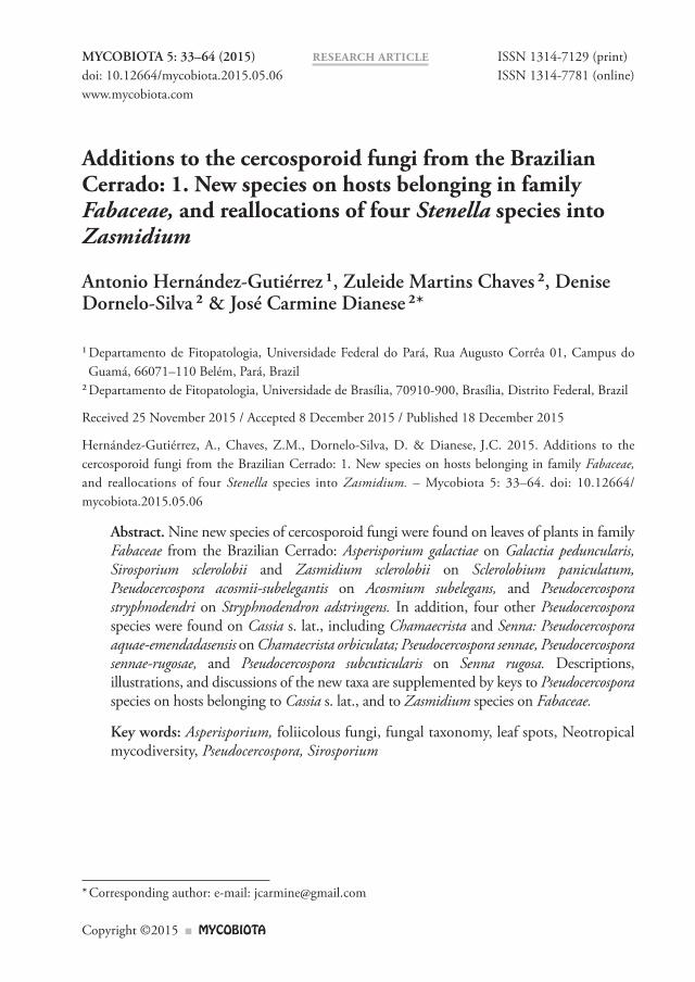
Pseudocercospora sennae A. Hern.-Gut., Z.M. Chaves & Dianese, sp. nov. Fig. 4MycoBank MB 814801
Holotype: BRAZIL. Distrito Federal: Brasília, North Peninsula, on leaves of Senna rugosa (G. Don.) H.S. Irwin & Barneby (Fabaceae), 31 May 1992, J.C. Dianese (UB – Mycol Col. 1132).
Etymology: Specifi c epithet derived from the host genus.
Diagnosis: Diff ers from P. taichungensis in having much longer, 1–5-septate conidiophores, 30–76 μm long, and much longer conidia, 24–132 μm (vs. conidiophores 10–25 × 1–3 μm, 0–2-septate, conidia, 20–55 μm long) in P. taichungensis).
Description: Lesions 1–6 mm diam., amphigenous, irregular, sometimes coalescent, brown with a darker margin and surrounded by a yellowish brown halo. Mycelium internal. Colonies amphigenous, brown. Stromata 42–183 μm diam., well-developed, textura angularis, erumpent, brown. Conidiophores in dense and compact layers arising from the stromata, unbranched, straight or slightly curved, cylindrical, 30–76 μm long, 4–6 μm broad at the widest part, reddish brown, 1–5-septate, sometimes percurrently proliferating, smooth, thin-walled. Conidiogenous cells terminal, integrated, mono- or polyblastic, sympodial; conidiogenous loci fl at, unthickened, 3–3.5 μm wide. Conidia solitary, curved or straight, narrowly obclavate, sometimes cylindrical or fi liform, obconically truncate to truncate at the base, hilum not thickened. 1–2.5 μm wide; obtuse to rounded or broadly rounded at the apex, 24–132 μm long, 3–5 μm broad at the widest part, 1–2 μm at the base, 1–3 μm near the apex, 1–8-septate, light brown to subhyaline, smooth, thin-walled.
Additional specimens examined: On leaves of Senna rugosa (Fabaceae) BRAZIL. Distrito Federal: Brasília, North Peninsula, 11 May 1992, J.C. Dianese (UB – Mycol Col. 1219). Planaltina, Águas Emendadas Ecological Station, 10 Jun 1992, R.B. Medeiros (UB – Mycol Col. 1139). Brazlândia, 18 Jun 1992, R.B. Medeiros (UB – Mycol Col. 1232). PADF, 18 Feb 1993, J.C. Dianese (UB – Mycol Col. 3177). Minas Gerais: Divinopolis, Expo Park, J.C. Dianese (UB – Mycol Col. 5514).
Pseudocercospora sennae-rugosae A. Hern.-Gut., Z.M. Chaves & Dianese, sp. nov. Fig. 5MycoBank MB 814802
Holotype: BRAZIL. Distrito Federal: Brasília, North Peninsula, on leaves of Senna rugosa (G. Don.) H.S. Irwin & Barneby (Fabaceae), 9 Jan 1993, J.C. Dianese (UB – Mycol Col. 3033).

MycoBIOTA 5 (2015) 39
Etymology: Epithet referring to the host species, Senna rugosa.
Diagnosis: Diff ers from P. sennae in having 2–12-septate conidiophores and longer, 3–18-septate conidia, 17–193 × 3–4 μm (vs. conidiophores 1–5-septate and conidia shorter, 24–132 × 3–5 μm, with 1 to 8 septa in P. sennae ).
Description: Lesions 2–8 mm diam., amphigenous, irregular, reddish brown, surrounded by a somewhat darker margin. Colonies mainly epiphyllous, occasionally hypophyllous, caespitose, shining grey. Stromata 152–245 μm diam., well-developed formed by cells of textura angularis at the lower part and textura prismatica at the upper part due to the compactly aggregated conidiophores, brown, subcuticular to subepidermal, erumpent. Conidiophores in very dense and compact fascicles to form a prismatic texture, individual conidiophores only evident at the apex or at conidiogenous cells, unbranched, straight or slightly curved, cylindrical, 50–66 μm long, 4–5 μm broad at the widest part, 2–12-septate, brown, smooth, thin-walled. Conidiogenous cells terminal, integrated, monoblastic, narrowly lageniform to cylindrical, light brown; conidiogenous loci fl at, unthickened, 2–2.5 μm wide. Conidia solitary, straight, slightly curved to strongly curved by being almost U-shaped, fi liform to slightly obclavate; obconically truncate to truncate at the base, hilum unthickened, 1–2 μm wide, obtuse to rounded at the apex, 17–193 μm long, 3–4 μm broad at the widest part, 1–3 μm at the base and near the apex, 3–18-septate, light brown to light olivaceous-brown, smooth, thin-walled.
Additional specimen examined: On leaves of Senna rugosa (Fabaceae) BRAZIL. Distrito Federal: Planaltina, Núcleo Rural do Pipiripau, 10 Jun 1994, C.A. Inácio (UB – Mycol Col. 6191).
Pseudocercospora stryphnodendri A. Hern.-Gut., Z.M. Chaves & Dianese, sp. nov. Fig. 6MycoBank MB 814803
Holotype: BRAZIL. Minas Gerais: Divinópolis, Barrinha Farm, right side of Highway from Divinopólis to Formiga, 20°13’54.9”S, 45°05’33.7”W, on leaves of Stryphnodendron adstringens (Mart.) Coville (Fabaceae), 31 Dec 1991, J.C. Dianese (UB – Mycol Col. 894).
Etymology: Epithet referring to the host genus.
Diagnosis: Diff ers from the similar P. chamaecristae in having fasciculate, but non-synnematous, much shorter conidiophores, 12–22 μm long and much longer and narrower, 3–9-septate conidia, 26–75 × 2–3 μm (vs. longer, synnematous conidiophores and much shorter and wider, 1–4-septate conidia, 25–35 × 5–8 μm, in P. chamaecristae.
Description: Lesions 1–16 mm diam., amphigenous, irregular, sometimes circular, coalescent, purplish brown, without margin or surrounding halo. Colonies amphigenous, caespitose, yellowish grey. Stromata 13–26 μm diam., poorly developed, formed by cells of textura
40 Hernández-Gutiérrez, A. et al. — Additions to the cercosporoid fungi of the Cerrado
globosa, substomatal, light olivaceous to subhyaline. Conidiophores in compact stomatal fascicles, unbranched, straight or slightly curved, lageniform or ampulliform, 12–22 μm long, 3–4 μm broad at the widest part, 0–1-septate, light olivaceous to subhyaline, smooth, thin-walled. Conidiogenous cells terminal, integrate, mainly forming a unicellular conidiophore, polyblastic, sympodial, geniculate, conidiogenous loci fl at, unthickened, not pigmented, 1–1.5 μm wide. Conidia solitary, curved, fl exuous or straight, fi liform, obclavate, obconically truncate to truncate at the base, hilum unthickened, 1 μm wide; acute, obtuse to rounded at the apex, 26–75 × 2–3 μm diam. at the widest part, 1–2 μm at the base and near the apex, 3–9-septate, light olivaceous to subhyaline, smooth, thin-walled.
Additional specimens examined: on leaves of Stryphnodendron adstringens (Fabaceae), BRAZIL. Distrito Federal: Brasília, Vargem Bonita, Cerrado das Mansões, 26 Jun 1994, M. Sanchez (UB – Mycol Col. 6222, 6223, 6242, 6243, and 6244). Planaltina, Águas Emendadas Ecological Station, 26 Jun 1995, Z.M. Chaves (UB – Mycol Col. 8880).
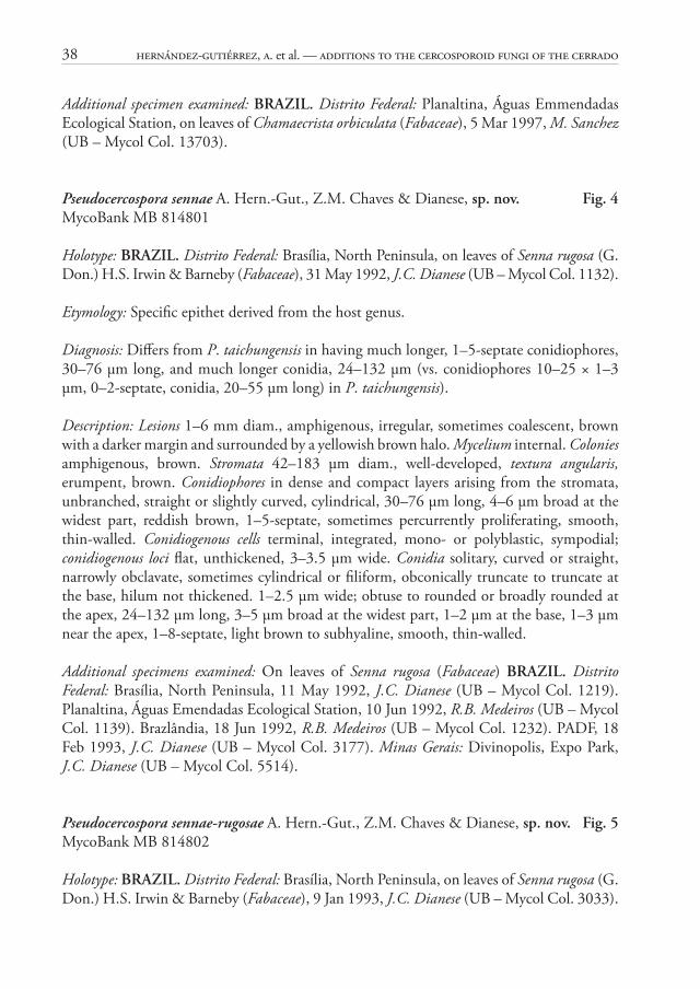
Pseudocercospora subcuticularis A. Hern.-Gut., Z.M. Chaves & Dianese, sp. nov. Fig. 7MycoBank MB 814804
Holotype: BRAZIL. Distrito Federal: Brasília, University of Brasília, Biological Experiment Station, on leaves of Senna rugosa (G. Don.) H.S. Irwin & Barneby (Fabaceae), 11 Jul 1993, J.C. Dianese (UB – Mycol Col. 4092).
Etymology: Epithet referring to the subcuticular origin of the conidiophores.
Diagnosis: Diff ers from the similar P. sennae-rugosae in having 2–7-septate, shorter conidiophores (to 56 μm long), and much shorter, 0–13-septate conidia (26–92 μm long) (vs. 2–12-septate conidiophores, 50–66 × 4–5 μm, conidia 3–18-septate, 17–193 × 3–4 μm in P. sennae-rugosae).
Description: Lesions 3–18 mm diam., amphigenous, angular or irregular, limited by the leaf veins, brown, surrounded by a darker margin. Colonies amphigenous, dark greyish or black, mostly subcuticular. Stromata 125–225 μm diam., well-developed, intraepidermal, formed by cells of textura angularis, light brown or olivaceous. Conidiophores in dense fascicles, unbranched, straight or slightly curved, cylindrical, inclined towards the direction of the cuticle fi ssure, 33–56 μm long, 4–7 μm broad at the widest part, 2–7-septate, light olivaceous, smooth, thin-walled. Conidiogenous cells terminal, sometimes intercalary, integrated, monoblastic, cylindrical or slightly ampulliform, sometimes percurrently proliferating; condiogenous loci 2–3 μm wide. Conidia solitary, straight, sometimes curved, obclavate or fusoid to cylindrical, obconically truncate to truncate at the base, hilum unthickened, 1–4 μm wide; rounded to broadly rounded at the apex, 26–92 μm long, 4–7 μm broad at the widest part, 2–4 μm at the base, 2–3 μm near the apex, 0–13-septate, light olivaceous, smooth, thin-walled.
MycoBIOTA 5 (2015) 41
Comments to the new Pseudocercospora species: Six new Pseudocercospora species found on four host species belonging to three diff erent genera, Acosmium, Chamaecrista, Stryphnodendron, and Senna, are described herein. Th e fi rst species, P. acosmii-subelegantis, is characterized by the marked conidial polymorphism, all with thickened septa and walls, as present in some Pseudocercospora species previously allocated to Prathighada s. lat. Th e type species of the latter genus and other species with unthickened conidiogenous loci proved to belong to Pseudocercospora (Braun et al. 2013). Th us, the new species, which is clearly separated from Passalora acosmii A. Hern.-Gut & Dianese, also found on A. subelegans (Hernández-Gutiérrez & Dianese 2009), is here accommodated in Pseudocercospora s. str. Th ere is no morphologically comparable species on allied hosts of the Amorpheae (dalbergioid clade, see Bruneau et al. 2013).
Pseudocercospora stryphnodendri is well-characterized by its short, pale, 0–1-septate conidiophores and narrow, very pale conidia. Th is is the fi rst Pseudocercospora described on a host belonging to the genus Stryphnodendron. Th ere are no comparable Pseudocercospora species on phylogenetically allied hosts of the Mimosoideae (Microlobium, Parapiptadenia, Pseudopiptadenia, Stryphnodendron lineage, see Jobson & Luckow 2007).
Th e other new species occur on hosts of the genera Chamaechrista and Senna, both pertaining to a well-supported clade that is taxonomically usually referred to as Caesalpinioideae, tribe Cassieae, subtribe Cassiinae, comprising Cassia, Chamaecrista, and Senna, although the latter group probably warrants to be considered as tribe Cassieae s. str. (Bruneau et al. 2013). Th ere are numerous Pseudocercospora spp. described on hosts of Cassia s. lat. (incl. Chamaecrista and Senna). A fi rst comprehensive survey of and key to cercosporoid fungi on Cassia s. lat. was published by Braun (1989). He reduced Pseudocercospora cassia-occidentalis and P. singaporensis to synonymy with P. nigricans. An updated key to Pseudocercospora species, refl ecting the morphological diff erences, is herein provided taking into account the summary shown in Table 1.
Table 1. Some morphotaxonomic features of the Pseudocercospora species found on hosts belonging to Cassia s. lat.
Pseudocercospora species Stromata(μm)
Conidiophores / Fascicles
Conidiophores(μm) Septation
Conidia(μm) Septation
P. angustata (2) S Mono, densely fas-ciculate
10–50 × 2–3.5
Rare 15–75 × 2–4
3–7
P. aquae-emendadasensis (1) 17–50, text. globo-sa, substomatal
Mono, stomatal 50–130 × 5–7
1–5 28–65 × 5–7
0–4Th ick-ened wall and septa
P. cassiae-alatae (3) A Mono on external hyphae
0–6 15–90 1–10
P. cassiae-diphyllae (4) S, 3–5, substo-matal
Mono, stomatal and laterally on creeping hyphae
5–70 × 2.5–7
Asep-tate
12–55 × 3–6
1–5

42 Hernández-Gutiérrez, A. et al. — Additions to the cercosporoid fungi of the Cerrado
Pseudocercospora species Stromata(μm)
Conidiophores / Fascicles
Conidiophores(μm) Septation
Conidia(μm) Septation
P. cassiae-occidentalis (5) A Mono, fascicle with 2–6 conidiophores, stomatal
60–130 × 4–5 μm
2–6 62–100 × 3.5–5
3–6
P. cassiae-siameae (2) 17–24, substo-matal
Mono, fascicles com-pact
15–27 × 3.5–4 non ge-niculate
0–1 29–94 × 3.5–4.5
3–10
P. cassiae-sophorae (6) A or small on ex-ternal hyphae
Mono, conidio-phores verruculose single on external hyphae, sometimes subsynnematous
6.5–65 × 2–3.5
0–4 14–55 × 2–3.5
2–5
P. cassiigena (3) Globose 25–40, erumpent
Mono, conidio-phores densely fas-ciculate
6—11 × 2–3
0 22–36 × 2–2.5
Strict-ly 3
P. cassia-fi stulae (7) Elongate, erum-pent, 45 wide
Mono, fasciculate on stromata, secondary conidiophores sim-ple on creeping hy-phae originated from stromata
10–30 × 2.5–5
0–2 25–65 × 3–4
2–8
P. caesalpiniicola (8) 30–45, substo-matal
Synnematous, fasci-cle with up to 35 co-nidiophores
50–251 × 4–6
Multi 40–105 × 5–6.5
2–9
P. chamaecristae (8) 30–50, substoma-tal to intraepider-mal
Synnematous, synnemata 120–280 × 15–60
Multi 25–35 × 5–8
1–4
P. chamaecristigena (8) 35–83, substoma-tal, textura globosa
Synnematous, tex-tura parallela, synnemata 208–335 × 20–83, with 16–65 conidiophores
208–335 × 3–5
4–12 35–79 × 5–8
P. exilis (8) 25–50, substo-matal
Synnematous, synnemata 149–332 × 7–23, with 5–13 conidiophores
149–332 × 5–7
6–14 38–103 × 6–9
4–10
P. luzianensis (8) 50–88, substo-matal
Synnematous, synnemata 315–600 × 12–47, with 6–41 conidiophores
141–600 × 3–5
8–21 22–89 × 5–7
1–8
P. nigricans (2) A Mono, stomatal, Fas-cicle with 2–12 co-nidiophores
15–125 × 3.5–5
Multi 30–80 × 3.5–5
3–5
P. sennae (1) P, 42–183, text. ang., erumpent
Mono, fascicle com-pact on stromata
30–76 × 4–6
1–5 24–132 × 3–5
1–8
Table 1. (continued)

MycoBIOTA 5 (2015) 43
Pseudocercospora species Stromata(μm)
Conidiophores / Fascicles
Conidiophores(μm) Septation
Conidia(μm) Septation
P. sennae-rugosae (1) 152–245/ textura angularis at base, prismatica abovesubepidermal erumpent
Mono, fascicle com-pact on stromata
50–66 × 4–5
2–12 17–193 × 3–4Strong-ly curved almost u-shaped
3–18
P. sieberiana (9) A Mono, fascicle with up to 12 conidio-phores, stomatal
37.5 × 2–2.5
27.5–72.5 × 2.5–3
1–6
P. simulata (10) A Mono, fascicle su-perfi cial with 2–20 conidiophores
50–300 × 3–5
Multi 20–80 × 3–7
1–5 (3)
P. singaporensis (3) A Mono, fascicles with 2–10 conidiophores, stomatal
31–77 × 4.5–5.5
1–4 30–67 × 3.5
1–4 (3)
P. subcuticularis (1) 125–225, textu-ra angularis, erum-pent intraepider-mal
Mono, fascicles densely compacted, on stromata
33–56 × 4–7
2–7 26–92 × 4–7
0–13
P. taichungensis (7) 30–100 wide, erumpent
Mono, fascicles densely compact, on stromata
10–25 × 1–3
0–2 20–55 × 1.5–3
1–6
(1) Present study, (2) Deighton (1976), (3) Yen & Lim (1980), (4) Braun (1989), (5) Yen (1981), (6) Singh et al. (2000), (7) Hsieh & Goh (1990), (8) Hernández-Gutiérrez & Dianese (2009), (9) Ram & Mallaiah (1992), (10) Castañeda-Ruiz & Braun (1989)A – absentS – small, no dimensions off eredMono – mononematousMulti – multiseptate, no number off ered
Key to Pseudocercospora species on hosts of the genus Cassia s. lat., including Chamaecrista and Senna species
1 Conidiophores synnematous . . . . . . . . . . . . . . . . . . . . . . . . . . . . . . . . . . . . . . . 2 1 Conidiophores mononematous . . . . . . . . . . . . . . . . . . . . . . . . . . . . . . . . . . . . . 5 2(1) Synnemata 315–600 μm long; conidiophores 8–21-septate; conidia 1–8-septate,
22–89 × 5–7 μm . . . . . . . . . . . . . . . . . . . . . . . . . . . . . . . . . . . . . . P. luzianiensis 2 Synnemata < 400 μm long . . . . . . . . . . . . . . . . . . . . . . . . . . . . . . . . . . . . . . . . 3 3(2) Synnemata 120–280 × 15–60; conidia 1–4-septate, 25–35 × 5–8 μm; stromata
30–50 μm diam., substomatal to intraepidermal . . . . . . . . . . . . P. chamaecristae 3 Synnemata > 300 μm long; conidia 4–12-septate . . . . . . . . . . . . . . . . . . . . . . . 4
Table 1. (continued)

44 Hernández-Gutiérrez, A. et al. — Additions to the cercosporoid fungi of the Cerrado
4(3) Synnemata composed of 5–13 conidiophores, 149–332 × 7–23 μm; conidiophores 6–14-septate; conidia 4–10-septate, 38–103 × 6–9 μm; stromata 25–50 μm, substomatal, textura globosa . . . . . . . . . . . . . . . . . . . . . . . . . . . . . . . . . . P. exilis
4 Synnemata composed of 16–65 conidiophores, 208–335 × 20–83 μm; conidia 4–12-septate, 35–79 × 5–8 μm; stromata, 35–83 μm diam., substomatal, textura globosa . . . . . . . . . . . . . . . . . . . . . . . . . . . . . . . . . . . . . . . . . . . . . . . P. chamaecristigena
5(1) Stromata present . . . . . . . . . . . . . . . . . . . . . . . . . . . . . . . . . . . . . . . . . . . . . . . . 6 5 Stromata absent . . . . . . . . . . . . . . . . . . . . . . . . . . . . . . . . . . . . . . . . . . . . . . . . 8 6(5) Stromata minute . . . . . . . . . . . . . . . . . . . . . . . . . . . . . . . . . . . . . . . . . . . . . . . . 7 6 Stromata well-developed . . . . . . . . . . . . . . . . . . . . . . . . . . . . . . . . . . . . . . . . . 12 7(6) Stromata small; condiophores mostly on external hyphae, densely fasciculate; conidia
3–7-septate, up to 75 μm long . . . . . . . . . . . . . . . . . . . . . . . . . . . . P. angustata 7 Stromata 3–5 μm diam.; conidiophores on stomatal fascicles and laterally on creeping
hyphae, conidia 1–5-septate, up to 55 μm long . . . . . . . . . . . P. cassiae-diphyllae 8(5) Conidiophores in fascicles, exclusively emerging through stomata . . . . . . . . . . . 9 8 Conidiophores also solitary, on external hyphae . . . . . . . . . . . . . . . . . . . . . . . 10 9(8) Conidiophores short, 37.5 × 2–2.5 μm; conidia 1–6 septate, narrow, 27.5–72.5 ×
2.5–3 μm. . . . . . . . . . . . . . . . . . . . . . . . . . . . . . . . . . . . . . . . . . . . . P. sieberiana 9 Conidiophores 15–130 × 3.5–5.5 μm; conidia 1–6-septate, 30–100 × 3.5–5 μm
. . . . . . . . . . . . . . . . . . . P. nigricans (incl. P. cassiae-occidentalis, P. singaporensis)10(8) Conidiophores up to 300 μm long; conidia mostly 1–5-septate, 20–80 × 3–7 μm
. . . . . . . . . . . . . . . . . . . . . . . . . . . . . . . . . . . . . . . . . . . . . . . . . . . . . .P. simulata10 Conidiophores up to 80 μm long . . . . . . . . . . . . . . . . . . . . . . . . . . . . . . . . . . 1111(10) Conidia 1–10-septate, 15–90 μm long . . . . . . . . . . . . . . . . . . . . P. cassiae-alatae11 Conidia 2–5-septate, 14–55 μm long . . . . . . . . . . . . . . . . . . P. cassiae-sophorae12(6) Stromata substomatal . . . . . . . . . . . . . . . . . . . . . . . . . . . . . . . . . . . . . . . . . . . 1312 Stromata intradermal, erumpent . . . . . . . . . . . . . . . . . . . . . . . . . . . . . . . . . . . 1413(12) Stromata up to 50 μm diam.; conidiophores 1–5-septate, 50–130 × 5–7 μm, conidia
thick walled, 0–4-septate, 28–65 × 5–7 μm . . . . . . . . P. aquae-emendadasensis13 Stromata up to 25 μm diam.; conidiophores 0–1-septate, 15–27 × 3.5–4 μm,
conidia 3–10-septate, 29–94 × 3.5–4.5 μm . . . . . . . . . . . . . . . P. cassiae-siameae14(12) Stromata globose, 25–40 μm diam.; condioiphores aseptate, 6–11 × 2–3 μm; conidia
strictly 3-septate, 22–36 × 2–2.5 μm . . . . . . . . . . . . . . . . . . . . . . . . P. cassiigena14 Stromata elongated, 45 μm wide, or globose, reaching more than 100 μm in diam.
. . . . . . . . . . . . . . . . . . . . . . . . . . . . . . . . . . . . . . . . . . . . . . . . . . . . . . . . . . . . 1515(14) Stromata elongated; primary conidiophores fasciculate on stromata, secondary
conidiophores on superfi cial hyphae originating from stromata; conidiophores 0–2-septate, 10–30 × 2.5–5 μm; conidia 2–8-septate, 25–65 × 3–4 μm . . . . . . . . . . . . . . . . . . . . . . . . . . . . . . . . . . . . . . . . . . . . . . . . P. cassiae-fi stulae

MycoBIOTA 5 (2015) 45
15 Stromata globose reaching over 200 μm in diam. . . . . . . . . . . . . . . . . . . . . . . 1616(15) Conidia up to 18-septate; stromata over 200 μm diam. . . . . . . . . . . . . . . . . . . 1716 Conidia up to 8-septate; stromata reaching up to 183 μm diam. . . . . . . . . . . . 1817(16) Conidiophores 2–12-septate, 50–66 × 4–5 μm; conidia 3–18-septate, 17–193 ×
3–4 μm . . . . . . . . . . . . . . . . . . . . . . . . . . . . . . . . . . . . . . . . . . P. sennae-rugosae17 Conidiophores 2–7-septate, 33–56 × 4–7 μm; conidia 0–13-septate, 26–92 × 4–7
μm . . . . . . . . . . . . . . . . . . . . . . . . . . . . . . . . . . . . . . . . . . . . . . P. subcuticularis18(16) Conidiophores 1–5-septate, 30–76 × 4–6 μm, sometimes showing percurrent
proliferations; conidia 1–8-septate, 24–132 × 3–5 μm . . . . . . . . . . . . . P. sennae18 Conidiophores 0–2–septate, 10–25 × 1–3 μm; conidia 1–6-septate, 20–55 × 1.5–
3 μm . . . . . . . . . . . . . . . . . . . . . . . . . . . . . . . . . . . . . . . . . . . . . . P. taichungensis
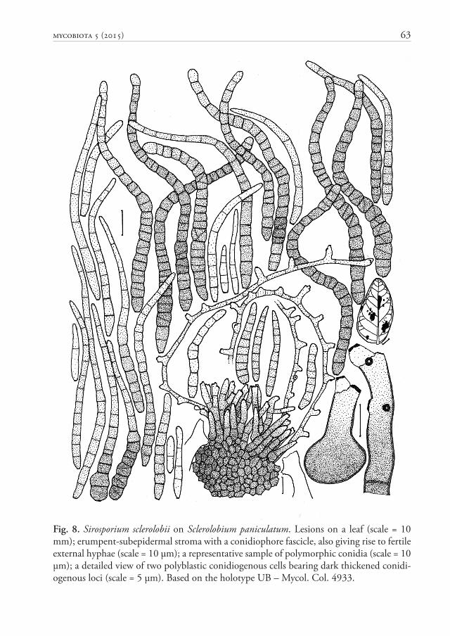
Sirosporium sclerolobii A. Hern.-Gut., Z.M. Chaves & Dianese, sp. nov. Fig. 8MycoBank MB 814805
Holotype: BRAZIL. Goiás: Cristalina, Fazenda Nova Índia, on leaves of Sclerolobium paniculatum var. rubiginosum (Tul.) Benth. (Fabaceae), 10 Apr 1996, J.C. Dianese (UB – Mycol Col. 4933).
Etymology: Specifi c epithet referring to the host genus, Sclerolobium.
Diagnosis: Diff ers from the similar S. munduleae in having 1–29-septate conidia, 15–107 μm long (vs. conidia up to 14 septate and up to 80 μm long in S. munduleae).
Description: Lesions 2–13 mm diam., irregular, coalescent, amphigenous, dark brown, without a limiting margin. Colonies exclusively hypophyllous, caespitose, yellowish brown. Mycelium internal and external. Stromata 33–66 μm diam., well-developed, textura angularis, erumpent, light brown, originating from the internal mycelium. Primary conidiophores on top of the stromata, in loose fascicles, simple or branched, straight or slightly curved, narrowly lageniform to cylindrical, 16–33 μm long, 4–5 μm broad at the widest part, 0–5-septate, brown-olivaceous, smooth, thin-walled; conidiogenous cells terminal, integrated, mono- or polyblastic, sympodial; conidiogenous loci conspicuous, thickened and dark. Secondary conidiophores formed as single polyblastic conidiogenous cells laterally diff erentiated on external hyphae, arising from stromata. Conidia solitary, curved, straight when young, later fl exuous or sinuous, subcylindrical to obclavate, sometimes cylindrical, young conidia show thin walls, but mature conidia are thick-walled, often constricted at the septa; obconically truncate at the base, with a moderately thick, dark hilum, 1–2 μm wide, rounded to broadly rounded at the apex, 15–107 μm long, 3–9 μm diam. at the widest part, 1–2 μm at the base, 2–4 μm near the apex, 1–29-septate, occasionally with some oblique septa, olive-brown to light olive-brown, smooth.

46 Hernández-Gutiérrez, A. et al. — Additions to the cercosporoid fungi of the Cerrado
Additional specimen examined: on leaves of Sclerolobium paniculatum var. rubiginosum (Fabaceae). BRAZIL. Goiás: Cristalina, Fazenda Nova Índia, 26 Jun 1994, 10 Apr 1996, J.C. Dianese (UB – Mycol Col. 4934).
Comment: Braun et al. (2013) indicated that “the phylogenetic meaning and value of thick conidial walls and oblique to longitudinal septa as distinguishing characters between Sirosporium and Passalora are unclear”, and further emphasized that the phylogenetic position of Sirosporium antenniforme (Berk. & M.A. Curtis) Bubák & Serebrian., the type species of genus, being still unknown. As the specimen on Sclerolobium paniculatum morphologically fi ts the traditional concept of the genus Sirosporium (Ellis 1971, 1976), and considering the currently unresolved phylogenetical position of this genus (Braun et al. 2013), it is advisable to place the new species in Sirosporium, at least tentatively. One of the most conspicuous characteristics of the specimen studied is the presence of fasciculate conidiophores on top of erumpent stromata and, at the same time, the presence of secondary conidiophores born laterally or apically on external hyphae, also originating from the stromata.
Amongst the 29 validly published Sirosporium names (http://indexfungorum.org, ac-ces sed on September 22, 2015), only three species were found on fabaceous hosts, viz. S. gliricidiae (Syd. & P. Syd.) Deighton on Gliricidia sepium from the Philippines, S. munduleae (Syd. & P. Syd.) M.B. Ellis on Mundulea suberosa from South Africa, and S. pluriseptatum (Gadp., C.D. Sharma, Firdousi, A.N. Rai & K.M. Vyas) Kamal on Cassia fi stula from India. Many of the known species associated with members of 16 other families show a similar conidial morphology, but all of them are easily separated from the new species based upon dimensional diff erences.
In Table 2 clear diff erences indicate that S. sclerolobii sp. nov., the fourth species found on Fabaceae, is easily distinguishable from the other three previously detected. Besides that, S. gliricidiae has dark stromata and the conidiophores on the adaxial leaf surface originate directly from the stromata, forming loose fascicles, but those of the abaxial surface, are solitary and originated directly from the superfi cial mycelium (Ellis 1976). Here in the case of S. sclerolobii the colonies are exclusively hypophyllous with both, conidiophore fascicles and external mycelium, formed on the same leaf surface.
Table 2. Comparison of Sirosporium species infecting Fabaceae, subfamily Faboideae
Sirosporium species ConidiophoresSize (μm) Septa
ConidiaSize (μm) Septa
S. gliricidiae –80 × 4–8 2–4 40–85 × 5–6 5–13
S. munduleae –80 × 5–8 0–2 22–45 × 9–12 1–6
S. pluriseptatum –101 × 3–6.5 up to 14 37–208 × 4–9 2–35
S. sclerolobii, sp. nov. –66 × 4–5 0–5 15–107 × 3–9 1–29

MycoBIOTA 5 (2015) 47
Sirosporium munduleae has longer (up to 80 μm) conidiophores with rugose to verrugose upper portion (Ellis 1976), diff erent from the smooth, shorter ones (up to 66 μm) formed by S. sclerolobii. Finally, S. pluriseptatum has larger, up to 14-septate conidiophores (–101 × 3–6.5 μm) and much larger, 2–35-septate conidia (37–208 × 4–9 μm).
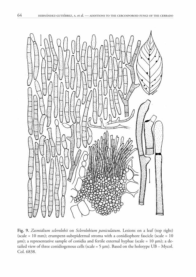
Zasmidium sclerolobii A. Hern.-Gut., Z.M. Chaves & Dianese, sp. nov. Fig. 9MycoBank MB 814806
Holotype: BRAZIL. Goiás: Luziânia, Roberto Ronald Farm on road to Unaí, on leaves of Sclerolobium paniculatum Vogel (Fabaceae), 15 Nov 1994, J.C. Dianese (UB – Mycol. Col. 6838).
Etymology: Specifi c epithet referring to the host genus.
Diagnosis: Diff erent from the similar Z. buteae by its very large stromata, shorter conidiophores, 30–74 μm long, and shorter conidia 13–69 μm long, with 0–10 septa (vs. lacking stromata, much longer conidiophores, up to 150 μm, and much longer, 5–12 septate conidia, 64–157 μm long, in Z. buteae.
Description: Lesions 1–3 mm diam., amphigenous, circular or irregular, sometimes coalescent, dark brown, surrounded by a reddish margin. Colonies mainly hypophyllous, sometimes epiphyllous, caespitose, dark brown. Mycelium external and internal; external hyphae light brown, septate, verruculose to verrucose, branched, giving rise to holoblastic conidiogenous cells; internal hyphae subhyaline, septate, giving rise to stromata. Stromata well-developed, 44–125 μm diam., textura globosa, subepidermal, erumpent, subhyaline to hyaline. Primary conidiophores on top of the stromata, in dense fascicles, simple, sometimes branched, straight or slightly curved, cylindrical, 30–74 μm long, 2–3 μm broad at the widest part, 2–8-septate, olive-brown, fi nely verruculose at the apex, thin-walled; secondary conidiophores diff erentiated as simples conidiogenous cells, arising laterally or terminally on creeping external verruculose hyphae. Conidiogenous cells terminal or intercalary, integrate, or conidiophores reduced to conidiogenous cells, mono- or polyblastic, sympodial, geniculate, verruculose, olivaceous; conidiogenous loci aplanate, thickened and dark, 1.5–2 μm wide. Conidia in simple or branched chains, straight to slightly curved, cylindrical, sometimes slightly obclavate, obconically truncate at the base with a thickened dark hilum, 1.5–2 μm wide, rounded to broadly rounded or conical-truncate at the apex, with 1–2 thick, dark apical scars in catenate conidia, 13–69 μm long, 3–5 μm broad at the widest part, 1–2 μm at the base, 1–3 μm near the apex, 0–10-septate, olivaceous, verruculose to verrucose, thin-walled.
Comment: Arzanlou et al. (2007) showed that although Zasmidium morphologically resembles Stenella, the type species of the latter genus clustered in the Teratosphaeriaceae, whereas Zasmidium in the Mycosphaerellaceae, as confi rmed by Crous et al. (2009a, b) who described and molecularly characterized new Zasmidium species. Furthermore, conidia of Stenella (type species: S. araguata Syd.) have pileate conidiogenous loci (David

48 Hernández-Gutiérrez, A. et al. — Additions to the cercosporoid fungi of the Cerrado
1991), while those of Zasmidium [type-species: Z. cellare (Pers.) Fr.] and the Stenella-like species belonging in the Mycosphaerellaceae show thickened dark conidiogenous loci, a morphological character that correlates with the molecular data available (Arzanlou et al. 2007; Crous et al. 2009a, b; Braun et al. 2010).
In the following years a series of Stenella species with Cercospora-like conidiogenous loci, were recombined into Zasmidium, most of them based on morphological characteristics, reaching 198 species names in 2015 (http://www.indexfungorum.org/Names/Names.asp, Accessed on September 28, 2015), distributed in 68 diff erent host families. Th e main host families with seven or more Zasmidium species are Fabaceae with 19 species, 13 in Myrtaceae and Rubiaceae, 10 in Rutaceae, 8 in Apocynaceae, 7 in Araceae and Asteraceae. Th e Zasmidium species on Fabaceae are distributed among 14 diff erent host genera, six of them on Cassia species (Braun & Urtiaga 2013).
Th ere are no previous records of Zasmidium species on S. paniculatum. Th e following dichotomous key, based on Table 3, segregates those Zasmidium species that infect hosts belonging in family Fabaceae, most of them from India.
Table 3. Morphotaxonomic features of Zasmidium species, including the new Z. sclerolobii, found on host belonging in family Fabaceae
Zasmidium species Stromata Conidiophores / Fascicle*
Conidiophores(μm) Septation
Conidia(μm) Septation Catenate /
Single
Z. bauhiniae (1) Present 10–14 condio-phores/fascicle
16.5–69 × 2.5–3.5 0–5 5–102.5
× 1.5–3 0–10 S and C
Z. browneicola (2) Lacking S from EH* 4.5–62.5 × 2.5–3 0–7 20–71
× 2.5–5.5 1–15 C
Z. buteae (3) 13–16 μm diam. S from EH 4–30
× 2–5 0–5 64–157 × 1–5 5–12 C
Z. canavaliae (4) Lacking Fasciculate 30–300 × 3–4.5
10–50 × 3.0–5.5* 1–5 C
Z. cassiae-fi stulae (5)Substomatal10–30 μm diam.
Small loose fas-cicles on super-fi cial stromata
40–150 × 2–4.5
Plu-risep-tate
15–100 ×2~6 3–22 S
Z. cassiae-grandis (6) Lacking S from EH 5–50 × 2.5–7 0–3 15–90
× 3–7 0–10 S and C
Z. cassiae-occidentalis (7) Present 15–20 μm diam.
Fasciculate on stromata or EH
48–140 × 2.5–5 2–6 20–80
× 2–7 1–4 C
Z. cassiae-torae (8) Small 7–10 μm diam.
Solitary or fas-ciculate
3.5–54 × 3–5 1–5 17–76
× 3.5–5 1–5 C
Z. cassiicola (9) Lacking S from EH 33–90 × 1.5–4 4–5 15–51
× 1.5–4.5 2–9 C
Z. citri-griseum (10) Lacking or small S from EH 5–80
× 2.5–6 0–6 6–120 × 2–4.5 0–10 S and C
Z. crotalariicola (11) Lacking S from EH 80–140 × 4.0–5.0 2–4 22–75
× 3.5–5.0* C

MycoBIOTA 5 (2015) 49
Zasmidium species Stromata Conidiophores / Fascicle*
Conidiophores(μm) Septation
Conidia(μm) Septation Catenate /
Single
Z. dalbergiae (12) Lacking S from EH 45– 290 × 3–4 3–11 48–105
× 2–3 1–3 S and C
Z. fabaceaerum (13) Lacking S from EH 100–310 × 2–4
12–60 × 2–6 0–4 C
Z. fabaceicola (14)Substomatal17–19 μm diam.
Fascicle from stromata or ex-ternal hyphae
75–210 × 1.5–5 4–12 13.5–40
× 1.5–5 0–3 C
Z. millettiae (2) Lacking S from EH 54–70 × 3.5–5.0 7–9 20–80
× 2.5–5.5 3–17 C
Z. periandrae (15) Lacking S from EH 10–80 × 3–5
Pluri-sep-tate
15–50 × 3–4 1–4 C
Z. prosopidis (16) 10–75 μm diam.
Dense sporodo-chial fascicles on erumpent stro-mata
5–40 × 2–5 0–1 20–110
× 2.5–5 0–14 S
Z. pterocarpigenum (17) Lacking Small fascicles 30–110 × 3–6
20–55 × 4–7 1–5 C
Z. satpurense (18) 2–8 cellsFascicles with 2–4 conidio-phores
25–72.5 × 3–4
10–42.5 × 2.5–5 1–8 S
Z. sclerolobii sp. nov. 44–125 μm diam.
Large stromatal fascicles
30–74 × 2–3 2–8 13–69
× 3–5 0–10 C
Z. tephrosiae (19) Lacking
Solitary or on fascicle with 2–15 condio-phores
15–150 × 3.5–5
Plu-risep-tate
40–130 × 3–5
Plu-risep-tate
S
(1) Haldar et al (2003), (2) Chaudhary et al. (2001), (3) Misra et al. (1997), (4) Deighton (1971), (5) Braun et al. (2003), (6) Braun & Urtiaga (2013), (7) Kumar et al. (2006), (8) Singh et al. (2001), (9) Misra et al. (1999), (10) Fisher (1961), Braun et al. (2014), Huang et al. (2015), (11) Chaudhary et al. (1991), (12) Phengsintham et al. (2013), (13) Srivastava et al. (1994), (14) Kharwar et al. (2015), (15) Braun & Freire (2006), (16) Heald & Wolf (1911), and Braun et al. (2010), (17) Braun (2003), (18) Sharma et al. (2006), (19) Braun et al. (2010)S from EH = Solitary (mononematous) conidiophores originated from external hyphae
Key to Zasmidium species on fabaceous hosts
1 Stromata well-structured, reaching 125 μm diam; conidiophores 2–8-septate, 30–74 × 2–3 μm, forming dense fascicle on top of the stromata; on Sclerolobium paniculatum, Brazil . . . . . . . . . . . . . . . . . . . . . . . . . . . . . . . . . . . . . . . . . . . . . . . . . . Z. sclerolobii
1 Stromata when present with a maximum of 75 μm diam. . . . . . . . . . . . . . . . . . . . 2 2 Conidiophores fasciculate on top of stromata . . . . . . . . . . . . . . . . . . . . . . . . . . . . . 3
Table 3. (continued)

50 Hernández-Gutiérrez, A. et al. — Additions to the cercosporoid fungi of the Cerrado
2 Conidiophores with loose fascicles on poorly structured stromata, or single and mostly arising from external hyphae . . . . . . . . . . . . . . . . . . . . . . . . . . . . . . . . . . . . . . . . . 5
3 Conidiophores 2–6-septate, up to 140 μm long; conidia 1–4-septate, 20–80 μm long; on Cassia occidentalis, Varanasi, India . . . . . . . . . . . . . . . . . . . Z. cassiae-occidentalis
3 Conidiophores less than 70 μm long; conidia up to 14-septate, over 100 μm long . . . 4 4 Conidia catenate, 0–10-septate, 5–102.5 × 1.5–3 μm; on Bauhinia vahlii, Bengal,
India . . . . . . . . . . . . . . . . . . . . . . . . . . . . . . . . . . . . . . . . . . . . . . . . . . Z. bauhiniae 4 Conidia solitary, 0–14-septate, 20–110 × 2.5–5 μm; on Prosopis, USA . . . Z. prosopidis 5 Conidiophores on poorly structured stromata . . . . . . . . . . . . . . . . . . . . . . . . . . . . 6 5 Condiophores solitary on external hyphae; stromata lacking . . . . . . . . . . . . . . . . . 8 6 Conidiophores up to 150 μm long; conidia solitary, 5–12 septate, 64–157 × 1–5 μm;
on Butea parvifl ora, Uttar Pradesh, India . . . . . . . . . . . . . . . . . . . . . . . . . Z. buteae 6 Conidia up to 8-septate; on other hosts . . . . . . . . . . . . . . . . . . . . . . . . . . . . . . . . . 7 7 Conidia 1–5 septate, 17–76 × 3.5–5 μm; on Cassia tora, Uttar Pradesh, India
. . . . . . . . . . . . . . . . . . . . . . . . . . . . . . . . . . . . . . . . . . . . . . . . . . . . . Z. cassiae-torae7 Conidia 1–8-septate, 10–42.5 × 2.5–5 μm; on Cassiia fi stula, Madhia Pradesh, India
. . . . . . . . . . . . . . . . . . . . . . . . . . . . . . . . . . . . . . . . . . . . . . . . . . . . . . Z. satpurense 8 Conidiophores over 300 μm long; conidia 0–4-septate, 12–60 × 2–6 μm; on Dolichos
sp., Uttar Pradesh, India . . . . . . . . . . . . . . . . . . . . . . . . . . . . . . . . . . Z. fabacearum 8 Conidiophores < 300 μm long . . . . . . . . . . . . . . . . . . . . . . . . . . . . . . . . . . . . . . . . 9 9 Conidiophores less than 100 μm long . . . . . . . . . . . . . . . . . . . . . . . . . . . . . . . . . 10 9 Conidiophores over 100 μm long . . . . . . . . . . . . . . . . . . . . . . . . . . . . . . . . . . . . . 1510 Conidia over 100 μm long, 0–10-septate, 6–120 × 2–4.5 μm; conidiophores
0–6-septate, 5–80 × 2.5–6 μm; on Acacia mangium, Gabon, Africa Z. citri-griseum10 Conidia less than 100 μm long . . . . . . . . . . . . . . . . . . . . . . . . . . . . . . . . . . . . . . . 1111 Conidia reaching 80 to 90 μm long . . . . . . . . . . . . . . . . . . . . . . . . . . . . . . . . . . . 1211 Conidia reaching 50 to 71 μm long . . . . . . . . . . . . . . . . . . . . . . . . . . . . . . . . . . . 1312 Conidiophores 7–9-septate, 54–70 × 3.5–5.0 μm; conidia 3–17-septate, 20–80 ×
2.5–5.5 μm; on Millettia ovalifolia, Uttar Pradesh, India . . . . . . . . . . . Z. millettiae12 Conidiophores 0–3-septate, 5–50 × 2.5–7 μm; conidia 0–10-septate, 15–90 × 3–7
μm; on Cassia grandis, Cuba . . . . . . . . . . . . . . . . . . . . . . . . . . . . . Z. cassiae-grandis13 Conidiophores up to 90 μm, 4–5-septate, 33–90 × 1.5–4 μm; conidia 2–9-septate,
15–51 × 1.5–4.5 μm; on Cassia fi stula, Uttar Pradesh, India . . . . . . . . Z. cassiicola13 Conidiophores up to 80 μm . . . . . . . . . . . . . . . . . . . . . . . . . . . . . . . . . . . . . . . . . 1414 Conidiophores 4.5–62.5 × 2.5–3 μm; conidia 1–15-septate, 20–71 × 2.5–5.5 μm; on
Brownea hybrida, Uttar Pradesh, India . . . . . . . . . . . . . . . . . . . . . . . Z. browneicola

MycoBIOTA 5 (2015) 51
14 Conidiophores 10–80 × 3–5 μm; conidia 1–4-septate, 15–50 × 3–4 μm; on Periandra coccinea, Ceará, Brazil . . . . . . . . . . . . . . . . . . . . . . . . . . . . . . . . . . . . . Z. periandrae
15 Conidia 100 μm or more long . . . . . . . . . . . . . . . . . . . . . . . . . . . . . . . . . . . . . . . 1615 Conidia up to 75 μm long . . . . . . . . . . . . . . . . . . . . . . . . . . . . . . . . . . . . . . . . . . 1816 Conidia up to 100 μm long; conidiophores 40–150 × 2–4.5 μm; on Cassia-fi stulae,
Uttar Pradesh, India . . . . . . . . . . . . . . . . . . . . . . . . . . . . . . . . . . . . Z. cassia-fi stulae16 Conidia over 100 μm long . . . . . . . . . . . . . . . . . . . . . . . . . . . . . . . . . . . . . . . . . . 1717 Conidia pluriseptate, 40–130 × 3–5 μm; conidiophores 15–150 × 3.5–5 μm; on
Tephrosia hispidatae, Alabama, USA . . . . . . . . . . . . . . . . . . . . . . . . . . . Z. tephrosiae17 Conidia, 48–105 × 2–3 μm; conidiophores 3–11-septate, 45–290 × 3–4 μm; on
Dalbergia cultrata, Laos . . . . . . . . . . . . . . . . . . . . . . . . . . . . . . . . . . . Z. dalbergiae18 Conidiophores over 200 μm long . . . . . . . . . . . . . . . . . . . . . . . . . . . . . . . . . . . . . 1918 Conidiophores up to 140 μm long . . . . . . . . . . . . . . . . . . . . . . . . . . . . . . . . . . . . 2019 Conidiophores 75–210 × 1.5–5 μm; conidia 0–3-septate, 13.5–40 × 1.5–5 μm; on
Vigna unguiculata, Chitwan, Nepal . . . . . . . . . . . . . . . . . . . . . . . . . . Z. fabaceicola19 Conidiophores 30–300 × 3–4.5 μm; conidia 1–5-septate, 10–50 × 3.0–5.5 μm; on
Canavalia ensiformis, Los Banos, Philippines . . . . . . . . . . . . . . . . . . . Z. canavaliae20 Conidiophores 80–140 × 4.0–5.0 μm; conidia 22–75 × 3.5–5.0 μm; on Crotalaria
sp., Gorakhpur, India . . . . . . . . . . . . . . . . . . . . . . . . . . . . . . . . . . . Z. crotalariicola20 Conidiophores 30–110 × 3–6 μm; conidia 1–5-septate, 20–55 × 4–7 μm; on
Pterocarpus santalinus, Kerala, India . . . . . . . . . . . . . . . . . . . . . Z. pterocarpigenum
Finally, following Braun et al. (2010), four cercosporoid Stenella species described on non-fabaceous hosts from the Brazilian Cerrado are herein transferred to Zasmidium because they are characterized by having typically cercosporoid conidiogenous loci and conidial hila, and rather cercosporoid conidia:
Zasmidium erythroxylicolum (Dorn.-Silva & Dianese) Dorn.-Silva, A. Hern.-Gut. & Dianese, comb. nov. (MycoBank, MB 814807)Bas.: Stenella erythroxylicola Dorn.-Silva & Dianese, Mycologia 99: 755, 2007.
Zasmidium erythroxyli-campestris (Dorn.-Silva, Pereira-Carv. & Dianese) Dorn.-Silva, A. Hern.-Gut. & Dianese, comb. nov. (MycoBank, MB 814808)Bas.: Stenella erythroxyli-campestris Dorn.-Silva, Pereira-Carv. & Dianese, Mycologia 99: 753, 2007.
Zasmidium erythroxyli-suberosi (Dorn.-Silva, Pereira-Carv. & Dianese) Dorn.-Silva, A. Hern.-Gut. & Dianese, comb. nov. (MycoBank, MB 814809)Bas.: Stenella erythroxyli-suberosi Dorn.-Silva, Pereira-Carv. & Dianese, Mycologia 99:755, 2007.
52 Hernández-Gutiérrez, A. et al. — Additions to the cercosporoid fungi of the Cerrado
Zamidium ocotei (Dorn.-Silva & Dianese) Dorn.-Silva, A. Hern.-Gut. & Dianese, comb. nov. (MycoBank, MB 814810)Bas.: Stenella ocoteae Dorn.-Silva & Dianese, Mycologia 99:759, 2007.
Acknowledgements. Th e authors thank CNPq for earlier support to the senior author and for fellowships via PPBIO/Cerrado-CNPq for the second and third authors. To Prof. Mariza Sanchez the authors show their appreciation for the work in the fungarium.
References
Arzanlou, M., Groenewald, J.Z., Gams, W., Braun, U., Shin, H-D. & Crous, P.W. 2007. Phylogenetic and morphotaxonomic revision of Ramichloridium and allied genera. – Studies in Mycology 58: 57–93. http://dx.doi.org/10.3114/sim.2007.58.03
Barreto, R.W. & Evans, H.C. 1995. Th e mycota of the weed Mikania micrantha in Southern Brazil with particular reference to fungal pathogens for biological control. – Mycological Research 99: 343–352. http://dx.doi:10.1016/S0953-7562(09)80911-8
Batista, A.C., Souza, R.G. & Peres, G.E.P. 1960. Alguns Cercospora estudados no IMUR. – Publicações do Instituto de Micologia da Universidade do Recife 266: 1–36.
Braun, U. 1989. Cercospora-like fungi on Cassia. – International Journal of Mycology and Lichenology 4: 191–204.
Braun, U. 2003. Miscellaneous notes on some cercosporoid hyphomycetes. – Bibliotheca Lichenologica 86: 79–98.
Braun, U. & Freire, F.C.O. 2006. Some cercosporoid hyphomycetes from Brazil – IV. – Criptogamie, Mycologie 27: 231–248.
Braun, U. & Urtiaga, R. 2013. New species and new records of cercosporoid hyphomycetes from Cuba and Venezuela (Part 3) – Mycosphere 4: 591–614. http://dx.doi.org/10.5943/mycosphere/4/2/3
Braun, U., Crous, P.W., Schubert, K. & Shin, H-D. 2010. Some reallocations of Stenella species to Zasmidium. – Schlechtendalia 20: 99–104.
Braun, U., Nakashima, C. & Crous, P.W. 2013. Cercosporoid fungi (Mycosphaerellaceae) 1. Species on other fungi, Pteridophyta and Gymnospermae. – IMA Fungus 4: 265–345. http://dx.doi.org/10.5598/imafungus.2013.04.02.12
Braun, U., Crous, P.W. & Nakashima, C. 2014. Cercosporoid fungi. (Mycosphaerellaceae) 2. Species on monocots (Acoraceae to Xyridaceae, excluding Poaceae). – IMA Fungus 5: 203–390. http://dx.doi.org/10.5598/imafungus.2014.05.02.04
Bruneau, A., Doyle, J.J., Herendeen, P. et al. 2013. Legume phylogeny and classifi cation in the 21st century: Progress, prospects and lessons for other species-rich clades. – Taxon 62: 217–248. http://dx.doi.org/10.12705/622.8
Castañeda-Ruiz, R. & Braun, U. 1989. Cercospora and allied genera of Cuba (I). – Cryptogamic Botany 1: 42–55.
Chaudhary, R., Chaturbhuji, G. & Kamal. 1991. New species of Heteroconium, Pseudocercospora and Stenella from India. – Mycological Research 95: 1070–1073. http://dx.doi.org/10.1016/S0953-7562(09)80548-0

MycoBIOTA 5 (2015) 53
Chaudhary, R.K., Tripathi, M.S., Singh, P.N. & Chaudhary, S. 2001. Novel taxa of Stenella from forest fl ora of Norh-Eastern Uttar Pradesh. – Indian Phytopahology 54: 226–232.
Chupp, J.C. 1954. A monograph of the fungus genus Cercospora. By the Author. Ithaca, New York.Crous, P.W. & Braun, U. 2003. Mycosphaerella and its anamorphs. 1. Names published in Cercospora and
Passalora. CBS Biodiversity Series 1. Ponsen & Looyen, Wageningen.Crous, P.W., Alfenas, A.C. & Barreto, R.W. 1997. Cercosporoid fungi from Brazil. 1. – Mycotaxon 64:
405–430.Crous, P.W., Summerell, B.A., Carnegie, A.J., Wingfi eld, M.J. & Groenewald, J.Z. 2009a. Novel
species of Mycosphaerellaceae and Teratosphaeriaceae. – Persoonia 23: 119–146. http://dx.doi.org/10.3767/003158509X479531
Crous, P.W., Summerell, B.A., Carnegie, A.J., Wingfi eld, M.J., Hunter, G.C., Burgess, T.I., Andijc, V., Barber, P.A. & Groenewald, J.Z. 2009b. Unravelling Mycosphaerella: do you believe in genera? – Persoonia 23: 99–118. http://dx.doi.org/10.3767/003158509X479487
Crous, P.W., Schumacher, R.K., Wingfi eld, M.J., Lombardi, L., Giraldo, A., Christensen, M., Gardiennet, A., Nakashima, C., Pereira, O.L., Smith, A.J. & Groenewald, J.Z. 2015. Fungal systematics and evolution: Fuse 1. – Sydowia 67: 81–118. http://dx.doi.org/10.3767/003158509X479487
David, J.C. 1991. Parastenella, a new generic name for Heterosporium magnolia. – Mycological Research 95: 123–128. http://dx.doi.org/10.1016/S0953-7562(09)81369-5
Deighton, F.C. 1971. Brown leaf mould of Canavalia caused by Stenella canavaliae (H. & P. Syd.) comb. nov. – Transactions of the British Mycological Society 56: 411–418. http://dx.doi.org/10.1016/S0007-1536(71)80133-X
Deighton, F.C. 1976. Studies on Cercospora and allied genera, VI. Pseudocercospora Speg., Pantospora Cif. and Cercoseptoria Petr. – Mycological Papers 140: 1–168. http://dx.doi.org/10.1016/S0007-1536(71)80133-X
Dianese, A.C., Costa, A.M. & Dianese, J.C. 2008. A new Pseudocercospora species on Passifl ora setácea. – Mycotaxon 105: 1–5.
Dianese, J.C. & Câmara, M.P.S. 1994. Pseudocercospora aspidospermatis, a new combination for Bactrodesmiella aspidospermatis. – Sydowia 46: 225–232.
Dornelo-Silva, D. & Dianese, J.C. 2003. Hyphomycetes on Vochysiaceae from the Brazilian Cerrado. – Mycologia 95: 1239–1251. http://dx.doi.org/10.2307/3761924
Dornelo-Silva, D., Pereira-Carvalho, R.C. & Dianese, J.C. 2007. New Stenella and Parastenella species from the Brazilian Cerrado. – Mycologia 99: 753–764. http://dx.doi.org/10.3852/mycologia.99.5.753
Ellis, M.B. 1971. Dematiaceous Hyphomycetes. Commonwealth Mycological Institute, Kew, Surrey.Ellis, M.B. 1976. More Dematiaceous Hyphomycetes. Commonwealth Mycological Institute, Kew, Surrey.Fisher, F.E. 1961. Greasy spot and tar spot of citrus in Florida. – Phytopathology 51: 297–303.Furlanetto, C. & Dianese, J.C. 1999. Some Pseudocercospora species and a new Prathigada species
from the Brazilian cerrado. – Mycological Research 103: 1203–1209. http://dx.doi.org/10.1017/S0953756299008394
Haldar, D., Ray, J.B. & Das, A.K. 2003. Two new Stenella species from India. – Journal of Mycopathological Research 41: 63–66.
Heald, F.D. & Wolf, F.A. 1911. New species of Texas fungi. – Mycologia 3: 5–22. http://dx.doi.org/10.2307/3753651
54 Hernández-Gutiérrez, A. et al. — Additions to the cercosporoid fungi of the Cerrado
Hernández-Gutiérrez, A. & Dianese, J.C. 2008. New cercosporoid fungi from the Brazilian Cerrado. 1. Species on hosts of the families Anacardiaceae, Araliaceae, Bombacaceae, Burseraceae and Celastraceae. – Mycotaxon 106: 41–63.
Hernández-Gutiérrez, A. & Dianese, J.C. 2009. New cercosporoid fungi from the Brazilian Cerrado 2. Species on hosts of the subfamilies Caesalpinoideae, Faboideae, and Mimosoideae (Leguminosae s. lat.). – Mycotaxon 107: 1–24. http://dx.doi.org/10.5248/107.1
Hernández-Gutiérrez, A. & Dianese, J.C. 2014a. Cercosporoid hyphomycetes on malpighiaceous hosts from the Brazilian Cerrado: New Passalora and Pseudocercospora species on hosts of the genus Banisteriopsis. – Mycological Progress 13: 365–371. http://dx.doi.org/10.1007/s11557-013-0922-6
Hernández-Gutiérrez, A. & Dianese, J.C. 2014b. New Passalora species on Peixotoa (Malpighiaceae) from the Brazilian Cerrado. – Mycological Progress 13: 75–79. http://dx.doi.org/10.1007/s11557-013-0894-6
Hernández-Gutiérrez, A., Braun, U. & Dianese, J.C. 2014. Cercosporoid hyphomycetes on malpighiaceous hosts from the Brazilian Cerrado: Species of Pseudocercospora on hosts belonging to Byrsonima. – Mycological Progress 13: 193–210. http://dx.doi.org/10.1007/s11557-014-0971-5
Hsieh, W-H. & Goh, T.-K. 1990. Cercospora and similar fungi from Taiwan. Maw Chang Book Co., Taipei.Huang, F., Groenewald, J.Z., Zhu, L., Crous, P.W. & Li, H. 2015. Cercosporoid diseases of Citrus. –
Mycologia: preliminary version published on October 2, 2015, as doi:10.3852/15-059.Inácio, C.A. & Dianese, J.C. 1998. Foliicolous fungi on Tabebuia species. – Mycological Research 102:
695–708. http://dx.doi.org/10.1017/S0953756297005856Inácio, C.A. & Dianese, J.C. 1999. A new Mycovellosiella species on Myracrodruon urundeuva. – Mycotaxon
72: 251–254.Inácio, C.A. & Dianese, J.C. 2006. Foliicolous fungi on Tabebuia species from the cerrado. – Mycological
Progress 5: 120–127. http://dx.doi.org/10.1007/s11557-006-0507-8Inácio, C.A., Furlanetto, C., Hernández-Gutiérrez, A. & Dianese, J.C. 1996. Some Cercospora species
originally described by Ahmés Pinto Viégas. – Fitopatologia Brasileira 21: 405–409.Jobson, R.W. & Luclow, M. 2007. Phylogenetic studies of the genus Piptadenia (Mimosoideae: Leguminosae)
using plastid trnL-F and trnK/matK sequence data. – Systematic Botany 32: 569–575. http://dx.doi.org/10.1600/036364407782250544
Kharwar, R.N., Singh, A., Singh, R. & Kumar, S. 2015. Two new species of Zasmidium from Nepal. – Mycotaxon 130: 241–246. http://dx.doi.org/10.5248/130.241
Kumar, A., Kumar, A. & Kharwar, R.N. 2006. Two new phytoparasitic hyphomycetes from Varanasi, India. – Indian Phytopathology 59: 85–90.
Medeiros, R.B. & Dianese, J.C. 1994. Passalora eitenii sp. nov. on Syagrus comosa (Mart.) Mart. in Central Brazil, and a key for identifi cation of Passalora species. – Mycotaxon 51: 509–513.
Mendonça, R.C., Felfili, J.M., Walter, B.M.T., Silva-Júnior, M.C., Rezende, A.V., Filgueiras, T.S. & Nogueira, P.E. 2008. Flora vascular do Cerrado. – In: S.M. Sano & S.P. Almeida (eds). Cerrado: ambiente e flora. Pp. 289–556. EMBRAPA-CPAC, Planaltina.
Minnis, A.M., Kennedy, A.H., Grenier, D.B., Rehner, S.A. & Bischoff , J.F. 2011. Asperisporium and Pantospora (Mycosphaerellaceae): epitypifi cations and phylogenetic placement. – Persoonia 27: 1–8. http://dx.doi.org/10.3767/003158511X602071
Misra, S., Srivastava, N. & Srivastava, A.K. 1997. New species of Stenella from India. – Mycological Research 101: 278–280. http://dx.doi.org/10.1017/S0953756296002572

MycoBIOTA 5 (2015) 55
Misra, S., Srivastava, A.K. & Kamal. 1999. Further additions to Stenella from India and Nepal. – Mycological Research 103: 268–270. http://dx.doi.org/10.1017/S0953756298006108
Phengsintham, P., Chukeatirote, E., McKenzie, E.H.C., Hyde, K.D. & Braun, U. 2013. Monograph of Cercosporoid fungi from Laos. – Current Research in Environmental & Applied Mycology 3: 34–158. http://dx.doi.org/10.5943/cream/3/1/2.
Ram, M.R. & Mallaiah, K.V. 1992. Pseudocercospora sieberiana sp. nov. from India. – Mycotaxon 45: 405–408.
Sharma, N., Soni, K.K. & Verma, R.K. 2006. Some new hyphomycetes from forests of Satpura. – Indian Journal of Tropical Biodiversity 14: 34–40.
Singh, S.Y.K. & Bhalla, K. 2000. New Pseudocercospora species causing foliar diseases in plain forests of Vindhya region in India. – Indian Phytopathology 53: 399–403.
Singh, S.Y.K. & Bhalla, K. 2001. Four new foiiicolous hyphomycetes from Vindhya Hills, India. – Journal of Mycology and Plant Pathology 31: 277–286.
Srivastava, K., Srivastava, A.K. & Kamal. 1994. New species of Stenella from India. – Mycological Research 98: 516–520. http://dx.doi.org/10.1016/S0953-7562(09)80470-X
Viégas, A.P. 1945. Alguns fungos do Brasil: Cercosporae. – Boletim Sociedade Brasileira de Agronomia 8: 1–160.
Yen, J.M. 1981. Étude sur les champignons parasites de Sud-Est asiatique, 41. Les Cercospora de Formosae. – Bulletin de la Société Mycologique de France 97: 91–96.
Yen, J.M. & Lim, G. 1980. Cercospora and allied genera of Singapore and the Malay Peninsula. – Th e Gardens’ Bulletin Singapore 33: 151–263.

56 Hernández-Gutiérrez, A. et al. — Additions to the cercosporoid fungi of the Cerrado
Fig. 1. Asperisporium chapadensis on Galactia peduncularis. Lesions on a leaf (top right) (scale = 10 mm); erumpent-subcuticular stroma with a fascicle of primary conidiophores (scale = 10 μm); detailed view of a conidiophore bearing conidiogenous cells with protu-berant thickened loci (right) (scale 5 μm); representative sample of the conidia. (scale = 10 μm). Based on the holotype UB – Mycol. Col. 11332.

MycoBIOTA 5 (2015) 57
Fig. 2. Pseudocercospora acosmii-subelegantis on Acosmium subelegans. Lesions on a leaf (top right) (scale = 10 mm); stomatal condiophore fascicle originated from internal hyphae (scale = 10 μm); a representative sample of conidia with thickened walls and septa (scale = 10 μm); at the right side of the conidiophore fascicle, a detailed view of two conidiogenous cells showing aplanate conidiogenous loci (scale = 5 μm). Based on the holotype UB – My-col. Col. 7397.

58 Hernández-Gutiérrez, A. et al. — Additions to the cercosporoid fungi of the Cerrado
Fig. 3. Pseudocercospora aquae-emendadasensis on Chamaecrista orbiculata. Lesions on a leaf (top right) (scale = 10 mm); stomatal condiophore fascicle originated from internal hyphae (scale = 10 μm); a representative sample of conidia with thickened walls and septa (scale = 10 μm); at the right side of the conidiophore fascicle, a detailed view of a conidiogenous cell showing aplanate conidiogenous loci (scale = 5 μm). Based on the holotype UB – My-col. Col. 13421.

MycoBIOTA 5 (2015) 59
Fig. 4. Pseudocercospora sennae on Senna rugosa. Lesions on a leaf (top right) (scale = 10 mm); erumpent-subepidermal wide stroma bearing thightly packed conidiophores (scale = 10 μm); a representative sample of polymorphic conidia (scale = 10 μm); at the bottm right, detailed view of a conidiogenous cell (scale = 5 μm). Based on the holotype UB – Mycol. Col. 1132.

60 Hernández-Gutiérrez, A. et al. — Additions to the cercosporoid fungi of the Cerrado
Fig. 5. Pseudocercospora sennae-rugosae on Senna rugosa. Lesions on a leaf (top right) (scale = 10 mm); erumpent subepidermal stroma bearing a compact fascicle of highly septate co-nidiophores (scale = 10 μm); a representative sample of conidia (scale = 10 μm); a plurisep-tate conidiophore with an apical conidiogenous cell (bottom right) (scale = 5 μm). Based on the holotype UB – Mycol. Col. 3033.

MycoBIOTA 5 (2015) 61
Fig. 6. Pseudocercospora stryphnodendri on Stryphnodendron adstringens. Lesions on a leaf (top right) (scale = 10 mm); stomatal poorly developed stroma with a lax fascicle of short 1-septate conidiophores (scale = 10 μm); a representative sample of conidia (scale = 10 μm); a detailed view of two conidiophores, one reduced to a poliblastic conidiogenous cell (scale = 5 μm). Based on the holotype UB – Mycol. Col. 894.

62 Hernández-Gutiérrez, A. et al. — Additions to the cercosporoid fungi of the Cerrado
Fig. 7. Pseudocercospora subcuticularis on Senna rugosa. Lesions on a leaf (top right) (scale = 10 mm); erumpent-subepidermal stroma with a compact fascicle of conidiophores (scale = 10 μm); a representative sample of conidia (scale = 10 μm); a detailed view of three conidi-ogenous cells (bottom right) (scale = 5 μm). Based on the holotype UB – Mycol. Col. 4092.
MycoBIOTA 5 (2015) 63
Fig. 8. Sirosporium sclerolobii on Sclerolobium paniculatum. Lesions on a leaf (scale = 10 mm); erumpent-subepidermal stroma with a conidiophore fascicle, also giving rise to fertile external hyphae (scale = 10 μm); a representative sample of polymorphic conidia (scale = 10 μm); a detailed view of two polyblastic conidiogenous cells bearing dark thickened conidi-ogenous loci (scale = 5 μm). Based on the holotype UB – Mycol. Col. 4933.
64 Hernández-Gutiérrez, A. et al. — Additions to the cercosporoid fungi of the Cerrado
Fig. 9. Zasmidium sclerolobii on Sclerolobium paniculatum. Lesions on a leaf (top right) (scale = 10 mm); erumpent-subepidermal stroma with a conidiophore fascicle (scale = 10 μm); a representative sample of conidia and fertile external hyphae (scale = 10 μm); a de-tailed view of three conidiogenous cells (scale = 5 μm). Based on the holotype UB – Mycol. Col. 6838.