acute kidney injury report a 71 year old woman with a history of cll, began chemotherapy for...
TRANSCRIPT

ACUTE KIDNEY INJURY
Harry O. Senekjian, M.D.
Ogden Surgical/Medical Society
May 14, 2015

CASE REPORT
A 71 year old woman with a history of CLL, began chemotherapy for treatment of massively elevated WBC. One week later began having weakness, shortness of breath, nausea. Had no prior history of kidney disease. Seen in the emergency department with the following labs:
BUN/creatinine 115/3.8, Potassium 9.5, CO2 10, pH 7.02, uric acid 36.8

An elevated serum creatinine during hospitalisation is an
independent risk factor for mortality, progression to CKD,
end-stage renal disease, and reduced long-term survival.
Patients with chronically elevated serum creatinine (i.e.,
impaired baseline renal function) have a higher risk for
acute kidney injury during hospital stays and are more
often dialysis-dependent at hospital discharge than those
without.
http://bestpractice.bmj.com/best-practice/monograph/935.html

ACUTE KIDNEY INJURY-DEFINITION
A decrease in glomerular filtration rate (GFR) occurring over hours to days resulting in failure of the kidneys
• To excrete nitrogenous waste products
• To maintain fluid and electrolyte balance
• To metabolize and eliminate drugs
• To synthesize EPO and calcitriol


Figure 2 RIFLE criteria for diagnosing AKI
Murugan, R. & Kellum, J. A. (2011) Acute kidney injury: what’s the prognosis?
Nat. Rev. Nephrol. doi:10.1038/nrneph.2011.13
Permission obtained from BioMed Central ©
Bellomo, R. et al. Crit. Care 8, R204–R212 (2004)


Table 1 AKIN staging system for AKI
Murugan, R. & Kellum, J. A. (2011) Acute kidney injury: what’s the prognosis? Nat. Rev. Nephrol. doi:10.1038/nrneph.2011.13
Permission obtained from BioMed Central © Mehta, R. L. et al. Crit. Care. 11, R31 (2007)

Incidence
• Approximately 7% of all hospitalized patients
• 65-70% of critically ill patients
– RIFLE Stage F 10-20% of ICU admissions
• AKI requiring RRT: Mortality range 50-70%
• Sepsis most common cause
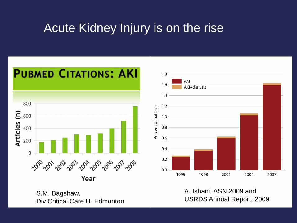
Acute Kidney Injury is on the rise
A. Ishani, ASN 2009 and
USRDS Annual Report, 2009 S.M. Bagshaw,
Div Critical Care U. Edmonton

Markers of AKI-1
Creatinine-most common, least sensitive
Nutrition Muscle mass Infection
Edema Protein metabolism
Volume of Serum creatinine Hepatic function
distribution
Renal excretion

Markers of AKI-2
Cystatin-C • 13 K Dalton proteinase produced by all nucleated
cells
• Production is not influenced by race, gender or inflammation
• Eliminated strictly by GFR
• Somewhat better than serum creatinine, but still not very sensitive
• Much more expensive

Potential Biomarkers of Kidney Damage
• Urinary IL-18
• Urinary IL-6
• Urinary TNF
• Urinary Kidney Injury Molecule (KIM-1)
• Urinary Tubular Enzymes
• Urinary Proteases
• Plasma Granzyme B
• NGAL-Neutrophil Gelatinase Associated Lipocalin

NephroCheck
• Detects the presence of insulin-like growth factor binding protein 7 (IGFBP7) and tissue inhibitor of metalloproteinases (TIMP-2) in the urine.
• Provides a score to determine the risk of developing AKI within 12 hours of the test
• Accurately detected 92% of AKI patients in one study and 76% in a second study. False positive in 50% of patients without AKI
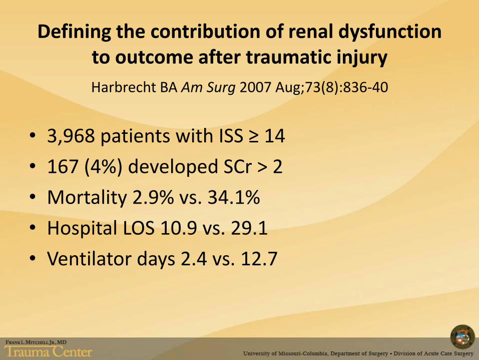
Defining the contribution of renal dysfunction to outcome after traumatic injury
Harbrecht BA Am Surg 2007 Aug;73(8):836-40
• 3,968 patients with ISS ≥ 14
• 167 (4%) developed SCr > 2
• Mortality 2.9% vs. 34.1%
• Hospital LOS 10.9 vs. 29.1
• Ventilator days 2.4 vs. 12.7

AKI and Mortality
• Independent risk factor
• “AKI appears to increase the risk of developing severe non-renal complications that lead to death”
• Respiratory failure 20.7% vs 57.4%
• ICU mortality 14% vs 42.8%
• In-hospital mortality 7% vs 34%

Causes of AKI
Top 5
• Sepsis
• Major surgery
• Low cardiac output
• Hypovolemia
• Medications
Other common causes
• Cardiopulmonary bypass
• IAH-ACS
• Trauma
• Rhabdomyolysis
• Obstruction


Pre-Renal AKI
The problem may lie anywhere between the heart and the glomerulus
• LV failure
• Cardiac tamponade
• Constrictive pericarditis
• Coarctation
• Renal artery disease
• Renal vasoconstriction
• Volume depletion/hemorrhage

Urine Sodium
• In the setting of oliguria, urine sodium below 20 mEq/L usually indicates a prerenal disorder
• Elevated urine sodium can occur when a prerenal disorder is superimposed on intrinsic renal dysfunction (or diuretic therapy)
One of the most reliable parameters to determine difference: FENa

FENa
• FENa < 1% = Prerenal disorder
• FENa > 2% = Intrinsic renal disorder

Pre-Renal AKI
• GFR is poor, but tubules function normally
• Characterized by:
– Concentrated urine (sg>1.020)
– High BUN:creatinine ratio (>20)
– Bland urine sediment
– Avid sodium reabsorption
• Urine sodium <20
• FE sodium <1%

Treatment for Pre-Renal AKI
Fix the underlying problem

Post Renal AKI
• Should always be considered, even if just to dismiss it
– Bladder outlet obstruction
– Solitary kidney
– Large stones
– Women with pelvic malignancy
• There is not much easier or less invasive test than an ultrasound
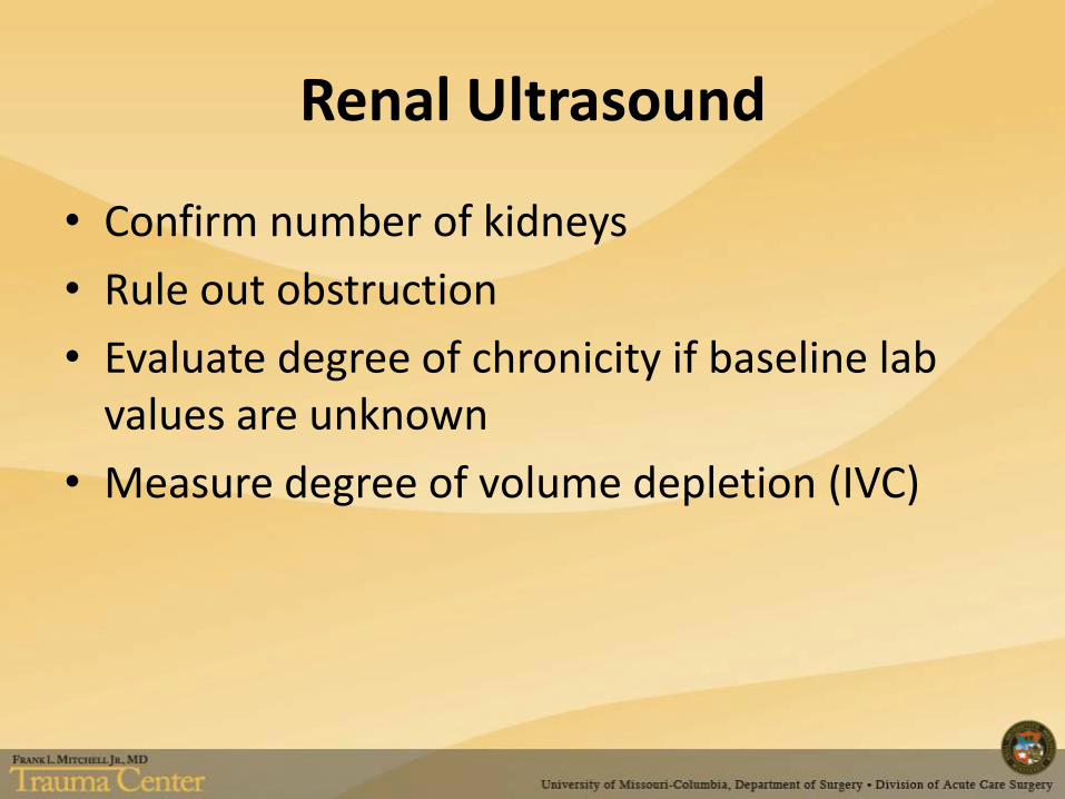
Renal Ultrasound
• Confirm number of kidneys
• Rule out obstruction
• Evaluate degree of chronicity if baseline lab values are unknown
• Measure degree of volume depletion (IVC)

Intrinsic Renal Disorders
• Impaired glomerular filtration, renal tubular dysfunction, or both
• UNa > 40 mEq/L, FENa > 2%
• Described as three entities:
– Acute glomerulonephritis
– Acute tubular necrosis (most common)
– Acute interstitial nephritis

Intrinsic Renal AKI
• Vasculitis or glomerulonephritis
– Characterized by proteinuria, hematuria, casts
– Diagnosed by renal biopsy
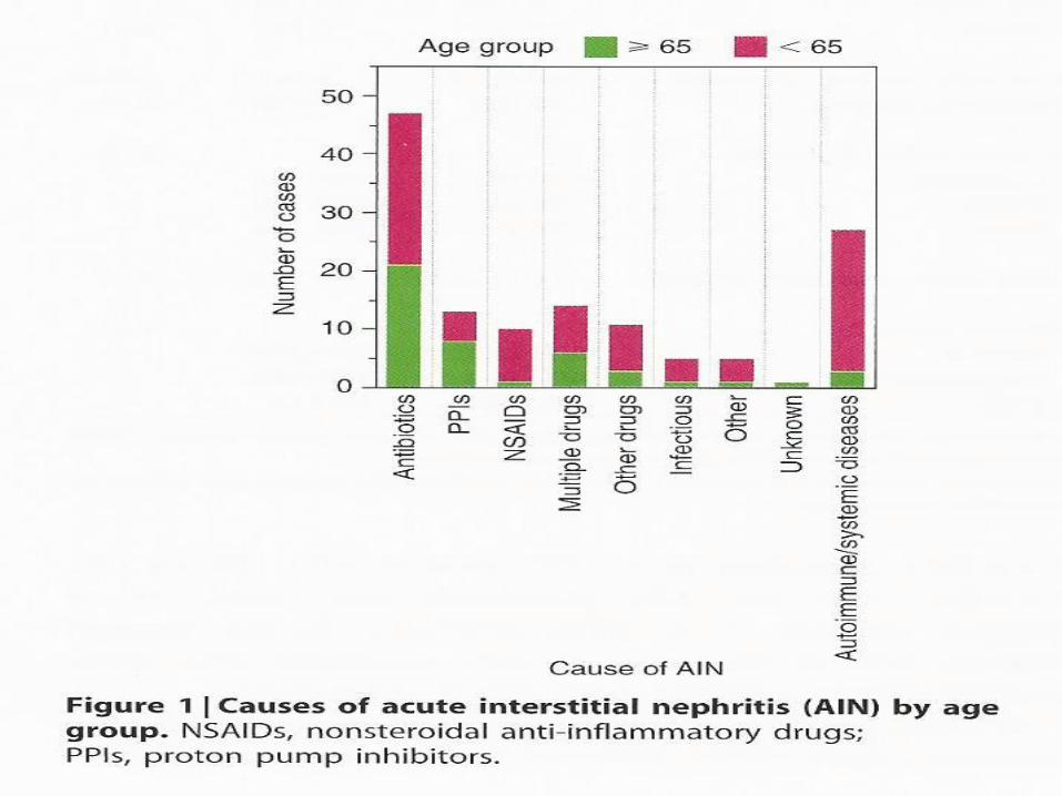
• Acute interstitial nephritis (AIN)
– Rash (15%), fever (27%), eosinophilia (23%)
– Modest proteinuria
– Most commonly drug-induced (>70%)
– Other causes include autoimmune disease, infections
– Definitive diagnosis by renal biopsy

Drugs Commonly Causing AIN
NSAID’s Penicillins, cephalosporins
Rifampin Sulfa
Quinolones (Cipro) H2 receptor blockers
PPI’s Allopurinol


Acute Tubular Necrosis (ATN)
• Most common cause of AKI in hospital or ICU setting
• Sepsis and ischemia are the most common causes
• Clinical manifestations: – Urine output may vary from complete anuria to
polyuria
– Characterized by high urine Na (>40) and high FENa (>2%)
– Urinalysis can demonstrate deeply pigmented granular casts and renal tubular epithelial cells

AKI Associated With Cardiac Surgery
• Pathogenesis – Nephrotoxins
– Regional Hypoxia
– Mechanical Blood Trauma
– Inflammation
• Preoperative Risk Factors Preexisting CKD Reduced LV Function
COPD Diabetes
Older Age Women
Prior Cardiac Surgery Emergency Surgery

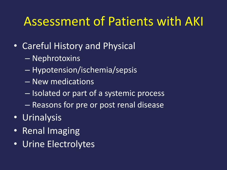
Assessment of Patients with AKI
• Careful History and Physical – Nephrotoxins
– Hypotension/ischemia/sepsis
– New medications
– Isolated or part of a systemic process
– Reasons for pre or post renal disease
• Urinalysis
• Renal Imaging
• Urine Electrolytes

Urine Microscopy
• Urine Microscopy
– Examination of sediment, easy, cost-effective
• Abundant tubular epithelial cells (ATN)
• White cell casts (interstitial nephritis)
• Pigmented casts (myoglobinuria)
If unrevealing, urinary sodium determination may be helpful

Consequences of AKI
• Inability to excrete sodium water, potassium, hydrogen ion, nitrogenous wastes
• Uremic syndrome
– Encephalopathy
– Pericarditis
– Platelet dysfunction
– Immune dysfunction


Indications for Renal Replacement Therapy (RRT)
• Volume overload, usually with respiratory insufficiency
• Acidosis (pH<7.2) • Hyperkalemia • “Uremic symptoms”
– Pericarditis – Altered mental status – Hyperuricemia
• Poisonings – Ethylene glycol, methanol, aspirin

CASE REPORT
A 71 year old woman with a history of CLL, began chemotherapy for treatment of massively elevated WBC. One week later began having weakness, shortness of breath, nausea. Had no prior history of kidney disease. Seen in the emergency department with the following labs:
BUN/creatinine 115/3.8, Potassium 9.5, CO2 10, pH 7.02, uric acid 36.8
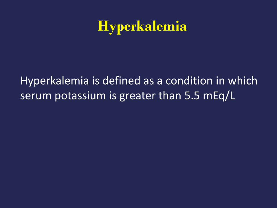
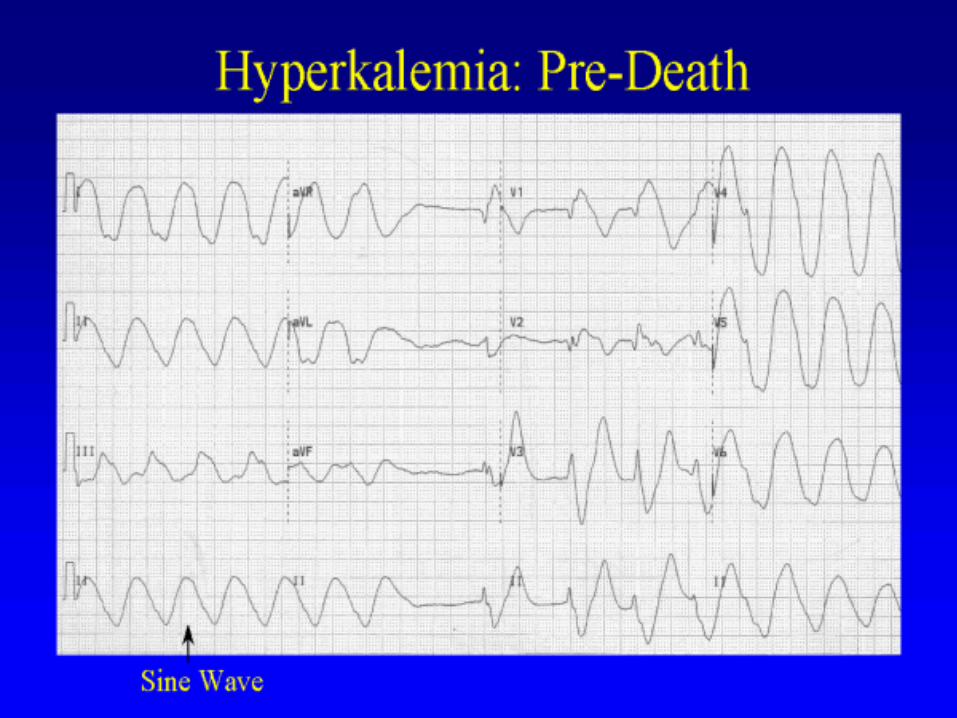
Hyperkalemia is defined as a condition in which serum potassium is greater than 5.5 mEq/L
Hyperkalemia

Causes Shift from (ICF to
ECF) Decreased renal excretion Excessive intake
Hyperosmolality
Rhabdomyolysis
Tumor lysis
Succinylcholine
Insulin deficiency
acute acidosis.
Diabetes mellitus (esp diabetic
nephropathy
Renal failure
Congestive heart failure
SLE
Sickle cell anemia
NSAID
ACE Inhibitor
Potassium sparing Diuretics
Multiple Myeloma
Chronic partial urinary tract obstruction
Oral or IV
Potassium
Supplementation
Salt substitute
Blood transfusion

Modes of RRT
• Intermittent hemodialysis: 3-5 hours, 3-6 times weekly
• Continuous renal replacement therapy (CRRT)
• Slow low efficiency daily dialysis (SLEDD). Hybrid of IDH and CRRT, 8-12 hours per day
• Acute peritoneal dialysis
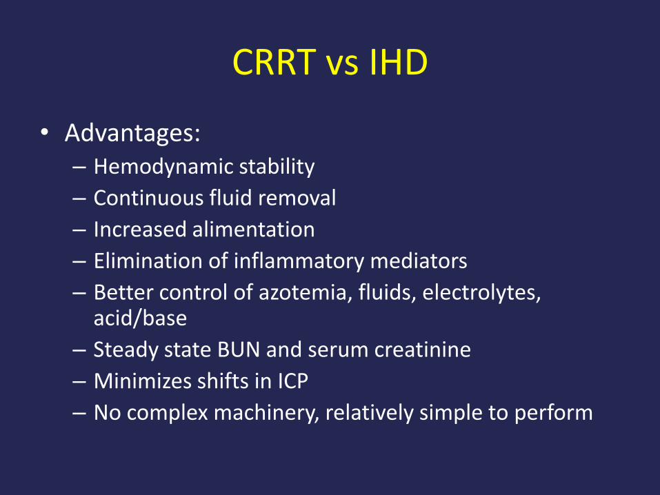
CRRT vs IHD
• Advantages: – Hemodynamic stability
– Continuous fluid removal
– Increased alimentation
– Elimination of inflammatory mediators
– Better control of azotemia, fluids, electrolytes, acid/base
– Steady state BUN and serum creatinine
– Minimizes shifts in ICP
– No complex machinery, relatively simple to perform

CRRT vs IHD
• Disadvantages
– Immobilization
– Continuous anticoagulation
– Time and labor intensive for ICU nurses
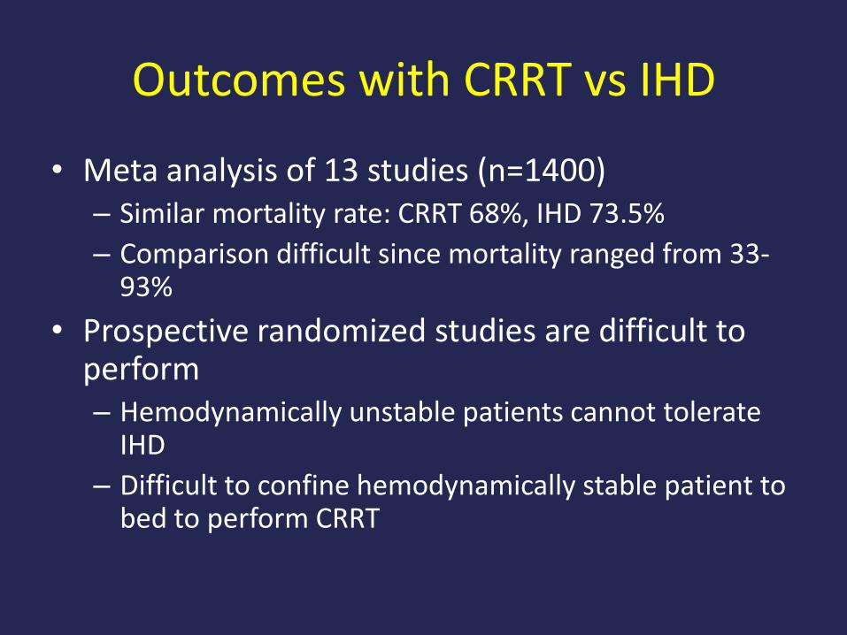
Outcomes with CRRT vs IHD
• Meta analysis of 13 studies (n=1400) – Similar mortality rate: CRRT 68%, IHD 73.5%
– Comparison difficult since mortality ranged from 33-93%
• Prospective randomized studies are difficult to perform – Hemodynamically unstable patients cannot tolerate
IHD
– Difficult to confine hemodynamically stable patient to bed to perform CRRT

RRT: Early vs Late
• There is no data to indicate that early initiation of RRT is associated with superior outcomes
• Risks of starting RRT too early
– Risks of catheter placement procedure
– Line associated sepsis
– Immobilization
– Prolonged ICU stay

Diuretics in AKI
Diuretics or no diuretics at nephrology consultation
Diuretic Group Odds Ratio
In hospital mortality 1.65 (1.05-2.55)
Non-recovery of kidney function 1.60 (1.14-2.53)

“Renal Dose” Dopamine
• No proven benefit in AKI
• Associated with harmful arrhythmias, bowel ischemia, increased myocardial oxygen consumption, decreased oxygen saturation, suppressed pituitary hormones
• Should not be routinely used

Table 2 Long-term consequences of AKI
Murugan, R. & Kellum, J. A. (2011) Acute kidney injury: what’s the prognosis?
Nat. Rev. Nephrol. doi:10.1038/nrneph.2011.13
©2012 MFMER | slide-54
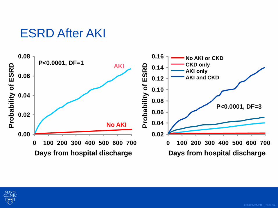
ESRD After AKI
0.00
0.02
0.04
0.06
0.08
0 100 200 300 400 500 600 700
Pro
bab
ilit
y o
f E
SR
D
Days from hospital discharge
0.02
0.04
0.06
0.08
0.10
0.12
0.14
0.16
0 100 200 300 400 500 600 700P
rob
ab
ilit
y o
f E
SR
D
Days from hospital discharge
No AKI
AKI P<0.0001, DF=1
P<0.0001, DF=3
No AKI or CKD
CKD only
AKI only
AKI and CKD
