acute effects thomas m seed, phd - nasa · 11/07/2011 · acute effects thomas m seed, phd...
TRANSCRIPT

Article Reviewed Posted to THREE 7/11/11
Acute Effects Thomas M Seed, PhD
Principal Consultant, Tech Micro Services, Bethesda, MD 20814
Outline Introduction and background Space radiation environments Exposure scenarios Description of acute radiation syndrome Radiobiological and temporal constraints Pathogenesis Countermeasures Summary and conclusions References Listing of Tables and Figures Acknowledgment The author gratefully acknowledges the reviews of Drs. Michael Fry and Michael Weil and for the very thoughtful and substantive editing by Dr. Fry. Their contribution is very much appreciated and valued. Introduction and background Acute (a-kut’) [L. acutus, sharp]. 1. Of short and sharp course, not chronic; said of a disease. (Stedman’s Medical Dictionary, Williams & Wilkins Co, Baltimore, 1976). Radiation is a hazard in all space activities but the great majority of radiation is at a low dose rate, and the concern is late effects from prolonged chronic exposure and not from acute exposures. Acute or early radiation effects are induced by radiation doses above a threshold level that is determined by the tissue sensitivity, the radiation quality, dose rate, and total dose. The effects of high doses at high dose rates are due to radiation-induced cell loss, whereas, the less severe effects, such as nausea and vomiting, are caused by less well understood processes not involving cell loss. Missions in space beyond the magnetosphere may experience solar particle events (SPE) in which large bursts of particles, mainly protons of a wide range of energies, are emitted from the sun. In the large events the dose rate of the radiation may rise, and the total dose may exceed the threshold dose for acute or early effects. The type, severity, and time of occurrence of the acute effects are determined by the total dose. Although the probability of acute effects is low, their impact, if they were to occur, would be significant (Hu et al. 2009). Therefore, an understanding of the risks of acute effects and how they can be prevented or limited is essential.
Acute effects. Seed TM. https://three.jsc.nasa.gov/articles/SeedAcuteEffects.pdf. Date posted: 07/11/2011.
Article Reviewed Posted to THREE 7/11/11
The aim of this report is to describe acute effects and countermeasures that should be considered in the design of spacecraft and planning missions. The paper complements Chapter 5 in Human Research Requirements Document (Wu et al. 2009). Space radiation environments For the purposes of description, the environment in space can be divided into two main environments separated by the geomagnetosphere. In Low-Earth Orbit (LEO) the environment consists of various energetic charged particles: electrons, protons, helium and heavier ions. There are three main categories of radiation: a) trapped particles, b) galactic cosmic rays (GCR), and c) solar particles. The trapped particles are electrons and protons trapped by the Earth’s magnetic field. GCR and the solar particles are predominately protons and a small contribution of helium and heavier ions (Badhwar 2000; Schimmerling 2000; NCRP-153 2006). External hazards of major concern, especially, on long duration flights beyond LEO, are galactic cosmic radiation (GCR) and sporadic, solar-particle events (SPEs). Earth-orbiting missions involve traversals of the Van Allen radiation belts that result in elevated exposures to energetic protons and electrons (NCRP-153 2006). For example, typical exposures on the International Space Station (ISS) are in the range of 0.5 to1.2 mSv per day with ~75% of the doses coming from GCR background, while the remainder come from energetic protons encountered during passage of the South Atlantic Anomaly regions of the Van Allen belts (NCRP-153, 2006). In addition to the external radiation hazards, a small contribution to the radiation hazard exposure may come from a variety of radiation sources that the spacecraft may carry (NCRP-153 2006). At the exposure levels cited above, acute injuries are not of concern. Of concern, however, are the rarely occurring very large solar particle events that would expose the spacecraft, and in turn the astronauts, to large numbers of very energetic (> 10 MeV) protons in a relatively short period of time. Such large SPEs with fluxes of high energy protons in excess of 30 MeV and at densities of ~5x109 cm2 have been well documented; e.g., in November 1960, August 1972, and again in October 1989. Absorbed dose-rates as high as 1.4 Gy per hour have been estimated for missions beyond LEO for an event similar to the large event of August 1972 (Parsons and Townsend 2000). More recently, dosimetry of latter SPEs of August 1972, October 1989, along with the SPE of September 1989 have been reevaluated (Hu et al. 2009), with more conservative exposure rate estimates reported for specific organ systems of astronauts situated either within the aluminum-shielded (5 g cm2) spacecraft itself or exterior to the space vehicle during extravehicular activity (EVA). For IR-sensitive, blood-forming and gastro-intestinal tissues, these average estimates are as follows: a) within the spacecraft, 6.3 cGy-Eq per hour (1.93-12.3 cGy-Eq per hour) and 3.8 cGy-Eq per hour (1.1-7.7 cGy-Eq per hour), respectively; and b) during EVA, 11.9 cGy-Eq
Acute effects. Seed TM. https://three.jsc.nasa.gov/articles/SeedAcuteEffects.pdf. Date posted: 07/11/2011.

Article Reviewed Posted to THREE 7/11/11
per hour (22.2-3.8 cGy-Eq per hour) and 6.1 cGy-Eq per hour (12.0-1.9 cGy-Eq per hour), respectively. These SPE-related exposures pale when compared to still larger SPEs of the past, such as the so-called Carrington event that occurred approximately 150 years ago, with exposure estimates, based on nitrate analysis of ice core samples, of ~10 Gy to bodily surfaces and ~1 Gy to deep, internal organs (Stephens et al. 2005). The estimated probability of a large SPE event occurring during an extended space mission (e.g., ~2 yrs) beyond the LEO based on recent experience is approximately 0.1 (Feynman et al. 1993). Significantly lower probabilities for the occurrence of multiple, massive SPEs have been recently reported for space missions of comparably long durations (Kim et al. 2009). Exposure scenarios There is exposure to radiation on all missions in space and the shielding of the spacecraft and the mission are designed to ensure that the exposure of the astronauts is limited to a level that is appropriate considering the risk and benefit. Importantly these exposures are at a dose rate too low to cause acute effects. It is the high dose rates that may occur during very large SPEs that are of concern. Astronauts on EVA are at potential risk if a SPE occurs and they cannot return to the space ship or shelter within a couple of hours because the space suit does not provide adequate shielding from the radiation as the dose rate increases. Although the occurrence of the SPE cannot be predicted, the rate at which the dose rate increases can be used to predict if it is likely that the dose rate will exceed the level required to cause acute effects. Consequently, astronauts will usually have time to return to safety. A considerable fraction of the radiation of a SPE consists of low energy protons that can penetrate the space suit, skin and superficial organs such as the eye and testes but not deeper tissues such as bone marrow. The probability of acute effects is highest in the tissues that are superficial and have radiosensitive cells, such as spermatogonia. The threshold dose temporary reduced fertility is about 15 cSv .The cells of the skin are not as radiosensitive as the spermatogonia, but are at risk because of the magnitude of the dose that they may that receive. The dose to the skin can be several times that of the deeper tissues, such as bone marrow, and a transient erythema may occur within hours after about 2 Gy, but the threshold for significant damage is considerably higher. Of particular concern is the onset of nausea and vomiting that characterize what is known as the prodromal syndrome discussed below. The most likely outcome is that the astronauts will be able to return to the spacecraft before they have received a dose sufficient to cause ARS and the only consequence is that their total lifetime radiation exposure will have been
Acute effects. Seed TM. https://three.jsc.nasa.gov/articles/SeedAcuteEffects.pdf. Date posted: 07/11/2011.

Article Reviewed Posted to THREE 7/11/11
increased. Description of the Acute Radiation Syndrome Acute Radiation Syndrome (ARS) is not a single disease entity, but rather a complex of interrelated pathologies brought about by intense and acute levels of ionizing radiation exposure (Figure 1) (Young 1987; Anno et al. 1989; Cerveny et al. 1989; Jarrett 1999; Hall 2000;Guskova et al. 2001;Singh and Seed 2003; Walselenko et al. 2004). ARS is a highly predictable clinical outcome of acute, sufficiently intense, whole-body or nearly whole-body, exposure. The full disease complex consists of three major organ system-specific sub-syndromes: (i) the hematopoietic system syndrome (HSS); (2) the gastrointestinal system syndrome (GIS); and (3) the cerebral, neurovascular system syndrome, commonly referred to as the central nervous system syndrome (CNS). Effects on other vital tissues, for example, lung and heart often accompany ARS, especially at the higher levels of exposure.
Figure 1. Major clinical responses associated with ARS. Preceding ARS, even at doses of 0.5-1 Gy, an initial prodromal phase (Gerstner 1960, Lushbaugh1974)) that consists of anorexia, nausea and vomiting may occur within 12 hours of exposure. The prodromal phase of ARS is attributed to IR-sensitive neural centers and communication tracts both within and between the stomach, respiratory tissue, and brain. The sensitive area within the brain lies within the medulla under the floor of the 4th ventricle in the area postrema (Wang
Acute effects. Seed TM. https://three.jsc.nasa.gov/articles/SeedAcuteEffects.pdf. Date posted: 07/11/2011.
Article Reviewed Posted to THREE 7/11/11
and Borison 1952). Experimental ablation of the postrema abolishes IR-induced emesis in the dog, in non-human primates and in man (Gunter-Smith 1987). There is insufficient data to estimate the threshold dose at the low dose rates of the radiation in the early hours of a SPE and at what level of the rate the effects increase. There is some evidence that at low dose rates the threshold dose may be about three times higher than at the dose rate of the background radiation. In contrast to the stochastic nature of radiation-induced cancer, the main sub-syndromes of ARS are deterministic in nature. That is to say, all individuals exposed to doses above a threshold will suffer acute effects, the severity of which will depend on the exposure conditions. The clinical outcome of ARS will vary, from full recovery to death. Expression of the sub-syndromes is dependent on a) the magnitude and extent of bodily IR exposure, b) the dose rate and quality of radiation, and c) the time following exposure. HSS is the most commonly observed major sub-syndrome occurring in the range of 0.5-8 Gy. This syndrome takes weeks to be fully expressed, with different survival outcomes. In the absence of medical treatments, responses range from full recovery with long-term survival at the lowest of IR doses to progressive failure of the hematopoietic system, with subsequent death within 15-60 days due to bleeding and infections after high doses. GIS occurs more quickly than HSS, within days rather than weeks, but requires substantially higher doses (4 ~15 Gy). At lower IR doses (4~8 Gy), GIS often occurs simultaneously with HSS, and as a consequence, greatly complicates the clinical picture and reduces the prospect of survival. At still higher doses, (~10-20 Gy) and in the absence of appropriate therapeutic intervention, GIS is lethal within a week following exposure due to extreme diarrhea, water loss and electrolyte imbalances, and infections. The ARS associated neurovascular/CNS responses are limited only to those individuals who have been exposed to extremely large doses, (>20-30 Gy) in, for example, a criticality accident. In such cases, there is no effective therapy and death occurs within hours to a day or so following exposure. Extreme weakness, loss of motor-control, disorientation, and convulsions characterize the clinical nature of the terminal neurovascular syndrome. Providing support, comfort and pain relief to these heavily exposed individuals are currently the only medical options available. Radiobiological and temporal constraints The dose-dependent and temporal patterns of expression of ARS-related syndromes as discussed above are shown in Figures 2 and 3 and Table 1.
Acute effects. Seed TM. https://three.jsc.nasa.gov/articles/SeedAcuteEffects.pdf. Date posted: 07/11/2011.

Article Reviewed Posted to THREE 7/11/11
The temporal patterns are influenced by the variation in susceptibility that influences the time of onset of symptoms, the severity and the threshold dose. In the case of HSS after exposure to 1-2 Gy up to 50% of those exposed will show in 2-4 weeks a moderate reduction in various types of blood cells. While GIS requires higher doses, it occurs earlier, within days. The CNS sub-syndrome occurs only after very large acute doses >20 Gy. It occurs in a matter of hours and results in an early death.
Radiation Dose in Gy0 41 2 6 8 1210 14
Performance decrement
Neurovascular syndrome
Gastrointestinal syndrome
Hematopoietic syndrome
Dose-dependent patterns of expression ofARS-related syndromes
0.5 >16
Prodromal responses
Hematopoietic syndrome Figure 2. Radiation dose-dependent expression of major clinical responses.
Time(days)Exposure event
0 2 4 6 8 10 12 1614 60
Performance decrement
Neurovascular syndrome
Gastrointestinal syndrome
Hematopoietic syndrome
Late effects
Temporal patterns of expression ofARS-related syndromes
Acute effects. Seed TM. https://three.jsc.nasa.gov/articles/SeedAcuteEffects.pdf. Date posted: 07/11/2011.

Article Reviewed Posted to THREE 7/11/11
Figure 3 Time-dependent expression of major clinical responses.
IR level (Gy)
ARS Sub-syndrome
Incidence (%)
Prodrome latency
(min-hrs-d)
Peak/critical response period
(hrs-wks)
Clinical characteristics
Est. Performance Degradation
< 1 Hematopoietic 0-5 >3hrs 2-4 wks Very mild blood cytopenias, with no other major clinical responses
none
1-2 Hematopoietic 0-50 >3hrs 2-4 wks Mild/moderate cytopenias- lymphopenia/ granulocytopenia/ thrombocytopenia. Mildly elevated risk of infection & uncontrolled bleeding.
Minimal/transient- due to nausea w. possible vomiting, & minimal fatigue
2-5 Hematopoietic 50-90 1-2hrs 2-6 wks Severe cytopenias- lymphopenia/ granulocytopenia/ thrombocytopenia. Moderate/marked increased susceptibility to infection and uncontrolled bleeding. Mild/moderate GI disturbances- diarrhea
Moderate/transient- due to moderate nausea w. vomiting & moderate fatigue
5-10 Hematopoietic/ Gastrointestinal
100 0.5- 2hrs 3d – 6 wks Very severe cytopenias- lymphopenia/ granulocyteopenia/ thrombocytopenia. Marked increased susceptibility of infections and uncontrolled bleeding. Moderate-severe GI disturbances- diarrhea.
Moderate-severe/extended- due to nausea, vomiting, w. moderate-significant fatigue
10-20 Gastrointestinal/pulmonary/ cardiovascular/
100 0.5- 1hrs 3 –14 d Severe early cytopenias- lymphopenia/ granulocyteopenia/ thrombocytopenia. Marked increased susceptibility of infection and uncontrolled bleeding. Severe GI disturbances- diarrhea
Severe/extended incapacitation- due to nausea, vomiting, diarrhea, significant & moderate fatigue
>20 Neurovascular/ central nervous system
100 <30 min 1 – 48 hrs Severe prodromal responses- nausea, vomiting, diarrhea. Rapidly developing, severe lymphopenia. Respiratory distress/hypotensive/dizziness. CNS responses- disorientation, convolutions, loss of consciousness, coma.
Severe/extended incapacitation- due to marked prodromal responses
Table 1. Major early acute effects of ionizing radiation exposure.
Acute effects. Seed TM. https://three.jsc.nasa.gov/articles/SeedAcuteEffects.pdf. Date posted: 07/11/2011.

Article Reviewed Posted to THREE 7/11/11
Performance decment Neurovascular syndromHematopoietic syndrome Health consequences: In terms of ARS, these IR exposure levels are at the lower end of the ‘ARS induction curve,’ producing only a short-lived prodrome of mild nausea with the possibility of vomiting, some fatigue and weakness in ~5% or less of the more radiosensitive astronauts. The onset of the prodrome would be delayed by several hours due to the protracted nature of these relatively low dose/dose-rate type exposures. By contrast, virtually all of the exposed astronauts will experience a reduction in the levels of lymphocytes, indicating a degree of suppression of the body’s lymphohematopoietic system as a consequence of IR-exposure. At these relatively low IR doses, lymphopenia would be the only significant blood cytopenia that would likely manifest clinically: the cytopenia would present early, starting 1-3 days post-exposure, and would last for weeks, prior to recovery of blood cell counts. By contrast, clinically significant granulocytopenia and thromobocytopenia would not likely occur at these low IR-exposure levels, and therefore, would not be subject to elevated risks of infection, sepsis, and/or various hemorrhagic syndromes that are normally associated with the more severe cytopenias induced at much higher IR exposure levels. At these relatively low exposure levels, ARS, if it occurred at all, would be of minimal severity and not life-threatening in nature; the probability of IR-induced acute lethality being < 1%. Countermeasures: Two basic strategies of countermeasures might be considered: a) minimizing the exposure by reducing extravehicular mission time and returning to the maximally shielded zones within the spacecraft; and b) applying safe and effective pharmacologics in order to prevent and minimize the radiation effects. Such antiemetics could quench the major prodromal responses of nausea and vomiting; and possibly, prophylactic doses of broad-spectrum antibiotics would be useful in the unlikely situation of a significantly severe granulocytopenia. By contrast, administration of recombinant growth factors or cytokines would probably not be recommended, as the level of IR-induced marrow injury would be sufficiently small, allowing for endogenous repair and recovery to occur in the absence of any additional therapeutic intervention(s). Pathogenesis General features: ARS is a disease complex that is driven by aberrant responses of several vital, radiosensitive organ systems of the body following sufficiently intense ionizing irradiation. The first two of these major organ systems at risk, namely the hematopoietic and gastrointestinal systems, are hierarchically structured in such a way as to produce vast numbers of essential, fully differentiated and functional end cells from an extremely small number of
Acute effects. Seed TM. https://three.jsc.nasa.gov/articles/SeedAcuteEffects.pdf. Date posted: 07/11/2011.
Article Reviewed Posted to THREE 7/11/11
primitive, undifferentiated and self-renewing stem cells and progenitorial daughters that have committed to given differentiative tracks specific for these tissues. Because of the generally short life span of functional end cells, a continuous stream of replacement cells is needed. Any imbalance in the production stream, either in terms of reproduction, maturation, and loss, has the potential to impact critically on the number of circulating blood cells. Both acute and chronic exposures not only to radiation but also to a wide variety of physicochemical toxicants have the capacity to induce such imbalances. . Major stages: The prodromal phase of ARS, of nausea, vomiting, and anorexia, is attributed to IR-sensitive neural centers and communication tracts both within and between the stomach, respiratory tissue, and brain. The sensitive area within the brain lies within the medulla under the floor of the 4th ventricle in the area postrema (Wang and Borison 1952). Experimental ablation of the postrema abolishes IR-induced emesis in the dog, in non-human primates, and in man (Gunter-Smith 1987). In terms of manifest ARS, there are three major clinical responses of interest and of concern: hematopoietic syndrome (HSS), gastrointestinal syndrome (GIS), and the neurovascular syndrome that encompasses both the cardiovascular syndrome and the central nervous system (CNS) syndrome.
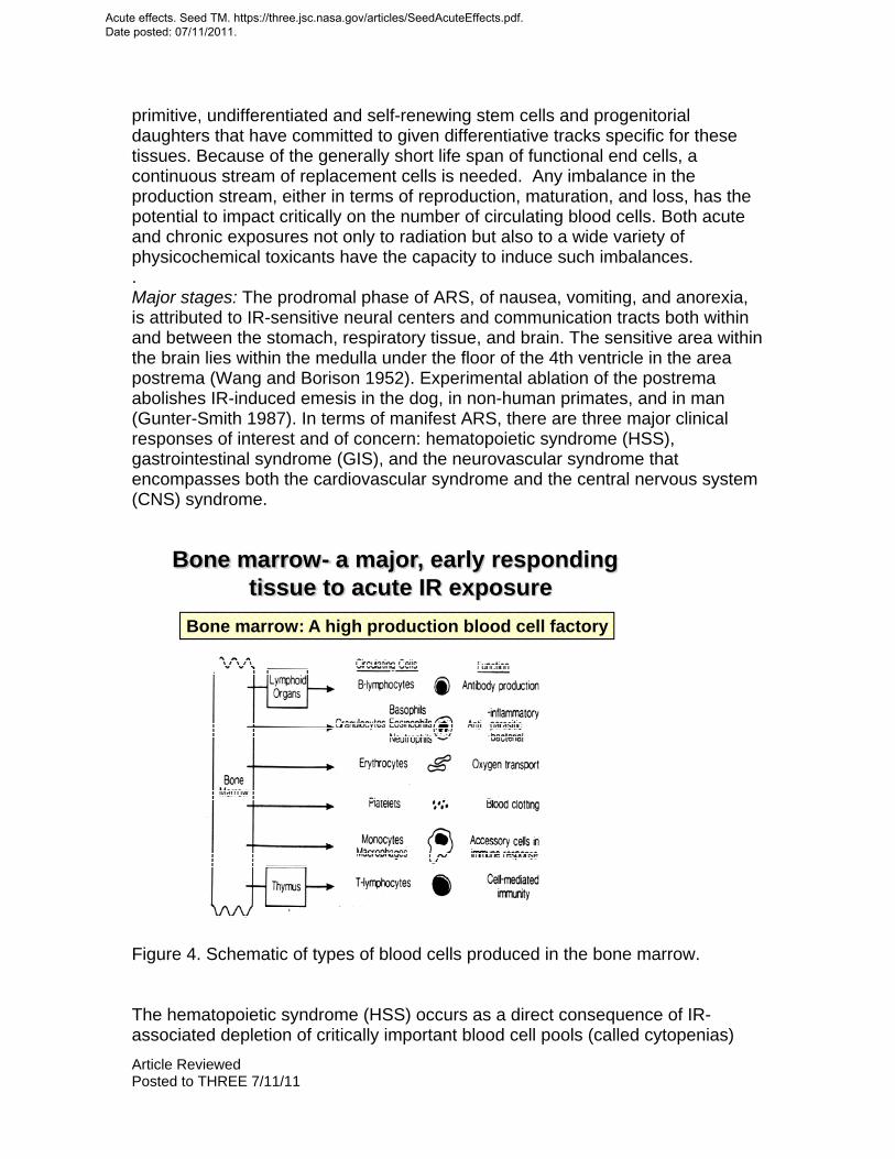
Bone marrow- a major, early respondingtissue to acute IR exposure
Bone marrow: A high production blood cell factory
Figure 4. Schematic of types of blood cells produced in the bone marrow. The hematopoietic syndrome (HSS) occurs as a direct consequence of IR-associated depletion of critically important blood cell pools (called cytopenias)
Acute effects. Seed TM. https://three.jsc.nasa.gov/articles/SeedAcuteEffects.pdf. Date posted: 07/11/2011.

Article Reviewed Posted to THREE 7/11/11
essential for a variety of vital bodily functions; e.g., tissue oxygenation by blood erythrocytes; innate immune defense against microbial agents by blood granulocytes and monocytes; acquired immune functions by select classes of blood lymphocytes; and bleeding control and wound healing processes by blood platelets (thrombocytes). If the IR exposure is sufficiently intense and vital blood cell pools are depleted below critical threshold levels essential for these vital functions, then the affected individual becomes at risk for developing life-threatening pathological sequelae associated with ARS (e.g., septicemias, uncontrolled bleeding, anoxia-mediated organ failure, etc.). To maintain a steady state, the hematopoietic system must produce daily some ~1..510 functional blood cells per kg of body weight of the average individual (Figure 4). This blood cell production occurs in bone-encapsulated marrow, comprised of ~1012 cells in functionally active marrow, of which ~108 cells (or ~0.008%) are core stem cells, comprised of both long-term and short-term marrow repopulating stems cells (Lt-HSCs / St-HSCs). Sufficiently intense IR exposure can impact circulating blood cell levels in multiple ways (see text below), especially by causing cytopenia by a restriction of the reproduction of marrow stem cells and early progenitors. Ionizing radiation can limit self-renewal of these cells by either cell killing, or indirectly by fostering commitment to differentiation over renewal of stem cells. The general temporal pattern of bone marrow depletion following acute, high-dose IR exposures is shown in Figure 5 (Fliedner et al. 1956); while in Figure 6, the hematological consequences of severe granulocytopenia and thromobocytopenia after acute IR exposures are illustrated in terms of blood responses of the acutely IR-exposed Chernobyl fire fighters (Guskova et al. 2001).
Acute effects. Seed TM. https://three.jsc.nasa.gov/articles/SeedAcuteEffects.pdf. Date posted: 07/11/2011.
Article Reviewed Posted to THREE 7/11/11
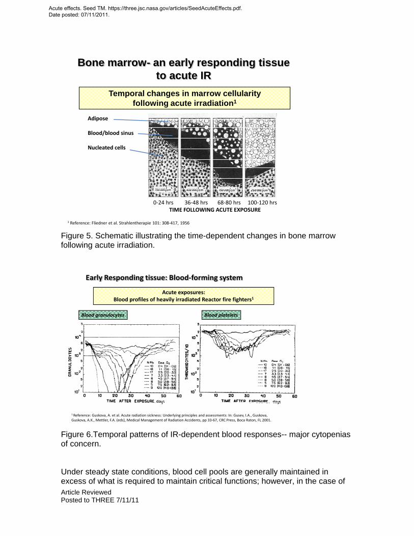
Temporal changes in marrow cellularityfollowing acute irradiation1
Adipose
Blood/blood sinus
Nucleated cells
0‐24 hrs 36‐48 hrs 68‐80 hrs 100‐120 hrsTIME FOLLOWING ACUTE EXPOSURE
1 Reference: Fliedner et al. Strahlentherapie 101: 308‐417, 1956
Bone marrow- an early responding tissue to acute IR
Figure 5. Schematic illustrating the time-dependent changes in bone marrow following acute irradiation.
Early Responding tissue: Blood‐forming system
Acute exposures: Blood profiles of heavily irradiated Reactor fire fighters1
Blood granulocytes Blood platelets
1 Reference: Guskova, A. et al. Acute radiation sickness: Underlying principles and assessments: In: Gusev, I.A., Guskova, Guskova, A.K., Mettler, F.A. (eds), Medical Management of Radiation Accidents, pp 33‐67, CRC Press, Boca Raton, FL 2001.
Figure 6.Temporal patterns of IR-dependent blood responses-- major cytopenias of concern. Under steady state conditions, blood cell pools are generally maintained in excess of what is required to maintain critical functions; however, in the case of
Acute effects. Seed TM. https://three.jsc.nasa.gov/articles/SeedAcuteEffects.pdf. Date posted: 07/11/2011.

Article Reviewed Posted to THREE 7/11/11
intense IR exposure, blood cell pools are selectively-- and dose-dependently--depleted by a number of processes. The major processes include: a) excessive cell loss, due to promotion of cell senescence, vascular margination due to tissue damage, with or without tissue sequestration; and b) markedly reduced influx of new cells produced within lymphohematopoietic tissues and released into the circulation. Intense IR is effective in disrupting this cell production via a number of well documented processes; for example, by increasing transit times across amplifying and maturing compartments, but most strikingly by direct killing of the more rare, vital, and radiosensitive progenitor cells in the system. Considering the very radiosensitive nature of the progenitor cells in the marrow, with rather low capacity for handling potentially lethal damage, reflected by mean Do values in the range of ~1 Gy, coupled with minimal sub-lethal damage repair capacity, as suggested by Dq values of <0.1-0.15 Gy, it is quite understandable that the HSS component of ARS is elicited at relatively low dose levels especially in more radiosensitive individuals. Therefore, without the benefit of having the requisite population of progenitors with the potential of self-renewal and differentiation, the production of functional blood cells soon becomes limited and ultimately fails to produce enough cells needed to replenish cells lost daily. The gastrointestinal syndrome (GIS) shares several prominent, common pathological processes with the HSS component of ARS described above. Like the HSS, IR elicits its damage by depleting vital cell compartments via direct and indirect mechanisms. Unlike the hematopoietic system, however, the progenitor cells are compartmentalized deep within discrete crypts, producing generations of differentiating and maturing daughters that move upward to replace the cells of the villi. This contrasts with hematopoietic tissue with its more randomly positioned niches of progenitors and surrounding patches of differentiating and maturing lineage-specific cells. Under steady state conditions, and within a 3-5 day life-cycle, epithelial cells are produced in the intestinal crypts, differentiate, mature, and migrate upward toward the tips of the villi where they are shed into the lumen of the intestine (Gunter-Smith 1987). The fully mature epithelial cells covering the intestinal villi, apart from their role in absorption, serve an essential role, namely, to provide a tight, physical barrier that cordons off the external milieu of the gut from the internal tissue spaces. This cell barrier serves as an essential component of the individual’s innate immune defense, and damage to its integrity poses a significant health hazard, especially from infections and septicemia (Gunter-Smith 1987; Gunter-Smith and Dubois 1996; Cerveny et al. 1989; Griffiths et al. 1995, 2002; Brennan et al. 1998; Friberg 1980; Carr 1981). In the GIS of ARS, intense IR disrupts the life cycle of the cells of the intestinal epithelium by directly killing or restricting differentiation of epithelial progenitors within the crypts that may lead to severe loss of the cells lining the villi. Also, the restriction of the maturation and migration of villus epithelia, while fostering premature aging and senescence, also contributes to the damage (Potten et al. 2001; Hall 2000; Matsuzawa and Tsubouchi 1969). Despite the latter, the
Acute effects. Seed TM. https://three.jsc.nasa.gov/articles/SeedAcuteEffects.pdf. Date posted: 07/11/2011.

Article Reviewed Posted to THREE 7/11/11
temporal pattern and the range of IR doses required to fully manifest GIS are related to the basic radiobiological characteristics of the crypt stem cells, which help to explain the differences in the temporal pattern of this syndrome and the level of dose required to cause it. The crypt stem cells are somewhat less sensitive than hematopoeitic stem cells and appear to have a greater repair capacity as indicated by a Do of 1-2 Gy and a Dq of 3-5 Gy (Withers et al.1970; Thames et al. 1981; Matsuzawa and Tsubouchi 1969; Cai et al. 1997). A pathological consequence of the cellular damage is that the normally impenetrable villus epithelial layer becomes leaky and less efficient in nutrient absorption. There is also a component of damage to the submucosal vasculature that leads to loss of electrolytes and the loss of the cell barrier with increased risk of bacterial invasion that could result in potentially fatal septicemia. Pathogenesis of the neurovascular/CNS syndrome is the least understood of the major clinical responses that comprise ARS. This is due to its rarity and the lack of clinical subjects to evaluate, as well as to the minimal amount of experimental work done to date in characterizing and elucidating basic pathologic mechanisms. Extremely high IR doses >20Gy are required to elicit the syndrome, which has a short latent period of< 1-2 days and a uniformly lethal outcome. The major signs and symptoms that characterize the syndrome include weakness, disorientation, loss of coordination and motor control, respiratory problems, convulsions, and terminal coma. A terminal, cardiovascular shock syndrome has been described as well (Lushbaugh 1969). It was considered that this resulted from a massive loss of electrolytes and plasma by leakage into the tissues that causes increased intracranial pressure, cerebral anoxia with death within two days.
Figure 7. Structural changes in GI tissues three days following escalating doses of acute irradiation.
Acute effects. Seed TM. https://three.jsc.nasa.gov/articles/SeedAcuteEffects.pdf. Date posted: 07/11/2011.

Article Reviewed Posted to THREE 7/11/11
Acute IR
Killing/limiting mitosis of crypt stem cells Limiting epithelial differentiation & cell migrationFostering epithelial cell senescenceFostering enhanced apical epithelial cell loss
Denuding of villiInvasion by opportunistic gut microbes Local & systemic infectionsEndotoxemiaMassive fluid & electrolyte loss
Death
IR IR‐induced GIS
Epithelial transitions‐ senescent cells‐maturing cells ‐ transitional cells‐stem cells Capillary/vascular beds
TM Seed TSM 8/28/09
Cell birth > m
aturation > cell death
Tissue invading gut microbes w/o septicemia/endotoxemia
Excessive loss of epithelia w/o breeching epithelial barrier
IR‐induced crypt cell death
Capillary coalescence w/o hemorrhage
Fluid/electrolyte losses
Figure 8. Schematic listing of major cellular responses of GI tissues following acute irradiation. Countermeasures for ARS The countermeasure strategies for acute, IR-related injuries during space travel are basically the same as those that would be used for terrestrial IR exposures (Singh and Seed, 2003; Seed 2005). In general, these strategies encompass adherence to basic principles of radiation protection such as minimizing the exposure and maximizing shielding. The degree of physical shielding is largely dependent on the structure of the spacecraft, but positioning of the astronauts within the spacecraft can provide some additional shielding. Of course, the most rapid return from an EVA or to shelter in the case of a lunar mission is imperative. Radiation monitoring will indicate the need for additional action such as the administration of anti-emetics, anti-diarrheals, antibiotics, fluids and electrolytes. CNS Increasing exposure (dose) levels Increasing severity of injuries w. increasing lethality risks
Acute effects. Seed TM. https://three.jsc.nasa.gov/articles/SeedAcuteEffects.pdf. Date posted: 07/11/2011.

Article Reviewed Posted to THREE 7/11/11
CNS
Increasing exposure (dose) levels
Increasing severity of injuries w. increasing lethality risks
COUNTERMEASURES FOR ARS
GISHSSProdome
2# Anti-emetics(Granesitron)
3# Basic clinical support regimens (Fluids, electrolytes, antibiotics, anti-emetic/antidiarrheals)
1# Exposure prevention(Shielding/sheltering)
TM Seed TMS 9/29
Preventive agents(ROS scavengers-phosphorothioates, tocopherol-succinate; anti-apoptotic agents-Toll-like receptor agonists- CBLI502)
HSS-injury Mitigators(GFs/cytokines-rG-CSF, GM-CSF, TPO, Epo; Anti-apoptotic agents; autologous stem cell infusions)
GIS-injury Mitigators(GFs- rKGF, TGFb; tocopherol analogs; Somatostatins-octreotide; probiotics; 2nd&3rd generation antibiotics)
CNS-injury mitigators(Provide comfort -w. anti-inflammatories, pain relievers, and sedatives)
Experimental strategies and agents currently under development
Primary, operationally viable approaches in preventing/managing ARS
Prodome H GIS Figure 9. Schematic listing of possible medical countermeasures. The daily use of anti-emetics (e.g., Kytril®/granisetron; Zolfran®/odansetron) throughout periods of high SPE risk would represent a simple, highly effective and safe way to prevent or limit acute IR-associated nausea and vomiting, in turn limiting IR-associated physical/cognitive performance degradations. Despite the proven effectiveness of these anti-emetic pharmacologics, they only serve to mask the root injury. Their benefit would be limited to the prevention of vomiting, which would be extremely important to an astronaut wearing an EVA space suit. In principle, effective prophylaxis and therapeutic options do exist for both limiting and managing ARS. However, in practice, only a few of these options have been actually approved by regulatory authorities for medical use under such conditions. Nevertheless, for the sake of discussion and information, the following prophylactic agents are listed. First, the phosphoroaminothioate agent, Amifostine (Ethyol®) is a well-documented, clearly effective, FDA-approved radioprotective pharmacologic that is currently being used to mitigate normal tissue damage that might occur as a consequence of radiotherapy for cancer. However, due to the drug’s pronounced hypotensive and emetic side effects, it has not been approved for the prevention of non-medical types of accidental exposures. Second, the use of a variety of ROS-quenching agents, such as select tocopherol compounds might be of some benefit in limiting the extent of IR-injury. Although these agents are clearly less effective than amifostine/Ethyol® or other types of phosphorothioates, they currently lack regulatory constraints for this type of use. If early signs and symptoms of ARS
Acute effects. Seed TM. https://three.jsc.nasa.gov/articles/SeedAcuteEffects.pdf. Date posted: 07/11/2011.
Article Reviewed Posted to THREE 7/11/11
were to manifest in space, basic elements of standard clinical protocols developed specifically for terrestrial-based settings (Walselenko et al. 2004) could be applied, and with few exceptions, with little difficulty. Basic clinical support would be essential, including administration of fluids and electrolytes, plus a spectrum of antimicrobials (Brook et al.2002). For the hematopoietic component of ARS, the administration of the newer, recombinant hematopoietic growth factors (GFs) such as G-CSF (Neupogen®) could prove to be life-saving in terms of enhancing the recovery of granulocytopoiesis within the bone marrow and in addressing the dangerous decrease in the levels of blood granulocytes influencing immune defense. The use of other specific GFs might be considered; e.g., Epo and Tpo for restoration of erythropoiesis and megakaryocytopoiesis, in severe anemia and thrombocytopenia, respectively. The GF/cytokine-therapy is only appropriate if the IR exposure is not >8 Gy: GF therapy loses its effectiveness at extremely high IR exposures, due to the massive loss of marrow stem cells, the primary cellular targets for the recombinant GFs. Since the likelihood of such acute IR doses is remote, provision for such therapy could be included in contingency planning. However for such therapy to be effective, infusion of hematopoietic stem cells would be required. Such treatments are not trivial medical procedures, but would perhaps be essential in order to save the life of a markedly overexposed astronaut. Preferably, the stem cell transplant would be autologous in nature, making use of banked, onboard, cryopreserved HSCs that had been initially harvested from the astronauts during the preflight medical evaluation period just for such critical life threatening contingencies. GIS starts to manifest following acute IR exposures of ~4 Gy, dose levels that are unlikely to occur, but if they did occur, anti-emetic and anti-diarrheal medicinals would be required in addition to nutritional, fluid and electrolyte supplements. Several new classes of agents, specifically designed to either prevent or to mitigate select stages of GIS, are currently under test and development. These include novel ROS scavengers and nutritional-based antioxidants; e.g., gamma-tocotrienol (Berbee et al. 2009). The established phosophorothioate-type radioprotectors need to be considered as well as selective inhibitors of IR-induced enterocyte/enterocyte progenitor depletion; e.g., the anti-apoptotic toll-like receptor 5 agonist-CBLB502 (Burdelya et al. 2008); recombinant GF-TGFB (Booth and Potten 2001); recombinant enterocyte growth stimulators-rKGF, rIL11, SCF, etc. (Farrell et al. 2002; Booth and Potten 2001); mitigators of mucosal barrier breakdown-prostaglandin analogs, effective in blocking gastric hemorrhage (Jarrett 1996); mitigators of mucositis-recombinant KGF; (Farrell et al. 2002); sulfasalazine, a 5-aminosalycilates; octreotide, asomatostatin (Stone et al. 2004); anti-diarrheals; e.g., soluble fiber nutrient supplements; octreotide; loperamide, aopiate agonist (Muehlbauer et al. 2009; Zimmerer et al. 2008; Yeoh et al. 1993); and agents effective in either blocking bacterial translocation or minimizing subsequent infectious complications; e.g., antibiotics, probiotics; (Brook et al. 2004; Isolauri et al. 2002).
Acute effects. Seed TM. https://three.jsc.nasa.gov/articles/SeedAcuteEffects.pdf. Date posted: 07/11/2011.

Article Reviewed Posted to THREE 7/11/11
At the extreme IR exposure levels that elicit CNS and related neurovascular syndromes, no preventive or treatment options currently exist other than the provision of comfort and support, along with relief of pain and sedation. Summary and conclusions Major clinical features of ARS and its temporal and radiological constraints: • The acute radiation syndrome, or ARS, preceded by a prodromal phase and a latent period, is an IR-associated disease complex, composed of three major, distinct clinical syndromes: hematopoietic syndrome (HSS), gastrointestinal syndrome (GIS), and neurovascular syndrome, or more commonly, the central nervous system syndrome (CNS). • Expression of these syndromes is dependent on: a) the magnitude and extent of bodily IR exposure, b) the acuteness of exposure, and c) the time following exposure. The HSS clinical response is the most common component of ARS observed: it occurs at the lowest range of IR doses (e.g., 0.5-10 Gy), takes weeks to fully express and is variably linked to dramatically different survival outcomes. Survival outcomes are linked to the two major clinical concerns associated with HSS presentation, namely a) systemic microbial infections; and b) uncontrolled bleeding. • GIS is observed less frequently than HSS because it is caused by a substantially higher range of doses (~4->15 Gy), but it is more quickly expressed, within 4-6 days. At the lower IR doses, GIS often occurs concurrently with HSS, and as a consequence, greatly complicates the clinical picture. At higher IR doses in the absence of appropriate therapeutic intervention, GIS is rapidly fatal within a week of IR exposure due to extreme diarrhea, water loss, electrolyte imbalances, and infections. • The neurovascular/CNS component of ARS is rarely observed and limited to those extremely over exposed individuals (IR doses >20Gy). The onset of neurovascular sub-syndrome is exceedingly rapid, minutes to hours and characterized clinically by weakness, respiratory distress, loss of motor-control, disorientation, convulsions, and coma. CNS is uniformly fatal due to precipitating cerebral vascular hemorrhage and edema, and currently, has no known effective therapy. Space radiation environments and associated exposure scenarios of major concern: • Periodic solar particle events and a chronic flux of galactic particles represent the two major, external IR sources of concern within the space environment. Acute effects are a concern from exposure during large solar events. These events are poorly predictable in terms of magnitude, duration, and time of occurrence. Dose-rates and total doses associated with the rather constant flux of GCR are considered too small to cause acute effects. The high densities of
Acute effects. Seed TM. https://three.jsc.nasa.gov/articles/SeedAcuteEffects.pdf. Date posted: 07/11/2011.

Article Reviewed Posted to THREE 7/11/11
highly energetic protons emanating from the larger solar events could yield doses with the potential to cause the prodromal stage and possibly the HSS component of ARS. The probability of having IR exposures sufficiently high to elicit GIS is unlikely, and the even more extreme case of CNS is improbable. • The estimated probability of an extremely large solar event containing >30 MeV fluence of ~5x10⁹ protons per cm-2 with ARS-yielding potential occurring over the span of a 2-yr deep space mission is approximately 10%. The probability of multiple, massive SPEs occurring during extended space missions is directly related to mission duration, but is significantly less. The estimates of risk require better estimates of the effect of dose rate on the incidence and severity of the effects in the prodromal phase during SPEs. Pathogenesis: • The major IR-associated prodrome of ARS, namely the complex of nausea, vomiting, and diarrhea, is attributed to IR-sensitive neural centers and communication tracts both within and between the stomach, respiratory tissue, and brain. • Several 5-HT3 receptor antagonistpharmacologics (e.g., granisetron/Kytril® or ondansetron/Zofran®) are effective in limiting/blocking IR-associated emesis. • HSS/ARS manifests as a direct consequence of IR-associated depletion of critically important blood cell pools (so called blood cytopenias) essential for a variety of vital bodily functions, erythrocytes for tissue oxygenation, granulocytes and monocytes for innate immune defense against microbial agents, select subsets of lymphocytes for acquired immune functions, and blood platelets (thrombocytes) for the control of bleeding and wound-healing processes. • With sufficiently intense IR, vital blood cell pools are depleted below critical threshold levels and the exposed individual becomes ‘at risk’ to develop life-threatening pathological sequelae associated with ARS such as septicemias, uncontrolled bleeding, and anoxia-mediated organ failure. • Under steady state conditions, blood cell pools are maintained in excess of what is required to maintain critical functions. IR-induced selective and dose dependent damage to the blood cell system depletes by a number of processes: - i) excessive cell loss due to promotion of cell senescence, ii) vascular margination due to tissue damage, with/without tissue sequestration; and iii) markedly reduced influx of new cells produced by lymphohematopoietic tissues. • Sufficiently intense IR is effective in disrupting or limiting cell production by lymphohematopoietic tissues, by well documented processes i) extending transit times across amplifying and maturing compartments and by ii) direct killing of progenitors. HSS is elicited at relatively low levels of IR due to the highly radiosensitive nature of major progenitor cell compartments within hematopoietic tissues. • GIS shares several prominent, common pathological processes with HSS. For example, IR exerts damage by depleting, by direct and indirect mechanisms, vital cell compartments of the GI tissue’s self-renewing, amplifying, and differentiating cell-producing system.
Acute effects. Seed TM. https://three.jsc.nasa.gov/articles/SeedAcuteEffects.pdf. Date posted: 07/11/2011.

Article Reviewed Posted to THREE 7/11/11
• Mature epithelia covering intestinal villi serve an essential and vital role -- to provide a tight physical barrier between the external milieu of the gut and the intestacies of intestinal tissues. In addition to its role in absorption, the epithelial cell barrier serves as an essential role in innate immune defense. Any damage to the integrity of the barrier poses a significant predisposing health hazard, especially in terms of infections. • GIS is caused by intense IR mainly by cell killing or restricting differentiation of epithelial progenitors within the crypts, but also by restricting maturation and migration of villus epithelia, while fostering premature aging and senescence. As a consequence, the normally tight and impenetrable villus epithelial layer becomes loose and ‘leaky’ and susceptible to invasion and colonization by select microbial species within the normally benign gut microflora. • Neurovascular CNS is the rarest and least understood of the major ARS sub-syndromes in terms of its pathogenesis. The sub-syndrome is unique in terms of the extremely high IR doses (>20Gy and up) required for induction, its extremely short latency, and its uniformly lethal nature. • The signs and symptoms that characterize neurovascular/CNS include weakness, disorientation, loss of coordination and motor control, respiratory problems, convulsions, and terminal comas, and are commonly attributed to cerebral hemorrhage and edema. • No effective treatment options currently exist for subjects suffering from neurovascular/CNS. Providing comfort, pain relief, and possibly sedation are the only medical options currently available. Countermeasures: • Two major strategies exist for countering space IR-associated acute injury: i) physical measures and ii) medical interventions. A guiding principle of effective radio hygiene--in deep space or on earth-is ‘exposure avoidance.’ Sheltering within shielded zones within the spacecraft is a practical option. • Pharmacologic intervention is a further option to counter IR-associated health effects. Major prodromal responses of ARS--nausea and vomiting--can be effectively prevented or minimized by daily administration of one of several anti-emetics, e.g., Kytril® or Zolfran®. • Effective prophylaxis and therapeutic options exist for both limiting and managing manifest ARS. [Note- these ‘medical options’ might be limited due to current regulatory constraints]. In terms of prophylaxis, the phosphorothioate agent, amifostine (Ethyol®) might be considered, as the drug is highly effective, but short-acting, and has substantial, potential side-effects of concern (i.e., hypotensive and emetic). Other less effective and better tolerated, but without regulatory limits or restrictions, might include a variety of neutraceuticals that are strongly antioxidative and free radical quenching. • In the event of manifest ARS, basic elements of standard clinical protocols developed specifically for terrestrial-based settings could be applied. Basic clinical support would be essential, including administration of fluids, plus broad spectrum antibiotics.
Acute effects. Seed TM. https://three.jsc.nasa.gov/articles/SeedAcuteEffects.pdf. Date posted: 07/11/2011.

Article Reviewed Posted to THREE 7/11/11
• To manage HSS, the administration of the newer, recombinant hematopoietic growth factors (rGFs), such as G-CSF (Neupogen®) could be used to supplement basic clinical support procedures. • The GF/cytokine-therapy option is only be effective if IR exposures are not extremely high (> 8-10 Gy). Following extremely high IR exposures, in-flight autologous stem cell infusions might be the only viable treatment option, and in turn, hope for long-term survival. • GIS manifests at higher IR exposures, requiring GI-specific medicinals--antidiarrheals, nutritionals, fluids, and electrolyte supplementations. • At extreme, but highly unlikely IR exposures in space that would elicit CNS and related neurovascular syndromes, no effective preventive or treatment options currently exist, other than simply providing comfort and support, along with administered pharamacologics to relieve pain and to sedate. References Anno GH, Baum SJ, Wither HR, Young RW. Symptomatology of acute radiation
effects in humans after exposure to exposure to doses of 0.5-3.0 Gy. Health Physics:56(6) 821-838, 1989.
Badhwar GD, Radiation environment for interplanetary travel, In: Radiation Research- Volume 2, )M Moriarty, C Mothersill, C Seymour, M Edington, JF Ward, RJM Fry, eds Proceedings of the 11th International Congress of Radiation Research, Dublin, Ireland, July 18-23, 1999, p 737-740, Allen Press, Lawrence, KS 2000.
Berbee M, Fu Q, Boerma M, Wang J, Kumar KS, Hauer-Jensen M. Gamma-tocotrienol ameliorates intestinal oxidative stress after total body irradiation by an HMG-CoAreductase-dependent mechanism, Radiat Res 171: 596-605, 2009.
Booth D, Potten CS. Protection against mucosal injury by growth factors and cytokines. J Natl Cancer Inst Monogr 29: 16-20, 2001.
Brennan PC, Carr KE, Seed T, McCullough JS, Acute and protracted radiation effects on small intestinal morphological parameters, Int J Radiat Biol 73 : 691-698, 1998.
Brook I, Elliott TB, Ledney GD, Shoemaker MO, Knudson GB. Management of postirradiation infection: lessons learned from animal models. Mil Med. 169(3): 194-197, 2002.
Burdelya LG, Krivokrysenko VI, Tallant TC, Strom E, Gleiberman AS, Gupta D, Kurnasov OV, Osterman AL, Didonato JA, Feinstein E, Gudkov AV. An agonist of toll-like receptor 5 has radioprotective activity in mouse and primate models. Science 320 (5873):226-230, 2008.
Cai WB, Roberts SA, Potten CS. The number of clonogenic cells in crypts in three regions of murine large intestine. Int J Radiat Biol 71(5): 573-579, 1997.
Carr KE Scanning electron microscopy of tissue response to irradiation, Scan Elect Microscopy 1981: 35-46, 1981.
Acute effects. Seed TM. https://three.jsc.nasa.gov/articles/SeedAcuteEffects.pdf. Date posted: 07/11/2011.

Article Reviewed Posted to THREE 7/11/11
Cerveny TJ, MacVittie TJ, Young RW. Acute radiation syndrome in humans, In: R I Walker, TJ Cerveny eds, Medical Consequences of Nuclear Warfare, pp 15-36, Textbook of Military Medicine, Part 1, Vol 2, TMM Publications, Office of the Surgeon General, Falls Church, VA (1989).
Farrell CL, Rex KL, Chen JN, Bready JV, DiPalma CR, Kaufman SA, Rattan A, Scully S, Lacey DL. The effects of keratinocyte growth factor in preclinical models of mucositis. Cell Prolif 35 (suppl 1): 78-85, 2002.
Feynman J, Spitale G, Wang J, Gabriel S. Interplanetary proton fluence model. J Geophys Res 98: 13281-13294, 1993.
Fliedner TM, Sandkuhler S, Stodmeister R. Significance of vascular wall lesions in the pathogenesis of hematopoietic disorders in rats after total-body irradiation with 15 MeV electrons. Strahlentherapie101(2): 308-317, 1956.
Friberg LG, Effects of irradiation on the small intestine of the rat: A SEM study, Thesis at Lund University, Sweden, 1980.
Griffiths NM, Linard C, Dublineau I, Scanff P, Jourbert C, Gourmelon P, Acute gastrointestinal dysfunction and pathology following accidental irradiation, pp 117-126, In: L’Homme Blesse,’ (eds) LA Court, J Lallemand, LeCentre de Recherche du Service de Sante’ des Arme’es (CRSSA), Grenoble, FR 1995.
Griffiths NM, Scanff P, Nenot J-C, Modification of gastrointestinal tract function following prolonged, intermittent or fractionated exposure to ionizing radiation. In: (eds) TM Fliedner, LE Feinendegen, JW Hopewell, Chronic Irradiation: Tolerance and Failure in Complex Biological Systems, pp 242-246, The British Institute of Radiology, supplement 26, London UK 2002.
Gestner HB. Reaction to short-term radiation in man. Ann. Rev. Med. 11. 289-302, 1960.
Gunter-Smith PJ. Effects on gastrointestinal physiology, In: JJ Conklin, RI Walker, eds. Military Radiobiology, Chapter 7, pp 135-151, Academic Press, New York, 1987.
Gunter-Smith PJ, Dubois A. Treatment strategies for radiation-induced gastrointestinal injuries, In: Advances in the treatment of radiation injuries, (eds) TJ MacVittie, JF Weiss, D Browne, Advances in the Biosciences, volume 94, p 195-200, Pergamon/Elsevier Science Ltd, White Plains, NY 1996.
Guskova A, Baranov AE, Gusev IA. Acute radiation sickness: Underlying principles and assessments, In: Gusev IA, Guskova, AK, Mettler FA eds, Medical Management of Radiation Accidents, pp 33-67, CRC Press, Boca Raton, FL 2001.
Hall EJ, Chapter 8 Acute effects of total body irradiation, In: Radiobiology for the Radiologist--5th Edition, pp 124-135, Lippincott Williams & Wilkins, Philadelphia, 2000.
Hu S, Kim M-H Y, McClellan GE, Cucinotta FA. Modeling the acute health effects of astronauts from exposure to large solar particle events. Health Phys J 96(4): 465-476, 2009.
Isolauri E, Kirjavainen PV, Salminen S. Probiotics: a role in the treatment of intestinal infection and inflammation. Gut 50 (suppl 3): 11154-11159,
Acute effects. Seed TM. https://three.jsc.nasa.gov/articles/SeedAcuteEffects.pdf. Date posted: 07/11/2011.

Article Reviewed Posted to THREE 7/11/11
2002. Jarrett DG. Medical management of radiological casualties handbook. Armed
Forces Radiobiology Research Institute, Bethesda, MD 1999. Lushbaugh CC. Theoretical and practical aspects of models explaining
“gastrointestinal death” and other lethal radiation syndromes. In: Comparative Cellular and Species Radiosensitivity, pp 288-297 Eds., VP Bond and T Sugahara, Igaku-Shoin, Tokyo, 1969.
Lushbaugh CC. Human Radiation Tolerance. In: Space Radiation Biology and Related Topics pp. 478-522 Eds CA Tobias and P Todd, Academic Press, London, 1974.
Matsuzawa T, Tsubouchi S. Injury and recovery patterns of intestinal epithelium in irradiated germfree and conventional animals. In: Sugahara, Comparative Cellular and Species Radiosensitivity, pp. 271-88-297, Eds., VP Bond, T Sugahara, Igaku-Shoin, Tokyo, 1969.
Muehlbauer PM, Thorpe D, Davis A, Drabot R, Rawlings BL, Kiker E. Putting evidence into practice: evidence-based interventions to prevent, manage, and treat chemotherapy- and radiotherapy-induced diarrhea. Clin J Oncol.Nurs. 13(3): 336-341, 2009.
Kim M-H Y, Hayat MJ, Feiveson AH, McClellan GE, Cucinotta FA. Prediction of frequency and exposure level of solar particle events. Health Phys J, 97(1): 68-81, 2009.
NCRP-153, Information needed to make radiation protection recommendations for space missions beyond low-earth orbit, NCRP Report No. 153, National Council on Radiation Protection and Measurements, Bethesda, MD 2006.
Parsons JL, Townsend LW. Interplanetary crew dose rates for the August 1972 solar particle event. Rad Res 153: 729-733, 2000.
Potten CS, Martin K, Kirkwood TB. Ageing of murine small intestinal stem cells. Novartis Found Symp. 235: 66-79 (and discussion 79-84, 101-114), 2001.
Schimmerling W, NASA space program for interplanetary missions: Challenges and opportunities, In: Radiation Research- Volume 2, (eds) M Moriarty, C Mothersill, C Seymour, M Edington, JF Ward, RJM Fry. Proceedings of the 11th International Congress of Radiation Research, Dublin, Ireland, July 18-23, 1999, p 733-736, Allen Press, Lawrence, KS 2000.
Seed TM, Tolle DV, Fritz TE. Hematological response to chronic irradiation: the past Argonne experience and future AFRRI initiatives In: Chronic irradiation tolerance and failure in complex biological systems. Eds TM Fliedner, LE Feinendegen, JW Hopewell. Brit. J. Radiology Suppl. 26, pp 94-102, 2002.
Seed TM. Radioprotectants: Current status and future prospects. Health Phys. 89(5):531-545, 2005.
Singh V, Seed TM Radiation Effects, Chapter 24, In: (ed) MJ Roy, Physician’s Guide to Terrorist Attack, pp 339-362, Humana Press Inc., Totowa, NJ (2003).
Stephens DL Jr, Townsend LW, Hoff JL. Interplanetary crew dose estimates for worst case solar particle events based on historical data for the Carrington
Acute effects. Seed TM. https://three.jsc.nasa.gov/articles/SeedAcuteEffects.pdf. Date posted: 07/11/2011.
Article Reviewed Posted to THREE 7/11/11
flare of 1859. Acta Astronaut 56(9-12):969-974, 2005. Stone HB, Moulder JE, Coleman CN, Ang KK, Anscher MS, Barcellos-Hoff MH,
Dynan WS, Fike JR, Grdina DJ, Greenberger JS, Hauer-Jensen M, Hill RP, Kolesnick RN, Macvittie TJ, Marks C, McBride WH, Metting N, Pellmar T, Purucker M, Robbins ME, Schiestl RH, Seed TM, Tomaszewski JE, Travis EL, Wallner PE, Wolpert M, Zaharevitz D. Models for evaluating agents intended for the prophylaxis, mitigation and treatment of radiation injuries. Report of an NCI Workshop, December 3-4, 2003. Radiat Res 162(6):711-728, 2004.
Thames HD Jr, Withers R, Mason KA, Reid BO. Dose-survival characteristics of mouse jejunal crypt cells. Int J Radiat Oncol Biol Phys7(11): 1591-1591, 1981.
Wang SC, Borison HL. A new concept of organization of the central emetic mechanism: Recent studies on the sites of action of apomorphine, copper sulfate and cardiac glycosides. Gastroent 22: 1-12, 1952.
Waselenko JK, MacVittie TJ, Blakely WF, Pesik N, Wiley AL, Dickerson WE, Tsu H, Confer DL,Coleman CN, Seed T, Lowry P, Armitage JO, Dainiak N. Medical management of acute radiation syndrome: recommendations of the Strategic National Stockpile Radiation Working Group. Ann Intern Med 140(12): 1037-1051, 2004.
Withers HR, Brennan JR, Elkind MM, The response of stem cells of intestinal mucosa to irradiation with 14 MeV neutrons. Br J Radiol 43: 796-801, 1970.
Wu H, Huff JL, Casey R, Kim M-H, Cucinotta FA. Risk of radiation syndromes due to solar particle events. Chapter 5 in: Human research program requirements document HRP-47952, 2009.
Young RW. Acute radiation syndrome, In: Military Radiobiology, Chapter 9, Eds. JJ Conklin, RI Walker, pp 165-190, Academic Press, New York, 1987.
Yeoh EK, Horowitz M, Russo A, Muecke T, Robb T, Chatterton BE. Gastrointestinal function in chronic radiation enteritis--effects of loperamide-N-oxide. Gut. 34(4): 476-482, 1993.
Zimmerer T, Böcker U, Wenz F, Singer MV. Medical prevention and treatment of acute and chronic radiation induced enteritis--is there any proven therapy? a short review. Z Gastroenterol. 46(5): 441-448, 2008.
Table and Figures Table Table 1. Major early acute effects of ionizing radiation exposure. Figures Figure 1. Major clinical responses associated with ARS. Figure 2. Radiation dose-dependent expression of major clinical responses. Figure 3. Time-dependent expression of major clinical responses. Figure 4. Schematic of types of blood cells produced in the bone marrow.
Acute effects. Seed TM. https://three.jsc.nasa.gov/articles/SeedAcuteEffects.pdf. Date posted: 07/11/2011.
Article Reviewed Posted to THREE 7/11/11
Figure 5. Schematic illustrating the time-dependent changes in bone marrow following acute irradiation. Figure 6. Temporal patterns of IR-dependent blood responses-- major cytopenias of concern. Figure 7. Structural changes in GI tissues three days following escalating doses of acute irradiation. Figure 8. Schematic listing of major cellular responses of GI tissues following acute irradiation. Figure 9. Schematic listing of possible medical countermeasures.
Acute effects. Seed TM. https://three.jsc.nasa.gov/articles/SeedAcuteEffects.pdf. Date posted: 07/11/2011.