activities - linn–benton community collegecf.linnbenton.edu/mathsci/bio/jacobsr/upload/11 -...
TRANSCRIPT

Complete Diagrams PNS 18 and 19
Complete PNS 23 Worksheet 3 #1 only
Complete PNS 24 Practice Quiz
ACTIVITIES
Introduction
Vision
THE SPECIAL SENSES
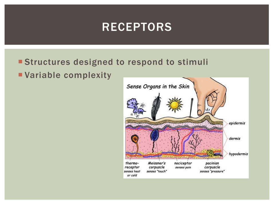
Structures designed to respond to stimuli
Variable complexity
RECEPTORS

Transducers
Receptor potential
Generator potential
GENERAL PROPERTIES OF RECEPTORS
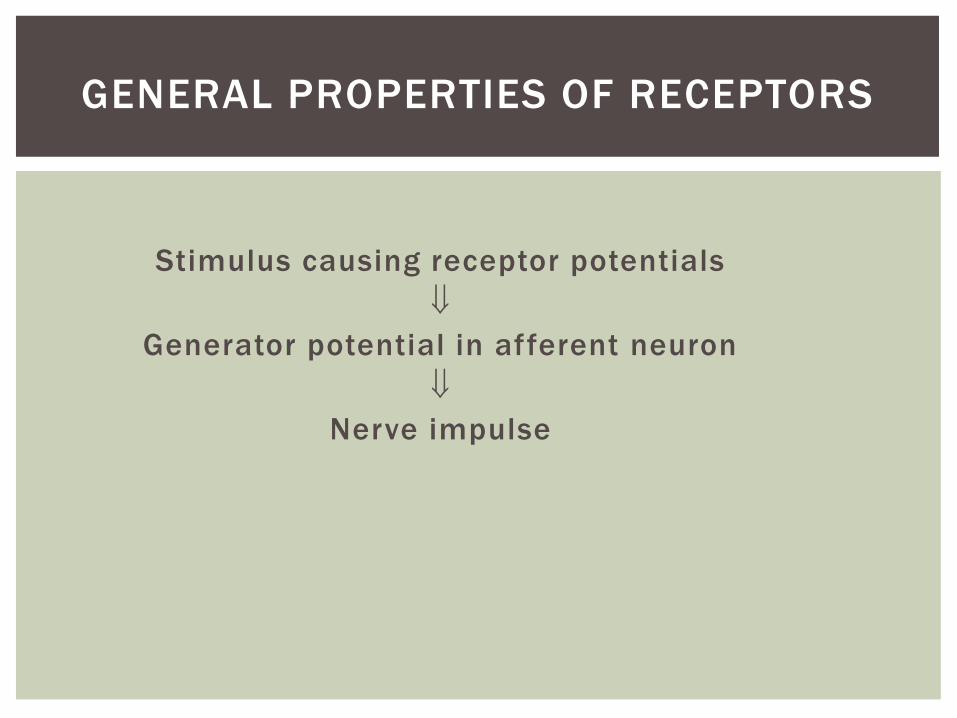
Stimulus causing receptor potentials
Generator potential in afferent neuron
Nerve impulse
GENERAL PROPERTIES OF RECEPTORS

SENSATION AND PERCEPTION
Stimulatory input
Conscious level = perception
Awareness = sensation
Information conveyed by receptors
Modality
Location
Intensity
Duration
GENERAL PROPERTIES OF RECEPTORS

ADAPTATION
Reduction in rate of impulse transmission when stimulus is prolonged

Stimulus Modality
Chemoreceptors
Thermoreceptors
Nociceptors
Mechanoreceptors
Photoreceptors
CLASSIFICATION OF RECEPTORS
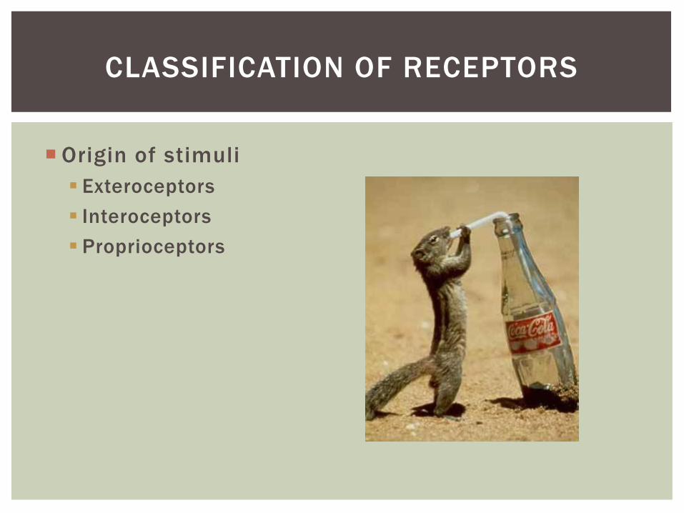
Origin of stimuli
Exteroceptors
Interoceptors
Proprioceptors
CLASSIFICATION OF RECEPTORS

Vision
Hearing
Olfaction
Gustation
SPECIAL SENSES
70% of all sensory receptors are in the eye
Nearly half of the cerebral cortex is involved in
processing visual information
Optic nerve is one of body’s largest nerve tracts
VISION INTRODUCTION
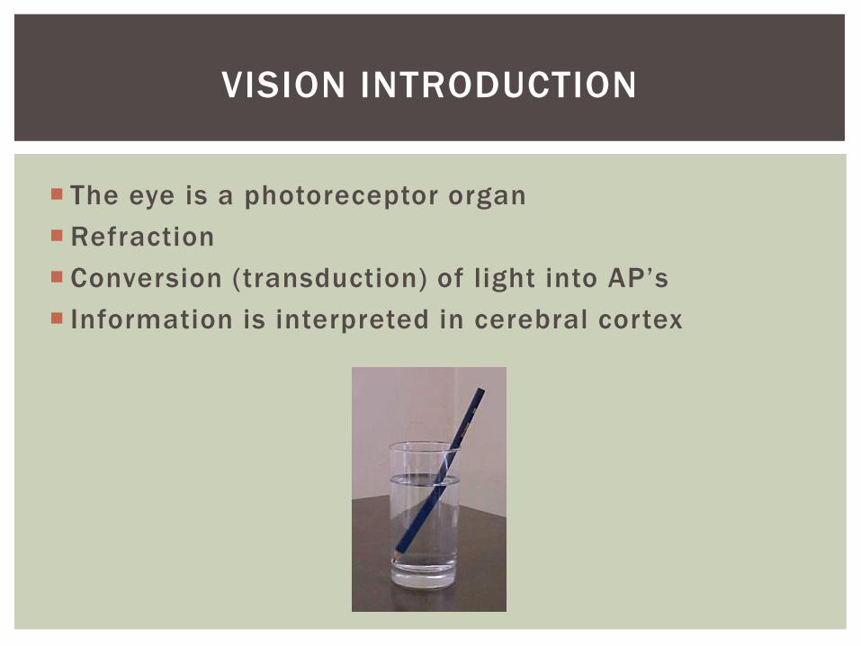
The eye is a photoreceptor organ
Refraction
Conversion (transduction) of light into AP’s
Information is interpreted in cerebral cortex
VISION INTRODUCTION
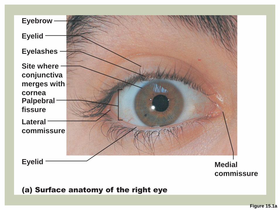
Figure 15.1a
Eyelashes
Site where
conjunctiva
merges with
cornea
Lateral
commissure
Medial
commissure
Eyelid
Eyelid
Eyebrow
Palpebral
fissure
(a) Surface anatomy of the right eye
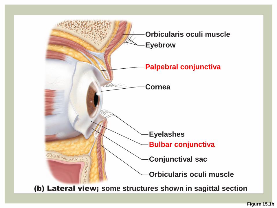
Figure 15.1b
(b) Lateral view; some structures shown in sagittal section
Orbicularis oculi muscle
Eyebrow
Palpebral conjunctiva
Cornea
Eyelashes
Bulbar conjunctiva
Conjunctival sac
Orbicularis oculi muscle

Lacrimal apparatus
Lacrimal gland and ducts that connect to nasal cavity
Lacrimal secretion (tears)
Dilute saline solution
Mucus, antibodies, and lysozyme
Blinking spreads the tears toward medial commissure
Drain into the nasolacrimal duct
ACCESSORY STRUCTURES
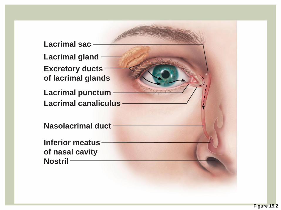
Figure 15.2
Lacrimal gland
Excretory ducts
of lacrimal glands
Lacrimal punctum
Lacrimal canaliculus
Nasolacrimal duct
Inferior meatus
of nasal cavity
Nostril
Lacrimal sac

Six extrinsic eye muscles
ACCESSORY STRUCTURES
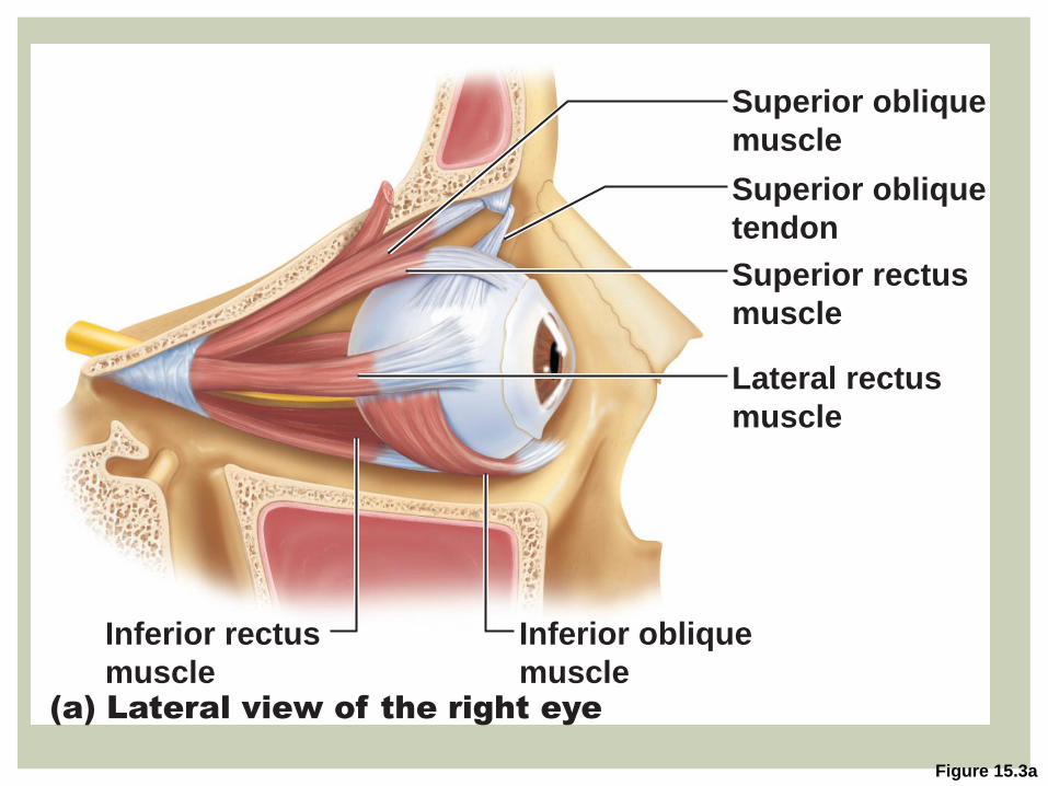
Figure 15.3a
Inferior rectus
muscle
Inferior oblique
muscle
Superior oblique
muscle
Superior oblique
tendon
Superior rectus
muscle
Lateral rectus
muscle
(a) Lateral view of the right eye
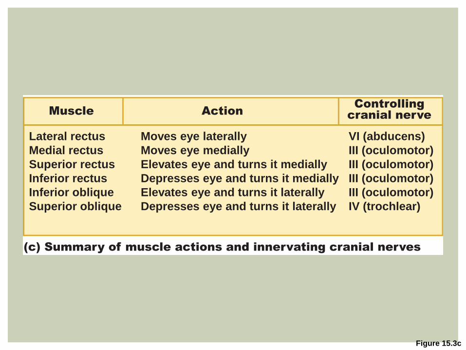
Figure 15.3c
(c) Summary of muscle actions and innervating cranial nerves
Lateral rectus
Medial rectus
Superior rectus
Inferior rectus
Inferior oblique
Superior oblique
Moves eye laterally
Moves eye medially
Elevates eye and turns it medially
Depresses eye and turns it medially
Elevates eye and turns it laterally
Depresses eye and turns it laterally
VI (abducens)
III (oculomotor)
III (oculomotor)
III (oculomotor)
III (oculomotor)
IV (trochlear)
Muscle Action Controlling
cranial nerve

Wall of eyeball contains three layers (tunics)
Fibrous
Vascular
Sensory (retinal)
THE EYEBALL
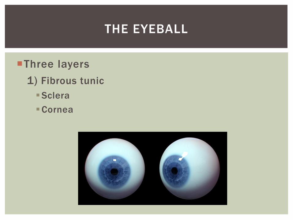
Three layers
1) Fibrous tunic
Sclera
Cornea
THE EYEBALL

Figure 15.4a
Sclera
Cornea
Fibrous Tunic
Scleral venous sinus
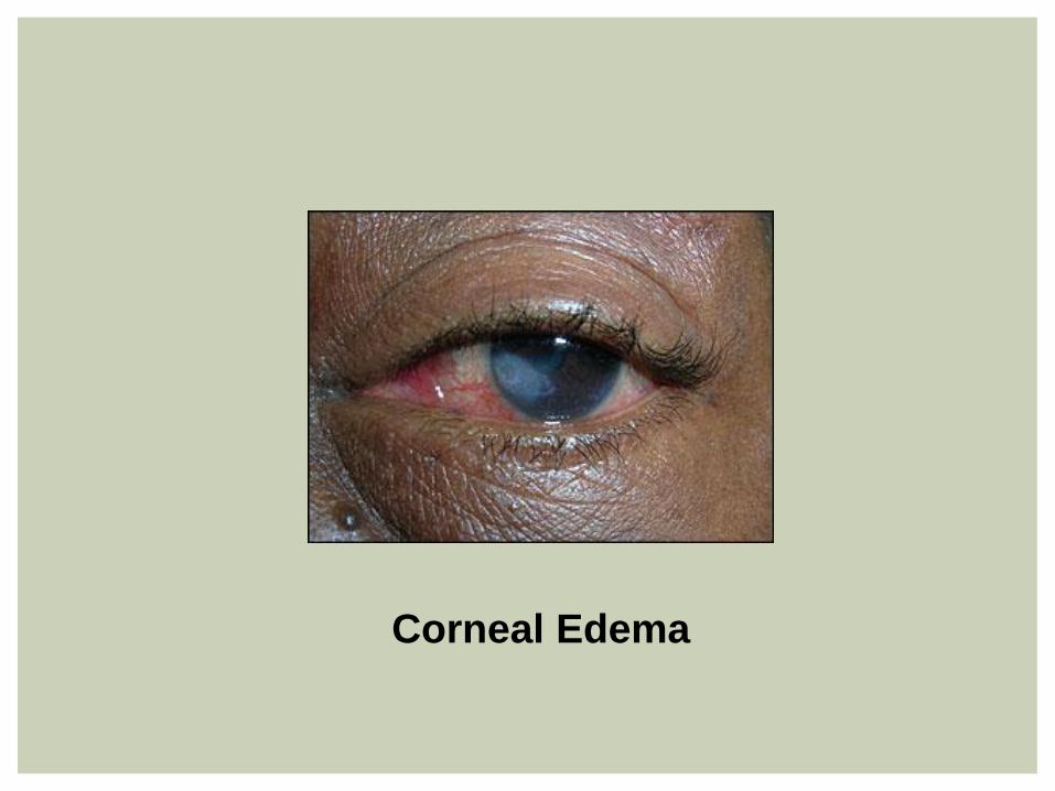
Corneal Edema
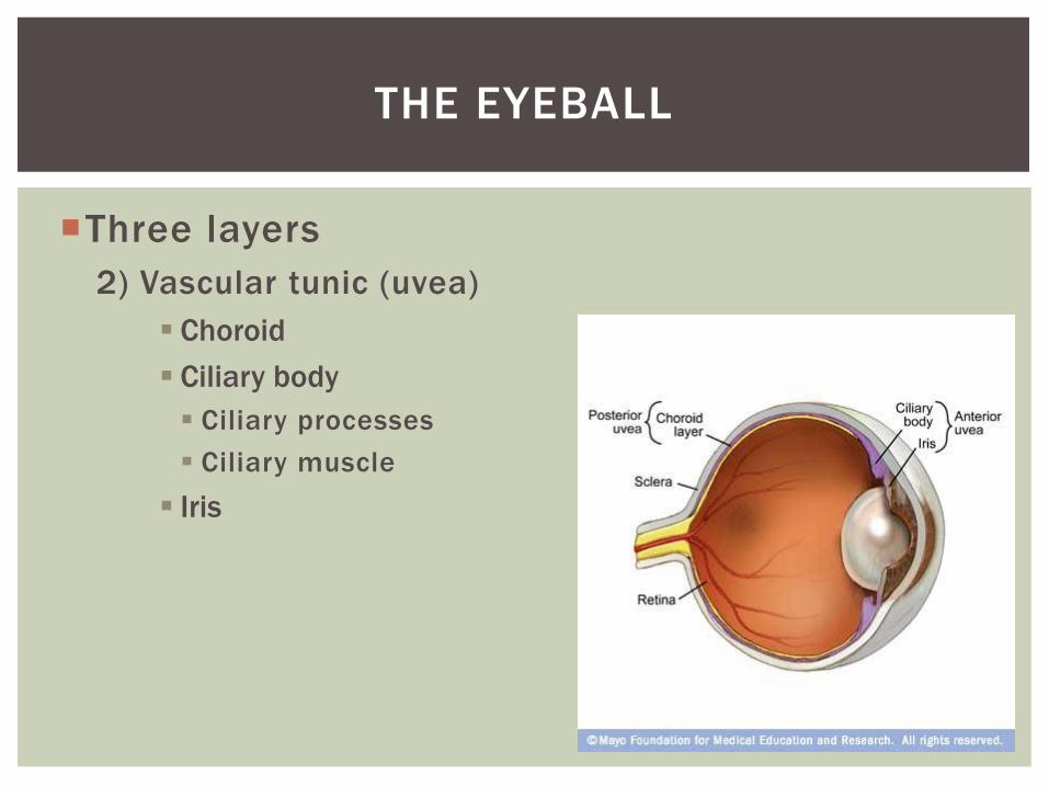
Three layers
2) Vascular tunic (uvea)
Choroid
Ciliary body
Ciliary processes
Ciliary muscle
Iris
THE EYEBALL

Figure 15.4a
Choroid
Sclera Ciliary body Ciliary zonule
(suspensory
ligament)
Iris Pupil
Lens
Scleral venous
sinus
VascularTunic
Cornea
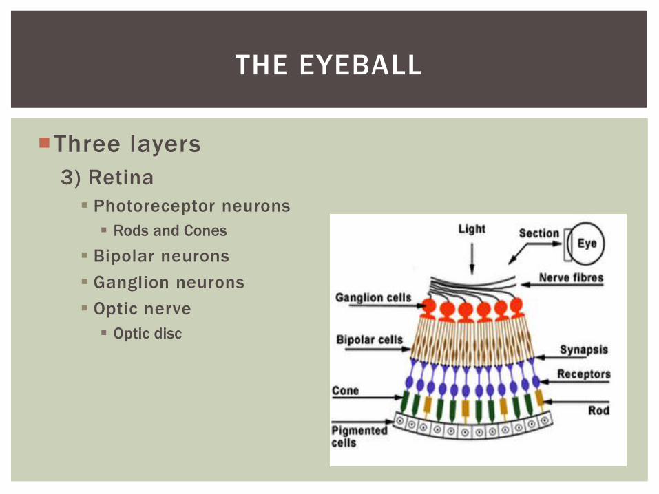
Three layers
3) Retina
Photoreceptor neurons
Rods and Cones
Bipolar neurons
Ganglion neurons
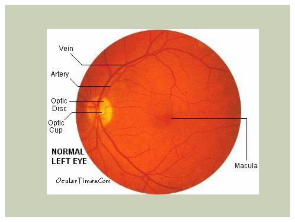
Optic nerve
Optic disc
THE EYEBALL
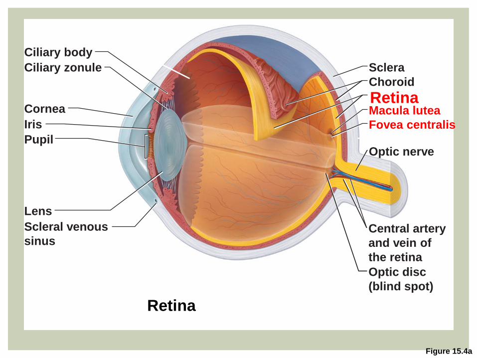
Figure 15.4a
Central artery
and vein of
the retina
Optic disc
(blind spot)
Optic nerve
Fovea centralis
Macula lutea Retina Choroid
Sclera
Ciliary body
Ciliary zonule
Cornea
Iris
Pupil
Lens
Scleral venous
sinus
Retina
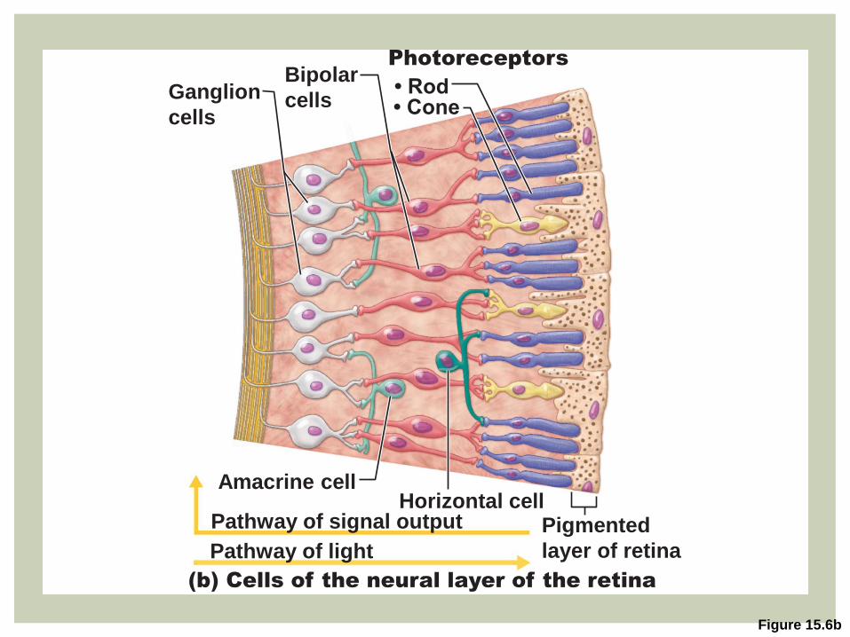
Figure 15.6b
Pigmented
layer of retina Pathway of light
Pathway of signal output
(b) Cells of the neural layer of the retina
Amacrine cell Horizontal cell
• Rod
Photoreceptors
• Cone
Bipolar
cells Ganglion
cells
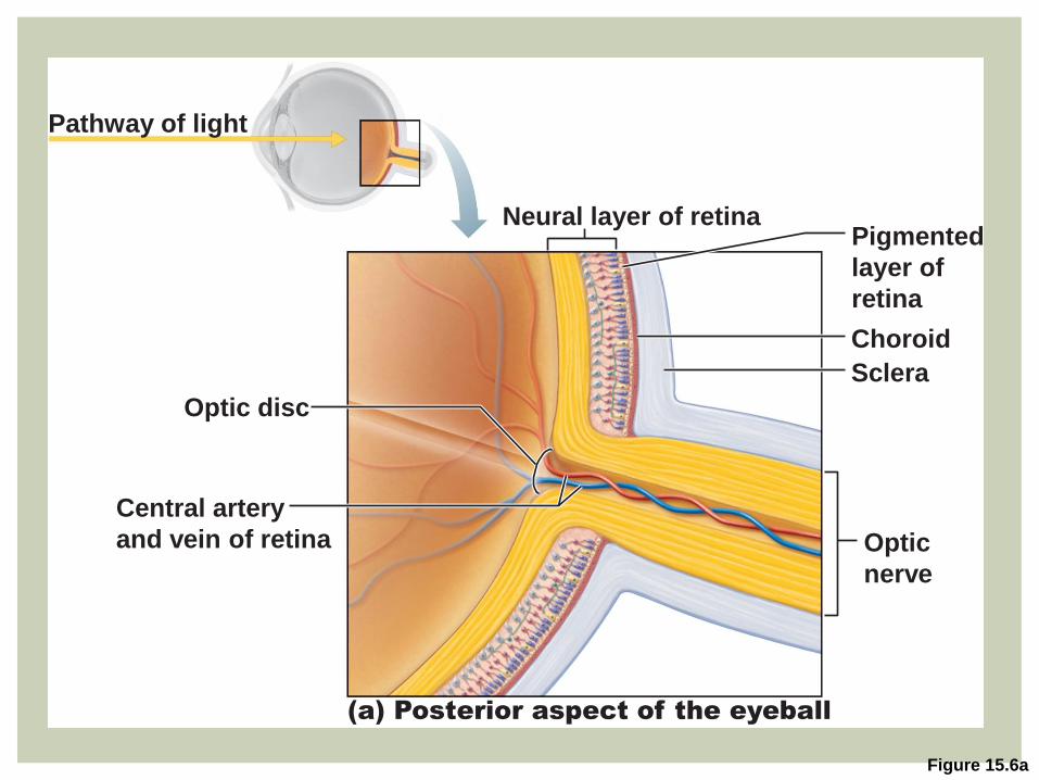
Figure 15.6a
(a) Posterior aspect of the eyeball
Neural layer of retina Pigmented
layer of
retina
Central artery
and vein of retina Optic
nerve
Sclera
Choroid
Optic disc
Pathway of light
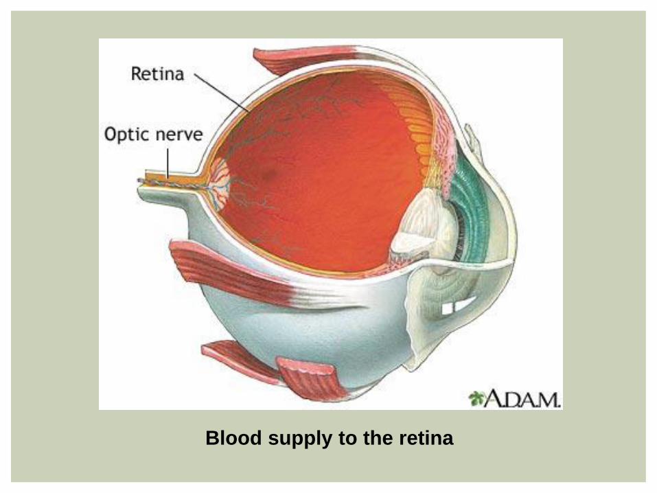
Blood supply to the retina
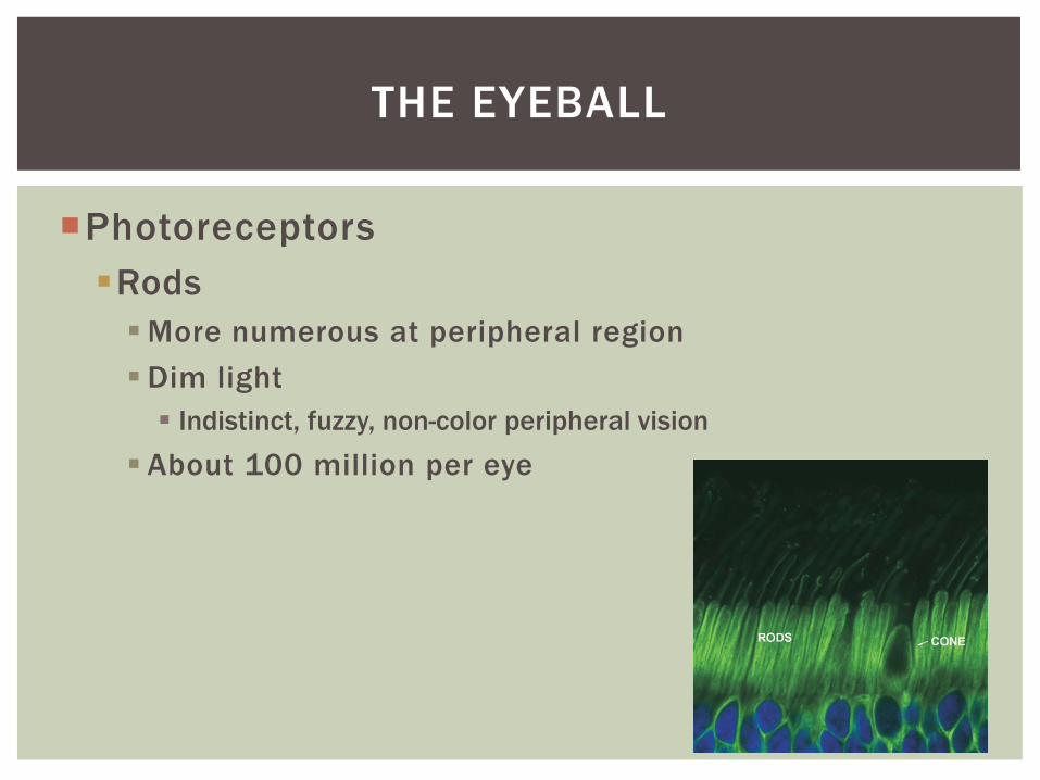
Photoreceptors
Rods
More numerous at peripheral region
Dim light
Indistinct, fuzzy, non-color peripheral vision
About 100 million per eye
THE EYEBALL
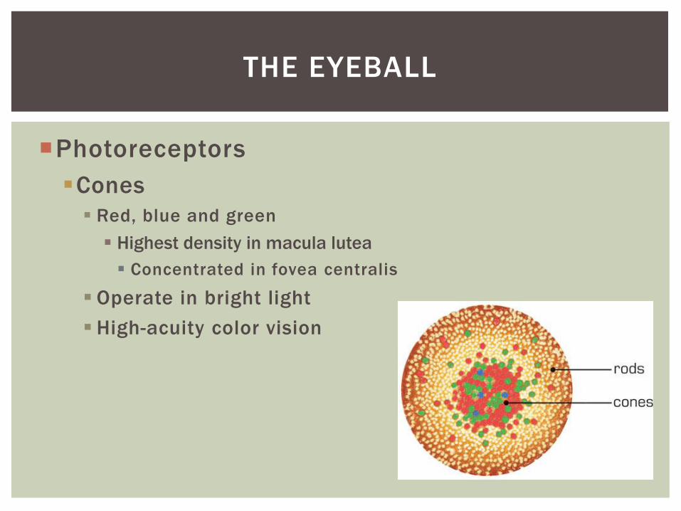
Photoreceptors
Cones
Red, blue and green
Highest density in macula lutea
Concentrated in fovea centralis
Operate in bright light
High-acuity color vision
THE EYEBALL
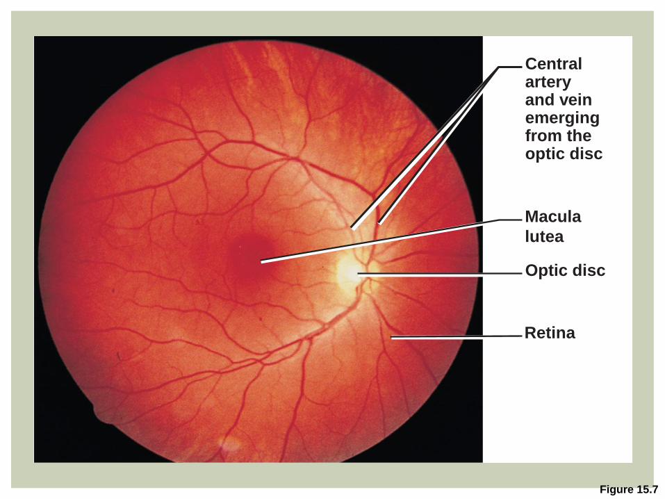
Figure 15.7
Macula
lutea
Central artery and vein emerging from the optic disc
Optic disc
Retina
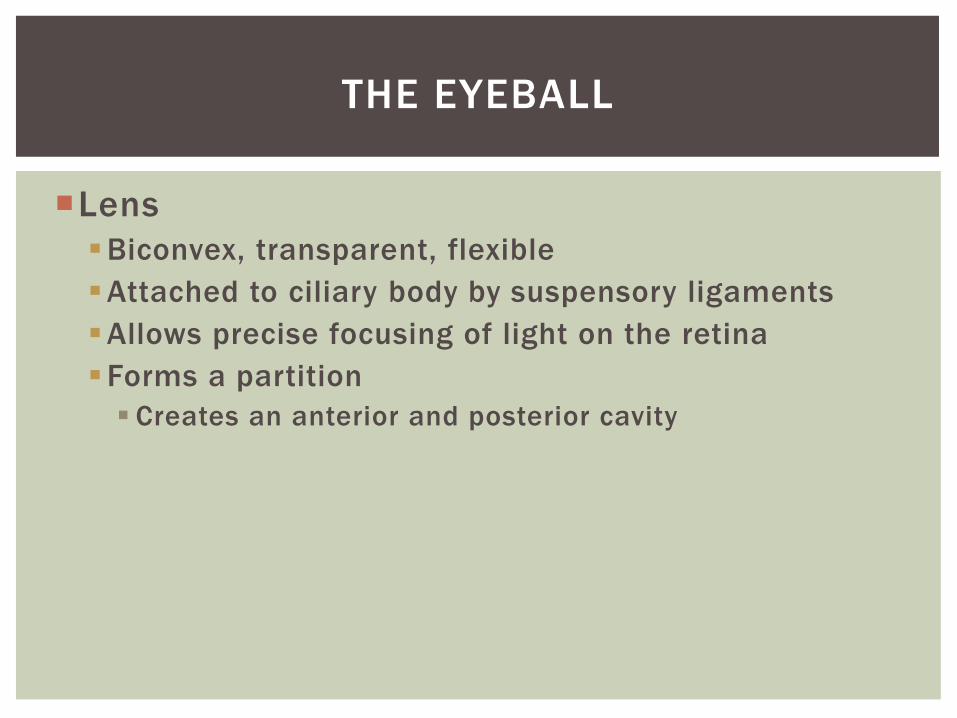
Lens
Biconvex, transparent, flexible
Attached to ciliary body by suspensory ligaments
Allows precise focusing of light on the retina
Forms a partition
Creates an anterior and posterior cavity
THE EYEBALL
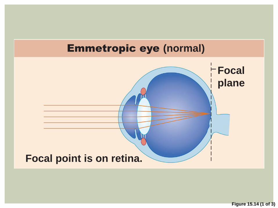
Figure 15.14 (1 of 3)
Focal
plane
Focal point is on retina.
Emmetropic eye (normal)
Figure 15.8
Sclera
Bulbar
conjunctiva
Scleral venous sinus
Posterior chamber
Anterior chamber
Anterior
segment
(contains
aqueous
humor)
Corneal- scleral junction
Cornea
Cornea
Corneal epithelium
Corneal endothelium
Aqueous humor
Iris
Lens
Lens epithelium
Lens
Posterior
segment
(contains vitreous
humor)
Ciliary zonule
(suspensory
ligament)
Ciliary
processes
Ciliary
muscle
Ciliary body
1 Aqueous humor is formed by filtration from the capillaries in the ciliary processes. 2 Aqueous humor flows from the posterior chamber through the pupil into the anterior chamber. Some also flows through the vitreous humor (not shown). 3 Aqueous humor is reabsorbed into the venous blood by the scleral venous sinus.
1
2
3
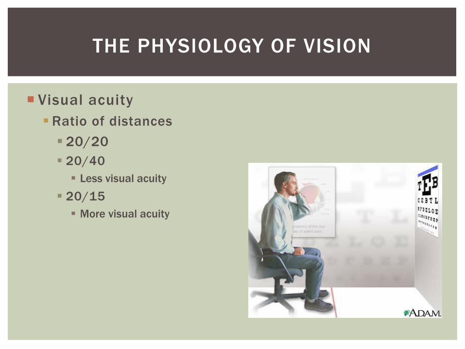
Visual acuity
Ratio of distances
20/20
20/40
Less visual acuity
20/15
More visual acuity
THE PHYSIOLOGY OF VISION
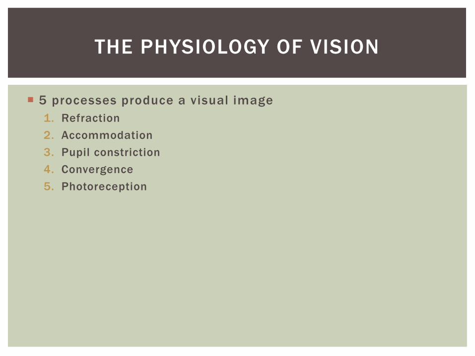
5 processes produce a visual image
1. Refraction
2. Accommodation
3. Pupil constriction
4. Convergence
5. Photoreception
THE PHYSIOLOGY OF VISION
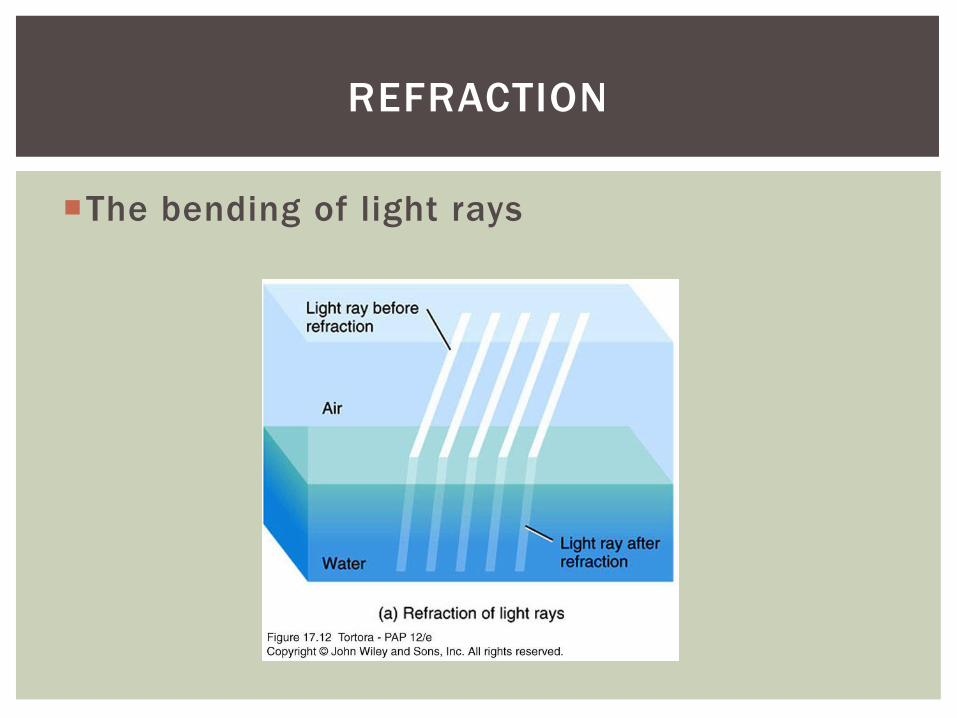
The bending of light rays
REFRACTION

Figure 15.14 (1 of 3)
Focal
plane
Focal point is on retina.
Emmetropic eye (normal)
Focal length is fixed in the eye
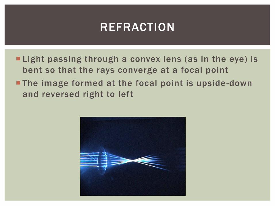
Light passing through a convex lens (as in the eye) is
bent so that the rays converge at a focal point
The image formed at the focal point is upside -down
and reversed right to left
REFRACTION
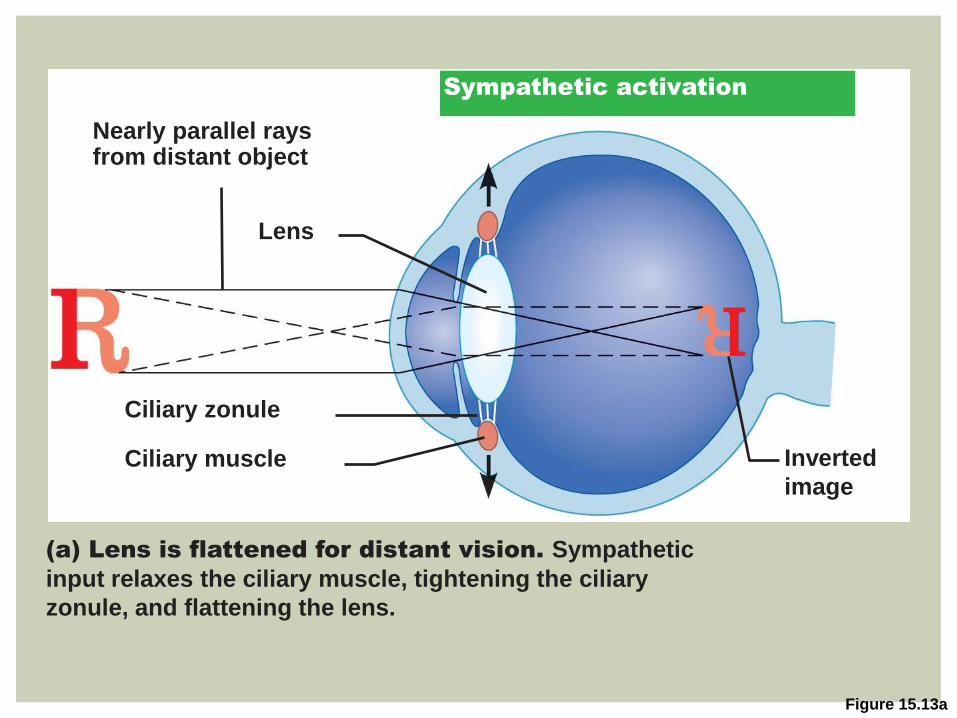
Figure 15.13a
Lens
Inverted
image
Ciliary zonule
Ciliary muscle
Nearly parallel rays from distant object
(a) Lens is flattened for distant vision. Sympathetic
input relaxes the ciliary muscle, tightening the ciliary
zonule, and flattening the lens.
Sympathetic activation

Refracting media
Light is refracted by
Cornea
Aqueous humor
Lens
Vitreous humor
Change in lens curvature
Allows for fine focusing of an image
REFRACTION
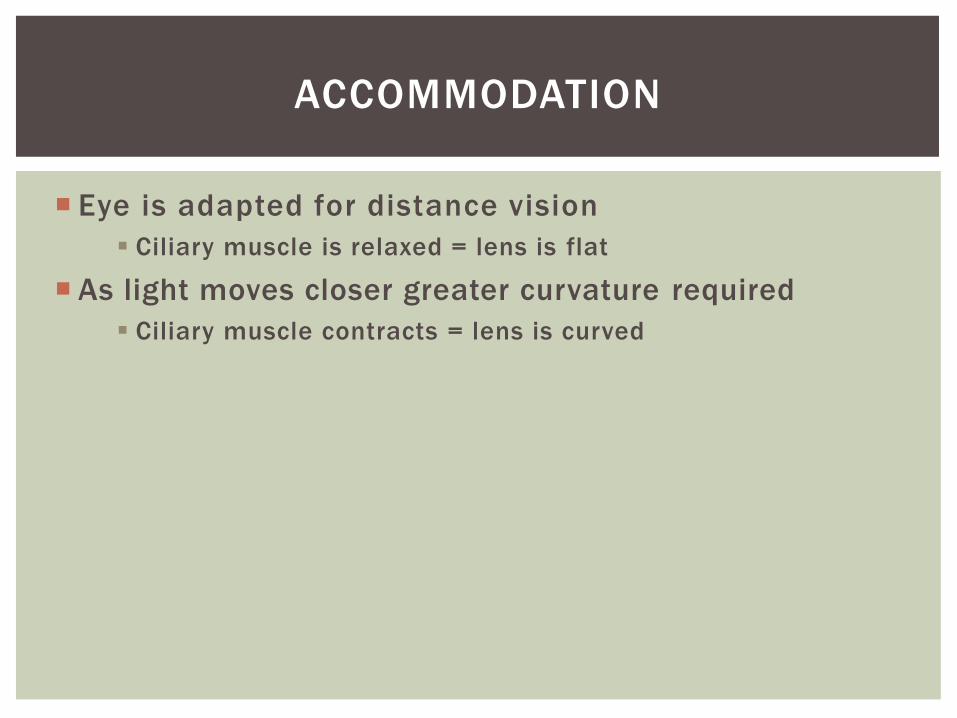
Eye is adapted for distance vision
Ciliary muscle is relaxed = lens is flat
As light moves closer greater curvature required
Ciliary muscle contracts = lens is curved
ACCOMMODATION

Figure 15.13a
Lens
Inverted
image
Ciliary zonule
Ciliary muscle
Nearly parallel rays from distant object
(a) Lens is flattened for distant vision. Sympathetic
input relaxes the ciliary muscle, tightening the ciliary
zonule, and flattening the lens.
Sympathetic activation
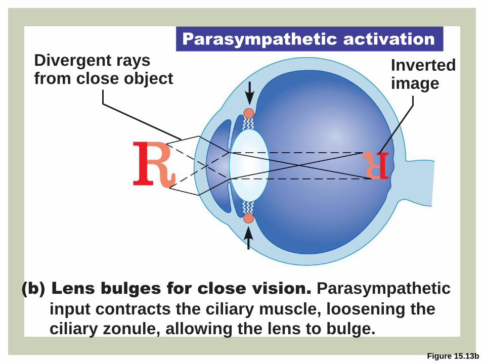
Figure 15.13b
Divergent rays from close object
(b) Lens bulges for close vision. Parasympathetic
input contracts the ciliary muscle, loosening the ciliary zonule, allowing the lens to bulge.
Inverted image
Parasympathetic activation

Near vision
Accommodation
Ciliary muscles contract
Eye strain
Near point
Maximum bulge
Aging
ACCOMMODATION


Close vision requires
Accommodation
Pupil constriction Prevents most divergent light rays from entering the eye
Convergence Medial rotation of the eyeballs toward the object being
viewed (binocular vision)
CLOSE VISION
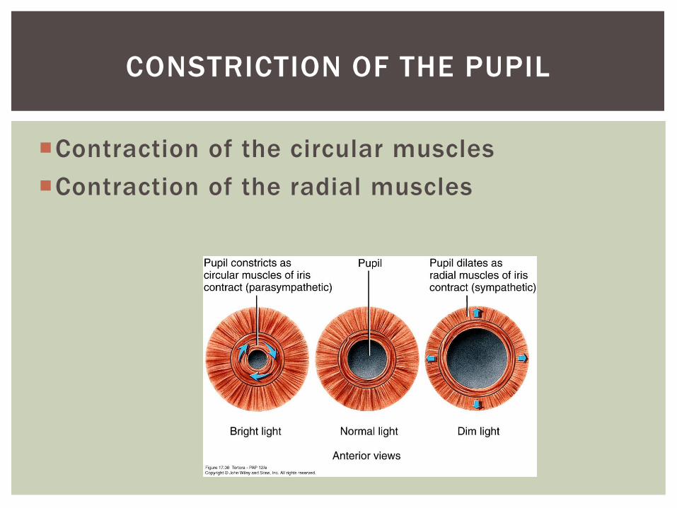
Contraction of the circular muscles
Contraction of the radial muscles
CONSTRICTION OF THE PUPIL

Sensory transduction
Light energy is converted into nerve impulses
Takes place in retina
Photoreceptors
PHOTORECEPTION

Rods and cones
Outer segment of each contains visual pigments
Molecules that change shape as they absorb light
PHOTORECEPTION

Figure 15.15a
Process of
bipolar cell
Outer fiber
Apical microvillus
Discs containing
visual pigments
Melanin
granules
Discs being
phagocytized
Pigment cell nucleus
Inner fibers
Rod cell body
Cone cell body
Synaptic terminals
Rod cell body
Nuclei
Mitochondria
Connecting
cilia
Basal lamina (border
with choroid)
The outer segments
of rods and cones
are embedded in the
pigmented layer of
the retina.
Pig
me
nte
d la
ye
r
Ou
te
r se
gm
en
t
In
ne
r
se
gm
en
t
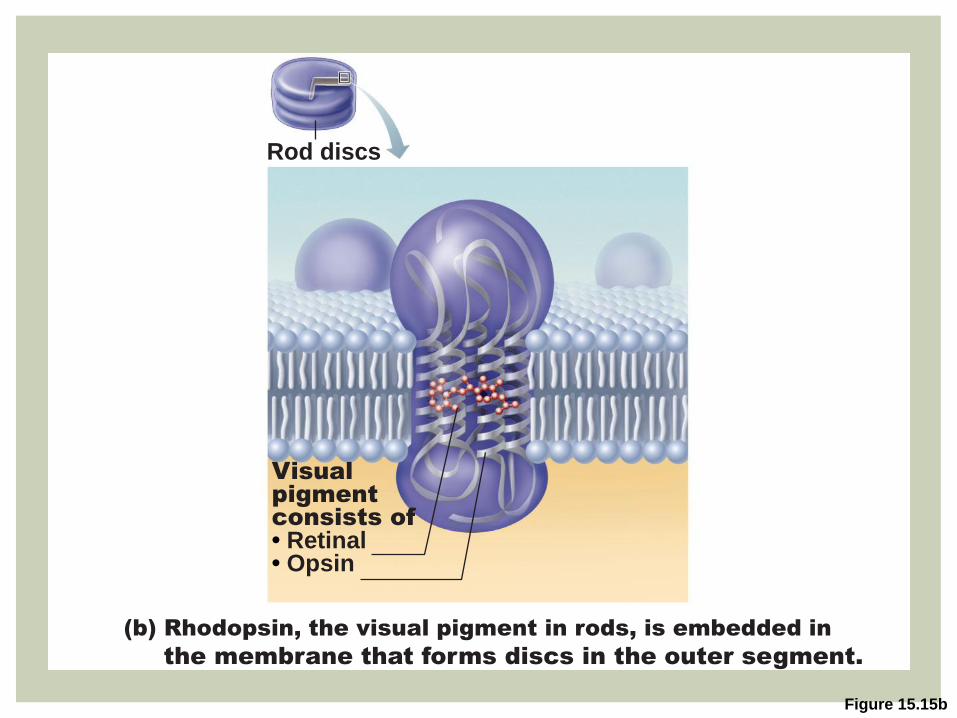
Figure 15.15b
Rod discs
Visual
pigment
consists of
• Retinal • Opsin
(b) Rhodopsin, the visual pigment in rods, is embedded in
the membrane that forms discs in the outer segment.
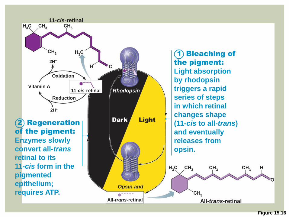
Figure 15.16
11-cis-retinal
Bleaching of
the pigment:
Light absorption
by rhodopsin
triggers a rapid
series of steps
in which retinal
changes shape
(11-cis to all-trans)
and eventually
releases from
opsin.
1
Rhodopsin
Opsin and
Regeneration
of the pigment:
Enzymes slowly
convert all-trans
retinal to its
11-cis form in the
pigmented
epithelium;
requires ATP.
Dark Light
All-trans-retinal
Oxidation
2H+
2H+
Reduction
Vitamin A
2
11-cis-retinal
All-trans-retinal
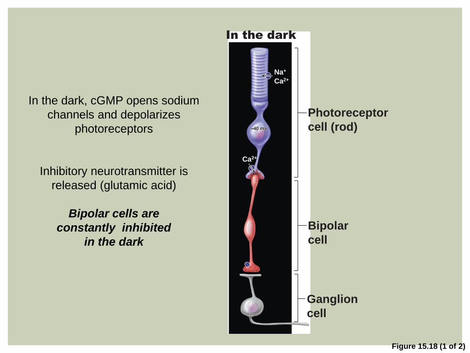
Figure 15.18 (1 of 2)
Na+
Ca2+
Ca2+
Photoreceptor
cell (rod)
Bipolar
cell
Ganglion
cell
In the dark
In the dark, cGMP opens sodium
channels and depolarizes
photoreceptors
Inhibitory neurotransmitter is
released (glutamic acid)
Bipolar cells are
constantly inhibited
in the dark
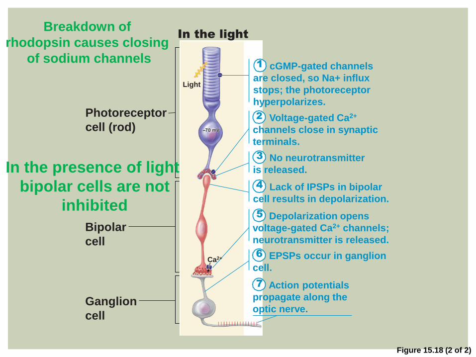
Figure 15.18 (2 of 2)
1 cGMP-gated channels
are closed, so Na+ influx
stops; the photoreceptor
hyperpolarizes.
Voltage-gated Ca2+
channels close in synaptic
terminals.
2
No neurotransmitter
is released.
3
Lack of IPSPs in bipolar
cell results in depolarization.
4
Depolarization opens
voltage-gated Ca2+ channels;
neurotransmitter is released.
5
EPSPs occur in ganglion
cell.
6
Action potentials
propagate along the
optic nerve.
7
Photoreceptor
cell (rod)
Bipolar
cell
Ganglion
cell
Light
Ca2+
In the light
In the presence of light
bipolar cells are not
inhibited
Breakdown of
rhodopsin causes closing
of sodium channels
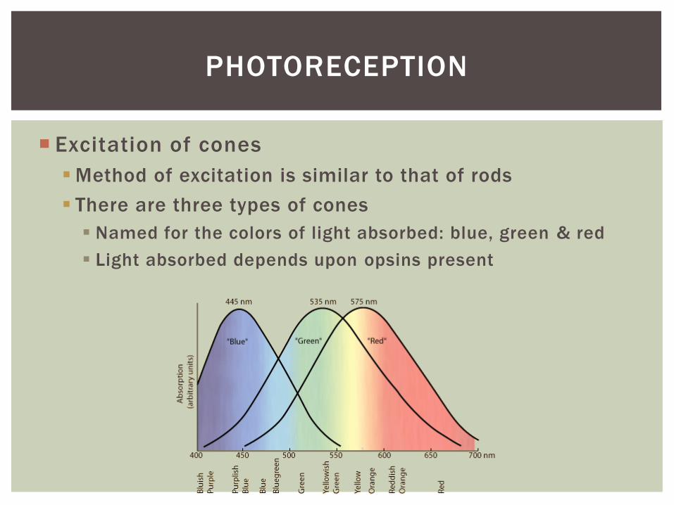
Excitation of cones
Method of excitation is similar to that of rods
There are three types of cones
Named for the colors of light absorbed: blue, green & red
Light absorbed depends upon opsins present
PHOTORECEPTION
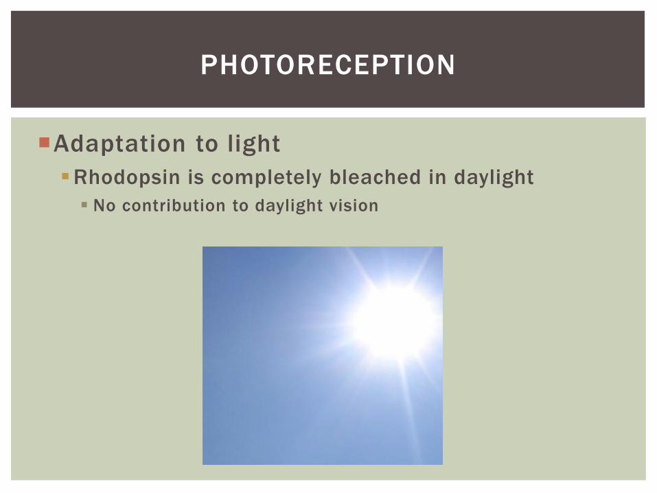
Adaptation to light
Rhodopsin is completely bleached in daylight
No contribution to daylight vision
PHOTORECEPTION
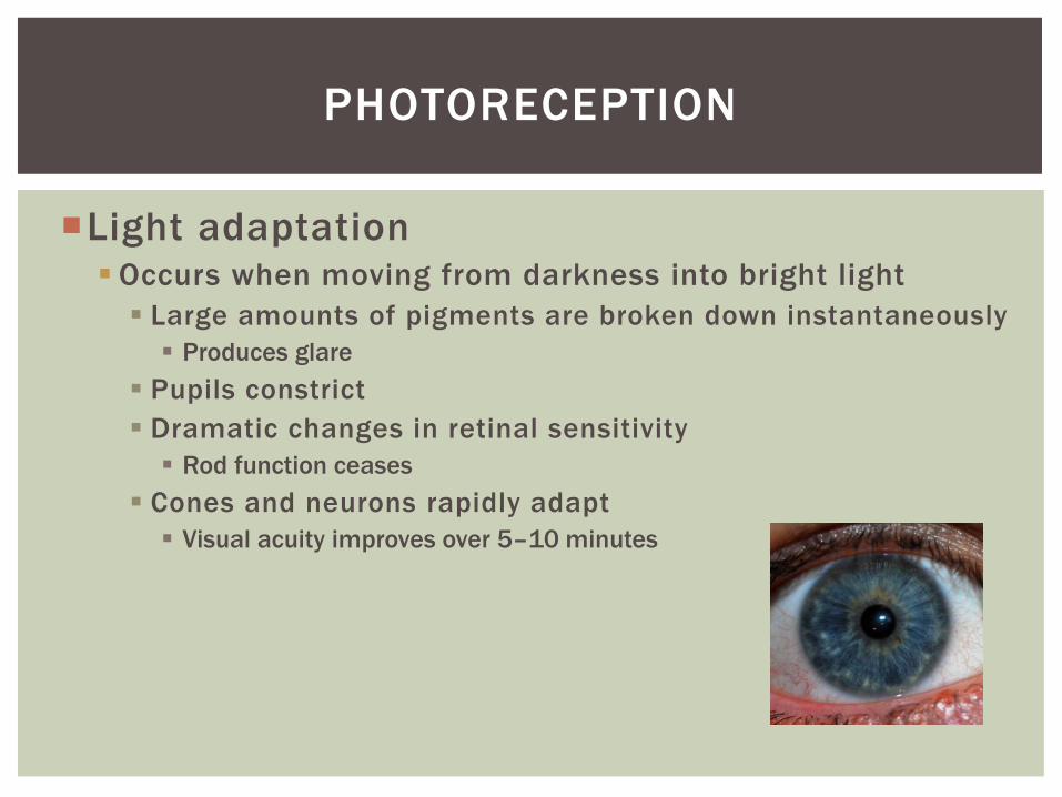
Light adaptation Occurs when moving from darkness into bright light
Large amounts of pigments are broken down instantaneously
Produces glare
Pupils constrict
Dramatic changes in retinal sensitivity
Rod function ceases
Cones and neurons rapidly adapt
Visual acuity improves over 5–10 minutes
PHOTORECEPTION
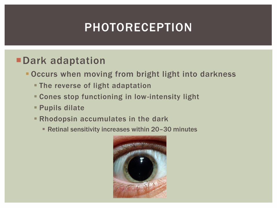
Dark adaptation
Occurs when moving from bright light into darkness
The reverse of light adaptation
Cones stop functioning in low-intensity light
Pupils dilate
Rhodopsin accumulates in the dark
Retinal sensitivity increases within 20–30 minutes
PHOTORECEPTION
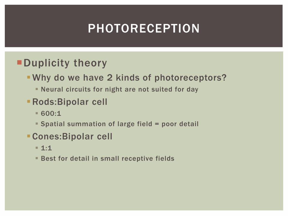
Duplicity theory
Why do we have 2 kinds of photoreceptors?
Neural circuits for night are not suited for day
Rods:Bipolar cell
600:1
Spatial summation of large field = poor detail
Cones:Bipolar cell
1:1
Best for detail in small receptive fields
PHOTORECEPTION
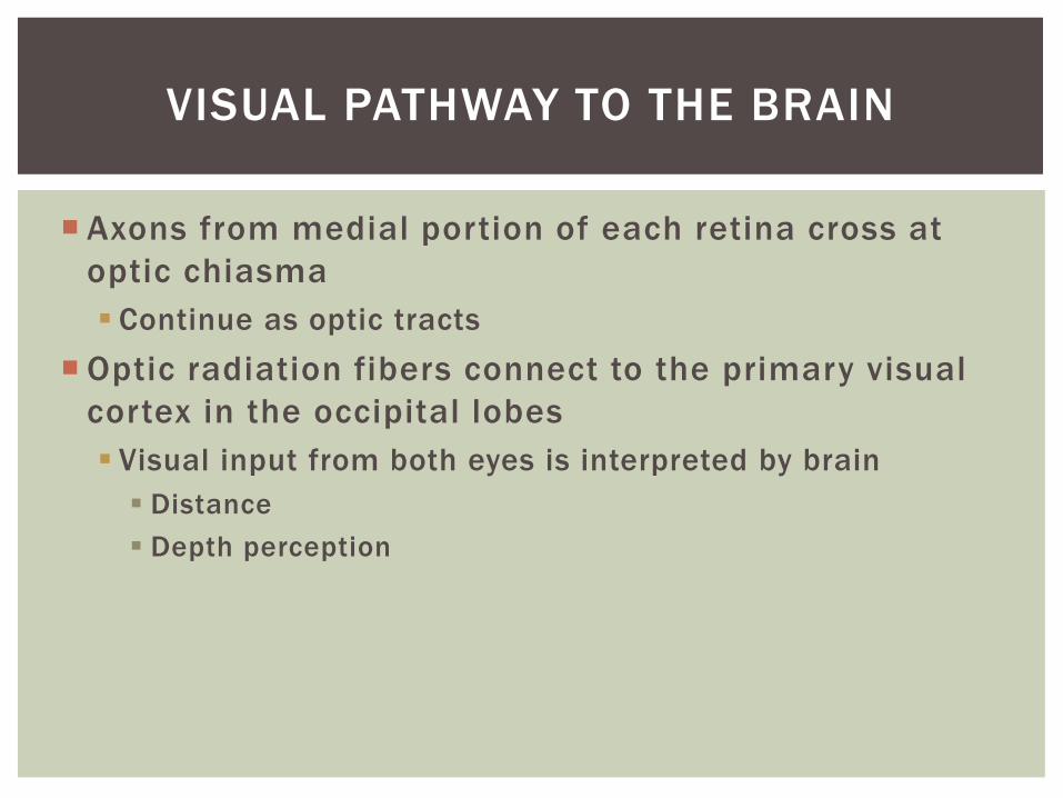
Axons from medial portion of each retina cross at
optic chiasma
Continue as optic tracts
Optic radiation fibers connect to the primary visual
cortex in the occipital lobes
Visual input from both eyes is interpreted by brain
Distance
Depth perception
VISUAL PATHWAY TO THE BRAIN

Figure 15.19a
Pretectal nucleus
Fixation point
Optic radiation
Optic tract
Optic chiasma
Uncrossed (ipsilateral) fiber Crossed (contralateral) fiber
Optic nerve
Lateral geniculate
nucleus of
thalamus
Superior colliculus Occipital lobe (primary visual cortex)
The visual fields of the two eyes overlap considerably.
Note that fibers from the lateral portion of each retinal field do not cross at the optic chiasma.
Suprachiasmatic
nucleus
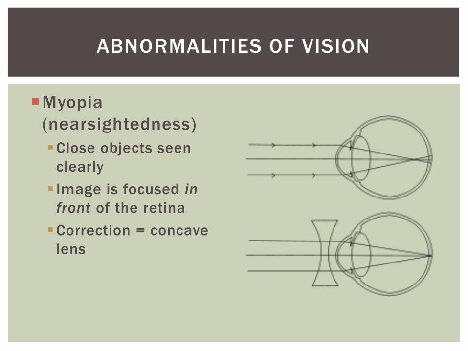
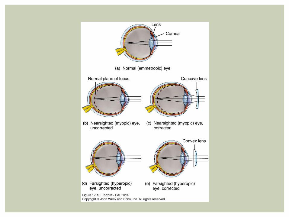
Myopia
(nearsightedness)
Close objects seen
clearly
Image is focused in
front of the retina
Correction = concave
lens
ABNORMALITIES OF VISION
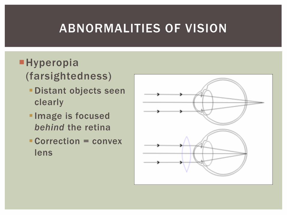
Hyperopia
(farsightedness)
Distant objects seen
clearly
Image is focused
behind the retina
Correction = convex
lens
ABNORMALITIES OF VISION
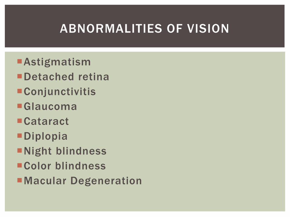
Astigmatism
Detached retina
Conjunctivitis
Glaucoma
Cataract
Diplopia
Night blindness
Color blindness
Macular Degeneration
ABNORMALITIES OF VISION

ABNORMALITIES OF VISION
Astigmatism

Detached retina
ABNORMALITIES OF VISION
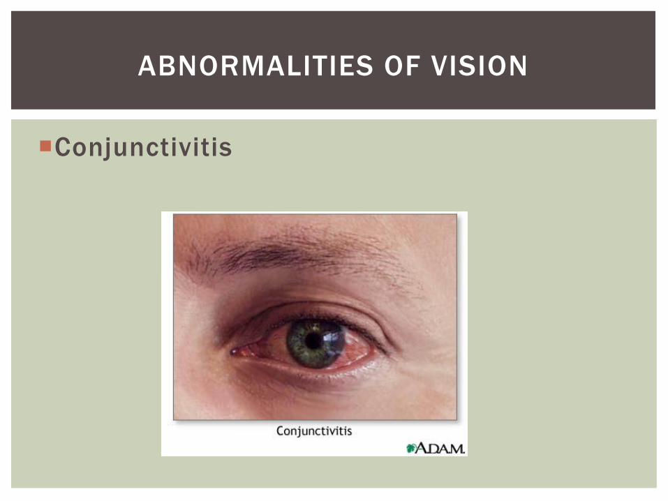
Conjunctivitis
ABNORMALITIES OF VISION
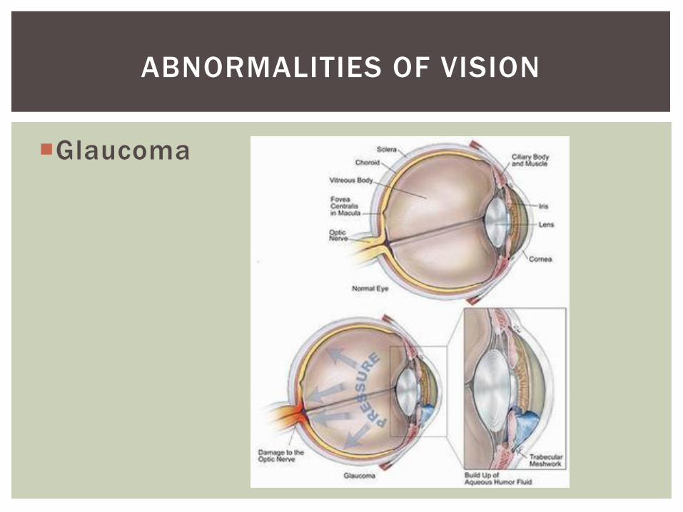
Glaucoma
ABNORMALITIES OF VISION
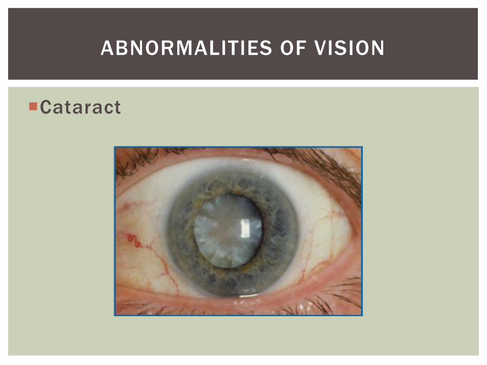
Cataract
ABNORMALITIES OF VISION

Diplopia
ABNORMALITIES OF VISION
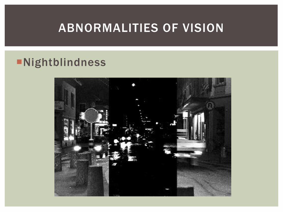
Nightblindness
ABNORMALITIES OF VISION
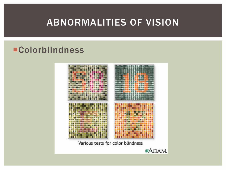
Colorblindness
ABNORMALITIES OF VISION
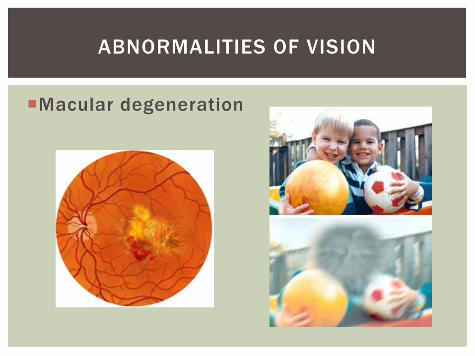
Macular degeneration
ABNORMALITIES OF VISION

QUESTIONS?
Activity
Special Senses 20, #11-25