activation of the unfolded protein response via inhibition ... · 17-04-2019 · inhibition in...
TRANSCRIPT

1
Activation of the unfolded protein response via inhibition of protein disulfide
isomerase decreases the capacity for DNA repair to sensitize glioblastoma to
radiotherapy
Yajing Liu1, Wenbin Ji1, Andrea Shergalis2, Jiaqi Xu1,3, Amy M. Delaney1, Andrew
Calcaterra1, Anupama Pal1, Mats Ljungman1,4, Nouri Neamati2, Alnawaz Rehemtulla1
1. Department of Radiation Oncology, University of Michigan Medical School and
Rogel Cancer Center, Ann Arbor, MI, USA.
2. Department of Medicinal Chemistry, College of Pharmacy, and Rogel Cancer
Center, University of Michigan, Ann Arbor, MI, USA.
3. Weill Cornell Graduate School of Medical Sciences, New York, NY, USA.
4. Department of Environmental Health Sciences, School of Public Health,
University of Michigan, Ann Arbor, MI, USA.
Corresponding Author:
Address correspondence to Alnawaz Rehemtulla, Department of Radiation Oncology,
University of Michigan Medical School and Rogel Cancer Center, 2800 Plymouth Road,
NCRC, Building 520-1342, Ann Arbor, MI, USA. Telephone: 734-764-4209; Email:
Running title: PDI inhibition impairs DNA repair capacity in GBM
Key words: PDI, GBM, RAD51, UPR, radiosensitization
Financial Support: A. Rehemtulla and N. Neamati received NIH CA193690 grant. This
work was supported in part by the National Institute of Health Cancer Center Support
Grant to the Rogel Cancer Center at the University of Michigan (P30 CA046592-29).
Conflict of Interest: The authors declare no potential conflicts of interest.
Manuscript Information:
Word count: Abstract = 232; Main text = 4151 (Introduction, Materials and Methods,
Results, Discussion).
Abbreviation:
GBM: glioblastoma multiforme P4HB: prolyl 4-hydroxylase subunit beta TMZ: temozolomide MGMT: O6-methylguanine–DNA methyltransferase EGFR: epidermal growth factor receptor
on March 16, 2021. © 2019 American Association for Cancer Research.cancerres.aacrjournals.org Downloaded from
Author manuscripts have been peer reviewed and accepted for publication but have not yet been edited. Author Manuscript Published OnlineFirst on April 17, 2019; DOI: 10.1158/0008-5472.CAN-18-2540

2
NF1: neurofibromin 1 PDFDRA: platelet derived growth factor receptor alpha IDH1: isocitrate dehydrogenase (NADP(+)) 1 ER: endoplasmic reticulum PDI: protein disulfide isomerase PACMAs: propynoic acid carbamoyl methyl amides KD: knock down GSEA: Gene set enrichment analysis DSB: double strand breaks MGMT: O-6-methylguanine–DNA methyltransferase UPR: unfolded protein response ERAD: ER-associated degradation CHX: cycloheximide
on March 16, 2021. © 2019 American Association for Cancer Research.cancerres.aacrjournals.org Downloaded from
Author manuscripts have been peer reviewed and accepted for publication but have not yet been edited. Author Manuscript Published OnlineFirst on April 17, 2019; DOI: 10.1158/0008-5472.CAN-18-2540
3
Abstract:
Patients with glioblastoma multiforme (GBM) survive on average 12-14 months after
diagnosis despite surgical resection followed by radiation and temozolomide therapy.
Intrinsic or acquired resistance to chemo- and radiation-therapy is common and
contributes to a high rate of recurrence. To investigate the therapeutic potential of
protein disulfide isomerase (PDI) as a target to overcome resistance to chemoradiation,
we developed a GBM tumor model wherein conditional genetic ablation of prolyl 4-
hydroxylase subunit beta (P4HB), the gene that encodes PDI, can be accomplished.
Loss of PDI expression induced the unfolded protein response (UPR) and decreased
cell survival in two independent GBM models. Nascent RNA Bru-seq analysis of PDI-
depleted cells revealed a decrease in transcription of genes involved in DNA repair and
cell cycle regulation. Activation of the UPR also led to a robust decrease in RAD51
protein expression as a result of its ubiquitination-mediated proteosomal degradation.
Clonogenic survival assays demonstrated enhanced killing of GBM cells in response to
a combination of PDI knockdown and ionizing radiation (IR) compared to either modality
alone, which correlated with a decreased capacity to repair IR-induced DNA damage.
Synergistic tumor control was also observed with the combination of PDI inhibition and
IR in a mouse xenograft model compared to either single agent alone. These findings
provide a strong rationale for the development of PDI inhibitors and their use in
combination with DNA damage-inducing, standard-of-care therapies such as IR.
on March 16, 2021. © 2019 American Association for Cancer Research.cancerres.aacrjournals.org Downloaded from
Author manuscripts have been peer reviewed and accepted for publication but have not yet been edited. Author Manuscript Published OnlineFirst on April 17, 2019; DOI: 10.1158/0008-5472.CAN-18-2540
4
Significance: Findings identify PDIA1 as a therapeutic target in GBM by demonstrating efficacy of its
inhibition in combination with radiation therapy through a novel mechanism involving
downregulation of DNA repair genes.
Introduction:
Glioblastoma multiforme (GBM) is the most common and lethal primary malignant brain
tumor (1). The standard of care treatment for GBM involves maximal tumor resection
followed by adjuvant temozolomide (TMZ) combined with radiation therapy. This
combination demonstrates a modest improvement in median survival to 15 months
compared to 12 months with radiation alone (2,3). Tumor recurrence occurs in most
GBM patients, resulting in an average 5-year survival rate of only 5% (4). Genetic
profiling has identified recurrent copy number alterations and/or mutations in epidermal
growth factor receptor (EGFR), neurofibromin 1 (NF1), platelet derived growth factor
receptor alpha (PDGFRA)/ isocitrate dehydrogenase (NADP(+)) 1 (IDH1), TP53,
phosphoinositide 3-kinase (PI3K) complex and cyclin dependent kinase inhibitor 2A
(CDKN2A) (3,5-7). Although the mechanistic basis for the contribution of these
mutations to oncogenesis is becoming better understood, therapeutic approaches to
target several of these pathways have limited benefit in clinical trials (3,8-13). The fact
that GBM tumors show intratumoral heterogeneity may at least partially contribute to the
dismal outcomes of therapeutic targeting of commonly dysregulated oncogenic
pathways. Therefore, there is an urgent need to develop therapies that target pathways
required for survival of glioblastoma, irrespective of oncogenic mutation status.
Due to the high rate of protein synthesis in cancer cells, an enhanced capacity for
protein folding is required which puts major demand on the protein folding machinery in
the endoplasmic reticulum (ER). To accommodate such demand, upregulation of
proteins such as PDI is often observed (3). The PDI family consists of 21 enzymes that
catalyze disulfide bond formation, reduction and isomerization to ensure proper folding
of nascent polypeptides (14), and also act as chaperones to assist protein folding (15).
on March 16, 2021. © 2019 American Association for Cancer Research.cancerres.aacrjournals.org Downloaded from
Author manuscripts have been peer reviewed and accepted for publication but have not yet been edited. Author Manuscript Published OnlineFirst on April 17, 2019; DOI: 10.1158/0008-5472.CAN-18-2540
5
Several, but not all, of the PDI family members are primarily localized to the ER, the
central compartment for protein folding and degradation, to maintain physiological
homeostasis (16). In response to ER stress, an imbalance between the unfolded protein
load and the protein folding machinery in the ER, initiates a collection of signaling
cascades termed the unfolded protein response to restore a productive ER protein-
folding environment by enhancing the capacity for protein folding and transport (17,18).
Expression of PDIA1, the canonical member of the PDI family, is upregulated in brain
and central nervous system cancers compared to matched normal tissues (19). In
addition, proteomic analysis has revealed up-regulation of the PDI in many cancers (20-
22). Furthermore, serial in vivo transplantation of primary glioma reveals PDI
overexpression in invasive low-generation tumors (23). We hypothesized that the
dependence of tumor cells on PDI activity provides a rationale for its therapeutic
targeting in GBM, irrespective of oncogenic mutation status. We recently described
propynoic acid carbamoyl methyl amides (PACMAs) as irreversible inhibitors of PDI, a
first-in-class, safe and efficacious targeted anticancer agents (24), as well as second
generation PDI inhibitors BAP2 (25). Since the prolyl 4-hydroxylase subunit beta
(P4HB) gene family that encodes PDI proteins, comprises 21 genes, varying in size,
expression, localization, and enzymatic function (26), of which PDIA1 is believed to be
the most highly expressed in GBM (19) and the primary target of our current small
molecule agents, it is essential that we demonstrate the selective dependence of GBM
cells on PDI to validate it as a therapeutic target.
Materials and Methods:
Cell lines and treatment
U87 (purchased from ATCC) and D54 (27,28) cells were maintained in DMEM (VWR,
Corning®) and RPMI (Life Technologies, Gibco®) respectively, supplemented with 10%
(v/v) fetal bovine serum (GE Healthcare, Hyclone®). The cells were authenticated using
Short Tandem Repeat profiling (tested on 12/3/18), and routinely tested for mycoplasma
contamination (latest test on 11/26/18) with MycoAlert™ mycoplasma detection kit
(Lonza). All cells used in the experiments were thawn within 10 passages and were
maintained in vitro no more than 2 months.
on March 16, 2021. © 2019 American Association for Cancer Research.cancerres.aacrjournals.org Downloaded from
Author manuscripts have been peer reviewed and accepted for publication but have not yet been edited. Author Manuscript Published OnlineFirst on April 17, 2019; DOI: 10.1158/0008-5472.CAN-18-2540

6
For tunicamycin and MG132 (both from Sigma-Aldrich) treatment, cells were seeded
overnight, and tunicamycin (5 μg/mL) was added with or without MG132 (10 μM) for 16
hours. For DNA repair studies, D54MG and U87MG cells were plated at 100,000
cells/well onto 0.1% Poly-L-lysine (Sigma) coated 4-well Millicell® EZ slides (Millipore
Sigma) overnight followed by 3 days of PDIA1 shRNA induction. Subsequently, 2 Gy IR
was delivered using an IC-320 orthovoltage irradiator (Kimtron Medical) and the cells
were fixed at 0.5, 4, 8, 16, and 24 hours post irradiation. Cycloheximide (Calbiochem)
was used at 100 μg/mL for 0, 2, 4, 6 and 8 hours for the time-course assay to study
protein degradation.
LentiCRISPR sgP4HB cloning and production
Two sgRNAs for P4HB: 5’-CACCGCCGCGCACGCCGTACTGCT-3’ and 5’-
CACCGAAGCAACTTCGCGGAGGCGC-3’ identified computationally
(https://zlab.bio/guide-design-resources), were inserted into the lentiCRISPR v2 plasmid
(Addgene). Briefly, vector and annealed oligos were digested by BsmBI (NEB) and
ligated using the quick ligation kit (NEB), and transformed into Stbl3 bacteria
(Invitrogen). Single colonies were expanded and sequenced to confirm sgRNA
insertion, and identified recombinant clones were packaged in HEK293T cells with
psPAX2 and pCMV-VSV-G as previously described (Addgene, (29,30).
Lentivirus infection
The inducible pTripz-PDIA1 shRNA vectors were obtained from Dharmacon Open
Biosystems. Lentivirus was produced using HEK293T cells at the University of Michigan
Vector Core Facility. U87 and D54 cells were infected in the presence of polybrene (8
μg/mL, AmericanBio), and stable cells were selected by single colony isolation in the
presence of puromycin (1 μg/mL, InvivoGen).
Bru-Seq
Nascent RNA Bru-seq was performed as previously described (31). GSEA analyses
based on a transcriptional cut-off of >0.1 RPKM and >100 counts per gene.
Clonogenic assay
Doxycycline (2 μg/mL, Sigma-Aldrich) was added to stable D54 and U87 cells to induce
PDIA1 shRNA for 3 days. Cells were irradiated with 0, 2, 4, 6, or 8 Gy as a single dose
and plated the following day at a clonogenic density with fresh medium (without
on March 16, 2021. © 2019 American Association for Cancer Research.cancerres.aacrjournals.org Downloaded from
Author manuscripts have been peer reviewed and accepted for publication but have not yet been edited. Author Manuscript Published OnlineFirst on April 17, 2019; DOI: 10.1158/0008-5472.CAN-18-2540
7
doxycycline) for 7-14 days before the colonies were fixed with 4% paraformaldehyde
(Electron Microscopy Science) and stained using 0.1% Crystal Violet (Sigma-Aldrich).
Quantitative polymerase chain reaction
PDI shRNA was induced for 1-3 days in D54 and U87 stable cells and total RNA was
extracted using the Qiagen RNeasy kit (Qiagen). 1 μg of total RNA was reverse-
transcribed into cDNA using the High-Capacity cDNA Reverse Transcription Kit (Applied
Biosystems). Quantitative PCR was performed using SYBR Green Master Mix (Bio-
Rad) on a Mastercycler® RealPlex2 (Eppendorf) with denaturation at 95ºC for 15 mins
followed by 40 cycles of amplification at 94ºC for 15 seconds; 55 ºC for 30 seconds; 72
ºC for 30 seconds. Sequences of the primers are listed in Supplementary Table 1.
Relative expression levels were normalized to GAPDH and fold changes in mRNA
expression level were evaluated using the ΔΔCt method.
Western blot
Cells or snap-frozen tissues were lysed in RIPA buffer consisting of 150 mM NaCl, 1
mM EDTA (pH=8.0), 50 mM Tris-HCl (pH=8.0), 1% NP-40 (v/v), 0.25% sodium
deoxycholate (w/v), 0.1% SDS (w/v) (all from Sigma-Aldrich), supplemented with
PhosSTOP™ Phosphatase inhibitor cocktail and Complete™ protease inhibitor cocktail
(Roche), and sonicated with a Sonic Dismembrator model 100 (Fisher Scientific).
Western blotting was performed as described previously (32), using primary antibodies
to PDI, BIP, elF2α, p-elF2α (S51), RAD51, phospho-Histone H2AX (Ser139) (all from
Cell Signaling), and β-actin (Sigma). Horseradish peroxidase-conjugated goat anti-
rabbit IgG (H+L) and goat anti-mouse (H+L) (Jackson ImmunoResearch) were applied
as secondary antibodies and Pierce™ ECL (Thermal Scientific) or ECLTM prime (GE
Healthcare) were used as substrate (detailed antibody working concentrations are listed
in Supplementary Table 2).
Immunofluorescent staining
Cells were fixed in 4% paraformaldehyde (Electron Microscopy Science) for 15 minutes
at room temperature and washed with 1X PBS before permeabilization in 0.3% Triton X-
100 (Sigma-Aldrich) for 10 minutes at room temperature. The slides were blocked in
10% goat serum (Reagent A from Invitrogen Histostain-Plus kit) for 30 minutes and
γH2AX-AF488 (Millipore) was applied at 1:100 dilution in 1% blocking solution
on March 16, 2021. © 2019 American Association for Cancer Research.cancerres.aacrjournals.org Downloaded from
Author manuscripts have been peer reviewed and accepted for publication but have not yet been edited. Author Manuscript Published OnlineFirst on April 17, 2019; DOI: 10.1158/0008-5472.CAN-18-2540

8
overnight. ProLong Gold with DAPI (Invitrogen) were used to prepare the slides for
analysis using an Olympus BX-51 scope. γ-H2AX signal was quantified using ImageJ
software. More than 100 cells were analyzed per experiment per condition.
Xenograft mouse model of glioblastoma
A total of 1 × 10⁶ U87-pTripz-PDIA1 shRNA stable cells in 100 μL DMEM (VWR,
Corning®): Matrigel (BD Bioscience) (1:1) suspension were subcutaneously injected
into 6-8 weeks old NCRNU sp/sp mice (Taconic). Tumor size was monitored twice
weekly and tumor volume was defined as (L x W x W)/2, where W is tumor width and L
is tumor length. Mice were randomized into 4 groups when tumors reached around 100
mm3 (5-8 animals/group), and two groups were switched to doxycycline (2 mg/mL) in
5% sucrose (both from Sigma-Aldrich) water (renewed thrice per week). Radiation was
given at 2 Gy per day, 5 days a week for 2 weeks only to tumors using a IC-320
orthovoltage irradiator (Kimtron Medical). The rest of the body was protected by lead
shielding. All animal experiments were approved by the University of Michigan
Committee on the Use and Care of Animals.
Statistical analysis
ImageJ was used for protein quantification and all proteins were normalized against
loading control. The statistical significance between two groups was evaluated based on
two-tailed Student’s t-Test using GraphPad Prism (Version 7). P values < 0.05 were
considered statistically significant.
Results
PDI knockdown leads to ER stress in GBM cells
To elucidate the cellular response to PDI knockdown (KD), we generated stable GBM
cell lines using D54 and U87 wherein doxycycline-inducible PDI shRNA expression can
be achieved. Two shRNAs targeting distinct sequences were used for PDI knockdown.
In D54 cells, downregulation of PDI with shRNA2 was evident as early as 12 hours after
doxycycline treatment (1.2-fold decrease compared to control, p=0.0013), with a 5.9-fold
decrease at 48 hours post-induction (Fig. 1A, B). In U87 cells, the decrease in PDI
protein levels started at 24 hours (1.1-fold decrease compared to control) but only
became significant after 48 hours (1.7-fold deduction compared to control, p=0.0047)
on March 16, 2021. © 2019 American Association for Cancer Research.cancerres.aacrjournals.org Downloaded from
Author manuscripts have been peer reviewed and accepted for publication but have not yet been edited. Author Manuscript Published OnlineFirst on April 17, 2019; DOI: 10.1158/0008-5472.CAN-18-2540

9
(Fig. 1C, D). We next explored whether downregulation of PDI leads to ER stress.
Increased levels of BiP, a well characterized maker for ER stress (33), was observed as
early as 12 hours after doxycycline treatment (1.4-fold increase compared to control,
p=0.0105) in D54 cells (Fig.1A, C) and remained elevated at 24 hours (1.7-fold
increase, p=0.0048) post shRNA2 induction. However, BiP returned to basal levels
around 48 hours post doxycycline treatment, despite a further decline in PDI protein
expression. In U87 cells (Fig. 1C, D), upregulation of BiP was detected 24 hours after
doxycycline addition (1.3-fold increase, p=0.0023), and expression was maintained up
to 72 hours (1.8-fold increase, p=0.0240).
Upon ER stress, EIF2α is phosphorylated (p-EIF2α) at serine 51, leading to inhibition of
protein translation and preventing further entry of nascent polypeptides into the ER (34).
p-EIF2α levels were elevated 12 hours after initiation of PDI knockdown in D54 cells
(1.7-fold increase compared to control, p=0.0026) and remained elevated at 24 hours
(2.2-fold increase, p=7 × 10⁻⁴) but returned to basal levels after 48 hours (Fig. 1A, B). In
U87MG cells, p-EIF2α levels were significantly upregulated at 48 hours (2.0-fold
increase, p=0.0090) and remained elevated at 72 hours (1.8-fold increase, p=0.0380)
(Fig. 1C, D), a pattern that correlated to changes observed for BiP and the dynamics of
PDI downregulation in both cell lines.
PDI knockdown leads to decreased transcription of DNA repair genes
In response to unfolded proteins, dissociation of BiP from the ER membrane-spanning
UPR receptor proteins, PERK, IRE1, and ATF6, results in transcriptional, translational
and post-translational changes in gene expression to restore cellular homeostasis (35).
To investigate immediate changes in gene transcription in response to PDI knockdown
and resulting accumulation of unfolded proteins, we conducted genome-wide nascent
RNA Bru-seq analysis (31). We found that knockdown of PDI expression for 72h
resulted in transcriptional upregulation of 153 and 58 genes by greater than 2-fold, while
135 and 41 genes were downregulated greater than 2-fold in D54 and U87 cells,
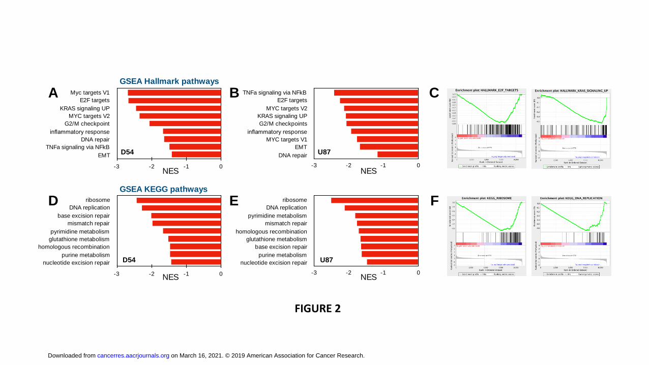
respectively. Gene set enrichment analysis (GSEA) revealed that among the Hallmark
pathways, MYC and E2F1 targets were suppressed as was KRAS signaling, G2/M
checkpoint, inflammatory response and DNA repair in both cell lines (Fig. 2A-C).
on March 16, 2021. © 2019 American Association for Cancer Research.cancerres.aacrjournals.org Downloaded from
Author manuscripts have been peer reviewed and accepted for publication but have not yet been edited. Author Manuscript Published OnlineFirst on April 17, 2019; DOI: 10.1158/0008-5472.CAN-18-2540
10
Furthermore, GSEA analysis of KEGG pathways showed a marked suppression of
ribosome and DNA replication as well as of four major DNA repair pathways and
purine/pyrimidine and glutathione metabolism (Fig. 2D-F). RAD51, which plays a central
role in homologous recombination repair (HR), showed a modest but significant
reduction in transcription (Supplementary Figure 1). To further investigate the effect of
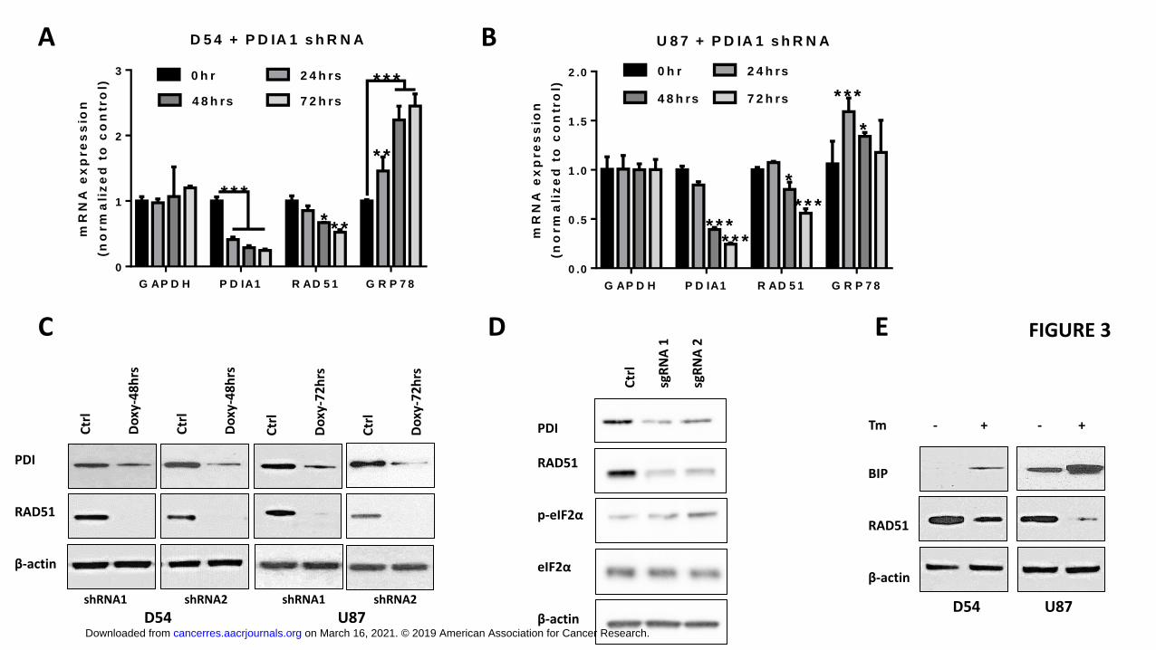
PDI knockdown on RAD51 gene expression, total RNA was extracted 24, 48 and 72
hours after doxycycline treatment and analyzed for steady-state PDIA1 and RAD51
mRNA levels by qRT-PCR. Maximum KD of PDIA1 mRNA was achieved in D54 and
U87 cells after 72 hours of shRNA2 induction with a 5.6-fold and 4-fold downregulation
of PDIA1 mRNA (p<0.0001), respectively. Downregulation of RAD51 steady-state RNA
expression was detected at 48 hours after PDI KD and reached approximately a 2.0-fold
decrease compared to control at 72 hours (p<0.0001). In addition, GRP78, the gene
encoding BiP, showed significantly upregulated RNA expression (p<0.01 and p<0.001
in D54 and U87 cells respectively) (Fig. 3A-B), indicating induction of ER stress in
response to PDI KD. While Bru-seq and qRT-PCR analysis revealed a modest
reduction of transcription and steady-state RNA levels, Western blot analysis revealed a
robust downregulation of RAD51 protein levels (10-fold) after 48 hours and 72 hours in
both D54 and U87 cells (Fig. 3C). To independently confirm the observation that PDI
knockdown leads to ER stress and downregulation of RAD51, we performed
CRISPR/Cas9, where sg RNA targeting P4HB exon was inserted into a lentiCRISPR
vector (36). Analysis of cells after a short term enrichment of infected cells using
puromycin revealed that CRISPR/Cas9 mediated deletion of the PDI locus led to a
simultaneous increase in ER stress as detected by an up-regulation of phosphorylated
eIF2α and a concomitant decrease in Rad51 levels (Figure 3D). To investigate whether
the downregulation of RAD51 occurred specifically as a consequence of PDI KD or as a
general response of the UPR, GBM cells were treated with tunicamycin, a potent
inhibitor of N-linked glycosylation and inducer of the UPR. Tunicamycin treatment
resulted in BiP upregulation and a concomitant decrease in RAD51 protein levels after
12 hours (Fig. 3E). Thus, mRNA levels of RAD51, a key mediator of homologous
recombination mediated DNA repair, were reduced in response to accumulation of
unfolded proteins induced by either PDI KD or tunicamycin.
on March 16, 2021. © 2019 American Association for Cancer Research.cancerres.aacrjournals.org Downloaded from
Author manuscripts have been peer reviewed and accepted for publication but have not yet been edited. Author Manuscript Published OnlineFirst on April 17, 2019; DOI: 10.1158/0008-5472.CAN-18-2540
11
RAD51 is targeted by ubiquitin-mediated proteasomal degradation following ER
stress
Upon ER stress, the UPR triggers three major cellular responses: inhibition of
transcription and protein translation, upregulation of protein-folding capacity to maintain
ER homeostasis and ER-associated protein degradation to eliminate misfolded proteins
(ERAD) (18). Since the robust (10-fold) decrease in RAD51 protein expression (Figure
3C) was not in agreement with results from analysis of RAD51 transcript levels using
qPCR and Bru-Seq analysis, we investigated whether PDI KD causes ERAD-mediated
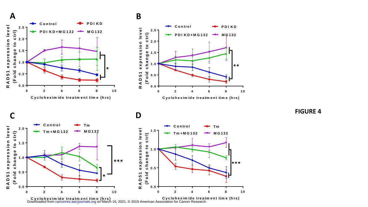
degradation of RAD51. Inhibition of protein synthesis using cycloheximide (CHX)
followed by evaluation of RAD51 protein levels would reveal protein decay rates in
control and PDI KD cells. As shown in Fig. 4A-B, PDI KD resulted in a RAD51 protein
half-life of approximately 2 hours and 6.5 hours in U87 and D54 cells, respectively,
compared to 4 hours and 8 hours in control cells. Addition of the proteasome inhibitor
MG132 rescued RAD51 protein levels in both control and PDI KD cells suggesting that
RAD51 is subjected to proteasome-mediated degradation (Supplementary Fig. 2). To
assess whether the increased rate of RAD51 decay in the PDI KD cells is due to the
activation of ER stress-associated degradation, we induced ER stress by treating
parental GBM cells with tunicamycin and similarly observed increased protein decay
rates of RAD51 (Fig. 4C-D and Supplementary Fig. 3). Taken together, our results show
that induction of ER stress via either PDI KD or tunicamycin resulted in the reduction of
RAD51 by a combination of reduced transcription and increased proteasome-mediated
degradation. A decrease in RAD51 activity, as a result of transcriptional or post-
translational mechanisms, significantly compromises the ability of cells to repair DNA
damage with high fidelity.
PDI knockdown sensitizes GBM cells to radiation
An enhanced capacity to repair damaged DNA in response to TMZ and IR underlies the
therapeutic resistance and high rate of recurrence in GBM (37). We therefore
hypothesized that the observed downregulation of DNA repair enzymes (Figures 2 and
3) in response to PDI KD may sensitize GBM cells to radiation therapy (IR). PDI KD
on March 16, 2021. © 2019 American Association for Cancer Research.cancerres.aacrjournals.org Downloaded from
Author manuscripts have been peer reviewed and accepted for publication but have not yet been edited. Author Manuscript Published OnlineFirst on April 17, 2019; DOI: 10.1158/0008-5472.CAN-18-2540

12
alone inhibited cell growth by 2-fold and 1.3-fold in D54 and U87, respectively
(Supplementary Fig. 4A). Importantly, PDI KD sensitized GBM cells to radiation (Fig.
5A-B) with an enhancement ratio of 1.4 ± 0.11 and 1.3 ± 0.12 in U87MG and D54MG
cells, respectively (Supplementary Fig. 4B). Based on the central role for RAD51 in
DNA double strand break repair (38), we explored the capacity of cells to repair IR-
induced DNA damage in the presence or absence of PDI expression, using γH2AX foci
formation as a surrogate for DNA double strand breaks (DSB). γH2AX foci were
detected 30 min after irradiation with 2 Gy of ionizing radiation in both control and PDI
KD cells. These foci were then resolved over a 24-hour recovery period as the cells
repaired the IR-induced DSBs. Importantly, the rate at which the γH2AX foci were
resolved was significantly slower in the PDI KD cells compared to the control cells (Fig.
5C-F). Thus, the reduced transcription of DNA repair genes such as RAD51 as well as
ERAD-mediated protein degradation following PDI KD correlates with reduced rates of
DSB repair and increased sensitivity to ionizing radiation.
PDI knockdown sensitizes GBM xenografts to radiation
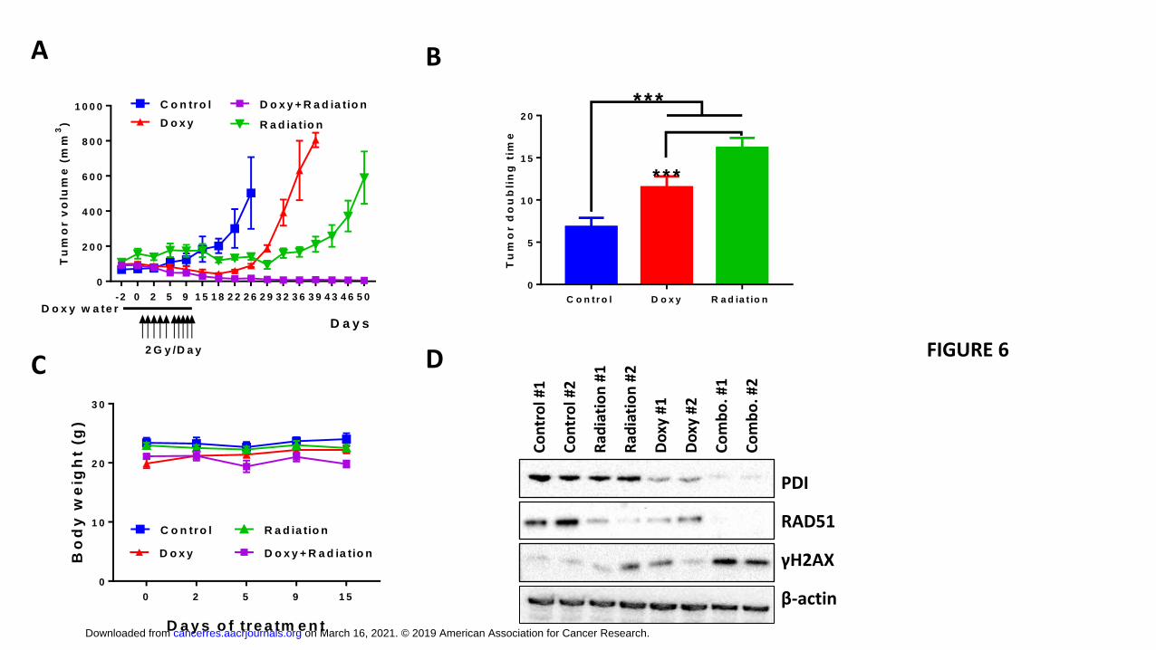
To investigate if PDI knockdown could also enhance the efficacy of radiation therapy in
an in vivo GBM model, U87 cells expressing inducible PDI-targeting shRNA were
implanted into the flanks of nude mice. Induction of PDI shRNA alone extended the
tumor doubling time, from 6.8 ± 1.10 days in the control group to 11.5 ± 1.29 days in the
PDI KD group (p<0.001), while IR alone increased the doubling time to 16.2 ± 1.17 days
(Fig. 6A-B). Despite the 3.5-fold decrease in tumor volume after doxycycline-mediated
shRNA expression, the tumors eventually regrew at 12 days post treatment termination,
whereas in mice treated with IR, tumor growth was controlled until 26 days after the last
treatment. Similar to our in vitro findings using clonogenic survival assays, in vivo
induction of PDI-targeting shRNA combined with radiation therapy led to a synergistic
tumor control as determined by tumor volume (4.8 ± 2.53 mm3). In addition, this
combination treatment prevented tumor re-growth for greater than 40 days post
treatment. Importantly, the combination treatment regimen was well tolerated in mice as
determined by limited body weight loss (Fig. 6C). To study the mechanistic basis of PDI
knockdown-mediated radiosensitization in vivo, two tumors from each group were
harvested 1 hour after the last dose of treatment and analyzed by Western blot (Fig.
on March 16, 2021. © 2019 American Association for Cancer Research.cancerres.aacrjournals.org Downloaded from
Author manuscripts have been peer reviewed and accepted for publication but have not yet been edited. Author Manuscript Published OnlineFirst on April 17, 2019; DOI: 10.1158/0008-5472.CAN-18-2540
13
6D). PDI as well as RAD51 expression was reduced in the doxycycline-treated tumors,
and especially in the combination group. In addition, consistent with our in vitro findings,
accumulation of DNA damage as evidenced by γH2AX levels, was most apparent in
tumors after the combination treatment indicating a compromised ability to repair IR-
induced DNA damage following PDIA1 knockdown.
Discussion
Markers of ER stress are often upregulated in solid tumors and correlate with tumor
stage (17). Additionally, the UPR is thought to be actively involved in promoting tumor
initiation and aggressive phenotypes as it participates in pro-survival processes. The
pro-survival functions of the UPR are accomplished through direct regulation of protein
synthesis and enhanced capacity for protein folding and posttranslational modifications
within the ER (e.g. enhanced PDI activity). Furthermore, degradation of
unfolded/misfolded or damaged proteins is induced by the ERAD system. Cancer cells
are hyper-sensitive to agents that augment ER stress due to a sustained elevation of
ER stress signals. ER-stress-inducing compounds induce apoptosis when ER stress
levels are already high (17). For example, bortezomib, a proteasome inhibitor that was
approved for the treatment of multiple myeloma and mantle cell lymphoma, is an ER
stress inducer. Unfortunately, studies using bortezomib as single agent or as
combination therapy have not been effective in solid tumors. A promising alternative
approach to induce excessive ER stress in cancer cells is to target PDI and folding of
newly synthesized proteins in GBM. Indeed, we (24) and others (39,40) have developed
PDI-specific small molecule inhibitors that demonstrate significant anticancer activity.
The results presented here validate PDI as a therapeutic target in GBM and
demonstrate its efficacy in combination with radiation therapy through a novel
mechanism involving the downregulation of DNA repair genes important in DSB repair.
PDI knockdown in a mouse xenograft model of GBM increased tumor doubling time
from 6.8 to 11.5 days, and radiation as a single agent resulted in a tumor doubling time
of 16.2 days. The poor response to radiation in GBM is consistent with pre-clinical and
clinical findings of the refractory nature of the disease that has been attributed to an
enhance capacity of these cancer cells to repair damaged DNA (41). Our xenograft
on March 16, 2021. © 2019 American Association for Cancer Research.cancerres.aacrjournals.org Downloaded from
Author manuscripts have been peer reviewed and accepted for publication but have not yet been edited. Author Manuscript Published OnlineFirst on April 17, 2019; DOI: 10.1158/0008-5472.CAN-18-2540

14
studies confirmed this with the observation that tumor recurrence occurred soon after
completion of radiation therapy. Similarly, withdrawal of doxycycline in xenografts with
PDIA1 knockdown caused rapid tumor repopulation, indicating that as single agents,
either modality may not significantly improve the management of GBM. However, when
the modalities are combined, a dramatic and prolonged effect on tumor growth rates
was observed such that even at the end of the study (day 50), a majority of the animals
did not have detectable tumor growth and as such, the tumor doubling time could not be
determined. The enhanced efficacy of the combination therapy was also observed in
vitro with clonogenic survival assays conducted using two independent GBM cell lines.
These findings provide a rationale and guidance for lead optimization studies of current
PDI inhibitors.
In an effort to delineate the mechanistic basis for the robust synergistic effect observed
upon the combination of PDI inhibition with radiation, we evaluated immediate changes
in gene transcription as well as hallmarks of ER stress. In D54 and U87 cells,
expression of PDIA1-targeting shRNA or CRIPR/Cas9 mediated deletion of PDI
expression, resulted in an increase in ER stress as detected by upregulation of BiP and
phosphorylated-eIF-2α. Nascent RNA Bru-seq analysis (7) was conducted to evaluate
changes in genome transcription in an unbiased manner in response to PDI inhibition
and surprisingly, transcription of several DNA repair genes was downregulated following
PDI knockdown (Fig. 2). Of these, RAD51 has been reported to be elevated in GBM
(42), and inhibition of RAD51 using small molecules as well as siRNA sensitizes cells to
DNA damaging agents (43-45). Our findings that PDI inhibition decreased expression of
DNA repair genes and compromised ability to repair damaged DNA in response to
ionizing radiation are consistent with PDI being a promising therapeutic target for
radiosensitization in GBM.
qRT-PCR studies measuring steady-state mRNA levels validated that RAD51
transcription was slightly downregulated upon PDI inhibition. Subsequent Western blot
analysis for RAD51 protein confirmed that RAD51 was strongly downregulated, and that
the induction of ER stress upon tunicamycin treatment also resulted in decreased
RAD51 levels. A previous study showed that RAD51 was downregulated in response to
ER stress induced by tunicamycin (46). This downregulation at the protein level
on March 16, 2021. © 2019 American Association for Cancer Research.cancerres.aacrjournals.org Downloaded from
Author manuscripts have been peer reviewed and accepted for publication but have not yet been edited. Author Manuscript Published OnlineFirst on April 17, 2019; DOI: 10.1158/0008-5472.CAN-18-2540

15
appeared to be caused by ubiquitin-mediated proteasomal degradation and correlated
with radio-sensitization of A549 lung cancer cells. In concordance with those studies,
we show that ER stress caused by inhibition of PDI expression resulted in increased
proteasome-mediated degradation of RAD51. Our protein stability results in the
presence of cycloheximide further revealed that, despite an observed decrease in
RAD51 transcription and steady state mRNA levels upon ER stress, the contribution of
proteasomal degradation machinery was a dominant determinant of RAD51 levels,
since MG132 treatment restored RAD51 to levels greater than in non-ER stressed
control cells. A possible explanation is that RAD51 protein is under dynamic
translational and post-translational regulation where it is under constant proteasomal
degradation as we demonstrated higher RAD51 protein level after MG132 treatment
alone (Supplementary Fig. 4). In addition, the cycloheximide treatment was started after
16-hour MG132 rescue experiment, which might contribute to the higher RAD51 protein
level observed compared to those without MG132 treatment.
RAD51 foci are detected in response to DNA damaging therapies that induce double
strand breaks, including ionizing radiation. Increased RAD51 protein levels are
associated with therapeutic resistance and targeting of RAD51 using siRNA or Gleevec,
a c-Abl inhibitor that has been shown to reduce RAD51 protein expression via inhibition
of its transcription, sensitizes GBM to DNA damaging therapeutics (47,48). RAD51
binds to single stranded DNA at sites of the lesion to promote recombination repair in
chromosomes containing a sister chromatid. Failure to repair damaged DNA in the S
and G2 phases of the cell cycle prior to mitotic entry can induce G2 arrest or mitotic
catastrophe leading to the induction of apoptosis. Our results demonstrate that PDI
knockdown is synergistic with radiation and specific targeting of PDI leading to ER
stress and diminished levels of RAD51 provides a new therapeutic opportunity to
sensitize GBM to radiation therapy.
Acknowledgements: We would like to thank Drs. Dipanka Ray, Meredith Morgan and Mr. Steven Kronenberg from Department of Radiation Oncology, University of Michigan on their guidance on in vitro studies and manuscript preparation. We also want to thank University of Michigan Vector Core, Proteomics & Peptide Synthesis Core and Microscopy & Image Analysis Laboratory for their services.
on March 16, 2021. © 2019 American Association for Cancer Research.cancerres.aacrjournals.org Downloaded from
Author manuscripts have been peer reviewed and accepted for publication but have not yet been edited. Author Manuscript Published OnlineFirst on April 17, 2019; DOI: 10.1158/0008-5472.CAN-18-2540
16
on March 16, 2021. © 2019 American Association for Cancer Research.cancerres.aacrjournals.org Downloaded from
Author manuscripts have been peer reviewed and accepted for publication but have not yet been edited. Author Manuscript Published OnlineFirst on April 17, 2019; DOI: 10.1158/0008-5472.CAN-18-2540

17
References: 1. Thakkar JP, Dolecek TA, Horbinski C, Ostrom QT, Lightner DD, Barnholtz-Sloan JS, et al.
Epidemiologic and molecular prognostic review of glioblastoma. Cancer Epidemiol Biomarkers Prev 2014;23(10):1985-96.
2. Stupp R, Hegi ME, Mason WP, van den Bent MJ, Taphoorn MJ, Janzer RC, et al. Effects of radiotherapy with concomitant and adjuvant temozolomide versus radiotherapy alone on survival in glioblastoma in a randomised phase III study: 5-year analysis of the EORTC-NCIC trial. Lancet Oncol 2009;10(5):459-66.
3. Shergalis A, Bankhead A, 3rd, Luesakul U, Muangsin N, Neamati N. Current Challenges and Opportunities in Treating Glioblastoma. Pharmacol Rev 2018;70(3):412-45.
4. Ostrom QT, Gittleman H, Xu J, Kromer C, Wolinsky Y, Kruchko C, et al. CBTRUS Statistical Report: Primary Brain and Other Central Nervous System Tumors Diagnosed in the United States in 2009-2013. Neuro Oncol 2016;18(suppl_5):v1-v75.
5. Verhaak RG, Hoadley KA, Purdom E, Wang V, Qi Y, Wilkerson MD, et al. Integrated genomic analysis identifies clinically relevant subtypes of glioblastoma characterized by abnormalities in PDGFRA, IDH1, EGFR, and NF1. Cancer Cell 2010;17(1):98-110.
6. Cancer Genome Atlas Research N. Comprehensive genomic characterization defines human glioblastoma genes and core pathways. Nature 2008;455(7216):1061-8.
7. Parsons DW, Jones S, Zhang X, Lin JC, Leary RJ, Angenendt P, et al. An integrated genomic analysis of human glioblastoma multiforme. Science 2008;321(5897):1807-12.
8. Chinot OL, Wick W, Mason W, Henriksson R, Saran F, Nishikawa R, et al. Bevacizumab plus radiotherapy-temozolomide for newly diagnosed glioblastoma. N Engl J Med 2014;370(8):709-22.
9. Friedman HS, Prados MD, Wen PY, Mikkelsen T, Schiff D, Abrey LE, et al. Bevacizumab alone and in combination with irinotecan in recurrent glioblastoma. J Clin Oncol 2009;27(28):4733-40.
10. Rich JN, Reardon DA, Peery T, Dowell JM, Quinn JA, Penne KL, et al. Phase II trial of gefitinib in recurrent glioblastoma. J Clin Oncol 2004;22(1):133-42.
11. Uhm JH, Ballman KV, Wu W, Giannini C, Krauss JC, Buckner JC, et al. Phase II evaluation of gefitinib in patients with newly diagnosed Grade 4 astrocytoma: Mayo/North Central Cancer Treatment Group Study N0074. Int J Radiat Oncol Biol Phys 2011;80(2):347-53.
12. Cloughesy TF, Yoshimoto K, Nghiemphu P, Brown K, Dang J, Zhu S, et al. Antitumor activity of rapamycin in a Phase I trial for patients with recurrent PTEN-deficient glioblastoma. PLoS Med 2008;5(1):e8.
13. Hainsworth JD, Shih KC, Shepard GC, Tillinghast GW, Brinker BT, Spigel DR. Phase II study of concurrent radiation therapy, temozolomide, and bevacizumab followed by bevacizumab/everolimus as first-line treatment for patients with glioblastoma. Clin Adv Hematol Oncol 2012;10(4):240-6.
14. Wilkinson B, Gilbert HF. Protein disulfide isomerase. Biochim Biophys Acta 2004;1699(1-2):35-44. 15. Cai H, Wang CC, Tsou CL. Chaperone-like activity of protein disulfide isomerase in the refolding
of a protein with no disulfide bonds. J Biol Chem 1994;269(40):24550-2. 16. Clarke HJ, Chambers JE, Liniker E, Marciniak SJ. Endoplasmic reticulum stress in malignancy.
Cancer Cell 2014;25(5):563-73. 17. Wang M, Kaufman RJ. The impact of the endoplasmic reticulum protein-folding environment on
cancer development. Nat Rev Cancer 2014;14(9):581-97. 18. Smith MH, Ploegh HL, Weissman JS. Road to ruin: targeting proteins for degradation in the
endoplasmic reticulum. Science 2011;334(6059):1086-90.
on March 16, 2021. © 2019 American Association for Cancer Research.cancerres.aacrjournals.org Downloaded from
Author manuscripts have been peer reviewed and accepted for publication but have not yet been edited. Author Manuscript Published OnlineFirst on April 17, 2019; DOI: 10.1158/0008-5472.CAN-18-2540
18
19. Xu S, Sankar S, Neamati N. Protein disulfide isomerase: a promising target for cancer therapy. Drug Discov Today 2014;19(3):222-40.
20. Shin BK, Wang H, Yim AM, Le Naour F, Brichory F, Jang JH, et al. Global profiling of the cell surface proteome of cancer cells uncovers an abundance of proteins with chaperone function. J Biol Chem 2003;278(9):7607-16.
21. Rho JH, Roehrl MHA, Wang JY. Glycoproteomic Analysis of Human Lung Adenocarcinomas Using Glycoarrays and Tandem Mass Spectrometry: Differential Expression and Glycosylation Patterns of Vimentin and Fetuin A Isoforms. Protein Journal 2009;28(3-4):148-60.
22. Zhang DH, Tai LK, Wong LL, Chiu LL, Sethi SK, Koay ESC. Proteomic study reveals that proteins involved in metabolic and detoxification pathways are highly expressed in HER-2/neu-positive breast cancer. Molecular & Cellular Proteomics 2005;4(11):1686-96.
23. Goplen D, Wang J, Enger PO, Tysnes BB, Terzis AJ, Laerum OD, et al. Protein disulfide isomerase expression is related to the invasive properties of malignant glioma. Cancer Res 2006;66(20):9895-902.
24. Xu S, Butkevich AN, Yamada R, Zhou Y, Debnath B, Duncan R, et al. Discovery of an orally active small-molecule irreversible inhibitor of protein disulfide isomerase for ovarian cancer treatment. Proc Natl Acad Sci U S A 2012;109(40):16348-53.
25. Shili Xu YL, Kai Yang, Hanxiao Wang, Andrea Shergalis, Anahita Kyani, Armand Bankhead III, Shuzo Tamura, Suhui Yang, Xi Wang, Chih-chen Wang, Alnawaz Rehemtulla, Mats Ljungman, Nouri Neamati Inhibition of protein disulfide isomerase in glioblastoma causes marked downregulation of DNA repair and DNA damage response genes. Theranostics 2019;In press.
26. Galligan JJ, Petersen DR. The human protein disulfide isomerase gene family. Hum Genomics 2012;6:6.
27. Bao S, Wu Q, McLendon RE, Hao Y, Shi Q, Hjelmeland AB, et al. Glioma stem cells promote radioresistance by preferential activation of the DNA damage response. Nature 2006;444(7120):756-60.
28. Bigner SH, Bullard DE, Pegram CN, Wikstrand CJ, Bigner DD. Relationship of in vitro morphologic and growth characteristics of established human glioma-derived cell lines to their tumorigenicity in athymic nude mice. J Neuropathol Exp Neurol 1981;40(4):390-409.
29. Sanjana NE, Shalem O, Zhang F. Improved vectors and genome-wide libraries for CRISPR screening. Nat Methods 2014;11(8):783-84.
30. Shalem O, Sanjana NE, Hartenian E, Shi X, Scott DA, Mikkelson T, et al. Genome-scale CRISPR-Cas9 knockout screening in human cells. Science 2014;343(6166):84-87.
31. Paulsen MT, Veloso A, Prasad J, Bedi K, Ljungman EA, Magnuson B, et al. Use of Bru-Seq and BruChase-Seq for genome-wide assessment of the synthesis and stability of RNA. Methods 2014;67(1):45-54.
32. Liu Y, Burness ML, Martin-Trevino R, Guy J, Bai S, Harouaka R, et al. RAD51 Mediates Resistance of Cancer Stem Cells to PARP Inhibition in Triple-Negative Breast Cancer. Clin Cancer Res 2017;23(2):514-22.
33. Lee AS. The ER chaperone and signaling regulator GRP78/BiP as a monitor of endoplasmic reticulum stress. Methods 2005;35(4):373-81.
34. Teske BF, Wek SA, Bunpo P, Cundiff JK, McClintick JN, Anthony TG, et al. The eIF2 kinase PERK and the integrated stress response facilitate activation of ATF6 during endoplasmic reticulum stress. Mol Biol Cell 2011;22(22):4390-405.
35. Han J, Kaufman RJ. Physiological/pathological ramifications of transcription factors in the unfolded protein response. Genes Dev 2017;31(14):1417-38.
on March 16, 2021. © 2019 American Association for Cancer Research.cancerres.aacrjournals.org Downloaded from
Author manuscripts have been peer reviewed and accepted for publication but have not yet been edited. Author Manuscript Published OnlineFirst on April 17, 2019; DOI: 10.1158/0008-5472.CAN-18-2540
19
36. Adamson B, Smogorzewska A, Sigoillot FD, King RW, Elledge SJ. A genome-wide homologous recombination screen identifies the RNA-binding protein RBMX as a component of the DNA-damage response. Nat Cell Biol 2012;14(3):318-28.
37. Erasimus H, Gobin M, Niclou S, Van Dyck E. DNA repair mechanisms and their clinical impact in glioblastoma. Mutat Res Rev Mutat Res 2016;769:19-35.
38. Baumann P, Benson FE, West SC. Human Rad51 protein promotes ATP-dependent homologous pairing and strand transfer reactions in vitro. Cell 1996;87(4):757-66.
39. Vatolin S, Phillips JG, Jha BK, Govindgari S, Hu J, Grabowski D, et al. Novel Protein Disulfide Isomerase Inhibitor with Anticancer Activity in Multiple Myeloma. Cancer Res 2016;76(11):3340-50.
40. Kaplan A, Gaschler MM, Dunn DE, Colligan R, Brown LM, Palmer AG, 3rd, et al. Small molecule-induced oxidation of protein disulfide isomerase is neuroprotective. Proc Natl Acad Sci U S A 2015;112(17):E2245-52.
41. Atkins RJ, Ng W, Stylli SS, Hovens CM, Kaye AH. Repair mechanisms help glioblastoma resist treatment. J Clin Neurosci 2015;22(1):14-20.
42. Welsh JW, Ellsworth RK, Kumar R, Fjerstad K, Martinez J, Nagel RB, et al. Rad51 protein expression and survival in patients with glioblastoma multiforme. Int J Radiat Oncol Biol Phys 2009;74(4):1251-5.
43. Lim YC, Roberts TL, Day BW, Stringer BW, Kozlov S, Fazry S, et al. Increased sensitivity to ionizing radiation by targeting the homologous recombination pathway in glioma initiating cells. Mol Oncol 2014;8(8):1603-15.
44. Short SC, Giampieri S, Worku M, Alcaide-German M, Sioftanos G, Bourne S, et al. Rad51 inhibition is an effective means of targeting DNA repair in glioma models and CD133+ tumor-derived cells. Neuro Oncol 2011;13(5):487-99.
45. King HO, Brend T, Payne HL, Wright A, Ward TA, Patel K, et al. RAD51 Is a Selective DNA Repair Target to Radiosensitize Glioma Stem Cells. Stem Cell Reports 2017;8(1):125-39.
46. Yamamori T, Meike S, Nagane M, Yasui H, Inanami O. ER stress suppresses DNA double-strand break repair and sensitizes tumor cells to ionizing radiation by stimulating proteasomal degradation of Rad51. FEBS Lett 2013;587(20):3348-53.
47. Russell JS, Brady K, Burgan WE, Cerra MA, Oswald KA, Camphausen K, et al. Gleevec-mediated inhibition of Rad51 expression and enhancement of tumor cell radiosensitivity. Cancer Res 2003;63(21):7377-83.
48. Golding SE, Rosenberg E, Khalil A, McEwen A, Holmes M, Neill S, et al. Double strand break repair by homologous recombination is regulated by cell cycle-independent signaling via ATM in human glioma cells. J Biol Chem 2004;279(15):15402-10.
on March 16, 2021. © 2019 American Association for Cancer Research.cancerres.aacrjournals.org Downloaded from
Author manuscripts have been peer reviewed and accepted for publication but have not yet been edited. Author Manuscript Published OnlineFirst on April 17, 2019; DOI: 10.1158/0008-5472.CAN-18-2540
20
Figure legends: Figure 1. PDI inhibition induces ER stress in GBM cells. PDIA1 knockdown was induced in (A) D54 by adding doxycycline (2 μg/mL) Lysates were collected after 12, 16, 24, and 48 hours, and ET stress markers was quantifiyied (B). Doxycycline (2 μg/mL) was added to U87 cells (C) for 24, 48 and 72hrs to induce PDIA1 shRNA and the lysates were collected for ER stress marker quantification (D). Protein expression level was normalized against β-actin. Data are means ± S.D. from three independent experiments. *, p<0.05; **, p<0.01; ***, p<0.001. Figure 2. GSEA analysis of PDI knockdown reveals reduced transcription of DNA repair genes, E2F1, MYC and targets, and KRAS signaling. Nascent RNA Bru-seq was used to assess the effect of 72h dox-induced PDI knockdown on genome-wide transcription in D54 and U87 cells. Gene set enrichment analysis (GSEA) of Hallmark pathways using log2-fold rank ordered genes from 10,687 and 11,030 genes expressed in D54 (A) and U87 cells (B), respectively. (C) GSEA plots for the top downregulated pathways: E2F1 targets and KRAS signaling. GSEA of KEGG pathways for D54 (D) and U87 cells (E) and (F) GSEA plots for the top downregulated pathways: ribosome and DNA replication. Figure 3. PDI KD induces ER stress that downregulates RAD51. qPCR (A-B) and Western blot (C) analysis for PDI and RAD51 expression level after doxycycline induction as indicated. qPCR data are presented as means ± S.D. *, p<0.05; **, p<0.01; ***, p<0.001 from three independent experiments. (D) Immunoblot analysis of CRISPR/Cas9 targeting P4HB U87 cells after 48hrs of infection. (E) Tunicamycin (Tm) was added at 5 μg/ul to D54MG and U87MG cells for 12 hrs and RAD51 and BiP protein expression were assessed. Figure 4. ER stress leads to RAD51 decreased RAD51 stability. PDI shRNA expression was induced in (A) U87MG and (B) D54MG cells for 48 hours in response to 2 μg/mL doxycycline. MG132 (10 μM) was added for the last 16 hours of the 48-hour doxycycline treatment and the cells were prepared for Western blot analysis. Parental (C) U87MG and (D) D54MG cells were treated with tunicamycin (TM, 5 μg/mL) for 16 hours prior to addition of MG132 (10 μM) and cycloheximide (CHX, 100 ug/mL), and cells were harvested at indicated time points for Western blot analysis. All bands were normalized against β-actin and then against corresponding control sample without CHX. Data are presented as means ± S.E.M. from three independent experiments. *, p<0.05; **, p<0.01; ***, p<0.001. Figure 5. Knockdown of PDI sensitizes GBM cells to radiation. U87MG (A) and D54MG (B) cells were treated with 0, 2, 4, 6, or 8 Gy radiation doses and plated for clonogenic survival analysis. PDIA1 knockdown was induced with 2 μg/ul doxycycline for 72 hrs in U87MG (C) and D54MG (D). DsRed is an indicator for PDIA1 shRNA induction. Cells were irradiated at 2 Gy, fixed, and stained with γH2AX antibody at indicated time points. Cells with ≥ five γH2AX foci/nucleus were counted as positive and at least 100 cells were counted for each time point from each experiment. Quantification of γH2AX foci in U87MG (E) and D54MG (F). Data are presented from three independent experiments is presented as means ± S.D. *, p<0.05; **, p<0.01; ***, p<0.001. Figure 6. Knockdown of PDI sensitizes GBM xenografts to radiation.
on March 16, 2021. © 2019 American Association for Cancer Research.cancerres.aacrjournals.org Downloaded from
Author manuscripts have been peer reviewed and accepted for publication but have not yet been edited. Author Manuscript Published OnlineFirst on April 17, 2019; DOI: 10.1158/0008-5472.CAN-18-2540

21
Approximately a million U87MG cells with inducible PDI shRNA were implanted into the flanks of Nu/Nu mice. When tumor volumes reached around 100 mm3, mice were randomized into four groups and treatments: 1) control, 2) doxycycline water (2mg/mL in 5% sucrose water), 3) radiation (2Gy per day, five days per week), 4) doxycycline water and radiation. The treatments were administered for two weeks. Doxycycline water was administered three days prior to radiation for combination groups. (A) Tumors were monitored twice per week by caliper and volume was calculated as (L x W x W)/2, where W is tumor width and L is tumor length. Values were plotted as means ± S.D. from ≥ 5 mice per treatment group. (B) Tumor doubling time was calculated according to an online calculator (http://www.chestx-ray.com/index.php/calculators/doubling-time). Tumors in the combination group never grew back, thus the doubling time for this group was infinite. (C) Bodyweight recorded during treatment. (D) Tumors from two mice from each treatment group were processed within one hour after the last dose of radiation and prepared for Western blot analysis.
on March 16, 2021. © 2019 American Association for Cancer Research.cancerres.aacrjournals.org Downloaded from
Author manuscripts have been peer reviewed and accepted for publication but have not yet been edited. Author Manuscript Published OnlineFirst on April 17, 2019; DOI: 10.1158/0008-5472.CAN-18-2540

A B
D
Ctr
l
Do
xy-2
4h
rs
Do
xy-4
8h
rs
Do
xy-7
2h
rs
Ctr
l
Do
xy-2
4h
rs
Do
xy-4
8h
rs
Do
xy-7
2h
rs
PDI
BiP
p-elF2α
elF2α
β-actin
shRNA1 shRNA2
PDI
BiP
p-elF2α
elF2α
β-actin
Ctr
l
Do
xy-1
2h
rs
Do
xy-1
6h
rs
Do
xy-2
4h
rs
Do
xy-4
8h
rs
Ctr
l
Do
xy-1
2h
rs
Do
xy-1
6h
rs
Do
xy-2
4h
rs
Do
xy-4
8h
rs
C
P DI
BiP
p -elF
2 (S
e r5 1 )
T o tal-e
lF2
0
1
2
3
Ex
pre
ss
ion
le
ve
l
(Fo
ld c
ha
ng
e t
o c
trl)
0 h rs 1 2 h rs 1 6 h rs
2 4 h rs 4 8 h rs
* * *
* * *
* ** * *
*
* * *
* ** * * *
* *
P DI
BiP
p -elF
2 (S
e r5 1 )
T o tal-e
lF2
0
1
2
3
Ex
pre
ss
ion
le
ve
l
(Fo
ld c
ha
ng
e t
o c
trl)
0 h r 2 4 h rs
4 8 h rs 7 2 h rs
*
** *
* ** * *
* *
* *
FIGURE 1
shRNA1 shRNA2 on March 16, 2021. © 2019 American Association for Cancer Research.cancerres.aacrjournals.org Downloaded from
Author manuscripts have been peer reviewed and accepted for publication but have not yet been edited. Author Manuscript Published OnlineFirst on April 17, 2019; DOI: 10.1158/0008-5472.CAN-18-2540
Myc targets V1
E2F targets
KRAS signaling UP
MYC targets V2
G2/M checkpoint
inflammatory response
DNA repair
TNFa signaling via NFkB
EMT
-3 -2 -1 0
D54
TNFa signaling via NFkB
E2F targets
MYC targets V2
KRAS signaling UP
G2/M checkpoints
inflammatory response
MYC targets V1
EMT
DNA repair
-3 -2 -1 0
U87
ribosome
DNA replication
base excision repair
mismatch repair
pyrimidine metabolism
glutathione metabolism
homologous recombination
purine metabolism
nucleotide excision repair
-3 -2 -1 0
ribosome
DNA replication
pyrimidine metabolism
mismatch repair
homologous recombination
glutathione metabolism
base excision repair
purine metabolism
nucleotide excision repair
-3 -2 -1 0
D54 U87
GSEA Hallmark pathways
GSEA KEGG pathways
A B
NES NES
NES NES
Figure 2
C
FD E
FIGURE 2
on March 16, 2021. © 2019 American Association for Cancer Research.cancerres.aacrjournals.org Downloaded from
Author manuscripts have been peer reviewed and accepted for publication but have not yet been edited. Author Manuscript Published OnlineFirst on April 17, 2019; DOI: 10.1158/0008-5472.CAN-18-2540
shRNA1 shRNA2 shRNA1 shRNA2
D54 U87
PDI RAD51 β-actin
Ctr
l
Do
xy-4
8h
rs
Ctr
l
Do
xy-4
8h
rs
Ctr
l
Do
xy-7
2h
rs
Ctr
l
Do
xy-7
2h
rs
A
C D E
Tm - + - +
D54 U87
BIP RAD51 β-actin
B
G AP D H P D IA1 R AD 5 1 G R P 7 8
0
1
2
3
D 5 4 + P D IA 1 s h R N A
mR
NA
ex
pre
ss
ion
(no
rm
ali
ze
d t
o c
on
tro
l)
0 h r 2 4 h rs
4 8 h rs 7 2 h rs
* * *
* * *
* *
* * *
G AP D H P D IA1 R AD 5 1 G R P 7 8
0 .0
0 .5
1 .0
1 .5
2 .0
U 8 7 + P D IA 1 s h R N A
mR
NA
ex
pre
ss
ion
(no
rm
ali
ze
d t
o c
on
tro
l)
0 h r 2 4 h rs
4 8 h rs 7 2 h rs
* * ** * *
* * *
* * *
*
*
FIGURE 3
Ctr
l
sgR
NA
1
sgR
NA
2
PDI RAD51 p-eIF2α eIF2α β-actin
on March 16, 2021. © 2019 American Association for Cancer Research.cancerres.aacrjournals.org Downloaded from
Author manuscripts have been peer reviewed and accepted for publication but have not yet been edited. Author Manuscript Published OnlineFirst on April 17, 2019; DOI: 10.1158/0008-5472.CAN-18-2540
A B
C D FIGURE 4
0 2 4 6 8 1 0
0 .0
0 .5
1 .0
1 .5
2 .0
2 .5
C y c lo h e x im id e tre a tm e n t t im e (h rs )
RA
D5
1 e
xp
re
ss
ion
le
ve
l
(Fo
ld c
ha
ng
e t
o c
trl)
C o n tro l P D I K D
P D I K D + M G 1 3 2 M G 1 3 2
*
0 2 4 6 8 1 0
0 .0
0 .5
1 .0
1 .5
2 .0
2 .5
C y c lo h e x im id e tre a tm e n t t im e (h rs )
RA
D5
1 e
xp
re
ss
ion
le
ve
l
(Fo
ld c
ha
ng
e t
o c
trl)
C o n tro l P D I K D
P D I K D + M G 1 3 2 M G 1 3 2
* *
0 2 4 6 8 1 0
0 .0
0 .5
1 .0
1 .5
2 .0
C y c lo h e x im id e tre a tm e n t t im e (h rs )
RA
D5
1 e
xp
re
ss
ion
le
ve
l
(Fo
ld c
ha
ng
e t
o c
trl)
C o n tro l Tm
T m + M G 1 3 2 M G 1 3 2
*
* * *
0 2 4 6 8 1 0
0 .0
0 .5
1 .0
1 .5
C y c lo h e x im id e tre a tm e n t t im e (h rs )
RA
D5
1 e
xp
re
ss
ion
le
ve
l
(Fo
ld c
ha
ng
e t
o c
trl)
C o n tro l Tm
T m + M G 1 3 2 M G 1 3 2
* * *
on March 16, 2021. © 2019 American Association for Cancer Research.cancerres.aacrjournals.org Downloaded from
Author manuscripts have been peer reviewed and accepted for publication but have not yet been edited. Author Manuscript Published OnlineFirst on April 17, 2019; DOI: 10.1158/0008-5472.CAN-18-2540

Figure 3
0 0 .5 4 8 2 4
0
2 0
4 0
6 0
8 0
1 0 0
U 8 7
T im e p o s t ra d ia t io n (h rs )
H
2A
X f
oc
i +
ve
ce
lls
(%
)
C trl
D o x y
*
* * *
* *
* *
0 0 .5 4 8 2 4
0
3 0
6 0
9 0
1 2 0
D 5 4
T im e p o s t ra d ia t io n (h rs )
H
2A
X f
oc
i +
ve
ce
lls
(%
)
C trl
D o x y
** * *
A B C
D
E F
FIGURE 5
0 2 4 6 8
0 .0 1
0 .1
1
U 8 7
D o s e (G y )
Su
rv
ivin
g F
ra
cti
on
C o n tro l
D o x y
*
* *
0 2 4 6
0 .0 0 1
0 .0 1
0 .1
1
D 5 4
D o s e (G y )
Su
rv
ivin
g F
ra
cti
on
C o n tro l
D o x y*
on March 16, 2021. © 2019 American Association for Cancer Research.cancerres.aacrjournals.org Downloaded from
Author manuscripts have been peer reviewed and accepted for publication but have not yet been edited. Author Manuscript Published OnlineFirst on April 17, 2019; DOI: 10.1158/0008-5472.CAN-18-2540
Co
ntr
ol #
1
Co
ntr
ol #
2
Rad
iati
on
#1
Rad
iati
on
#2
Do
xy #
1
Do
xy #
2
Co
mb
o. #
1
Co
mb
o. #
2
PDI
RAD51
γH2AX
β-actin 0 2 5 9 1 5
0
1 0
2 0
3 0
D a y s o f tre a tm e n t
Bo
dy
we
igh
t (g
)
C o n tro l
D o x y
R a d ia tio n
D o x y + R a d ia tio n
C o n tr o l D o x y R a d ia t io n
0
5
1 0
1 5
2 0
Tu
mo
r d
ou
bli
ng
tim
e
* * *
* * *
B
C D
A
-2 0 2 5 9 1 5 1 8 2 2 2 6 2 9 3 2 3 6 3 9 4 3 4 6 5 0
0
2 0 0
4 0 0
6 0 0
8 0 0
1 0 0 0
D a y s
Tu
mo
r v
olu
me
(m
m3
)
C o n tro l
D o x y R a d ia tio n
D o x y + R a d ia tio n
D o x y w a te r
2 G y /D a y FIGURE 6
on March 16, 2021. © 2019 American Association for Cancer Research.cancerres.aacrjournals.org Downloaded from
Author manuscripts have been peer reviewed and accepted for publication but have not yet been edited. Author Manuscript Published OnlineFirst on April 17, 2019; DOI: 10.1158/0008-5472.CAN-18-2540

Published OnlineFirst April 17, 2019.Cancer Res Yajing Liu, Wenbin Ji, Andrea Shergalis, et al. repair to sensitize glioblastoma to radiotherapyprotein disulfide isomerase decreases the capacity for DNA Activation of the unfolded protein response via inhibition of
Updated version
10.1158/0008-5472.CAN-18-2540doi:
Access the most recent version of this article at:
Material
Supplementary
http://cancerres.aacrjournals.org/content/suppl/2019/04/17/0008-5472.CAN-18-2540.DC1
Access the most recent supplemental material at:
Manuscript
Authoredited. Author manuscripts have been peer reviewed and accepted for publication but have not yet been
E-mail alerts related to this article or journal.Sign up to receive free email-alerts
Subscriptions
Reprints and
To order reprints of this article or to subscribe to the journal, contact the AACR Publications
Permissions
Rightslink site. Click on "Request Permissions" which will take you to the Copyright Clearance Center's (CCC)
.http://cancerres.aacrjournals.org/content/early/2019/04/17/0008-5472.CAN-18-2540To request permission to re-use all or part of this article, use this link
on March 16, 2021. © 2019 American Association for Cancer Research.cancerres.aacrjournals.org Downloaded from
Author manuscripts have been peer reviewed and accepted for publication but have not yet been edited. Author Manuscript Published OnlineFirst on April 17, 2019; DOI: 10.1158/0008-5472.CAN-18-2540