activation of mtorc2 by association with the ribosome
TRANSCRIPT

Activation of mTORC2by Association with the RibosomeVittoria Zinzalla,1 Daniele Stracka,1 Wolfgang Oppliger,1 and Michael N. Hall1,*1Biozentrum, University of Basel, CH-4056 Basel, Switzerland*Correspondence: [email protected] 10.1016/j.cell.2011.02.014
SUMMARY
The target of rapamycin (TOR) is a highly conservedprotein kinase and a central controller of growth.Mammalian TOR complex 2 (mTORC2) regulatesAGC kinase family members and is implicated invarious disorders, including cancer and diabetes.Here, we investigated the upstream regulation ofmTORC2. A genetic screen in yeast and subsequentstudies in mammalian cells revealed that ribosomes,but not protein synthesis, are required for mTORC2signaling. ActivemTORC2was physically associatedwith the ribosome, and insulin-stimulated PI3Ksignaling promoted mTORC2-ribosome binding,suggesting that ribosomes activate mTORC2 di-rectly. Findings with melanoma and colon cancercells suggest that mTORC2-ribosome association isimportant in oncogenic PI3K signaling. Thus,TORC2-ribosome interaction is a likely conservedmechanism of TORC2 activation that is physiologi-cally relevant in both normal and cancer cells. Asribosome content determines growth capacity ofa cell, this mechanism of TORC2 regulation ensuresthat TORC2 is active only in growing cells.
INTRODUCTION
Target of rapamycin (TOR) is a central controller of cell growthand metabolism in response to nutrients, growth factors, andenergy status. TOR is found in two structurally and functionallydistinct multiprotein complexes termed TOR complex 1(TORC1) and TORC2 (Wullschleger et al., 2006). The TOR com-plexes, originally described in yeast (Loewith et al., 2002), areconserved across all eukaryotes and regulate a wide spectrumof cellular processes that mediate cell growth (Laplante andSabatini, 2009; Soulard et al., 2009; Wullschleger et al., 2006;Yang and Guan, 2007). In mammalian cells, mammalian TORC1(mTORC1) contains mTOR, raptor, and mLST8 and is sensitiveto the immunosuppressant and anticancer drug rapamycin(Hara et al., 2002; Kim et al., 2002; Loewith et al., 2002). mTORC1controls transcription, ribosome biogenesis, protein synthesis,lipid synthesis, nutrient transport, autophagy, and othergrowth-related processes. The best-characterized substratesof mTORC1 are S6K and 4E-BP via which mTORC1 controls
translation (Sonenberg and Hinnebusch, 2009). mTORC2consists of mTOR, rictor, mSIN1, mLST8, and PRR5/PRR5L(also known as protor1 and 2) and is insensitive to rapamycin,although long-term rapamycin treatment can indirectly inhibitmTORC2 in some cell types (Cybulski and Hall, 2009; Jacintoet al., 2004; Sarbassov et al., 2004; Sarbassov et al., 2006;Sparks and Guertin, 2010). mTORC2 directly phosphorylatesand activates the AGC kinases Akt (also known as PKB),SGK1, and likely PKC (Facchinetti et al., 2008; Garcıa-Martınezand Alessi, 2008; Hresko and Mueckler, 2005; Ikenoue et al.,2008; Jacinto and Lorberg, 2008; Sarbassov et al., 2004; Sar-bassov et al., 2005). mTORC2 promotes cell survival via Aktand mediates organization of the actin cytoskeleton (Cybulskiand Hall, 2009; Sparks and Guertin, 2010).Both mTORC1 and mTORC2 are activated by growth factors,
including insulin, IGF-1, and others. Growth factors activatemTORC1 via phosphatidylinositol 3-kinase (PI3K), PDK1, Akt,the TSC1-TSC2 complex, and Rheb, a small guanosine triphos-phate (GTP)-binding protein that binds and activates mTORC1directly (Avruch et al., 2009; Manning and Cantley, 2003). Incontrast, the mechanism via which growth factors activatemTORC2 has been elusive. Growth factors activate mTORC2via PI3K (Frias et al., 2006; Garcıa-Martınez and Alessi, 2008;Yang et al., 2006), but signaling steps beyond PI3K are distinctfrom those upstream of mTORC1 and unknown (Cybulski andHall, 2009; Sparks and Guertin, 2010). The nature of theupstream regulators of TORC2 in unicellular model organismssuch as yeasts, which lack a growth factor signaling pathway,is completely unknown (Soulard et al., 2009).Here, we describe a genetic screen in yeast and subsequent
studies in mammalian cells that identify the ribosome as an acti-vator of TORC2. We demonstrate that the ribosome, indepen-dent of protein synthesis, is required for mTORC2 signalingin vivo and mTORC2 kinase activity in vitro. Active mTORC2 isassociated with the ribosome. Insulin stimulates the associationof mTORC2 with the ribosome via PI3K signaling. Findings withcancer cells suggest that ribosome-dependent mTORC2 activa-tion is physiologically relevant in tumors with hyperactive PI3Ksignaling.
RESULTS
A Genetic Screen Reveals that NIP7 Is Requiredfor TORC2 Signaling in YeastTo identify upstream activators of TORC2, we performeda genetic screen in the budding yeast S. cerevisiae. Furthermore,
Cell 144, 757–768, March 4, 2011 ª2011 Elsevier Inc. 757

we assumed that whatever activates TORC2 in yeast would alsobe upstream of mTORC2 in mammals. In other words, weassumed that growth factor signaling was grafted onto a hereto-fore unknown ancestral input controlling TORC2 in unicellularyeast. This reasoning was supported by the fact that growthfactor signaling was grafted onto the ancestral nutrient input inthe case of TORC1.
mTORC2 phosphorylates the hydrophobic motif in the AGCkinase SGK1 and thereby activates SGK1. In yeast, TORC2 simi-larly phosphorylates and activates the SGK1 ortholog YPK2. Ourgenetic screen was based on the observation that overexpres-sion of constitutively active YPK2 (YPK2D239A, hereafter referredto as YPK2*) suppresses the lethality of a TORC2 defect(Aronova et al., 2008; Kamada et al., 2005). Assuming thatYPK2* would also suppress lethality caused by a defect in anupstream activator of TORC2, we performed a so-called reversesuppressor screen to isolate yeast mutants that depend onYPK2* for viability (Figure 1A and Experimental Procedures).This is referred to as a reverse suppressor screen because itstarts with a suppressor mutation (YPK2*) to identify unknown‘‘suppressee’’ mutations—the reverse order of a normal(forward) suppressor screen. The screen was predicted to iden-tify loss-of-function mutations in an essential upstream activatorof TORC2 or in the essential components of TORC2, includingTOR2, AVO1 (mSIN1 ortholog), and AVO3 (rictor ortholog). Thescreen yielded a total of 44 independent mutants defective inTORC2, thereby validating the screen. The 44mutants consistedof 25, 13, and 6 tor2, avo1, and avo3 mutants, respectively. Inaddition, we obtained a temperature-sensitive nip7 mutant thatwe hereafter refer to as nip7-1 (Figure 1A and Figures S1A andS1B). Sequence analysis of the nip7-1 allele identified a pointmutation that converts glycine 71 to aspartic acid. Westernblot analysis of extracts from nip7-1 cells showed that the pointmutation mildly and strongly decreased NIP7 protein levels atpermissive temperature (25!C) and nonpermissive temperature(37!C), respectively (Figure S1A). YPK2*, but not wild-typeYPK2, suppressed the growth defect of the nip7-1 mutant atsemipermissive temperature (30!C and 34!C) (Figure S1B).NIP7 is an essential protein required for maturation of rRNA ofthe 60S ribosomal subunit (Zanchin et al., 1997). Confirmingthis role of NIP7 in ribosomebiogenesis,we observed a reductionin the amounts of the 60S subunit, the 80S ribosome, and poly-somes, with a concomitant appearance of halfmer polysomes, inextracts of the nip7-1 mutant grown at semipermissive temper-ature (30!C) (Figure S1C).
To examine further whether NIP7 is required for TORC2signaling, we investigated whether the nip7-1mutant phenocop-ied TORC2mutants. The nip7-1mutant indeed exhibited severaldefects similar to those observed in temperature-sensitiveTORC2 mutants (avo3-1 and tor2-21) (Aronova et al., 2008;Beeler et al., 1998; Facchinetti et al., 2008; Helliwell et al.,1998; Kamada et al., 2005; Schmidt et al., 1997). First, thenip7-1 mutant exhibited reduced signaling through the cell wallintegrity pathway, as evidenced by decreased MPK1 phosphor-ylation and PKC1 protein levels, depolarization of the actin cyto-skeleton, and restoration of growth in the presence of theosmotic stabilizer sorbitol (Figure 1B and Figure S1D). Second,the nip7-1 mutant showed impaired sphingolipid biosynthesis,
including hypersensitivity to myriocin, an inhibitor of the firststep in the sphingolipid biosynthetic pathway, and restorationof growth in the presence of Ca2+ in a csg2D background(Figure S1E). Third, the nip7-1 mutant showed decreasedYPK2 kinase activity, as measured by an in vitro kinase assaywith immunopurified YPK2 and, as a control, kinase-deadYPK2K373A (Figure 1C and data not shown for kinase dead).Finally and most importantly, TORC2 kinase activity wasreduced in the nip7-1 mutant, as measured by an in vitro kinaseassay with TORC2 immunopurified from wild-type cells andnip7-1mutant cells grown at 30!C (Figure 1D). The above resultsstrongly suggest that NIP7 is required, directly or indirectly, forTORC2 kinase activity and signaling.
mNIP7 Is Required for mTORC2 Activityand Ribosome Maturation in MammalsYeast NIP7 shares 75% identity with an uncharacterizedmammalian protein also termedNIP7 (Figure S2A).We examinedwhether mammalian NIP7 (mNIP7) is required for mTORC2signaling. mTORC2 directly phosphorylates Ser473 in the hydro-phobicmotif of Akt and Ser422 in the hydrophobicmotif of SGK1and thereby activates Akt and SGK1 toward substrates such asFoxO3a (Thr32) and NDRG1 (Thr346), respectively (Garcıa-Martınez and Alessi, 2008; Sarbassov et al., 2005). mTORC2also autophosphorylates sites in rictor (Sarbassov et al., 2004;Jacinto et al., 2004) and is required for phosphorylation ofThr450 in the turn motif of Akt (Facchinetti et al., 2008; Ikenoueet al., 2008). Knockdown of mNIP7 in HeLa and HEK293 cellsstrongly decreased basal and insulin-stimulated phosphoryla-tion of Ser473 and Thr450 in Akt, Thr32 in FoxO3a, Thr346 inNDRG1, and rictor in mTORC2 (Figure 2A, Figure S2B, anddata not shown). mNIP7 knockdown had no effect on Erk phos-phorylation or mTORC1-dependent S6K phosphorylation (Fig-ure 2A). These findings suggest that mNIP7 is specificallyrequired for mTORC2 signaling. Finally, we observed thatmNIP7 knockdown reduced (by 58%) mTORC2 kinase activityin vitro, with no effect on mTORC2 amount or integrity (Fig-ure 2B). These findings indicate that mNIP7 is required formTORC2 activity and signaling, and not for mTORC2 synthesis,assembly, or stability. Furthermore, NIP7 is required for TORC2signaling in both yeast and mammalian cells, suggestinga conserved mechanism of TORC2 activation.Is mNIP7 a 60S maturation factor like NIP7 in yeast? Knock-
down of mNIP7 reduced the amounts of the 60S ribosomalsubunit and the 80S ribosome (80S), with no effect on theamount of the 40S subunit (Figure S2C). Knockdown ofmTORC2component mSIN1 had no effect on the ribosome profile (Fig-ure S2C), indicating that the effect of mNIP7 on ribosome matu-ration was not due to a defect in signaling downstream ofmTORC2. Thus, NIP7 is conserved from yeast to human asa 60S ribosome maturation factor and a TORC2 activator.
Ribosomes, but Not Protein Synthesis, Are Requiredfor mTORC2 SignalingDoes mNIP7 control mTORC2 via its role in ribosome matura-tion? To address this question, we examined whether ribosomecontent affects mTORC2 signaling. Knockdown of ribosomalprotein Rpl7 (60S subunit) reduced the amounts of the 60S
758 Cell 144, 757–768, March 4, 2011 ª2011 Elsevier Inc.

subunit and assembled ribosomes (80S monosomes andpolysomes), whereas knockdown of ribosomal protein Rps16(40S subunit) reduced the amounts of the 40S subunit andassembled ribosomes (Figure S2C). We also observed thatknockdown of Rpl7 or Rps16 reduced the amounts of other
proteins in the corresponding ribosomal subunit (Figure 3A andFigure S3A), consistent with the published finding that ribosomalsubunit assembly determines the level of ribosomal proteins(Idol et al., 2007). Like knockdown of mNIP7, knockdown ofeither Rpl7 or Rps16 decreased basal and insulin-stimulated
Figure 1. NIP7, Identified by a Reverse Suppressor Screen, Is Required for TORC2 Activity In Vivo and In Vitro(A) Schematic representation of the reverse suppressor screen. A wild-type strain (JK9-3da) overexpressing constitutively active YPK2* (YPK2D239A) on an
URA3-based plasmid was mutagenized with ethyl-methanesulfonate (EMS). Mutants that could not grow on plates containing 5-FOA were chosen for further
analysis. A plasmid-borne NIP7 gene (pNIP7) was isolated by complementation of the nip7-1 mutation.
(B) nip7-1mutant exhibits depolarization of actin cytoskeleton.Wild-type (JK9-3da), avo3-1 (BAS65-2a), and nip7-1 (DS1) cells grown in YPD at 25!Cwere shifted
to 37!C for 6 hr. The actin cytoskeleton was stained with rhodamine-coupled phalloidin. A representative figure of three independent experiments for each strain
is shown.
(C) nip7-1 mutant shows decreased YPK2 activity. Wild-type cells (JK9-3da) (mock) and wild-type, avo3-1, or nip7-1 cells expressing plasmid-borne YPK2-HA
(pYPK2-HA) were grown in YPD at 30!C. YPK2-HA was immunoprecipitated and subjected to in vitro kinase assay using cross-tide as a substrate. Substrate
phosphorylation was quantified, and the average ± standard deviation from the mean based on three independent experiments is shown. Immunoprecipitated
YPK2-HA was detected by western blotting (bottom).
(D) nip7-1 mutant shows decreased TORC2 kinase activity in vitro. Wild-type cells (JK9-3da; mock) and wild-type (DS2; WT) or nip7-1 (DS3; nip7-1) cells
expressing AVO3-HA were grown in YPD at 30!C. AVO3-HA was immunoprecipitated and subjected to a TORC2 kinase assay in vitro using recombinant YPK2
protein as a substrate. Also shown are Coomassie blue-stained total YPK2 protein and immunoblot of immunoprecipitated AVO3-HA.
See also Figure S1.
Cell 144, 757–768, March 4, 2011 ª2011 Elsevier Inc. 759

phosphorylation of Akt (Ser473 and Thr450), FoxO3a (Thr32),NDRG1 (Thr346), and rictor, with no effect on Erk or S6K phos-phorylation (Figure 3A, Figure S2B, and Figure S3A). Finally,we observed that Rpl7 knockdown reduced mTORC2 kinaseactivity, as determined by an in vitro kinase assay with immuno-purified mTORC2 and recombinant, kinase-dead Akt as asubstrate. Rpl7 knockdown had no effect on mTORC2 amountor integrity, as determined by unaffected coimmunoprecipitationof mTOR, mSIN1, or mLST8 with rictor (Figure 3B). Thus, assem-bled ribosomes (not 60S or 40S ribosomal subunits alone) arerequired for mTORC2 kinase activity and signaling. Furthermore,the above findings suggest that mNIP7 controls mTORC2 via itsrole in ribosome maturation.
mTORC2 promotes cell survival via phosphorylation andactivation of Akt, which in turn phosphorylates and inhibitsproapoptotic Bad (Brazil et al., 2004; Datta et al., 1997; Jacintoet al., 2006; Yang et al., 2006). To investigate further the physio-logical relevance of ribosome-mediated mTORC2 regulation, weexamined the effect of mNIP7, Rpl7, or Rps16 knockdown oninduction of apoptosis by etoposide or hydrogen peroxide(H2O2). Both treatments induce cell death in an Akt-sensitivemanner (He et al., 2010; Kim et al., 2001; Wang et al., 2000).Knockdown of mNIP7, Rpl7, or Rps16 enhanced the inductionof apoptosis by etoposide or hydrogen peroxide, as indicatedby an increase in caspase 3 and PARP cleavage and a decreasein cell viability (Figure 4A and Figure S4). Knockdown of mSIN1(mTORC2) similarly enhanced apoptosis. Knockdown of Bad
Figure 2. mNIP7 Is Required for mTORC2 ActivityIn Vivo and In Vitro(A) siRNA-mediated knockdown of mNIP7 inhibits
mTORC2 signaling. HeLa cells, 48 hr after transfection
with the indicated siRNA, were harvested (left) or serum
starved for 3 hr and then restimulated with insulin for the
indicated times before harvesting (right). Phosphorylation
and protein levels were determined by immunoblotting
with the appropriate antibodies, as indicated.
(B) Knockdown of mNIP7 inhibits mTORC2 kinase activity
toward Akt with no effect on mTORC2 integrity. rictor
immunoprecipitates were immunoblotted with the indi-
cated antibodies to determine mTORC2 integrity.
mTORC2 in vitro kinase assay was performed using
immunopurified mTORC2 (rictor) and recombinant,
kinase-dead Akt as a substrate.
See also Figure S2.
blocked the proapoptotic effect of mSIN1,mNIP7, Rpl7, or Rps16 knockdown (Figure 4B).Thus, ribosomes appear to regulate mTORC2signaling in a physiologically relevant manner.Although ribosomal knockdown reduces
mTORC2 kinase activity and signaling withoutaffecting mTORC2 synthesis, is the process ofprotein synthesis per se required for mTORC2activation? To address this question, we exam-ined whether the protein synthesis inhibitorssalubrinal, cycloheximide, anisomycin, or puro-mycin acutely inhibit mTORC2 signaling. Salu-brinal blocks translation initiation by selective
inhibition of eIF2a dephosphorylation (Boyce et al., 2005; Cnopet al., 2007) (Figure S3B). Cycloheximide, anisomycin, and puro-mycin inhibit translation elongation. Unlike knockdown ofmNIP7, Rpl7, or Rps16, salubrinal, cycloheximide, anisomycin,and puromycin had no effect on basal or insulin-stimulated AktSer473 phosphorylation (Figures S3B–S3D and Figure S5C). Alldrugs were used under conditions known to affect proteinsynthesis in the expected manner, as determined by polysomegradient analysis (Figures S3B–S3D and data not shown).Thus, ribosomes mediate mTORC2 signaling independently ofprotein synthesis.
Active mTORC2 Is Associated with the RibosomeTo investigate the molecular mechanism by which ribosomesactivate mTORC2 signaling, we examined by four unrelatedmethods whether ribosomes and mTORC2 physically interact.First, we determined whether mTORC2 coimmunoprecipitateswith ribosomal protein Rpl26. Endogenous Rpl26 coimmunopre-cipitated with endogenous mTOR, rictor, mSIN1, and ribosomalprotein Rpl7 (Figure 5A), but not with raptor. This suggests thatmTORC2, but not mTORC1, associates with the ribosome.Second, we determined whether mTORC2 copurifies with totalribosomes isolated by sedimentation through a sucrose cushion(see Experimental Procedures). mTOR and rictor, but not raptor,cosedimented with Rpl26, Rpl7, and Rps16 (Figure 5B). Third,although ribosomes activate mTORC2 independently of proteinsynthesis, we also determined whether mTORC2 cosediments
760 Cell 144, 757–768, March 4, 2011 ª2011 Elsevier Inc.

with polysomes in a sucrose gradient. Lysates were fractionatedin a sucrose gradient to separate polysomes from 80S, 60S, and40S ribosomes. mTOR, rictor, andmSIN1 were found in both thepolysomal and ribosomal fractions, to the same extent as Rpl7,Rpl26, and Rps16 (Figure S5A). Finally, we determined whethermTORC2 copurifies with mRNA-bound ribosomes. mRNA-bound ribosomes were purified by pull-down of poly(A) mRNAwith oligo(dT) cellulose (Figure S5B) (Ceci et al., 2003). In thisexperiment, mTOR and rictor copurified with Rpl26, Rpl7, andthe 40S ribosomal protein RACK1. RNase A treatment of a lysatebefore pull-down prevented isolation of any of the aboveproteins (Figure S5B), confirming interaction of mTORC2 withmRNA-bound ribosomes. The above data taken togethersuggest that mTORC2 physically interacts with translating(mRNA-bound) and nontranslating 80S ribosomes. An interac-tion between mTORC2 and the 80S ribosome is also supportedby published mass spectrometry studies that identified severallarge and small subunit ribosomal proteins in mTORC2 immuno-precipitates (Pearce et al., 2007; Thedieck et al., 2007). We alsonote that nonionic detergent was required during any of our fourabove ribosome purifications to detect copurification ofmTORC2 and that protein synthesis inhibitors had no effect onthe interaction between mTORC2 and Rpl26 (Figure S5C).To obtainmore insight into themTORC2-ribosome interaction,
we performed coimmunoprecipitation experiments after knock-down of mTORC2 subunits or ribosomal proteins. First, theamount of mTOR in Rpl26 immunoprecipitates from rictor ormSIN1 knockdown cells was significantly reduced comparedto control cells (Figure S6A), suggesting that mTORC2 interacts
Figure 3. Ribosomes Are Required for mTORC2Activity and Signaling(A) siRNA-mediated knockdown of Rpl7 inhibits mTORC2
signaling. HeLa cells, 24 hr after transfection with the
indicated siRNA, were harvested (left, basal activity) or
serum starved for 3 hr and then restimulated with insulin
for the indicated times before harvesting (right, insulin-
stimulated activity). Phosphorylation and protein levels
were determined by immunoblotting with the appropriate
antibodies, as indicated.
(B) Knockdown of Rpl7 inhibits mTORC2 kinase activity
toward Akt with no effect on mTORC2 integrity. rictor
immunoprecipitates were immunoblotted with the indi-
cated antibodies. mTORC2 in vitro kinase assay was
performed using immunopurified mTORC2 (rictor) and
recombinant, kinase-dead Akt as a substrate.
See also Figure S3.
with the ribosome via rictor and/or mSIN1. Wecould not distinguish a requirement specificallyfor rictor or mSIN1 because knockdown ofeither rictor or mSIN1 alone also results in lossof the other. Second, knockdown of Rpl7 (60Ssubunit) abolished the interaction betweenmTORC2 and Rpl26, whereas knockdown ofRps16 (40S subunit) only moderately decreasedthe interaction between mTORC2 and Rpl26(60S subunit) (Figure S6B). These results
suggest that mTORC2 associates with the ribosome via rictorand/or mSIN1 binding to the 60S subunit.The above findings suggest that ribosomes bind and activate
mTORC2. To test this model further, we investigated whetherribosome-bound mTORC2 is indeed active. We performed anmTORC2 kinase assay with ribosomes purified either by Rpl26immunoprecipitation or by sedimentation through a sucrosecushion,asdescribedabove. Inbothcases,a ribosome-associatedkinase phosphorylated Ser473 in recombinant, kinase-dead Akt(Figure 5C). ThemTOR inhibitor PP242 (Feldman et al., 2009) abol-ished the in vitro phosphorylation of Ser473 (Figure 5C), confirmingthat the ribosome-associated Ser473 kinase was mTORC2. Thus,ribosome-associatedmTORC2 isactive.Furthermore,weanalyzedwhethermTORC2kinaseactivity is required formTORC2-ribosomeinteraction. Treatment of HeLa cells with PP242 abolished Ser473phosphorylation, as expected, but had no effect on mTORC2-Rpl26 interaction (Figure S6D), suggesting that mTORC2 activityis not required for mTORC2-ribosome association.
Insulin-PI3K Signaling Stimulates mTORC2-RibosomeAssociationWe next investigated whether mTORC2 association with theribosome is regulated by insulin. Insulin treatment of serum-starved cells significantly increased the amount of mTOR andrictor that coimmunoprecipitated with Rpl26 (Figure 6A). Impor-tantly, this stimulation of the interaction between mTORC2 andRpl26 correlated with phosphorylation of the mTORC2 targetsite Ser473 in endogenous Akt. Thus, insulin stimulatesmTORC2-ribosome association.
Cell 144, 757–768, March 4, 2011 ª2011 Elsevier Inc. 761

Insulin activates mTORC2 via PI3K (Frias et al., 2006; Garcıa-Martınez and Alessi, 2008; Yang et al., 2006). To investigatewhether PI3K signaling regulates mTORC2 association withthe ribosome, we examined whether the mTORC2-Rpl26 inter-action was affected upon inhibition or hyperactivation of PI3Ksignaling. Inhibition of PI3K strongly decreased mTORC2-Rpl26 interaction, as determined by a reduction in the amountof mTOR and rictor that coimmunoprecipitated with Rpl26 in
lysates from cells treated with the PI3K inhibitor LY294002 (Fig-ure 6B and Figure S6D). In contrast, hyperactivation of PI3Ksignaling by knockdown of PTEN, a negative regulator of PI3Ksignaling, increased the amount of mTOR and rictor in Rpl26immunoprecipitates, compared to control cells (Figure 6C).Again, the change in mTORC2-Rpl26 interaction upon PI3K inhi-bition or hyperactivation was paralleled by a correspondingchange in phosphorylation of the mTORC2 target site Ser473
Figure 4. Ribosome or mTORC2 Knockdown Enhances Stress-Induced, Bad-Dependent Apoptosis(A) siRNA-mediated knockdown of mSIN1, mNIP7, Rpl7, or Rps16 increases etoposide-induced apoptosis. HeLa cells were transfected with the various siRNAs
and then treated with 25 mM etoposide for the indicated times (top) or for 24 hr (bottom). Extracts were analyzed by immunoblotting to assess efficiency of siRNA
knockdown and induction of apoptosis with the indicated antibodies (top). Apoptosis was assessed by blotting for cleaved PARP (cPARP) and cleaved caspase 3
(cCaspase 3). Cells were fixed and stained with DAPI and cleaved PARP antibody to detect apoptotic cells (bottom).
(B) Bad is required for enhanced apoptosis in cells with knockdown of mSIN1, mNIP7, Rpl7, or Rps16. HeLa cells were transfected with the indicated siRNA and
then treated with 25 mM etoposide for 24 hr. Extracts were analyzed by immunoblotting to assess the induction of apoptosis with cleaved PARP antibody. The
efficiency of Bad knockdown is shown.
See also Figure S4.
762 Cell 144, 757–768, March 4, 2011 ª2011 Elsevier Inc.

in endogenous Akt (Figures 6B and 6C). Taken together, theabove data suggest that insulin stimulates mTORC2-ribosomeassociation in a physiologically relevant manner via PI3K.
mTORC2-Ribosome Interaction Promotes Akt Signalingin Cancer CellsPI3K-dependent association of mTORC2 with the ribosome,ribosome-mediated mTORC2 activation, and the physiological
relevance of mTORC2-ribosome interaction in promoting cellsurvival prompted us to analyze mTORC2-ribosome associationin cancer cells with hyperactive PI3K signaling. First, we exam-ined whether PTEN-deficient metastatic melanoma cells haveelevated mTORC2 activity. Approximately 60% of metastaticmelanomas have reduced PTEN expression and elevated Aktphosphorylation (Robertson, 2005; Stahl et al., 2004). Weanalyzed PTEN expression and Akt Ser473 phosphorylation in
Figure 5. Active mTORC2 Is Associated with theRibosome(A) Endogenous Rpl26 coimmunoprecipitates with
endogenous mTOR, rictor, and mSIN1. Rpl26 and mock
immunoprecipitations were performed with HeLa cell
extracts and analyzed by immunoblotting with the indi-
cated antibodies.
(B) mTORC2 associates with ribosomes. Ribosomes were
purified from HeLa cells by sedimentation through a
sucrose cushion. Ribosomes were probed with the indi-
cated antibodies (left) or were resedimented through a
sucrose gradient to monitor 40S, 60S, and 80S ribosomes
and polysomes. The absorbance profile of the sucrose
gradient was determined at 254nm (right).
(C) Ribosome-associated mTORC2 is active. mTORC2
kinase assays were performed with Rpl26 or mock
immunoprecipitates using recombinant, kinase-dead Akt
as a substrate and in the presence or absence of the
mTOR inhibitor PP242 (left). mTORC2 kinase assay was
performed with ribosomes purified as described in (B),
with recombinant, kinase-dead Akt as a substrate and in
the presence or absence of PP242 (right).
See also Figure S5.
Figure 6. Insulin-PI3K Signaling StimulatesmTORC2-Ribosome Association(A) Insulin stimulates mTORC2-ribosome association.
HeLa cells were serum starved and then restimulated with
insulin for the indicated time. Rpl26 immunoprecipitates
(IP: Rpl26) and cell extracts (lysate) were immunoblotted
with the indicated antibodies.
(B) PI3K inhibition decreases mTORC2-ribosome associ-
ation. HeLa cells were treated with LY294002 (50 mM
final concentration) for 30 min before harvesting. Rpl26
immunoprecipitates and cells extracts (lysate) were im-
munoblotted with the indicated antibodies.
(C) siRNA-mediated knockdown of PTEN increases
mTORC2-ribosome association. HeLa cells were trans-
fected with the indicated siRNA and then harvested after
48 hr. Rpl26 immunoprecipitates and cell extracts (lysate)
were analyzed by immunoblotting with the indicated
antibodies.
See also Figure S6.
Cell 144, 757–768, March 4, 2011 ª2011 Elsevier Inc. 763
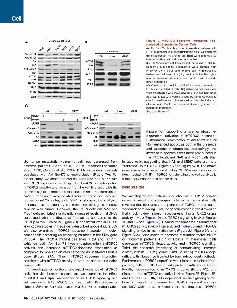
six human metastatic melanoma cell lines generated fromdifferent patients (Certa et al., 2001; Deschodt-Lanckmanet al., 1990; Gervois et al., 1996). PTEN expression inverselycorrelated with Akt Ser473 phosphorylation (Figure 7A). Forfurther study, we chose the two cell lines NA8 and ME67 withlow PTEN expression and high Akt Ser473 phosphorylation(mTORC2 activity) and, as a control, the cell line Juso with theopposite signaling profile. To examinemTORC2-ribosome asso-ciation, ribosomes were isolated from the three cell lines andprobed for mTOR, rictor, and mSIN1. In all cases, the total yieldof ribosomes obtained by sedimentation through a sucrosecushion was similar. However, the PTEN-deficient NA8 andME67 cells exhibited significantly increased levels of mTORC2associated with the ribosomal fraction as compared to thePTEN-positive Juso cells (Figure 7B), consistent with our PTENknockdown studies in HeLa cells described above (Figure 6C).We also examined mTORC2-ribosome interaction in coloncancer cells harboring an activating mutation in the PI3K genePIK3CA. The PIK3CA mutant cell lines HT29 and HCT116exhibited both Akt Ser473 hyperphosphorylation (mTORC2activity) and increased mTORC2-ribosome association ascompared to SW60 colon cells harboring an unaltered PIK3CAgene (Figure S7A). Thus, mTORC2-ribosome interactioncorrelates with mTORC2 activity in both melanoma and coloncancer cells.
To investigate further the physiological relevance of mTORC2activation via ribosome association, we examined the effectof mSIN1 and Rpl7 knockdown on mTORC2 signaling andcell survival in NA8, ME67, and Juso cells. Knockdown ofeither mSIN1 or Rpl7 decreased Akt Ser473 phosphorylation
Figure 7. mTORC2-Ribosome Interaction Pro-motes Akt Signaling in Cancer Cells(A) Akt Ser473 phosphorylation inversely correlates with
PTEN expression in human melanoma cells. Cell extracts
from six human melanoma cell lines were analyzed by
immunoblotting with indicated antibodies.
(B) PTEN-deficient cell lines exhibit increased mTORC2-
ribosome association. Ribosomes were purified from
PTEN-deficient (NA8 and ME67) and PTEN-positive
melanoma cell lines (Juso) by sedimentation through a
sucrose cushion. Ribosomes were probed with the indi-
cated antibodies.
(C) Knockdown of mSIN1 or Rpl7 induces apoptosis in
PTEN-deficient (NA8 andME67) melanoma cell lines. Cells
were transfected with the indicated siRNA and harvested
after 72 hr. Extracts were analyzed by immunoblotting to
check the efficiency of the knockdown and the induction
of apoptosis (PARP and caspase 3 cleavage) with the
indicated antibodies.
See also Figure S7.
(Figure 7C), supporting a role for ribosome-dependent activation of mTORC2 in cancer.Furthermore, knockdown of either mSIN1 orRpl7 enhanced apoptosis both in the presenceand absence of etoposide. Interestingly, theincrease in apoptosis was more pronounced inthe PTEN-deficient NA8 and ME67 cells than
in Juso cells, suggesting that NA8 and ME67 cells are more‘‘addicted’’ to mTORC2 (Figure 7C and Figure S7B). The aboveresults taken together suggest that mTORC2-ribosome associa-tion, mediating PI3K-mTORC2-Akt signaling and cell survival, isfunctionally important in cancer cells.
DISCUSSION
We investigated the upstream regulation of TORC2. A geneticscreen in yeast and subsequent studies in mammalian cellsrevealed that ribosomes are upstream of TORC2. In particular,we found the following. First, the genetic screen in yeast revealedthat knocking down ribosome biogenesis inhibits TORC2 kinaseactivity in vitro (Figure 1D) and TORC2 signaling in vivo (Figures1B and 1C and Figure S1). Second, the ribosome is required formTORC2 activity in vitro (Figure 2B and Figure 3B) and mTORC2signaling in vivo in mammalian cells (Figure 2A, Figure 3A, andFigure S3A). Knockdown of ribosome maturation factor mNIP7or ribosomal proteins (Rpl7 or Rps16) in mammalian cellsdecreased mTORC2 kinase activity and mTORC2 signaling.Third, the ribosome (translating or nontranslating) interactsdirectly with mTORC2 (Figures 5 and Figure S5). mTORC2 cop-urified with ribosomes isolated by four independent methods.Furthermore, mTORC2 copurified with ribosomes isolated fromgrowing cells or cells treated with protein synthesis inhibitors.Fourth, ribosome-bound mTORC2 is active (Figure 5C), andribosome-free mTORC2 is inactive in vitro (Figure 2B, Figure 3B,and Figure S6B). Fifth, PI3K-dependent insulin signaling stimu-lates binding of the ribosome to mTORC2 (Figure 6 and Fig-ure S6D) with the same kinetics that it stimulates mTORC2
764 Cell 144, 757–768, March 4, 2011 ª2011 Elsevier Inc.

activation (Figure 6A). Finally, the mTORC2-ribosome interactioncorrelates with mTORC2 activity in both melanoma and coloncancer cells (Figure 7 and Figure S7). Melanoma and coloncancer cells with high PI3K activity (due to loss of PTEN or anactivating mutation in the PI3K gene) exhibited both enhancedmTORC2-ribosome interaction and increased mTORC2 activity.Our findings suggest that the translating or nontranslating 80Sribosome binds and activates mTORC2 in response to growthfactor-stimulated PI3K signaling (Figure S7C). TORC2-ribosomeassociation is a mechanism of TORC2 activation that is likelyconserved from unicellular yeast to human. The ribosome ispresumably a primordial activator of TORC2 onto which growthfactor signaling was grafted during the evolution ofmulticellularity.In a parallel and complementary study, Oh et al. also showed
that mTORC2 associates with the ribosome (Oh et al., 2010).Furthermore, they showed that mTORC2 phosphorylates theAkt turn motif cotranslationally and the Akt hydrophobic motifposttranslationally. Thus, Oh et al. investigated the role of themTORC2-ribosome interaction in downstream signaling bymTORC2. Our study addresses the separate issue of upstreamregulation of mTORC2. We show that an mTORC2-ribosomeinteraction activates mTORC2, and this activation is indepen-dent of translation. In other words, an mTORC2-ribosomeinteraction activates mTORC2 regardless of whether mTORC2is phosphorylating a substrate co- or posttranslationally. Wenote that Oh et al. did not examine a requirement for the ribo-some in posttranslational phosphorylation.A connection between ribosomes and TOR signaling is well
established. TORC1 activates ribosome biogenesis and proteinsynthesis and inhibits autophagy as key readouts in the controlof cell growth. Why should ribosomes control TORC2? Ribo-some content determines growth capacity of the cell andTORC2 regulates growth-related processes. Thus, regulationof TORC2 by ribosomes ensures that TORC2 is not inappropri-ately activated in cells that are unable to grow. The above alsoimplies that TORC1, via activation of ribosome biogenesis andinhibition of autophagy-mediated ribosome turnover, indirectlycontrols TORC2. Indeed, Sarbassov et al. (Sarbassov et al.,2006) have shown that inhibition of mTORC1 by long-term rapa-mycin treatment indirectly inhibits mTORC2. Our findingssuggest that the effect of rapamycin on mTORC2 is due, at leastin part, to a reduction in ribosome content. Interestingly, the liter-ature also indicates that ribosomal defects induce apoptotic celldeath, although the underlying mechanism is not understood(Warner and McIntosh, 2009). We find that ribosomal defectsinhibit mTORC2 and its downstream effector Akt, which in turnleads to Bad-dependent apoptosis. Thus, our findings alsoprovide a mechanism for the induction of apoptosis by a ribo-somal defect.Our findings suggest that ribosomes bind and activate
mTORC2 directly. The fraction of total mTORC2 that associateswith ribosomes varies depending on the cell type and the growthconditions. For example, under normal growth conditions,"20%of total rictor (mTORC2) was associated with ribosomes in HeLacells, whereas "30%–40% of rictor was associated with ribo-somes in PTEN-deficient cells such as melanoma and PTENknockdown cells. Thus, ribosome association appears to be
a major if not the sole mechanism of TORC2 activation. Consid-eration of the fraction of total ribosomes that associate withmTORC2 is also potentially informative. Given that ribosomesare 100- to 1000-fold more abundant than signaling kinasessuch as mTORC2, only a small fraction of total ribosomes bindmTORC2. This excess of ribosomeswould require strong regula-tion ofmTORC2-ribosome binding by upstreamPI3K signaling toachieve physiologically relevant regulation of mTORC2. Alterna-tively, TORC2 could be regulated by a specific subpopulation ofribosomes. Previous studies have demonstrated that TORC2 isassociated with membranes, including the endoplasmic retic-ulum (ER) and Golgi apparatus, and that mTORC2 isolated fromER microsomes phosphorylates Akt Ser473 in vitro (Drenanet al., 2004; Hresko and Mueckler, 2005; Liu and Zheng, 2007;Schroder et al., 2007; Sturgill et al., 2008). These findings suggestthat TORC2 might associate specifically with membrane-boundribosomes. In support of this notion, we observed that mTORC2copurifies with ribosomes only when the ribosomes are isolatedin the presence of detergent. Membrane-bound ribosomes con-stitute"10% of total ribosomes and include both translating andnontranslating ribosomes (Seiser and Nicchitta, 2000). It is alsointeresting to note that Komili et al. (Komili et al., 2007) haveproposed a ribosome code in which there is a specialization ofribosomes for specific cellular process.Our findings are consistent with other studies proposing the
ribosome as a kinase platform. The two kinases PKCbII andPim-1 are associated with the ribosome via RACK1 andRps19, respectively (Ceci et al., 2003; Chiocchetti et al., 2005;Grosso et al., 2008b). Overall, the ribosome appears to bea signaling platform for mTORC2 and other kinases. Further-more, ribosomal proteins have been shown to modulate theactivity of NF-kB, p53, and c-Myc (Lindstrom, 2009).PI3K-Akt signaling is upregulated and contributes to tumori-
genesis in "60% of advanced-stage melanomas (Stahl et al.,2003, 2004). PTEN expression or Akt inhibition increasessensitivity of melanoma cells to apoptosis-inducing agents andprevents tumor development (Madhunapantula and Robertson,2009; Stahl et al., 2004). Our data, with melanoma, cancer colon,and HeLa cells, suggest that PI3K signaling promotes Aktphosphorylation via stimulation of mTORC2-ribosome binding.Furthermore, disruption of the mTORC2-ribosome supercom-plex selectively induces apoptosis in PTEN-deficient melanomacells (Figure 7C). The extent to which cells of other cancersrequire ribosome-dependent mTORC2 activation is unclear,although we expect that other cancers driven by mutationspromoting PI3K signaling may also depend on mTORC2-ribo-some association. Disrupting the mTORC2-ribosome interactionmay be a useful strategy in the treatment of melanomas, coloncarcinomas, and possibly other cancers.Several findings suggest that upregulation of the protein
synthesis machinery contributes to the development of cancerand other diseases (Ruggero and Pandolfi, 2003). Consistentwith our findings, a ribosomal protein deficiency inhibits Akt-driven tumorigenesis (Hsieh et al., 2010). Furthermore, the myconcogene enhances ribosome biogenesis, and myc oncoge-nicity in mice can be blocked by mutations in ribosomal proteingenes (Barna et al., 2008; Ruggero, 2009). Our findings and theobservation that mTORC2 is required for tumor progression in
Cell 144, 757–768, March 4, 2011 ª2011 Elsevier Inc. 765

at least some cancers (Guertin et al., 2009; Gulhati et al., 2009;Masri et al., 2007) suggest that myc and increased ribosomalcontent may promote tumorigenicity via stimulation of mTORC2and its downstream effector Akt.
Curiously, our genetic screen in yeast yielded 44 TORC2mutants but only a single mutant (nip7-1) that was defective inribosome biogenesis. Why did we not obtain more mutantsthat were defective in ribosomal maturation factors or ribosomalproteins? First, ribosomal genes are duplicated in yeast, therebyprecluding identification of recessive, loss-of-function mutationsin these genes. Second, YPK2 is downstream of the ribosome inactivation of TORC2 but is not downstream of the ribosome inmediating protein synthesis, precluding full suppression of a ribo-some biogenesis defect by YPK2*. YPK2* only partly suppressesthe nip7-1 mutation and only at semipermissive temperature.
EXPERIMENTAL PROCEDURES
Detailed protocols for apoptosis assays and statistical analyses can be found
in the Extended Experimental Procedures.
Yeast Strains, Media, Kinase Assays, Actin Staining, and AntibodiesYeast strains used in this study are listed in the Table S1. All strains are
isogenic derivatives of JK9-3da. Plasmids used in this study are described
in Table S2. Standard techniques and media were used for yeast manipulation
(Kamada et al., 2005; Loewith et al., 2002). Unless indicated otherwise, cells
were grown in rich YPD medium. YPK2 and TORC2 kinase assays were
performed as described previously (Casamayor et al., 1999; Kamada et al.,
2005; Loewith et al., 2002; Wullschleger et al., 2005). Rhodamine phalloidin
staining of polymerized actin was performed as described (Loewith et al.,
2002; Mulet et al., 2006). Immunoprecipitations were performed as described
previously (Loewith et al., 2002; Wullschleger et al., 2005).
Reverse Suppressor ScreenA wild-type strain (JK9-3da) was transformed with an URA3-based plasmid
overexpressing YPK2* (pYPK2*) (Figure 1A). Cells were randomlymutagenized
with 100 mM ethyl-methanesulfonate (EMS) (Sigma) for 15 min in SD-Ura
medium, washed, and then allowed to recover in SD-Ura medium without
mutagen for 2–4 hr. Cells were plated on solid SD-Ura or SD supplemented
with 5-FOA and incubated at 30!C. The SD medium supplemented with
5-FOA counterselected againstURA3 such that only those cells that had spon-
taneously lost the URA3-based pYPK2* plasmid were able to form a colony.
Mutants that were unable to grow on SD 5-FOA (and hence in the absence
of YPK2*) were isolated from the master SD-Ura plate. "45,000 colonies
from mutagenized cells were screened, from which 45 mutants were isolated
and the corresponding mutations were identified. Mutated genes were iso-
lated by complementation with a LEU2 centromeric plasmid-based yeast
genomic library. Complementing members of the genomic library were
selected by growth on SD-Leu medium containing 5-FOA. Genomic inserts
of library-derived plasmids were identified by sequencing. Complementation
with subclones of isolated inserts identified the complementing ORF within
a given insert. Sequencing of the genomic copy of NIP7 and meiotic segrega-
tion studies confirmed that NIP7 was indeed the relevant mutant gene. nip7-1
was found to be temperature sensitive upon subsequent characterization. For
experiments at semipermissive temperature (30!C), nip7-1 cells were grown in
YPD at permissive temperature (25!C), diluted to OD600 = 0.1, and grown at
30!C to approximately OD600 = 0.6–0.8. For experiments at the nonpermissive
temperature (37!C), nip7-1mutant was grown in YPD at 25!C and then shifted
to 37!C for 6 hr.
Cell Culture, Immunoprecipitations, Immunoblotting,and mTORC2 Kinase AssayHeLa, melanoma cells, and colon cancer cells were cultured, transfected,
stimulated, and harvested as described previously (Jacinto et al., 2004;
Thedieck et al., 2007). In brief, cells were seeded and grown for 48 hr in
DMEM supplemented with 10% serum (basal conditions). Cells were starved
of serum for 3 hr before restimulation with 100 nM insulin (Sigma).
For mSIN1, mNIP7, Rpl7, Rps16, PTEN, or Bad knockdown, a pool of four
different synthetic siRNA or of the appropriate control siRNA (Dharmacon)
was used as described (Thedieck et al., 2007). All transfections were done
according to the manufacturer’s instructions (Lipofectamine, Invitrogen
transfection).
Protein extracts were prepared as previously described (Jacinto et al., 2004;
Thedieck et al., 2007), resolved on SDS-PAGE, and transferred to nitrocellu-
lose membranes (Protran, Whatman). Immunoprecipitation, immunoblotting,
and mTORC2 kinase assays were performed as previously described (Jacinto
et al., 2004; Thedieck et al., 2007).
Polysome Profiles, Ribosome Purification, and Poly(A) mRNAPull-DownPolysome analysis using sucrose gradients was performed as described previ-
ously (Grosso et al., 2008b; Idol et al., 2007). For ribosome purification by sedi-
mentation through a sucrose cushion, HeLa ormelanoma cells were washed in
PBS, trypsinized, and lysed in buffer A (50mMTris-HCl [pH 7.4], 100mMNaCl,
30 mMMgCl2, 0.3% CHAPS, 100 ug/ml cycloheximide, 40 U/ml RNase inhib-
itor, protease inhibitor cocktail, and 100 ug/ml cycloheximide). Whole-cell
extracts were clarified at 4!C, 10 min at 15,000 3 g. Extracts were loaded
on a 30% sucrose cushion in 50 mM Tris-acetate (pH 7.5), 50 mM NH4Cl,
12 mMMgCl2, and 1 mMDTT and ultracentrifuged for 17 hr in a SW41Ti Beck-
man rotor at 39,000 rpm. For the mTORC2 kinase assay, the ribosomal pellet
was resuspended in mTORC2 kinase buffer (Jacinto et al., 2004; Thedieck
et al., 2007). Ribosome-sucrose gradient fractionation was performed as
previously described (Grosso et al., 2008a). For poly(A) pull-down, HeLa cells
were washed with PBS, trypsinized, and lysed in buffer A. Whole-cell extracts
were clarified at 4!C, 10 min at 8000 3 g. Lysates corresponding to 5 3 107
cells were incubated with oligo(dT) cellulose (Invitrogen) for 1 hr at room
temperature. After incubation, the oligo(dT) cellulose was pelleted andwashed
five times with buffer A. The bound fraction was eluted with elution buffer
(100mMTris [pH 7.4], 500mMNaCl, 10mMEDTA, 1% sodium dodecyl sulfate
(SDS), and 5 mM DTT). Purified ribosome fractions and the bound and
unbound fractions after poly(A) pull-down were concentrated with Vivaspin
500 (Sartorius Stedim) and analyzed by immunoblotting with the indicated
antibodies.
SUPPLEMENTAL INFORMATION
Supplemental Information includes Extended Experimental Procedures,
seven figures, and two tables and can be found with this article online at
doi:10.1016/j.cell.2011.02.014.
ACKNOWLEDGMENTS
We thank Rachel Idol and Stefano Grosso for valuable discussions and
technical assistance and Giulio Spagnoli for the melanoma cell lines. We
acknowledge support from the Swiss National Science Foundation, Sys-
temsX.ch, the Swiss Cancer League, the Louis Jeantet Foundation, and the
Canton of Basel. We declare that no competing interests exist.
Received: July 14, 2010
Revised: October 20, 2010
Accepted: February 7, 2011
Published: March 3, 2011
REFERENCES
Aronova, S., Wedaman, K., Aronov, P.A., Fontes, K., Ramos, K., Hammock,
B.D., and Powers, T. (2008). Regulation of ceramide biosynthesis by TOR
complex 2. Cell Metab. 7, 148–158.
766 Cell 144, 757–768, March 4, 2011 ª2011 Elsevier Inc.

Avruch, J., Long, X., Ortiz-Vega, S., Rapley, J., Papageorgiou, A., and Dai, N.
(2009). Amino acid regulation of TOR complex 1. Am. J. Physiol. Endocrinol.
Metab. 296, E592–E602.
Barna, M., Pusic, A., Zollo, O., Costa, M., Kondrashov, N., Rego, E., Rao, P.H.,
and Ruggero, D. (2008). Suppression of Myc oncogenic activity by ribosomal
protein haploinsufficiency. Nature 456, 971–975.
Beeler, T., Bacikova, D., Gable, K., Hopkins, L., Johnson, C., Slife, H., and
Dunn, T. (1998). The Saccharomyces cerevisiae TSC10/YBR265w gene
encoding 3-ketosphinganine reductase is identified in a screen for tempera-
ture-sensitive suppressors of the Ca2+-sensitive csg2Delta mutant. J. Biol.
Chem. 273, 30688–30694.
Boyce, M., Bryant, K.F., Jousse, C., Long, K., Harding, H.P., Scheuner, D.,
Kaufman, R.J., Ma, D., Coen, D.M., Ron, D., and Yuan, J. (2005). A selective
inhibitor of eIF2alpha dephosphorylation protects cells from ER stress.
Science 307, 935–939.
Brazil, D.P., Yang, Z.Z., and Hemmings, B.A. (2004). Advances in protein
kinase B signalling: AKTion on multiple fronts. Trends Biochem. Sci. 29,
233–242.
Casamayor, A., Torrance, P.D., Kobayashi, T., Thorner, J., and Alessi, D.R.
(1999). Functional counterparts of mammalian protein kinases PDK1 and
SGK in budding yeast. Curr. Biol. 9, 186–197.
Ceci, M., Gaviraghi, C., Gorrini, C., Sala, L.A., Offenhauser, N., Marchisio, P.C.,
and Biffo, S. (2003). Release of eIF6 (p27BBP) from the 60S subunit allows 80S
ribosome assembly. Nature 426, 579–584.
Certa, U., Seiler, M., Padovan, E., and Spagnoli, G.C. (2001). High density
oligonucleotide array analysis of interferon- alpha2a sensitivity and transcrip-
tional response in melanoma cells. Br. J. Cancer 85, 107–114.
Chiocchetti, A., Gibello, L., Carando, A., Aspesi, A., Secco, P., Garelli, E.,
Loreni, F., Angelini, M., Biava, A., Dahl, N., et al. (2005). Interactions between
RPS19, mutated in Diamond-Blackfan anemia, and the PIM-1 oncoprotein.
Haematologica 90, 1453–1462.
Cnop, M., Ladriere, L., Hekerman, P., Ortis, F., Cardozo, A.K., Dogusan, Z.,
Flamez, D., Boyce, M., Yuan, J., and Eizirik, D.L. (2007). Selective inhibition
of eukaryotic translation initiation factor 2 alpha dephosphorylation potentiates
fatty acid-induced endoplasmic reticulum stress and causes pancreatic
beta-cell dysfunction and apoptosis. J. Biol. Chem. 282, 3989–3997.
Cybulski, N., and Hall, M.N. (2009). TOR complex 2: a signaling pathway of its
own. Trends Biochem. Sci. 34, 620–627.
Datta, S.R., Dudek, H., Tao, X., Masters, S., Fu, H., Gotoh, Y., and Greenberg,
M.E. (1997). Akt phosphorylation of BAD couples survival signals to the
cell-intrinsic death machinery. Cell 91, 231–241.
Deschodt-Lanckman, M., Vanneste, Y., Loir, B., Michel, A., Libert, A.,
Ghanem, G., and Lejeune, F. (1990). Degradation of alpha-melanocyte stimu-
lating hormone (alpha-MSH) by CALLA/endopeptidase 24.11 expressed by
human melanoma cells in culture. Int. J. Cancer 46, 1124–1130.
Drenan, R.M., Liu, X., Bertram, P.G., and Zheng, X.F. (2004). FKBP12-rapamy-
cin-associated protein or mammalian target of rapamycin (FRAP/mTOR)
localization in the endoplasmic reticulum and the Golgi apparatus. J. Biol.
Chem. 279, 772–778.
Facchinetti, V., Ouyang,W., Wei, H., Soto, N., Lazorchak, A., Gould, C., Lowry,
C., Newton, A.C., Mao, Y., Miao, R.Q., et al. (2008). The mammalian target of
rapamycin complex 2 controls folding and stability of Akt and protein kinase C.
EMBO J. 27, 1932–1943.
Feldman, M.E., Apsel, B., Uotila, A., Loewith, R., Knight, Z.A., Ruggero, D., and
Shokat, K.M. (2009). Active-site inhibitors of mTOR target rapamycin-resistant
outputs of mTORC1 and mTORC2. PLoS Biol. 7, e38.
Frias, M.A., Thoreen, C.C., Jaffe, J.D., Schroder, W., Sculley, T., Carr, S.A.,
and Sabatini, D.M. (2006). mSin1 is necessary for Akt/PKB phosphorylation,
and its isoforms define three distinct mTORC2s. Curr. Biol. 16, 1865–1870.
Garcıa-Martınez, J.M., and Alessi, D.R. (2008). mTOR complex 2 (mTORC2)
controls hydrophobic motif phosphorylation and activation of serum- and
glucocorticoid-induced protein kinase 1 (SGK1). Biochem. J. 416, 375–385.
Gervois, N., Guilloux, Y., Diez, E., and Jotereau, F. (1996). Suboptimal activa-
tion of melanoma infiltrating lymphocytes (TIL) due to low avidity of TCR/MHC-
tumor peptide interactions. J. Exp. Med. 183, 2403–2407.
Grosso, S., Volta, V., Sala, L.A., Vietri, M., Marchisio, P.C., Ron, D., and Biffo,
S. (2008a). PKCbetaII modulates translation independently from mTOR and
through RACK1. Biochem. J. 415, 77–85.
Grosso, S., Volta, V., Vietri, M., Gorrini, C., Marchisio, P.C., and Biffo, S.
(2008b). Eukaryotic ribosomes host PKC activity. Biochem. Biophys. Res.
Commun. 376, 65–69.
Guertin, D.A., Stevens, D.M., Saitoh, M., Kinkel, S., Crosby, K., Sheen, J.H.,
Mullholland, D.J., Magnuson, M.A., Wu, H., and Sabatini, D.M. (2009).
mTOR complex 2 is required for the development of prostate cancer induced
by Pten loss in mice. Cancer Cell 15, 148–159.
Gulhati, P., Cai, Q., Li, J., Liu, J., Rychahou, P.G., Qiu, S., Lee, E.Y., Silva, S.R.,
Bowen, K.A., Gao, T., and Evers, B.M. (2009). Targeted inhibition of mamma-
lian target of rapamycin signaling inhibits tumorigenesis of colorectal cancer.
Clin. Cancer Res. 15, 7207–7216.
Hara, K., Maruki, Y., Long, X., Yoshino, K., Oshiro, N., Hidayat, S., Tokunaga,
C., Avruch, J., and Yonezawa, K. (2002). Raptor, a binding partner of target of
rapamycin (TOR), mediates TOR action. Cell 110, 177–189.
He, H.N., Wang, X., Zheng, X.L., Sun, H., Shi, X.W., Zhong, Y.J., Huang, B.,
Yang, L., Li, J.K., Liao, L.C., et al. (2010). Concurrent blockade of the
NF-kappaB and Akt pathways potently sensitizes cancer cells to chemother-
apeutic-induced cytotoxicity. Cancer Lett. 295, 38–43.
Helliwell, S.B., Howald, I., Barbet, N., and Hall, M.N. (1998). TOR2 is part of two
related signaling pathways coordinating cell growth in Saccharomyces cerevi-
siae. Genetics 148, 99–112.
Hresko, R.C., andMueckler, M. (2005). mTOR.RICTOR is the Ser473 kinase for
Akt/protein kinase B in 3T3-L1 adipocytes. J. Biol. Chem. 280, 40406–40416.
Hsieh, A.C., Costa, M., Zollo, O., Davis, C., Feldman, M.E., Testa, J.R.,
Meyuhas, O., Shokat, K.M., and Ruggero, D. (2010). Genetic dissection of
the oncogenic mTOR pathway reveals druggable addiction to translational
control via 4EBP-eIF4E. Cancer Cell 17, 249–261.
Idol, R.A., Robledo, S., Du, H.Y., Crimmins, D.L., Wilson, D.B., Ladenson, J.H.,
Bessler, M., and Mason, P.J. (2007). Cells depleted for RPS19, a protein
associated with Diamond Blackfan Anemia, show defects in 18S ribosomal
RNA synthesis and small ribosomal subunit production. Blood Cells Mol.
Dis. 39, 35–43.
Ikenoue, T., Inoki, K., Yang, Q., Zhou, X., and Guan, K.L. (2008). Essential
function of TORC2 in PKC and Akt turn motif phosphorylation, maturation
and signalling. EMBO J. 27, 1919–1931.
Jacinto, E., Facchinetti, V., Liu, D., Soto, N., Wei, S., Jung, S.Y., Huang, Q.,
Qin, J., and Su, B. (2006). SIN1/MIP1 maintains rictor-mTOR complex integrity
and regulates Akt phosphorylation and substrate specificity. Cell 127,
125–137.
Jacinto, E., Loewith, R., Schmidt, A., Lin, S., Ruegg, M.A., Hall, A., and Hall,
M.N. (2004). Mammalian TOR complex 2 controls the actin cytoskeleton and
is rapamycin insensitive. Nat. Cell Biol. 6, 1122–1128.
Jacinto, E., and Lorberg, A. (2008). TOR regulation of AGC kinases in yeast and
mammals. Biochem. J. 410, 19–37.
Kamada, Y., Fujioka, Y., Suzuki, N.N., Inagaki, F., Wullschleger, S., Loewith,
R., Hall, M.N., and Ohsumi, Y. (2005). Tor2 directly phosphorylates the AGC
kinase Ypk2 to regulate actin polarization. Mol. Cell. Biol. 25, 7239–7248.
Kim, A.H., Khursigara, G., Sun, X., Franke, T.F., and Chao, M.V. (2001). Akt
phosphorylates and negatively regulates apoptosis signal-regulating kinase
1. Mol. Cell. Biol. 21, 893–901.
Kim, D.H., Sarbassov, D.D., Ali, S.M., King, J.E., Latek, R.R., Erdjument-
Bromage, H., Tempst, P., and Sabatini, D.M. (2002). mTOR interacts with
raptor to form a nutrient-sensitive complex that signals to the cell growth
machinery. Cell 110, 163–175.
Komili, S., Farny, N.G., Roth, F.P., and Silver, P.A. (2007). Functional specificity
among ribosomal proteins regulates gene expression. Cell 131, 557–571.
Cell 144, 757–768, March 4, 2011 ª2011 Elsevier Inc. 767

Laplante, M., and Sabatini, D.M. (2009). mTOR signaling at a glance. J. Cell
Sci. 122, 3589–3594.
Lindstrom, M.S. (2009). Emerging functions of ribosomal proteins in gene-
specific transcription and translation. Biochem. Biophys. Res. Commun.
379, 167–170.
Liu, X., and Zheng, X.F. (2007). Endoplasmic reticulum and Golgi localization
sequences for mammalian target of rapamycin. Mol. Biol. Cell 18, 1073–1082.
Loewith, R., Jacinto, E., Wullschleger, S., Lorberg, A., Crespo, J.L., Bonenfant,
D., Oppliger, W., Jenoe, P., and Hall, M.N. (2002). Two TOR complexes, only
one of which is rapamycin sensitive, have distinct roles in cell growth control.
Mol. Cell 10, 457–468.
Madhunapantula, S.V., and Robertson, G.P. (2009). The PTEN-AKT3 signaling
cascade as a therapeutic target in melanoma. Pigment Cell Melanoma Res.
22, 400–419.
Manning, B.D., and Cantley, L.C. (2003). Rheb fills a GAP between TSC and
TOR. Trends Biochem. Sci. 28, 573–576.
Masri, J., Bernath, A., Martin, J., Jo, O.D., Vartanian, R., Funk, A., and Gera, J.
(2007). mTORC2 activity is elevated in gliomas and promotes growth and cell
motility via overexpression of rictor. Cancer Res. 67, 11712–11720.
Mulet, J.M., Martin, D.E., Loewith, R., and Hall, M.N. (2006). Mutual antago-
nism of target of rapamycin and calcineurin signaling. J. Biol. Chem. 281,
33000–33007.
Oh, W.J., Wu, C.C., Kim, S.J., Facchinetti, V., Julien, L.A., Finlan, M., Roux,
P.P., Su, B., and Jacinto, E. (2010). mTORC2 can associate with ribosomes
to promote cotranslational phosphorylation and stability of nascent Akt poly-
peptide. EMBO J. 29, 3939–3951.
Pearce, L.R., Huang, X., Boudeau, J., Paw1owski, R., Wullschleger, S., Deak,
M., Ibrahim, A.F., Gourlay, R., Magnuson, M.A., and Alessi, D.R. (2007).
Identification of Protor as a novel Rictor-binding component of mTOR
complex-2. Biochem. J. 405, 513–522.
Robertson, G.P. (2005). Functional and therapeutic significance of Akt dereg-
ulation in malignant melanoma. Cancer Metastasis Rev. 24, 273–285.
Ruggero, D. (2009). The role of Myc-induced protein synthesis in cancer.
Cancer Res. 69, 8839–8843.
Ruggero, D., and Pandolfi, P.P. (2003). Does the ribosome translate cancer?
Nat. Rev. Cancer 3, 179–192.
Sarbassov, D.D., Ali, S.M., Kim, D.H., Guertin, D.A., Latek, R.R., Erdjument-
Bromage, H., Tempst, P., and Sabatini, D.M. (2004). Rictor, a novel binding
partner of mTOR, defines a rapamycin-insensitive and raptor-independent
pathway that regulates the cytoskeleton. Curr. Biol. 14, 1296–1302.
Sarbassov, D.D., Guertin, D.A., Ali, S.M., and Sabatini, D.M. (2005).
Phosphorylation and regulation of Akt/PKB by the rictor-mTOR complex.
Science 307, 1098–1101.
Sarbassov, D.D., Ali, S.M., Sengupta, S., Sheen, J.H., Hsu, P.P., Bagley, A.F.,
Markhard, A.L., and Sabatini, D.M. (2006). Prolonged rapamycin treatment
inhibits mTORC2 assembly and Akt/PKB. Mol. Cell 22, 159–168.
Schmidt, A., Bickle, M., Beck, T., and Hall, M.N. (1997). The yeast phosphati-
dylinositol kinase homolog TOR2 activates RHO1 and RHO2 via the exchange
factor ROM2. Cell 88, 531–542.
Schroder, W.A., Buck,M., Cloonan, N., Hancock, J.F., Suhrbier, A., Sculley, T.,
and Bushell, G. (2007). Human Sin1 contains Ras-binding and pleckstrin
homology domains and suppresses Ras signalling. Cell. Signal. 19, 1279–
1289.
Seiser, R.M., and Nicchitta, C.V. (2000). The fate of membrane-bound
ribosomes following the termination of protein synthesis. J. Biol. Chem. 275,
33820–33827.
Sonenberg, N., and Hinnebusch, A.G. (2009). Regulation of translation initia-
tion in eukaryotes: mechanisms and biological targets. Cell 136, 731–745.
Soulard, A., Cohen, A., and Hall, M.N. (2009). TOR signaling in invertebrates.
Curr. Opin. Cell Biol. 21, 825–836.
Sparks, C.A., and Guertin, D.A. (2010). Targeting mTOR: prospects for mTOR
complex 2 inhibitors in cancer therapy. Oncogene 29, 3733–3744.
Stahl, J.M., Cheung, M., Sharma, A., Trivedi, N.R., Shanmugam, S., and
Robertson, G.P. (2003). Loss of PTEN promotes tumor development in
malignant melanoma. Cancer Res. 63, 2881–2890.
Stahl, J.M., Sharma, A., Cheung, M., Zimmerman, M., Cheng, J.Q.,
Bosenberg, M.W., Kester, M., Sandirasegarane, L., and Robertson, G.P.
(2004). Deregulated Akt3 activity promotes development of malignant mela-
noma. Cancer Res. 64, 7002–7010.
Sturgill, T.W., Cohen, A., Diefenbacher, M., Trautwein, M., Martin, D.E., and
Hall, M.N. (2008). TOR1 and TOR2 have distinct locations in live cells.
Eukaryot. Cell 7, 1819–1830.
Thedieck, K., Polak, P., Kim, M.L., Molle, K.D., Cohen, A., Jeno, P.,
Arrieumerlou, C., and Hall, M.N. (2007). PRAS40 and PRR5-like protein are
new mTOR interactors that regulate apoptosis. PLoS ONE 2, e1217.
Wang, X., McCullough, K.D., Franke, T.F., and Holbrook, N.J. (2000).
Epidermal growth factor receptor-dependent Akt activation by oxidative stress
enhances cell survival. J. Biol. Chem. 275, 14624–14631.
Warner, J.R., and McIntosh, K.B. (2009). How common are extraribosomal
functions of ribosomal proteins? Mol. Cell 34, 3–11.
Wullschleger, S., Loewith, R., Oppliger, W., and Hall, M.N. (2005). Molecular
organization of target of rapamycin complex 2. J. Biol. Chem. 280, 30697–
30704.
Wullschleger, S., Loewith, R., and Hall, M.N. (2006). TOR signaling in growth
and metabolism. Cell 124, 471–484.
Yang, Q., and Guan, K.L. (2007). Expanding mTOR signaling. Cell Res. 17,
666–681.
Yang, Q., Inoki, K., Ikenoue, T., and Guan, K.L. (2006). Identification of Sin1 as
an essential TORC2 component required for complex formation and kinase
activity. Genes Dev. 20, 2820–2832.
Zanchin, N.I., Roberts, P., DeSilva, A., Sherman, F., and Goldfarb, D.S. (1997).
Saccharomyces cerevisiae Nip7p is required for efficient 60S ribosome
subunit biogenesis. Mol. Cell. Biol. 17, 5001–5015.
768 Cell 144, 757–768, March 4, 2011 ª2011 Elsevier Inc.

Supplemental Information
EXTENDED EXPERIMENTAL PROCEDURES
The following commercially available antibodies were used for immunoblots and immunoprecipitations: Rpl26, Rpl7, mSIN1 (Bethyl);mNIP7 (Aviva Systems Biology); Erk1, phospho-Erk1,2 (Thr202, Thr204) (Santa Cruz); RACK1 (Upstate); Rps16 (GeneTex, Inc);mTOR, mLst8, rictor, raptor, S6, NDRG1, phospho-S6 (Ser235,Ser236), S6K, phospho-S6K (Thr389), Akt, phospho-Akt (Thr308),FoxO3, phospho-FoxO3 (Thr32) phospho-Akt (Ser473), phospho-Akt (Thr450), phospho-NDRG1 (Thr346), cleaved Caspase 3(cCaspase 3), cleaved PARP (cPARP), Bad, and PTEN (Cell Signaling).For protein synthesis inhibitor studies, cells were incubated with cycloheximide (10 mg/ml) (Sigma), anisomycin (10 mg/ml) (Sigma),
salubrinal (10, 50, and 100 mM) (Tocris) and puromycin (500 nM) (Sigma). LY294002 and etoposide were purchased fromCalbiochem.PP242 (Chemdea) was added at a final concentration of 0.05 mM to the in vitro mTORC2 kinase assay.Apoptosis was monitored by a cleaved PARP immunofluorescence assay (Cell Signaling) or by immunoblotting with anti-cleaved
Caspase 3 and anti-cleaved PARP antibody. After induction of apoptosis, cells were fixed with pre-chilled 100%methanol for 5 minand then washed with 0.1% Triton X-100 and PBS sequentially. Cells were then incubated with anti-cleaved PARP antibody (1/200dilution) overnight at 4!C, washed and incubated for 1 hr with Alexa 568 goat anti-Rabbit antibody (1/500 dilution). Immunofluores-cence images were captured by microscopy (Jacinto et al., 2004; Thedieck et al., 2007). For analysis by immunoblotting, cleavedcaspase 3 and cleaved PARP levels were detected by a human specific antibody. Cell viability of H2O2- or etoposide-treated cellswas quantified by trypan blue exclusion.For the statistical analysis, all values are expressed as mean ± SEM. For all other comparisons, paired Student’s t test was used.
Differences were considered statistically significant at p < 0.05.
SUPPLEMENTAL REFERENCES
Helliwell, S.B., Howald, I., Barbet, N., andHall, M.N. (1998). TOR2 is part of two related signaling pathways coordinating cell growth in Saccharomyces cerevisiae.
Genetics 148, 99–112.
Kamada, Y., Fujioka, Y., Suzuki, N.N., Inagaki, F., Wullschleger, S., Loewith, R., Hall, M.N., and Ohsumi, Y. (2005). Tor2 directly phosphorylates the AGC kinase
Ypk2 to regulate actin polarization. Mol. Cell. Biol. 25, 7239–7248.
Thedieck, K., Polak, P., Kim, M.L., Molle, K.D., Cohen, A., Jeno, P., Arrieumerlou, C., and Hall, M.N. (2007). PRAS40 and PRR5-like protein are new mTOR in-
teractors that regulate apoptosis. PLoS ONE 2, e1217.
Yang, Q., Inoki, K., Ikenoue, T., andGuan, K.L. (2006). Identification of Sin1 as an essential TORC2 component required for complex formation and kinase activity.
Genes Dev. 20, 2820–2832.
Cell 144, 757–768, March 4, 2011 ª2011 Elsevier Inc. S1

Figure S1. Characterization of nip7-1 Mutant, Related to Figure 1(A) Growth curves of wild-type and nip7-1mutant. Wild-type (JK9-3da) and the nip7-1mutant (DS1) were grown in YPD at 25!C and shifted to 37!C. NIP7 protein
level was analyzed by immunoblotting (lower panel).
S2 Cell 144, 757–768, March 4, 2011 ª2011 Elsevier Inc.

(B) The nip7-1 mutation is complemented by wild-type NIP7 (pNIP7) and partly suppressed by overexpression of YPK2* (pYPK2*), but not by wild-type YPK2
(pYPK2) at semi-permissive temperature (30!C and 34!C).
(C) nip7-1 mutant exhibits a decrease in the amounts of 60S subunit, 80S ribosomes and polysomes, with a concomitant appearance of halfmer polysomes.
Polysome profiles of wild-type and nip7-1mutant grown at 30!C, are shown. A representative figure of three independent experiments for each strain is shown.
(D) nip7-1 mutant exhibits reduced cell wall integrity pathway signaling, as evidenced by suppression of the growth defect by sorbitol, decreased MPK1
phosphorylation, and decreased PKC1 protein levels. Wild-type (JK9-3da) and tor1D (MH350-3a), avo3-1 (BAS65-2a), nip7-1 (DS1) mutants, and nip7-1mutant
expressing plasmid-borne NIP7 (pNIP7) were grown on YPDmedium or YPDmedium containing 0.8M or 1M sorbitol at 30!C or 37!C, as indicated (upper panel).
Wild-type, avo3-1 and nip7-1 cells were grown in YPD at 25!C and then shifted to 37!C for 6 hr (middle panel). Wild-type, avo3-1, tor2-21 (SH121) and nip7-1 cells
expressing plasmid-encoded PKC1-HA were grown in SD-Leu medium at 25!C and then shifted to 37!C for 6 hr (lower panel). Cell extracts were analyzed by
immunoblotting with the indicated antibodies.
(E) nip7-1 mutant shows impaired sphingolipid biosynthesis, including hypersensitivity to myriocin and restoration of growth in the presence of Ca2+ in a csg2D
background. The indicated strains were grown on YPD plates or YPD plates containing myriocin (100ng/ml and 250ng/ml) and incubated at 30!C (upper panel).
Wild-type (JK9-3da), csg2D (DS4), avo3-1 (BAS65-2a), avo3-1 csg2D (DS5), nip7-1 (DS1), and nip7-1 csg2D (DS6) cells were grown on YPD medium or YPD
medium containing Ca2+ (50mM) at 34!C (lower panel).
Cell 144, 757–768, March 4, 2011 ª2011 Elsevier Inc. S3
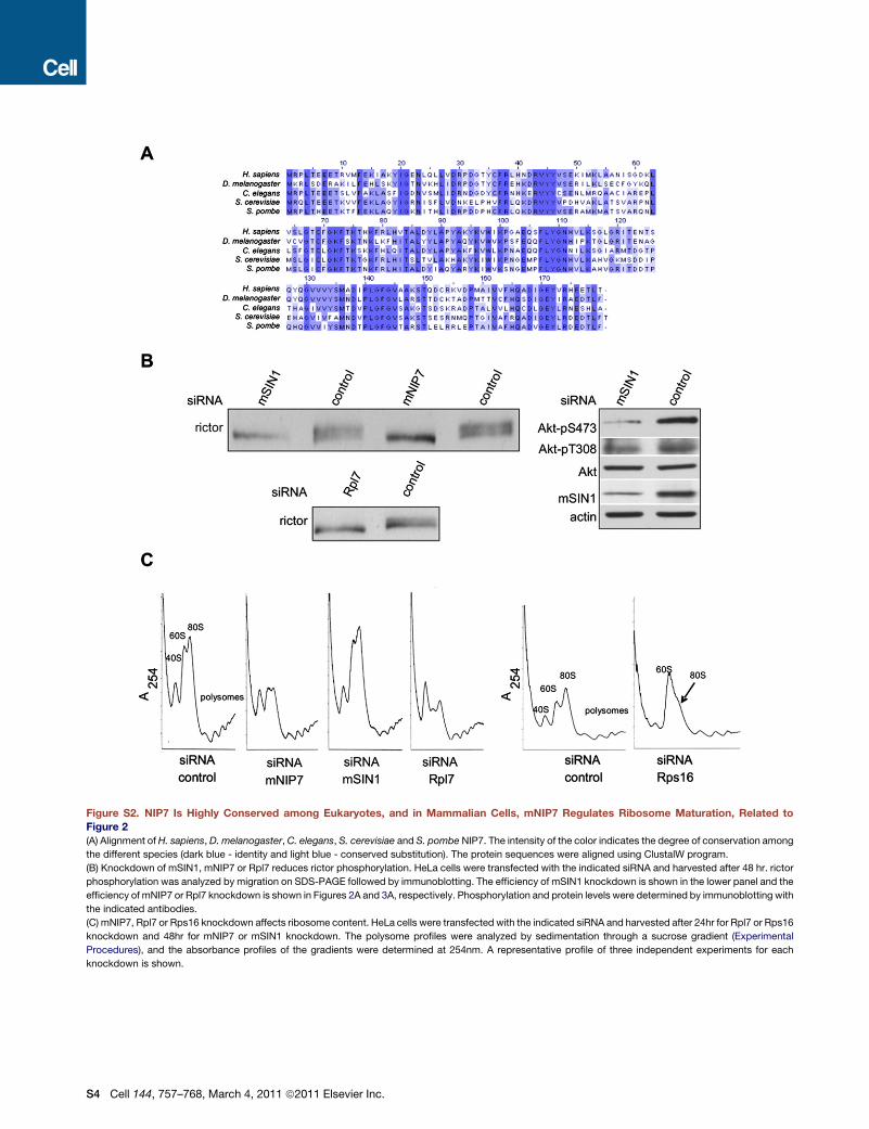
Figure S2. NIP7 Is Highly Conserved among Eukaryotes, and in Mammalian Cells, mNIP7 Regulates Ribosome Maturation, Related toFigure 2(A) Alignment of H. sapiens, D. melanogaster,C. elegans, S. cerevisiae and S. pombe NIP7. The intensity of the color indicates the degree of conservation among
the different species (dark blue - identity and light blue - conserved substitution). The protein sequences were aligned using ClustalW program.
(B) Knockdown of mSIN1, mNIP7 or Rpl7 reduces rictor phosphorylation. HeLa cells were transfected with the indicated siRNA and harvested after 48 hr. rictor
phosphorylation was analyzed by migration on SDS-PAGE followed by immunoblotting. The efficiency of mSIN1 knockdown is shown in the lower panel and the
efficiency of mNIP7 or Rpl7 knockdown is shown in Figures 2A and 3A, respectively. Phosphorylation and protein levels were determined by immunoblotting with
the indicated antibodies.
(C) mNIP7, Rpl7 or Rps16 knockdown affects ribosome content. HeLa cells were transfected with the indicated siRNA and harvested after 24hr for Rpl7 or Rps16
knockdown and 48hr for mNIP7 or mSIN1 knockdown. The polysome profiles were analyzed by sedimentation through a sucrose gradient (Experimental
Procedures), and the absorbance profiles of the gradients were determined at 254nm. A representative profile of three independent experiments for each
knockdown is shown.
S4 Cell 144, 757–768, March 4, 2011 ª2011 Elsevier Inc.

Figure S3. Knockdown of Rps16, but Not Protein Synthesis Inhibition, Decreases mTORC2 Signaling, Related to Figure 3(A) siRNA mediated knockdown of Rps16 decreases basal and insulin-stimulated phosphorylation of Ser473 in Akt and of Thr32 in FoxO3a. HeLa cells were
transfected with the indicated siRNA, harvested after 24 hr (left panel, basal activity) or serum starved for 3 hr and then re-stimulated with insulin for the indicated
times before harvesting (right panel, insulin-stimulated activity). Phosphorylation and protein levels were determined by immunoblotting with the appropriate
antibodies, as indicated.
(B–D) Inhibition of protein synthesis by cycloheximide, anisomycin or salubrinal treatment does not affect mTORC2 signaling.
Cell 144, 757–768, March 4, 2011 ª2011 Elsevier Inc. S5

(B) HeLa cells were treated for the indicated times with cycloheximide (10 mg/ml) (left panel). HeLa cells were serum starved for 3hr, treated with cycloheximide or
DMSO for 20min and then re-stimulatedwith insulin for the indicated times before harvesting (right panel). Phosphorylation and protein levels were determined by
immunoblotting with the indicated antibodies.
(C) HeLa cells were treated with anisomycin (10 mg/ml) for the indicated times (left panel). HeLa cells were serum starved for 3hr, treated with anisomycin (10 mg/
ml) or DMSO for 30 min and then re-stimulated with insulin for the indicated times before harvesting (right panel). Phosphorylation and protein levels were
determined by immunoblotting with the indicated antibodies.
(D) HeLa cells were treated with different concentrations of salubrinal for the indicated times (left panel). HeLa cells were treatedwith salubrinal (100 mM) or DMSO
for 24 hr, serum starved for 3hr and then re-stimulated with insulin for the indicated times before harvesting (right panel). Phosphorylation and protein levels were
determined by immunoblotting with the appropriate antibodies, as indicated.
S6 Cell 144, 757–768, March 4, 2011 ª2011 Elsevier Inc.

Figure S4. A Decrease in Ribosome Content Leads to Increased Sensitivity to Stress-Induced Apoptosis, Related to Figure 4HeLa cells were transfected with the indicated siRNA. After 48 hr, cells were treated with 0.5mM H2O2. Cell viability was quantified by a trypan blue exclusion
assay (Experimental Procedures). The average ± standard deviation from the mean based on two independent experiments is shown.
Cell 144, 757–768, March 4, 2011 ª2011 Elsevier Inc. S7

Figure S5. mTORC2 Cosediments with Polysomes and Associates with mRNA-Bound Ribosomes, Related to Figure 5(A) Cell extracts from HeLa cells were fractionated onto a 15%–45% sucrose gradient after incubation with or without RNase A. The absorbance profile and the
polysomal (P) and ribosomal (M) fractions are indicated. Fractions were concentrated and analyzed by Western blot. Phosphorylation and protein levels were
determined by immunoblotting with the indicated antibodies.
(B) Poly(A) mRNA-bound ribosomes were purified from HeLa cell extracts by oligo(dT) pull-down (Experimental Procedures). The bound fraction (bound) and the
supernatant (sup; 1:10 dilution) were analyzed by immunoblotting with the indicated antibodies (left panel). Poly(A) mRNA binding proteins were purified by oligo
(dT) pull-down after incubation of cell extracts with or without RNase A. The bound fractions were analyzed by immunoblotting with the indicated antibodies (right
panel).
(C) Inhibition of protein synthesis has no effect on mTORC2-ribosome association. HeLa cells were treated with salubrinal (100 mM) or DMSO for 12 hr before
harvesting (left panel). HeLa cells were treated with puromycin (500 mM) or vehicle for 30min before harvesting (right panel). Rpl26 immunoprecipitates (IP: Rpl26)
and cell extracts (lysate) were analyzed by immunoblotting with the indicated antibodies.
S8 Cell 144, 757–768, March 4, 2011 ª2011 Elsevier Inc.

Figure S6. mTOR Interacts with the 60S Ribosomal Subunit via Rictor and/or mSIN1, Related to Figure 6(A) mTOR interacts with the ribosome via rictor and/or mSIN1. HeLa cells were transfected with the indicated siRNA. Cells were harvested 48 hr after transfection.
Rpl26 immunoprecipitates (IP: Rpl26) and cell extracts (lysate) were analyzed by immunoblotting with the indicated antibodies. As reported previously (Thedieck
et al., 2007; Yang et al., 2006), we observed a reduction in the amount of rictor when mSIN1 was knocked down, supporting the earlier suggestion that these two
proteins stabilize each other.
(B) mTORC2 interacts with the ribosome via the 60S ribosomal subunit. HeLa cells were transfected with the indicated siRNA. Cells were harvested 24 hr after
transfection. Rpl26 immunoprecipitates and cell extracts (lysate) were immunoblotted with the indicated antibodies.
(C) mTORC2 kinase activity is not required for mTORC2-ribosome interaction. HeLa cells were treated with PP242 (500nM final concentration) for 60 min before
harvesting. Rpl26 immunoprecipitates and cells extracts (lysate) were immunoblotted with the indicated antibodies.
(D) PI3K inhibition decreases mTORC2-ribosome association. HeLa cells were treated with LY294002 (10 mM final concentration) for 30 min before harvesting.
Rpl26 immunoprecipitates and cells extracts (lysate) were immunoblotted with the indicated antibodies.
Cell 144, 757–768, March 4, 2011 ª2011 Elsevier Inc. S9

Figure S7. mTORC2-Ribosome Interaction Promotes Akt Signaling in Melanoma and Colon Cancer Cells, Related to Figure 7(A) Akt Ser473 phosphorylation andmTORC2-ribosome association are increased in colon cancer cells harboring activating mutations in PI3K. Cell extracts from
colon cancer cells harboring activating mutation in PIK3CA (HT29 and HCT116) and wild-type colon cancer cells (SW620) were analyzed by immunoblotting with
indicated antibodies. Ribosomes were purified from the three cell lines by sedimentation through a sucrose cushion. Ribosomes were probed with the indicated
antibodies.
S10 Cell 144, 757–768, March 4, 2011 ª2011 Elsevier Inc.

(B) Knockdown of mSIN1 or Rpl7 enhances stress-induced apoptosis in PTEN deficient (NA8 and ME67) melanoma cell lines. Melanoma cells (NA8, Juso and
ME67) were transfected with the indicated siRNA and, after 24 hr, were treated with etoposide (25 mM) for 24 hr. Cell viability was determined by trypan blue
exclusion assay and quantified. The average ± standard deviation from the mean based on three independent experiments is shown.
(C) Model of ribosome-dependent mTORC2 activation. Growth factors, via PI3K signaling, induce mTORC2 activation via ribosome association. See text for
details.
Cell 144, 757–768, March 4, 2011 ª2011 Elsevier Inc. S11