asmallribosomalsubunit(ssu)processomecomponent ... · tified a novel pathway for p53 activation...
TRANSCRIPT
A Small Ribosomal Subunit (SSU) Processome Component,the Human U3 Protein 14A (hUTP14A) Binds p53 andPromotes p53 Degradation*□S
Received for publication, June 23, 2010, and in revised form, November 12, 2010 Published, JBC Papers in Press, November 15, 2010, DOI 10.1074/jbc.M110.157842
Lelin Hu‡§, Jiangnan Wang‡§, Yun Liu‡§, Ying Zhang§¶, Liangliang Zhang‡§, Ruirui Kong§¶, Zongfang Zheng§¶,Xiaojuan Du‡§1, and Yang Ke§¶�2
From the ‡Department of Cell Biology and the §Cancer Research Center, Peking University Health Science Center, Beijing 100191, Chinaand the ¶Key Laboratory of Carcinogenesis and Translational Research (Ministry of Education) and the �Genetics Laboratory, PekingUniversity School of Oncology, Beijing Cancer Hospital & Institute, Beijing 100142, China
Ribosome biogenesis is required for normal cell function,and aberrant ribosome biogenesis can lead to p53 activation.However, how p53 is activated by defects of ribosome biogene-sis remains to be determined. Here, we identified humanUTP14a as an SSU processome component by showing thathUTP14a is nucleolar, associated with U3 snoRNA and in-volved in 18 S rRNA processing. Interestingly, ectopic expres-sion of hUTP14a resulted in a decrease and knockdown ofhUTP14a led to an increase of p53 protein levels. We showedthat hUTP14a physically interacts with p53 and functionallypromotes p53 turn-over, and that hUTP14a promotion of p53destabilization is sensitive to a proteasome inhibitor butindependent of ubiquitination. Significantly, knockdown ofhUTP14a led to cell cycle arrest and apoptosis. Our data iden-tified a novel pathway for p53 activation through a defect inrRNA processing and suggest that a ribosome biogenesis factoritself could act as a sensor for nucleolar stress to regulate p53.
Ribosome biogenesis is required for normal cell function,and aberrant ribosome biogenesis can lead to p53 activation.In eukaryotes ribosome biogenesis including transcription ofribosome RNA genes (rDNA),3 rRNA processing, and assem-bly of 40 S and 60 S ribosome subunits takes place in the nu-cleolus. A 47 S rRNA precursor (pre-rRNA) containing thesequences for the mature rRNAs (18 S, 5.8 S, and 28 S rRNA),two external transcribed spacers (ETS) and two internal tran-scribed spacers (ITS) is transcribed by RNA polymerase I (pol
I). After chemical modification at numerous sites, the 47 Spre-rRNA is cleaved to produce 18 S, 5.8 S, and 28 S rRNAs.The 18 S rRNA is incorporated in the 40 S small subunit(SSU) with small subunit ribosomal proteins. The 5.8 S rRNAand 28 S rRNA are incorporated into the 60 S large subunitwith 5 S rRNA, which is independently transcribed by RNApolymerase III elsewhere. Both the large and small subunitsare assembled in the nucleolus and transferred to the cyto-plasm for protein synthesis.Polymerase I transcription and pre-rRNA processing can
be disrupted by serum starvation, depletion of nucleotides,chemical reagents, and malfunction of nucleolar proteins. Allof these nucleolar functional disruptions have been shown toresult in nucleolar stress signaling to p53. For example, acti-nomycin D selectively inhibits RNA pol I-dependent tran-scription at low concentrations (5–10 nM) (1, 2) and stimu-lates stabilization of p53. The chemotherapeutic agent5-fluorouracil (5-FU), which blocks pre-rRNA processing byincorporating newly synthesized rRNA activates p53. Func-tional disruption of nucleolar proteins, which are required forrRNA processing, also activates p53. Bop1 is an essential fac-tor in 28 S rRNA processing and its dominant negative mu-tant inhibits ribosomal biogenesis and elicits p53 activation(3). For 18 S rRNA processing, the U3 snoRNA base pairswith the 47 S pre-rRNA at A0, A1, and A2 sites (4, 5) and me-diates 18 S rRNA maturation (6). If a protein is nucleolar, as-sociated with U3 snoRNA and required for 18 S rRNA pro-cessing, it is identified as UTP (U3 protein). UTPs are alsoknown as SSU processome as they play important roles in 40S subunit biogenesis (7, 8). Recently, disruption of humanUTP18 was found to induce p53 activation (9). In addition,inhibition of nucleophosmin (B23) activity by ARF (10) andreduction of ribosomal protein S6 (11) also activates p53.All of these nucleolar or ribosomal stresses activate p53,
which induces cell cycle arrest and/or apoptosis. The in vivosignificance of this p53 activation has been determined from anumber of mouse models. For example, A mouse model withthe juvenile spermatogonial depletion phenotype (jsd) showedthat utp14b is required for spermatogenesis in mice (12, 13).A p53-dependent pathway has been found to mediate apopto-sis in spermatogonial differentiation in utp14bjsd mice (14).However, the mechanisms by which nucleolar disruptionsdirect p53 activation are largely undefined.
* This work was supported by grants from the National Natural ScienceFoundation of China (Grants No. 30571038 and 30771224), the Key Con-struction Program of the National “985” Project (Grant No. 985-2-016-24),Grants from the National High Technology Research and DevelopmentProgram of China (863 Program) (Grant No. 2008AA02Z131 and2006AA02A402), and from the National Basic Research Program of China(973 Program) (Grant No. 2010CB529303).Author’s Choice—Final version full access.
□S The on-line version of this article (available at http://www.jbc.org) con-tains supplemental Fig. S1.
1 To whom correspondence may be addressed: Cancer Research Center, 38Xue-Yuan Road, Hai-Dian District, Beijing 100191, China. Fax: 86-10-82801130; E-mail: [email protected].
2 To whom correspondence may be addressed: Genetics Laboratory, 52Fu-Cheng Road, Hai-Dian District, Beijing 100142, China. Fax: 86-10-62015681; E-mail: [email protected].
3 The abbreviations used are: rDNA, ribosome RNA genes; SSU, small sub-unit; snoRNA, small nucleolar RNA; hUTP, human U3 protein.
THE JOURNAL OF BIOLOGICAL CHEMISTRY VOL. 286, NO. 4, pp. 3119 –3128, January 28, 2011Author’s Choice © 2011 by The American Society for Biochemistry and Molecular Biology, Inc. Printed in the U.S.A.
JANUARY 28, 2011 • VOLUME 286 • NUMBER 4 JOURNAL OF BIOLOGICAL CHEMISTRY 3119
by guest on August 24, 2019
http://ww
w.jbc.org/
Dow
nloaded from

In unstressed cells, the p53 protein level remains lowthrough regulation of its protein stability by a number of neg-ative regulators. MDM2 serves as a key negative feedback reg-ulator for p53 and various stresses activate distinct cellularsignaling pathways leading to the suppression of MDM2 ac-tivity and activation of p53 (15, 16). Thus, the p53-MDM2feedback loop plays an essential role in response to a multi-tude of genotoxic and cytotoxic stressors. It has been foundthat 5 S rRNA and ribosomal protein (RP) of the large subunitRPL5 interact with MDM2 (17) and RPL5 participates inMDM2 nuclear export (18). It was thought that p53-MDM2might “hitch a ride” on the ribosome for cytoplasmic degrada-tion (19). Therefore, nucleolar stress was thought to inducep53 accumulation due to a failure in nucleolus-dependentexport and degradation of p53 in the cytoplasm (20). Laterstudies found that treating cells with either a lower dose ofactinomycin D or serum starvation inhibits ribosome assem-bly and consequently releases free ribosomal proteins fromthe nucleolus to the nucleoplasm (21). Moreover, it has beenfound that several ribosomal 60 S proteins including RPL11(22, 23), RPL23 (24, 25), and RPL5 (23) interact with MDM2.This binding inhibits the MDM2 E3 ligase function, resultingin p53 accumulation and activation. A small ribosomal sub-unit protein RPS7 (27, 28) has also been shown to interactwith MDM2. In addition, RPL26 was found to increase thetranslational rate of p53 mRNA by binding to its 5�-untrans-lated region (29). All these findings identify p53 as a moleculewhich is critical in sensing nucleolar stress, and suggest thatRPs may play a pivotal role in the p53 response to nucleolarstress. A recent study revealed that defects in 18 S and 28 SrRNA processing activate p53 by an RPL11-dependent path-way (9). However, the specific linkage between pre-rRNAprocessing and p53 activation has not been determined.Recently, mutations in human UTP14C have been found in
3 of 234 nonobstructive and azoospermic/severely oligosper-mic males, suggesting that the human UTP14C gene is associ-ated with human spermatogenesis and fertility, and raisingthe possibility UTP14Cmay be functionally equivalent tomouse utp14b (30). More importantly, expression of UTP14CmRNA has also been found to be associated with humanovarian cancer (31). Mouse utp14b and human UTP14C areretrogenes of the X-linked utp14a gene and UTP14A, respec-tively. Thus, functional study of human UTP14a will deter-mine the fundamental molecular mechanisms by whichUTP14c functions in spermatogenesis deficiency and ovariancancer. Therefore, we set out to investigate the function ofhuman UTP14a (hUTP14a) and explore the mechanism bywhich nucleolar stress activates the p53 pathway. In thisstudy, we identified hUTP14a as the mammalian homolog ofyeast Utp14 which functions in 18S rRNA processing, andfound that hUTP14a itself acts as a nucleolar stress sensor,which signals to p53.
EXPERIMENTAL PROCEDURES
Plasmids and Antibodies—The expression plasmids codingpCI-neo-Flag-hUTP14a and series deletion mutants Del-1(amino acids 1–267), Del-2 (amino acids 268–645), and Del-3(amino acids 646–771) were obtained by RT-PCR-cloning
using total RNA extracted from HeLa cells as a template. Theplasmids were verified by DNA sequencing. Plasmids codingpEGFP-hUTP14a and its series deletion mutants were con-structed by inserting hUTP14a cDNA fragments from pCI-neo-Flag-hUTP14a into the pEGFP plasmid. Plasmids codingGST-hUTP14a and its series deletion mutants were con-structed by inserting hUTP14a cDNA fragments from pCI-neo-Flag-hUTP14a into the pGEX-4T1 plasmid. Plasmidscoding MDM2, p53 and p53 deletion mutants were kindlyprovided by Dr. Yongfeng Shang (Peking University HealthScience Center). Plasmid coding HA-Ub was constructed byinserting an ubiquitin cDNA fragment into a pCMV-HA plas-mid. Antibodies directed against p53 or MDM2 were pur-chased from Santa Cruz Biotechnology, p53 phosphorylationantibodies including anti-Ser15, anti-Thr18, anti-Ser20, anti-Ser33, and anti-Ser37 are fromCell Signaling. Anti-p21Waf1/Cip1,anti-nucleolin, anti-PARP, and anti-�-actin were from SantaCruz Biotechnology, and anti-HA and anti-Flag monoclonalantibody M2 were from Sigma. Anti-1A6/DRIM monoclonalantibody was raised in our laboratory. TRITC-conjugatedgoat anti-mouse IgG and FITC-conjugated goat anti-rabbitIgG were from the Zhongshan Goldenbridge BiotechnologyCo., Ltd.Generation of an Anti-hUTP14a Antibody—A rabbit poly-
clonal antibody was generated against peptides derived fromthe C-terminal 18 amino acids (amino acids 751–768: QRN-PKRITTRHKKQLKKC) of the hUTP14a protein and usedafter affinity purification.Cell Culture and Transfections—Cell lines were grown in
DMEM or RPMI 1640 medium supplemented with 10% FCSaccording to the instructions of the ATCC (American TypeCulture Collection). For knockdown of hUTP14a expression,three hUTP14a-specific siRNAs (small interference RNA)(A-1: 5�-CAGGAAGAACUAGCGGAUUtt-3�; A-2: 5�-GACGCCGGUUUCUCAUUAAtt-3�; A-3: 5�- GAGAUU-GAACGGAUCCACAtt-3�) together with an unrelated con-trol siRNA (siNC: 5�-ACUACCGUUGUUAUAGGUG-3�)were chemically synthesized (Shanghai GenePharma Co.,Ltd). These synthesized siRNAs were transfected into cells ata concentration of 100 nM. Transfections were carried outwith LipofectamineTM 2000 (Invitrogen) according to themanufacturer’s instructions.Immunofluorescence Staining—Cells were plated on cover-
slips in 6-well plates 1 day before harvest. 1A6/DRIM,hUTP14a, Flag-hUTP14a, and nucleolin were detected, re-spectively, with the monoclonal anti-1A6/DRIM antibody6D9, the polyclonal antibody against hUTP14a, and themonoclonal antibody against Flag or nucleolin-specific poly-clonal antibody after cells were fixed with methanol/acetone.The 1A6/DRIM- or Flag-hUTP14a-specific immunocom-plexes were identified with TRITC-conjugated goat anti-mouse IgG. The hUTP14a and nucleolin specific immuno-complexes were detected with FITC-conjugated goatanti-rabbit IgG. Immunofluorescence signals were recordedwith confocal laser scanning microscopy (Leica TCS-ST2).Newly Synthesized rRNA Analysis—Pulse-chase labeling
was performed as described previously (32). In brief, 72 h af-ter transfection of chemically synthesized siRNA, HeLa cells
hUTP14a Promotes p53 Degradation
3120 JOURNAL OF BIOLOGICAL CHEMISTRY VOLUME 286 • NUMBER 4 • JANUARY 28, 2011
by guest on August 24, 2019
http://ww
w.jbc.org/
Dow
nloaded from
were labeled with L-[methyl-3H]methionine (PerkinElmer LifeSciences) for 30 min. RNA was evaluated at various timepoints. Total RNA was isolated with Trizol reagent (Invitro-gen) and resolved on a 1% agarose-glyoxal gel (Ambion). TheRNA was transferred to a nylon membrane and the newlysynthesized rRNAs were detected by fluorography.Immunoprecipitation Analyses of Protein-RNAAssociation—
Immunoprecipitation was performed as described previously(33). Briefly, HeLa cell lysates were prepared in buffer A (25mM Tris-Cl, pH 7.5, 100 mM KCl, 1 mM dithioerythreitol, 2mM EDTA, 0.5 mM phenylmethylsulfonyl fluoride, 0.05%Nonidet P-40, 1 unit/ml RNasin) and used directly for immu-noprecipitation. Antibody was coupled with a 50% suspensionof protein A-Sepharose beads (Amersham Biosciences) inIPP500 (500 mM NaCl, 10 mM Tris-Cl, pH 8.0, 0.05% NonidetP-40). Coupled beads were incubated with cellular extracts for2 h at 4 °C. After washes, precipitated proteins were analyzedby Western blotting and co-precipitated RNA was isolatedwith Trizol� reagent (Invitrogen).Immunoblot—Proteins from cellular fractions were sepa-
rated on SDS-PAGE and transferred onto PVDF membrane.Membranes were probed with corresponding primary anti-bodies after blocking with 5% milk in PBS/T (0.5% Tween-20in PBS buffer). After extensive washing with PBS/T, the mem-brane was incubated with HRP-conjugated secondary anti-body followed by detection using the ECL-Kit (AmershamBiosciences).Northern Blot—To visualize U3 snoRNA, Northern blot
was performed as described previously (34) with minor modi-fications. In brief, the RNA sample was loaded onto 7% poly-acrylamide-8.3 M urea gel and blotted onto a BrightStar�-PLUS positively charged Nylon membrane (Ambion) byelectroblotting (Bio-Rad). The U3 snoRNA-specific RNAprobe was labeled with biotin-UTP using the Riboprobe� Sys-tem (Promega) with SalI-linearized pGEM-T-U3 (34). Hy-bridization was carried out for 16 h at 65 °C after 3 h of prehy-bridization. Detection was carried out with the BrightStar�BioDetectTM Kit (Ambion) according to the manufacturer’sinstructions.Reverse Transcription PCR—For U3 snoRNA amplification,
RT-PCR was performed as previously described (34).GST Pull-down—GST or GST fusion proteins were ex-
pressed in E. coli and crosslinked to glutathione-Sepharosebeads (BD Product). In vitro transcription/translation wasperformed with a rabbit reticular lysis-coupled transcription/translation kit (Promega). GST pull-down was carried out byincubating the GST fusion proteins with in vitro transcribed/translated proteins. The GST fusion protein interacting pro-teins were detected by Western blotting probed with appro-priate antibodies.Flow Cytometric Analysis—For DNA content analysis, cells
were trypsinized, washed with PBS, and fixed in 75% ice-coldethanol at 4 °C overnight. Cells were rehydrated in PBS on thesecond day. Following RNase digestion, cells were stainedwith propidium iodide (PI). Flow cytometry analysis was per-formed using red (PI) emission (at 630 nm). Data from 104cells were collected and analyzed by using Cellquest software(Becton Dickinson). For apoptosis analysis, cells were double-
stained with annexin V and PI and subjected to flow cytomet-ric analysis.BrdU Incorporation Assay—The cell population in S-phase
was determined with a BD PharmingenTM BrdU Flow kit ac-cording to the manufacturer’s instruction. Briefly, cells werepulsed for 16 h with bromodeoxyuridine (BrdU), and werethen stained with fluorescein isothiocyanate (FITC)-conju-gated anti-BrdU and 7-amino-actinomycin D (7-AAD). Posi-tive BrdU staining and 7-AAD staining were analyzed byFACScan (Becton Dickinson). Data from 104 cells were ana-lyzed by using Cellquest software (Becton Dickinson). Cells inS-phase were quantitated by comparing FITC (DNA incorpo-ration) versus 7-AAD (total DNA) staining.Growth Curve—A growth curve was plotted with the Cell
Counting Kit-8 (CCK-8, Dojindo) according to the manufac-turer’s instructions. In brief, cells were seeded in 96-wellplates. WST-8 [2-(2-methoxy-4-nitrophenyl)-3-(4-nitrophe-nyl)-5-(2,4-disulfophenyl)-2H-tetrazolium, monosodium salt]was added to these cells and incubated for 4 h at differenttime points. Absorbance at 450 nm was measured using a mi-croplate reader. The experiment was repeated three times induplicate. Growth curves were plotted using the mean � S.D.of absorbance at 450 nm versus the time points.
RESULTS
Expression Profile of hUTP14a in Different Human CellLines—Sequence alignment showed that hUTP14a is con-served among Saccharomyces cerevisiae, mice, and humans(supplemental Fig. S1). To evaluate the function of hUTP14aprotein and to detect endogenous hUTP14a expression, weraised a rabbit polyclonal antibody against hUTP14a and con-firmed its specificity by Western blotting utilizing whole celllysates of Flag-hUTP14a or hUTP14a-transfected HeLa cells(Fig. 1A). A band at the expected molecular mass forhUTP14a of 88 kDa was observed in HeLa cells andhUTP14a-transfected cells. Flag-tagged hUTP14a was recog-nized by both anti-hUTP14a antibody and anti-Flag antibodyM2 as a band of the same size. In addition, we analyzed theexpression of hUTP14a in various cell lines. Cellular fractionswere prepared and proteins from the fractions were separatedon SDS-PAGE, and transferred onto a PVDF membrane. Theblot was probed with anti-hUTP14a. Fig. 1B shows thathUTP14a was ubiquitously expressed in the nuclear extractsof the cell lines under evaluation. Subcellular localization ofendogenous hUTP14a was determined by indirect immuno-fluorescence performed with the polyclonal anti-hUTP14aantibody and an anti-1A6/DRIM monoclonal antibody wasused as a nucleolar protein marker. As shown in Fig. 1C,hUTP14a was predominantly localized in the nucleolus andco-localized with 1A6/DRIM (also known as humanUTP20). To investigate the localization of ectopically ex-pressed hUTP14a, GFP-hUTP14a was transfected intoU2OS cells and GFP fluorescence showed the same local-ization pattern with endogenous hUTP14a (Fig. 1D). Flag-hUTP14a was transfected into U2OS cells and doubleindirect immunofluorescence staining was performed withanti-Flag antibody and anti-nucleolin antibody. The confo-
hUTP14a Promotes p53 Degradation
JANUARY 28, 2011 • VOLUME 286 • NUMBER 4 JOURNAL OF BIOLOGICAL CHEMISTRY 3121
by guest on August 24, 2019
http://ww
w.jbc.org/
Dow
nloaded from

cal image showed Flag-hUTP14a co-localized with nucleo-lin in the nucleolus (Fig. 1E).hUTP14a Is an SSU Processome Component—To investi-
gate whether hUTP14a is required for pre-rRNA processing,hUTP14a-specific siRNA was transfected into HeLa cells, andpulse chase labeling was performed to analyze newly synthe-sized rRNA 72-h post-transfection. Efficiency of hUTP14asilencing was determined by Western blotting using anti-hUTP14a antibody (Fig. 2A). The pulse chase results showedthat at the chase time points 15 min, 30 min, 1 h, and 1.5 h,the level of 41 S rRNA decreased (Fig. 2B, right). In addition,the 18 S rRNA level decreased markedly at chase time points15 min, 30 min, 1 h, and 1.5 h in hUTP14a-deficient cells. Todetermine whether hUTP14a is associated with U3 snoRNA,immunoprecipitation was performed using anti-hUTP14aantibody. Protein and RNA from the precipitates were ana-lyzed in parallel. Proteins extracted from one-half of the pre-cipitate were subjected to Western blotting to analyze forhUTP14a. RNA was extracted from the remaining half ofthe precipitate and was used for RT-PCR to analyze for U3snoRNA. As shown in Fig. 2C (left panel), U3 snoRNA wasamplified from the hUTP14a-specific immunoprecipitates. Toconfirm this observation, RNA was extracted from thehUTP14a-specific immunoprecipitates and subjected toNorthern blotting to analyze for U3 snoRNA. The resultsshowed that U3 snoRNA was present in the hUTP14a-specificimmunoprecipitates (Fig. 2C, right panel) demonstrating invivo association between hUTP14a and U3 snoRNA. In addi-tion, Northern blotting showed that U3 snoRNA levels werenot affected by silencing of hUTP14a expression (Fig. 2A,lower panel). These results demonstrated that depletion ofhUTP14a inhibited 18 S rRNA processing. Taken these resultstogether, we identified hUTP14a as an SSU processomecomponent.
FIGURE 1. Expression profile of hUTP14a in different human cell lines.A, specificity of anti-hUTP14a antibody was verified by Western blotting.Whole cell extracts were prepared from HeLa cells and Flag-hUTP14a- orhUTP14a-transfected HeLa cells. Equal amount of proteins from the ex-tracts were separated by SDS-PAGE, and transferred onto PVDF mem-branes. Blots were probed with anti-hUTP14a antibody. Endogenousand exogenous hUTP14a are indicated (upper panel). Blots as describedabove were stripped and reprobed with anti-Flag antibody M2 (lowerpanel). B, expression of hUTP14a in various human cell lines. Cytosolicand nuclear extracts were fractionated from multiple cell lines. The sameamount of protein was separated on SDS-PAGE and transferred onto aPVDF membrane. Blots were probed with anti-hUTP14a antibody. Cellu-lar fractionation was controlled by using nuclear protein topoisomeraseI (Topo I) as a nuclear marker and cytoplasmic protein RhoA as a cytoso-lic marker. N represents nuclear extract and C represents cytosolic ex-tract. Cell lines are indicated at the top of the blots. C, endogenoushUTP14a is localized to the nucleolus. Immunofluorescence was per-formed with 1A6/DRIM-specific monoclonal antibody and anti-hUTP14apolyclonal antibody on U2OS cells. 1A6/DRIM immuno-signals were de-tected with TRITC-conjugated goat anti-mouse IgG and hUTP14a im-muno-complexes were recognized with FITC-conjugated goat anti-rab-bit IgG. Nucleus was stained with DAPI. D, GFP-hUTP14a displayed thesame localization pattern as endogenous hUTP14a. Plasmid coding GFP-hUTP14a was transfected into U2OS cells. Localization of GFP proteinwas observed and recorded with confocal microscopy. E, ectopicFlag-hUTP14a is localized to the nucleolus. U2OS cells were transfectedwith Flag-hUTP14a. Immunofluorescence was performed using anti-Flagmonoclonal antibody and anti-nucleolin polyclonal antibody. Flag-hUTP14a immuno-signals were detected with TRITC-conjugated goatanti-mouse IgG and nucleolin immuno-complexes were recognized withFITC-conjugated goat anti-rabbit antibody. The nuclei were stained with DAPI.
hUTP14a Promotes p53 Degradation
3122 JOURNAL OF BIOLOGICAL CHEMISTRY VOLUME 286 • NUMBER 4 • JANUARY 28, 2011
by guest on August 24, 2019
http://ww
w.jbc.org/
Dow
nloaded from
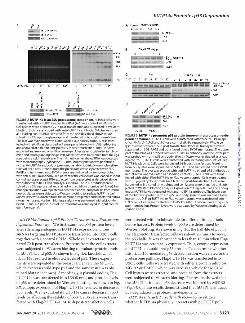
hUTP14a Promotes p53 Protein Turnover via a Proteasome-dependent Pathway—We first examined p53 protein levelsafter silencing endogenous hUTP14a expression. ThreesiRNAs targeting hUTP14a were transfected into U2OS cellstogether with a control siRNA. Whole cell extracts were pre-pared 72 h post-transfection. Proteins from the cell extractswere subjected to Western blotting to evaluate protein levelsof hUTP14a and p53. As shown in Fig. 3A, knockdown ofhUTP14a resulted in elevated levels of p53. These experi-ments were repeated in the breast cancer cell line MCF-7,which expresses wild-type p53 and the same result was ob-tained (data not shown). Accordingly, a plasmid coding Flag-hUTP14a was transfected into U2OS cells, and protein levelsof p53 were determined by Western blotting. As shown in Fig.3B, ectopic expression of Flag-hUTP14a resulted in decreasedp53 levels. We next asked if hUTP14a causes decrease in p53levels by affecting the stability of p53. U2OS cells were trans-fected with Flag-hUTP14a. At 16-h post-transfection, cells
were treated with cycloheximide for different time periodsbefore harvest. Protein levels of p53 were determined byWestern blotting. As shown in Fig. 3C, the half-life of p53 inthe Flag vector transfected cells was about 20 min. However,the p53 half-life was shortened to less than 10 min when Flag-hUTP14a was ectopically expressed. Thus, ectopic expressionof hUTP14a destabilized p53 protein. To determine whetherthis hUTP14a-mediated p53 destabilization was related to theproteasome pathway, Flag-hUTP14a was transfected intoU2OS cells. Cells were treated with either a protein inhibitor,MG132 or DMSO, which was used as a vehicle for MG132.Cell lysates were extracted, and proteins from the extractswere subjected to Western blotting. The results showed thatthe hUTP14a-induced p53 decrease was blocked by MG132(Fig. 3D). These results demonstrated that hUTP14a-inducedp53 degradation is proteasome-dependent.UTP14a Interacts Directly with p53—To investigate
whether hUTP14a physically interacts with p53, GST pull-
FIGURE 2. hUTP14a is an SSU processome component. A, HeLa cells weretransfected with a hUTP14a-specific siRNA (A-1) or a control siRNA (siNC).Cell lysates were prepared 72-h post-transfection and subjected to Westernblotting. Blots were probed with anti-hUTP14a antibody. �-Actin was usedas a loading control. RNA extracted from the cells described above was re-solved on a 1% agarose-glyoxal gel and transferred onto a nylon membrane.The blot was hybridized with biotin-labeled U3 snoRNA probe. B, cells trans-fected with siRNAs as described in A were pulse labeled with [3H]methionineand analyzed at different time points 72-h post-transfection. Total RNA wasextracted and resolved on a 1% agarose gel. After staining with ethidium bro-mide and photographing the gel (left panel), RNA was transferred from the aga-rose gel to a nylon membrane. The [3H]methionine-labeled RNA was detectedwith radioautography (right panel). C, immunoprecipitation was performedwith anti-hUTP14a antibody or pre-immune rabbit IgG (rIgG) on whole cell ex-tracts of HeLa cells. Proteins from the precipitates were separated with SDS-PAGE and transferred onto PVDF membranes followed by immunoprobingwith anti-hUTP14a antibody. Ten percent of the cell extract was loaded as inputcontrol (left upper panel). RNA extracted from precipitates as described abovewas subjected to RT-PCR to amplify U3 snoRNA. The PCR products were re-solved on a 2% agarose gel and stained with ethidium bromide (left lower). Im-munoprecipitation was repeated as described above, and proteins from immu-noprecipitates were subjected to Western blotting to analyze hUTP14a (rightupper). RNA was extracted from the immunoprecipitates and transferred onto anylon membrane. Northern blotting analysis was performed with a biotin-la-beled U3 snoRNA probe. 2.5% of the total RNA was employed as input control(right lower panel).
FIGURE 3. hUTP14a promotes p53 protein turnover in a proteasome-de-pendent manner. A, U2OS cells were transfected with three hUTP14a-spe-cific siRNAs (A-1, A-2, and A-3) or a control siRNA, respectively. Whole celllysates were prepared 72-h post-transfection. Proteins from lysates wereseparated on SDS-PAGE and transferred onto a PVDF membrane. The upperpart of the blot was probed with anti-hUTP14a antibody, and the lower partwas probed with anti-p53 antibody. �-Actin (Actin) was evaluated as a load-ing control. B, U2OS cells were transfected with increasing amounts of Flag-hUTP14a plasmid. Cells were harvested 24-h post-transfection. Proteinsfrom cell lysates were separated on SDS-PAGE and transferred onto a PVDFmembrane. The blot was probed with anti-hUTP14a or anti-p53 antibody asin A. �-Actin was evaluated as a loading control. C, U2OS cells were trans-fected with either Flag-hUTP14a or Flag vector plasmid. Cells were treatedwith 15 �g/ml cycloheximide for 4 h at 16-h post-transfection. Cells wereharvested at indicated time points, and cell lysates were prepared and sub-jected to Western blotting analysis. Expression of Flag-hUTP14a and endog-enous hUTP14a was detected with anti-hUTP14a antibody. The lower partof the blot was probed with anti-p53 antibody. �-Actin was used as a load-ing control. D, Flag-hUTP14a or Flag vector plasmid was transfected intoU2OS cells. Cells were treated with DMSO or MG132 before harvesting 20 hpost-transfection. Protein levels were evaluated by Western blotting as de-scribed above.
hUTP14a Promotes p53 Degradation
JANUARY 28, 2011 • VOLUME 286 • NUMBER 4 JOURNAL OF BIOLOGICAL CHEMISTRY 3123
by guest on August 24, 2019
http://ww
w.jbc.org/
Dow
nloaded from

down experiments were performed with E. coli expressedGST-p53 fusion protein or GST protein and in vitro tran-scribed/translated Flag-hUTP14a. The GST fusion protein-bound Flag-hUTP14a was determined by Western blottingusing anti-UTP14a antibody. These results showed thathUTP14a specifically interacted with p53 (Fig. 4A). To mapthe interaction domain in p53, GST pull-down experimentswere performed with in vitro transcribed/translated Flag-hUTP14a and GST-p53 deletion mutants. Fig. 4B shows thathUTP14a interacted with the central core region and C termi-nus of p53. Conversely, the interacting domain in hUTP14awas mapped using in vitro translated p53 and GST-hUTP14adeletion mutants with GST pull-down experiments. Fig. 4Cshows that both the N and C terminus of hUTP14a were re-quired for interaction with p53. Further, the cellular localiza-tion of hUTP14a deletion mutants was examined. The plas-mids coding GFP-hUTP14a deletion mutants weretransfected into U2OS cells separately. The localization ofGFP proteins was observed under confocal microscopy afternuclear staining with DAPI. The results showed that bothp53-interacting domains in hUTP14a were localized in thenucleus (Fig. 4D). To confirm the interaction betweenhUTP14a and p53, Flag-hUTP14a and p53 plasmids weretransfected into a p53-null cell line H1299. Immunoprecipita-tion was performed with anti-p53 antibody, and proteins fromthe precipitates were separated with SDS-PAGE and trans-ferred onto a PVDF membrane. The blot was hybridized withanti-hUTP14a antibody and anti-p53 antibody. Fig. 4E showsthat both Flag-hUTP14a and endogenous hUTP14a werepresent in the p53-specific immunoprecipitates, confirmingthat hUTP14a was associated with p53 in cells.hUTP14a Promotes p53 Degradation Possibly through a
Ubiquitin-independent Pathway—The next question raisedwas whether hUTP14a-induced p53 degradation depends onubiquitination. To this end, H1299 cells were co-transfectedwith p53 and HA-Ub plasmids in the presence of either Flag-hUTP14a or Flag vector or MDM2, which is known to ubiq-uitinate p53 protein. Cells were treated with MG132 beforeharvest. Polyubiquitination of p53 was identified by Westernblotting. As shown in Fig. 5A, polyubiquitination of p53 wasobserved when p53 was co-transfected with HA-Ub in thepresence ofMDM2. However, co-expression of Flag-hUTP14awith HA-Ub did not show any effect on p53 polyubiquitina-tion. To confirm that hUTP14a induces p53 degradation in-dependent of ubiquitination, H1299 cells were co-transfectedwith p53 and Flag-hUTP14a or MDM2 expression plasmidsin the presence of HA-Ub. Immunoprecipitation experimentswere performed with anti-p53 antibody and Western blottingwas performed with anti-HA antibody. As shown in Fig. 5B,p53 polyubiquitination was enhanced when p53 was co-trans-fected with HA-Ub and MDM2. In contrast, Flag-hUTP14a
FIGURE 4. hUTP14a specifically interacts with p53 in vitro and in cells.A, GST pull-down was performed with E. coli expressed GST-p53 fusion pro-tein or GST and in vitro transcribed/translated Flag-hUTP14a. The GST fusionprotein-bound Flag-hUTP14a proteins were analyzed by Western blot probedwith anti-hUTP14a antibody. Ten percent of the Flag-hUTP14a protein wasloaded as an input control. B, GST pull-down was performed with GST or GST-p53 deletion mutant fusion proteins and in vitro translated Flag-hUTP14a. TheGST-p53 bound Flag-hUTP14a proteins were analyzed by Western blot probedwith anti-hUTP14a antibody. Ten percent of the Flag-hUTP14a protein wasloaded as an input control. A schematic structure of p53 deletion mutants isshown in the lower panel. C, GST pull-down was performed with GST or GST-hUTP14a deletion mutant fusion proteins and in vitro translated p53. The GST-hUTP14a-bound p53 proteins were analyzed by Western blot probed with anti-p53 antibody. Ten percent of the p53 protein was loaded as an input control. Aschematic structure of hUTP14a deletion mutants is shown in the lower panel.D, plasmids coding GFP-hUTP14a deletion mutants were transfected into U2OScells, and GFP proteins were observed under confocal microscopy after thenucleus was stained with DAPI. E, Flag-hUTP14a and p53 plasmids were
transfected into H1299 cells. Cell lysates were extracted 24 h post-transfec-tion. Immunoprecipitation was performed with anti-p53 antibody, and pro-teins from the immunoprecipitates were subjected to Western blotting. Theupper part of the blot was probed with anti-hUTP14a antibody, and thelower part was hybridized with anti-p53 antibody. The expression level ofhUTP14a and p53 in whole cell extracts (WCE) was determined by Westernblotting.
hUTP14a Promotes p53 Degradation
3124 JOURNAL OF BIOLOGICAL CHEMISTRY VOLUME 286 • NUMBER 4 • JANUARY 28, 2011
by guest on August 24, 2019
http://ww
w.jbc.org/
Dow
nloaded from

showed no effect on p53 polyubiquitination. We further de-termined that hUTP14a had no effect on p53 polyubiquitina-tion in the absence of ubiquitin overexpression. H1299 cellswere co-transfected with p53 and Flag-hUTP14a or MDM2expression plasmids. Cells were treated with MG132 beforeharvest. Polyubiquitination of p53 was examined by Westernblotting. As shown in Fig. 5C, p53 is polyubiquinated byMDM2 but not by Flag-hUTP14a. These results suggestedthat Flag-hUTP14a may induce p53 degradation in an ubiq-uitin-independent manner.To determine whether knockdown of hUTP14a can stabi-
lize p53 by affecting p53 phosphorylation, p53 phosphoryla-tion at Ser-15, Thr-18, Ser-20, Ser-33, and Ser-37 wasevaluated after silencing hUTP14a. Cell lysates from p53-transfected H1299 cells were used as positive control. Asshown in Fig. 5D, DNA damage reagent doxorubicin inducedp53 phosphorylation at Ser-15, Ser-20, Ser-33, and Ser-37sites, while no detectable p53 phosphorylation at these phos-phorylation sites was induced by hUTP14a depletion. More-over, treatment of cells with actinomycin D or 5-FU (FUrd)did not show detectable phosphorylation of these phosphory-lation sites.Knockdown of hUTP14a Inhabits Cell Growth and Induces
Apoptosis—We then asked whether hUTP14a modulates p53activity. As hUTP14a is ubiquitously expressed in various hu-man cell lines, most of these experiments were conductedwith knockdown of hUTP14a. First, we examined cell growthwhen hUTP14a was silenced in U2OS cells. Colony formationwas performed with U2OS cells following knockdown ofhUTP14a by siRNA. As shown in Fig. 6A, a significant reduc-tion in both the size and the number of colonies was observedin hUTP14a-deficient cells. To confirm the effect of hUTP14aknockdown on cell growth, a growth curve was plotted afterhUTP14a was depleted by siRNA in U2OS cells. As shown inFig. 6B, cell growth was significantly inhibited in the
FIGURE 5. hUTP14a promotes p53 degradation possibly through anubiquitin-independent pathway. A, H1299 cells were co-transfected withp53 and Flag-hUTP14a or MDM2 or Flag vector plasmid in the presence ofHA-Ub. Cells were treated with 10 �M MG132 for 4 h before harvest. Equalamount of protein of whole cell extracts were subjected to Western blottingfor detection of p53. B, H1299 cells were co-transfected with p53 plasmidand HA-Ub with either Flag-hUTP14a or MDM2 or Flag vector plasmid. Cellswere treated with 10 �M MG132 for 4 h before harvesting at 20 h post-transfection. Immunoprecipitation was performed with anti-p53 antibody.The proteins from the precipitates were separated on SDS-PAGE and trans-ferred to a PVDF membrane. The blot was probed with anti-HA antibody(upper panel). Expression of hUTP14a or MDM2 was detected by immuno-blot with anti-Flag antibody or anti-MDM2 antibody (middle panels). Equalamounts of protein from whole cell extracts (WCE) were subjected to West-ern blotting and probed for HA-Ub with anti-HA antibody (lower panel).C, H1299 cells were co-transfected with p53 and Flag-hUTP14a or MDM2expression plasmid or Flag vector. Cells were treated with 10 �M MG132 for4 h before harvest. Equal amount of proteins of whole cell extracts weresubjected to Western blotting for detection of p53. D, U2OS cells weretransfected with hUTP14a specific siRNAs (A-1, A-1, or A-3) or a controlsiRNA. Cells were harvested and whole cell extracts were prepared 72-hpost-transfection. Proteins from the cell extracts were separated on SDS-PAGE and transferred onto a PVDF membrane. Blots were probed with p-Ser15, p-Thr18, p-Ser20, p-Ser33, or p-Ser37 p53 antibody. Proteins ex-tracted from U2OS cells treated with 5 �M of doxorubicin for 16 h, 5 �M ofFUrd for 15 min, or 5 nM of actinomycin D for 4 h or from p53-transfectedH1299 cells were loaded as controls. Total p53 levels were determined byWestern blotting and probing with anti-p53. �-Actin was used as a loadingcontrol.
hUTP14a Promotes p53 Degradation
JANUARY 28, 2011 • VOLUME 286 • NUMBER 4 JOURNAL OF BIOLOGICAL CHEMISTRY 3125
by guest on August 24, 2019
http://ww
w.jbc.org/
Dow
nloaded from

hUTP14a-deficient cells. Accordingly, the cell cycle was ana-lyzed with flow cytometry when hUTP14a was silenced inU2OS cells. The result showed that silencing of hUTP14a re-sults in cell cycle arrest at G1. To verify this phenomenon, thesame experiments were performed in another p53-expressingcell line MCF-7 and identical results were obtained (Fig. 6C).Cell cycle analyses after hUTP14a knockdown were repeatedthree times in duplicate in MCF-7 cells, and the results aresummarized in Fig. 6D. The percentage of the cells was 58.3%in G1, 32.0% in S, and 9.7% in G2/M in hUTP14a-depletedcells, while it was 38.5% in G1, 48.4% in S, and 13.1% in G2/Min control siRNA-transfected cells. Knockdown of hUTP14aresulted in a significant accumulation of cells in G1 and con-comitantly fewer cells in S and G2/M (p � 0.05), indicatingthat knockdown of hUTP14a arrested cells in G1. In accor-dance with these cell cycle analysis results, Western blottingshowed that knockdown of hUTP14a caused increased levelsof p53 and consequently elevated p21 expression (Fig. 6E). Toconfirm that depletion of hUTP14a caused G1 arrest, BrdUincorporation was determined with the BrdU Flow kit (BDProduct). Cells were pulsed with BrdU for 16 h after transfec-tion with hUTP14a-specific siRNA. Cells were stained withFITC-conjugated anti-BrdU to evaluate for DNA incorpora-tion during the pulse and with 7-AAD to determine totalDNA content. As shown in Fig. 6F (left panel), the cell num-ber of FITC-BrdU-staining positive in hUTP14a-depletedcells decreased dramatically compared with that in controlsiRNA-treated cells. The S-phase population was quantitatedas the percentage of cells staining FITC (BrdU) positive versus7-AAD stained cells. Data from three independent experi-ments in duplicate are shown in Fig. 6F (right panel). Cells
FIGURE 6. Knockdown of hUTP14a inhibits cell growth and induces apo-ptosis. A, U2OS cells were transfected with hUTP14a siRNA (A-1). Two thou-sand cells were seeded in 6-mm plates 72-h post-transfection. Cells weregrown for 10 days, and cell colonies were stained with Coomassie BrightBlue after fixation with ethanol/acetic acid. B, U2OS cells were transfectedwith hUTP14a siRNA (A-1), and cells were seeded into 96-well plates 72-hpost-transfection. WST-8 was added to cells at different time points, andabsorbance at 450 nm was measured. This experiment was repeated threetimes in duplicate, and growth curves were plotted with the mean � S.D. ofabsorbance at 450 nm versus time points. C–E, MCF-7 cells were transfectedwith hUTP14a siRNA (A-1). Cell cycle was analyzed by flow cytometry 72-hpost-transfection. A representative result is shown in C and a summary ofresults from three independent experiments in duplicate is shown in D.Whole cell extracts were prepared from the siRNA-transfected cells. Proteinsfrom the extracts were separated by SDS-PAGE and transferred onto a PVDFmembrane. The blot was probed with antibodies directed against hUTP14a,p53, or p21. �-Actin was used as a loading control (E). F, U2OS cells weretransfected with hUTP14a siRNA (A-1) or a control siRNA. BrdU incorpora-tion was determined with a BrdU Flow kit 72-h post-transfection as de-scribed under “Experimental Procedures.” A representative result of cellnumber of positive BrdU staining and 7-AAD staining is shown (left panel).The S-phase population was plotted by comparing BrdU staining versus7-AAD-stained cells. Data are presented as mean � S.D. from three inde-pendent experiments in duplicate (right panel). G, U2OS cells were trans-fected with hUTP14a siRNA (A-1) or a control siRNA and cells were thendouble-stained with annexin V and PI and cell apoptosis was determined byflow cytometry 72-h post-transfection. A summary of the results from threeindependent experiments in duplicate is shown. Bars represent the mean �S.D. Whole cell extracts were prepared from hUTP14a siRNA-transfectedcells 72-h post-transfection. Proteins from whole cell extracts were sepa-rated on SDS-PAGE and transferred onto a PVDF membrane. The upper partof the blot was probed with anti-PARP antibody, and the lower part wasprobed with anti-�-actin antibody (right panel). Statistical analyses wereperformed with one-tailed unpaired t test.
hUTP14a Promotes p53 Degradation
3126 JOURNAL OF BIOLOGICAL CHEMISTRY VOLUME 286 • NUMBER 4 • JANUARY 28, 2011
by guest on August 24, 2019
http://ww
w.jbc.org/
Dow
nloaded from
transfected with hUTP14a siRNA had about 6.8% of cells inS-phase while cells treated with control siRNA had 40.6% ofcells in S-phase. The S-phase population in hUTP14a-defi-cient cells was significantly reduced compared with the con-trol siRNA-treated cells (p � 0.05). This experiment furtherindicated that knockdown of hUTP14a arrested cells in G1.Taking these findings together, we conclude that knockdownof hUTP14a stabilized p53 and inhibited cell growth by ar-resting cells in G1. We then examined the effect of hUTP14aknockdown on cell apoptosis. U2OS cells were transfectedwith hUTP14a-specific siRNA, and cells were then doublestained with annexin V and propidium iodide and subjectedto flow cytometric analysis 72-h post-transfection. Asshown in Fig. 6G (left panel), hUTP14a knockdown caused16.4% of the cells to undergo apoptosis, while controlsiRNA-transfected cells only resulted in 7.2% apoptotic cells,indicating that knockdown of hUTP14a significantly in-creased cell apoptosis. To further confirm hUTP14a-inducedapoptosis, PARP cleavage was determined by Western blot-ting after hUTP14a was depleted. As shown in Fig. 6G (rightpanel), knockdown of hUTP14a enhanced PARP cleavage,while control siRNA resulted in a modest level of cleavedPARP and p53. Thus, we demonstrate that knockdown ofhUTP14a-induced apoptosis.
DISCUSSION
It has previously been found that nucleolar stress causesstabilization of p53. The mechanism of nucleolar stress-in-duced p53 activation has focused on the MDM2 interactingRPs including RPL5, RPL11, and RPL23. On one hand, theseribosomal proteins bind MDM2 and inhibit the E3 ubiquitinligase activity resulting in stabilization of p53. On the otherhand, RPL5 and RPL26 bind p53 mRNA to enhance p53 syn-thesis. The significance of multiple MDM2 interacting RPs inthe p53 response to nucleolar stress has been discussed byZhang and Lu (35). It was thought that multiple RPs mightsense different growth inhibitory or ribosomal stresses so thatthe various steps of ribosome biogenesis can be effectivelymonitored. These findings suggest that nucleolar stress-in-duced p53 activation is far more complex than previouslythought. This prompted us to consider the possibility thatdefects in ribosome biogenesis may trigger p53 activationthrough other mechanisms.We first identified hUTP14a as an SSU processome compo-
nent by demonstrating that hUTP14a is nucleolar, associatedwith U3 snoRNA, and that knockdown of hUTP14a inhibits18 S rRNA processing. Upon further investigation we foundthat knockdown of hUTP14a simultaneously caused increasein p53 levels and ectopic expression of hUTP14a induced de-crease in p53 levels. Interestingly, ectopic expression ofhUTP14a shortened the half-life of p53 from 20 min to lessthan 10 min. In addition, hUTP14a-induced p53 decrease wasinhibited by the proteasome inhibitor MG132. These findingsindicate that hUTP14a induces p53 degradation through aproteasome-dependent pathway. GST pull-down experimentsshowed that hUTP14a interacts directly with p53. This inter-action was further confirmed by immunoprecipitation per-formed with p53-specific antibody on cell lysates from cells
transfected with Flag-hUTP14a and p53. Thus, we provide thefirst evidence for a pre-rRNA processing factor binding p53directly and promoting p53 degradation.In proteasomes, proteins can be degraded either via ubiq-
uitin-dependent or ubiquitin-independent pathways (36). Inubiquitin-dependent protein degradation, the substrate ispolyubiquitinated by specific E3 ubiquitin ligases, marking itfor degradation by 26 S proteasomes. p53 protein has beenfound to be degraded in ubiquitin-dependent and ubiquitin-independent pathways (37). For example, the human papil-loma virus (HPV) E6 protein interacts with the central DNAbinding region and the C terminus region of p53 (38, 39).Binding of E6 to the DNA binding region enhances p53 deg-radation through the ubiquitin pathway in the presence ofE6-associated protein (E6AP), whereas binding to the C ter-minus enhances ubiquitin-independent degradation (40). Ourin vivo ubiquitination experiments demonstrated thathUTP14a had no effect on p53 ubiquitination, whereas p53ubiquitination was promoted by MDM2. This raises the pos-sibility of an ubiquitin-independent pathway for hUTP14a-induced p53 degradation. Phosphorylation of p53 usuallymodulates its stability (41). Because p53 phosphorylation atSer-15, Thr-18, Ser-20, Ser-33, and Ser-37 stabilizes p53 (42–44), we therefore evaluated p53 phosphorylation at Ser-15,Thr-18, Ser-20, Ser-33, and Ser-37 when hUTP14a was de-pleted. However, no p53 phosphorylation was found at thesesites in the absence of hUTP14a. Moreover, treatment of cellswith actinomycin D or 5-FU failed to induce p53 phosphory-lation at Ser-15, Thr-18, Ser-20, Ser-33, and Ser-37 under ourexperimental conditions. It is therefore unlikely that thesenucleolar stresses cause p53 stabilization through modifica-tion of p53 phosphorylation at these DNA damage-inducedphosphorylation sites. In the present study, the biologic resultof hUTP14a deficiency-induced p53 activation was arrest ofcells at G1 and induction of apoptosis. Acetylation of p53 isinduced in response to stress, and p53 acetylation causes p53activation and stabilization (for review, see Ref. 45). An acety-lation-defective p53–8KR mutant is completely unable toinduce cell cycle and apoptotic regulators. Therefore, whetherdepletion of hUTP14a can induce p53 acetylation needs to bedetermined.In the present study, protein binding experiments showed
that association of hUTP14a and p53 requires the centralDNA binding domain and the C terminus of p53, the N ter-minus domain (amino acids 1–267) and the C terminus(amino acids 646–771) of hUTP14a. It is as yet unclear howthe two p53-interaction domains of hUTP14a function in acoordinated manner to induce p53 degradation. A model ofdual-site regulation of MDM2 E3-ubiquitin ligase activity hasbeen proposed by Wallace et al. (46). In this model, the inter-action between the p53-BOX-I domain and the N terminus ofMDM2 promotes conformational changes in MDM2. Theseconformational changes stabilize the interaction of theMDM2 acid domain and the DNA binding domain of p53 andfurther facilitate MDM2-mediated p53 ubiquitination. The Cterminus of p53 has been demonstrated to mediate ubiquitin-independent p53 degradation by E6 (40). We would like toraise the possibility that the interaction between hUTP14a
hUTP14a Promotes p53 Degradation
JANUARY 28, 2011 • VOLUME 286 • NUMBER 4 JOURNAL OF BIOLOGICAL CHEMISTRY 3127
by guest on August 24, 2019
http://ww
w.jbc.org/
Dow
nloaded from

and the DNA binding region of p53 might induce a conforma-tional change to facilitate the binding of hUTP14a to the Cterminus of p53 and thereby mediate ubiquitin-independentdegradation of p53. Our GST pull-down experiments showedthat hUTP14a also interacted with MDM2 in vitro (data notshown). However, we failed to find hUTP14a/MDM2/p53complexes in in vivo experiments. Because RPs bind and tar-get MDM2 for degradation in response to nucleolar stress, wespeculate that hUTP14a as a novel nucleolar stress sensor,may interact with MDM2 and affect MDM2 level and activityunder nucleolar stress. Whether hUTP14a affects MDM2-p53interaction is currently under investigation.Our demonstration of direct p53 targeting by a ribosome
biogenesis factor is a novel finding. We propose thathUTP14a is required for 18 S rRNA processing and that itcontributes to keeping p53 at low levels in unstressed cells.When hUTP14a is deficient, 18 S rRNA processing is inhib-ited, and hUTP14a-induced p53 degradation is also simulta-neously inhibited. Thus, hUTP14a itself functions as a nucleo-lar stress sensor in addition to its function in ribosomebiogenesis. Our findings provide a novel mechanism for p53activation in which a ribosome biogenesis factor itself func-tions as a nucleolar stress sensor serving to signal stress di-rectly to p53.An RPL11-dependent pathway has recently been demon-
strated to mediate p53 degradation by defects in 18 S and 28 SrRNA processing. Moreover, it has been shown that RPL26binds the 5�-UTR of p53 mRNA and activates p53 translationafter DNA damage (29). We therefore cannot rule out thepossibility that other alternative pathways also function in p53activation caused by depletion of hUTP14a. We also speculatethat p53 acetylation may be involved in the mechanisms bywhich cells survey ribosome biogenesis and activate p53 toensure normal cell proliferation.
Acknowledgments—We thank Dr. Michael A McNutt for editing theEnglish in this manuscript. We would also like to thank Dr. QihuaHe and Dr. Hounan Wu for assistance with the confocal microscopyand flow cytometry analysis.
REFERENCES1. Iapalucci-Espinoza, S., and Franze-Fernandez, M. T. (1979) FEBS Lett.
107, 281–2842. Perry, R. P., Cheng, T. Y., Freed, J. J., Greenberg, J. R., Kelley, D. E., and
Tartof, K. D. (1970) Proc. Natl. Acad. Sci. U.S.A. 65, 609–6163. Pestov, D. G., Strezoska, Z., and Lau, L. F. (2001)Mol. Cell. Biol. 21,
4246–42554. Kass, S., Tyc, K., Steitz, J. A., and Sollner-Webb, B. (1990) Cell 60,
897–9085. Hughes, J. M., and Ares, M., Jr. (1991) EMBO J. 10, 4231–42396. Dragon, F., Gallagher, J. E., Compagnone-Post, P. A., Mitchell, B. M.,
Porwancher, K. A., Wehner, K. A., Wormsley, S., Settlage, R. E., Sha-banowitz, J., Osheim, Y., Beyer, A. L., Hunt, D. F., and Baserga, S. J.(2002) Nature 417, 967–970
7. Fournier, M. J., and Maxwell, E. S. (1993) Trends Biochem. Sci. 18,131–135
8. Venema, J., and Tollervey, D. (1995) Yeast 11, 1629–16509. Holzel, M., Orban, M., Hochstatter, J., Rohrmoser, M., Harasim, T.,
Malamoussi, A., Kremmer, E., Langst, G., and Eick, D. (2010) J. Biol.Chem. 285, 6364–6370
10. Itahana, K., Bhat, K. P., Jin, A., Itahana, Y., Hawke, D., Kobayashi, R., andZhang, Y. (2003)Mol. Cell 12, 1151–1164
11. Volarevic, S., Stewart, M. J., Ledermann, B., Zilberman, F., Terracciano,L., Montini, E., Grompe, M., Kozma, S. C., and Thomas, G. (2000) Sci-ence 288, 2045–2047
12. Bradley, J., Baltus, A., Skaletsky, H., Royce-Tolland, M., Dewar, K., andPage, D. C. (2004) Nat. Genet. 36, 872–876
13. Rohozinski, J., and Bishop, C. E. (2004) Proc. Natl. Acad. Sci. U.S.A. 101,11695–11700
14. Shetty, G., Shao, S. H., and Weng, C. C. (2008) Endocrinology 149,2773–2781
15. Vogelstein, B., Lane, D., and Levine, A. J. (2000) Nature 408, 307–31016. Vousden, K. H., and Lu, X. (2002) Nat Rev Cancer 2, 594–60417. Marechal, V., Elenbaas, B., Piette, J., Nicolas, J. C., and Levine, A. J.
(1994)Mol. Cell. Biol. 14, 7414–742018. Roth, J., Dobbelstein, M., Freedman, D. A., Shenk, T., and Levine, A. J.
(1998) EMBO J. 17, 554–56419. Sherr, C. J., and Weber, J. D. (2000) Curr. Opin. Genet. Dev. 10, 94–9920. Rubbi, C. P., and Milner, J. (2003) EMBO J. 22, 6068–607721. Scheer, U., and Hock, R. (1999) Curr. Opin Cell Biol. 11, 385–39022. Lohrum, M. A., Ludwig, R. L., Kubbutat, M. H., Hanlon, M., and Vous-
den, K. H. (2003) Cancer Cell 3, 577–58723. Zhang, Y., Wolf, G. W., Bhat, K., Jin, A., Allio, T., Burkhart, W. A., and
Xiong, Y. (2003)Mol. Cell. Biol. 23, 8902–891224. Dai, M. S., Zeng, S. X., Jin, Y., Sun, X. X., David, L., and Lu, H. (2004)
Mol. Cell. Biol. 24, 7654–766825. Jin, A., Itahana, K., O’Keefe, K., and Zhang, Y. (2004)Mol. Cell. Biol. 24,
7669–768026. Dai, M. S., and Lu, H. (2004) J. Biol. Chem. 279, 44475–4448227. Chen, D., Zhang, Z., Li, M., Wang, W., Li, Y., Rayburn, E. R., Hill, D. L.,
Wang, H., and Zhang, R. (2007) Oncogene 26, 5029–503728. Zhu, Y., Poyurovsky, M. V., Li, Y., Biderman, L., Stahl, J., Jacq, X., and
Prives, C. (2009)Mol. Cell 35, 316–32629. Takagi, M., Absalon, M. J., McLure, K. G., and Kastan, M. B. (2005) Cell
123, 49–6330. Rohozinski, J., Lamb, D. J., and Bishop, C. E. (2006) Biol. Reprod. 74,
644–65131. Rohozinski, J., Anderson, M. L., Broaddus, R. E., Edwards, C. L., and
Bishop, C. E. (2009) PLoS One 4, e506432. Strezoska, Z., Pestov, D. G., and Lau, L. F. (2002) J. Biol. Chem. 277,
29617–2962533. Pluk, H., Soffner, J., Luhrmann, R., and van Venrooij, W. J. (1998)Mol.
Cell. Biol. 18, 488–49834. Wang, Y., Liu, J., Zhao, H., Lu, W., Zhao, J., Yang, L., Li, N., Du, X., and
Ke, Y. (2007) Biochim. Biophys. Acta 1773, 863–86835. Zhang, Y., and Lu, H. (2009) Cancer Cell 16, 369–37736. Asher, G., Tsvetkov, P., Kahana, C., and Shaul, Y. (2005) Genes Dev. 19,
316–32137. Asher, G., and Shaul, Y. (2005) Cell Cycle 4, 1015–101838. Lechner, M. S., and Laimins, L. A. (1994) J. Virol. 68, 4262–427339. Li, X., and Coffino, P. (1996) J. Virol. 70, 4509–451640. Camus, S., Menendez, S., Cheok, C. F., Stevenson, L. F., Laín, S., and
Lane, D. P. (2007) Oncogene 26, 4059–407041. Bode, A. M., and Dong, Z. (2004) Nat. Rev. Cancer 4, 793–80542. Shieh, S. Y., Ikeda, M., Taya, Y., and Prives, C. (1997) Cell 91, 325–33443. Sakaguchi, K., Saito, S., Higashimoto, Y., Roy, S., Anderson, C. W., and
Appella, E. (2000) J. Biol. Chem. 275, 9278–928344. Unger, T., Juven-Gershon, T., Moallem, E., Berger, M., Vogt Sionov, R.,
Lozano, G., Oren, M., and Haupt, Y. (1999) EMBO J. 18, 1805–181445. Kruse, J. P., and Gu, W. (2009) Cell 137, 609–62246. Wallace, M., Worrall, E., Pettersson, S., Hupp, T. R., and Ball, K. L.
(2006)Mol. Cell 23, 251–263
hUTP14a Promotes p53 Degradation
3128 JOURNAL OF BIOLOGICAL CHEMISTRY VOLUME 286 • NUMBER 4 • JANUARY 28, 2011
by guest on August 24, 2019
http://ww
w.jbc.org/
Dow
nloaded from
Zongfang Zheng, Xiaojuan Du and Yang KeLelin Hu, Jiangnan Wang, Yun Liu, Ying Zhang, Liangliang Zhang, Ruirui Kong,
14A (hUTP14A) Binds p53 and Promotes p53 DegradationA Small Ribosomal Subunit (SSU) Processome Component, the Human U3 Protein
doi: 10.1074/jbc.M110.157842 originally published online November 15, 20102011, 286:3119-3128.J. Biol. Chem.
10.1074/jbc.M110.157842Access the most updated version of this article at doi:
Alerts:
When a correction for this article is posted•
When this article is cited•
to choose from all of JBC's e-mail alertsClick here
Supplemental material:
http://www.jbc.org/content/suppl/2010/11/15/M110.157842.DC1
http://www.jbc.org/content/286/4/3119.full.html#ref-list-1
This article cites 46 references, 18 of which can be accessed free at
by guest on August 24, 2019
http://ww
w.jbc.org/
Dow
nloaded from