ace2-spike (rbd) protein-protein interaction (svnt) based
TRANSCRIPT
Page 1/15
A SARS-CoV-2 surrogate virus neutralization test(sVNT) based on antibody-mediated blockage ofACE2-spike (RBD) protein-protein interactionChee Wah Tan
Duke-NUS Medical SchoolWan Ni Chia
Duke-NUS Medical SchoolMark I-C Chen
National Centre for Infectious DiseasesZhiliang Hu
Nanjing University of Chinese MedicineBarnaby E. Young
National Centre for Infectious DiseasesYee-Joo Tan
National University of SingaporeYongxiang Yi
Nanjing University of Chinese MedicineDavid C. Lye
National Centre for Infectious DiseasesDanielle E. Anderson ( [email protected] )
Duke-NUS Medical SchoolLin-Fa Wang ( [email protected] )
Duke-NUS Medical School
Research Article
Keywords: COVID-19, serological test, detection of neutralizing antibodies , SARS-CoV-2 surrogate virusneutralization test
Posted Date: April 23rd, 2020
DOI: https://doi.org/10.21203/rs.3.rs-24574/v1
License: This work is licensed under a Creative Commons Attribution 4.0 International License. Read Full License

Page 2/15
Version of Record: A version of this preprint was published at Nature Biotechnology on July 23rd, 2020.See the published version at https://doi.org/10.1038/s41587-020-0631-z.
Page 3/15
AbstractAt this critical moment of the international response to the COVID-19 outbreak, there is an urgent need fora robust serological test to detect neutralizing antibodies to SARS-CoV-2. Such a test is not onlyimportant for contact tracing, but for determining infection rate, herd immunity and predicted humoralprotection. The current gold standard is a virus neuralization test (VNT) requiring live virus and abiosafety level 3 (BSL3) laboratory. On the other hand, the ELISA- or lateral �ow-based assays are for thedetection of binding antibodies, which does not directly correlate with their neutralizing ability. Here wereport a SARS-CoV-2 surrogate virus neutralization test (sVNT) that is designed to detect total neutralizingantibodies in an isotype- and species-independent manner. Our simple and rapid test is based onantibody-mediated blockage of virus-host interaction between the ACE2 receptor protein and the receptorbinding domain (RBD) of the viral spike protein. The test has been validated with two COVID-19 patientcohorts in two different countries, achieving 100% speci�city and 95-100% sensitivity and is capable ofdifferentiating antibody responses from other known human coronaviruses. Importantly, the sVNT doesnot require BSL3 containment, thereby making the test immediately accessible to the global community.
IntroductionThe COVID-19 outbreak was �rst recognized in December 2019 in Wuhan, China1, which has since spreadto all parts of the world resulting in a total 2,160,207 infections with 146,088 deaths as of 18 April, 20202.The causative agent was identi�ed as 2019-nCoV, subsequently designated SARS-CoV-23,4, whichbelongs to the species SARS-related coronavirus (SARSr-CoV), same as for SARS-CoV, the causative agentof the SARS outbreak 17 years ago5.
While molecular detection, such as polymerase chain reaction (PCR) and next generation sequencing(NGS), played and continue to play an important role in acute diagnosis and monitoring of geneticchanges of the virus, there is now an urgent need for a reliable and versatile serological or antibody test.Such a test is needed for retrospective contact tracing, investigation of asymptomatic infection rate,accurate determination of case fatality rate, assessment of herd immunity and humoral protectiveimmunity in recovered patients and recipients of vaccine candidates, and in the search for the naturalreservoir host and intermediate host(s)6. Research laboratories and pharmaceutical companies are racingto produce antibody tests that can detect COVID-19 infection with su�cient speci�city and sensitivity6.There are two types of antibody tests one can aim for. The �rst type is the virus neutralization test (VNT)which detects neutralizing antibodies (NAbs) in a patient’s blood. VNT requires handling live SARS-CoV-2in a specialized biosafety level 3 (BSL3) containment facility which is tedious and time consuming,taking 2-4 days to complete. Pseudovirus-based virus neutralization test (pVNT) is similar, but stillrequires the use of live viruses and cells although handled in a BSL2 laboratory7,8. All other assays, suchas ELISA and lateral �ow rapid tests, represent the second assay type which detect only bindingantibodies, and not NAbs6,9-11.
Page 4/15
In this study, we established a surrogate virus neutralization test (sVNT) which detects NAbs, but withoutthe need to use any live virus or cells and can be completed in 1-2 hours in a BSL2 lab. Using puri�edreceptor binding domain (RBD) protein from the viral spike (S) protein and the host cell receptor ACE2, ourtest is designed to mimic the virus-host interaction by direct protein-protein interaction in a test tube or anELISA plate well. This highly speci�c interaction can then be neutralized, i.e., blocked by highly speci�cNAbs in patient or animal sera in the same manner as in a conventional VNT.
ResultsBiochemical simulation of virus-receptor interaction and antibody-mediated neutralization
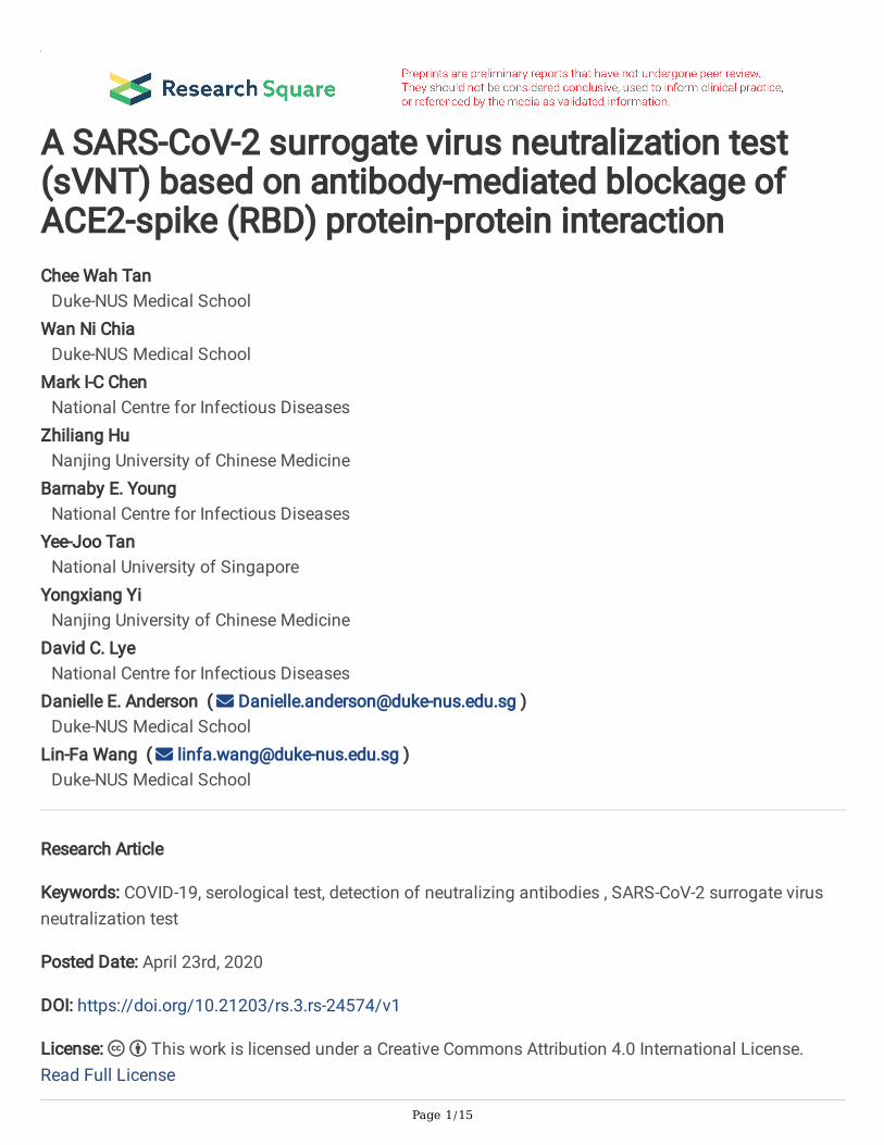
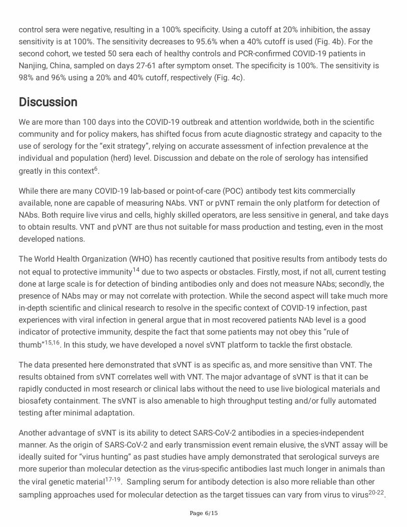
Immediately after SARS-CoV-2 was identi�ed as the causative agent of the COVID-19 outbreak, it wasshown that the human angiotensin converting enzyme-2 (hACE2) is the main functional receptor for viralentry3. We hypothesized that the virus-receptor binding can be mimicked in vitro via a protein-proteininteraction using puri�ed recombinant hACE2 and the RBD of the SARS-CoV-2 S protein. This interactioncan be blocked by virus NAbs present in the test serum, using the same principle as a conventional VNTconducted using live virus inside a BSL3 facility (Fig. 1a and b).
In our study, the direct binding was demonstrated using different SARS-CoV-2 proteins conjugated withhorseradish peroxidase (HRP). There is a dose-dependent speci�c binding between hACE2 and RBD or S1,but not with the nucleocapsid (N) protein, with the RBD producing the best binding characteristics (Fig.1c). The HRP-RBD protein was chosen for subsequent studies. We then demonstrated that the speci�cRBD-hACE2 binding can be blocked or neutralized by COVID-19 sera in a dose-dependent manner, but notby sera from healthy controls (Fig. 1d). To prove that the same principle works with the closely relatedSARS-CoV, which also uses hACE2 as the entry receptor12, we repeated the similar experiments andproved that the SARS-CoV RBD performed in an almost identical manner in this new test format (Fig. 1e,f), termed surrogate virus neutralization test (sVNT).
Isotype- and species-independent neutralization
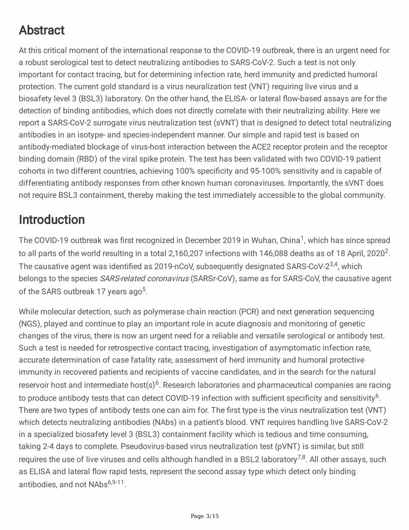
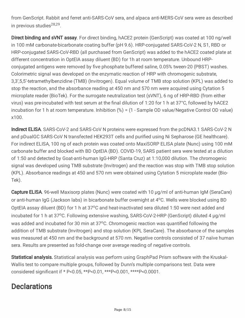
One of the advantages of sVNT is its ability to detect total antibodies in patient sera, in contrast to mostSARS-CoV-2 antibody tests published or marketed, which are almost all isotype-speci�c, mostly for IgM orIgG, with some for IgA9-11. From a panel of 77 COVID-19 positive sera from patients in Singapore, wehave designated four groups based on IgM or IgG ELISA levels, determined by our in-house capture ELISAassays (see Methods), present in the patient convalescent sera: a) high IgM/low IgG; b) low IgM/high IgG;c) low IgM/ low IgG; and d) high IgM/high IgG. All groups showed strong neutralization activity in thesVNT (Fig. 2), demonstrating the isotype-independent performance of the assay. It is worth to note thatfor panel c with low IgM/IgG, the % inhibition in sVNT is still signi�cant at 70-75%, demonstrating itssuperior sensitivity as this group of sera were deemed negative or weakly positive with isotype-speci�ccapture ELISA based on IgM or IgG alone.
Page 5/15
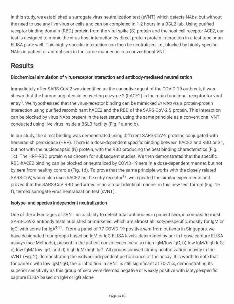
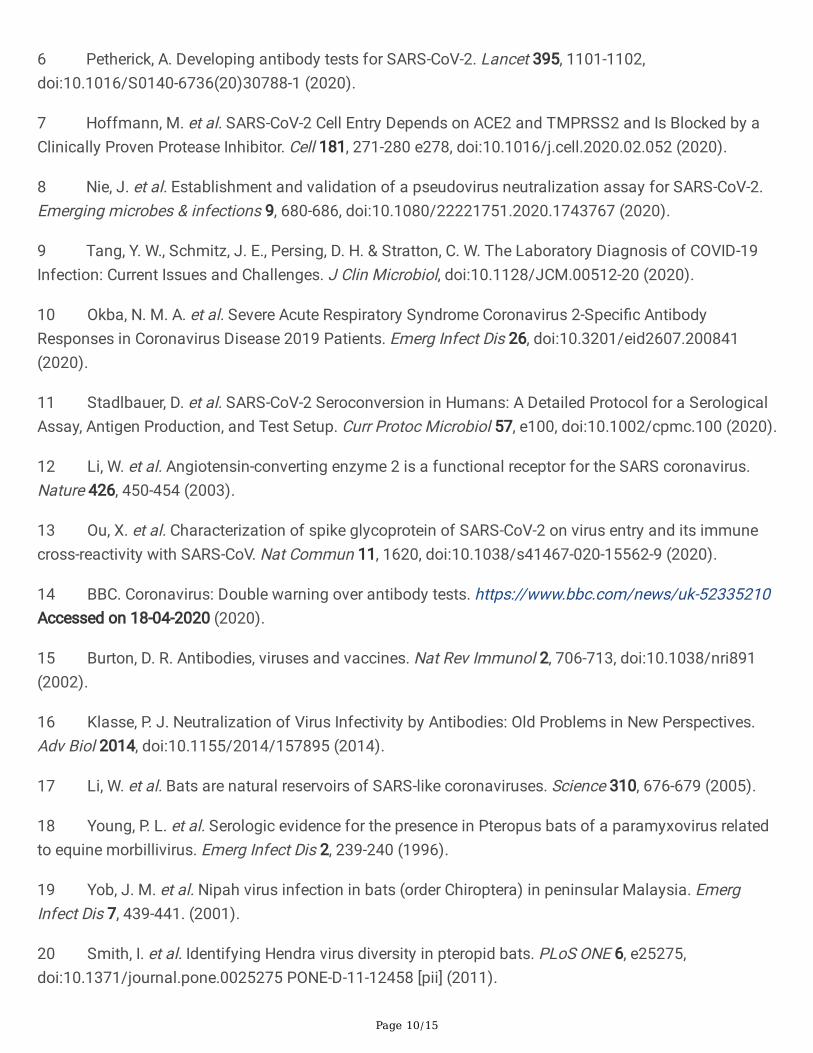
We then tested different animal sera in the sVNT assays to demonstrate species-independentperformance. Results from three independent rabbits immunized with the SARS-CoV-2 RBD protein,demonstrate very potent neutralizing activity in the SARS-CoV-2 sVNT (Fig. 3a). Similarly, sera fromferrets infected with SARS-CoV (Fig. 3b) and rabbits immunized with inactivated SARS-CoV (Fig. 3c) alsodemonstrate an e�cient dose-dependent inhibition of the hACE2-SARS-CoV RBD interaction in the SARS-CoV sVNT.
Speci�city against other hCoVs and comparison of SARS sera collected in 2003 vs 2020
To demonstrate speci�city, we tested different panels of sera against other known human coronaviruses(hCoVs) and con�rmed that the SARS-CoV-2 sVNT can differentiate antibody responses between COVID-19 and other coronavirus infections (Fig. 3d). For SARS sera, there is some level of cross reactivity asexpected from their close genetical relatedness and previous published studies3,7. But the difference inneutralization is statistically signi�cant, and hence the sVNT can be used to differentiate COVID-19infection from past SARS infection. For human sera from patients with 229/NL63 or OC43 infection andalpaca sera from experimental MERS-CoV infection, there is no detectable cross neutralization.
During the investigation of potential cross reactivity between SARS sera and SARS-CoV-2 virus, we madeseveral important observations. Firstly, despite the lack of cross neutralization by SARS sera against thelive SARS-CoV-2 virus in VNT observed by us and other groups13, we detected some level of crossneutralization in sVNT (Fig. 3d), indicating sVNT is more sensitive than VNT; secondly, SARS NAbs aredetectable for at least 17 years in recovered patients (Fig. 3f); thirdly, the cross neutralization level ishigher in the 2020 SARS sera than the 2003 samples (Fig. 3d) although the homologous neutralizinglevel of the 2020 SARS sera (Fig. 3f) is lower than the 2003 SARS sera (Fig. 3e); lastly, we have found thatthe N-speci�c antibody level is much lower in the 2020 SARS sera than the 2003 samples (Fig. 3g).
Correlation between live virus VNT and biochemical sVNT
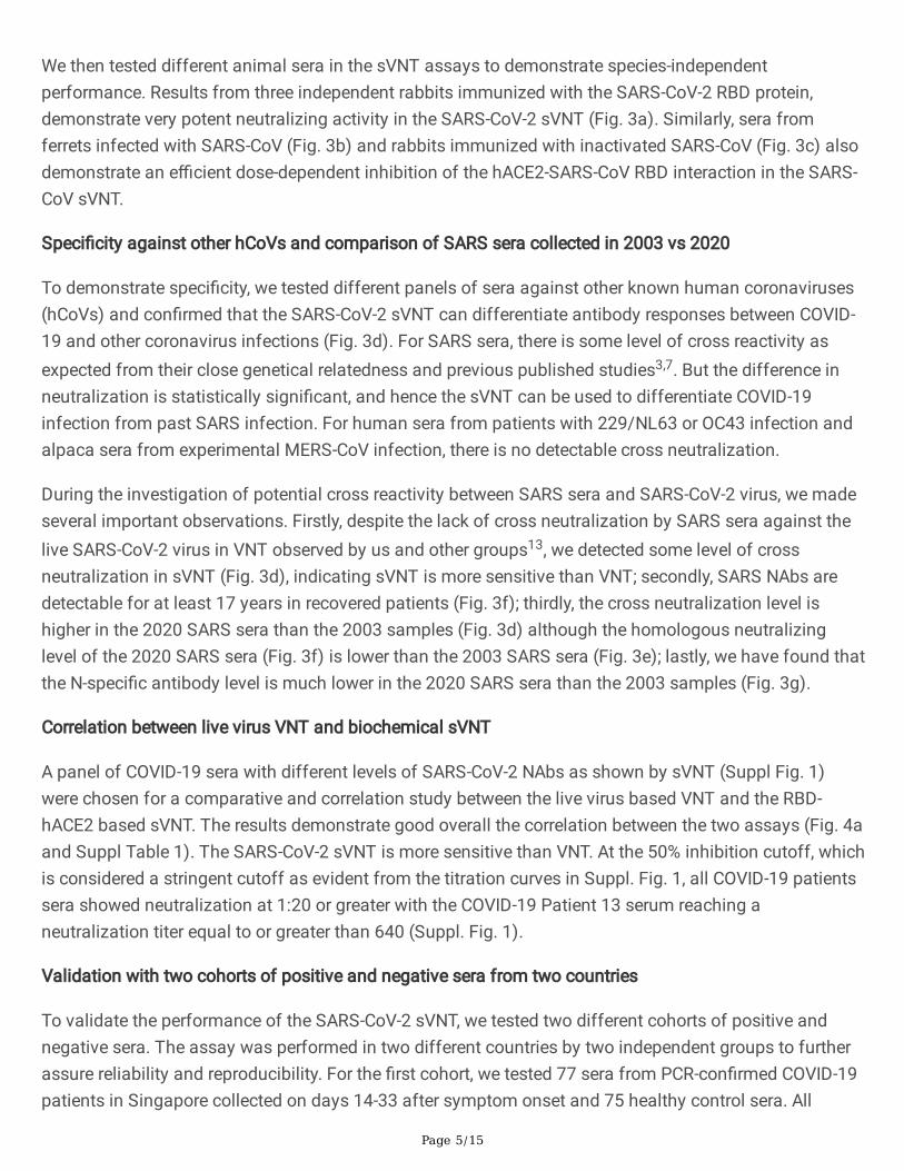
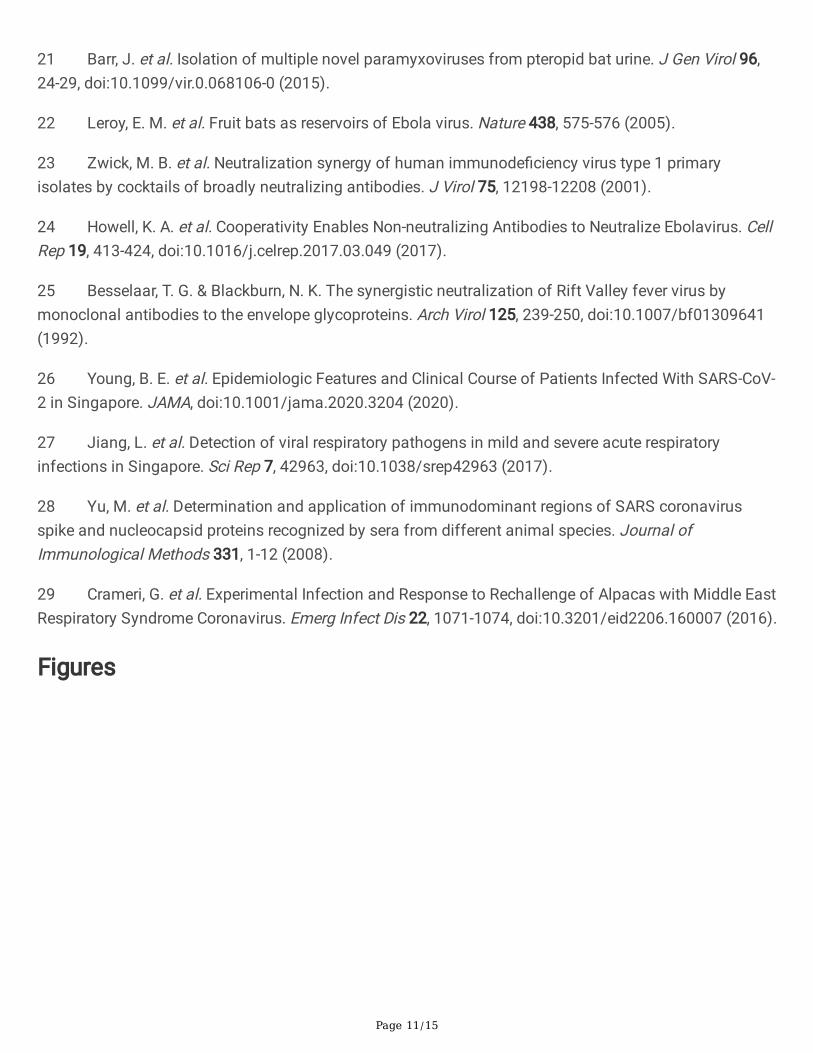
A panel of COVID-19 sera with different levels of SARS-CoV-2 NAbs as shown by sVNT (Suppl Fig. 1)were chosen for a comparative and correlation study between the live virus based VNT and the RBD-hACE2 based sVNT. The results demonstrate good overall the correlation between the two assays (Fig. 4aand Suppl Table 1). The SARS-CoV-2 sVNT is more sensitive than VNT. At the 50% inhibition cutoff, whichis considered a stringent cutoff as evident from the titration curves in Suppl. Fig. 1, all COVID-19 patientssera showed neutralization at 1:20 or greater with the COVID-19 Patient 13 serum reaching aneutralization titer equal to or greater than 640 (Suppl. Fig. 1).
Validation with two cohorts of positive and negative sera from two countries
To validate the performance of the SARS-CoV-2 sVNT, we tested two different cohorts of positive andnegative sera. The assay was performed in two different countries by two independent groups to furtherassure reliability and reproducibility. For the �rst cohort, we tested 77 sera from PCR-con�rmed COVID-19patients in Singapore collected on days 14-33 after symptom onset and 75 healthy control sera. All
Page 6/15
control sera were negative, resulting in a 100% speci�city. Using a cutoff at 20% inhibition, the assaysensitivity is at 100%. The sensitivity decreases to 95.6% when a 40% cutoff is used (Fig. 4b). For thesecond cohort, we tested 50 sera each of healthy controls and PCR-con�rmed COVID-19 patients inNanjing, China, sampled on days 27-61 after symptom onset. The speci�city is 100%. The sensitivity is98% and 96% using a 20% and 40% cutoff, respectively (Fig. 4c).
DiscussionWe are more than 100 days into the COVID-19 outbreak and attention worldwide, both in the scienti�ccommunity and for policy makers, has shifted focus from acute diagnostic strategy and capacity to theuse of serology for the “exit strategy”, relying on accurate assessment of infection prevalence at theindividual and population (herd) level. Discussion and debate on the role of serology has intensi�edgreatly in this context6.
While there are many COVID-19 lab-based or point-of-care (POC) antibody test kits commerciallyavailable, none are capable of measuring NAbs. VNT or pVNT remain the only platform for detection ofNAbs. Both require live virus and cells, highly skilled operators, are less sensitive in general, and take daysto obtain results. VNT and pVNT are thus not suitable for mass production and testing, even in the mostdeveloped nations.
The World Health Organization (WHO) has recently cautioned that positive results from antibody tests donot equal to protective immunity14 due to two aspects or obstacles. Firstly, most, if not all, current testingdone at large scale is for detection of binding antibodies only and does not measure NAbs; secondly, thepresence of NAbs may or may not correlate with protection. While the second aspect will take much morein-depth scienti�c and clinical research to resolve in the speci�c context of COVID-19 infection, pastexperiences with viral infection in general argue that in most recovered patients NAb level is a goodindicator of protective immunity, despite the fact that some patients may not obey this “rule ofthumb”15,16. In this study, we have developed a novel sVNT platform to tackle the �rst obstacle.
The data presented here demonstrated that sVNT is as speci�c as, and more sensitive than VNT. Theresults obtained from sVNT correlates well with VNT. The major advantage of sVNT is that it can berapidly conducted in most research or clinical labs without the need to use live biological materials andbiosafety containment. The sVNT is also amenable to high throughput testing and/or fully automatedtesting after minimal adaptation.
Another advantage of sVNT is its ability to detect SARS-CoV-2 antibodies in a species-independentmanner. As the origin of SARS-CoV-2 and early transmission event remain elusive, the sVNT assay will beideally suited for “virus hunting” as past studies have amply demonstrated that serological surveys aremore superior than molecular detection as the virus-speci�c antibodies last much longer in animals thanthe viral genetic material17-19. Sampling serum for antibody detection is also more reliable than othersampling approaches used for molecular detection as the target tissues can vary from virus to virus20-22.

Page 7/15
In addition, sVNT offers a key advantage over most ELISA or POC tests in its ability to detect total NAbsin an isotype-independent manner. This will not only simplify the testing strategy, but also furtherincrease test sensitivity. As shown in Fig. 2c for the serum panel of COVID-19 patients showing low IgMand IgG in the isotype-speci�c ELISAs, the sVNT assay still detected signi�cant level of NAbs. Althoughthe mechanism needs further investigation, there are at least two possibilities: the presence of other Igisotypes or neutralization synergy or cooperativity from the combination of different isotype antibodiestargeting different neutralization critical epitopes, as previously observed for HIV and other viruses23-25.
Results obtained for the two SARS serum panels are very interesting. The long lasting NAbs 17 yearsafter initial infection is encouraging news for recovered COVID-19 patients considering the closerelationship of the two viruses. The mechanism and biological signi�cance of the increased crossneutralization towards SARS-COV-2 coupled with the decrease/disappearance of N-speci�c antibodies 17years after infection warrants further investigation in the context of better understanding SARSr-CoVimmune response dynamics.
In summary, we have addressed the challenge of COVID-19 serology with a new approach that enablesthe detection of NAbs in an easy, safe, rapid and inexpensive manner with enhanced speci�city andsensitivity. While the sVNT assay may never be able to completely replace the conventional VNT, our dataindicate that their performance is generally well correlated. Its application can cover many aspects ofCOVID-19 investigation from contact tracing, sero-prevalence survey, reservoir/intermediate animaltracking to assessment of herd immunity, longevity of protective immunity and e�cacy of differentvaccine candidates.
MethodsCells and virus. Human embryonic kidney (HEK293T) cells (ATCC# CRL-3216) and African green monkeykidney, clone E6 (Vero-E6) cells (ATCC# CRL-1586) were maintained in Dulbecco’s modi�ed EagleMedium (DMEM) supplemented with 10% fetal bovine serum. SARS-CoV-2, isolateBetaCoV/Singapore/2/2020 (Accession ID EPI_ISL_406973), was used for virus neutralization test onVero-E6 cells as described previously26.
Panels of human and animal sera used in this study. In Singapore, COVID-19 patient sera used in thisstudy was from the Singapore PROTECT study as described [13]. Sera from recovered SARS patientsfrom 2003 were as previously described [15]. For SARS recall sampling in 2020, we contacted and thenobtained blood from consenting individuals previously admitted for SARS (ethics approval number: NHGDSRB E 2020/00091). The hCoV serum panel included post-infection samples from subjects con�rmedCoV 229/NL63 and CoV OC43 positive using the SeeGene RV12 respiratory multiplex kit in a previousstudy (ethics approval number: NUS-IRB 11-3640)27. Negative control sera were obtained from residualserum samples from previous unrelated studies. In Nanjing, China, COVID-19 convalescent sera werecollected with written informed consent and approved by the ethics committee of the Second Hospital ofNanjing (ethics approval number: 2020-LS-ky003). Rabbit anti-SARS-CoV-2 RBD sera were purchased
Page 8/15
from GenScript. Rabbit and ferret anti-SARS-CoV sera, and alpaca anti-MERS-CoV sera were as describedin previous studies28,29.
Direct binding and sVNT assay. For direct binding, hACE2 protein (GenScript) was coated at 100 ng/wellin 100 mM carbonate-bicarbonate coating buffer (pH 9.6). HRP-conjugated SARS-CoV-2 N, S1, RBD orHRP-conjugated SARS-CoV-RBD (all purchased from GenScript) was added to the hACE2 coated plate atdifferent concentration in OptEIA assay diluent (BD) for 1h at room temperature. Unbound HRP-conjugated antigens were removed by �ve phosphate buffered saline, 0.05% tween-20 (PBST) washes.Colorimetric signal was developed on the enzymatic reaction of HRP with chromogenic substrate,3,3’,5,5’-tetramethylbenzidine (TMB) (Invitrogen). Equal volume of TMB stop solution (KPL) was added tostop the reaction, and the absorbance reading at 450 nm and 570 nm were acquired using Cytation 5microplate reader (BioTek). For the surrogate neutralization test (sVNT), 6 ng of HRP-RBD (from eithervirus) was pre-incubated with test serum at the �nal dilution of 1:20 for 1 h at 37°C, followed by hACE2incubation for 1 h at room temperature. Inhibition (%) = (1 - Sample OD value/Negative Control OD value)x100.
Indirect ELISA. SARS-CoV-2 and SARS-CoV N proteins were expressed from the pcDNA3.1 SARS-CoV-2 Nand pDualGC SARS-CoV N transfected HEK293T cells and puri�ed using Ni Sepharose (GE healthcare).For indirect ELISA, 100 ng of each protein was coated onto MaxiSORP ELISA plate (Nunc) using 100 mMcarbonate buffer and blocked with BD OptEIA (BD). COVID-19, SARS patient sera were tested at a dilutionof 1:50 and detected by Goat-anti-human IgG-HRP (Santa Cruz) at 1:10,000 dilution. The chromogenicsignal was developed using TMB substrate (Invitrogen) and the reaction was stop with TMB stop solution(KPL). Absorbance readings at 450 and 570 nm were obtained using Cytation 5 microplate reader (Bio-Tek).
Capture ELISA. 96-well Maxisorp plates (Nunc) were coated with 10 µg/ml of anti-human IgM (SeraCare)or anti-human IgG (Jackson labs) in bicarbonate buffer overnight at 4oC. Wells were blocked using BDOptEIA assay diluent (BD) for 1 h at 37oC and heat-inactivated sera diluted 1:50 were next added andincubated for 1 h at 37oC. Following extensive washing, SARS-CoV-2-HRP (GenScript) diluted 4 µg/mlwas added and incubated for 30 min at 37oC. Chromogenic reaction was quanti�ed following theaddition of TMB substrate (Invitrogen) and stop solution (KPL SeraCare). The absorbance of the sampleswas measured at 450 nm and the background at 570 nm. Negative controls consisted of 37 naïve humansera. Results are presented as fold-change over average reading of negative controls.
Statistical analysis. Statistical analysis was perform using GraphPad Prism software with the Kruskal-Wallis test to compare multiple groups, followed by Dunn’s multiple comparisons test. Data wereconsidered signi�cant if * P<0.05, **P<0.01, ***P<0.001, ****P<0.0001.
Declarations

Page 9/15
Online content. Any methods, additional references, Nature Research reporting summaries,supplementary information, acknowledgements; details of author contributions and competing interestsare available at [Article DOI].
Acknowledgements
We thank Xijian Qin, Shuangshuang Tang, Pei Liu and Weihui Shao for technical advice and assistancewith assay development and testing; Yilong Peng, Charles Tiu, Akshamal Gamage, Beng Lee Lim, VivianChen, Wan Rong Sia and Xin Mei Ong for assistance in protein puri�cation, sample management andtesting; Yazid Abdad and Linda Wei Lin Tan for help with hCoV sera collection; Viji Vijayan, Benson Ngand Velraj Sivalingam of the Duke-NUS Medical School ABSL3 facility for logistics management andassistance. L-FW and DEA are supported by grants from the Singapore National Research Foundation(NRF2016NRF-NSFC002-013) and National Medical Research Council (STPRG-FY19-001 andCOVID19RF-003).
Author contributions
L-FW conceived and guided the study. CWT, WNC and DEA performed laboratory work including dataanalysis. MI-CC, ZH, BEY, Y-JT, YY and DCL provided necessary samples and coordination for the study. L-FW initiated the manuscript writing with input from all authors.
Con�ict of interest
A patent application has been �led for the content disclosed in this study and a SARS-CoV-2 sVNT Kit isunder development with an industrial partner for commercialization.
References1 Wang, C., Horby, P. W., Hayden, F. G. & Gao, G. F. A novel coronavirus outbreak of global healthconcern. Lancet 395, 470-473, doi:10.1016/S0140-6736(20)30185-9 (2020).
2 WHO. COVID-19 Status Report. https://www.who.int/emergencies/diseases/novel-coronavirus-2019/situation-reports. Accessed on 18 April 2020 (2020).
3 Zhou, P. et al. A pneumonia outbreak associated with a new coronavirus of probable bat origin.Nature 579, 270-273, doi:10.1038/s41586-020-2012-7 (2020).
4 Coronaviridae Study Group of the International Committee on Taxonomy of, V. The species Severeacute respiratory syndrome-related coronavirus: classifying 2019-nCoV and naming it SARS-CoV-2. NatMicrobiol 5, 536-544, doi:10.1038/s41564-020-0695-z (2020).
5 Peiris, J. S. et al. Coronavirus as a possible cause of severe acute respiratory syndrome. Lancet361, 1319-1325 (2003).
Page 10/15
6 Petherick, A. Developing antibody tests for SARS-CoV-2. Lancet 395, 1101-1102,doi:10.1016/S0140-6736(20)30788-1 (2020).
7 Hoffmann, M. et al. SARS-CoV-2 Cell Entry Depends on ACE2 and TMPRSS2 and Is Blocked by aClinically Proven Protease Inhibitor. Cell 181, 271-280 e278, doi:10.1016/j.cell.2020.02.052 (2020).
8 Nie, J. et al. Establishment and validation of a pseudovirus neutralization assay for SARS-CoV-2.Emerging microbes & infections 9, 680-686, doi:10.1080/22221751.2020.1743767 (2020).
9 Tang, Y. W., Schmitz, J. E., Persing, D. H. & Stratton, C. W. The Laboratory Diagnosis of COVID-19Infection: Current Issues and Challenges. J Clin Microbiol, doi:10.1128/JCM.00512-20 (2020).
10 Okba, N. M. A. et al. Severe Acute Respiratory Syndrome Coronavirus 2-Speci�c AntibodyResponses in Coronavirus Disease 2019 Patients. Emerg Infect Dis 26, doi:10.3201/eid2607.200841(2020).
11 Stadlbauer, D. et al. SARS-CoV-2 Seroconversion in Humans: A Detailed Protocol for a SerologicalAssay, Antigen Production, and Test Setup. Curr Protoc Microbiol 57, e100, doi:10.1002/cpmc.100 (2020).
12 Li, W. et al. Angiotensin-converting enzyme 2 is a functional receptor for the SARS coronavirus.Nature 426, 450-454 (2003).
13 Ou, X. et al. Characterization of spike glycoprotein of SARS-CoV-2 on virus entry and its immunecross-reactivity with SARS-CoV. Nat Commun 11, 1620, doi:10.1038/s41467-020-15562-9 (2020).
14 BBC. Coronavirus: Double warning over antibody tests. https://www.bbc.com/news/uk-52335210Accessed on 18-04-2020 (2020).
15 Burton, D. R. Antibodies, viruses and vaccines. Nat Rev Immunol 2, 706-713, doi:10.1038/nri891(2002).
16 Klasse, P. J. Neutralization of Virus Infectivity by Antibodies: Old Problems in New Perspectives.Adv Biol 2014, doi:10.1155/2014/157895 (2014).
17 Li, W. et al. Bats are natural reservoirs of SARS-like coronaviruses. Science 310, 676-679 (2005).
18 Young, P. L. et al. Serologic evidence for the presence in Pteropus bats of a paramyxovirus relatedto equine morbillivirus. Emerg Infect Dis 2, 239-240 (1996).
19 Yob, J. M. et al. Nipah virus infection in bats (order Chiroptera) in peninsular Malaysia. EmergInfect Dis 7, 439-441. (2001).
20 Smith, I. et al. Identifying Hendra virus diversity in pteropid bats. PLoS ONE 6, e25275,doi:10.1371/journal.pone.0025275 PONE-D-11-12458 [pii] (2011).
Page 11/15
21 Barr, J. et al. Isolation of multiple novel paramyxoviruses from pteropid bat urine. J Gen Virol 96,24-29, doi:10.1099/vir.0.068106-0 (2015).
22 Leroy, E. M. et al. Fruit bats as reservoirs of Ebola virus. Nature 438, 575-576 (2005).
23 Zwick, M. B. et al. Neutralization synergy of human immunode�ciency virus type 1 primaryisolates by cocktails of broadly neutralizing antibodies. J Virol 75, 12198-12208 (2001).
24 Howell, K. A. et al. Cooperativity Enables Non-neutralizing Antibodies to Neutralize Ebolavirus. CellRep 19, 413-424, doi:10.1016/j.celrep.2017.03.049 (2017).
25 Besselaar, T. G. & Blackburn, N. K. The synergistic neutralization of Rift Valley fever virus bymonoclonal antibodies to the envelope glycoproteins. Arch Virol 125, 239-250, doi:10.1007/bf01309641(1992).
26 Young, B. E. et al. Epidemiologic Features and Clinical Course of Patients Infected With SARS-CoV-2 in Singapore. JAMA, doi:10.1001/jama.2020.3204 (2020).
27 Jiang, L. et al. Detection of viral respiratory pathogens in mild and severe acute respiratoryinfections in Singapore. Sci Rep 7, 42963, doi:10.1038/srep42963 (2017).
28 Yu, M. et al. Determination and application of immunodominant regions of SARS coronavirusspike and nucleocapsid proteins recognized by sera from different animal species. Journal ofImmunological Methods 331, 1-12 (2008).
29 Crameri, G. et al. Experimental Infection and Response to Rechallenge of Alpacas with Middle EastRespiratory Syndrome Coronavirus. Emerg Infect Dis 22, 1071-1074, doi:10.3201/eid2206.160007 (2016).
Figures
Page 12/15
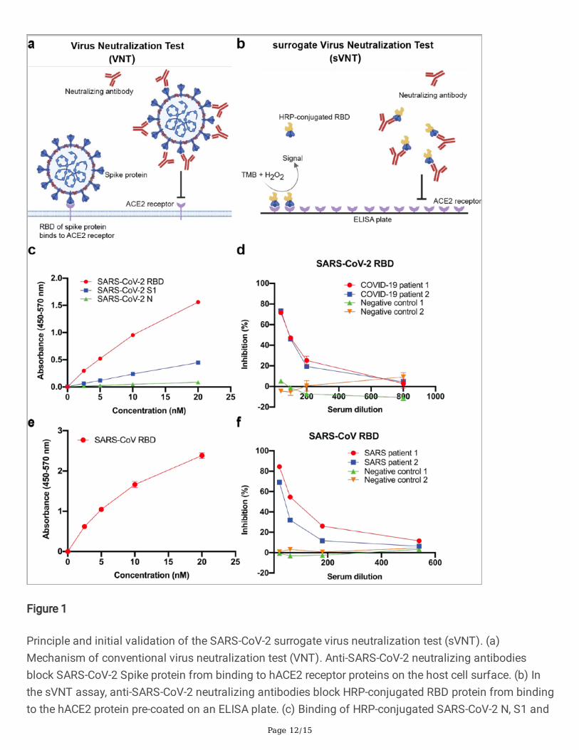
Figure 1
Principle and initial validation of the SARS-CoV-2 surrogate virus neutralization test (sVNT). (a)Mechanism of conventional virus neutralization test (VNT). Anti-SARS-CoV-2 neutralizing antibodiesblock SARS-CoV-2 Spike protein from binding to hACE2 receptor proteins on the host cell surface. (b) Inthe sVNT assay, anti-SARS-CoV-2 neutralizing antibodies block HRP-conjugated RBD protein from bindingto the hACE2 protein pre-coated on an ELISA plate. (c) Binding of HRP-conjugated SARS-CoV-2 N, S1 and

Page 13/15
RBD proteins to hACE2. (d) Inhibition of SARS-CoV-2 RBD-hACE2 interaction by COVID-19 patient sera. (e)Binding of HRP-conjugated SARS-CoV RBD to hACE2. (f) Inhibition of SARS-CoV RBD-hACE2 interactionby SARS patient sera
Figure 2
Isotype-independent neutralization by human sera with different levels of IgM and IgG antibodies. (a)High IgM/Low IgG (n = 5); (b) Low IgM/High IgG (n = 3); (c) Low IgM/Low IgG (n = 9); (d) High IgM/HighIgG (n = 5). The IgM and IgG levels were determined by isotype-speci�c capture ELISA detailed inMethods.
Page 14/15
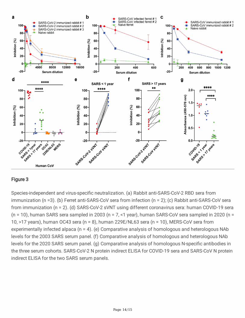
Figure 3
Species-independent and virus-speci�c neutralization. (a) Rabbit anti-SARS-CoV-2 RBD sera fromimmunization (n =3). (b) Ferret anti-SARS-CoV sera from infection (n = 2); (c) Rabbit anti-SARS-CoV serafrom immunization (n = 2). (d) SARS-CoV-2 sVNT using different coronavirus sera: human COVID-19 sera(n = 10), human SARS sera sampled in 2003 (n = 7, <1 year), human SARS-CoV sera sampled in 2020 (n =10, >17 years), human OC43 sera (n = 8), human 229E/NL63 sera (n = 10), MERS-CoV sera fromexperimentally infected alpaca (n = 4). (e) Comparative analysis of homologous and heterologous NAblevels for the 2003 SARS serum panel. (f) Comparative analysis of homologous and heterologous NAblevels for the 2020 SARS serum panel. (g) Comparative analysis of homologous N-speci�c antibodies inthe three serum cohorts. SARS-CoV-2 N protein indirect ELISA for COVID-19 sera and SARS-CoV N proteinindirect ELISA for the two SARS serum panels.
Page 15/15
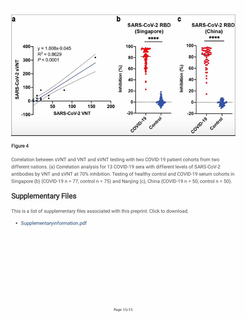
Figure 4
Correlation between sVNT and VNT and sVNT testing with two COVID-19 patient cohorts from twodifferent nations. (a) Correlation analysis for 13 COVID-19 sera with different levels of SARS-CoV-2antibodies by VNT and sVNT at 70% inhibition. Testing of healthy control and COVID-19 serum cohorts inSingapore (b) (COVID-19 n = 77, control n = 75) and Nanjing (c), China (COVID-19 n = 50, control n = 50).
Supplementary Files
This is a list of supplementary �les associated with this preprint. Click to download.
Supplementaryinformation.pdf