the nucleocapsid protein of sars-cov-2 abolished pluripotency … · pluripotent stem cells....
TRANSCRIPT

The Nucleocapsid Protein of SARS-CoV-2 Abolished Pluripotency
in Human Induced Pluripotent Stem Cells
Lin Zebin1,2#, Gao Qiang3#, Fang Qian4, Mai Jinlian1, Zhou Lishi1, Lin Xianming1,
Qian Yu2, Cai Tian5, Chen Zhenhua5, Wang Ping1 and Lin Bin1*
1 Guangdong Beating Origin Regenerative Medicine Co. Ltd., Foshan, Guangdong 528231, China
2 School of Pharmaceutical Sciences, Sun Yat-Sen University, Guangzhou, Guangdong 510275,
China
3 Department of Cardiac Surgery, Guangdong Cardiovascular Institute, Guangdong Provincial
People’s Hospital, Guangdong Academy of Medical Sciences, Guangzhou, Guangdong 510100,
China
4 Department of Medical Ultrasonics, Guangzhou Women and Children's Medical Center,
Guangzhou Medical University, Guangzhou, Guangdong 510120, China
5 Nanhai District People’s Hospital of Foshan, Foshan, Guangdong 528200, China
#These authors contributed equally to the work.
*Correspondence to: Lin Bin, Ph.D., Guangdong Beating Origin Regenerative Medicine Co. Ltd..
Tel: +86 0757-81184899. Fax: +86 0757-81184899. E-mail: [email protected].
.CC-BY-NC 4.0 International license(which was not certified by peer review) is the author/funder. It is made available under aThe copyright holder for this preprintthis version posted March 28, 2020. . https://doi.org/10.1101/2020.03.26.010694doi: bioRxiv preprint

Abstract
The COVID-19 pandemic caused by severe acute respiratory syndrome coronavirus 2 (SARS-CoV-
2) is raging across the world, leading to a mortality rate of 3.4%. As a potential vaccine and
therapeutic target, the nucleocapsid protein of SARS-CoV-2 (nCoVN) functions in packaging the
viral genome and viral self-assembly. To investigate the biological effect of nCoVN to human
induced pluripotent stem cells (iPSC), genetically engineered iPSC overexpressing nCoVN (iPSC-
nCoVN) were generated by lentiviral expression systems. Unexpectedly, the morphology and
proliferation rate of iPSC were changed after nCoVN expressing for two weeks. The pluripotency
markers SSEA4 and TRA-1-81 were not detectable in iPSC-nCoVN. Meanwhile, iPSC-nCoVN lost
the ability for differentiation into cardiomyocytes when using a routine differentiation protocol. Our
data suggested that nCoVN disrupted the pluripotent properties of iPSC and turned them into
fibroblasts, which provided a new insight to the pathogenic mechanism of SARS-CoV-2.
Keywords:
SARS-CoV-2; nucleocapsid protein; human induced pluripotent stem cell; pluripotency; fibroblast
Introduction
Right now, the COVID-19 pandemic is sweeping the world, causing a huge crisis in public health
and economics globally. According to the continuously updated data from World Health
Organization, to date, more than 300,000 infected cases were confirmed, while more than 13,000
individuals died because of COVID-19 (https://www.who.int/emergencies/diseases/novel-
coronavirus-2019). Severe acute respiratory syndrome coronavirus 2 (SARS-CoV-2), which was
proved to be the pathogen of COVID-19, has 79% identity in genomes with severe acute respiratory
syndrome coronavirus (SARS-CoV)[1]. 12 coding regions were predicted in SARS-CoV-2,
including spike protein, nucleocapsid protein, envelope protein, and membrane protein[1-3]. The
Cryo-EM structure of spike protein had been determined[4], and more and more evidences showed
.CC-BY-NC 4.0 International license(which was not certified by peer review) is the author/funder. It is made available under aThe copyright holder for this preprintthis version posted March 28, 2020. . https://doi.org/10.1101/2020.03.26.010694doi: bioRxiv preprint
that the spike protein binds human ACE2 to entry into host cells[4,5], which indicated that SARS-
CoV-2 might share similar pathogenic mechanisms with SARS-CoV. Because of the very limited
knowledge of SARS-CoV-2, we sought to understand the biology of SARS-CoV-2 based on the
previous studies about SARS-CoV.
As one of most studied proteins in SARS-CoV, the nucleocapsid protein binds to viral RNA to
package the genome in a ribonucleoprotein particle[6]. Unlike the spike protein with a certain
mutation frequency, the sequence of nucleocapsid protein was more stable[7], which meant it was
an ideal target for diagnostic tools[8-10] and antiviral therapy[11,12]. The pathogenic effects in host
cells caused by the nucleocapsid protein were also studied. It was reported that the nucleocapsid
protein inhibited type I interferon production after virion infected the host cells[13], which was
considered as a possible mechanism of immune escape. The nucleocapsid protein inhibited cell
cytokinesis and proliferation[14], and regulated several pathways, such as transforming growth
factor-beta signaling[15], AP-1 signal transduction pathway[16], and NF-KappaB pathway[17].
Besides, the nucleocapsid protein was reported as an apoptosis inducer in COS-1 cells[18,19] and
HPF cells[20].
As the nucleocapsid protein of SARS-CoV-2 (nCoVN) has 88.1% identity with the nucleocapsid
protein of SARS-CoV[1], it is reasonable to speculate that they share a same pathogenic pathway
in host cells. The original goal of this study is to determine the physiological malfunctions in human
cardiomyocytes overexpressing nCoVN by using human induced pluripotent stem cells (iPSC) and
direct differentiation protocols. Unexpectedly, the morphology of iPSC altered obviously when
nCoVN had been expressed for 14 days. We turned to investigate whether nCoVN obstructed the
pluripotency maintenance in iPSC. Here, we present a new deleterious effect of nCoVN to human
pluripotent stem cells.
Results
ACE2 was expressed in various of stem cells
As ACE2 is the major receptor of SARS-CoV-2 on the cell membrane[4,5], we first examined
whether ACE2 were expressed in the stem cells. Thanks to the gene expression data collection in
Gene Expression Omnibus (GEO, https://www.ncbi.nlm.nih.gov/geo/), it is convenient to analyze
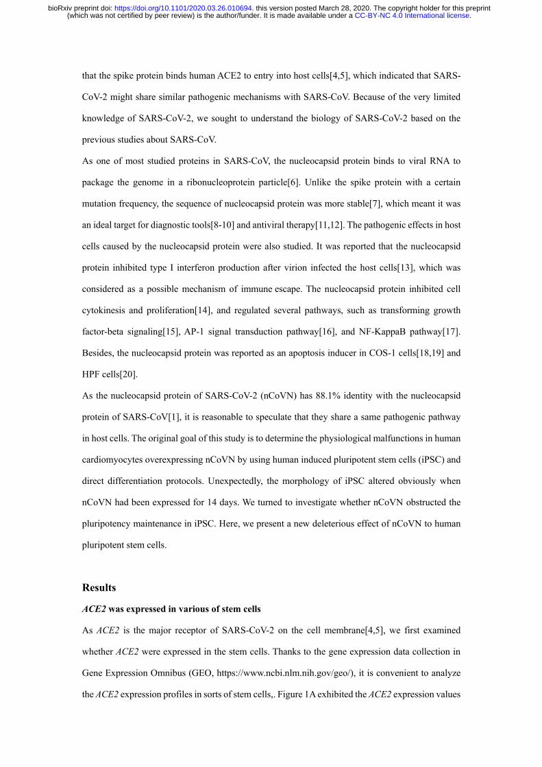
the ACE2 expression profiles in sorts of stem cells,. Figure 1A exhibited the ACE2 expression values
.CC-BY-NC 4.0 International license(which was not certified by peer review) is the author/funder. It is made available under aThe copyright holder for this preprintthis version posted March 28, 2020. . https://doi.org/10.1101/2020.03.26.010694doi: bioRxiv preprint
in different stem cells from different projects, including human embryonic stem cells[21], iPSC[22],
human epithelial stem cells[22], human adipose stem cells[23], human hematopoietic stem cells[24],
and human mesenchymal stem cells[25]. The expression values of a housekeeping gene GAPDH
were simultaneously collected as controls. ACE2 was expressed in each kind of stem cells, though
the expression values were relatively low compared with GAPDH. The reverse transcription-PCR
(RT-RCR) results showed that ACE2 was expressed in iPSC, iPSC-derived cardiomyocytes (iPSC-
CM) and human coronary artery endothelial cells (HCAEC), suggesting these cells were the
potential targets of SARS-CoV-2 (Figure 1B).
Overexpression of nCoVN drove iPSC to fibroblasts
To study whether physiological activities in iPSC were disturbed by nCoVN, a human induced
pluripotent stem cell line (iPSC-nCoVN) in which the expression of nCoVN could be modulated by
a Tet-On system was generated by a lentiviral expression system. In this system, nCoVN cDNA
sequence was conjugated to puromycin resistance gene through a T2A peptide encoding sequence,
and the transcription was relied on the induction of tetracycline or doxycycline (Dox). After
puromycin selection, iPSC-nCoVN were divided into two groups: one was induced by Dox for
nCoVN expression (Dox+), the other was added with DMSO as a control set (DMSO+). The
transcriptional level of nCoVN was detected by Real-time PCR in iPSC, Dox+ and DMSO+ groups.
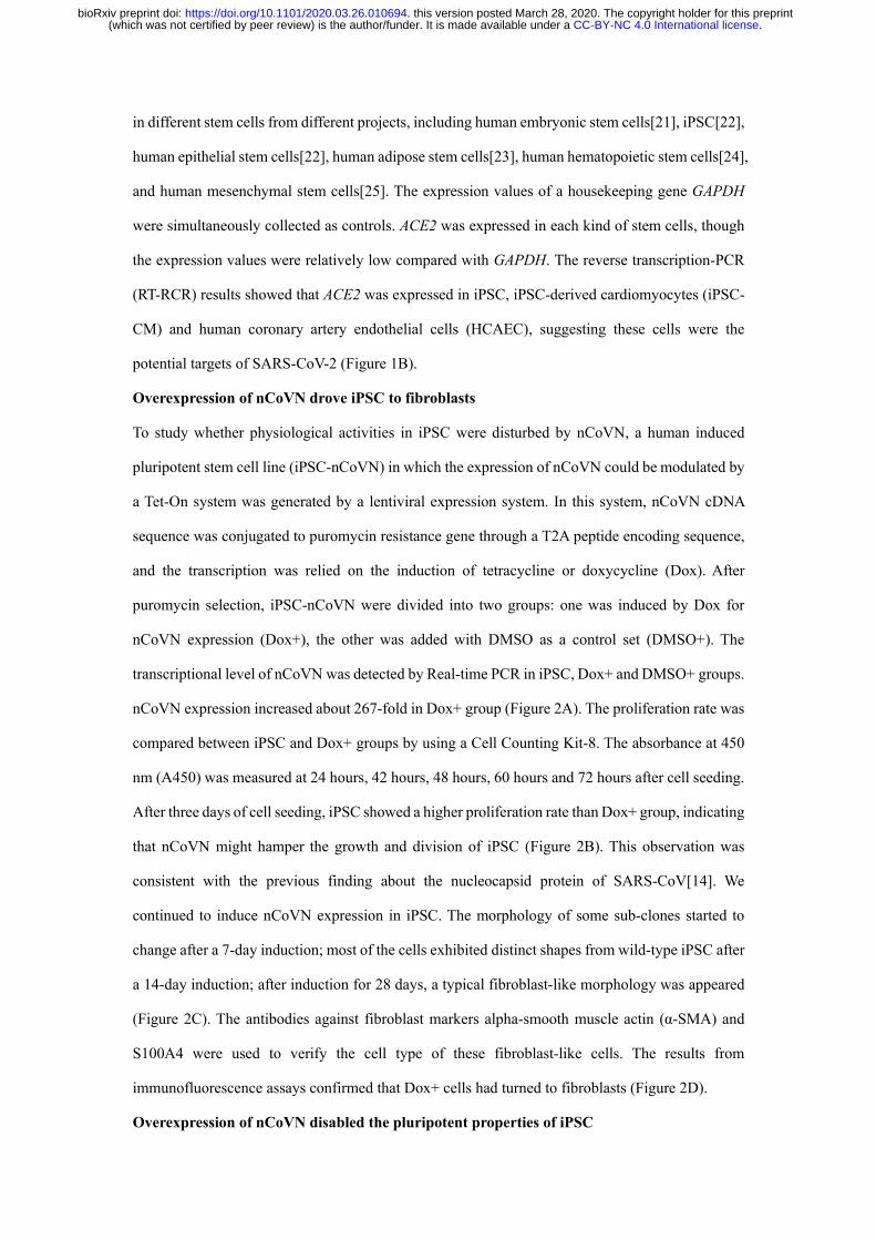
nCoVN expression increased about 267-fold in Dox+ group (Figure 2A). The proliferation rate was
compared between iPSC and Dox+ groups by using a Cell Counting Kit-8. The absorbance at 450
nm (A450) was measured at 24 hours, 42 hours, 48 hours, 60 hours and 72 hours after cell seeding.
After three days of cell seeding, iPSC showed a higher proliferation rate than Dox+ group, indicating
that nCoVN might hamper the growth and division of iPSC (Figure 2B). This observation was
consistent with the previous finding about the nucleocapsid protein of SARS-CoV[14]. We
continued to induce nCoVN expression in iPSC. The morphology of some sub-clones started to
change after a 7-day induction; most of the cells exhibited distinct shapes from wild-type iPSC after
a 14-day induction; after induction for 28 days, a typical fibroblast-like morphology was appeared
(Figure 2C). The antibodies against fibroblast markers alpha-smooth muscle actin (α-SMA) and
S100A4 were used to verify the cell type of these fibroblast-like cells. The results from
immunofluorescence assays confirmed that Dox+ cells had turned to fibroblasts (Figure 2D).
Overexpression of nCoVN disabled the pluripotent properties of iPSC
.CC-BY-NC 4.0 International license(which was not certified by peer review) is the author/funder. It is made available under aThe copyright holder for this preprintthis version posted March 28, 2020. . https://doi.org/10.1101/2020.03.26.010694doi: bioRxiv preprint

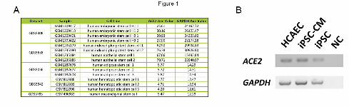
Next, we examined the pluripotency markers in iPSC and iPSC-nCoVN. The pluripotency markers
SSEA4 and TRA-1-81, which were expressed on the membranes of human embryonic stem cells
and iPSC, were widely applied in identification of pluripotent stem cells[26,27]. The images
obtained from immunofluorescence assays clearly illustrated that iPSC-nCoVN completely lost the
expression of SSEA4 and TRA-1-81, namely, iPSC-nCoVN lost the pluripotency in the presence of
nCoVN (Figure 3). To further test the pluripotency in iPSC and iPSC-nCoVN, we directly
differentiated these cells to cardiomyocytes by using a routine protocol and under same conditions.
As expected, the differentiation efficiency could reach 60% in iPSC; however, on differentiation
day 12, only a very small portion of cells were expressed cardiac Troponin T, accompanied by most
of cells death (Figure 4). This differentiation assay provided solid evidence that the pluripotency
maintenance of iPSC-nCoVN was disrupted by nCoVN.
Discussion
According to the current knowledge about the life cycle of SARS-CoV, the nucleocapsid protein
was translated by the host cell translation protein synthesis machinery[28,29], and was localized
mainly in the cytoplasm[30]. The primary function of nucleocapsid protein was to package the viral
genome into nucleocapsids to protect the genomic RNA[28]. During the formation of nucleocapsids,
numerous nucleocapsid proteins bound to the viral RNA and started oligomerization. The viral
reproductive strategies would synthesize nucleocapsid proteins as many as possible to meet the
requirements of viral assembly, which meant the nucleocapsid proteins were overproduced. The
findings that redundant nucleocapsid proteins interfered with the normal physiology of host cells
were reported[13-15,18-20]. In this study, we first presented that nCovN abolished pluripotency,
reduced the proliferation rate, but did not cause apoptosis in human induced pluripotent stem cells.
Long-term expression of nCovN drove iPSC to fibroblasts in spite of using the stem cell culture
conditions. It was reported that the nucleocapsid protein of SARS-CoV facilitated TGF-β-induced
PAI-1 expression to promote lung fibrosis[15], which was also the possible pathway that nCovN
turned iPSC to fibroblasts.
In addition, how nCovN breaks the pluripotency maintenance of iPSC is still a riddle. The
pluripotency maintenance in stem cells requires delicate regulations to maintain the balance of
pluripotency gene expression in a complicated network. Since nCovN is able to bind RNAs, it is
.CC-BY-NC 4.0 International license(which was not certified by peer review) is the author/funder. It is made available under aThe copyright holder for this preprintthis version posted March 28, 2020. . https://doi.org/10.1101/2020.03.26.010694doi: bioRxiv preprint

possible that nCovN suppresses the key pluripotency gene’s translation through occupying the
particular sites of RNAs. Although the mechanism is unknown, the toxic effects of nCovN are clear,
which reminds us that SARS-CoV-2 could impair the reproductive system and hematopoietic system.
More functional experiments are ongoing and we will provide detailed data to illustrate the
deleterious mechanism of nCovN in iPSC later.
Materials and Methods
Cell culture and differentiation assay
Human induced pluripotent stem cells (iPSC) DYR0100 (The American Type Culture Collection,
ATCC) were plated on Matrigel matrix (hESC-Qualified, LDEV-Free, Corning, 354277)-coated
plates, and then were cultured in DMEM/F-12 medium (Gibco, 11320033) supplemented with
STEMUP® ES/iPS cell culture medium supplement (Nissan Chemical Corporation). STEMUP
Medium was changed every two days. IPSC were passaged every three to four days or when the
cell culture was 80-90% confluent. During passages, iPSC were rinsed with 1× DPBS (Gibco,
14040133) for one time then were treated with 0.5mM EDTA (Invitrogen, 15575020) in 1× DPBS
(Gibco, 14190144) for 10 mins at room temperature. The split ratio was 1:3-1:6. The detailed
differentiation protocol was described in the previous published reports[31,32]. Briefly, iPSC were
treated with small molecule CHIR99021 (Tocris, 4423, final concentration 10 μM) in the RPMI-
BSA medium [RPMI 1640 Medium (HyClone, SH30027.01) supplemented with 213 μg/ml AA2P
(l-ascorbic acid 2-phosphate magnesium) (Sigma, A8960) and 0.1% bovine serum albumin (BSA)
(Sigma, A1470)] for 24 hours, then were incubated with RPMI-BSA medium for 48 hours. On
differentiation day 4, cells were treated with the small molecule IWP2 (Tocris, 3533, final
concentration 5 μM) in RPMI-BSA medium. After 48 hours, medium was changed to RPMI-BSA
medium. Then, RPMI 1640 Medium supplemented with 3% KnockOut Serum Replacement (Gibco,
10828-028) was used to culture the cardiomyocytes in the following experiments. For nCoVN
expression induction, doxycycline hyclate (Sigma, D9891) was supplemented in the cardiomyocyte
culture medium at a final concentration of 2 μg/mL.
Generation of iPSC-nCoVN
The cDNA of nCoVN (Sangon Biotech) and puromycin resistance gene were sub-cloned into the
plasmid pCW-Cas9-Blast (Addgene, 83481) to replace Cas9 and Blast cDNA, respectively.
.CC-BY-NC 4.0 International license(which was not certified by peer review) is the author/funder. It is made available under aThe copyright holder for this preprintthis version posted March 28, 2020. . https://doi.org/10.1101/2020.03.26.010694doi: bioRxiv preprint

Lentivirus preparation using a third generation lentivirus packaging system were referred to the
previous report[33]. We followed and modified the protocol from Zhang lab to detect MOI of the
lentivirus and perform transduction[34]. After 24 hours of transduction, medium was changed to
fresh STEMUP medium supplemented with doxycycline hyclate (Sigma, D9891) for induction.
Three days later, puromycin (InvivoGen, ant-pr-1, final concentration 2 µg/mL) was added into the
STEMUP medium supplemented with doxycycline hyclate. After 2-3 days' selection, which
resulting in a transduction efficiency of ~30%, cells were dissociated and re-plated in new 6-well
plates for future culture.
Reverse transcription-PCR and Quantitative Real-time PCR
Total RNA was extracted using the UNlQ-10 Column Trizol Total RNA Isolation Kit (Sangon
Biotech, B511321-0100) prior to the treatment with DNase I (Sangon Biotech, B618252) for 30
minutes. mRNA was reverse transcribed using iScript Reverse Transcription Supermix (Bio-Rad,
1708841). Quantitative Real-time PCR was performed using a PikoReal Real-Time PCR
System (Thermo Fisher) with SsoAdvanced™ Universal SYBR® Green Supermix (Bio-Rad,
1725271). The primers for Reverse transcription-PCR and Quantitative Real-time PCR are as
followed (from 5′ to 3′):
ACE2-RT-F: GGTCTTCTGTCACCCGATTT;
ACE2-RT-R: ACCACCCCAACTATCTCTCG;
nCoVN-RT-F: CATTGGCATGGAAGTCACAC;
nCoVN-RT-R: TCTGCGGTAAGGCTTGAGTT;
GAPDH-RT-F: TGGGTGTGAACCATGAGAAG;
GAPDH-RT-R: GTGTCGCTGTTGAAGTCAGA.
The proliferation assay
IPSC and iPSC-nCoVN were seeded in 96-well plates with the same cell number. After 24 hours,
CCK-8 reagent was added in the medium to monitor the proliferation rate (Beyotime, C0038). The
absorbance at 450 nm was measured at 24 hours, 42 hours, 48 hours, 60 hours and 72 hours. The
cell-free medium with CCK-8 reagent were used as blank control sets. The data were analyzed and
plotted using GraphPad Prism 6.
Immunofluorescence Staining
Cells were fixed with 4% paraformaldehyde at room temperature for 20 minutes and washed three
.CC-BY-NC 4.0 International license(which was not certified by peer review) is the author/funder. It is made available under aThe copyright holder for this preprintthis version posted March 28, 2020. . https://doi.org/10.1101/2020.03.26.010694doi: bioRxiv preprint

times with 1× PBS. Cells were then permeabilized with PBS containing 0.25% Triton X-100 at room
temperature for 10 minutes. After incubating in the blocking buffer (1× PBS with 10% goat serum),
cells were stained with different primary antibodies at 4°C overnight. These primary antibodies were
[target, dilution, species, company, product number]: Troponin T Cardiac Isoform, 1:100, mouse,
Thermo Fisher, MA5-12960; alpha-smooth muscle actin, 1:100, mouse, Bioss, bsm-33187M;
S100A4, 1:100, rabbit, Bioss, bs-3759R; SSEA4, 1:250, mouse, Invitrogen, 14-8843-80; TRA-1-
81, 1:250, mouse, Invitrogen, 14-8883-80. Cells were washed three times with PBS containing 0.1%
Triton X-100, then incubated with the Alexa Fluor 488 goat anti-mouse or Alexa Fluor 555 goat
anti-rabbit IgG secondary antibodies at room temperature for 1 hour. Nuclei were labeled with DAPI
(4′,6-diamidino-2-phenylindole, 1 μg/ml) for 5 min. Images were obtained by using the DMi6000
B inverted microscope (Leica) and analyzed by using ImageJ software.
Statistic
Values were expressed as mean ± SD (standard deviation). Statistical significances were evaluated
using one-way ANOVA with Bonferroni correction or Student’s T-Test. P<0.05 was considered
statistically significant.
Figure Legends
Figure 1. ACE2 was expressed in human stem cells. (A) Expression values of ACE2 and GAPDH
derived from the Gene Expression Omnibus database. (B) Images from agarose gel electrophoresis
for analyzing the Reverse transcription-PCR products. ACE2 was expressed in iPSC, iPSC-CM and
HCAEC. IPSC, human induced pluripotent stem cell; iPSC-CM, human induced pluripotent stem
cell-derived cardiomyocyte; HCAEC, human coronary artery endothelial cell.
Figure 2. nCoVN affected the proliferation and morphology of iPSC. (A) The mRNA expression
level of nCoVN was significantly elevated in iPSC-nCoVN for a long-term induction (n=3). **,
p<0.001. (B) The time course of cellular proliferation from iPSC and iPSC-nCoVN (n=6). #, p<0.05;
**, p<0.001. (C) The morphology of iPSC, iPSC-nCoVN under a 14-day induction, and iPSC-
nCoVN under a 28-day induction. The scale bar is 100 μm. (D) Representative immunofluorescent
staining images of fibroblast markers alpha-smooth muscle actin (α-SMA, green) and S100A4 (red)
in iPSC-nCoVN for a 28-day induction. The cell nuclei were stained by DAPI (blue). IPSC-nCoVN
exhibited fibroblast-like morphology. The scale bar represents 50 μm.
.CC-BY-NC 4.0 International license(which was not certified by peer review) is the author/funder. It is made available under aThe copyright holder for this preprintthis version posted March 28, 2020. . https://doi.org/10.1101/2020.03.26.010694doi: bioRxiv preprint

Figure 3. IPSC-nCoVN lost the expression of pluripotency markers. Representative
immunofluorescent staining images of pluripotency markers (A) SSEA4 (green) and, (B) TRA-1-
81 (green) in iPSC and iPSC-nCoVN for a 14-day induction. The cell nuclei were stained by DAPI
(blue). Scale bars represent 50 μm.
Figure 4. IPSC-nCoVN lost the ability for cardiac differentiation. (A) Representative
immunofluorescent staining images of cardiomyocyte marker cardiac Troponin T (Red) in iPSC-
and iPSC-nCoVN-derived cardiomyocytes. The cell nuclei were stained by DAPI (blue). The scale
bar represents 50 μm. (B) The cardiac differentiation efficiency of iPSC and iPSC-nCoVN. Images
taken from (A) were analyzed by using ImageJ software. The efficiency was calculated as the
portion of cardiac Troponin T positive cells in all the cells. Approximately 6,000 cells were counted
in each group. **, p<0.001.
Acknowledgement
This work is dedicated to all the medical staff who are still fighting against COVID-19 in China.
Your efforts make us safer.
Declaration of conflict of interest: None.
References
1. Lu R, Zhao X, Li J, Niu P, Yang B, et al. (2020) Genomic characterisation and epidemiology of
2019 novel coronavirus: implications for virus origins and receptor binding. Lancet 395:
565-574.
2. Xu X, P. C, J. W, J. F, H. Z, et al. (2020) Evolution of the novel coronavirus from the ongoing
Wuhan outbreak and modeling of its spike protein for risk of human transmission. Sci China
Life Sci 63.
3. Wu A, Peng Y, Huang B, Ding X, Wang X, et al. (2020) Genome Composition and Divergence
of the Novel Coronavirus (2019-nCoV) Originating in China. Cell Host Microbe 27: 325-
328.
4. Wrapp D, Wang N, Corbett KS, Goldsmith JA, Hsieh CL, et al. (2020) Cryo-EM structure of the
2019-nCoV spike in the prefusion conformation. Science 367: 1260-1263.
5. Hoffmann M, Kleine-Weber H, Schroeder S, Kruger N, Herrler T, et al. (2020) SARS-CoV-2 Cell
Entry Depends on ACE2 and TMPRSS2 and Is Blocked by a Clinically Proven Protease
Inhibitor. Cell.
6. Chang CK, Hou MH, Chang CF, Hsiao CD, Huang TH (2014) The SARS coronavirus
nucleocapsid protein--forms and functions. Antiviral Res 103: 39-50.
.CC-BY-NC 4.0 International license(which was not certified by peer review) is the author/funder. It is made available under aThe copyright holder for this preprintthis version posted March 28, 2020. . https://doi.org/10.1101/2020.03.26.010694doi: bioRxiv preprint

7. Chinese SMEC (2004) Molecular evolution of the SARS coronavirus during the course of the
SARS epidemic in China. Science 303: 1666-1669.
8. Das D, Kammila S, Suresh MR (2010) Development, characterization, and application of
monoclonal antibodies against severe acute respiratory syndrome coronavirus nucleocapsid
protein. Clin Vaccine Immunol 17: 2033-2036.
9. Suresh MR, Bhatnagar PK, Das D (2008) Molecular targets for diagnostics and therapeutics of
severe acute respiratory syndrome (SARS-CoV). J Pharm Pharm Sci 11: 1s-13s.
10. Severance EG, Bossis I, Dickerson FB, Stallings CR, Origoni AE, et al. (2008) Development of
a nucleocapsid-based human coronavirus immunoassay and estimates of individuals
exposed to coronavirus in a U.S. metropolitan population. Clin Vaccine Immunol 15: 1805-
1810.
11. Cheung YK, Cheng SC, Sin FW, Chan KT, Xie Y (2008) Investigation of immunogenic T-cell
epitopes in SARS virus nucleocapsid protein and their role in the prevention and treatment
of SARS infection. Hong Kong Med J 14 Suppl 4: 27-30.
12. Chang CK, Lo SC, Wang YS, Hou MH (2016) Recent insights into the development of
therapeutics against coronavirus diseases by targeting N protein. Drug Discov Today 21:
562-572.
13. Hu Y, Li W, Gao T, Cui Y, Jin Y, et al. (2017) The Severe Acute Respiratory Syndrome
Coronavirus Nucleocapsid Inhibits Type I Interferon Production by Interfering with
TRIM25-Mediated RIG-I Ubiquitination. J Virol 91.
14. Zhou B, Liu J, Wang Q, Liu X, Li X, et al. (2008) The nucleocapsid protein of severe acute
respiratory syndrome coronavirus inhibits cell cytokinesis and proliferation by interacting
with translation elongation factor 1alpha. J Virol 82: 6962-6971.
15. Zhao X, Nicholls JM, Chen YG (2008) Severe acute respiratory syndrome-associated
coronavirus nucleocapsid protein interacts with Smad3 and modulates transforming growth
factor-beta signaling. J Biol Chem 283: 3272-3280.
16. He R, Leeson A, Andonov A, Li Y, Bastien N, et al. (2003) Activation of AP-1 signal
transduction pathway by SARS coronavirus nucleocapsid protein. Biochem Biophys Res
Commun 311: 870-876.
17. Zhang X, Wu K, Wang D, Yue X, Song D, et al. (2007) Nucleocapsid protein of SARS-CoV
activates interleukin-6 expression through cellular transcription factor NF-kappaB.
Virology 365: 324-335.
18. Zhang L, Wei L, Jiang D, Wang J, Cong X, et al. (2007) SARS-CoV nucleocapsid protein
induced apoptosis of COS-1 mediated by the mitochondrial pathway. Artif Cells Blood
Substit Immobil Biotechnol 35: 237-253.
19. Surjit M, Liu B, Jameel S, Chow VT, Lal SK (2004) The SARS coronavirus nucleocapsid protein
induces actin reorganization and apoptosis in COS-1 cells in the absence of growth factors.
Biochem J 383: 13-18.
20. Zhao G, Shi SQ, Yang Y, Peng JP (2006) M and N proteins of SARS coronavirus induce
apoptosis in HPF cells. Cell Biol Toxicol 22: 313-322.
21. Kim JJ, Khalid O, Namazi A, Tu TG, Elie O, et al. (2014) Discovery of consensus gene signature
and intermodular connectivity defining self-renewal of human embryonic stem cells. Stem
Cells 32: 1468-1479.
22. Yang R, Zheng Y, Burrows M, Liu S, Wei Z, et al. (2014) Generation of folliculogenic human
.CC-BY-NC 4.0 International license(which was not certified by peer review) is the author/funder. It is made available under aThe copyright holder for this preprintthis version posted March 28, 2020. . https://doi.org/10.1101/2020.03.26.010694doi: bioRxiv preprint

epithelial stem cells from induced pluripotent stem cells. Nat Commun 5: 3071.
23. Onate B, Vilahur G, Camino-Lopez S, Diez-Caballero A, Ballesta-Lopez C, et al. (2013) Stem
cells isolated from adipose tissue of obese patients show changes in their transcriptomic
profile that indicate loss in stemcellness and increased commitment to an adipocyte-like
phenotype. BMC Genomics 14: 625.
24. Pang WW, Price EA, Sahoo D, Beerman I, Maloney WJ, et al. (2011) Human bone marrow
hematopoietic stem cells are increased in frequency and myeloid-biased with age. Proc Natl
Acad Sci U S A 108: 20012-20017.
25. Bernstein P, Sticht C, Jacobi A, Liebers C, Manthey S, et al. (2010) Expression pattern
differences between osteoarthritic chondrocytes and mesenchymal stem cells during
chondrogenic differentiation. Osteoarthritis Cartilage 18: 1596-1607.
26. Trusler O, Huang Z, Goodwin J, Laslett AL (2018) Cell surface markers for the identification
and study of human naive pluripotent stem cells. Stem Cell Res 26: 36-43.
27. Abujarour R, Valamehr B, Robinson M, Rezner B, Vranceanu F, et al. (2013) Optimized surface
markers for the prospective isolation of high-quality hiPSCs using flow cytometry selection.
Sci Rep 3: 1179.
28. McBride R, van Zyl M, Fielding BC (2014) The coronavirus nucleocapsid is a multifunctional
protein. Viruses 6: 2991-3018.
29. Song Z, Xu Y, Bao L, Zhang L, Yu P, et al. (2019) From SARS to MERS, Thrusting
Coronaviruses into the Spotlight. Viruses 11.
30. Rowland RR, Chauhan V, Fang Y, Pekosz A, Kerrigan M, et al. (2005) Intracellular localization
of the severe acute respiratory syndrome coronavirus nucleocapsid protein: absence of
nucleolar accumulation during infection and after expression as a recombinant protein in
vero cells. J Virol 79: 11507-11512.
31. Lin B, Lin X, Stachel M, Wang E, Luo Y, et al. (2017) Culture in Glucose-Depleted Medium
Supplemented with Fatty Acid and 3,3',5-Triiodo-l-Thyronine Facilitates Purification and
Maturation of Human Pluripotent Stem Cell-Derived Cardiomyocytes. Front Endocrinol
(Lausanne) 8: 253.
32. Shekhar A, Lin X, Lin B, Liu FY, Zhang J, et al. (2018) ETV1 activates a rapid conduction
transcriptional program in rodent and human cardiomyocytes. Sci Rep 8: 9944.
33. Jiang W, Hua R, Wei M, Li C, Qiu Z, et al. (2015) An optimized method for high-titer lentivirus
preparations without ultracentrifugation. Sci Rep 5: 13875.
34. Shalem O, Sanjana NE, Hartenian E, Shi X, Scott DA, et al. (2014) Genome-scale CRISPR-
Cas9 knockout screening in human cells. Science 343: 84-87.
.CC-BY-NC 4.0 International license(which was not certified by peer review) is the author/funder. It is made available under aThe copyright holder for this preprintthis version posted March 28, 2020. . https://doi.org/10.1101/2020.03.26.010694doi: bioRxiv preprint
.CC-BY-NC 4.0 International license(which was not certified by peer review) is the author/funder. It is made available under aThe copyright holder for this preprintthis version posted March 28, 2020. . https://doi.org/10.1101/2020.03.26.010694doi: bioRxiv preprint
.CC-BY-NC 4.0 International license(which was not certified by peer review) is the author/funder. It is made available under aThe copyright holder for this preprintthis version posted March 28, 2020. . https://doi.org/10.1101/2020.03.26.010694doi: bioRxiv preprint

.CC-BY-NC 4.0 International license(which was not certified by peer review) is the author/funder. It is made available under aThe copyright holder for this preprintthis version posted March 28, 2020. . https://doi.org/10.1101/2020.03.26.010694doi: bioRxiv preprint

.CC-BY-NC 4.0 International license(which was not certified by peer review) is the author/funder. It is made available under aThe copyright holder for this preprintthis version posted March 28, 2020. . https://doi.org/10.1101/2020.03.26.010694doi: bioRxiv preprint