acceleration of wound repair and regeneration by nigella ... · pdf fileside effects and can...
TRANSCRIPT
American Research Journal of Medicine and Surgery Original Article
ISSN 2379-8955 Volume 1, Issue 3, 2015
www.arjonline.org 1
Acceleration of Wound Repair and Regeneration by Nigella
Sativa in the Deep Dermal Excision Wound of Mice Whole
Body Exposed to Different Doses of γ-radiation
Ganesh Chandra Jagetia1, P. B. Ravikiran
2
1Department of Zoology, Mizoram University, Aizawl-796 004 (India)
2Metabolomics core, Molecular and cellular Biology, Baylor College of Medicine
Jewish Building, Baylor Plaza, Houston
Abstract: The effect of kalonji (Nigella sativa) extract (NSE) was studied on dermal excision wound of mice
whole body exposed to different doses of γ-radiation. The optimum dose was selected by administering mice
with different doses of NSE for five consecutive days before whole-body exposure to 6 Gy γ-radiation and
creating a full thickness deep dermal wound of 15 mm diameter on the dorsum of mice after irradiation. The
estimation of wound contraction and mean wound healing time showed that 150 mg/kg NSE was the optimum
dose as it caused an early closure of the wound and reduced the mean wound healing time to a maximum extent
than the other NSE doses. Irradiation of mice to 0, 2, 4, 6 and 8 Gy resulted in a dose dependent delay in the
wound contraction and mean wound healing time, whereas administration of mice with 150 mg/kg b. wt. NSE
before exposure to 0, 2, 4, 6 and 8 Gy γ-radiation, reduced the radiation induced delay in the wound contraction
and mean healing time, significantly especially for 6 and 8 Gy irradiation. The estimation of total collagen and
hexosamine contents at 3, 5, 10 and 15 days post-wounding in the granulation tissues of regenerating wounds,
revealed that irradiation of mice to 6 Gy γ-radiation resulted in a significant decline in the collagen, and
hexosamine contents at all post-irradiation times, whereas pre-treatment of mice with 150 mg/kg b. wt. NSE
resulted in a significant elevation in the collagen and hexosamine synthesis when compared to the concurrent
irradiation alone group. The irradiation caused a reduction in glutathione concentration in the granulation tissue at
6, 12, 48 and 72 h post-treatment, whereas glutathione peroxidase and superoxide dismutase activities increased
at 6 h post-irradiation. NSE treatment arrested the decline in GSH concentration, and, further elevated glutathione
peroxidase and superoxide dismutase activity. The irradiation resulted in a dose dependent elevation in lipid
peroxidation and pretreatment of mice with NSE significantly reduced radiation induced lipid peroxidation.
The NSE administration reduced the radiation induced delay in wound contraction and mean wound healing time,
which may be due to enhanced synthesis of collagen and hexosamine and increased anti-oxidants and alleviation
in the lipid peroxidation.
Keywords: Mice, Radiation, Nigella sativa, Collagen, antioxidants, and lipid peroxidation.
I. INTRODUCTION
Since the discovery of ionizing radiations, ionizing radiations have emerged as one of the most powerful tools in the
diagnosis of diseases, and treatment of cancer or other similar conditions preoperatively or postoperatively. Many a
times irradiation is the only modality for the treatment of cancer (Ryan 2012; Liauw et al., 2013). Apart from
therapy, humans may experience combined injuries during nuclear accidents, atomic bomb explosion and release of
radioactive material. Irradiation along with other injuries acts synergistically, resulting in much greater morbidity
and/or mortality than the radiation injury alone (Kumar and Jagetia 1994, Jarrett 1999, Jagetia and Rajanikant 2004,
2012; Hall and Giaccia 2006, Cox and Ang, 2010, Ryan, 2012). In normal course, the wounds heal naturally as the
body works to heal itself. However, the healing process may be delayed or complicated in conditions of radiation
exposure (for treatment or otherwise), age, body size, chronic diseases, low nutritional status, vascular
insufficiencies, and immunosuppression that may require specialized treatment and care (Lee and Thiele 2010, Guo
and DiePietro, 2010, Maryna et al., 2013).
Irradiation inhibits inflammatory reactions, connective tissue proliferation, formation and maturation of granulation
tissue, transcription of collagen mRNAs, secretion of collagen and neovascularization (Jagetia et al., 2003; Jagetia
1Corresponding Author: [email protected]

American Research Journal of Medicine And Surgery, Volume 1, Issue 3, 2015
ISSN 2379-8955
www.arjonline.org 2
and Rajanikant, 2004, 2012; McQuestion, 2011; Liauw et al., 2013). The irradiation initiates acute degenerative
changes in the basement membrane, edema of vascular walls and thrombosis with the progressive loss of vessels and
decreased neovascularization (Doyle 1996; Mack and Maytin 2010; Lee and Thiele 2010). Interaction of ionizing
radiation with wounded tissue disrupts normal responses to injury leading to a protracted recovery period (Power et
al., 1967; Jagetia et al., 2003; Jagetia and Rajanikant 2004, 2012; Ryan 2012).
Numerous attempts have been made to reduce the radiation-induced complications in healing of irradiated wounds.
Bacterial cellulose impregnated membranes, poviargol, and autologous non-irradiated fibroblasts have been shown
to accelerate the healing of irradiated wounds earlier (Ferguson 1999; Legeza et al., 2004). Vascular endothelial
growth factor (VEGF-A) and stromal cell–derived growth factor stromal cell–derived growth factor 1α (SDF-1α)
have been also shown to accelerate wound healing in animal models (Hunt et al., 1984; Beaudry et al., 2010),
whereas platelet derived growth factor (PDGF) has been successfully used to clinically treat wounds (Ma et al.,
2015). A major shortcoming of these approaches is that these treatments only supply individual factors in short
boluses, whereas wound healing is a complex interplay of multitude of factors that requires pleotropic agent to
tackle wound healing effectively. Phenytoin sodium, vitamin A, C and curcumin have been reported to inhibit
radiation-induced defects in wound healing (Levenson et al., 1984; Song and Cheng 1997; Jagetia et al., 2003, 2007;
Jagetia and Rajanikant 2004, 2005, 2012).
Natural plant products have been used by humans since the advent of human history for various purposes including
healthcare. Having coevolved with life, these natural products are billions of years old. Tens of thousands of them
are produced as secondary metabolites by the higher plants as a natural defense against disease and infection (Dixon
2001). Medicines derived from plants have played a pivotal role in the human healthcare since the ancient times and
continue to do so in the modern era. Therefore, use of herbal products in the regeneration and restoration of
irradiated wounds is an attractive proposition, because, they have wide acceptability, better tolerance, do not have
side effects and can be safely manipulated for human use (Jagetia and Venkatesha 2005). The kalonji, seeds of
Nigella sativa Linn. (family: Ranunculaceae), commonly known as black seed or black cumin, are used in folk
(herbal) medicine all over the world for the treatment and prevention of a number of diseases including asthma,
diarrhoea and dyslipidaemia. It is also an antioxidant, antiparasitic, antimicrobial, antiulcer, anti-inflammatory, and
analgesic in nature (Ali and Blunden, 2003). Seeds of N. sativa contain 36–38% fixed oil, proteins, alkaloids,
saponins and 0.4–2.5% essential oils. The anti-oxidant property of essential oils obtained from seeds of N. sativa has
been investigated in cell-free systems (Burits and Bucar 2000, Ramadan et al., 2003, Abdel-Wahhab and Aly 2005,
Kanter et al., 2006). It has been reported to modify the oxidative damage in irradiated rats after intra-peritoneal
injection of N. sativa oil (Cemek et al., 2006). Ethanol extract of N. sativa showed significant free radical
scavenging activity and protection against DNA damage in cell free systems. In addition, ex vivo treatment of
mouse splenic lymphocytes with an ethanol extract of N. sativa showed significant prevention of the formation of
lipid-peroxides and intracellular reactive oxygen species (Rastogi et al., 2010). The effect of N. sativa on the
irradiated wound has not been evaluated. Therefore, the present study has been undertaken to obtain an insight into
the wound healing and regenerative potential of kalonji in Swiss albino mice whole body exposed to different doses
of γ-radiation and then inflicted with deep dermal excision wound.
II. MATERIALS AND METHODS
2.1. Chemicals
Chloramine-T, ρ-dimethylaminobenzaldehyde, hydroxyproline, acetylacetone, hexosamine, glutathione, 2-
thiobarbituric acid (TBA), 5,5-dithiobis(2-nitrobenzoic acid) (DTNB), phenazine methosulphate, ethylene diamine
terta acetic acid (EDTA), diethylenetriaminopentaacetic acid (DETAPAC) nitroblue tetrazolium (NBT), butylated
hydroxytoluene (BHT), sodium azide and tetraethoxypropane were purchased from Sigma Aldrich Chemicals Co.,
Bangalore, India. The other routine chemicals were procured from Sd Fine Chemcals, Mumbai, India.
2.2. Animal Care and Handling
The animal care and handling were done according to the guidelines issued by the World Health Organization,
Geneva, Switzerland and the INSA (Indian National Science Academy, New Delhi, India). Usually, six to eight
weeks old Swiss albino mice weighing 22 to 24 g of either sex were selected from an inbred colony maintained
under the controlled conditions of temperature (23 ± 2°C), humidity (50 ± 5%) and 12 h of light and dark cycle,
respectively. The animals had free access to the sterile food and water, five animals were housed in a polypropylene
cage containing sterile paddy husk (procured locally) as bedding throughout the experiment. The study was
approved by the Institutional Animal Ethical Committee of Manipal University, Manipal, India.

American Research Journal of Medicine and Surgery, Volume 1, Issue 3, 2015
ISSN 2379-8955
www.arjonline.org 3
2.3. Preparation of Extract
The dried seeds of kalonji, Nigella sativa (family: Ranunculaceae) were commercially procured and the details are
given elsewhere (Jagetia and Ravikiran, 2014).
2.4. Mode of Administration
NSE was weighed, dissolved in double distilled water (DDW) and 0.01 ml/g b. wt. of NSE or DDW was administered
orally.
2.5. Experimental Design
The wound healing and regenerative potential of NSE was studied by undertaking various experiments described
below.
DDW+irradiation: This group of animals received 0.01 ml/g body weight of DDW prior to 6 Gy γ- radiation.
NSE+irradiation: The animals of this group were administered orally NSE once daily for 5 consecutive days before
6 Gy γ-radiation.
2.6. Irradiation
One hour after the last administration of DDW or NSE, each animal was placed into a specially designed well-
ventilated acrylic restrainer and the whole body of the prostrate animals was exposed to 0 or 6 Gy of γ-radiation,
given at a dose rate of 1.35 Gy/min from a 60
Co Teletherapy source (Theratron, Atomic Energy Agency, Ontario,
Canada).
2.7. Production of Full-Thickness Skin Wound
The fur of the dorsum (below the rib cage) of each animal was removed with a cordless electric mouse clipper
(Wahl Clipper Corporation, Illinois, USA) before exposure to different doses of γ-radiation and a full-thickness skin
wound was created on the dorsum of each animal within 1 h of irradiation as described earlier (Jagetia et al., 2003,
2007). Briefly, the animals were anesthetized using diethyl ether and the skin of the entire body was cleaned and
decontaminated by wiping the whole animal body with sterillium disinfectant solution (Bode Chemie, Germany).
The cleared dorsal surface of the skin was marked with a sterile circular (15-mm-diameter) stainless steel stencil. A
full-thickness wound was created by excising the skin flap including penniculus carnosus in an aseptic environment
using sterile scissors and forceps. The whole procedure was carried out in an aseptic environment. Each wounded
animal was housed in an individual sterile polypropylene cage.
The wound healing and regenerative potential of NSE was determined as described below:-
2.8. Selection of Optimum Dose
An experiment was performed to select the optimum wound healing dose of NSE in the irradiated wounds, where
the grouping and other conditions were essential similar as described in the experimental design section, except that
the animals of NSE+irradiation group were orally administered with 25, 50, 100, 150, 250 or 500 mg/kg b. wt. of
NSE before 6 Gy of whole body γ-radiation and monitored regularly for wound contraction until complete
contraction of the wound/s.
2.9. Wound Contraction
Wound contraction was monitored by capturing the video images of each full-thickness wound with a charged
coupled device (CCD) camera connected to a computer (Jagetia et al., 2003, 2004, and 2007; Jagetia and
Rajanikant, 2012). The first image of each wound from different groups was obtained one day after wounding, and
that day was considered as day one. The subsequent images were captured on 2, 3, 5, 7, 9, 11, 13, 15, 17, 19, 21, 23,
25 and 27 days post wounding. The wound area was calculated using Auto CAD R14 (Autodesk Inc., San Rafael,
CA) software. Six animals were used in each group at each exposure dose and a total of eighty four animals were
used for the whole experiment.
2.10. Mean Wound Healing Time
A separate experiment was performed to determine the mean wound healing time in mice receiving NSE or not
before exposure to 0 or 6 Gy γ-radiation, where grouping and other conditions were essentially similar to that
described above, except that all the animals in each group were monitored regularly until complete healing of

American Research Journal of Medicine And Surgery, Volume 1, Issue 3, 2015
ISSN 2379-8955
www.arjonline.org 4
wounds and the day by which each wound healed completely was recorded. Mean of all healed wounds was
calculated and expressed as mean wound healing time in days. Five animals were used in each group at each
exposure dose and a total of seventy animals were used for the whole experiment.
2.11. Effect of NSE on Irradiated Wound
Two individual experiments were conducted to evaluate the wound healing potential of 150 mg/kg b. wt. of NSE in
mice exposed to different doses of γ-radiation, where the grouping and other conditions remained essentially similar
to that described above, except that the animals were exposed to 0, 2, 4, 6 and 8 Gy of γ-radiation before creation of
the wound. Ten animals were used for each group and a total of 100 animals were used for each experiment and a
total of 200 animals were used for both the experiments.
The wound contraction and mean wound healing time were determined for both the experiments as described above
for optimum dose selection experiments.
2.12. Biochemical Analyses
To understand the alteration in various biochemical parameters by 150 mg/kg b. wt. of NSE in irradiated wound,
separate experiment was carried out, where the grouping and other conditions were essentially similar to that
described in experimental section, except that the animals were exposed to 0 or 6 Gy γ-radiation after treatment with
150 mg/kg b. wt. NSE for collagen and hexosamine estimations.
2.13. Preparation of Animals
The hairs were depilated from the lower half of the dorsal surface of the animals before irradiation and a full
thickness wound was created as described previously (Jagetia et al., 2003, 2007). The granulation tissue from
regenerating wounds was collected at 3, 5, 10 and 15 days after the exposure. The skin was freed from panniculus
carnosus and flash frozen in the liquid nitrogen. The skin was weighed and homogenized in the phosphate buffered
saline. A total of five animals were used in each group at each interval and a total of 40 animals were used for the
whole experiment. The following estimations were carried out:-
2.14. Estimation of Collagen
As an indication of total collagen content, hydroxyproline concentration was determined as described by Woessner,
(1961). The details of procedure are given elsewhere (Jagetia and Rajanikant, 2012).
2.15. Estimation of Hexosamine
For estimation of hexosamine, the weighed granulation tissues were hydrolyzed in 6N HCl for 8 h at 98°C,
neutralized to pH 7 with 4 N NaOH and diluted with Milli-Q water. Hexosamine contents of granulation tissues
were estimated by the method of Elson and Morgan (1933) with minor modifications.
2.16. Estimation of Antioxidant Status
A separate experiment was conducted to evaluate the intracellular antioxidant status in irradiated wounds treated
with 150 mg/kg b. wt. of NSE, where animal grouping and other conditions were essentially similar to that described
in experimental design section, except that the animals were whole body exposed to 2, 4, 6, or 8 Gy γ-radiation. The
skin biopsies from the healing wounds were collected on 6, 12, 24, 48 and 72 h post treatment and the proteins,
glutathione (GSH), glutathione peroxidase (GSHPx), superoxide Dismutase (SOD) and lipid peroxidation (LOO)
were estimated using standard protocols (Jagetia and Rajanikant, 2015)
III. ANALYSIS OF DATA
Statistical significance between the treatments was determined using one way ANOVA and Bonferroni‟s post-hoc
test was applied for multiple comparisons wherever necessary. The Student‟s „t‟ test was used for biochemical
estimations. The Solo 4 Statistical Package (BMDP Statistical Software Inc., Los Angles, CA, USA) was used for
data analysis, and the data are expressed as mean ± SEM (Standard error of mean).
IV. RESULTS
The results of wound contraction, mean wound healing time and different biochemical analyses are expressed as
mean ±standard error of the mean in Figures 1-10.
4.1. Optimum Dose Selection
The results of the experiments conducted to select the optimum dose of NSE for wound contraction are described
below.

American Research Journal of Medicine and Surgery, Volume 1, Issue 3, 2015
ISSN 2379-8955
www.arjonline.org 5
4.2. Wound Contraction
Progression of the healing of excision wounds can be determined by the periodic computation of wound area which
is a measure of contraction. The area of each wound at a specific time has been expressed as the percentage of its
original size on day 1. The mean corresponding area of wound for each group was plotted as a function of days after
wounding.
Treatment of mice with DDW or various doses of NSE alone resulted in a steady contraction of excision wounds
with the elapse of time after wounding. The administration of 25, 50, 100, 150, 200 or 500 mg/kg NSE enhanced the
wound contraction significantly when compared to untreated sham-irradiated controls depending on the NSE dose.
NSE treatment showed a progressive reduction in the scab formation with increasing dose, and the scab formation
was almost absent for 150 mg/kg NSE and thereafter. In contrast the non-drug treated animals (NSE+sham-
irradiation) showed a thick scab formation. The irradiation of animals to 6 Gy retarded the wound contraction
causing delayed wound healing in this group when compared to sham-irradiated group (0 Gy). The administration of
different doses of NSE resulted in a progressive acceleration in the wound contraction as evidenced by a continuous
reduction in the open wound area with time and an early closure of wound when compared with DDW+irradiation
group. The wound contraction accelerated with an increase in NSE dose up to 150 mg/kg, where a significant
enhancement of wound contraction was observed at day 3 (p < 0.001), 6 (p < 0.01) and 9 (p < 0.05) post-irradiation
when compared to DDW+irradiation (Figure 1). Therefore, this dose of NSE was considered as an optimum dose
and further experiments were performed using this dose.
a b
0 7 14 21
0
25
50
75
100
Wo
un
d a
rea
(%
) o
f d
ay
1
Post-irradiation time (days)0 7 14 21
0
20
40
60
80
100
Wo
un
d a
rea
(%
) o
f d
ay
1
Post-irradiation time(days)
c d
0 7 14 21
0
25
50
75
100
Wo
un
d a
rea
(%
) o
f d
ay
1
Post-irradiation time (days)
0 7 14 21
0
25
50
75
100
Wo
un
d a
rea
(%
) o
f d
ay
1
Post-irradiation time (days)

American Research Journal of Medicine And Surgery, Volume 1, Issue 3, 2015
ISSN 2379-8955
www.arjonline.org 6
e f
0 7 14 21
0
25
50
75
100
Wo
un
d a
rea
(%
) o
f d
ay
1
Post-irradiation time (days)
0 7 14 21
0
25
50
75
100
Wo
un
d a
rea
(%
) o
f d
ay
1
Post-irradiation time (days)
Fig 1: Alteration in the contraction of skin excision wound of mice treated with different doses Nigella sativa extract
(NSE) before 6 Gy of whole-body γ-radiation. Closed Squares: DDW+Sham-irradiation; Open Squares:
NSE+Sham-irradiation; Closed circles: DDW+irradiation and Open circles: NSE+irradiation. a. 25 mg/kg NSE
Gy, b. 50 mg/kg NSE, c. 100 mg/kg NSE d. 150 mg/kg NSE; e. 250 mg/kg NSE and f. 500 mg/kg NSE. Error bars
indicate standard error of mean. (n = 8 animals in each group).
4.3. Mean Wound Healing Time
The complete closure of wounds was observed by 19.17 days post-irradiation in DDW+sham irradiation group,
whereas treatment of mice with 25, 50, 100, 250 or 500 mg/kg NSE did not alter the mean wound healing time
significantly when compared with the DDW+sham-irradiation group. However, a significant reduction in the mean
wound healing time was observed (17 days post-irradiation) for 150 mg/kg NSE when compared with the other
doses of NSE. A further increase in NSE dose did not alter the mean healing time significantly.
Whole-body exposure of mice to 6 Gy γ-radiation resulted in a significant delay in the complete closure of wounds
as a result the mean wound healing time was also increased by 5 days (24.33 days post-irradiation) in
DDW+irradiation group when compared with the DDW+sham-irradiation group (19.17 days post-irradiation).
Treatment of animals with different doses of NSE before 6 Gy irradiation reduced the mean wound healing time in a
dose dependent manner until 150 mg/kg NSE where a shortest wound healing time of 21.67 days was recorded when
compared with concurrent DDW+irradiation group. A further increase in the NSE dose did not change the mean
healing time significantly that remained almost unaltered up to a dose of 500 mg/kg, the highest NSE dose evaluated
(Figure 2).
0 25 50 100 150 250 5000
7
14
21
28
Mea
n w
ound
hea
ling
time (
days
)
Nigella sativa extract (mg/kg b. wt.)
DDW+NSE
NSE+IR
Fig 2: Effect of different doses of Nigella sativa extract (NSE) on the contraction of excision wound in the skin of
mice exposed to 6 Gy of whole-body γ-radiation.

American Research Journal of Medicine and Surgery, Volume 1, Issue 3, 2015
ISSN 2379-8955
www.arjonline.org 7
4.4. Effect Of NSE on Irradiated Wound Contraction
Irradiation resulted in a dose dependent scab formation and it was thicker at higher doses, especially 6 and 8 Gy
irradiation. Treatment of mice with 150 mg/kg b. wt. NSE reduced the thickness of scab formation, which was very
thin for 2 and 4 Gy irradiation. Whole-body exposure of mice resulted in a irradiation dose dependent delay in the
wound contraction (Figure 3), and the longest delay in wound contraction was recorded for 8 Gy. However, the
difference in wound contraction between DDW+irradiation and DDW+sham-irradiation group was statistically non-
significant. A further increase in exposure dose to 6 or 8 Gy resulted in a significant delay in the wound contraction
at all the post-irradiation times studied when compared to DDW+sham-irradiation group (Figure 3). Treatment of
mice with 150 mg/kg NSE before exposure to 2, 4, 6 or 8 Gy irradiation resulted in a significant rise in the
contraction of wounds at 3, 6 and 9 days post-irradiation, when compared with the DDW+irradiation group.
However, NSE treatment prior to 8 Gy irradiation resulted in a significant contraction of the wound only at day 6
post-irradiation (Figure 3). The wound contraction profile of NSE+6 Gy irradiation was almost similar to that of
DDW+sham-irradiation group as the both curves are almost superimposed to each other (Figure 3).
0 7 14 21 28
0
20
40
60
80
100
Wo
un
d a
rea
(%
) o
f d
ay
1
Post-irradiation time (days)
0 7 14 21 28
0
25
50
75
100
Wo
un
d a
rea
(%
) o
f d
ay
1
Post-irradiation time(days)
0 7 14 21 28
0
20
40
60
80
100
Wou
nd
are
a (%
) of
day
1
Post-irradiation time (days)
0 7 14 21 28
0
25
50
75
100
Wo
un
d a
rea
(%
) o
f d
ay
1
Post-irradiation time (days)
Fig3. Effect of Nigella sativa extract (NSE) treatment on the contraction of excision wound in the skin of mice
exposed to different doses of whole-body γ-radiation. Closed Squares: DDW+Sham-irradiation; Open Squares:
NSE+Sham-irradiation; Closed circles: DDW+irradiation and Open circles: NSE+irradiation. A. 2 Gy, b. 4
Gy, c. 6 Gy and d. 8 Gy. Error bars indicate standard error of mean. (n = 8 animals in each group).

American Research Journal of Medicine And Surgery, Volume 1, Issue 3, 2015
ISSN 2379-8955
www.arjonline.org 8
4.5. Mean Wound Healing Time
The DDW+sham-irradiation group showed a complete closure of wounds by 19.17 day post-irradiation, whereas
oral administration of mice with 150 mg/kg NSE resulted in a significant decline in the mean wound healing time,
where the excision wound closed by 17.0 days post-irradiation. The whole-body exposure of mice to different doses
of γ-radiation delayed the complete closure of wounds in a dose dependent manner as a result the mean wound
healing time also increased in DDW+irradiation group when compared with the DDW+sham-irradiation group. This
delay in the mean wound healing time was 2 (21.2 ± 0.75), 3 (22.4 ± 0.42), 4 (23.1 ± 0.50) and 7 (26.1 ± 0.53) days,
for 2, 4, 6 and 8 Gy irradiation, respectively, in DDW+irradiation group (Figure 4). The oral administration of 150
mg/kg of NSE to mice before irradiation to various doses of γ-radiation accelerated the healing of irradiated excision
wounds leading to a reduction in the mean wound healing time. The mean wound healing time of 20.72 ± 0.45, 21.4
± 0.62, 22.3 ± 0.15 and 25.7 ± 0.25 was observed for 2, 4, 6 and 8 Gy, respectively in NSE+irradiation group
(Figure 3.4). This improvement in wound healing time was statistically significant (p < 0.05) for 2, 4 and 6 Gy,
respectively in NSE+irradiation group, except for 8 Gy irradiation, where this enhancement in wound healing time
was statistically non-significant.
0 2 4 6 80
5
10
15
20
25
30
*
**
Mea
n w
ou
nd
hea
lin
g t
ime (
da
ys)
Irradiation dose (Gy)
DDW+IR
NSE+IR
Fig4. Effect of optimal dose of NSE (150mg/kg) on the mean wound healing time of wounded mice exposed to 2, 4, 6
and 8Gy whole-body γ-radiation. All results are shown as mean ± SEM. The significance * = p< 0.05, when
compared with respective DDW+irradiation group.

American Research Journal of Medicine and Surgery, Volume 1, Issue 3, 2015
ISSN 2379-8955
www.arjonline.org 9
4.6. Estimation of Collagen
The amount of hydroxyproline is an index of collagen content and is also a measure of neo-collagen synthesis. The
synthesis of collagen increased in a time dependent manner up to day 10 post-irradiation, where the highest
hydroxyproline contents were estimated in sham-irradiation groups; thereafter, the synthesis of collagen declined on
day 15 post-irradiation. The pattern of collagen synthesis after 6 Gy irradiation was similar to that of sham-
irradiation groups, except that the amount of collagen synthesis was lower than the sham-irradiation groups at all the
post-irradiation time points evaluated. The NSE administration before 6 Gy exposure resulted in an elevation in the
collagen synthesis indicated by the increased hydroxyproline contents at all post-treatment times. However, this
increase in collagen synthesis was statistically significant at 5 and 10 days post-irradiation only (Figure 5).
0
10
20
30
40
50 DDW + 0Gy; NSE + 0Gy
DDW + 6Gy; NSE + 6Gy
*
*
Post - irradiation time (days) 3 5 10 15
**
**
Co
lla
gen
(m
g/g
dry
tis
sue
wei
gh
t)
Fig5. Effect of NSE treatment on biosynthesis of collagen in the wound exposed to 6 Gy γ-radiation. All results are
shown as mean ± SEM. The significance * = p< 0.05, ** = p< 0.01, *** = p< 0.001 and No symbol = Non
significant, when compared with respective DDW+irradiation group.
4.7. Estimation of Hexosamine
NSE treatment alone increased hexosamine, the ground substratum for collagen synthesis on day 3 post-irradiation
when compared to non-irradiated control (0 Gy), and continued to rise up to day 10 post-irradiation and declined
thereafter (Figure 6). Irradiation of animals to 6 Gy caused a significant decline in the hexosamine contents at all
post-irradiation days in DDW+irradiation group in comparison with the sham-irradiation group(Figure 6). Despite
this reduction, the hexosamine contents showed a time dependent increase up to day 10 post-irradiation and a
decline thereafter. The pattern of hexosamine synthesis was similar in the NSE+irradiation group, except that the
hexosamine synthesis was significantly higher at all post-irradiation days when compared to concurrent irradiation
group (Figure 6).

American Research Journal of Medicine And Surgery, Volume 1, Issue 3, 2015
ISSN 2379-8955
www.arjonline.org 10
0
10
20
30
*
**
**
*
Post -irradiation time (days) 3 5 10 15
H
exos
amin
e (m
g/g
dry
tis
sue
wt)
DDW + 0Gy; NSE + 0Gy
DDW + 6Gy; NSE + 6Gy
Fig6. Effect of NSE treatment on the biosynthesis of hexosamine in the wound exposed to 6 Gy γ-radiation. All
results are shown as mean ± SEM. The significance * = p< 0.05, ** = p< 0.01, *** = p< 0.001 and No symbol = Non significant, when compared with respective DDW+irradiation group.
4.8. Glutathione (GSH)
Irradiation of animals reduced the GSH contents in a dose dependent manner and a highest decline was observed for 8 Gy irradiation. The GSH concentration showed a maximum reduction at 6 h post-irradiation, where it was lowest than all other post-irradiation times. Thereafter the GSH concentration increased with time up to 72 h post-irradiation. Despite this elevation, the GSH concentration was lower than the DDW+ sham-irradiation group (Figure 7). The pattern of GSH decline and elevation in NSE+irradiation group was similar to that of DDW+irradiation group except that the administration of NSE significantly elevated the GSH concentration at all the post-irradiation times, when compared to the DDW+irradiation group (Figure 7).
150
225
300
375
1 6 12 24 48 72
G
SH
nm
ol/m
g p
rote
in
Post-irradiation time (h)
DDW + 0Gy; NSE + 0Gy
DDW + 2Gy; NSE + 2Gy
DDW + 4Gy; NSE + 4Gy
DDW + 6Gy; NSE + 6Gy
DDW + 8Gy; NSE + 8Gy
Fig7. Effect of NSE on the Glutathione activity in the mice skin exposed to different doses of whole-body γ-radiation.

American Research Journal of Medicine and Surgery, Volume 1, Issue 3, 2015
ISSN 2379-8955
www.arjonline.org 11
4.9. Glutathione Peroxidase (Gshpx)
The GSHpx declined after irradiation and a maximum decline was observed for 2 Gy, whereas increase in exposure
dose resulted in an elevation in the GSHpx activity, especially for 6 and 8 Gy which was greater than 0 h at 6 Gy
post-irradiation (Figure 8). With increase in assessment time this pattern was not followed and at 72 h post-
irradiation, all exposure doses showed a reduced activity of GSHpx (Figure 8). NSE pretreatment significantly
elevated the GSHpx activity for all exposure doses and at all post-irradiation times, when compared to the
DDW+irradiation group (Figure 8).
0.1
0.2
0.3
0.4
0.5
DDW + 0Gy; NSE + 0Gy
DDW + 2Gy; NSE + 2Gy
DDW + 4Gy; NSE + 4Gy
DDW + 6Gy; NSE + 6Gy
DDW + 8Gy; NSE + 8Gy
Post -irradiation time (h)1 6 12 24 48 72
GP
X n
mo
l/m
g p
ro
tein
Fig8. Effect of NSE on the Glutathione peroxidase activity in the mice skin exposed to different doses of whole-body
γ-radiation.
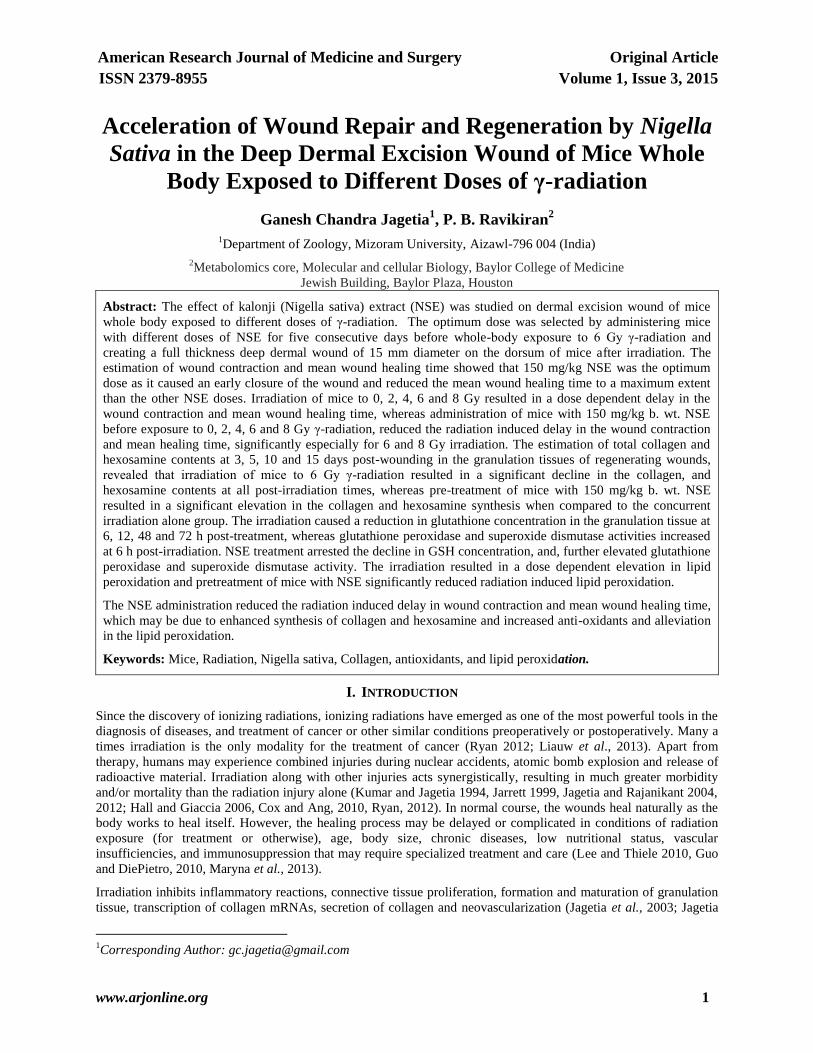
4.10. Superoxide Dismutase (Sod)
Infliction of excision wound itself increased the SOD activity in the sham- irradiation groups. Irradiation elevated
the SOD activity significantly and a maximum increase was observed for 2 Gy at 6 h post-irradiation. This increase
in SOD activity showed a radiation dose dependent decline and almost normal levels were restored by 72 h post-
irradiation (Figure9). The pattern of SOD activity in NSE+irradiation group was almost similar to that of
DDW+irradiation group, except that the activity was significantly higher in the former than the latter (Figure 9).
American Research Journal of Medicine And Surgery, Volume 1, Issue 3, 2015
ISSN 2379-8955
www.arjonline.org 12
15
30
45
60
75
1 6 12 24 48 72
DDW + 0Gy; NSE + 0Gy
DDW + 2Gy; NSE + 2Gy
DDW + 4Gy; NSE + 4Gy
DDW + 6Gy; NSE + 6Gy
DDW + 8Gy; NSE + 8Gy
Post - irradiation time (h)
SO
D n
mo
l/m
g p
rote
in
Fig9. Effect of NSE on the super oxide dismutase activity in the mice skin exposed to different doses of whole-body
γ-radiation.
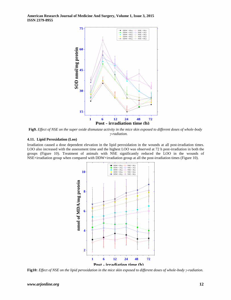
4.11. Lipid Peroxidation (Loo)
Irradiation caused a dose dependent elevation in the lipid peroxidation in the wounds at all post-irradiation times.
LOO also increased with the assessment time and the highest LOO was observed at 72 h post-irradiation in both the
groups (Figure 10). Treatment of animals with NSE significantly reduced the LOO in the wounds of
NSE+irradiation group when compared with DDW+irradiation group at all the post-irradiation times (Figure 10).
2
4
6
8
10
DDW + 0Gy; NSE + 0Gy
DDW + 2Gy; NSE + 2Gy
DDW + 4Gy; NSE + 4Gy
DDW + 6Gy; NSE + 6Gy
DDW + 8Gy; NSE + 8Gy
1 6 12 24 48 72
Post - irradiation time (h)
nm
ol
of
MD
A/m
g p
ro
tein
Fig10: Effect of NSE on the lipid peroxidation in the mice skin exposed to different doses of whole-body γ-radiation.
American Research Journal of Medicine and Surgery, Volume 1, Issue 3, 2015
ISSN 2379-8955
www.arjonline.org 13
V. DISCUSSION
Natural products have been the major source of medicine for human healthcare and several modern drugs have been
isolated from the plant products as they have been found to be nontoxic and have been in constant use since the
inception of human civilization. Kalonji, the seeds of Nigella Sativa Linn. (Ranunculaceae) has been used as a spice
and food preservative as well as a curative remedy for several disorders in the Ayurvedic system of medicine in
India. However, its healing activity in irradiated wounds remains unexplored, therefore, the present study was
undertaken to elucidate the effect of NSE on healing of irradiated excision wounds in mice.
Wound healing is a concerted effort of a sequence of various physiological processes including inflammation,
metabolism, regeneration and remodeling leading to complete wound closure (Haubner et al., 2012). Wound
contraction can be defined as the centripetal movement of the edges of a full thickness wound in order to facilitate
closure of the defect (Peacock, 1984; Tejero-Trujeque, 2001). The progression of wound healing can be determined
by the periodic assessment of contraction of excision wounds using various methods. In the present study, the
wound contraction was measured by capturing the video images of excision wound periodically (Jagetia and
Rajanikant, 2012). The exposure of mice to 6 Gy retarded the healing of wound and this delay in wound contraction
is in good agreement with earlier reports, where a similar effect has been reported (Grillo and Potsaid 1961,
Stromberg et al., 1968, Kumar and Jagetia, 1995; Jagetia et al., 2004, 2007, Jagetia and Rajanikant, 2012).
Treatment of mice with different doses of NSE prior to whole body 6 Gy irradiation resulted in a dose-related
acceleration in wound healing up to 150 mg/kg, as was evident by an early closure of wound in the NSE+irradiation
group. The studies regarding the amelioration of wound healing by NSE treatment after whole body irradiation are
lacking. The other nutrient factors including ascorbic acid and curcumin accelerated early repair of irradiated
wounds in a dose related manner and a maximum effect was observed up to a certain dose (Jagetia and Rajinikant,
2003;Jagetia et al., 2004). Similarly, vitamin A supplementation improved the acute radiation-induced delay in
wound healing (Levenson et al., 1984). Likewise, phenytoin sodium has been also reported to accelerate healing of
irradiated wounds earlier (Song and Cheng 1997).
Wound healing involves a cascade of well-orchestrated biochemical and cellular events leading to the growth and
regeneration of wounded tissue in a specific manner and ionizing radiation have been reported to produce multiple
negative effects on wound healing including diminished vascularity, impairment of the proliferate capacity of
fibroblasts and hematopoietic cells and decreased collagen synthesis (Rudolph et al., 1988, Doyle et al., 1996,
Kumar and Jagetia 1995; Jagetia et al., 2004,2007; Jagetia and Rajanikant, 2012). The retardation in wound healing
after irradiation may be due to the multiple negative effects elicited by irradiation on various important
physiochemical events leading to delayed healing of wounds.
The inflammatory response is generated immediately after wounding by neutrophils and macrophages that collects
in the wound bed after injury. This inflammation is caused by releasing H2O2 and other ROS species. (Woo et al.,
2012). The excess generation of ROS by irradiation in addition to wounding, may be one of the causes of delayed
wound healing in the irradiated wounds in the present study. The continued inflammation in the form of persistence
increase in ROS has been reported in chronic non healing wounds(Trabucchi et al., 2002; Williams and McBride,
2011; Ryan, 2012). Pretreatment of mice with NSE may have neutralized radiation-induced free radicals by
increasing the antioxidant enzymes and subsequently reduced the radiation-induced delay in irradiated wounds. The
NSE has been reported to scavenge various free radicals in vitro (Jagetia and Ravikiran, 2015)
Ionizing radiation induces severe damage to vital tissues, especially those with a high rate of cell division, such as
hematopoietic tissue (Levenson et al., 1984; Ryan, 2012) that play an essential role in healing of wounds. The loss
of significant number of bone marrow cells can lead to an immunocompromised state that could make the animals
more susceptible to bacterial infections, leading to complications in the healing of wounds. In fact shielding of bone
marrow during acute whole body X-irradiation has been reported to lower mortality and increase the closure of open
dorsal skin wounds of rats (Stromberg et al., 1967). These studies suggest that radiation-sensitive bone marrow-
derived cells play an important role in tissue repair and regeneration. Other possibility includes a delay in fixation of
the wound edge to the underlying tissue after irradiation, which may be due to lack of fibroblast proliferation and a
decrease in fibroblast synthetic function in the granulation bed. Recently, the role of bone marrow derived
mesenchymal stem cells has been confirmed in irradiated mice; where these cells have been reported to reduce
wound healing time (Liauw et al., 2013).
The contraction of open excised wounds has been found to be a function of contractile fibroblasts, known as
myofibroblasts (Gabbiani et al., 1972; Darby et al., 2014). Irradiation is thought to impair wound healing in skin
through its cytotoxic effect on fibroblasts, which may be due to the delay in the progression of cells through the cell
American Research Journal of Medicine And Surgery, Volume 1, Issue 3, 2015
ISSN 2379-8955
www.arjonline.org 14
cycle after irradiation (Rudolph et al., 1988). Irradiation may also induce adverse effects on the fibroblasts through
bone marrow depression, since some fibroblasts of the normal subcutaneous connective tissue, participating in
wound healing have been shown to originate from the bone marrow (Lange et al., 1979; Ryan 2012). The fibroblasts
play an important role during wound healing by secreting various collagens, fibronectins, heparin sulphate
proteoglycans, tenascin and connective tissue growth factors that are essential for healing of wounds (Kendall and
Feghali-Bostwick, 2014; Darby et al., 2014). Therefore, destruction of fibroblasts by irradiation may have been
responsible for delayed healing of irradiated wounds. Furthermore, fibroblasts have been reported to secrete various
angiogenic factors like VEGF, transforming growth factor-β (TGF-β) and platelet derived growth factor (PDGF)
that aid in the formation of new blood vessels during tissue repair (Haubner et al., 2012).
The proteins are one of the most important nutrient factors affecting wound healing. A deficiency of proteins can
impair capillary formation, fibroblast proliferation, proteoglycan synthesis, collagen synthesis, and wound
remodeling. A deficiency of protein also affects the immune system, with resultant decreased leukocyte
phagocytosis and increased susceptibility to infection (Gogia, 1995). Collagen is the major protein component of
connective tissue and is composed primarily of glycine, proline, and hydroxyproline. Collagen synthesis requires
hydroxylation of lysine and proline, and co-factors such as ferrous iron and vitamin C. Impaired wound healing
results from deficiencies in any of these co-factors (Campos et al., 2008). Also, reduction in collagen and DNA
syntheses may be another reason for delayed healing after irradiation. Irradiation has been reported to produce
negative effects on collagen and DNA syntheses (Reily 1994, Jagetia et al., 2003, 2005, 2007, Jagetia and
Rajanikant, 2005, 2012). In this study, the NSE administration resulted in a significant elevation in the collagen and
hexosamine synthesis when compared to the concurrent DDW+irradiation group, which may be one of the reasons
of early wound closure in NSE treated group.
Cells are equipped with antioxidant defence systems to detoxify the endogenous and exogenous oxidative challenges
during aerobic metabolism or when they encounter stress inducing agents (Mates et al., 1999). The increase in
GSHpx and SOD after irradiation is a short-term measure to counter the oxidative stress induced by wounding
coupled with irradiation. Alterations in the activity of these enzymes would modify the cellular redox state and the
net effect of radiation. Lipid hydroperoxides are a well-known marker of oxidative stress formed from unsaturated
phospholipids, glycolipids and cholesterol, through peroxidative reactions under oxidative stress (Girotti, 1998;
Jagetia et al., 2003; Rastogi et al., 2010). The NSE pretreatment has increased GSH concentration, GSHpx, catalase
and SOD activities in the present study, which may have contributed to reduced inflammation and early wound
repair. The ascorbic acid and curcumin treatment have been reported to elevate GSH, and GSHpx and superoxide
dismutase in irradiated wounds (Jagetia et al., 2003; Jagetia and Rajanikant, 2015) The induction of lipid peroxide
after irradiation is well known phenomenon and it has increased in a dose and time dependent manner. A similar
effect has been observed earlier in irradiated wounds treated with curcumin (Jagetia and Rajanikant, 2015). The
active constituents of Nigella sativa, thymoquinone could act as a free radical and superoxide radical scavenger, as
well as preserve the activity of various antioxidant enzymes such as catalase, glutathione peroxidase and
glutathione-S-transferase (Houghton et al., 1995). NSE treatment has reduced the formation of lipid peroxides and
the decline in the lipid peroxide may have accelerated the wound repair in NSE+irradiation group.
Thus acceleration in wound healing after NSE treatment may not be due to a single mechanism, but it could be due
to interplay of multiple mechanisms during the healing of irradiated wounds. The presence of NSE may have
scavenged the radiation induced free radicals thereby neutralizing their negative effect and accelerating the repair
and regeneration of irradiated wounds. NSE may have curtailed the inflammatory response elicited by wounding and
irradiation by increasing the antioxidant status and reducing the lipid peroxidation as observed in the present study.
The Nrf2 is involved in transcriptional regulation of antioxidant genes and irradiation has been reported to suppress
Nrf2 pathway genes (Canales-Aguirre, 2012; Khan et al., 2015). The thymoquinone the main phytochemical of NSE
has been recently reported to upregulate the transcription of Nrf2 gene (Kundu et al., 2014). NSE might have
alleviated the radiation-induced inhibition in Nrf2 leading to increased antioxidant status and reduced the delay in
healing of irradiated wounds in the present study. This may have accelerated wound repair. Although molecular
pathways employed by NSE during wound healing have not been investigated in the present study, the presence of
NSE before irradiation may have blocked the down modulation of cell cycle regulatory protein mRNAs retaining the
capacity of fibroblast and endothelial cell division that are essential for wound repair. Moreover, wounding and
irradiation have been reported to activate the transcription of NF-κB, COX- II and LOX mRNAs, and NSE may
have inhibited their transcriptional activation leading to an early repair and regeneration of wounds in this group.
The thymoquinone one of the active principles of NSE has been reported to block the transcriptional activation of
NF-κB, COX- II and LOX-5 (Haughton et al., 1995; Ryan 2012). NSE may have aided in the transcription of

American Research Journal of Medicine and Surgery, Volume 1, Issue 3, 2015
ISSN 2379-8955
www.arjonline.org 15
mRNAs related to collagen, DNA, NO syntheses therefore it would have helped in matrix remodeling and early
repair of irradiated wounds. NSE treatment may have also struck a balance between regeneration and apoptosis that
are essential for wound repair and thus accelerated the repair and regeneration of wounds.
The present study demonstrates that NSE retarded the radiation induced delay in healing of irradiated wounds in a
dose dependent manner and the greatest augmentation in wound healing was observed for 150 mg/kg. This healing
effect may be due to scavenging of radiation-induced free radicals, reduced inflammatory activity, increased
antioxidant status due to increased Nrf2 transcription and upregulation other mRNAs needed for wound healing. The
active principle of NSE, the thymoquinone may have suspended the transcription of NF-κB, COX- II and LOX5 and
accelerated wound repair and regeneration of irradiated wound in the present study. The NSE would have added in
early deposition of collagen and other proteins required during wound healing.
VI. ACKNOWLEDGEMENTS
We are thankful to Dr. M.S. Vidyasagar, Professor and Head, and Dr. J.G.R. Solomon, Department of Radiotherapy
and Oncology, Kasturba Medical College Hospital, Manipal, for providing the necessary irradiation facilities and for
dosimetric calculations, respectively. This work was carried out under a grant sanctioned to GCJ by the Council of
Scientific and Industrial Research, Govt. of India, New Delhi.
REFERENCES
[1] Abdel-Wahhab, M.A., Aly, S.E. Antioxidant property of Nigella sativa (black cumin) and Syzygium aromaticum (clove) in
rats during aflatoxicosis. J. Appl. Toxicol. 2005; 25:218–223.
[2] Ali B.H., Blunden G. Pharmacological and toxicological properties of Nigella sativa. Phytother. Res. 2003;17:299–305.
[3] Beaudry, V.G., Ihrie, R.A., Jacobs, S.B., Nguyen, B., Pathak, N., Park, E., Attardi, L.D. Loss of the desmosomal
component perp impairs wound healing in vivo. Dermatol. Res. Pract. 2010;2010:759731.
[4] Burits, M., Bucar, F. Antioxidant activity of Nigella sativa essential oil. Phytother. Res. 2000;14:323–328.
[5] Campos, A.C., Groth, A.K., Branco, A.B. Assessment and nutritional aspects of wound healing. Curr. Opin. Clin. Nutr.
Metab. Care 2008; 11:281-288.
[6] Canales-Aguirre, A.A.; Gomez-Pinedo, U.A.; Luquin, S.; Ramırez-Herrera, M.A.; Mendoza-Magana, M.L.; Feria-Velasco,
A. “Curcumin protects against the oxidative damage induced by the pesticide parathion in the hippocampus of the rat
brain,” Nutrit. Neurosci. 2012, 15(2): 62–69.
[7] Cemek, M., Enginar, H., Karaca, T., Unak, P. In vivo radioprotective effects of Nigella sativa L. oil and reduced
glutathione against irradiation-induced oxidative injury and number of peripheral blood lymphocytes in rats. Photochem.
Photobiol.. 2006;82:1691–1696.
[8] Cox, J.D., Ang, K. Radiation Oncology: Rationale, Technique, Results. 9th edn. Philadelphia, PA: Mosby Elsevier. 2010.
[9] Darby I.A., Laverdet B., Bonté F., Desmoulière A. Fibroblasts and myofibroblasts in wound healing Clin. Cosmet.
Investigat. Dermatol. 2014;7: 301–311.
[10] Dixon, R.A. Natural products and plant disease resistance. Nature, 2001;411:843–844.
[11] Doyle, J.W., Li, Y., Salloum, A. The effects of radiation on neo-vascularization in a rat model. Plast. Reconstr. Surg.
1996; 98:129-34.
[12] Ferguson, P.C., Boynton, E.L., Wunder, J.S., Hill, R.P., O'Sullivan, B., Sandhu, J.S., Bell, R.S: Intradermal injection of
autologous dermal fibroblasts improves wound healing in irradiated skin. J. Surg. Res. 1999, 85:331–8.
[13] Gabbiani, G., Chaponnier, C., Huttner, I: Cytoplasmic filaments and gap junctions in epithelial cells and myofibroblasts
during wound healing. J. Cell Biol. 1978;76:561-568.
[14] Girotti, A.W: Lipid hydroperoxide generate on, turnover, and effector action in biological systems. J. Lipid Res.
1998;39:1529–1542.
[15] Guo, S., DiPietro, L. A. Factors Affecting Wound Healing.J. Dental Res. 2010;89(3): 219–229.
[16] Hall, E., Giaccia, A. Radiobiology for the Radiologist. 6th Edn. Philadelphia, PA: Lippincott Williams & Wilkins. 2006.
[17] Haubner, F., Ohmann, E., Pohl, F., Strutz, J., Gassner, H.G. Wound healing after radiation therapy: review of the literature.
Radiat. Oncol. 2012;7:162.
[18] Houghton, P.J., Zarka, R., de Las, Heras. B., Hoult, JR. Fixed oil of Nigella sativa and derived thymoquinone inhibit
eicosanoid generation in leukocytes and membrane lipid peroxidation. Planta Med. 1995;61(1):33-36.
[19] Hunt, T.K, K.D., Thakra,l K.K: Cellular control of repair. In Soft and hard tissue repair: biological and clinical aspects.
Edited by Hunt TK Heppenstall RB, Pines E, Rovee D. New York: Praeger; 1984; pp 3–19.
[20] Jagetia G.C., Rajanikant G.K. Acceleration of wound repair by curcumin in the excision wound of mice exposed to
different doses of fractionated γ-radiation. Int. Wound J. 2012;.9(1):76-92.

American Research Journal of Medicine And Surgery, Volume 1, Issue 3, 2015
ISSN 2379-8955
www.arjonline.org 16
[21] Jagetia G.C., Rajanikant G.K. (2015) Curcumin Stimulates the Antioxidant Mechanisms in Mouse Skin Exposed to
Fractionated γ -Irradiation. Antioxidants, 2015; 4(1), 25-41.
[22] Jagetia G.C., Ravikiran P. B. (2014) Radioprotective Potential of Nigella Sativa Extract in Swiss Albino Mice Exposed to
Whole Body γ-Radiation. Altern. Integrat. Med. 3:4.
[23] Jagetia, G.C., Venkatesha, V.A. Effect of mangiferin on radiation-induced micronucleus formation in cultured human
peripheral blood lymphocytes. Environ. Mol. Mutagen. 2005;46(1):12-21.
[24] Jagetia, G.C Rajanikant, G.K. Role of curcumin, a naturally occurring phenolic compound of turmeric in accelerating the
repair of excision wounds in mice whole-body exposed to various doses of γ-radiation. J. Surg. Res. 2004;120:127-138.
[25] Jagetia, G.C., Rajanikant, G.K. Curcumin treatment enhances the repair and regeneration of wounds in mice hemi-body
exposed to γ-radiation. Plast. Reconstruct. Surg. 2005; 115(2):515-528.
[26] Jagetia, G.C., Rajanikant, G.K., Mallikarjun, Rao, K.V.N. Modulation of radiation-induced delay in the wound healing by
ascorbic acid in mice exposed to different doses of hemi-body γ-radiation. Wounds 2003; 15:324-338.
[27] Jagetia, G.C., Rajanikant, G.K., Rao, K.V.N.M. Ascorbic acid increases healing of excision wounds of mice whole body
exposed to different doses of γ-radiation. Burns 2007; 33(4):484-494.
[28] Jagetia, G.C., Rajanikant, G.K., Rao, S.K. Evaluation of the effect of ascorbic acid treatment in the artificially wounded
mouse exposed to different doses of fractionated gamma radiation. Radiat. Res. 2003; 159:371-380.
[29] Jagetia, G.C., Rajanikant, G.K., Rao, S.K., Baliga, S.M. Alteration in the glutathione, glutathione peroxidase, superoxide
dismutase and lipid peroxidation by ascorbic acid in the skin of mice exposed to fractionated γ-radiation. Clin. Chim. Acta
2003; 332:111-121.
[30] Jagetia, G.C., Rajanikant, G.K., Rao, S.K: Evaluation of the effect of ascorbic acid treatment on wound healing in mice
exposed to different doses of fractionated gamma radiation. Radiat. Res. 2003; 159:371–380.
[31] Kanter, M., Coskun, O., Uysal, H. The antioxidative and antihistaminic effect of Nigella sativa and its major constituent,
thymoquinone on ethanol-induced gastric mucosal damage. Arch. Toxicol. 2006; 80:217–224.
[32] Kendall R.T., Feghali-Bostwick C.A . (2014) Fibroblasts in fibrosis: novel roles and mediators. Front. Pharmacol. 5:123.
doi: 10.3389/fphar.2014.00123
[33] Khan A., Manna K., Das D.K., Kesh S.B., Sinha M., Das U., Biswas S., Sengupta A., Sikder K., Datta S., Ghosh M.,
Chakrabarty A., Banerji A., Dey S. Gossypetin ameliorates ionizing radiation-induced oxidative stress in mice liver-a
molecular approach. Free Radic. Res. 2015;49(10):1173-1186.
[34] Kumar, P., Jagetia, G.C. Modulation of wound healing in Swiss albino mice by different doses of gamma radiation. Burns
1995; 21:163-165.
[35] Kundu J., Kim D.H., Kundu J.K.,Chun K.S. Thymoquinone induces hemeoxygenase-1 expression in HaCaT cells via
Nrf2/ARE activation: Akt and AMPKα asupstream targets. Food Chem. Toxicol. 2014;65:18-26
[36] Lange, M.A., Vasil'eva, T.V., Michurina, T.V. Origin of fibroblasts and macrophages of skin wound granulation tissue.
Arkh. Anat., Gistol. Embriol. 1979;77:22-28.
[37] Lee, S., Thiele, C: Factors associated with free flap complications after head and neck reconstruction and the molecular
basis of fibrotic tissue rearrangement in preirradiated soft tissue. J Oral Maxillofac. Surg. 2010; 68:2169–2178.
[38] Legeza, VI., Galenko-Yaroshevskii, V.P., Zinov'ev, E.V., Paramonov, B.A., Kreichman, G.S., Turkovskii, II., Gumenyuk,
E.S., Karnovich, A.G., Khripunov, A.K: Effects of new wound dressings on healing of thermal burns of the skin in acute
radiation disease. Bull Exp. Biol. Med. 2004; 138:311–315.
[39] Levenson, S.M., Gruber, C.A., Rettura, G., Gruber, D.K., Demetriou, A.A., Seifter, A. Supplemental vitamin A prevents
the acute radiation-induced defect in wound healing. Ann. Surg. 1984; 200:494–512
[40] Liauw, S.L., Connell, P.P., Weichselbaum, R.R. New paradigms and future challenges in radiation oncology: an update of
biological targets and technology. Sci. Transl. Med. 2013;5(173):173sr2.
[41] Ma C., Hernandez M.A., Kirkpatrick V. E., Liang Li-J., Nouvong A. L., Gordon I.L., (2015) Topical-platelet-derived-
growth-factor-vs-placebo-therapy-diabetic-foot-ulcers:- Offloaded With Windowed Casts: A Randomized, Controlled Trial
- WOUNDS. 2015;27(4):83-91
[42] Mack J.A., Maytin E.V. Persistent inflammation and angiogenesis during wound healing in K14-directed Hoxb13
transgenic mice. J. Invest. Dermatol. 2010;130(3):856-865
[43] Maryna, S., Shkumat, Pavlo, P., Klymenko, Yuri., I. Leonov., Iryna, N., Pishel, Pavel, V., Glukhovskiy. Prolonged
inflammatory cytokine expression during the late phase of wound healing in the diabetic K14/mIGF1 transgenic mice. J.
Med. Biol. Sci. 2013;6 (1).
[44] Mates, J.M., Perez-Gomez, C., Nunez, de Castro, I. Antioxidant enzymes and human diseases. Clin. Biochem.
1999;32:595–603.
[45] McQuestion, M. Evidence-based skin care management in radiation therapy: clinical update. 2011. Semin. Oncol. Nurs.
27:e1–7.

American Research Journal of Medicine and Surgery, Volume 1, Issue 3, 2015
ISSN 2379-8955
www.arjonline.org 17
[46] Peacock, E.E. Contraction. In: Wound Repair, 3d Edition, Peacock EE (ed), Philadelphia, WB Saunders 1984; 39-55.
[47] Ramadan, M.F., Kroh, L.W., Morsel, J.T. Radical scavenging activity of black cumin (Nigella sativa L.), coriander
(Coriandrum sativum L.), and niger (Guizotia abyssinica Cass.) crude seed oils and oil fractions. J. Agr. Food Chem.
2003;51:6961–6969.
[48] Rastogi, L., Feroz, S., Pandey, B.N., Jagtap, A., Mishra, KP. Protection against radiation-induced oxidative damage by an
ethanolic extract of Nigella sativa L. Int J Radiat Biol. 2010;86(9):719-731.
[49] Reily, P.A. Free Radicals in Biology: Oxidative stress and the effect of ionizing radiation . Int J Radiat Biol 1994; 65: 27-
33.
[50] Rudolph, R., Vande Berg, J., Schneider, J.A., Fischer, J.C., Poolman, W.L. Slowed growth of cultured fibroblasts from
human radiation wounds. Plast. Recontruct. Surg. 1988; 82:669–77.
[51] Ryan, J.L. Ionizing radiation: the good, the bad, and the ugly. J. Invest. Dermatol. 2012;132(3 Pt 2):985-993.
[52] Song, S., Cheng, T. The effect of systemic and local irradiation on wound macrophage and repair promoting action of
phenytion sodium. Zhonghma Yi Xue Za Zhi 1997; 77:54–7.
[53] Stromberg, L.R., Woodward, K.T., Mahin, D.T., Donati, R.M. Altered wound healing in x-irradiated rats: the effect of
bone marrow shielding. Experientia, 1967;23:1064-1065.
[54] Tejero-Trujeque R. How do fibroblasts interact with the extracellular matrix in wound contraction? J. Wound Care,
2001;10(6): 237–242.
[55] Trabucch, E., Pallotta, S., Morini, M., Corsi, F., Franceschini, R., Casiraghi, A., Pravettoni, A., Foschi, D., Minghetti, P.
Low molecular weight hyaluronic acid prevents free radical damage to granulation tissue during wound healing. Int. J.
Tissue React. 2002;24:65-71.
[56] Williams, J.P., McBride, W.H. After the bomb drops: a new look at radiation-induced multiple organ dysfunction
syndrome (MODS). Int. J. Radiat. Biol. 2011;87:851–868.
[57] Woo, C.C., Kumar, A.P., Sethi, G., Tan, K.H.B. Thymoquinone: Potential cure for inflammatory disorders and cancer.
Biochem. Pharmacol. 2012;83. 443–451.