prophylactic effects of nigella sativa extract and ... · preparation of nigella sativa ethanolic...
TRANSCRIPT

BEPLS Vol 6 [9] September 2017 5 | P a g e ©2017 AELS, INDIA
Bulletin of Environment, Pharmacology and Life Sciences Bull. Env. Pharmacol. Life Sci., Vol 6[10] September 2017 : 05-16 ©2017 Academy for Environment and Life Sciences, India Online ISSN 2277-1808 Journal’s URL:http://www.bepls.com CODEN: BEPLAD Global Impact Factor 0.876 Universal Impact Factor 0.9804
NAAS Rating 4.95
ORIGINAL ARTICLE OPEN ACCESS
Prophylactic Effects of Nigella sativa Extract and Thymoquinone against Cyclophosphamide-induced Sperm Head Abnormalities
and Chromatin Instability in Mice
Suzanah Abdul-Rahman1 and Saheera Kamarzaman2 1-Department of Biomedical Science, Faculty of Allied Health Sciences, International Islamic University Malaysia (IIUM), Jalan Sultan Ahmad Shah, Bandar Indera Mahkota, 25200 Kuantan, Pahang, MALAYSIA
2-Division of Basic Medical Sciences, Faculty of Medicine, Cyberjaya University College, 3410, Jalan Teknokrat 3, Cyber 4, 63000 Cyberjaya, Selangor, MALAYSIA
Email: [email protected]
ABSTRACT
Genetic stability in male fertility is indicated by DNA integrity of sperm. Cyclophosphamide is cytotoxic to germ cells of the testes causing DNA breakage, azoospermia and infertility. This study focuses on effectiveness of Nigella sativa and thymoquinone in lowering chemotherapeutic-associated toxicity of cyclophosphamide on sperm head and chromatin stability of Balb/c mice. Thirty male mice were divided into 6 groups and administration of cyclophosphamide at 200 mg/kg body weight by intraperitoneal injections is followed similarly by either thymoquinone or ethanol extraction of Nigella sativa at 10 mg/kg, 6 hours post-cyclophosphamide exposure. Thymoquinone and Nigella sativa administration was continued on alternate days for 32 days. Sperm samples were assayed with Eosin Y and Toluidine Blue. Sperm head abnormalities were classified into normal, lack-hook, banana-like form, amorphous and folded on themselves. Chromatin condensation was categorized into unstained, completely and partially-stained. One hundred spermatozoa per animal were observed. Nigella sativa extract reduced the percentage of abnormal sperm head post-cyclophosphamide treatment and showed preservation of the normal chromatin condensation indicative of protection against sperm DNA alteration. This study indicates the potential of Nigella sativa and thymoquinone as prophylaxes against cyclophosphamide-induced damage to gametes in the attempt to salvage reproductive functions following chemotherapy. Key words: Cyclophosphamide; Thymoquinone; Nigella sativa extract; Sperm Head Abnormalities; Chromatin Condensation Received 01.04.2017 Revised 15.05.2017 Accepted 19.07.2017 INTRODUCTION The research conducted herein focused on the combination of drug-herbal therapy to provide more evidence on the chemoprotective potential of the beneficial plant, Nigella sativa, via its seed extract and active constituent, thymoquinone. Cyclophosphamide is a chemotherapeutic agent known to alter the reproductive capacity and fertility in many animal models. As N. sativa and thymoquinone are found to antagonise the genotoxic effects of this anticancer drug, it would be of importance for future application in the clinical setting as a new treatment option, able to minimise the toxic effects on reproductive function perhaps in the form of prophylaxes. Spermatogenesis is one of the most productive self-renewing systems of which four to sixty million spermatozoa are produced daily per gram of testis tissue in the mammalian species [1]. However, it has long been known that cell death during the process reduces the final production of sperm substantially. Stem cells persist throughout the reproductive life and are constantly under attack from DNA-damaging agents produced by endogenous and exogenous agents. They can accumulate a large amount of chemical exposure and may result in cell death if not repaired. Cells that survived radiation or chemicals continue to divide and differentiate, and mutations can be transmitted to the offspring if not repaired by the time of replication. Once the unrepaired lesions in male germ cells are transmitted to the zygote, it may lead to foetal death including pre- or post-implantation loss. New mutations that developed in the paternal genome will not be eliminated in the fertilised egg.

BEPLS Vol 6 [9] September 2017 6 | P a g e ©2017 AELS, INDIA
Anticancer drugs, in general, are mutagenic as they can interfere with DNA metabolism [2]. There is a strong indication that DNA damage could play an important role in male fertility and reproduction [3]. The commonly used anticancer drug, cyclophosphamide (CPA), is one of the most damaging alkylating agents that affect the DNA of replicating and rapidly multiplying cells especially in the gonads and pituitary leading to miscoding, cross-linking and DNA breakage. It acts by the transfer of alkyl groups to the guanine compound of the DNA. The genotoxic effects of cyclophosphamide on male germ cells showed that it can reach the spermatogonia in significant quantities. Germ cells are specifically sensitive to cyclophosphamide treatment due to its high proliferating activity. Administration of cyclophosphamide has been demonstrated to cause oligospermia, azoospermia, testicular damage and germ cell toxicity in male rodents [4 and 5]. It induces defects in mice foetuses [6] and increases the incidence of pre-implantation loss. Recent studies have investigated the deleterious effects of cyclophosphamide on chromosomal aneuploidy [7] and chromatin condensation [8]. Previous studies have reported significant correlations between chromatin abnormalities and morphological alterations [9]. Mammalian sperm heads consist almost totally of chromatin and as oxidative stress is associated with DNA strand breaks, the loosely condensed sperm chromatin that suffers DNA damage will lead to the weakening of the sperm chromatin condensation following alterations in the relative proportion of protamines. DNA denaturation has important implications on fertility outcomes. Some sperms with chromatin abnormalities are able to fertilise oocytes in vivo and in vitro, but the DNA damage can persist throughout the embryonic period which will induce apoptosis and embryo fragmentation that can ultimately lead to abortion. The use of a traditional medicinal plant is the basis of the intervention program in this study, in contributing to the amelioration of the toxicity effects shown by the toxicant. The interest in the prophylactic effects of natural-based drugs started mainly because of the wide spread belief that herbal therapy is healthier than the synthetic drugs. N. sativa has been demonstrated as an antioxidant in many in vitro and in vivo studies. The protective mechanism of action of antioxidants is often described according to their ability to act as free radical scavengers of various radical oxygen species including superoxide radical anion and hydroxyl radicals through different mechanisms [10, 11 and 12]. N. sativa, a medicinal plant from the family of Ranunculaceae and its active constituent, thymoquinone, have shown strong antioxidant activities in their ability to reduce oxidative stress. N. sativa oil has been reported to exhibit antiviral, anticancer, antibacterial, anticlastogenic, antidiabetic and antimicrobial effects [13, 14, 15, 16, 17 and 18]. Moreover, N. sativa oil protects cyclosporine A injury in rat heart demonstrated by normalised histopathology, a decrease in lipid peroxidation, improvement in the antioxidant enzyme status and cellular protein oxidation suggesting antioxidant activity [19]. N. sativa extract has been shown to ameliorate the cytotoxic effects of cyclophosphamide in the testis and acrosome reaction in mice [20]. Thymoquinone was previously reported to re-establish mouse spermatogenesis following testicular injury [21]. The hepatoprotective effects of thymoquinone against carbon tetrachloride (CCl4) in mice and its protective effect against doxorubicin induced nephrotoxicity and cardiotoxicity were found to be via antioxidant mechanisms [22 and 23]. The prime goal of this study is to test whether the provision of N. sativa extract and its active compound, thymoquinone, can significantly lower chemotherapeutic-associated toxicity on the reproductive capacity in mice following cyclophosphamide treatment. The high antioxidant content of N. sativa has been proven in other studies thus the plant extract is hoped to provide an essential rejuvenative environment for germ cell growth and cell repair following insult by mutagenic and alkylating agents. MATERIALS AND METHODS Experimental animals A total of 30 male Balb/c mice, weighing about 18 – 21 g, 5 - 7 weeks of age, were obtained from the local laboratory (Kuala Lumpur, Malaysia). Mice were housed under controlled temperatures of 22 ± 1 ºC with 12 hours light/dark cycle, and provided with food pellets and water ad libitum. Work done in this study was performed according to the Guidelines for Animal Study and was approved by the Ethics Committee of the Faculty of Medicine, International Islamic University Malaysia. Preparation of Nigella sativa Ethanolic Extract The Nigella sativa seeds (Syria) were purchased from a local herb store. Seeds of N. sativa were powdered in a mixer and 400 g of air-dried powder was extracted with ethanol (40 – 60°C) in a Soxhlet Extractor for 16 hours. The solution was then evaporated to dryness under reduced pressure and controlled temperature by using a rotary evaporator. The crude extract was stored in a refrigerator at 4°C in an air-tight bottle until further use. Determination of Thymoquinone in Nigella sativa Extract
Abdul-Rahman and Kamarzaman

BEPLS Vol 6 [9] September 2017 7 | P a g e ©2017 AELS, INDIA
A Liquid Chromatography tandem Mass Spectrometer (LCMS/MS – AB Sciex 3200QTrap) coupled to Ultra High Performance Liquid Chromatography (UHPLC- Perkin ElmarFlexar FX15) system using Multiple Reaction Monitoring (MRM) method was undertaken for quantification of the plant bioactive compound, thymoquinone, in the ethanolic extract of N. sativa. A Phenomenax Aqua C18 analytical column was employed using mobile phase of water with 0.1% formic acid and 5 µM ammonium formate: acetonitrile with 0.1% formic acid and 5 µM ammonium formate at a flow rate of 500 µL/min for 8 minutes. The parent masses of thymoquinone with its respective fragment masses were targeted. Any positive identification resulted in a positive chromatographic peak. Further confirmation was based on retention time compared with the reference standard. Reference standard at 10 ppm was prepared using ethanol: methanol and infused into the instrument for optimisation and compound tuning. Further confirmation was done by monitoring two fragment ions for both compounds. A positive result should have both fragment ions present in the sample with the correct retention time. A standard calibration curve was prepared for thymoquinone at low and high concentrations ranging from 0.05 to 50 ppm. Experimental Design Cyclophosphamide (Sigma-Aldrich), thymoquinone (Santa-Cruz) and N. sativa ethanolic extract were dissolved in sodium chloride 0.9% (normal saline). Cyclophosphamide was injected intraperitoneally (i.p) with a single dose of 200 mg/kg of body weight. Thymoquinone and N. sativa extract were administered via i.p. at a concentration of 10 mg/kg body weight and supplementation was continued on alternate days throughout the length of the study. Male mice were randomly divided into the following groups (n=5/group); (i) Vehicle-treated control (normal saline), (ii) cyclophosphamide alone, (iii) N. sativa extract alone, (iv) thymoquinone alone, (v) thymoquinone six-hours post cyclophosphamide treatment, and (vi) N. sativa extract six hours post cyclophosphamide treatment. Observation of the effects was done on day 33. Mice were weighed and sacrificed by cervical dislocation 24 hours after the last dose. Sperm Count The approximate number of sperm/ml was calculated using a haemocytometer (Hirschmann ®Laborgerate). Sperm suspension at the amount of 10 µl was added into an Eppendorf® tube that contained 90 µl of tap water (10X dilution). The water was used to immobilise the sperm to make the calculation easier. The mixture of sperm/water solution (10µl) was placed in the counting chamber of the haemocytometer, covered with a cover slip and viewed under a light microscope. Five (5) squares were counted to determine the concentration of sperm x dilution (10) x 5 squares x 10000.
Concentration of sperm/ml = Number of sperm in 5 squares x 10 x 5 x 10000
Sperm Motility Microscope slide was pre-warmed to retain motility and sperm survival. Sperm suspension of 10 µl was placed and covered with a cover slip. Sperm motility was assessed under a light microscope. Motility was estimated as a percentage (%). Sperm Head Abnormality Test Mice from each group were sacrificed by cervical dislocation and their cauda epididymides were removed. Sperm suspensions were prepared by mincing the cauda in 2 ml of normal saline and mixed (10:1) with 1% Eosin Y for 30 minutes, allowed to air dry and mounted onto a coverslip with Permount mounting medium. One hundred (100) spermatozoa were examined for each animal at 40X and 100X magnifications hence a total of 500 sperms was examined for each group. Sperm head abnormalities were categorised according to Wyrobek and Bruce (1975) as ‘normal’, ‘lack the usual hook’, ‘banana-like form’, ‘amorphous’ and ‘folded on themselves’ groups [24]. Sperm Chromatin Condensation Test Sperm samples from the epididymis were suspended in 0.5 ml saline and centrifuged at 200 g for 5 minutes. The pellets were re-suspended in 1.0 ml saline. The dense sperm suspension was spread on slides and air dried. The preparations were fixed in acetic alcohol (3 parts of absolute ethanol and 1 part of glacial acetic acid) for 2 minutes. Slides were then air dried, stained with 1% (w/v) aqueous Toluidine Blue (Sigma-Aldrich) for 15 minutes, washed with distilled water, covered with cover slips and observed under a light microscope (Olympus)37. One hundred (100) spermatozoa were examined for each animal at 40X and 100X magnifications hence a total of 500 sperms was examined for each group. Sperm chromatin was categorised according to Krzanowska (1982); unstained, partially stained and completely stained [25]. Toluidine blue staining gave positive reaction only with unstable chromatin. Unstained or partially stained sperm head was scored as possessing DNA of normal integrity and those completely stained were scored as having damaged DNA. Statistical Analysis The results were shown as mean ± standard deviation (SD) for each group. Statistical analysis was performed using IBM SPSS Statistics 21 software. Body weight was analysed by One-Way ANOVA. The data on sperm concentration and sperm head morphology do not meet the stringent assumptions of the
Abdul-Rahman and Kamarzaman
BEPLS Vol 6 [9] September 2017 8 | P a g e ©2017 AELS, INDIA
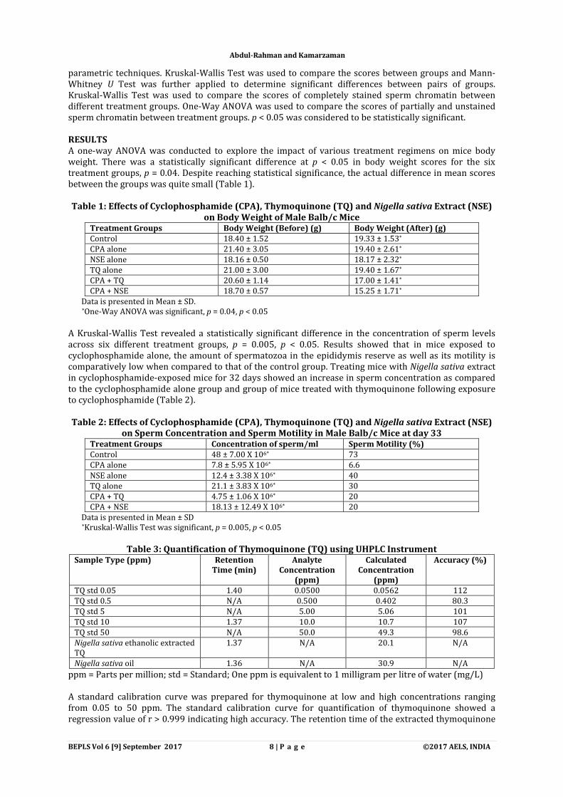
parametric techniques. Kruskal-Wallis Test was used to compare the scores between groups and Mann-Whitney U Test was further applied to determine significant differences between pairs of groups. Kruskal-Wallis Test was used to compare the scores of completely stained sperm chromatin between different treatment groups. One-Way ANOVA was used to compare the scores of partially and unstained sperm chromatin between treatment groups. p < 0.05 was considered to be statistically significant. RESULTS A one-way ANOVA was conducted to explore the impact of various treatment regimens on mice body weight. There was a statistically significant difference at p < 0.05 in body weight scores for the six treatment groups, p = 0.04. Despite reaching statistical significance, the actual difference in mean scores between the groups was quite small (Table 1). Table 1: Effects of Cyclophosphamide (CPA), Thymoquinone (TQ) and Nigella sativa Extract (NSE)
on Body Weight of Male Balb/c Mice Treatment Groups Body Weight (Before) (g) Body Weight (After) (g) Control 18.40 ± 1.52 19.33 ± 1.53* CPA alone 21.40 ± 3.05 19.40 ± 2.61* NSE alone 18.16 ± 0.50 18.17 ± 2.32* TQ alone 21.00 ± 3.00 19.40 ± 1.67* CPA + TQ 20.60 ± 1.14 17.00 ± 1.41* CPA + NSE 18.70 ± 0.57 15.25 ± 1.71*
Data is presented in Mean ± SD. *One-Way ANOVA was significant, p = 0.04, p < 0.05
A Kruskal-Wallis Test revealed a statistically significant difference in the concentration of sperm levels across six different treatment groups, p = 0.005, p < 0.05. Results showed that in mice exposed to cyclophosphamide alone, the amount of spermatozoa in the epididymis reserve as well as its motility is comparatively low when compared to that of the control group. Treating mice with Nigella sativa extract in cyclophosphamide-exposed mice for 32 days showed an increase in sperm concentration as compared to the cyclophosphamide alone group and group of mice treated with thymoquinone following exposure to cyclophosphamide (Table 2).
Table 2: Effects of Cyclophosphamide (CPA), Thymoquinone (TQ) and Nigella sativa Extract (NSE)
on Sperm Concentration and Sperm Motility in Male Balb/c Mice at day 33 Treatment Groups Concentration of sperm/ml Sperm Motility (%) Control 48 ± 7.00 X 106* 73 CPA alone 7.8 ± 5.95 X 106* 6.6 NSE alone 12.4 ± 3.38 X 106* 40 TQ alone 21.1 ± 3.83 X 106* 30 CPA + TQ 4.75 ± 1.06 X 106* 20 CPA + NSE 18.13 ± 12.49 X 106* 20
Data is presented in Mean ± SD *Kruskal-Wallis Test was significant, p = 0.005, p < 0.05
Table 3: Quantification of Thymoquinone (TQ) using UHPLC Instrument
Sample Type (ppm) Retention Time (min)
Analyte Concentration
(ppm)
Calculated Concentration
(ppm)
Accuracy (%)
TQ std 0.05 1.40 0.0500 0.0562 112 TQ std 0.5 N/A 0.500 0.402 80.3 TQ std 5 N/A 5.00 5.06 101 TQ std 10 1.37 10.0 10.7 107 TQ std 50 N/A 50.0 49.3 98.6 Nigella sativa ethanolic extracted TQ
1.37 N/A 20.1 N/A
Nigella sativa oil 1.36 N/A 30.9 N/A
ppm = Parts per million; std = Standard; One ppm is equivalent to 1 milligram per litre of water (mg/L) A standard calibration curve was prepared for thymoquinone at low and high concentrations ranging from 0.05 to 50 ppm. The standard calibration curve for quantification of thymoquinone showed a regression value of r > 0.999 indicating high accuracy. The retention time of the extracted thymoquinone
Abdul-Rahman and Kamarzaman

BEPLS Vol 6 [9] September 2017 9 | P a g e ©2017 AELS, INDIA
from N. sativa ethanolic extract was 1.37 minutes which was similar to the retention time of standard thymoquinone. N. sativa extract at 10 mg contained 20.1 ppm of thymoquinone which is equivalent to 50,250 mg/L of sample concentration at 2500 dilution factor (Table 3). The results on the percentage of sperm head abnormalities in different treatment groups are presented in Fig. 1. Kruskal-Wallis Test revealed a statistically significant difference in the percentage of abnormal sperms across six different treatment groups, p = 0.01, p < 0.05. Mann-Whitney U test revealed a significant increase in the percentage of abnormal sperm heads of cyclophosphamide alone group (66.33%) as compared to control group (39.74%), p < 0.05. There was no increase in the percentage of abnormal sperms in N. sativa extract treated groups when compared with control values. In contrast, there was an increase in the percentage of abnormal sperms in thymoquinone treated groups when compared with the control group. However, the difference is not significant. The test also indicated that there was a statistically significant reduction in the percentage of abnormal sperm heads in cyclophosphamide-treated group supplemented with N. sativa extract (49.27%), and groups of mice that received N. sativa extract and thymoquinone alone, 30.13% and 51.58% respectively, compared to cyclophosphamide alone group, p < 0.05. No significance difference in the percentage of sperm head abnormalities were found in group for cyclophosphamide-treated mice supplemented with thymoquinone (66.29%) when compared to cyclophosphamide alone group.
Fig. 1: Percentage of abnormal sperm heads in different treatment groups
aKruskal-Wallis Test was significant, p < 0.05 bSignificantly different from control treatment group, p < 0.05 (Mann-Whitney U Test) CPA = Cyclophosphamide, TQ = Thymoquinone, NSE = Nigella sativa extract
Table 4: Types of Sperm Head Morphology according to Wyrobek and Bruce (1975) Groups Normal Abnormal
Lack-Hook Banana-like Amorphous Folded Control 65.67 ± 7.37c (60) 1.67 ± 2.88c (1.53) 0.67 ± 0.57c
(0.61) 7.67 ± 0.57c (7.03) 33.33 ± 12.50a
(30.57) CPA alone 35.40 ± 6.58b (33.65) 15.80 ± 10.01b
(15.01) 6.00 ± 2.00b (5.7)
19.20 ± 6.05b (18.25)
28.80 ± 4.38a
(27.37) NSE alone 70.00 ± 3.67c (69.86) 8.40 ± 3.05b (8.38) 2.20 ± 2.28c
(2.19) 6.40 ± 3.50c (6.39) 13.20 ± 0.83bc
(13.17) TQ alone 49.50 ± 12.45a
(48.41) 5.75 ± 2.87c (5.62) 2.25 ± 1.89c
(2.2) 12.00 ± 4.89a (11.73)
32.75 ± 3.77a (32.03)
CPA + TQ 32.00 ± 15.56a (33.68)
10.50 ± 4.95a (11.05)
1.50 ± 0.70c (1.57)
40.00 ± 21.21a (42.10)
11.00 ± 5.65c (11.57)
CPA + NSE 53.00 ± 3.16bc (50.71) 7.75 ± 1.5b (7.41) 3.50 ± 1.29b
(3.35) 18.25 ± 2.75b (17.46)
22.00 ± 6.63a (21.05)
Data is presented in mean ± SD. The values in parantheses are percentages. Kruskal-Wallis Test was significant, p < 0.05 a No significant difference between treatment groups. b Significantly different from control treatment group, p < 0.05 (Mann-Whitney U Test). c Significantly different from cyclophosphamide alone treatment group, p < 0.05 (Mann-Whitney U Test). CPA = Cyclophosphamide, TQ = Thymoquinone, NSE = N. sativa Extract
a
a
a
a
Abdul-Rahman and Kamarzaman

BEPLS Vol 6 [9] September 2017 10 | P a g e ©2017 AELS, INDIA
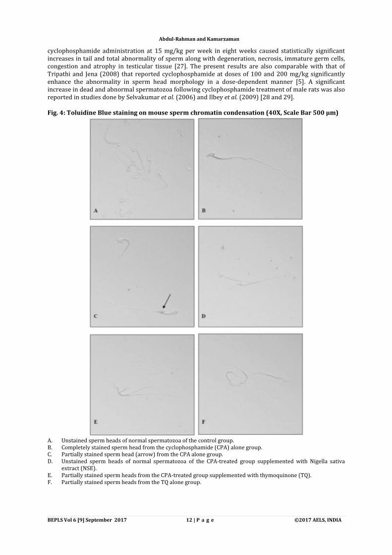
The results of the types of sperm head morphology are presented in Table 4. Kruskal-Wallis Test revealed a statistically significant difference in the mean scores of sperm head morphology across six different treatment groups, p < 0.05. Cyclophosphamide significantly induced sperm head abnormalities when compared to the control group as tested by Mann-Whitney U Test, p < 0.05. The highest mean score of sperm head abnormalities in the cyclophosphamide alone group was seen in folded sperms with 28.80 ± 4.38 (27.37%). Amorphous-shaped sperms made up 18.25%, followed by lack-hook sperms with 15.01% and banana-like sperms with 5.7% from the pool of sperm with abnormalities. There is a significant decrease in the mean scores of banana-like (1.57%) and folded (11.57%) sperms in cyclophosphamide-treated groups supplemented with thymoquinone as compared to the cyclophosphamide alone group (p < 0.05). Supplementation of N. sativa extract to cyclophosphamide-treated mice on alternate days indicated lower mean scores of overall lack-hook, banana-like, amorphous and folded sperms as compared to the cyclophosphamide alone group. However, the differences were not statistically significant. Fig. 2 shows a representation of normal and abnormal sperm heads of mice in various treatment groups. The mean percentages of sperm chromatin condensation are shown in Fig. 3. Kruskal-Wallis Test revealed a statistically significant difference in the score of completely stained sperm heads across six different treatment groups, p < 0.05. A higher percentage of completely stained sperm heads (cells with damaged DNA) was associated with sperms obtained from the group treated with cyclophosphamide alone (15.54%) as compared to the control group (6.37%). The completely stained sperm heads were also significantly abundant in the group co-treated with cyclophosphamide and thymoquinone (13.76%). There was a statistically significant difference at the p < 0.001 level in partially stained and unstained sperm head scores for the various treatment groups as measured by One-Way ANOVA. There was a higher proportion of partially stained sperm heads in the group of cyclophosphamide alone (70.62%) than in the control group (22.44%). In the group of cyclophosphamide-treated mice supplemented with N. sativa extract, the percentage of partially stained sperm heads was reduced to 22.47%. Mice co-treated with cyclophosphamide and N. sativa extract showed a significant increase in the percentage of unstained sperm heads (76.29%), as opposed to that in the cyclophosphamide alone group (13.85%). No significance difference in the proportion of unstained sperm heads was determined between the group of mice co-treated with cyclophosphamide and thymoquinone and group of cyclophosphamide alone. Fig. 4 showed a representation of sperm chromatin condensation of the three categories of unstained, partially stained and completely stained sperm heads in the various treatment groups.
Fig. 2: Eosin staining of sperm head morphology.
Figure shows (A) normal sperm heads of control group (40X); (B) sperm lacking the usual hook of cyclophosphamide (CPA) alone group (100X); (C) sperm folded on itself of CPA alone group (100X); (D) sperms with banana-like form of CPA alone group (100X); (E) amorphous sperm of CPA alone group (100X) and (F) normal sperm head of the CPA-treated groups supplemented with Nigella sativa extract (NSE) (100X), Scale Bar 500 µm
Abdul-Rahman and Kamarzaman

BEPLS Vol 6 [9] September 2017 11 | P a g e ©2017 AELS, INDIA
Fig. 3: Percentages of sperm chromatin condensation with Toluidine Blue staining in mice spermatozoa.
Data is presented in mean ± SD. The values in parentheses are percentages. The completely stained spermatozoa was significantly different by Kruskal-Wallis Test, p = 0.004, p< 0.05 The partly and unstained spermatozoa were significantly different by One-Way ANOVA, p = 0.000, p < 0.001 a No significant difference between treatment groups. b Significantly different from control group. c Significantly different from cyclophosphamide alone group, p<0.05 CPA = Cyclophosphamide, TQ = Thymoquinone, NSE = Nigella sativa extract. DISCUSSION In this study, we evaluated the possible chemoprotective effects of 10 mg/kg Nigella sativa extract and thymoquinone as prophylaxes in mice following exposure to 200 mg/kg of cyclophosphamide that distinctly induced changes in sperm head morphology and sperm chromatin condensation. The chemoprotective activity of N. sativa extract and thymoquinone against cyclophosphamide-induced sperm head abnormalities has not been evaluated previously. Mouse studies suggest that exposure of spermatogonia to low doses of alkylating agents, increases the frequency of germ line DNA damage, predisposing offspring to increased mutation rates. In our study, we observed a significant increase in the percentage of abnormal sperm heads to 66.33% following exposure to a single toxic dose of cyclophosphamide as compared to concurrent control values of 39.74%, a total increase of more than 25%. Cyclophosphamide and other mutagenic, teratogenic and carcinogenic chemicals such as methyl methanesulfonate, ethyl methanesulfonate, griseofulvin, benzo(a)pyrene, metepa and thiotepa are known to induce sperm abnormalities to 1.2 to 3.4% above that of control values [24]. Our findings are in agreement with results by Rudrama et al. (2013) which reported that treatment of male mice with 50 mg/kg of cyclophosphamide showed significant increase in the percentage of abnormal sperms to 12.80% when compared to control values of only 3.40% [26]. Ceribasi et al. (2010) reported that
Abdul-Rahman and Kamarzaman
BEPLS Vol 6 [9] September 2017 12 | P a g e ©2017 AELS, INDIA
cyclophosphamide administration at 15 mg/kg per week in eight weeks caused statistically significant increases in tail and total abnormality of sperm along with degeneration, necrosis, immature germ cells, congestion and atrophy in testicular tissue [27]. The present results are also comparable with that of Tripathi and Jena (2008) that reported cyclophosphamide at doses of 100 and 200 mg/kg significantly enhance the abnormality in sperm head morphology in a dose-dependent manner [5]. A significant increase in dead and abnormal spermatozoa following cyclophosphamide treatment of male rats was also reported in studies done by Selvakumar et al. (2006) and Ilbey et al. (2009) [28 and 29]. Fig. 4: Toluidine Blue staining on mouse sperm chromatin condensation (40X, Scale Bar 500 µm)
A. Unstained sperm heads of normal spermatozoa of the control group. B. Completely stained sperm head from the cyclophosphamide (CPA) alone group. C. Partially stained sperm head (arrow) from the CPA alone group. D. Unstained sperm heads of normal spermatozoa of the CPA-treated group supplemented with Nigella sativa
extract (NSE). E. Partially stained sperm heads from the CPA-treated group supplemented with thymoquinone (TQ). F. Partially stained sperm heads from the TQ alone group.
Abdul-Rahman and Kamarzaman

BEPLS Vol 6 [9] September 2017 13 | P a g e ©2017 AELS, INDIA
Cyclophosphamide has been associated with increased oxidative stress and DNA damage in mice [30]. Alterations in the morphology of the sperm head due to cyclophosphamide may be elucidated with the excessive production of reactive oxygen species (ROS) in association with decreased antioxidant capacity in the testis. Spermatocytes and spermatids are able to generate low levels of ROS which are essential for capacitation, hyperactivation and sperm-oocyte function. However, they are particularly susceptible to damage induced by excessive ROS due to large quantities of polyunsaturated fatty acids in the plasma membranes but low concentrations of scavenging enzymes in the cytoplasm [31 and 32]. Lipid peroxidation, which can be induced by direct or indirect toxicity of cyclophosphamide, is an oxidative deterioration of lipids containing a number of carbon-carbon double bonds, which later formed a large number of toxic-by products and behave as toxic ‘second messengers’ [33]. Sperm lipid peroxidation destroys the structure of the lipid matrix in the sperm membranes. It is associated with rapid loss of intracellular ATP leading to axonemal damage, decreased sperm viability and increased mid-piece morphological defects. In extreme cases, it completely inhibits spermatogenesis [34]. The elevation of frequency of sperm head abnormalities in exposed mice indicate that the active metabolite was in fact reaching the germ cells and may have caused damage to the pre-meiotic stages of spermatogenesis. DNA synthesis will be disturbed since the process occurs before pre-meiotic phase and no further DNA synthesis occurs throughout spermatogenesis in the cell cycle. Exposure to chemical and radiations was also reported to produce pituitary-hypothalamic hormonal effects which in turn could affect spermatogenesis. In addition, exposure to chemical mutagens could cause abnormalities in seminal fluid resulting in functional or structural impairment of sperm, significantly increasing the frequency of mistakes in the spermatozoon differentiating process during spermatogenesis [35]. Sperm head abnormalities have also been attributed to incidences of chromosomal aberration mutations to testicular DNA that occur during the packaging of genetic material in the sperm head. In the present study, administrations of N. sativa extract to cyclophosphamide-treated mice resulted in significant decreases in the percentage of total sperm head abnormalities. Decreases in overall abnormal sperm head morphology, although not significant, were also seen when compared with effects of cyclophosphamide alone. These results are comparable with other natural antioxidants such as carcumin, ascorbic acid, lycopene and ellagic acid as well as grape seed and linseed oils which exhibited significant reduction in the percentage and frequency of sperm abnormalities in mice and rats following exposure to cyclophosphamide [26, 27, 36 and 37]. Previous studies have designated N. sativa and its active metabolite, thymoquinone as efficient antioxidant quenchers. Chemoprotective effects of N. sativa may be due to the presence of the relevant biological active compounds. Some of the major compounds isolated from N. sativa plant include carvone, thymoquinone, thymol, nigellicine, nigellicimine and nigellicimine n-oxide [38]. Thymoquinone and N. sativa inhibit generation of ROS by protecting against lipid peroxidation and detoxification of cytotoxic radicals, and it is presumed that they also contribute to DNA repair [10]. Thymoquinone possesses anti-inflammatory properties and is known to have chemopreventive action [39 and 40]. In our earlier study, we found that thymoquinone protected testis and spermatozoa from toxicity and DNA fragmentation induced by cyclophosphamide [41]. Sperm of poor quality chromatin structure may be an indication of male sub-fertility regardless of the number, motility and morphology of the spermatozoa. Abnormal sperm chromatin condensation has been shown to correlate with the presence of DNA strand breaks while chromosomal structural abnormalities were reported to correlate with infertility and oligospermia [42].In this study, the quantitative analysis of sperm chromatin condensation revealed that chromatin changes were as high as 15.54% in animals treated with cyclophosphamide alone, but were infrequent in the control animals or those treated with N. sativa extract and thymoquinone alone as well as in those co-treated with cyclophosphamide and N. sativa extract. These results are in agreement with those obtained by Codrington et al. (2007) who reported paternal exposure to cyclophosphamide alters sperm chromatin structure as well as the composition of sperm head basic proteins involved in events during spermiogenesis and fertilization as measured by the sperm chromatin structure assay (SCSA) and acridine orange (AO) DNA denaturation assay [8]. Sperm chromatin is a highly organised structure consisting of DNA and heterogenous nucleoproteins. It is condensed and insoluble in nature to protect genetic integrity and facilitate transport of the paternal genome through the male and female reproductive tracts. This very tightly packaged genetic information ensures that the DNA is delivered in a physical and chemical form that allows the developing embryo to access the genetic information. The organisation of DNA into a highly compacted chromatin structure also helps to protect against double-strand breaks. Therefore, poorly compacted abnormal sperm chromatin frequently contains DNA strand breaks. In relation to these observations, previous study has suggested that infertile men possessed nuclear alterations including an abnormal chromatin structure, chromosomes with microdeletions, aneuploidies and DNA strand breaks. In the present results, unstabilised chromatin is indicated by positive staining with toluidine blue. In normal
Abdul-Rahman and Kamarzaman
BEPLS Vol 6 [9] September 2017 14 | P a g e ©2017 AELS, INDIA
sperm chromatin, due to its condensed structure, most of the ionised phosphates that bind to the toluidine blue dye molecules are blocked by protamines. Therefore, only a few dye molecules bind to DNA which results in green to light blue staining. In contrast, spermatozoa with less compacted chromatin have more binding sites allowing toluidine blue molecules to bind to DNA phosphates. This results in staining that varies from dark blue to magenta. Three main theories have been proposed in explaining chromatin abnormalities/ DNA damage in spermatozoa; (i) defective sperm chromatin packaging, (ii) apoptosis and (iii) oxidative stress. Cyclophosphamide is an alkylating agent that was said to cause significant systemic toxicity due to the overproduction of ROS that led to oxidative stress. It is suggested that cyclophosphamide effects on chromatin condensation may have led to stress in the chromatin structure and result in DNA strand breaks. This is in accordance with our previous results of the COMET assay which showed increase in the total DNA damage to 49% after exposure to cyclophosphamide [41]. In mouse spermatozoa, chromatin release was most likely due to the activation of endogenous nucleases which could be mediated by ROS. ROS was reported to induce alterations in mitochondrial membrane permeability and release of apoptosis inducing factor (AIF) which have a general effect on the sperm membrane as well as the chromatin. Robaireet al. (2007) reported that during spermiogenesis, the most damaging effects of cyclophosphamide occurred during a key point of sperm chromatin remodelling, as chromosomal histones are acetylated and ubiquitinated in mid-spermiogenic spermatids to allow transition proteins to bind to DNA [43]. These transition proteins facilitate the binding of protamines in late spermatids to fully condense the chromatin. Thus chromatin condensation is affected when increased DNA damage in mid-spermiogenic cells alters the binding of protamines to DNA. Furthermore, spermatozoa are particularly susceptible to oxidative stress-induced damage because of the large quantities of polyunsaturated fatty acids in their plasma membrane and low concentration of scavenging enzymes in the cytoplasm. It was also reported that DNA bases and phosphodiester backbones are very susceptible to peroxidation. Since ROS generation is a major source of sperm DNA damage, herbal therapy of high antioxidant activity may protect sperm DNA although not conclusively. The results of the current study showed an improvement in the percentage of completely stained chromatin in animals exposed to cyclophosphamide, where it is reduced to below the control values after 32 days of treatment with N. sativa extract. The present data indicated the protective effect of N. sativa extract against cyclophosphamide-induced chromatin abnormalities which can be explained by scavenging reactive oxygen species. Abou Gabal et al. (2007)showed that N. sativa seed at 50 mg/kg has a preventive effect against carbon tetrachloride-induced chromosomal aberrations and ultra-changes of bone marrow cells in mice [44]. The protective effects of N. sativa extract and thymoquinone on the cytogenetic damage caused by schistosomiasis infection in mice were also observed with significant reduction in the percentage of chromosomal aberrations in the infected animals. Previous studies on the role of antioxidants in protecting the spermatozoa from subsequent DNA damage showed great success. Furthermore, Yoshikawa et al. (2006) reported that addition of ascorbic acid (vitamin C) protects the DNA against double-strand breaks in a dose-dependent manner [45]. A reduction in the concentration of oxidised DNA was found in sperm after antioxidant supplementation. We conclude that Nigella sativa and thymoquinone therapies cause sperm morphologic improvement following testicular injuries in mice after cyclophosphamide exposure. These agents exhibited prophylactic effects by providing a dual protection against oxidative DNA damage i.e. enhancing the antioxidant levels and stimulating DNA repair. These defense systems against the ROS protect cells from damage and it is postulated that together, these effects would decrease the risk of mutagenic changes leading to infertility. Further pre-clinical research in the use of the products of this plant may indicate its usefulness as a potential treatment to ameliorate the genotoxic effects induced by cyclophosphamide on spermatogenesis. ACKNOWLEDGEMENT This study was supported by the International Islamic University Malaysia (IIUM) Research Endowment Fund and the Ministry of Higher Education of Malaysia, Exploratory Research Grant Scheme (ERGS 13-013-0046). The authors thank Norhafizah Ab Manan from Faculty of Medicine, Cyberjaya University College of Medical Sciences (CUCMS) for her assistance with statistical analysis. We also thank Nadia Hanis Abdul Samat for some technical help. CONFLICT OF INTEREST The authors declare no conflict of interest.
Abdul-Rahman and Kamarzaman

BEPLS Vol 6 [9] September 2017 15 | P a g e ©2017 AELS, INDIA
REFERENCES 1. Hess, R. A., and Renato de Franca, L. (2008). Spermatogenesis and cycle of the seminiferous epithelium.
Advances in Experimental Medicine and Biology. 636, 1-15. 2. John, J. M., and Timothy, J. G. (2007). Reproductive outcomes among men treated for cancer. In ‘Male-Mediated
Developmental Toxicity’. (Ed D. Anderson.) pp. 7-14. (RSC Publishing.) 3. Zitzmann, M., Rolf, C., Nordhoff, V., Schrader, G., Rickert-Fohring, M., Gassner, P., Behre, H. M., Greb, R. R., Kiesel,
L., and Nieschlag, E. (2003). Male smokers have a decreased success rate for in vitro fertilization and intracytoplasmic sperm injection. Fertility and Sterility. 79, 1550-1581.
4. Elangovan, N., Chiou, T. J., Tzeng, W. F., and Chu, S. T. (2006). Cyclophosphamide treatment causes impairment of sperm and its fertilizing ability in mice. Toxicology. 222, 60-70.
5. Tripathi, D. N., and Jena, G. B. (2008a). Astaxanthin inhibits cytotoxic and genotoxic effects of cyclophosphamide in mice germ cells. Toxicology. 248, 96-103.
6. Khaksary, M. M., Najafzadeh, V. H., and Bakhtiari, E. (2012). The Teratogenicity of Cyclophosphamide on Skeletal System and Neural Tube of Fetal Mice. World Applied Sciences Journal. 16, 831-834.
7. Barton, T. S., Wyrobek, A. J., Hill, F. S., Robaire, B., and Hales, B. F. (2003). Numerical chromosomal abnormalities in rat epididymal spermatozoa following chronic cyclophosphamide exposure. Biology of Reproduction. 69, 1150-1157.
8. Codrington, A. M., Hales, B. F., and Robaire, B. (2007). Exposure of male rats to cyclophosphamide alters the chromatin structure and basic proteome in spermatozoa. Human Reproduction. 22, 1431-1442.
9. Ostermeier, G. C., Sargeant, G. A., Yandell, B. S., Evenson, D. P., and Parrish, J. J. (2001). Relationship of bull fertility to sperm nuclear shape. Journal of Andrology. 22, 595-603.
10. Mansour, M. A., Nagi, M. N., El-Khatib, A.S., and Al-Bekairi, A. M. (2002). Effects of thymoquinone on antioxidant enzyme activities, lipid peroxidation and DT-diaphorase in different tissues of mice: A possible mechanism of action. Cell Biochemistry and Function. 20, 143-151.
11. Badary, O. A., Taha, R. A., Gamal el-Din, A. M., and Abdel-Wahab, M. H. (2003). Thymoquinone is a potent superoxide anion scavenger. Drug and Chemical Toxicology. 26, 87-98.
12. Mahgoub, A. A. (2003). Thymoquinone protects against experimental colitis in rats. Toxicol Letters. 143, 133-143.
13. Kawther, S. Z., Ahmed, W. M., and Zerizer, S. N. (2008). Observations on the Biological Effects of Black Cumin Seed (Nigella sativa) and Green Tea (Camellia sinensis). Global Veterinaria. 2, 198-204.
14. Wafaa, A. A., Sohair, A. H., Fayek, M. G., Maha, A. E. T., and Abu-Bedair, F. A. (2008). The In Vitro Promising Therapeutic Activity of Thymoquinone on Hepatocellular Carcinoma (HepG2) Cell Line. Global Veterinaria. 2, 233-241.
15. Halawani, E. (2009). Antibacterial activity of thymoquinone and thymohydroquinone of Nigella sativa L. and their interaction with some antibiotics. Advances in Biological Research. 3, 158-152.
16. Wagdy, K. B. K., Abdel-Gawad, F.K., Belattar, N., Senator, A., and Abdel-Wahhab, M. A. (2011). Protective Effects of Nigella sativa extract against Chromiumvi-Induced Genotoxicity in Nile Tilapia (Oreochromisniloticus) and Zebrafish (Danio rerio). Global Veterinaria. 7, 283-293.
17. Shohayeb, M., and Halawani, E. (2012). Comparative Antimicrobial Activity of Some Active Constituents of N. Sativa L. World Applied Sciences Journal. 20, 182-189.
18. Ali Hmza, A. J., Omar, E., Adnan, A., and Osman, M. T. (2013). Nigella sativa oil has significant repairing ability of damaged pancreatic tissue occurs in induced type 1 diabetes mellitus. Global Journal of Pharmacology. 7,14-19.
19. Ebru, U., Burak, U., Yusuf, S., Reyhan, B., Arif, K., Faruk, T. H., Emin, M., Aydin, K., Atilla, I. I., Semsettin, S., and Kemal, E. (2008). Cardioprotective effects of Nigella sativa oil on cyclosporine A-induced cardiotoxicity in rats. Basic and Clinical Pharmacology and Toxicology. 103, 574-580.
20. Abdul Rahman, S., ShaikDawood, N.F., Basha,and S.S., Kamarzaman, S. (2013). Protective effect of black seed Nigella sativa (L.) against cyclophosphamide-induced toxicity on reproductive and acrosomal function in mice. Middle-East Journal of Scientific Research. 17, 955-964.
21. Kanter, M. (2011). Thymoquinonereestablishes spermatogenesis after testicular injury caused by chronic toluene exposure in rats. Toxicology and Industrial Health. 27, 155-166.
22. Badary, O. A., Abdel-Naim, A. B., Abdel-Wahab, M. H., and Hamada, F. M. (2000). The influence of thymoquinone on doxorubicin-induced hyperlipidemic nephropathy in rats. Toxicology. 143, 219-226.
23. Nagi, M. N, and Mansour, M. A. (2000). Protective effect of thymoquinone against doxorubicin-induced cardiotoxicity in rats: a possible mechanism of protection. Pharmacological Research. 41, 283-289.
24. Wyrobek, A. J., and Bruce, W. R. (1975). Chemical induction of sperm abnormalities in mice. Proceedings of the National Academy of Sciences. 72, 4425-4429.
25. Krzanowska, H. (1982). Toluidine blue staining reveals changes in chromatin stabilization of mouse spermatozoa during epididymal maturation and penetration of ova. Journal of Reproduction and Fertility. 64, 97-101.
26. Rudrama, D. K., Yadamma, K., and Dilip, R. K. (2013). Protective Effects Of Curcumin In Cyclophosphamide Induced Sperm Head Abnormalities In Male Mice. International Journal of Pharma and Bio Sciences. 4, 1131 – 1137.
27. Ceribasi, A. O., Turk, G., Sonmez, M., Sakin, F., and Atessahin, A. (2010). Toxic effect of cyclophosphamide on sperm morphology, testicular histology and blood oxidant-antioxidant balance, and protective roles of lycopene and ellagic acid. Basic and Clinical Pharmacology and Toxicology. 107, 730-736.
28. Selvakumar, E., Prahalathan, C., Sudharsan, P. T., and Varalakshmi, P. (2006). Chemoprotective effect of lipoic acid against cyclophosphamide-induced changes in the rat sperm. Toxicology. 217, 71-78.
Abdul-Rahman and Kamarzaman

BEPLS Vol 6 [9] September 2017 16 | P a g e ©2017 AELS, INDIA
29. Ilbey, Y. O., Ozbek, E., Simsek, A., Otunctemur, A., Cekmen, M., and Somay, A. (2009). Potential chemoprotective effect of melatonin in cyclophosphamide- and cisplatin-induced testicular damage in rats. Fertility and Sterility. 92, 1124-1132.
30. Tripathi, D. N., and Jena, G. B. (2008b). Ebselen attenuates cyclophosphamide-induced oxidative stress and DNA damage in mice. Free Radical Research. 42, 966-977.
31. Aitken, R. J., and McLaughlin, E. A. (2007). Molecular mechanisms of sperm capacitation: Progesterone-induced secondary calcium oscillations reflect the attainment of a capacitated state. Society of Reproduction and Fertility Supplement. 63, 273-293.
32. Agarwal, A., Makker, K., and Sharma, R. (2008). Clinical relevance of oxidative stress in male factor infertility: An update. American Journal of Reproductive Immunology. 59, 2-11.
33. Raha, S., and Robinson, B. H. (2000). Mitochondria, oxygen free radicals, disease and ageing. Trends in Biochemical Sciences. 25, 502-508.
34. Turk, G., Atessahin, A., Sonmez, M., Yuce, A., and Ceribasi, A. O. (2007). Lycopene protects against cyclosporine A-induced testicular toxicity in rats. Theriogenology. 67, 778-785.
35. Bakare, A. A., Mosuro, A. A., and Osibanjo, O. (2005). An in vivo evaluation of induction of abnormal sperm morphology in mice by landfill leachates. Mutation Research. 582, 28-34.
36. Abeer, H. A. E., and Naglaa, A. H. (2009). Investigation on the protective effect of grape seed and linseed oils against cyclophosphamide induced genotoxicity in mice. Global Veterinaria. 3, 377-382.
37. Ikpeme, E. V., Udensi, O., Ekaluo, U. B., and Solomon, T. O. (2012). Efficacy of Ascorbic Acid in Reducing Glyphosphate-Induced Toxicity in Rats. British Biotechnology Journal. 2, 57-168.
38. Padmaa, M. P. (2010). Nigella sativa Linn. - A comprehensive review. Indian Journal of Natural Products and Resources. 1, 409-429.
39. Hoque, A., Lippman, S. M., Wu, T. T., Xu, Y., Liang, Z. D., Swisher, S., Zhang, H., Cao, L., Ajani, J. A., and Xu, X. C. (2005). Increased 5-lipoxygenase expression and induction of apoptosis by its inhibitors in esophageal cancer: a potential target for prevention. Carcinogenesis. 26, 785-791.
40. Sethi, G., Ahn, K. S., and Aggarwal, B. B. (2008). Targeting nuclear factor-kappa B activation pathway by thymoquinone: Role in suppression of antiapoptotic gene products and enhancement of apoptosis. Molecular Cancer Research. 6, 1059-1070.
41. Kamarzaman, S., Sha'ban, M., and Abdul Rahman, S. (2013). Effects on mouse spermatogenesis and DNA fragmentation following exposure to cyclophosphamide and thymoquinone. European International Journal of Science and Technology. 2, 119-136.
42. Schmid, T. E., Brinkworth, M. H., Hill, F., Sloter, E., Kamischke, A., Marchetti, F., Nieschlag, E., and Wyrobek, A. J. (2004). Detection of structural and numerical chromosomal abnormalities by ACM-FISH analysis in sperm of oligozoospermic infertility patients. Human Reproduction. 19, 1395-1400.
43. Robaire, B., Codrington, A. M., and Hales, B.F. (2007). Molecular Changes in Sperm and Early Embryos after paternal Exposure to a Chemotherapeutic Agent. In ‘Male-Mediated Developmental Toxicity’. (Ed D. Anderson.) pp. 124-133. (RSC Publishing.)
44. Abou Gabal, A. A., Essawy, A. E., Abdel-Moneim, A. M., Hamed, S. S., and Elzergy, A. A. (2007). The protective effect of black seed (Nigella sativa) against carbon tetrachloride-induced chromosomal aberrations and ultrastructural changes of bone marrow cells. Arab Journal of Biotechnology. 10, 275-288.
45. Yoshikawa, Y., Hizume, K., Oda, Y., Takeyasu, K., Araki, S., and Yoshikawa, K. (2006). Protective effect of vitamin C against double-strand breaks in reconstituted chromatin visualized by single-molecule observation. Biophysical Journal. 90, 993-999.
CITATION OF THIS ARTICLE Suzanah Abdul-Rahman and Saheera Kamarzaman. Prophylactic Effects of Nigella sativa Extract and Thymoquinone against Cyclophosphamide-induced Sperm Head Abnormalities and Chromatin Instability in Mice. Bull. Env. Pharmacol. Life Sci., Vol 6[10] September 2017: 05-16
Abdul-Rahman and Kamarzaman