accaddeemiicc sscie ennccess international journal of ... · complex of compounds have significant...
TRANSCRIPT
Research Article
EVALUATION OF DRUG CANDIDATURE OF SOME QUINAZOLINE- 4-(3H)-ONES AS INHIBITOR OF HUMAN DIHYDROFOLATE REDUCTASE ENZYME: MOLECULAR DOCKING AND IN SILICO
STUDIES
BIPRANSH KUMAR TIWARY1#, RAVI KANT PATHAK2#, KIRAN PRADHAN3, ASHIS KUMAR NANDA3, ASIM KUMAR BOTHRA4 , RANADHIR CHAKRABORTY1*
1Omics laboratory, Department of Biotechnology, University of North Bengal, Siliguri, West Bengal 734013, 2Genetics and Molecular Biology Domain, Department of Biotechnology, Lovely Professional University, Phagwara, Punjab, 144402, 3Department of Chemistry,
University of North Bengal, Siliguri, West Bengal 734013, 4Cheminformatics Bioinformatics Laboratory, Department of Chemistry, Raiganj College (University College), Raiganj-733134, Uttar Dinajpur, West Bengal , India. Email: [email protected], [email protected];
Received: 26 Nov 2013, Revised and Accepted: 02 Feb 2014
ABSTRACT
Objectives: Human dihydrofolate reductase (hDHFR) is one of the best targets for the anticancer drug because it plays an important role in the synthesis of purines and pyrimidines. It also maintains intracellular biochemically active reduced folate pools. Quinazoline-containing compounds are more noticed because of their some resemblance with folic acid and also have provided attractive scaffolds for designing anticancer drugs. In this study, molecular docking and In silico studies were carried out in an attempt to evaluate the drug candidature of some quinazoline-4-(3H) -ones as inhibitors of human dihydrofolate reductase enzyme.
Methods: The study comprised of 27 compounds belonging to quinazoline-4-(3H)-one along with one standard drug methotrexate. Automated molecular docking of some quinazoline-4(3H)-ones with human DHFR was performed by the AutoDock 4.0 suite. Molecular descriptor properties were predicted by Molinspiration and OSIRIS Property explorer. Ligand based pharmacophore has been generated by PharmaGist tools.
Results: All the derivatives have qualified the Lipinski’s Rule of Five and occupied the same cavity (as evidenced by the molecular docking results) in the protein molecule as is occupied by the natural ligand folic acid and the standard drug methotrexate. The binding energies of all the docked complex of compounds have significant negative values as compared to methotrexate.
Conclusion: The molecular docking study signified that the compounds can act as a putative inhibitor of hDHFR. The generated pharmacophore could further be used to design and develop new drugs. This study significantly supports a theoretical perception regarding the candidature of these compounds as inhibitors of human DHFR.
Keywords: Dihydrofolate reductase, Molecular docking, Drug likeliness, Drug score, Anticancer drug.
INTRODUCTION
In cancer chemotherapy, the folate metabolism has long been considered as an attractive target because of its obligatory role in the biosynthesis of nucleic acid precursor [1]. In folate metabolism, dihydrofolate reductase (5, 6, 7, 8 tetrahydrofolate: NADP+
oxidoreductase, EC 1.5.1.3, DHFR) catalyzes the reduction of folate or 7, 8-dihydrofolate to tetrahydrofolate and intimately couples with thymidylate synthase. DHFR plays a fundamental role in the maintenance of intracellular biochemically active reduced folate pools [2]. It is also an important target for the treatment of a wide range of diseases. The abilities of quinazolines to inhibit DHFR activities were reported earlier [3-6].
Quinazoline-containing compounds have provided attractive scaffolds for designing anticancer drugs [7]. They are more noticed because of their diverse biological activity notably as kinase inhibitors [8] and some resemblance with folic acid. [9,10] The 4-anilinoquinazoline derivatives have led to the development and marketing of a new series of antitumor agents, such as gefitinib, erlotinib and lapatinib [11-13]. The quinazoline ring provides a satisfactory backbone for inhibition of mammalian DHFR, establishing contact with the key amino acid residues in the enzyme pocket. The 3-amino-2-aryl-4(3H)-quinazolinone was found to be highly potential against the multiple-antibiotic-resistant bacteria [14] and later antifungal and anti viral properties were also reported [15]. But literatures revealing anticancer property of these compounds are inadequate.
A suitable screening of the compounds, using theoretical and computational approaches prior to real-time experiments, to generate pharmacophore is considered to be the most appropriate strategy in the context of drug discovery research. The physico-chemical properties closely related to drug absorption are used in predicting bioavailability and also to interpret in vitro and in vivo findings. However, it is also found that the intrinsic biological and
physiochemical parameters of the molecules depend on many of these properties. But, the complex structure of the whole drug molecule seems difficult to correlate with these parameters [16].
Additionally, modern drug design process helps to identify and develop new ligands with high binding affinity towards a target protein receptor. The molecular docking approaches help to reveal drug–receptor interaction to a greater detail. The study of receptor-ligand interaction is considered as one of the fundamental approaches for rational drug design and so the prediction of such interactions by molecular docking has been gaining importance [17].
In the present study, the molecular docking study was done for some quinazoline-4-3H-one against human DHFR. This was followed by ADMET prediction and drug likeliness as well as drug score analysis of the docked compounds to evaluate the status of some quinazoline-4-(3H) -one as inhibitors of human DHFR.
MATERIALS AND METHODS
The study comprised of 27 compounds belonging to quinazoline-4-(3H)-one (Fig.1) along with one standard drug methotrexate. The selected compounds have different substituents as shown in Table 1. Molinspiration (http://www.molinspiration.com) and OSIRIS Property explorer (http://www.organic-chemistry.org/prog/peo/) were used to calculate logP, solubility, drug likeliness, polar surface area, molecular weight, number of atoms, number of rotatable bonds, volume, drug score and number of violations to Lipinski’s rule. PreADMET (http://preadmet.bmdrc.org/) server was also used to test drug-likeliness and ADMET (Absorption, Distribution, Metabolism, Excretion and Toxicity) profile. The OSIRIS program was used to predict the overall toxicity of the most active derivatives (as it may reveal or indicate the presence of some fragments generally responsible for the irritant, mutagenic, tumorigenic, or reproductive effects of the tested compounds).
International Journal of Pharmacy and Pharmaceutical Sciences
ISSN- 0975-1491 Vol 6 suppl 2, 2014
AAccaaddeemmiicc SScciieenncceess

Tiwary et al. Int J Pharm Pharm Sci, Vol 6, Suppl 2, 393-400
394
The physical properties of the ligands were determined. The similarity coefficient of the ligands was compared with the standard drug methotrexate and a cluster tree representing the
similarity of the molecules was generated by ChemMine tools (http://chemmine.ucr.edu). Automated molecular docking was performed using the AutoDock 4.0 suite [18].
N
N
R
O
N
R2
R1
Fig. 1: Structure of 3-amino-2-aryl-4(3H)-quinazolinone
Fig. 2: LigPlot generated snapshot of the residues in the active site of 1DHF interacting with the natural ligand Folate.
Table 1: The substituents of the 3-amino-2-aryl-4(3H)-quinazolinone
Compounds R R1 R2 4a H 3,5-Cl Ph 4b H 3-NO2-4-Cl Ph 4c H 4-CF3 Ph 4d H 3-Cl Ph 4e H 2,3-Cl Ph 4f H 2,6-Cl Ph 4g H 3,4-F Ph 4h H 3-CF3 Ph 4i 6-Br 2-F Me 4j 6-Br 3-F Me 4k 6-Br 4-F Me 4l 6-Br 2-CF3 Me 4m 6-Br 3-Cl Me 4n 6-Br 2,4-Cl Me 4o 6-Br 2,6-Cl Me 4p 6-Br 3,4-F Me 4q 6-Br 2-Cl, 5-NO2 Me 4r 6-Br 4-Cl, 3-NO2 Me 4s 6-Br 2-F, 3-CF3 Me 4t 6-Br 3,4-OMe Me 4u 6-Br 2,3-OMe Me 4v 6-Br 2,5-OMe Me 4w 6-Br 3-NO2 Me 4x 6-Br 2-OH Me 4y 6-Br 2,4-OMe Me 4z 6-Br 5-Cl, 3-OH Me 4 H 2,3-Cl Me
The three dimensional structure (Fig 2) of the human dihydrofolate reductase was retrieved from the protein data bank (PDB ID: 1DHF) [19]. All water molecules and ligands were removed from the PDB file prior to docking. The receptor molecule was prepared by adding all missing hydrogen and side chain atoms, using the graphic user interface of AutoDock tools (ADT) [20]. The ligand files were also prepared from the 27 compounds used in this study, by adjusting the number of rotatable and non-rotatable bonds in the ligand molecules to assist in flexible docking process. The number of active torsions was set to the maximum number of atoms. As AutoDock requires pre-calculated grid maps, one for each atom type, present in the ligand being docked for storing the interaction potential energy, the grid was prepared in a way that it surrounded the active site based on the amino acid residues, which are involved in folate binding. The grid box size was set at 90, 90, and 90 A° (x, y, and z respectively) using AutoGrid 4.0 Program integrated in AutoDock 4.0. Twenty seven separate molecular docking experiments were set up using Lamarckian Genetic Algorithm (LGA) keeping all other parameters set in default mode. The top ranked model in the lowest energy cluster with maximum cluster size was considered for further interaction studies. Interaction has been compared based on
the amino acid residues interacting with the natural ligand folate in the active site of hDHFR [Fig 2]. The docking result was converted from .dlg format to .pdb format by using python script. The compounds were structurally aligned to get a ligand based pharmacophore using PharmaGist tool [21].
RESULTS
Twenty-seven compounds used in this study have successfully qualified Lipinski’s Rules, CMC like rule (except 4a and 4f), MDDR like rule and WDI like rule (Table 2). Ligands tested in this study were predicted to have good oral bioavailability (Table 3). Some of the compounds (4e, 4f and 4) have shown excellent permeability, while others have relatively less or poor (in some cases) permeability (Table 4). The physical properties like ionization potential, electronic energy and dipole plays an important role in activity of compounds (data not shown). The drug score and drug likeliness of the ligands were also predicted (Table 5). It revealed that drug score of compounds (4t, 4v, 4k, 4i, 4m, 4o and 4y) in the range of 0.5-0.66 and the rest of the compound in the range of 0.2 0.5.

Tiwary et al. Int J Pharm Pharm Sci, Vol 6, Suppl 2, 393-400
395
Table 2: Data representing the qualification of the substituents for drug likeliness using CMC like rule, MDDR like rule and WDI like rule along with Rule of Five as predicted using OSIRIS server
Compound CMC like rule MDDR like rule Rule of five WDI like rule
4a Not qualified Mid structure Suitable 90%
4b Qualified Mid structure Suitable 90%
4c Qualified Mid structure Suitable 90%
4d Qualified Mid structure Suitable 90%
4e Qualified Mid structure Suitable 90%
4f Not qualified Mid structure Suitable 90%
4g Qualified Mid structure Suitable 90%
4h Qualified Mid structure Suitable 90%
4i Qualified Mid structure Suitable 90%
4j Qualified Mid structure Suitable 90%
4k Qualified Mid structure Suitable 90%
4l Qualified Mid structure Suitable 90%
4m Qualified Mid structure Suitable 90%
4n Qualified Mid structure Suitable 90%
4o Qualified Mid structure Suitable 90%
4p Qualified Mid structure Suitable 90%
4q Qualified Mid structure Suitable 90%
4r Qualified Mid structure Suitable 90%
4s Qualified Mid structure Suitable 90%
4t Qualified Mid structure Suitable 90%
4u Qualified Mid structure Suitable 90%
4v Qualified Mid structure Suitable 90%
4w Qualified Mid structure Suitable 90%
4x Qualified Mid structure Suitable 90%
4y Qualified Mid structure Suitable 90%
4z Qualified Mid structure Suitable 90%
4 Qualified Mid structure Suitable 90%
Table 3: Molecular descriptor properties of the ligands
Compound miLogP TPSA nON nOHNH Nviolations nrotb volume natoms
4a 5.887 47.261 4 0 1 3 321.379 27.0
4b 5.16 93.08 4 0 0 3 317.532 27.0
4c 4.25 47.261 4 0 0 3 288.64 25.0
4d 5.25 47.26 4 0 1 3 307.843 26.0
4e 5.863 47.261 4 0 1 3 321.379 27.0
4f 5.86 47.26 4 0 1 3 321.37 27.0
4g 4.85 47.26 4 0 0 3 304.17 27.0
4h 5.47 47.26 4 0 1 4 325.60 29.0
4i 3.47 47.26 4 0 0 2 262.27 22.0
4j 3.5 47.26 4 0 0 2 262.27 22.0
4k 3.52 47.26 4 0 0 2 262.27 22.0
4l 4.20 47.26 4 0 0 3 288.64 25.0
4m 4.01 47.26 4 0 0 2 270.88 22.0
4n 4.64 47.26 4 0 0 2 284.41 23.0
4o 4.62 47.26 4 0 0 2 284.41 23.0
4p 3.61 47.261 4 0 0 2 267.20 23.0
4q 3.92 93.08 7 0 0 3 294.21 25.0
4r 3.52 47.261 4 0 0 2 262.27 22.0
4s 4.32 47.261 4 0 0 3 293.57 26.0
4t 3.00 65.729 6 0 0 4 308.43 25.0
4u 3.18 65.729 6 0 0 4 308.43 25.0
4v 3.40 65.729 6 0 0 4 308.43 25.0
4w 3.2 93.08 7 0 0 3 280.67 24.0
4x 3.3 67.489 5 1 0 2 265.36 22.0
4y 3.40 65.729 6 0 0 4 308.43 25.0
4z 3.954 67.489 5 1 0 2 278.898 23.0
4 4.62 47.261 4 0 0 2 284.41 23.0

Tiwary et al. Int J Pharm Pharm Sci, Vol 6, Suppl 2, 393-400
396
Table 4: preADME prediction of ligands
Compound name
HIA% Caco-2 nm/sec
MDCK nm/sec
In vitro plasma%
In vitro blood barrier
4a 98.06 45.898 15.94 96.47 0.84 4b 99.142 17.55 0.044 93.484 0.027 4c 97.68 27.43 0.044 92.11 0.135 4d 97.84 42.28 44.058 93.244 2.07 4e 98.06 45.54 25.09 96.07 1.107 4f 98.06 44.77 34.26 95.998 2.05 4g 97.62 44.77 0.182 93.022 0.269 4h 97.668 27.45 0.044 93.718 0.127 4i 97.589 35.543 0.0958 96.408 2.31 4j 97.589 35.46 0.053 100 1.39 4k 97.589 35.455 0.046 99.18 0.996 4l 97.63 42.27 0.020 100 0.1945 4m 97.809 38.752 0.094 100 1.319 4n 98.003 42.6359 0.0412 100 0.79 4o 98.033 47.7122 0.125 100 1.38 4p 97.592 35.998 0.025 98.44 0.491 4q 99.14 17.55 0.023 100 0.292 4r 99.143 17.34 0.0208 100 0.201 4s 97.64 43.077 0.021 98.35 0.159 4t 97.485 37.517 0.024 95.31 0.241 4u 97.485 37.62 0.026 92.21 1.88 4v 97.485 37.62 0.028 92.71 1.93 4w 99.38 18.775 0.0323 100 0.194 4x 96.169 21.197 0.138 94.513 0.623 4y 97.48 37.06 0.028 89.708 0.358 4z 96.56 22.355 0.037 98.24 0.49 4 97.64 39.17 75.66 91.17 1.67
Table 5: Fragment based drug-likeliness of the ligands
Compound cLogP Solubility MW Drug likeness Drug Score 4a 5.26 -6.27 393 1.71 0.23 4b 3.77 -5.44 404 5.07 0.45 4c 4.65 -5.44 393 5.19 0.4 4d 5.26 -5.54 359 5.51 0.31 4e 5.26 -6.27 394 5.59 0.4 4f 4.65 -6.27 394 6.1 0.31 4g 4.16 -5.43 361 2.68 0.42 4h 4.8 -5.58 393 -1.84 0.21 4i 3.42 -4.78 362 1.44 0.59 4j 3.42 -4.78 360 0.12 0.4 4k 3.42 -4.78 360 1.91 0.61 4l 4.12 -5.24 410 -6.99 0.27 4m 3.97 -5.2 376 2.72 0.55 4n 4.59 -5.2 411 3.17 0.44 4o 3.97 -5.94 411 3.76 0.56 4p 3.48 -5.09 378 0.19 0.47 4q 3.71 -5.84 421 1.5 0.45 4r 3.71 -5.84 428 3.16 0.49 4s 4.18 -5.56 402 -4.45 0.25 4t 3.15 -4.5 402 4.56 0.66 4u 3.15 -4.5 402 3.02 0.65 4v 3.15 -4.5 402 3.23 0.66 4w 3.09 -5.1 387 2.57 0.6 4x 3.06 -4.17 358 2.7 0.71 4y 3.15 -4.5 402 1.57 0.61 4z 3.67 -4.9 392 3.41 0.6 4 3.89 -5.1 332 4.94 0.49
The structural similarities of the compounds between each pair of molecules and also with the standard drug methotrexate were calculated. The cluster diagram revealing the relatedness amongst the molecules considering methotrexate as a reference has been shown in Fig. 3. The fate of a promising drug depends on its toxicity. The therapeutic index of a drug would be higher when it shows low
toxicity/side effects. Based on this we have performed toxicity predication using Osiris Property Explorer. Results revealed that the compounds have low toxicity. The prediction using Osiris Property Explorer was shown in color codes. Green color represents low toxicity, yellow represents the mediocre toxicity, and red represents high toxicity as shown in table 6.

Tiwary et al. Int J Pharm Pharm Sci, Vol 6, Suppl 2, 393-400
397
Fig. 3: The cluster image showing the structural relationship of ligands (4, 4a-4z) with Methotrexate (MTX).The cluster tree drawn by ChemMine tools (http://chemmine.ucr.edu).
Table 6: Toxicity prediction as per output of Orisis programme
Compound Mutagenic Tumorogenic Irritant Reproductive effect
4a Green Green Green Yellow 4b Green Green Green Yellow 4c Green Green Green Yellow 4d Green Green Green Yellow 4e Green Green Green Yellow 4f Green Green Green Yellow 4g Green Green Green Yellow 4h Green Green Green Yellow 4i Green Green Green Green 4j Green Green Green Yellow 4k Green Green Green Green 4l Green Green Green Green 4m Green Green Green Green 4n Green Green Green Green 4o Green Green Green Green 4p Green Green Green Green 4q Green Green Green Green 4r Green Green Green Green 4s Green Green Green Green 4t Green Green Green Green 4u Green Green Green Green 4v Green Green Green Green 4w Green Green Green Green 4x Green Green Green Green 4y Green Green Green Green 4z Green Green Green Green 4 Green Green Green Green
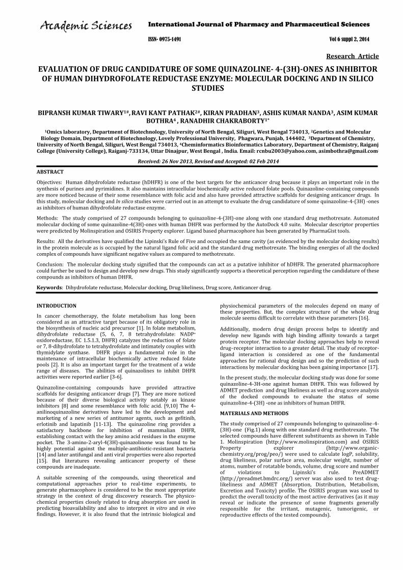
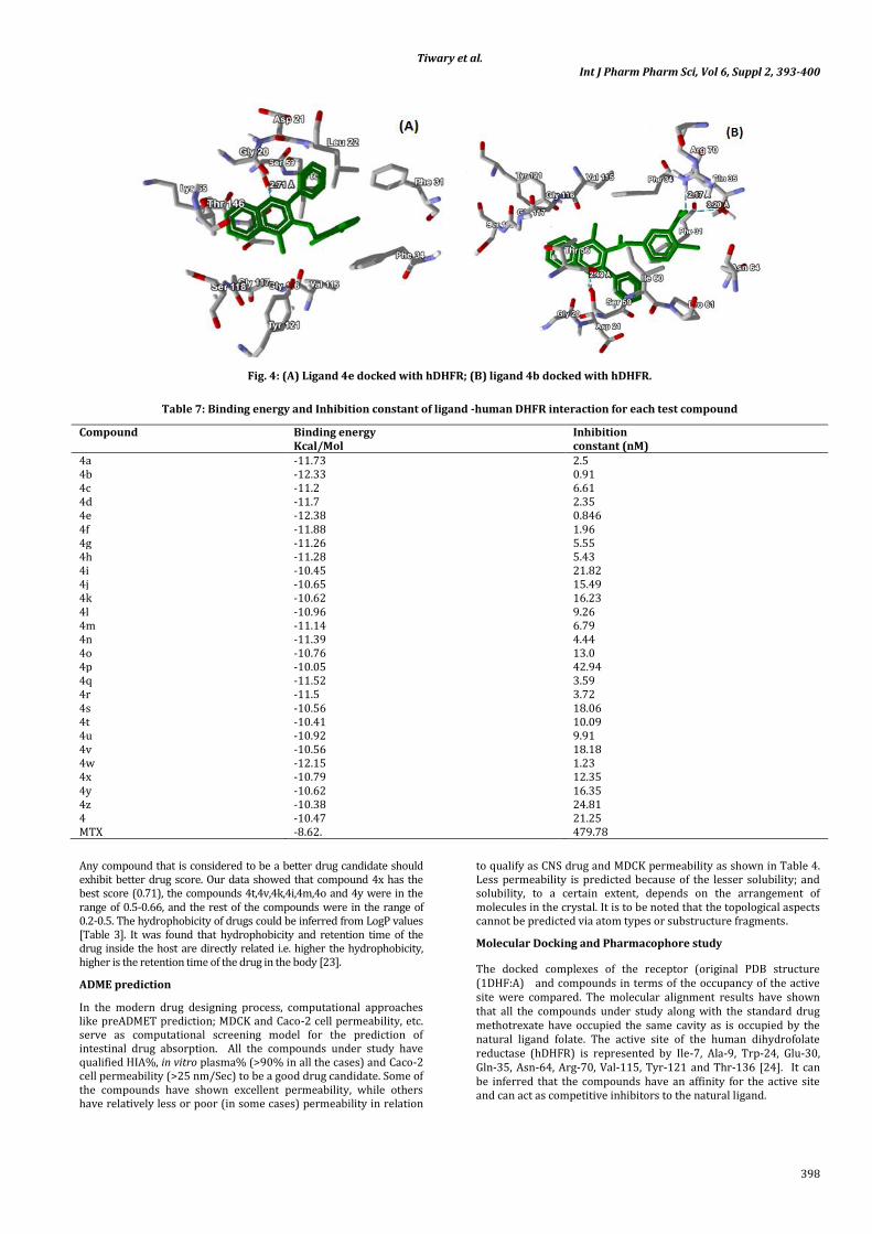
The molecular docking results revealed that the docked complex of 27 compounds had less binding energy than methotrexate as shown in table 7 .The docking model of the ligand and hDHFR are shown in Fig. 4. The molecular alignment results have shown that all the compounds under study along with the standard drug methotrexate have occupied the same cavity (Fig 5) as is occupied by the natural ligand folate. All the 27 compounds were used to develop a ligand based pharmacophore (Fig. 6) using PharmaGist tool. Pharmacophore with a score of 66.813 showed the following characteristics: five spatial features out of which three are aromatic rings and two are hydrogen bond acceptors. There are no negative or positive centers, hydrophobic groups or hydrogen bond donors.
DISCUSSION
Molecular descriptor properties
The selected compounds used in this study were evaluated as potential hDHFR inhibitors. The oral bioavailability of the compounds projected as potential drugs were evaluated by determining the molecular weight, number of rotatable bonds
(nrotb), number of hydrogen bonds (nON and nOHNH), and drug’s polar surface (TPSA). Since the individual molecular weights of all the compounds were less than 500, the number of the rotatable bond were <10, the number of hydrogen bond donors and acceptors were < 12, and TPSA values being <140, they qualified to be an ideal oral drug. Ligands tested in this study were also predicted to have good oral bioavailability.
Calculation of the fragment based drug-likeliness of the compounds signifies that the compounds have the same fragments as compared to the existing drug. The drug-likeliness values of all the compounds are reasonably acceptable (except 4h,4i and 4s) as shown in Table 2. The higher drug-likeliness values are found in the case of compounds 4c, 4d, 4e and 4f. Results indicated that these four compounds have the most fragments similar to existing potent drugs to fulfill the potentiality of being drugs.
The drug score values [Table 3] were also calculated which took into account the effect of drug-likeliness, LogP, solubility, molecular weight, and toxicity risk together.
Tiwary et al. Int J Pharm Pharm Sci, Vol 6, Suppl 2, 393-400
398
Fig. 4: (A) Ligand 4e docked with hDHFR; (B) ligand 4b docked with hDHFR.
Table 7: Binding energy and Inhibition constant of ligand -human DHFR interaction for each test compound
Compound Binding energy Kcal/Mol
Inhibition constant (nM)
4a -11.73 2.5 4b -12.33 0.91 4c -11.2 6.61 4d -11.7 2.35 4e -12.38 0.846 4f -11.88 1.96 4g -11.26 5.55 4h -11.28 5.43 4i -10.45 21.82 4j -10.65 15.49 4k -10.62 16.23 4l -10.96 9.26 4m -11.14 6.79 4n -11.39 4.44 4o -10.76 13.0 4p -10.05 42.94 4q -11.52 3.59 4r -11.5 3.72 4s -10.56 18.06 4t -10.41 10.09 4u -10.92 9.91 4v -10.56 18.18 4w -12.15 1.23 4x -10.79 12.35 4y -10.62 16.35 4z -10.38 24.81 4 -10.47 21.25 MTX -8.62. 479.78
Any compound that is considered to be a better drug candidate should exhibit better drug score. Our data showed that compound 4x has the best score (0.71), the compounds 4t,4v,4k,4i,4m,4o and 4y were in the range of 0.5-0.66, and the rest of the compounds were in the range of 0.2-0.5. The hydrophobicity of drugs could be inferred from LogP values [Table 3]. It was found that hydrophobicity and retention time of the drug inside the host are directly related i.e. higher the hydrophobicity, higher is the retention time of the drug in the body [23].
ADME prediction
In the modern drug designing process, computational approaches like preADMET prediction; MDCK and Caco-2 cell permeability, etc. serve as computational screening model for the prediction of intestinal drug absorption. All the compounds under study have qualified HIA%, in vitro plasma% (>90% in all the cases) and Caco-2 cell permeability (>25 nm/Sec) to be a good drug candidate. Some of the compounds have shown excellent permeability, while others have relatively less or poor (in some cases) permeability in relation
to qualify as CNS drug and MDCK permeability as shown in Table 4. Less permeability is predicted because of the lesser solubility; and solubility, to a certain extent, depends on the arrangement of molecules in the crystal. It is to be noted that the topological aspects cannot be predicted via atom types or substructure fragments.
Molecular Docking and Pharmacophore study
The docked complexes of the receptor (original PDB structure (1DHF:A) and compounds in terms of the occupancy of the active site were compared. The molecular alignment results have shown that all the compounds under study along with the standard drug methotrexate have occupied the same cavity as is occupied by the natural ligand folate. The active site of the human dihydrofolate reductase (hDHFR) is represented by Ile-7, Ala-9, Trp-24, Glu-30, Gln-35, Asn-64, Arg-70, Val-115, Tyr-121 and Thr-136 [24]. It can be inferred that the compounds have an affinity for the active site and can act as competitive inhibitors to the natural ligand.

Tiwary et al. Int J Pharm Pharm Sci, Vol 6, Suppl 2, 393-400
399
Fig. 5: (A) Docking model of ligands with hDHFR (PDB ID- 1DHF) protein (Folic Acid and methotrexate are represented in green and
red sticks respectively, and the other 27 molecules are shown in line representation)
Fig. 5: (B) Zoomed view of the active site showing all the docked molecules (ligands are represented in different color sticks
including folic acid and methotrexate represented in green and red sticks respectively)
Fig. 6: (A) Structural alignment of 27 molecules along with the reference compound, methotrexate (methotrexate is shown in
yellow color)
Fig. 6: (B) Structural representation of the derived Pharmacophore
The compounds were evaluated in terms of their binding mode to hDHFR. Based on the binding free energy (ΔG binding) of the protein-ligand interaction and inhibition constant (Ki), and one of the 10 models was chosen to be the best one. The docking result showed that all 27 compounds have low binding energy and inhibition constant as compared to the standard drug methotrexate. The minimum binding energy (maximum stability) was found in case of the compound 4e (-12.38 Kcal/Mol). The N1 of 4e forms hydrogen bond with the Ser-59 with a distance of 2.71Å. The amino acids Val-115, Phe-31, Phe-34, Tyr-121, Thr-136 and Asp-21 are found to be involved in making hydrophobic interactions with 4e. Interestingly, all these amino acids are also present in the active site of hDHFR, which infers that 4e binds to the active site region of the enzyme. The ligand 4b has also formed significant stable complex on docking. Similar to 4e, the N1 atom of 4b formed hydrogen bond with the Ser-59. It was reported that the tested quinazoline’s recognition with the key amino acid Glu-30 and Ser-59 are essential for binding and biological activity [25]. The maximum binding energy was found in 4p (-10.05 Kcal/mol) which did not form hydrogen bond with the residues of the receptor. It was observed from the calculated binding energies that incorporation of phenyl at 2-C increased the interaction with the enzyme in comparison to compounds substituted with methyl at 2-C. The compounds 4a- 4h have binding energies in the range of -11.28 to -12.38 Kcal/Mol. The inhibition constant is directly proportional to the binding energy as shown in table 7. Many authors have used ligand-based approach for pharmacophore modeling of species-specific DHFR inhibitors. Moreover, a pharmacophore model for hDHFR (human) inhibitors has also been modeled [26]. All the 27 compounds were used to develop a ligand based pharmacophore [Fig. 6 (B)] using PharmaGist
tool, which could be used further for the development of new, improved and optimized drug acting as inhibitor to hDHFR.
The pharmacophore has 3 aromatic rings and two hydrogen bond acceptors which enables in making several non covalent interactions like hydrophobic-hydrophobic interactions, hydrogen bonding, pi cloud interactions, etc.
This pharmacophore is qualifying all the four parameters of Lipinski‘s rule of five and thus could be considered as a lead molecule to generate new conformations for virtual screening library along with more modifications which could enhance its therapeutic index by enhancing the kind of interactions it could possibly make with the target protein.
CONCLUSIONS
Structure based drug design is significantly based on the protein-ligand interaction. The molecular docking study signified that the compounds can act as a putative inhibitor of hDHFR. The binding energy was found to be lesser than that of methotrexate. The compounds have also successfully qualified the rule of five, CMC like rule, WDI like rule and MDDR like rule. Every compound possessed apt pharmacological properties based on the results of Lipinski’s Rule, hydrophobicity (based on log P value), and good drug likeliness and drug score. Moreover, the compounds have low toxicity value. The compounds were predicted to be safe (non-mutagenic as well as non-carcinogenic). This study has enabled to broaden the vision for the generation of more specific drugs for hDHFR, and may pave the way for the production and identification of more effective drugs.
Conflicts of Interest: All authors have none to declare.

Tiwary et al. Int J Pharm Pharm Sci, Vol 6, Suppl 2, 393-400
400
ACKNOWLEDGEMENTS
Authors are thankful to Tamal Sarkar, Technical officer, USIC, University of North Bengal for assistance in computation. BKT was provided by fellowship funded by DBT Sanction No. BT/BIO-CARe/06/141/2010-2011.
REFERENCES
1. Abali EE, Celikkaya H, Yi-Ching H, Banerjee D, Bertino RJ Novel Therapeutic Approaches to Regulate Human Dihydrofolate Reductase Activity and Expression. Current enzyme inhibition 2012;8: 107-117.
2. MacKenzie RE Folates and Pterins. In. (Blakley R L & Benkovic S J eds), Wiley-Interscience, New York. 1984;Vol. 1 pp. 255-306
3. Wyss PC, Gerber P, Hartman PG, Hubschwerlen C, Locher H, Marty H, Stahl M Novel Dihydrofolate Reductase Inhibitors. Structure-Based versus Diversity-Based Library Design and High-Throughput Synthesis and Screening. J Med Chem 2003;46: 2304–2312.
4. Rastelli G, Pacchioni S, Sirawaraporn W, Sirawaraporn R, Parenti MD, Ferrari AM Docking and Database Screening Reveal New Classes of Plasmodium falciparum Dihydrofolate Reductase Inhibitors. J Med Chem 2003;46: 2834–2845.
5. Donkor IO, Li H, Queener SF Synthesis and DHFR inhibitory activity of a homologous series of 6-substituted 2,4-diaminoathieno[2,3-d]pyrimidines’. Eur J Med Chem 2003;38: 605–611.
6. Zink M, Lanig H, Troschutz R Structural variations of piritrexim, a lipophilic inhibitor of human dihydrofolate reductase: synthesis, antitumor activity and molecular modeling investigations. Eur J Med Chem 2004;39: 1079–1088.
7. Garofalo A, Goossens L , Baldeyrou B, Lemoine A , Ravez S , Six P , Cordonnier M, Bonte J, Depreux P , Lansiaux A, Goossens J Design, Synthesis, and DNA-Binding of N-Alkyl(anilino)quinazoline Derivatives. J Med Chem 2010;53: 8089–8103.
8. Klutchko SR, Zhou H, Winters RT, Tran TP, Bridges AJ, Althaus IW, Amato DM, Elliott WL, Ellis PA, Meade MA et al Tyrosine Kinase Inhibitors. 19. 6-Alkynamides of 4-Anilinoquinazolines and 4-Anilinopyrido[3,4-d]pyrimidines as Irreversible Inhibitors of the erbB Family of Tyrosine Kinase Receptors J Med Chem 2006;49: 1475-1485.
9. Hynes JB, Buch JM, Freisheim JH Quinazolines as inhibitors of dihydrofolate reductase. 3. Analogs of pteroic and isopteroic acids. J Med Chem 1975;18:1191-1194.
10. Hynes JB. Buch JM Freisheim JH Quinazolines as inhibitors of dihydrofolate reductase. 4. Classical analogs of folic and isofolic acids. J Med Chem 1977;20: 588-591.
11. Wakeling AE, Guy SP, Woodburn JR, Ashton SE, Curry BJ, Barker AJ, Gibson KH ZD1839 (Iressa): an orally active inhibitor of epidermal growth factor signaling with potential for cancer therapy. Cancer Research 2002;62: 5749–5754.
12. Akita RW, Sliwkowski MX Preclinical studies with Erlotinib (Tarceva). Seminars in Oncology. 2003;30: 15–24.
13. Rusnak DW, Lackey K, Affleck K, Wood ER, Alligood KJ, Rhodes N, Keith BR, Murray DM, Knight WB, Mullin RJ, Gilmer TM The effects of the novel, reversible epidermal growth factor
receptor/ErbB-2 tyrosine kinase inhibitor, GW2016, on the growth of human normal and tumor-derived cell lines in vitro and in vivo. Mol Cancer Ther 2001;1(2): 85–94.
14. Nanda AK, Ganguli S, Chakraborty R Antibacterial activity of some 3-(Arylideneamino)-2-phenylquinazoline-4(3H)-ones: synthesis and preliminary QSAR studies. Molecules 2007;12: 2413-2426.
15. Xingwen G, Xuejian C, Kai Y, Baoan S, Lili G, Zhuo C Synthesis and Antiviral Bioactivities of 2-Aryl- or 2-Methyl-3-(substituted- Benzalamino)-4(3H)-quinazolinone Derivatives. Molecules 2007;12: 2621-2642.
16. Kadam RU, Roy N Recent trends in Drug-Likeliness Prediction: A comprehensive review of In Silico methods. Indian J Pharm Sci 2009;14: 609-615.
17. Shoichet BK, McGovern SL, Wei B, Irwin JJ Lead discovery using molecular docking. Curr Opin Chem Biol 2002;6: 439-446.
18. Morris GM, Goodsell DS, Halliday RS, Huey R, Hart WE, Belew RK, Olson A J Automated Docking Using a Lamarckian Genetic Algorithm and and Empirical Binding Free Energy Function. J Comp Chem 1998;19(14): 1639-1662.
19. Davies JF, Delcamp TJ, Prendergad NJ, Ashford VA , Freisheim JH, Kraut J Crystal structures of recombinant human dihydrofolate reductase complexed with folate and 5-deazafolate. Biochemistry 1990;29: 9467-9479.
20. Sanner MF Python: a programming language for software integration and development. J Mol Graph Model 1999;17: 57-61.
21. Schneidman-Duhovny D, Dror O, Inbar Y, Nussinov R, Wolfson JH PharmaGist: a webserver for ligand-based pharmacophore detection. Nucleic Acids Research 2008;36 : W223–W228
22. Lipinski CA, Lombardo F, Dominy BW, Feeney PJ Experimental and computational approaches to estimate solubility and permeability in drug discovery and development settings. Adv Drug Deliv Rev 1997;23: 3-25.
23. Tambunan USF, Bramantya N, Parikesit AA. In silico modification of suberoylanilide hydroxamic acid (SAHA) as potential inhibitor for class II histone deacetylase (HDAC). BMC Bioinformatics 2011; 12(Suppl 13): S23.
24. Yamini L, Meena KK, Vijjulatha M Molecular Docking, 3D QSAR and Designing of New Quinazolinone Analogues as DHFR Inhibitors. Bull Korean Chem Soc 2011;32 : 2433-2442.
25. Al-Rashood Sarah T, Aboldahab Ihsan A, Nagi Mahmoud N, Laila A. Alaa Abouzeid A, Abdel-Aziz M., Sami G, Abdel-hamide, M. Youssef Khairia, M. Al-Obaid Abdulrahman I. El-Subbagh Hussein Synthesis, dihydrofolate reductase inhibition, antitumor testing, and molecular modeling study of some new 4(3H)-quinazolinone analogs. Bioorg Med Chem 2006;14: 8608–8621.
26. Al-Omary FAM, Abou-zeid LA, Nagi MN, Habib EE, Abdel-Aziz AAM., El-Azab AS, Abdel-Hamide SG, Al-Omar MA, Al-Obaid AM, El-Subbagh HI Non-classical antifolates. Part 2: Synthesis, biological evaluation, and molecular modeling study of some new 2,6-substituted-quinazolin-4-ones. Bioorg Med Chem 2010;18: 2849-2863.