acalculia: an fmri study with implications with respect … · an fmri study with implications with...
TRANSCRIPT

1505
Intern. J. Neuroscience, 113:1505–1523, 2003Copyright Taylor & Francis Inc.ISSN: 0020-7454 / 1543-5245 onlineDOI: 10.1080/00207450390231545
ACALCULIA:AN fMRI STUDY WITH IMPLICATIONS
WITH RESPECT TO BRAIN PLASTICITY
BYRON BERNALNeurologist, Miami Children’s HospitalDepartment of RadiologyMiami, Florida, USA
ALFREDO ARDILANeuropsychologist, Memorial Regional HospitalHollywood, Florida, USA
NOLAN R. ALTMANNeuroradiologist, Chief, Department of RadiologyMiami Children’s HospitalMiami, Florida, USA
We report the case of a 44-year-old right-handed man with severe acal-culia and 4 normal age-matched controls. The acalculia patient hadmagnetic resonance findings of a wedge shaped defect in the left poste-rior temporal-parietal cortex, involving the angular gyrus, and a lacu-nar infarct of the right thalamus. Functional magnetic resonance examswere performed utilizing four tasks consisting of serial subtracting bysevens, basic calculation, complex calculation, and abstract calcula-tion. Both patient and controls showed specific task-related activation
Received 3 March 2003.Our sincere gratitude to Dr. Mauricio Concha and Dr. Monica Rosselli for their significant
contribution when studying this case and for their valuable suggestions; to Dr. Rosario Rosalesfor her invaluable help with the bibliography work-up and review of the article.
Address correspondence to Nolan R Altman, MD, Miami Children’s Hospital, Departmentof Radiology, 3100 SW 62nd Avenue, Miami, Florida 33155-3009, USA. E-mail: [email protected]
Int J
Neu
rosc
i Dow
nloa
ded
from
info
rmah
ealth
care
.com
by
Flor
ida
Inte
rnat
iona
l Uni
vers
ity, M
edic
al L
ibra
ry o
n 03
/03/
11Fo
r pe
rson
al u
se o
nly.

1506 B. Bernal et al.
with some differences that may indicate brain plasticity, even though nosignificant recovery in calculations abilities was observed in the patient.
Keywords acalculia, brain, calculations, fMRI, functional, plasticity
Brain plasticity is defined as the ability of the circuitry of the brainto reorganize in response to a neural insult (Kaas, 1991; Simos etal., 2000). From this definition there is no causal relationship be-tween mutability of neural tissue and recovery. Brain plasticity mayinvolve pathway-redundancy, changes in synaptic strength, growthof new synapses, assumption of function by contralateral homolo-gous cortex, and substitution of uncrossed pathways (Frackowiak,1996; Lee & van Donkelaar, 1995). These brain capabilities usuallyoccur early in immature brains, but may be observed even late inadulthood (Kennard, 1940; Nass, 1984; Vargha-Khadem et al., 1985;Chollet et al., 1991). Thus, the neuronal circuitry involved in aspecific function may possibly be shifted, displaced, or relocatedafter an insult. Brain plasticity has been investigated by functionalmagnetic resonance imaging (Rossini et al., 1998; Musso et al.,1999; Thulborn et al., 1999; Cao et al., 1999) and positron emissiontomography (Chollet et al., 1991; Weiller et al., 1995), in patientswith brain lesions and cognitive impairments. These studies corre-late changes in brain activation to recovery of the lost function.However, no studies have addressed the functional changes that canoccur in the brain not related to recovery. The understanding ofthese “non-successful” changes can contribute to the knowledge offunctional recovery.
Functional magnetic resonance (fMR), a current tool used to mapbrain activity related to motor, sensory, and cognitive tasks, is basedon the chemical and vascular changes produced by brain corticalactivity. These minute tissue changes occur locally and modify thesignal of the MR images. fMRI has been utilized to study brainplasticity in patients after stroke. These studies have focused ondifferent types of brain functions: motor (Rijntjes et al., 1994; Weilleret al., 1993; Maegaki et al., 1995; Rossini et al., 1998), sensory(Maegaki et al., 1995), and language (Weiller et al., 1995; Heiss etal., 1999; Musso et al., 1999; Cappa et al., 1997; Cao et al., 1999).
To our knowledge, no prior investigations with functional images
Int J
Neu
rosc
i Dow
nloa
ded
from
info
rmah
ealth
care
.com
by
Flor
ida
Inte
rnat
iona
l Uni
vers
ity, M
edic
al L
ibra
ry o
n 03
/03/
11Fo
r pe
rson
al u
se o
nly.
Acalculia and Brain Plasticity 1507
have addressed brain plasticity in acalculia, a term that defines theloss or impairment in computation ability. Similar to language, cal-culation relies on a multi-modular network involving frontal andparietal lobes of both hemispheres (Fulbright et al., 2000), withimportant nodes located primarily in the left posterior parietal areas(Stanescu-Cosson et al., 2000). The understanding of brain plastic-ity in calculation is important, as calculation is a category-specificmodel less dependent on language input modalities (Le Clec’H etal., 2000).
Despite recent knowledge gained from functional and lesionalmodels, whether there is reorganization without recovery still re-mains an unanswered question. We present a case in which a right-handed patient, who suffered a left parietal stroke, showed right-parietal activation without significant recovery of the lost arithmeticskills. As in other reports with different cognitive deficits (Weilleret al., 1993; Weiller et al., 1995), the normal activation is assumedfrom results in normal volunteers, reported in the literature, andfrom our own service.
MATERIALS AND METHODS
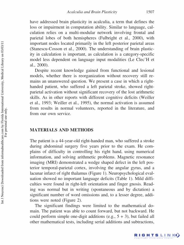
The patient is a 44-year-old right-handed man, who suffered a strokeduring abdominal surgery five years prior to the exam. He com-plains of difficulty in controlling his right hand, using numericalinformation, and solving arithmetic problems. Magnetic resonanceimaging (MRI) demonstrated a wedge shaped defect in the left pos-terior temporal-parietal cortex, involving the angular gyrus, and alacunar infarct of right thalamus (Figure 1). Neuropsychological eval-uation showed no important language deficits (Table 1). Mild diffi-culties were found in right-left orientation and finger gnosis. Read-ing was normal but in writing (spontaneous and by dictation) asignificant number of word omissions and, to a lesser degree, addi-tions were noted (Figure 2).
The significant findings were limited to the mathematical do-main. The patient was able to count forward, but not backward. Hecould perform simple one-digit additions (e.g., 5 + 3), but failed allother mathematical tests, including serial additions and subtractions,
Int J
Neu
rosc
i Dow
nloa
ded
from
info
rmah
ealth
care
.com
by
Flor
ida
Inte
rnat
iona
l Uni
vers
ity, M
edic
al L
ibra
ry o
n 03
/03/
11Fo
r pe
rson
al u
se o
nly.

1508 B. Bernal et al.
multiplying, and dividing. Solving arithmetical problems (e.g., “howmany books are there in two dozen and a half?”) was impossible.Similar difficulties were observed in oral and written calculations.However, he preserved the ability to calculate grossly the answer
FIGURE 1. Axial T1 weighted MRI of the brain shows generalized atrophy with in-volvement of cortical and subcortical white matter. A chronic infarct in the left parietaloccipitaljunction and lacunar right thalamic infarct is seen.
Int J
Neu
rosc
i Dow
nloa
ded
from
info
rmah
ealth
care
.com
by
Flor
ida
Inte
rnat
iona
l Uni
vers
ity, M
edic
al L
ibra
ry o
n 03
/03/
11Fo
r pe
rson
al u
se o
nly.
Acalculia and Brain Plasticity 1509
(e.g., “How much is 15 plus 12?”, “I think 25”). He had significantdifficulties in halving and doubling small quantities. He failed individing objects (e.g., pencils) into equal groups (e.g., to separatenine pencils in three groups, each one containing the same numberof pencils). He could read one and two-digit numbers, but the reading
TABLE 1. Language evaluation of the acalculia patient
Language testing Raw score Standard score*
Boston Naming Test 56 50Multilingual Aphasia Examination
Visual naming 56 64Sentence repetition 12 43Controlled oral word association 23 5Token test 39 15Reading comprehension 17 17
WAIS-IIIVocabulary 43 9Similarities 16 7Digit span 14 8Information 20 11Comprehension 15 7
*Standard scores rfer to percentiles (Boston Naming Test and Multilingual Aphasia Examination or scaledscores (WAIS-III).
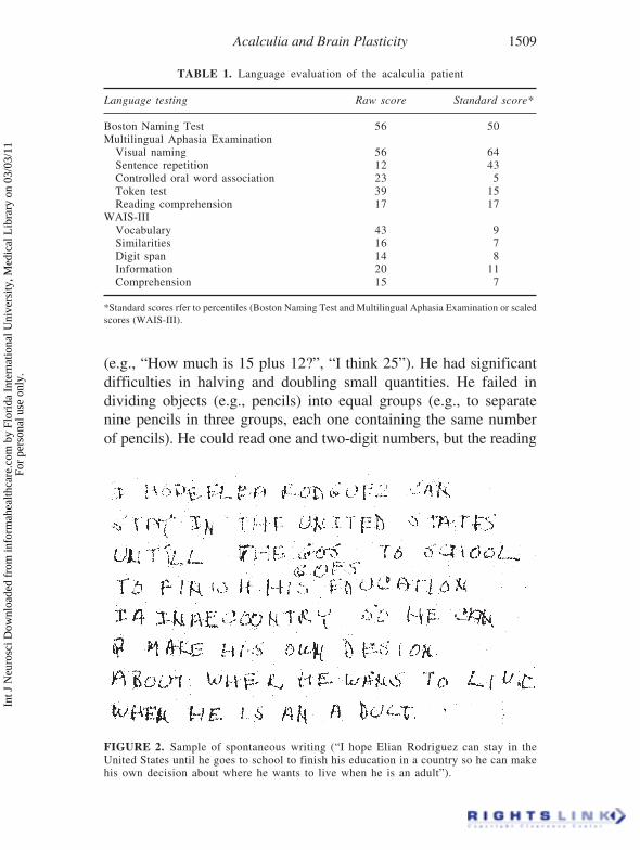
FIGURE 2. Sample of spontaneous writing (“I hope Elian Rodriguez can stay in theUnited States until he goes to school to finish his education in a country so he can makehis own decision about where he wants to live when he is an adult”).
Int J
Neu
rosc
i Dow
nloa
ded
from
info
rmah
ealth
care
.com
by
Flor
ida
Inte
rnat
iona
l Uni
vers
ity, M
edic
al L
ibra
ry o
n 03
/03/
11Fo
r pe
rson
al u
se o
nly.

1510 B. Bernal et al.
of numbers with more than three digits was difficult. Omissions(10003 → 1003), and inversions (4908 → 4098) would result. Therewere no errors in aligning numbers in columns. He could writequantities by dictation. Digit substitutions were noted in translatingfrom verbal to numerical (e.g., twenty-three → 25). Omissions andadditions of letters and decomposition errors (e.g., 67 → six seven)were observed when transcoding from numerical to verbal code (Figure3). Table 2 summarizes the results of the different calculation tests.
The patient was examined with fMR imaging. Prior to the study,he was instructed not to move during image acquisition and avoidvocalization or sub-vocalization. He was informed that this was atest involving mathematical skills. He was trained using short para-digms to assure comprehension of the task. We were more inter-ested in the continuation of the task rather than the answer. Hisperformance was known beforehand, and then debriefed after theexam, and we preferred not to ask the patient to verbalize an answerbecause of the risk of producing head motion. Four healthy adultright-handed males volunteers (age 24–35, mean 27.7), were re-cruited among coworkers; a signed, written informed consent wasobtained that was approved by the Institutional Review Board. Thesubjects were given the same tasks. This group served as a control.
Imaging was performed on a 1.5-T MR scanner (General Elec-tric, Milwaukee, WI, USA. Signa Horizon Echospeed 5X) using
FIGURE 3. Transcoding from a numerical to a verbal code.
Int J
Neu
rosc
i Dow
nloa
ded
from
info
rmah
ealth
care
.com
by
Flor
ida
Inte
rnat
iona
l Uni
vers
ity, M
edic
al L
ibra
ry o
n 03
/03/
11Fo
r pe
rson
al u
se o
nly.

Acalculia and Brain Plasticity 1511
birdcage quadrature-transmit-and-receive head coil designed for wholebrain imaging. A series of 16 localizer images were obtained in thesagittal plane. On the basis of these images, ten axial slices wereselected to obtain the functional and the anatomic images. The sliceswere parallel to the plane defined by a line between the basal aspectof the frontal and occipital lobes. The anatomic series were ob-tained with T1-spin-echo acquisitions, time of repetition/time of echo(TR/TE) of 300/14 ms, field of view (FOV) 240 mm, matrix: 256 ×256, 6 mm slice thickness, and 2 mm gap. The functional images
TABLE 2. Results of different calculation test in acalculia patient
Calculation ability testing Raw Score interpretation
WAIS-III Arithmetic 6 (Scaled score = 3) abnormalCounting forwards (1 to 20) 20/20 normalBackwards (30–20) 7/10 abnormalReading numbers 4/6 abnormalWriting numbers 6/6 normalTranscoding: numerical to a verbal 4/6 abnormalVerbal to a numerical 4/6 abnormalMultiplication tables (table of 3) 6/10 abnormalTo complete an arithmetical operation 7/8 abnormal (e.g., 23?18 = 30)
Successive arithmetical operationsAdding (l, 4, 7?37) 7/12 abnormalSubtracting (100, 93–86?16) 0/12 abnormal
Mental calculation (with two digits)Adding 0/4 abnormalSubtracting 0/4 abnormalMultiplying 0/4 abnormalDividing 0/4 abnormal
Written calculation (with two and three digits)Adding 0/4 adnormalSubtracting 0/4 adnormalMultiplying 0/4 adnormalDividing 0/4 adnormalAligning numbers in columns 5/5 normalFractions 3/3 normal e.g., “what is larger,
one-third or one-fourth?”General numerical knowledge 4/4 normal (e.g., “at what
temperature does waterboil?”
Personal numeral knowledge 4/4 normal (e.g., “what is yoursocial security number?”
Numerical problems 0/5 abnormalHalving and doubling small quantities 0/6 abnormalDividing objects into equal groups 0/5 abnormal
Int J
Neu
rosc
i Dow
nloa
ded
from
info
rmah
ealth
care
.com
by
Flor
ida
Inte
rnat
iona
l Uni
vers
ity, M
edic
al L
ibra
ry o
n 03
/03/
11Fo
r pe
rson
al u
se o
nly.

1512 B. Bernal et al.
with same thickness and gap were obtained using a gradient echo-single shot echo planar imaging sequence with TR/TE/Flip-angle of3750 ms/60/60°. The FOV was 240 mm, and the matrix 64 × 64voxels. Each voxel had a volume of 3.75 × 3.75 × 6 mm (84.375mm3). The experiment consisted of 48 time-points (sets) of 10 im-ages, and 2 extra sets at the beginning of the series, used to obtainthe steady-state of the transverse magnetization. The 48 sets weredivided in 6 epochs, 3 OFF (baseline or control condition) and 3ON (activated condition). Starting in OFF, the experiment proceededswitching every 30 s. Thus, each slice was scanned 48 times, result-ing in 240 image pairs for comparisons.
Four paradigms were applied: (1) serial subtracting by seven, (2)basic calculations, (3) complex calculations, and (4) abstract calcu-lations. Serial subtracting by seven was performed starting at 100for the first ON epoch, from 50 in the second, and from 75 in thethird. Basic calculations consisted of simple addition and subtractionproblems of one or two digits avoiding carrying over of numbers(e.g.: 7 – 5 = ?). Complex calculations were problems with the sametype of calculations involving two digits and requiring carrying,borrowing, and handling of place values (e.g.: 15 – 7 = ?). Abstractcalculations included problems involving rules and concepts relatedto logical and spatial analysis (e.g.: 2 – ¼ = ?). To prevent motionartifacts, the head was fixed with cushions and taped to the head-holder of the table. Problems were displayed with a black font onone horizontal line (e.g.: 23 – 7 = ?) over a white background using aback projector and a semitransparent screen placed at the foot ofthe imaging table. The image was delivered to the patient by a mirrorlocated over his eyes. Numbers were 3 × 2 cm and the screen waslocated 200 cm from the mirror. The numbers formed a 0.85° verticalangle with the eyes. Each problem was presented for 3 to 6 s (de-pending on the complexity of the task and previous experience withvolunteers). Two different types of baseline (OFF) conditions wereused: (1) counting forward from 1 for the serial subtracting by seven,and (2) reading strings of four elements (letter and digits), randomlymixed, for the remainder of paradigms (e.g., F 2 H 6). These OFFepochs were designed to match attention, ocular motion, visual stim-ulus, and verbal impute and decoding.
The fMRI data were analyzed off-line on a Sun-Ultrasparc work-
Int J
Neu
rosc
i Dow
nloa
ded
from
info
rmah
ealth
care
.com
by
Flor
ida
Inte
rnat
iona
l Uni
vers
ity, M
edic
al L
ibra
ry o
n 03
/03/
11Fo
r pe
rson
al u
se o
nly.

Acalculia and Brain Plasticity 1513
station (Sun Microsystems Inc., CA, USA) using MEDx 3.0 software(Sensor Systems, Inc., VA, USA). The functional images were firstanalyzed to detect motion, using the Intensity Weighted Method. Theimages were spatially smoothed with a Gaussian kernel of full-width-at-half-maximum (FWHM) of 10 mm to reduce the noise in theimages. Intensity normalization was applied to normalize the signalat each voxel and remove the effects of signal drift. Parametricalstatistical maps were obtained in each series using a t-test method. z-scores were used as activation reference maps, after thresholdingthem to an uncorrected p < .0005, corresponding to a z-score of 3.47.Anatomic localization was performed by superimposing the func-tional images on T1 weighted images at the same slice location.
RESULTS AND DISCUSSION
Table 3 summarizes the results of the control group. In the patient,the serial subtracting by sevens produced activation in the posterior
TABLE 3. fMRI results of the calculation tasks in the control group
Paradigm Areas of activation Cases
Counting backwards Left middle-inferior frontal gyrus 3Right middle-inferior frontal gyrus 3Left posterior inferior parietal lobule (PIPL) 3
(including angular gyrus)Left supplemental motor area (SMA) 3Left frontal eye field 2
Basic calculation Right middle frontal gyrus 2Right caudate nucleus 2Right SMA 2Left PIPL (including angular gyrus) 2Left broca I
Complex calculation Left middle-inferior frontal gyrus 4Left PIPL (including angular gyrus) 3Right PIPL (including mirror angular gyrus) 2Left cuneus 2
Abstract calculation Left PIPL (including angular gyrus) 4Right PIPL (including angular gyrus) 4Left middle-inferior frontal gyrus 3Left SMA 2Right SMA 1Left > right frontal eye field 3
Int J
Neu
rosc
i Dow
nloa
ded
from
info
rmah
ealth
care
.com
by
Flor
ida
Inte
rnat
iona
l Uni
vers
ity, M
edic
al L
ibra
ry o
n 03
/03/
11Fo
r pe
rson
al u
se o
nly.
1514 B. Bernal et al.
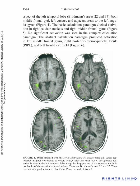
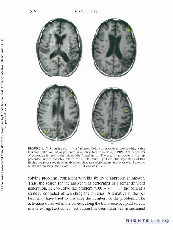
aspect of the left temporal lobe (Brodmann’s areas 22 and 37), bothmiddle frontal gyri, left cuneus, and adjacent areas to the left angu-lar gyrus (Figure 4). The basic calculation paradigm elicited activa-tion in right caudate nucleus and right middle frontal gyrus (Figure5). No significant activation was seen in the complex calculationparadigm. The abstract calculation paradigm produced activationin left middle frontal gyrus, right posterior-inferior-parietal lobule(PIPL), and left frontal eye field (Figure 6).
FIGURE 4. fMRI obtained with the serial subtracting by sevens paradigm. Areas rep-resented in green correspond to voxels with p value less than .0005. The greatest acti-vation is seen in the left temporal lobe along the deep portion of the superior and infe-rior banks of the superior temporal sulcus. These are Brodmann’s area 22 and 37. Thereis a left side predominance. (See Color Plate I at end of issue.)
Int J
Neu
rosc
i Dow
nloa
ded
from
info
rmah
ealth
care
.com
by
Flor
ida
Inte
rnat
iona
l Uni
vers
ity, M
edic
al L
ibra
ry o
n 03
/03/
11Fo
r pe
rson
al u
se o
nly.

Acalculia and Brain Plasticity 1515
Variation in the pattern of brain activation was found with thedifferent paradigms. Significant local changes were observed in thetemporal, posterior parietal, and frontal lobes. These local changeswe correlated with specific task demands.
Serial subtracting by seven resulted in a significant activation ofBrodmann’s 37 area. This area involves the region about the supe-rior and inferior temporal sulcus. This region has also been foundactivated in semantic verbal tasks (Vandenberghe et al., 1996). It isrelated to visual representation of words (Benson & Ardila, 1996).We may assume that the patient had adopted a semantic strategy to
FIGURE 5. fMRI obtained with basic calculation. Color corresponds to activation invoxels with p value less than .0005. The main activation is seen in the right caudatenucleus. Notice the lack of activation in the left hemisphere. (See Color Plate II at endof issue.)
Int J
Neu
rosc
i Dow
nloa
ded
from
info
rmah
ealth
care
.com
by
Flor
ida
Inte
rnat
iona
l Uni
vers
ity, M
edic
al L
ibra
ry o
n 03
/03/
11Fo
r pe
rson
al u
se o
nly.
1516 B. Bernal et al.
solving problems, consistent with his ability to approach an answer.Thus, the search for the answer was performed as a semantic wordgeneration, i.e., to solve the problem “100 – 7 = __,” the patient’sstrategy consisted of searching the nineties. Alternatively, the pa-tient may have tried to visualize the numbers of the problems. Theactivation observed in the cuneus, along the transverse occipital sulcus,is interesting. Left cuneus activation has been described in sustained
FIGURE 6. fMRI during abstract calculation. Color corresponds to voxels with p valueless than .0005. Activation presented in yellow is located in the right PIPL. A wider clusterof activation is seen in the left middle frontal gyrus. The area of activation in the leftprecentral area is probably related to the left frontal eye field. The asymmetry of thisfinding suggests a cognitive involvement, since an underlying motor process would producebilateral activation. (See Color Plate III at end of issue.)
Int J
Neu
rosc
i Dow
nloa
ded
from
info
rmah
ealth
care
.com
by
Flor
ida
Inte
rnat
iona
l Uni
vers
ity, M
edic
al L
ibra
ry o
n 03
/03/
11Fo
r pe
rson
al u
se o
nly.
Acalculia and Brain Plasticity 1517
visual attention (Le et al., 1998). The patient may have used visualclues to perform the task such as picturing the numbers and scan-ning them into a given category. Precuneus activation has been ob-served in aphasia patients with improvement in verbal comprehen-sion after training (Musso et al., 1999). The previous training of thepatient may also partially explain these results. Activation of themiddle frontal gyri has been reported already using this paradigm.This activation is felt to be related to working memory (Rueckert etal., 1996).
The activation seen in the head of the right caudate nucleus,during basic calculation paradigm, was also observed in two casesin the control group. These results are in agreement with prior casesof acalculia reported in patients with subcortical strokes (Corbett etal., 1986). Basic calculations are automatic memory-related pro-cesses, which could be performed by subcortical structures. There-fore, it is understandable that our acalculia patient preserved theability to perform simple basic calculations. Basic calculations (e.g.,5 + 2) represent kinds of verbal automatism.
The lack of activation in the complex paradigm is not well un-derstood; however, it is possible that the patient became frustratedand stopped performing the requested task.
Abstract calculation paradigm produced left middle frontal gy-rus, left frontal eye field, and right PIPL activation (in absence ofthe normal homologous contralateral area). The left middle frontalgyrus activation, as stated before, is related to verbal working memory,although pure (nonverbal) abstract tasks may elicit activation in thesame regional (Roland & Friberg, 1985). The frontal eye field acti-vation is probably related to pursuit and saccadic eye movements(Petit et al., 1997), and spatial short-term memory (Gaymard et al.,1999). However, the activation related to eye movement was notsupported in a recent study (Gitelman et al., 2000), raising the pos-sibility of an unknown role for this area in cognitive tasks. Theasymmetry of activation supports this hypothesis.
The right PIPL activation, in absence of the normal homologouscontralateral area, during the abstract paradigm is the most strikingfinding of this case. The four normal subjects activated both PIPL,although the left PIPL was activated more strongly. Therefore, inour case, activation on the homologous contralateral area could be
Int J
Neu
rosc
i Dow
nloa
ded
from
info
rmah
ealth
care
.com
by
Flor
ida
Inte
rnat
iona
l Uni
vers
ity, M
edic
al L
ibra
ry o
n 03
/03/
11Fo
r pe
rson
al u
se o
nly.

1518 B. Bernal et al.
anticipated. The left angular gyrus cannot be activated because ofencephalomalacia. The left PIPL, although preserved, did not showactivation possibly due to disconnection from input areas, as sug-gested by the deep extension of the hypointensity representing glio-sis of the white matter (Figure 1). The contralateral right-PIPL areais activated, with the left frontal networking that is intact. This mayexplain the patient’s abnormal performance indicating the ineffi-ciency of this new hardwiring, or the remaining network.
Activation of contralateral-homologous areas does not necessar-ily mean functional recovery. Asymmetric bilateral normal repre-sentation may explain previous results dealing with the shifting typeof brain plasticity. Using different techniques, bilateral activationhas been found in normal subjects involving basic unilateral motortasks, sensory stimulation (Hansson & Brismar, 1999; Babiloni etal., 1999), and language tasks (Binder, 1997; Weiller, 1997). Unilat-eral activation, which does occur in many cases, may be also artifi-cially obtained as a result of high threshold levels, which are usu-ally applied to the image post-processing. Often, statistical analysisis done using very strict p values, therefore, eliminating areas ofweaker activation. Conceivably, a left hemisphere lesion may ap-pear to have contralateral activation if the same statistical methodsare applied to a smaller sample with lower absolute values, or ifless conservative p values are utilized to depict activation. This couldgive the appearance of shifting plasticity.
There are no previous studies of brain plasticity in acalculia. Mostprevious reports of brain plasticity refer to language. However, lan-guage and calculation, as brain functions, share several features:they demand a network rather than a dedicated module, they aresymbolic, and both are located principally in the left hemisphere.Therefore, some overlap in brain plasticity seems reasonable.
Calculation and language functions are based on bilateral net-works with several levels of complexity. Understanding the reorga-nization of the scheme of the network is more important thanreplacement of a function in the study of recovery of cognitivefunctions (Weiller et al., 1997). This is stated by Thulborn et al.(1999): “When a key node of a large-scale cortical network is dam-aged network components (namely, contralateral homologous) areincreasingly recruited to increase their workload” (p. 753).
Int J
Neu
rosc
i Dow
nloa
ded
from
info
rmah
ealth
care
.com
by
Flor
ida
Inte
rnat
iona
l Uni
vers
ity, M
edic
al L
ibra
ry o
n 03
/03/
11Fo
r pe
rson
al u
se o
nly.

Acalculia and Brain Plasticity 1519
Bilateral temporal lobe activation has been elicited by verbal tasks,both in normal subjects and stroke patients (Binder, 1997). How-ever, the right hemisphere activation in stroke patients was greaterin areas contralateral to those found in normal subjects (Weiller etal., 1995; Weiller, 1998). Patients with left frontal strokes presentinitially with global metabolic reduction of the left hemisphere andnew activation in the right frontal area homologous to Broca’s area,with repetition tasks. When recovery takes place, activation of theleft posterior superior temporal gyrus returns, and the activation inthe right frontal region (Broca’s homologue) disappears (Heiss etal., 1999). Temporal lesions show activation in the right temporallobe in the early phase (without recovery) and later when recoveryoccurs (Heiss et al., 1999).
The fact that right perisylvian areas in aphasic patients showstronger activation than in controls (Weiller et al., 1995) suggeststhe idea of a substitution role. This supports the assumption that theright hemisphere plays a crucial role in recovery, as suggested bysome early PET studies (Ohyama et al., 1995; Price et al 1995).Clinical recovery is not only associated with the appearance of newactivation in the right hemisphere, but also with the regression offunctional depression in structurally unaffected regions of both hemi-spheres (Cappa et al., 1997). The recovery of the functional depres-sion agrees with the demonstration that better language recoveryoccurs in individuals who have bilateral rather than right hemisphere-predominant activation (Cao et al., 1999). These findings were alsorecently reported by Heiss et al. (1999) in a PET study. This groupfound that complete and satisfactory recovery from aphasia can onlybe achieved as long as structures of the left superior temporal gyrusare preserved and can be reintegrated into language processing andproduction. The final answer, however, is not fully known: Patientsin speech therapy improve their performance on the Token test astheir right superior temporal gyrus and left precuneus show increasein blood flow measures (Musso et al., 1999). These studies per-formed in aphasia patients may be useful in understanding our re-sults in calculation. Many reported cases of aphasia patients alsohave acalculia. We feel that theoretical conclusions from one maybe applied to the other.
Our patient showed activation in the right PIPL area, the contra-
Int J
Neu
rosc
i Dow
nloa
ded
from
info
rmah
ealth
care
.com
by
Flor
ida
Inte
rnat
iona
l Uni
vers
ity, M
edic
al L
ibra
ry o
n 03
/03/
11Fo
r pe
rson
al u
se o
nly.
1520 B. Bernal et al.
lateral homologous area involved in calculations in normal subjects.However, he did not significantly recover from his deficit. The pa-tient succeeded in some basic calculations because they representedautomatic memory-related processes, somehow mediated by the righthemisphere. Basic calculations involving small numbers (e.g., add-ing and subtracting one-digit numbers) have been demonstrated dis-sociated from other types of calculations (Stanescu-Cosson et al.,2000; Cohen et al., 2000), because of having a domain-specific rep-resentation (Dehaene et al., 1998). Therefore, this type of calculationcan be spared in primary acalculia, as was the case in our strokepatient. He possibly was also performing a spontaneous “cognitiveprosthesis” (i.e., he may have been using an alternative semanticcategory strategy to approximate the results in serial subtracting byseven paradigm, explaining activation of Brodmann’s area 37, other-wise not seen in the control group). Combining extrapolated infor-mation obtained from language studies, and our observations in thenormal subjects, we feel that the right PIPL activation representsthe remaining activation of a subsidiary circuitry. This circuitry pre-viously did subserve calculation, but now cannot accomplish thetask utilizing only the right PIPL module.
It may be assumed that the pattern of activation obtained corre-sponds to a redistribution of the workload over the pre-existing net-work, in which the right hemisphere was not crucial for calculation.This assumption has the support of previous reports in which bi-parietal activation was elicited in normal volunteers by calculationtasks (Chochon et al., 1999). Further investigations are needed toelucidate how redistribution is responsible for the so-called reloca-tion and shifting phenomena, and if recovery is necessarily contin-gent upon these changes.
CONCLUSION
We present a case of acalculia in a patient with a left angular gyruslesion, demonstrating on fMRI cortical activation related to math-ematical tasks. The distribution of the obtained activation may betaken as brain “plasticity.” Findings taken from other functional-cognitive studies of language suggest a rewiring can occur without
Int J
Neu
rosc
i Dow
nloa
ded
from
info
rmah
ealth
care
.com
by
Flor
ida
Inte
rnat
iona
l Uni
vers
ity, M
edic
al L
ibra
ry o
n 03
/03/
11Fo
r pe
rson
al u
se o
nly.

Acalculia and Brain Plasticity 1521
significant functional recovery. In some cases, the so-called rewiringis just the redistribution of workload in a pre-existent network whichmay not truly be plasticity.
REFERENCES
Babiloni, C., Carducci, F., Pizzella, V., Indovina, I., Romani, G. L., Rossini, P.M., & Babiloni,F. (1999). Bilateral neuromagnetic activation of human primary sensorimotor cortex inpreparation and execution of unilateral voluntary finger movements. Brain Research,827, 234–236.
Benson, D. F., & Ardila, A. (1996). Aphasia: A clinical perspective. New York: OxfordPress.
Binder, J. (1997). Functional magnetic resonance imaging. Language mapping. Neurosur-gery Clinics of North America, 8 ,383–392.
Cao, Y., Vikingstand, E. M., George, K. P., Johnson, A. F., & Welch, K. M. (1999). Cor-tical language activation in stroke patients recovering from aphasia with functionalMRI. Stroke, 30, 2331–2340.
Cappa, S. F., Perani, D., Grassi, F., Bressi, S., Alberoni, M., Franceschi, M., Bellinardi,V., Todde, S., & Fazio, F. (1997). A PET follow-up study of recovery after stroke inacute aphasics. Brain and Language, 56, 55–67.
Chochon, F., Cohen, L., van de Moortele, P. F., & Dehaene, S. (1999). Differential contri-butions of the left and right inferior parietal lobules to number processing. Journal ofCognitive Neuroscience, 11, 617–630.
Chollet, F., DiPiero, V., Wise, R. J., Brooks, D. J., Dolan, R. J., & Frackowiak, R. S.(1991). The functional anatomy of motor recovery after stroke in humans: A studywith positron emission tomography. Annals of Neurology, 29, 63–71.
Cohen, L., Dehaene, S., Chochon, F., Lehericy, S., & Naccache L. (2000). Language andcalculation within the parietal lobe: A combined cognitive, anatomical fMRI study.Neuropsychologia, 38, 1426–1440.
Corbett, A. J., McCusker, E. A., & Davidson, O. R. (1986). Acalculia following a domi-nant-hemisphere subcortical infarct. Archives of Neurology, 43, 964–966.
Dehaene, S., Dehaene-Lambertz, G., & Cohen, L. (1998). Abstract representations of numbersin the animal and human brain. Trends in Neurosciences, 21, 355–361.
Frackowiak, R. S. J. (1996). Plasticity and the human brain: Insights from functional im-aging. Neuroscientist, 2, 353–362.
Fulbright, R. K., Molfese, D. L., Stevens, A. A., Skudlarski, P., Lacadie, C. M., & Gore,J. C. (2000). Cerebral activation during multiplication: A functional MR imaging studyof number processing. American Journal of Neuroradiology, 21, 1048–54.
Gaymard, B., Ploner, C. J., Rivaud-Pechoux, S., & Pierrot-Deseilligny, C. (1999). Thefrontal eye field is involved in spatial short-term memory but not in reflexive saccadeinhibition. Experimental Brain Research, 129, 288–301.
Gitelman, D. R., Parrish, T. B., LaBar, K. S., & Mesuham, M. M. (2000). Real-time moni-toring of eye movements using infrared video-oculography during functional magneticresonance imaging of the frontal eye fields. Neuroimage, 11, 58–65.
Hansson, T., & Brismar, T. (1999). Tactile stimulation of the hand causes bilateral corticalactivation: A functional magnetic resonance study in humans. Neuroscience Letters,271, 29–32.
Heiss, W. D., Kessler, J., Thiel, A., Ghaemi, M., & Karbe, H. (1999). Differential capacityof left and right hemispheric areas for compensation of poststroke aphasia. Annals ofNeurology, 45, 430–438.
Int J
Neu
rosc
i Dow
nloa
ded
from
info
rmah
ealth
care
.com
by
Flor
ida
Inte
rnat
iona
l Uni
vers
ity, M
edic
al L
ibra
ry o
n 03
/03/
11Fo
r pe
rson
al u
se o
nly.

1522 B. Bernal et al.
Kass, J. H. (1991). Plasticity of sensory and motor maps in adult mammals. Annual Re-view of Neuroscience, 14, 137–167.
Kennard, M. A. (1940). Relation of age to motor impairment in man and in subhumanprimates. Archives of Neurology and Psychiatry, 44, 377–397.
Le, T. H., Pardo, J. V., & Hu, X. (1998). 4 T-fMRI study of nonspatial shifting of selec-tive attention: Cerebellar and parietal contributions. Journal of Neurophysiology, 79,1535–1548.
Le Clec’H, G., Dehaene, S., Cohen, L., Mehler, J., Dupoux, E., Poline, J. B., Lehericy, S.,van de Moortele, P. F., & Le Bihan, D. (2000). Distinct cortical areas for names ofnumbers and body parts independent of language input modality. Neuroimage, 12, 381–391.
Lee, R. G., & van Donkelaar, P. (1995). Mechanisms underlying functional recovery fol-lowing stroke. Canadian Journal of Neurological Sciences, 22, 257–263.
Maegaki, Y., Yamamoto, T., & Takeshita, K. (1995). Plasticity of central motor and sen-sory pathways in a case of unilateral extensive cortical dysplasia: Investigation of magneticresonance imaging, transcranial magnetic stimulation, and short-latency somatosensoryevoked potentials. Neurology, 45, 2255–2261.
Musso, M., Weiller, C., Kiebel, S., Müller, S. P., Bülau, P., & Rijntjes, M. (1999). Train-ing-induced brain plasticity in aphasia. Brain, 122, 1781–1790.
Nass, R. (1984). Case report: Recovery and reorganization after congenital unilateral braindamage. Perceptual and Motor Skills, 59, 867–874.
Ohyama, M., Senda, M., Terashi, A., Kitamura, S., Toyama, H., Ishii, K., & Mishina, M.(1995). A follow up PET activation study in aphasia due to cerebral infraction evalu-ates functional reorganization. Journal of Cerebral Blood Flow and Metabolism, 15(Suppl.), S697.
Petit, L., Clark, V. P., Ingeholm, J., & Haxby, J. V. (1997). Dissociation of saccade-re-lated and pursuit-related activation in human frontal eye fields as revealed by fMRl.Journal of Neurophysiology, 77, 3386–3390.
Price, C., Warburton, E., Swinburn, K., Wise, R., & Frackowiak, R. (1995). Monitoringthe recovery of aphasia using positron emission tomography. Journal of Cerebral BloodFlow and Metabolism, 15 (Suppl.), S696.
Rijntjes, M., Weiller, C., Krams, M., Bauermann, H., Diener, C., & Faiss, J. (1994). Func-tional magnetic resonance imaging in recovery from motor stroke. Stroke, 25, 60.
Roland, P. E., & Friberg, L. (1985). Localization of cortical areas activated by thinking.Journal of Neurophysiology, 53, 1219–1243.
Rossini, P. M., Caltagirone, C., Castriota-Scanderbeg, A., Cicinelli, P., Del Gratta, C.,Demartin, M., Pizzella, V., Traversa, R., & Romani, G. L. (1998). Hand motor corticalarea reorganization in stroke: A study with fMRl, MEG and TCS maps. Neuroreport,9, 2141–2146.
Rueckert, L., Lange, N., Partiot, A., Appollonio, I., Litvan, I., Le Bihan, D., & Grafman,J. (1996). Visualizing cortical activation during mental calculation with functional MRI.Neuroimage, 3, 97–103.
Simos, P. G., Papanicolaou, A. C., Breler, J. I., Fletcher, J. M., Wheless, J. W., Maggio,W. W., Gormley, W., Constantinou, J. E., & Kramer, L. (2000). Insights into brainfunction and neural plasticity using magnetic source imaging. Journal of Clinical Neu-rophysiology, 17, 143–162.
Stanescu-Cosson, R., Pinel, P., van De Moortele, P. F., Le Bihan, D., Cohen, L., & Dehaene,S. (2000). Understanding dissociations in dyscalculia: A brain imaging study of theimpact of number size on the cerebral networks for exact and approximate calculation.Brain, 123, 2240–2255.
Thulborn, K. R., Carpenter, P. A., & Just, M. A. (1999), Plasticity of language-relatedbrain function during recovery from stroke. Stroke, 30, 749–754.
Vandenberghe, R., Price, C., Wise, R., Josephs, O., & Frackowiak, R. S. (1996). Func-tional anatomy of a common semantic system for words and pictures. Nature, 383,254–256.
Int J
Neu
rosc
i Dow
nloa
ded
from
info
rmah
ealth
care
.com
by
Flor
ida
Inte
rnat
iona
l Uni
vers
ity, M
edic
al L
ibra
ry o
n 03
/03/
11Fo
r pe
rson
al u
se o
nly.
Acalculia and Brain Plasticity 1523
Vargha-Khadem, F., Watters, G. V., & O’Gorman, A. M. (1985). Development of speechand language following bilateral frontal lesions. Brain and Language, 25, 167–183.
Weiller, C. (1998). Imaging recovery from stroke. Experimental Brain Research, 123, 13–17.
Weiller, C., Chollet, F., & Frackowiak, R. (1997). Physiological aspects of functional re-covery from stroke. In J. Bogousslavsky, M. Ginsberg, & M. Hennerici (Eds.), Cerebro-vascular disease (pp. 2057–2067) Oxford: Blackwell.
Weiller, C., Isensee, C., Rijntjes, M., Huber, W., Müler, S., Bier, D., Dutschka, K., Woods,R. P., North, J., & Diener, H. C. (1995). Recovery from Wernicke’s aphasia: A positronemission tomographic study. Annals of Neurology, 37, 723–732.
Weiller C., Ramsey, S. C., Wise, R. J., Friston, K. J., & Frackowiak, R. S. J. (1993).Individual patterns of functional reorganization in the human cerebral cortex after cap-sular infarction. Annals of Neurology, 33, 181–189.
Int J
Neu
rosc
i Dow
nloa
ded
from
info
rmah
ealth
care
.com
by
Flor
ida
Inte
rnat
iona
l Uni
vers
ity, M
edic
al L
ibra
ry o
n 03
/03/
11Fo
r pe
rson
al u
se o
nly.